Édité par Elsevier Masson SAS. Tous droits réservés...
12
J Radiol 2008;89:751-62 © Éditions Françaises de Radiologie, Paris, 2008 Édité par Elsevier Masson SAS. Tous droits réservés review gastrointestinal Mucocele of the appendix and pseudomyxoma peritonei A Fairise, C Barbary, AL Derelle, S Tissier, P Granger, F Marchal, V Laurent and D Regent To cite the present paper, use exclusively the following reference. Fairise A, Barbary C, Derelle AL, Tissier S, Granger P, Marchal F, Laurent V, Regent D. Mucocèle appendiculaire et pseudomyxome péritonéal (full text in English on www.masson.fr/revues/jr). J Radiol 2008;89:751-62. mucocele of the appendix corres- ponds to mucinous distension of the appendix. Rupture, the most severe complication, leads to pseudomyxo- ma peritonei (PMP) which frequently is as- sociated with a poor prognosis. The main role of medical imaging is to provide presurgical diagnosis so that all precautions may be taken to avoid per- operative rupture and peritoneal seeding. Early diagnosis of pseudomyxoma is also important to allow early radical surgery in patients with peritoneal involvement to reduce recurrences. Radiologists must be familiar with the imaging features of this entity since imaging frequently is the fist step in the diagnosis and management of this pa- thology and may contribute in avoiding errors with devastating impact on pro- gnosis. In this pictorial essay, the imaging features of 12 cases of mucocele of the appendix and 8 cases of PMP will be reviewed. Anatomical and histological features Knowledge of the anatomical and patho- physiological features as well as epide- miology of this pathology is mandatory for diagnosis. Mucocele of the appendix A mucocele of the appendix is defined by the distension of the appendiceal lumen by mucus (1). Mucinous distension of the appendix lu- men may be tumoral or non-tumoral, benign or malignant in origin. Etio- logies, by increasing order of severity, include (2): – Retention cyst corresponding to the accumulation of mucus secondary to non- tumoral obstruction of the appendiceal lu- men by an appendicolith, inflammatory stricture, extrinsic compression… – Villous hyperplasia, diffuse or focal. Involvement is confined to the mucosa. – Mucinous cystadenoma, a benign tumor of the mucosa, sometimes with areas of dysplasia. – Mucinous cystadenocarcinoma, a mali- gnant tumor characterized by malignant cells, desmoplastic reaction, and invasion of the muscularis mucosa. Well differen- tiated cystadenocarcinoma, defined by the presence of more than 50% of mucinous component, may be difficult to differentiate from cystadenoma in the absence of muscularis invasion; the tumor is often called mucinous tumor of uncertain ma- lignant potential. Pseudomyxoma peritonei PMP corresponds to a clinico-pathological entity characterized by diffuse perito- neal involvement with mucinous ascites and multifocal mucinous epithelial im- plants; the diagnostic histological featu- re is the presence of extracellular mucin within the peritoneal cavity that may be as- sociated with mucinous epithelial cells, more or less well differentiated (3). It results from intraperitoneal rupture of a mucinous tumor, whose appendiceal or ovarian origin has been discussed for some time, and remains controversial. Early reports on PMP described a female predominance with frequent ovarian in- volvement, often bilateral, hence the hy- pothesis for primary ovarian origin of this entity. However, recent studies, based on immunohistochemical analysis and mole- cular biology, have demonstrated the appendiceal origin of nearly all cases of PMP, with eventual secondary ovarian involvement (4, 5). These results were partly based on a study of MUC2-expression, a gene coding for a mucin protein. This protein was present in PMP and mucinous tumors of the ap- pendix, but not in primary ovarian tumors. Primary ovarian tumors, initially consi- dered as the origin of PMP, may cause peritoneal carcinomatosis with peritoneal tumor implants, but no true PMP. Perito- neal carcinomatosis from ovarian prima- ry is characterized by a smaller amount of mucin relative to tumor cells compared to PMP (4). Based on these data, the number of PMP from definite ovarian origin would correspond to a small percentage. The only primary ovarian tumors capable of true pseudomyxomatous dissemination would be cystic mature teratomas, may- be due to the presence of gastrointesti- nal tissue in these embryonal tumors (6). Finally, a minority of PMP would result from rupture of non-appendiceal muci- nous tumors, or gastric or colonic origin. A few cases of PMP secondary to urachal mucinous cystadenocarcinoma have been reported (7). While prognosis of a non-ruptured mu- cocele is favorable, prognosis in cases of rupture with peritoneal dissemination is directly correlated to the cell type of the pseudomyxoma (8). This classification includes two main categories (table I): – Disseminated peritoneal adenomuci- nosis (DPAM) corresponding to ascites mainly composed of extracellular mucin, with few epithelial cells, few atypical cells, and low mitotic activity; the under- lying lesion is a mucinous adenoma of the appendix. The evolution is relatively benign with 10-year survival over 80%, and no metastatic potential. A Service de Radiologie Adultes, CHU Nancy-Brabois, Allée du Morvan, 54511 Vandœuvre-lès-Nancy Cedex Correspondence: A Fairise E-mail : [email protected] © 2011 Elsevier Masson SAS. Tous droits réservés. - Document téléchargé le 12/07/2011 par Centre Alexis Vautrin - (16126)
Transcript of Édité par Elsevier Masson SAS. Tous droits réservés...
J Radiol 2008;89:751-62© Éditions Françaises de Radiologie, Paris, 2008
Édité par Elsevier Masson SAS. Tous droits réservés review gastrointestinal
Mucocele of the appendix and pseudomyxoma peritonei
A Fairise, C Barbary, AL Derelle, S Tissier, P Granger, F Marchal, V Laurent and D Regent
To cite the present paper, use exclusively the following reference. Fairise A, Barbary C, Derelle AL, Tissier S, Granger P, Marchal F, Laurent V, Regent D. Mucocèle appendiculaire et pseudomyxome péritonéal (full text in English on www.masson.fr/revues/jr). J Radiol 2008;89:751-62.
mucocele of the appendix corres-ponds to mucinous distension ofthe appendix. Rupture, the most
severe complication, leads to pseudomyxo-ma peritonei (PMP) which frequently is as-sociated with a poor prognosis.The main role of medical imaging is toprovide presurgical diagnosis so that allprecautions may be taken to avoid per-operative rupture and peritoneal seeding.Early diagnosis of pseudomyxoma is alsoimportant to allow early radical surgeryin patients with peritoneal involvementto reduce recurrences.Radiologists must be familiar with theimaging features of this entity sinceimaging frequently is the fist step in thediagnosis and management of this pa-thology and may contribute in avoidingerrors with devastating impact on pro-gnosis. In this pictorial essay, the imaging featuresof 12 cases of mucocele of the appendixand 8 cases of PMP will be reviewed.
Anatomical and histological featuresKnowledge of the anatomical and patho-physiological features as well as epide-miology of this pathology is mandatoryfor diagnosis.
Mucocele of the appendixA mucocele of the appendix is defined bythe distension of the appendiceal lumenby mucus (1). Mucinous distension of the appendix lu-men may be tumoral or non-tumoral,
benign or malignant in origin. Etio-logies, by increasing order of severity,include (2): – Retention cyst corresponding to theaccumulation of mucus secondary to non-tumoral obstruction of the appendiceal lu-men by an appendicolith, inflammatorystricture, extrinsic compression…– Villous hyperplasia, diffuse or focal.Involvement is confined to the mucosa.– Mucinous cystadenoma, a benign tumorof the mucosa, sometimes with areas ofdysplasia.– Mucinous cystadenocarcinoma, a mali-gnant tumor characterized by malignantcells, desmoplastic reaction, and invasionof the muscularis mucosa. Well differen-tiated cystadenocarcinoma, defined by thepresence of more than 50% of mucinouscomponent, may be difficult to differentiatefrom cystadenoma in the absence ofmuscularis invasion; the tumor is oftencalled mucinous tumor of uncertain ma-lignant potential.
Pseudomyxoma peritoneiPMP corresponds to a clinico-pathologicalentity characterized by diffuse perito-neal involvement with mucinous ascitesand multifocal mucinous epithelial im-plants; the diagnostic histological featu-re is the presence of extracellular mucinwithin the peritoneal cavity that may be as-sociated with mucinous epithelial cells,more or less well differentiated (3).It results from intraperitoneal rupture ofa mucinous tumor, whose appendiceal orovarian origin has been discussed for sometime, and remains controversial. Early reports on PMP described a femalepredominance with frequent ovarian in-volvement, often bilateral, hence the hy-pothesis for primary ovarian origin of thisentity. However, recent studies, based onimmunohistochemical analysis and mole-cular biology, have demonstrated theappendiceal origin of nearly all cases of
PMP, with eventual secondary ovarianinvolvement (4, 5).These results were partly based on a studyof MUC2-expression, a gene coding for amucin protein. This protein was presentin PMP and mucinous tumors of the ap-pendix, but not in primary ovarian tumors. Primary ovarian tumors, initially consi-dered as the origin of PMP, may causeperitoneal carcinomatosis with peritonealtumor implants, but no true PMP. Perito-neal carcinomatosis from ovarian prima-ry is characterized by a smaller amount ofmucin relative to tumor cells compared toPMP (4).Based on these data, the number ofPMP from definite ovarian origin wouldcorrespond to a small percentage. Theonly primary ovarian tumors capable oftrue pseudomyxomatous disseminationwould be cystic mature teratomas, may-be due to the presence of gastrointesti-nal tissue in these embryonal tumors(6).Finally, a minority of PMP would resultfrom rupture of non-appendiceal muci-nous tumors, or gastric or colonic origin.A few cases of PMP secondary to urachalmucinous cystadenocarcinoma have beenreported (7).While prognosis of a non-ruptured mu-cocele is favorable, prognosis in cases ofrupture with peritoneal dissemination isdirectly correlated to the cell type of thepseudomyxoma (8).This classification includes two maincategories (table I): – Disseminated peritoneal adenomuci-nosis (DPAM) corresponding to ascitesmainly composed of extracellular mucin,with few epithelial cells, few atypicalcells, and low mitotic activity; the under-lying lesion is a mucinous adenoma of theappendix. The evolution is relatively benignwith 10-year survival over 80%, and nometastatic potential.
A
Service de Radiologie Adultes, CHU Nancy-Brabois, Allée du Morvan, 54511 Vandœuvre-lès-Nancy CedexCorrespondence: A FairiseE-mail : [email protected]
© 2011 Elsevier Masson SAS. Tous droits réservés. - Document téléchargé le 12/07/2011 par Centre Alexis Vautrin - (16126)
J Radiol 2008;89
752 Mucocele of the appendix and pseudomyxoma peritonei A Fairise et al.
– Peritoneal mucinous carcinomatosis(PMC) corresponding to peritoneal col-lections of extracellular mucin withabundant epithelial cells, more or lessstructured, with frequent cellular atypia,and high mitotic activity; the underlyinglesion is a mucinous adenocarcinoma ofthe appendix. The evolution is very poor,with 3-year survival inferior to 10%, andnodal, hepatic and pulmonary metastases.In addition to these two main categoriesof tumor, B. Ronnett has described twoadditional intermediate categories ofmalignant lesions, with intermediateprognosis (3-year survival inferior to60%):– Peritoneal mucinous carcinomatosiswith intermediate features (PMC-I):disseminated peritoneal adenomucino-sis with rare foci of well differentiatedperitoneal mucinous adenocarcinoma;the underlying lesion is a well differen-tiated mucinous adenocarcinoma asso-ciated to a mucinous adenoma of theappendix. – Peritoneal mucinous carcinomatosiswith discordant features (PMC-D): fo-ci of peritoneal mucinous adenocarci-noma, generally without low-gradecomponents; the underlying lesion typi-cally is a mucinous adenoma of theappendix with high-grade dysplasia orintramucosal carcinoma, without inva-sive carcinoma. Even though more complex forms of PMPmay sometimes occur, difficult tocategorize using this classification, thesystem by Ronnett remains valuablebecause of its simplicity. In 1995, Sugar-baker proposed a “surgical” classifica-
tion of secondary peritoneal involvement bybenign and malignant mucinous perito-neal lesions, that is valuable mainly forprognosis.
Pathophysiology: mechanism of peritoneal disseminationRupture of a mucocele of the appendixmost frequently is intraperitoneal; it maybe retroperitoneal in patients with retro-cecal appendix.After rupture, dissemination initially isloco-regional, with seeding in the vicinityof the ruptured appendix by extracellularmucin and mucinous epithelial cells,followed by diffuse seeding of the perito-neal cavity by mucin-producing cells. Knowledge of preferential paths of distri-bution of tumor cells is helpful for surgicalplanning, characterized by localized re-section of multiple sites of involved peri-toneal.The paths of peritoneal redistribution arethose for cells with low-adhesive potential(9, 10): – zones of dependent stasis: cul-de-sac ofDouglas, paracolic gutters;– zones of GI structures with low mobi-lity, zones of retroperitoneal attachmentor hypoperistaltic regions: antro-pyloricregion, duodenojejunal junction, ileo-cecal and rectosigmoid regions, with rela-tive sparing of segments where peristalsisis more important: mainly jejunum andileum;– regions with posttraumatic or post-surgical scarring. Preferential tumor
adhesions on scars explains the need forearly radical surgical resection, and poorprognosis related to multiple surgeries;– regions of peritoneal fluid reabsorption:right hemidiaphragm, followed by lefthemidiaphragm.
Clinical featuresMucocele of the appendix is most frequent-ly asymptomatic, but may present withabdominal pain, chronic or acute, nausea,vomiting, and rarely a palpable mass (11).Malignant mucoceles would be sympto-matic more frequently than mucoceles dueto benign lesions. Mucoceles may present in relation to acomplication: acute intussusception, tor-sion, or compression of an adjacent organ,especially hydronephrosis due to ureteralobstruction from retroperitoneal muco-cele. Acute appendicitis is more rare thanwith other appendiceal tumors, due to thechronic nature of luminal distension withmucocele. Finally, the most severe complicationfrom mucocele of the appendix is perito-neal rupture, spontaneous or preoperative(or from percutaneous punction thatshould obviously be avoided…) with riskof tumor dissemination.Clinical symptoms from PMP are non-specific, with heaviness, diffuse abdominalpain, variable gastrointestinal symp-toms, resulting in frequent diagnostic de-lay (11).
TreatmentThe treatment of non-ruptured mucoceleof the appendix is surgical, preferably
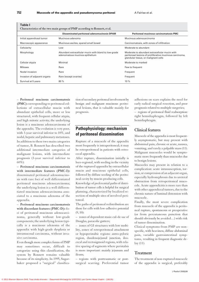
Table ICharacteristics of the two main groups of PMP according to Ronnett, et al.
Disseminated peritoneal adenomucinosis DPAM Peritoneal mucinous carcinomatosis PMC
Initial appendiceal tumor Mucinous adenoma Mucinous adenocarcinoma
Macroscopic appearance Mucinous ascites, spared small bowel Carcinomatosis, with zones of infiltration
Cellularity Low Moderate to abundant
Morphology Abundant extracellular mucin with bland to low-grade adenomatous mucinos epithelium
Moderate to abundant extracellular mucin with peritoneal lesions of proliferative mucinous carcinoma, glandular tissue, or malignant cells
Cellular atypia Minimal Moderate to marked
Mitoses Rare Few to frequent
Nodal invasion Rare Frequent
Invasion of adjacent organs Rare (except ovaries) Frequent
Survival at 5 years 80 % 10 %
© 2011 Elsevier Masson SAS. Tous droits réservés. - Document téléchargé le 12/07/2011 par Centre Alexis Vautrin - (16126)

J Radiol 2008;89
A Fairise et al. Mucocele of the appendix and pseudomyxoma peritonei 753
from laparotomy as opposed to laparoscopy;in case of laparoscopy, a collection bagmust absolutely be used to prevent perito-neal seeding (11). Appendectomy is per-formed, without appendiceal rupture, withcomplete resection of the meso-appendixand peritoneal fluid sampling for cyto-logy. Based on results from cytology, mucocelerupture, and involvement of the meso-appendix, additional treatment may berequired: right hemicolectomy, tumorcytoreduction, intraperitoneal chemo-hyperthermia.The treatment of PMP is similar for alltypes: radical tumor reduction surgerycharacterized by optimal gross total re-moval of mucus and tumor implantswith omentectomy, localized peritonealresection, right hemicolectomy, and bi-lateral oophorectomy in females, withperoperative intraperitoneal chemohy-perthermia, with improved prognosisby treating residual microscopic disease(10, 12, 13).
Imaging features
Mucocele of the appendixAbdomen radiographsRadiographs of the abdomen are rarelyhelpful for diagnosis. Mucoceles are ra-rely visible on plain radiographs as a rightflank or iliac fossa mass with curvilinear
mural calcifications (fig. 1). Calcificationsare not always present (1). Abdomen radio-graphs are not part of the work-up ofmucocele of the appendix.
Barium studiesBarium studies are no longer recommen-ded in the initial work-up of abdominalpain.The imaging features on barium enemadescribed in association with mucocele ofthe appendix included extrinsic compres-sion of the medial cecal wall, absence ofappendiceal opacification, and presenceof vertical folds on the cecal mucosa cor-responding to a concentric distribution ofmucosal folds around the obstructed ap-pendiceal orifice (1, 14).In rare cases of cecal intussusception (fig. 2),barium enema would demonstrate the pre-sence of an endoluminal mass at the levelof the cecum, right hemicolon, and eventransverse colon, to be differentiated fromintussusception due to carcinoma at theileocecal valve or colonic lipoma. The presence of thin mural calcificationswould in this case be valuable to suggest adiagnosis of intussusception due to muco-cele (1).
UltrasoundTypical mucoceles of the appendix arehypoechoic masses of the right lower ab-domen. They may have a layered appea-rance (onion skin sign) (fig. 3 and 4). Theyare well defined, cylindrical or lobulated
in shape (pear shaped), with increasedthrough transmission, and frequent thincurvilinear or punctate echogenic muralcalcifications (fig. 5). They are mobile butattached to the cecum (14, 15).In the absence of these characteristic fea-tures, the sonographic appearance ofmucoceles may be misleading. The USappearance of the mucocele content va-ries based on the presence of necrosis, celldebris, and especially the consistency ofthe mucus, which may cause the muco-cele to have a spectrum of appearancesfrom a cystic anechoic mass (fig. 6) to a“solid” mass with “soft tissue” echogeni-city (1).The wall may be of variable thickness,especially in the presence of mucosalhyperplasia, that may mimic acute ap-pendicitis or a malignant tumor of the ap-pendix. Septations (fig. 6) and endoluminal poly-poid nodules may be present. These fea-tures are not specific for diagnosis of theunderlying lesion. Mural calcifications may be present in lessthan 50% of cases (15).The size is variable, up to 10 cm in somepatients; the relationship to the cecummay be difficult to confirm for larger lesions(16).Finally, myxoglobulosis, a rare variant ofmucocele, is characterized by distensionof the appendiceal lumen by small rim-calcified spherical globules, visible on radio-graphs, US and CT (1, 14).
Fig. 1: Mucocele of the appendix (arrow) with calcified wall in the right lower abdomen.a Abdomen radiographb Axial CT image
a b
© 2011 Elsevier Masson SAS. Tous droits réservés. - Document téléchargé le 12/07/2011 par Centre Alexis Vautrin - (16126)
J Radiol 2008;89
754 Mucocele of the appendix and pseudomyxoma peritonei A Fairise et al.
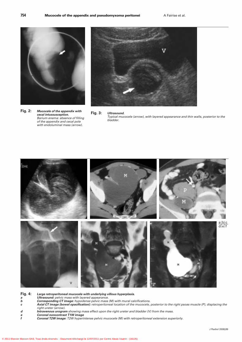
Fig. 2: Mucocele of the appendix with cecal intussusception. Barium enema: absence of filling of the appendix and cecal pole with endoluminal mass (arrow).
Fig. 3: Ultrasound. Typical mucocele (arrow), with layered appearance and thin walls, posterior to the
bladder.
Fig. 4: Large retroperitoneal mucocele with underlying villous hyperplasia.a Ultrasound: pelvic mass with layered appearance.b Corresponding CT image: hypodense pelvic mass (M) with mural calcifications.c Axial CT image (bowel opacification): retroperitoneal location of the mucocele, posterior to the right psoas muscle (P), displacing the
right ureter (arrow).d Intravenous urogram showing mass effect upon the right ureter and bladder (V) from the mass.e Coronal noncontrast T1W imagef Coronal T2W image: T2W hyperintense pelvic mucocele (M) with retroperitoneal extension superiorly.
a b cd e f
© 2011 Elsevier Masson SAS. Tous droits réservés. - Document téléchargé le 12/07/2011 par Centre Alexis Vautrin - (16126)
J Radiol 2008;89
755 Mucocele of the appendix and pseudomyxoma peritonei A Fairise et al.
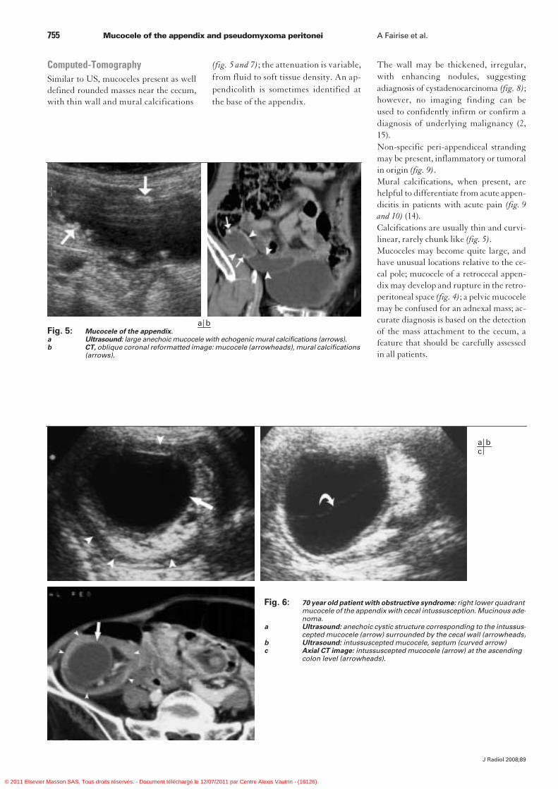
Computed-TomographySimilar to US, mucoceles present as welldefined rounded masses near the cecum,with thin wall and mural calcifications
(fig. 5 and 7); the attenuation is variable,from fluid to soft tissue density. An ap-pendicolith is sometimes identified atthe base of the appendix.
Fig. 5: Mucocele of the appendix.a Ultrasound: large anechoic mucocele with echogenic mural calcifications (arrows).b CT, oblique coronal reformatted image: mucocele (arrowheads), mural calcifications
(arrows).
a b
Fig. 6: 70 year old patient with obstructive syndrome: right lower quadrant mucocele of the appendix with cecal intussusception. Mucinous ade-noma.
a Ultrasound: anechoic cystic structure corresponding to the intussus-cepted mucocele (arrow) surrounded by the cecal wall (arrowheads)
b Ultrasound: intussuscepted mucocele, septum (curved arrow)c Axial CT image: intussuscepted mucocele (arrow) at the ascending
colon level (arrowheads).
a bc
The wall may be thickened, irregular,with enhancing nodules, suggestingadiagnosis of cystadenocarcinoma (fig. 8);however, no imaging finding can beused to confidently infirm or confirm adiagnosis of underlying malignancy (2,15).Non-specific peri-appendiceal strandingmay be present, inflammatory or tumoralin origin (fig. 9).Mural calcifications, when present, arehelpful to differentiate from acute appen-dicitis in patients with acute pain (fig. 9and 10) (14).Calcifications are usually thin and curvi-linear, rarely chunk like (fig. 5).Mucoceles may become quite large, andhave unusual locations relative to the ce-cal pole; mucocele of a retrocecal appen-dix may develop and rupture in the retro-peritoneal space (fig. 4); a pelvic mucocelemay be confused for an adnexal mass; ac-curate diagnosis is based on the detectionof the mass attachment to the cecum, afeature that should be carefully assessedin all patients.
© 2011 Elsevier Masson SAS. Tous droits réservés. - Document téléchargé le 12/07/2011 par Centre Alexis Vautrin - (16126)

J Radiol 2008;89
756 Mucocele of the appendix and pseudomyxoma peritonei A Fairise et al.
Complications may be detected bothon US and CT: intussusception (fig. 6),torsion, ureteral compression (fig. 4).
Magnetic Resonance ImagingLesion location and morphological featu-res are identical to CT; the mucocele isT1W hypointense and T2W hyperinten-se, but signal is variable based on mucincontent (fig. 4). Calcifications may be lessconspicuous (2, 3).
Pseudomyxoma PeritoneiOn US and CT, the diagnosis of PMP isbased on the identification of three le-sions: characteristic mucinous ascites,nodular peritoneal implants, and the pri-mary tumor; the latter is only rarely vi-sualized.
UltrasoundMucinous ascites typically is heteroge-neous, hypoechoic, poorly mobile, with
Fig. 7: Mucocele of the appendix with underlying diffuse hyperplasia.a Axial CT image: hypodense mass (M) behind the cecum (C), with thin calcified wall.
The presence of wall calcifications differentiates the mass from a retrocecal abscess.b Macroscopy.
a b
Fig. 8: Mucocele of the appendix with underlying adenocarcinoma; pericecal mass with enhancing irregular nodular walls (arrows), septations.
a more or less layered appearance. It may beloculated, with multiple septations, andthin septal calcifications (fig. 11).It is classically more heterogeneous, lessanechoic, and less mobile than simpleascitis. In some cases, it may even appearas an echogenic soft tissue mass. Mass effect with extrinsic compressionand scalloping of the liver and spleenand displacement of hollow structuresmay be seen both on US and CT (17,18).
Computed-TomographyOn CT, mucinous ascites typically ishypodense, but usually slightly denserthan a simple transsudate (fig. 10, 12and 13); it may be loculated (fig. 11 and14) and contain thin curvilinear calcifi-cations (17).Thick gelatinous ascites and solid nodularperitoneal implants may cause extrinsiccompression with typical scalloping ofupper abdominal solid organs (fig. 15). A characteristic feature of liver scal-loping is the discordance between thepresence of multiple or large hypodenseperipheral lesions and the absence of in-traparenchymal lesions, away from theliver capsule (fig. 15).Gelatinous ascites may also cause defor-mity and displacement of lower abdo-minal structures and vessels, with fixa-tion of small bowel loops (fig. 14 and 16)(9, 19).Peritoneal implants correspond to hete-rogeneous nodules that may enhancefollowing intravenous administrationof contrast. Their presence should beassessed at the level of the greateromentum, cul-de-sac of Douglas, ova-ries, paracolic gutters, and subphrenicregions (fig. 11, 12 and 15) (9).They may be disseminated and involvemultiple peritoneal and retroperitonealstructures.In patients with ascites and peritonealimplants, differentiation between PMPand peritoneal carcinomatosis may be dif-ficult; the presence of calcifications and/or septations, the degree of scallopingand the hypodense nature of the gelatinousmasses all favor a diagnosis of PMP (20).
© 2011 Elsevier Masson SAS. Tous droits réservés. - Document téléchargé le 12/07/2011 par Centre Alexis Vautrin - (16126)
J Radiol 2008;89
A Fairise et al. Mucocele of the appendix and pseudomyxoma peritonei 757
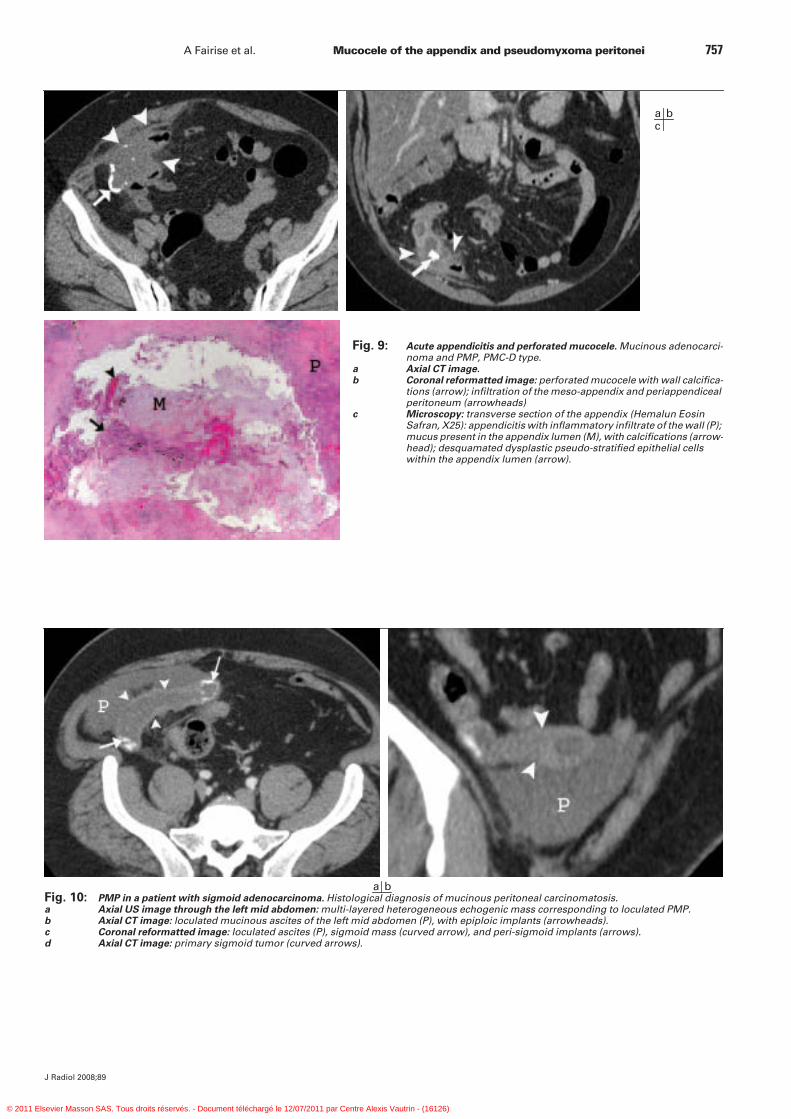
Fig. 9: Acute appendicitis and perforated mucocele. Mucinous adenocarci-noma and PMP, PMC-D type.
a Axial CT image.b Coronal reformatted image: perforated mucocele with wall calcifica-
tions (arrow); infiltration of the meso-appendix and periappendiceal peritoneum (arrowheads)
c Microscopy: transverse section of the appendix (Hemalun Eosin Safran, X25): appendicitis with inflammatory infiltrate of the wall (P); mucus present in the appendix lumen (M), with calcifications (arrow-head); desquamated dysplastic pseudo-stratified epithelial cells within the appendix lumen (arrow).
a bc
Fig. 10: PMP in a patient with sigmoid adenocarcinoma. Histological diagnosis of mucinous peritoneal carcinomatosis.a Axial US image through the left mid abdomen: multi-layered heterogeneous echogenic mass corresponding to loculated PMP.b Axial CT image: loculated mucinous ascites of the left mid abdomen (P), with epiploic implants (arrowheads).c Coronal reformatted image: loculated ascites (P), sigmoid mass (curved arrow), and peri-sigmoid implants (arrows).d Axial CT image: primary sigmoid tumor (curved arrows).
a b
© 2011 Elsevier Masson SAS. Tous droits réservés. - Document téléchargé le 12/07/2011 par Centre Alexis Vautrin - (16126)
J Radiol 2008;89
758 Mucocele of the appendix and pseudomyxoma peritonei A Fairise et al.
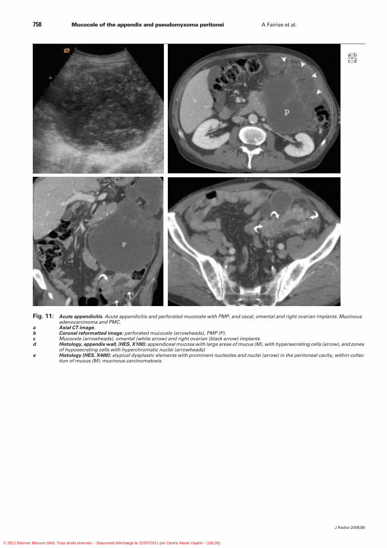
Fig. 11: Acute appendicitis. Acute appendicitis and perforated mucocele with PMP, and cecal, omental and right ovarian implants. Mucinous adenocarcinoma and PMC.
a Axial CT image.b Coronal reformatted image: perforated mucocele (arrowheads), PMP (P).c Mucocele (arrowheads), omental (white arrow) and right ovarian (black arrow) implantsd Histology, appendix wall, (HES, X100): appendiceal mucosa with large areas of mucus (M), with hypersecreting cells (arrow), and zones
of hyposecreting cells with hyperchromatic nuclei (arrowheads)e Histology (HES, X400): atypical dysplastic elements with prominent nucleoles and nuclei (arrow) in the peritoneal cavity, within collec-
tion of mucus (M): mucinous carcinomatosis.
a bc d
© 2011 Elsevier Masson SAS. Tous droits réservés. - Document téléchargé le 12/07/2011 par Centre Alexis Vautrin - (16126)
J Radiol 2008;89
A Fairise et al. Mucocele of the appendix and pseudomyxoma peritonei 759
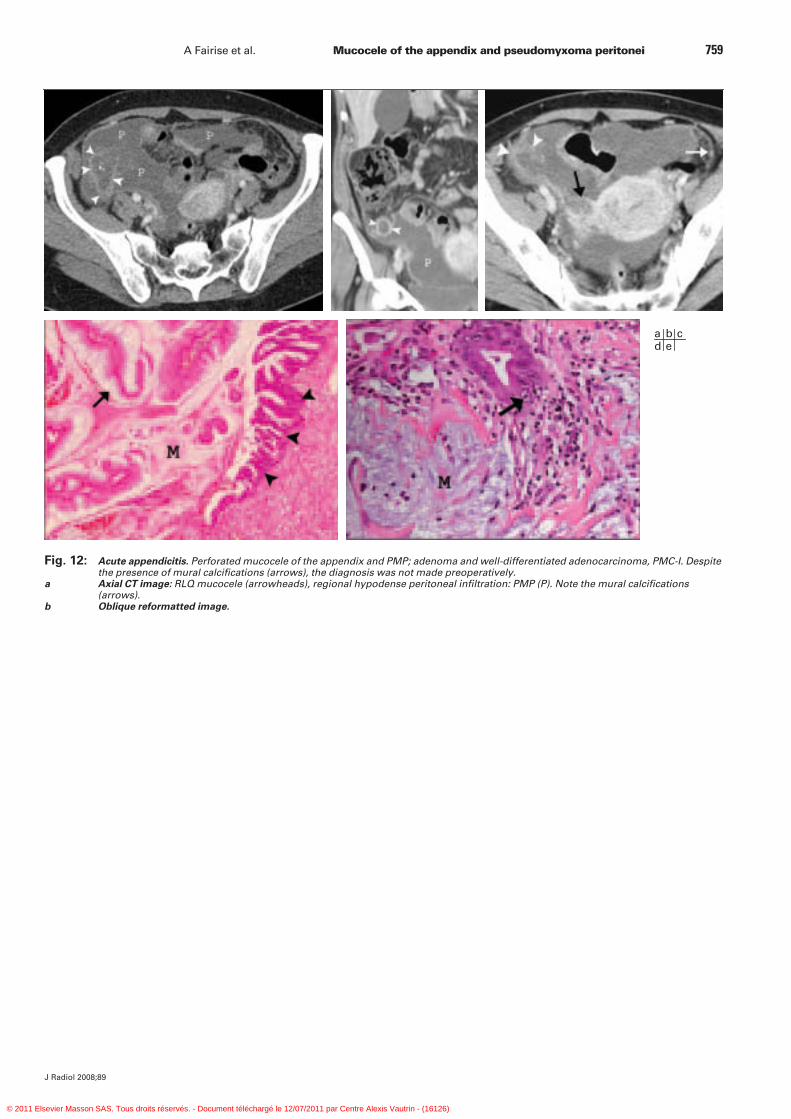
Fig. 12: Acute appendicitis. Perforated mucocele of the appendix and PMP; adenoma and well-differentiated adenocarcinoma, PMC-I. Despite the presence of mural calcifications (arrows), the diagnosis was not made preoperatively.
a Axial CT image: RLQ mucocele (arrowheads), regional hypodense peritoneal infiltration: PMP (P). Note the mural calcifications (arrows).
b Oblique reformatted image.
a b cd e
© 2011 Elsevier Masson SAS. Tous droits réservés. - Document téléchargé le 12/07/2011 par Centre Alexis Vautrin - (16126)
J Radiol 2008;89
760 Mucocele of the appendix and pseudomyxoma peritonei A Fairise et al.
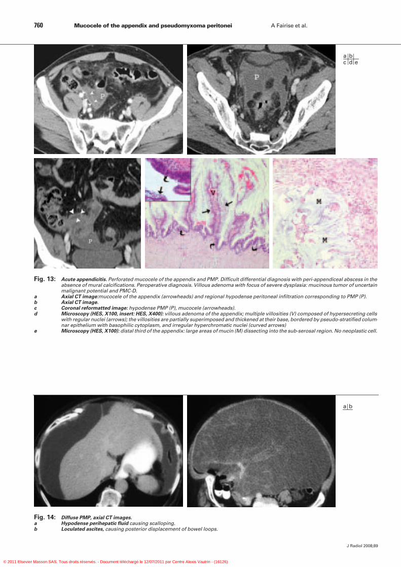
Fig. 13: Acute appendicitis. Perforated mucocele of the appendix and PMP. Difficult differential diagnosis with peri-appendiceal abscess in the absence of mural calcifications. Peroperative diagnosis. Villous adenoma with focus of severe dysplasia: mucinous tumor of uncertain malignant potential and PMC-D.
a Axial CT image:mucocele of the appendix (arrowheads) and regional hypodense peritoneal infiltration corresponding to PMP (P).b Axial CT image.c Coronal reformatted image: hypodense PMP (P), mucocele (arrowheads).d Microscopy (HES, X100, insert: HES, X400): villous adenoma of the appendix; multiple villosities (V) composed of hypersecreting cells
with regular nuclei (arrows); the villosities are partially superimposed and thickened at their base, bordered by pseudo-stratified colum-nar epithelium with basophilic cytoplasm, and irregular hyperchromatic nuclei (curved arrows)
e Microscopy (HES, X100): distal third of the appendix: large areas of mucin (M) dissecting into the sub-serosal region. No neoplastic cell.
a bc d e
Fig. 14: Diffuse PMP, axial CT images.a Hypodense perihepatic fluid causing scalloping.b Loculated ascites, causing posterior displacement of bowel loops.
a b
© 2011 Elsevier Masson SAS. Tous droits réservés. - Document téléchargé le 12/07/2011 par Centre Alexis Vautrin - (16126)

J Radiol 2008;89
A Fairise et al. Mucocele of the appendix and pseudomyxoma peritonei 761
ConclusionThe detection and diagnosis of a non-ruptured mucocele of the appendix onimaging studies is a significant prognosticfactor, allowing surgeons to take the ne-cessary precautions to avoid peroperativerupture with peritoneal seeding. Whilediagnosis on imaging studies may not
always be straightforward, the presenceof a few imaging features including ap-pendiceal distension, mural calcificationsand septations, should raise the possibilityof mucocele, irrespective of the presen-ting clinical symptoms. Early detection of a ruptured mucocele ofthe appendix with PMP plays a signifi-cant role by enabling tailored and radical
Fig. 15: Diffuse PMP:mucinous peritoneal carcinomatosis. History of appen-dectomy and mucinous adenocarcinoma two years previously.
a Axial CT images: Heterogeneous diffuse multinodular ascites; liver scalloping (arrowheads) and lesser sac involvement (P).
b, c Multiple nodular peritoneal, omental and parietal masses (arrows).
a bc
Fig. 16: Axial CT image: diffuse PMP with displacement and grouping of bowel loops at the center of the abdomen.
surgical intervention in order to reducethe number of subsequent surgeries withworsened prognosis.
References1. Dachman AH, Lichtenstein JE, Fried-
man AC. Mucocele of the appendix andpseudomyxoma peritonei. AJR Am JRoentgenol 1985;144:923-9.
2. Pickhardt PJ, Levy AD, Rohrmann CA Jr,Kende AI. Primary neoplasms of theappendix: radiologic spectrum of diseasewith pathologic correlation. Radiogra-phics 2003;23:645-62.
3. Derelle AL, Tissier S, Granger P et al.Diagnostic précoce de pseudomyxomepéritonéal localisé à la zone de ruptured’une mucocèle appendiculaire : image-rie et aspects anatomopathologiques. JRadiol 2007;88:289-95.
4. O’Connell JT, Tomlinson JS, Roberts AAet al. Pseudomyxoma peritonei is a di-sease of MUC2-expressing goblet cells.Am J Pathol 2002;161:551-64.
5. Szych C, Staebler A, Connolly DC et al.Molecular genetic evidence supportingthe clonality and appendiceal origin of
© 2011 Elsevier Masson SAS. Tous droits réservés. - Document téléchargé le 12/07/2011 par Centre Alexis Vautrin - (16126)
J Radiol 2008;89
762 Mucocele of the appendix and pseudomyxoma peritonei A Fairise et al.
pseudomyxoma peritonei in women. AmJ Pathol 1999;154:1849-55.
6. Stewart CJ, Tsukamoto T, Cooke B etal. Ovarian mucinous tumour arising inmature cystic teratoma and associatedwith pseudomyxoma peritonei: report oftwo cases and comparison with ovarianinvolvement by low-grade appendicealmucinous tumour. Pathology 2006;38:534-8.
7. Takeuchi M, Matsuzaki K, Yoshida S etal. Imaging findings of urachal mucinouscystadenocarcinoma associated withpseudomyxoma peritonei. Acta Radiol2004;45:348-50.
8. Ronnett BM, Zahn CM, Kurman RJ et al.Disseminated peritoneal adenomucinosisand peritoneal mucinous carcinomatosis.A clinicopathologic analysis of 109 caseswith emphasis on distinguishing patholo-gic features, site of origin, prognosis, and
relationship to 'pseudomyxoma perito-nei”. Am J Surg Pathol 1995;19:1390-408.
9. Régent D, Laurent V, Cannard L et al. Lepéritoine « témoin » de la pathologie abdo-mino-pelvienne. J Radiol 2004;85:555-71.
10. Sugarbaker PH. Pseudomyxoma perito-nei. A cancer whose biology is characteri-zed by a redistribution phenomenon.Ann Surg 1994;219:109-11.
11. Dixit A, Robertson JHP, Mudan SS et al.Appendiceal mucocoeles and pseudo-myxoma peritonei. World J Gastroente-rol 2007;13:2381-4.
12. Li C, Kanthan R, Kanthan SC. Pseud-myxoma peritonei – a revisit : report of2 cases and literature review. World JSurg Oncol 2006;4:60.
13. Qu ZB, Liu LX. Management of pseudo-myxoma peritonei. World J Gastroente-rol 2006;12:6124-7.
14. Madwed D, Mindelzun R, Jeffrey RB etal. Mucocele of the appendix : Imaging
findings. AJR Am J Roentgenol 1992;159:69-72.
15. Kim SH, Lim HK, Lee WJ et al. Mucoceleof the appendix : ultrasonographic and CTfindings. Abdom Imaging 1998;23: 292-6.
16. Francica G, Lapiccirella G, Giardiello C,et al. Giant mucocele of the appendix. JUltrasound Med 2006;25:643-8.
17. Walensky RP, Venbrux AC, Prescott CAet al. Pseudomyxoma peritonei. AJR AmJ Roentgenol 1996;167:471-4.
18. Hanbidge AE, Lynch D, Wilson SR. USof the Peritoneum. Radiographics 2003;23:663-85.
19. Seshul MB, Coulam CM. Pseudomyxo-ma Peritonei : Computed tomographyand sonography. AJR Am J Roentgenol1981;136:803-6.
20. Taourel P, Baud C, Lesnik A et al. Le pé-ritoine acteur de la pathologie abdomina-le. J Radiol 2004;85:574-90.
© 2011 Elsevier Masson SAS. Tous droits réservés. - Document téléchargé le 12/07/2011 par Centre Alexis Vautrin - (16126)