Structure of Sputnik, a virophage, at 3.5-Å · PDF filesuggested (9), to have a double...
6
Structure of Sputnik, a virophage, at 3.5-Å resolution Xinzheng Zhang a , Siyang Sun a , Ye Xiang a , Jimson Wong a , Thomas Klose a , Didier Raoult b , and Michael G. Rossmann a,1 a Department of Biological Sciences, Purdue University, West Lafayette, IN 47907; and b Unité de Recherche sur les Maladies Infectieuses et Tropicales Emergentes, Unité Mixte de Recherche 6236 Centre National de la Recherche Scientifique, L’Institut de Recherche pour le Développement 198, Faculté de Médecine, Université de la Méditerranée, 13385 Marseille Cedex 5, France Edited by Nikolaus Grigorieff, Howard Hughes Medical Institute, Brandeis University, Waltham, MA, and accepted by the Editorial Board September 26, 2012 (received for review July 10, 2012) “Sputnik” is a dsDNA virus, referred to as a virophage, that is coassembled with Mimivirus in the host amoeba. We have used cryo-EM to produce an electron density map of the icosahedral Sputnik virus at 3.5-Å resolution, sufficient to verify the identity of most amino acids in the capsid proteins and to establish the identity of the pentameric protein forming the fivefold vertices. It was also shown that the virus lacks an internal membrane. The capsid is organized into a T = 27 lattice in which there are 260 trimeric capsomers and 12 pentameric capsomers. The trimeric cap- somers consist of three double “jelly-roll” major capsid proteins creating pseudohexameric capsomer symmetry. The pentameric capsomers consist of five single jelly-roll proteins. The release of the genome by displacing one or more of the pentameric capsomers may be the result of a low-pH environment. These results suggest a mechanism of Sputnik DNA ejection that probably also occurs in other big icosahedral double jelly-roll viruses such as Adenovirus. In this study, the near-atomic resolution structure of a virus has been established where crystallization for X-ray crystallography was not feasible. dsDNA egress | virus assembly | Mimivirus satellite | polypeptide tracing | cryo-electron microscopy M imivirus is one of the largest known viruses, first discovered in a water tower during a search for the cause of a hospital- acquired pneumonia outbreak in 2003 (1). Mimivirus has a dsDNA genome and uses Acanthamoeba as its natural host (1). Sputnik virus was discovered to be associated with Mimivirus when isolated from amoeba (2). Sputnik can only be propagated when coinfected with Mimivirus. The amoeba host implies that both Mimivirus and Sputnik are frequently present in aquatic food, resulting in disease when humans eat raw food (3). Unlike most satellite viruses, coinfection decreases the production of Mimivirus by approximately 70% (2). The requirement of Mimi- virus for the propagation of Sputnik gave rise to the term “viro- phage” (4). EM images suggest that Sputnik virus matures in viral factories together with Mimivirus itself (5). This observation is supported by in situ hybridization experiments (6), which showed that Sputnik replicates its genome in viral factories. Other viroph- ages such as Mavirus and Organic Lake virus, which are associ- ated with giant dsDNA viruses, have been discovered recently (7, 8). These virophages might influence the ecosystem of aquatic environments (8). Sputnik virions have an AT-rich, circular 18-kb dsDNA genome with 21 ORFs (1). Genes V20, V19, and V8 were tentatively as- sociated with the major and two minor capsid proteins, respectively (1). Sequences homologous to the major capsid protein (MCP) of Sputnik have been found in Mavirus (19% sequence identity) and Organic Lake virus (28% sequence identity). Another eight ORFs of Spunik’s genome have similarity to eukaryal, archaeal, and bacterial virus genes. A 10.7-Å resolution cryo-EM study of the Sputnik virus (9) had shown that the capsid protein assembled into an icosahedral shell with a diameter of approximately 750 Å and T = 27 quasi- symmetry (SI Materials and Methods). Thus, the capsid is composed of 260 quasi-hexomeric “hexons” and 12 pentomeric “pentons.” The double “jelly-roll” hexon structure of the Paramecium bursaria Chlorella virus 1 (PBCV-1) MCP (Vp54) fitted well into the 10.7- Å resolution cryo-EM density map of Sputnik (9). A mushroom- like fiber was found associated with the center of each hexon, although the icosahedrally averaged density suggested that the fiber had only partial occupancy. The capsid protein appeared to surround a membrane that enclosed the genome, consistent with the presence of lipid in the virus. The capsid structure of numerous icosahedral viruses consists of several jelly-roll folds organized into pseudohexameric cap- somers. The jelly-roll structure is a β-barrel formed by two opposing antiparallel-stranded β-sheets. If the strands along the polypeptide are identified by B, C, D, E, F, G, H, and I, then the strands in the two sheets are BIDG and CHEF (10). The jelly-roll structure was first recognized in the capsid of small icosahedral plant viruses (11, 12). The structure of small RNA animal viruses such as picornaviruses were then discovered to have similar tertiary structures for their capsid proteins as well as similar quaternary structure for the organization of the viral capsid (13, 14). Some ssDNA viruses were also found to have a jelly-roll structure for their capsid proteins (15, 16). The structure of the dsDNA ad- enovirus capsid protein was found to have two consecutive jelly- rolls in its MCP (17), giving the base of the capsomer a hexago- nal shape. A similar double jelly-roll was found in the plant RNA cowpea mosaic virus (18) and later in PBCV-1 (19). Other viruses with double jelly-roll MCP structures include the dsDNA PRD1 bacterial phage (20), the marine phage PM2 (21, 22), the archaeal Sulfolobus turreted icosahedral virus (23, 24), and vaccinia virus (25, 26). Furthermore, there are numerous other viruses that most likely also have double jelly-roll MCPs according to sequence comparisons, as for instance Mimivirus (1) and African swine fever virus. Where the structure is known, the double jelly- roll forms a trimeric “capsomer” that has quasi-sixfold symmetry. In double jelly-roll structures, large insertions often occur be- tween β-strands B and D and between F and G. These insertions form surface features of the virus. The term “tower” has been used for especially large (approximately 300 amino acids) insertions between strands DE and FG in the first jelly-roll of the adeno- virus structure (17), giving the external part of the capsomer a triangular appearance. We report here a 3.5-Å near-atomic resolution structure of Sputnik determined by cryo-EM. The MCP was confirmed as being the product of gene V20 and was shown, as previously Author contributions: X.Z. and M.G.R. designed research; X.Z., S.S., and T.K. performed research; X.Z., J.W., and D.R. contributed new reagents/analytic tools; X.Z. and Y.X. ana- lyzed data; and X.Z. and M.G.R. wrote the paper. The authors declare no conflict of interest. This article is a PNAS Direct Submission. N.G. is a guest editor invited by the Editorial Board. Data deposition: The atomic coordinates and structure factors have been deposited in the Electron Microscopy Data Bank, www.ebi.ac.uk/pdbe/emdb [EMDB ID codes 5495 (3.5-Å resolution cryo-EM map of Sputnik) and 5496 (3.8-Å resolution cryo-EM map of empty Sputnik)]; and the Protein Data Bank, www.pdb.org [PDB ID code 3J26 (coordinates of the near-atomic resolution structure of Sputnik)]. 1 To whom correspondence should be addressed. E-mail: [email protected]. This article contains supporting information online at www.pnas.org/lookup/suppl/doi:10. 1073/pnas.1211702109/-/DCSupplemental. www.pnas.org/cgi/doi/10.1073/pnas.1211702109 PNAS | November 6, 2012 | vol. 109 | no. 45 | 18431–18436 BIOPHYSICS AND COMPUTATIONAL BIOLOGY
Transcript of Structure of Sputnik, a virophage, at 3.5-Å · PDF filesuggested (9), to have a double...
Structure of Sputnik, a virophage, at 3.5-Å resolutionXinzheng Zhanga, Siyang Suna, Ye Xianga, Jimson Wonga, Thomas Klosea, Didier Raoultb, and Michael G. Rossmanna,1
aDepartment of Biological Sciences, Purdue University, West Lafayette, IN 47907; and bUnité de Recherche sur les Maladies Infectieuses et TropicalesEmergentes, Unité Mixte de Recherche 6236 Centre National de la Recherche Scientifique, L’Institut de Recherche pour le Développement 198, Faculté deMédecine, Université de la Méditerranée, 13385 Marseille Cedex 5, France
Edited by Nikolaus Grigorieff, Howard Hughes Medical Institute, Brandeis University, Waltham, MA, and accepted by the Editorial Board September 26, 2012(received for review July 10, 2012)
“Sputnik” is a dsDNA virus, referred to as a virophage, that iscoassembled with Mimivirus in the host amoeba. We have usedcryo-EM to produce an electron density map of the icosahedralSputnik virus at 3.5-Å resolution, sufficient to verify the identityof most amino acids in the capsid proteins and to establish theidentity of the pentameric protein forming the fivefold vertices.It was also shown that the virus lacks an internal membrane. Thecapsid is organized into a T = 27 lattice in which there are 260trimeric capsomers and 12 pentameric capsomers. The trimeric cap-somers consist of three double “jelly-roll” major capsid proteinscreating pseudohexameric capsomer symmetry. The pentamericcapsomers consist of five single jelly-roll proteins. The release ofthe genome by displacing one or more of the pentameric capsomersmay be the result of a low-pH environment. These results suggest amechanism of Sputnik DNA ejection that probably also occurs inother big icosahedral double jelly-roll viruses such as Adenovirus.In this study, the near-atomic resolution structure of a virus hasbeen established where crystallization for X-ray crystallographywas not feasible.
dsDNA egress | virus assembly | Mimivirus satellite | polypeptide tracing |cryo-electron microscopy
Mimivirus is one of the largest known viruses, first discoveredin a water tower during a search for the cause of a hospital-
acquired pneumonia outbreak in 2003 (1). Mimivirus has adsDNA genome and uses Acanthamoeba as its natural host (1).Sputnik virus was discovered to be associated with Mimiviruswhen isolated from amoeba (2). Sputnik can only be propagatedwhen coinfected with Mimivirus. The amoeba host implies thatboth Mimivirus and Sputnik are frequently present in aquaticfood, resulting in disease when humans eat raw food (3). Unlikemost satellite viruses, coinfection decreases the production ofMimivirus by approximately 70% (2). The requirement of Mimi-virus for the propagation of Sputnik gave rise to the term “viro-phage” (4). EM images suggest that Sputnik virus matures in viralfactories together with Mimivirus itself (5). This observation issupported by in situ hybridization experiments (6), which showedthat Sputnik replicates its genome in viral factories. Other viroph-ages such as Mavirus and Organic Lake virus, which are associ-ated with giant dsDNA viruses, have been discovered recently(7, 8). These virophages might influence the ecosystem of aquaticenvironments (8).Sputnik virions have an AT-rich, circular 18-kb dsDNA genome
with 21 ORFs (1). Genes V20, V19, and V8 were tentatively as-sociated with the major and two minor capsid proteins, respectively(1). Sequences homologous to the major capsid protein (MCP)of Sputnik have been found in Mavirus (19% sequence identity)and Organic Lake virus (28% sequence identity). Another eightORFs of Spunik’s genome have similarity to eukaryal, archaeal,and bacterial virus genes.A 10.7-Å resolution cryo-EM study of the Sputnik virus (9)
had shown that the capsid protein assembled into an icosahedralshell with a diameter of approximately 750 Å and T = 27 quasi-symmetry (SI Materials and Methods). Thus, the capsid is composedof 260 quasi-hexomeric “hexons” and 12 pentomeric “pentons.”The double “jelly-roll” hexon structure of the Paramecium bursaria
Chlorella virus 1 (PBCV-1) MCP (Vp54) fitted well into the 10.7-Å resolution cryo-EM density map of Sputnik (9). A mushroom-like fiber was found associated with the center of each hexon,although the icosahedrally averaged density suggested that thefiber had only partial occupancy. The capsid protein appeared tosurround a membrane that enclosed the genome, consistent withthe presence of lipid in the virus.The capsid structure of numerous icosahedral viruses consists
of several jelly-roll folds organized into pseudohexameric cap-somers. The jelly-roll structure is a β-barrel formed by two opposingantiparallel-stranded β-sheets. If the strands along the polypeptideare identified by B, C, D, E, F, G, H, and I, then the strands in thetwo sheets are BIDG and CHEF (10). The jelly-roll structure wasfirst recognized in the capsid of small icosahedral plant viruses(11, 12). The structure of small RNA animal viruses such aspicornaviruses were then discovered to have similar tertiarystructures for their capsid proteins as well as similar quaternarystructure for the organization of the viral capsid (13, 14). SomessDNA viruses were also found to have a jelly-roll structure fortheir capsid proteins (15, 16). The structure of the dsDNA ad-enovirus capsid protein was found to have two consecutive jelly-rolls in its MCP (17), giving the base of the capsomer a hexago-nal shape. A similar double jelly-roll was found in the plant RNAcowpea mosaic virus (18) and later in PBCV-1 (19). Otherviruses with double jelly-roll MCP structures include the dsDNAPRD1 bacterial phage (20), the marine phage PM2 (21, 22), thearchaeal Sulfolobus turreted icosahedral virus (23, 24), andvaccinia virus (25, 26). Furthermore, there are numerous otherviruses that most likely also have double jelly-roll MCPs accordingto sequence comparisons, as for instance Mimivirus (1) and Africanswine fever virus. Where the structure is known, the double jelly-roll forms a trimeric “capsomer” that has quasi-sixfold symmetry.In double jelly-roll structures, large insertions often occur be-tween β-strands B and D and between F and G. These insertionsform surface features of the virus. The term “tower” has been usedfor especially large (approximately 300 amino acids) insertionsbetween strands DE and FG in the first jelly-roll of the adeno-virus structure (17), giving the external part of the capsomer atriangular appearance.We report here a 3.5-Å near-atomic resolution structure of
Sputnik determined by cryo-EM. The MCP was confirmed asbeing the product of gene V20 and was shown, as previously
Author contributions: X.Z. and M.G.R. designed research; X.Z., S.S., and T.K. performedresearch; X.Z., J.W., and D.R. contributed new reagents/analytic tools; X.Z. and Y.X. ana-lyzed data; and X.Z. and M.G.R. wrote the paper.
The authors declare no conflict of interest.
This article is a PNAS Direct Submission. N.G. is a guest editor invited by theEditorial Board.
Data deposition: The atomic coordinates and structure factors have been deposited in theElectron Microscopy Data Bank, www.ebi.ac.uk/pdbe/emdb [EMDB ID codes 5495 (3.5-Åresolution cryo-EM map of Sputnik) and 5496 (3.8-Å resolution cryo-EM map of emptySputnik)]; and the Protein Data Bank, www.pdb.org [PDB ID code 3J26 (coordinates of thenear-atomic resolution structure of Sputnik)].1To whom correspondence should be addressed. E-mail: [email protected].
This article contains supporting information online at www.pnas.org/lookup/suppl/doi:10.1073/pnas.1211702109/-/DCSupplemental.
www.pnas.org/cgi/doi/10.1073/pnas.1211702109 PNAS | November 6, 2012 | vol. 109 | no. 45 | 18431–18436
BIOPH
YSICSAND
COMPU
TATIONALBIOLO
GY
suggested (9), to have a double jelly-roll structure. The proteinthat constitutes the penton was identified as being a combinationof gene products V18 and V19, which had wrongly been assignedto have a frameshift relative to each other. The resultant V18/19was shown to be a single jelly-roll structure. A 3.8-Å resolutionstructure of the emptied Sputnik virus was calculated and com-pared with the full virus structure, which suggests a possiblemechanism of Sputnik DNA ejection that might apply to otherlarge dsDNA viruses with double jelly-roll MCPs.
Results and DiscussionOverall Structure of the Sputnik Virus. A 3.5-Å resolution, icosahe-drally averaged map (Fig. 1 A–D) was determined by image re-construction using approximately 12,000 cryo-EM particles. Inaddition, a 3.8-Å resolution map of empty capsids was calculatedusing approximately 13,000 empty particles. There are 4 and 1/3hexon capsomers in each icosahedral asymmetric unit of the virus,where the 1/3 hexon is from a hexon sitting on the icosahedralthreefold axis. In addition there is 1/5 of a penton capsomer ad-jacent to the fivefold vertex in the asymmetric unit. After thehexon and penton densities had been interpreted there remainedthree similar uninterpreted densities, each representing nine aminoacids, presumably corresponding to the same sequence of a minorcapsid protein. These densities were located on the inside of thecapsid, at the boundary between pairs of hexons (Fig. S1), but areabsent in the empty capsids and must have been associated withthe DNA so as to be able to exit along with the DNA.
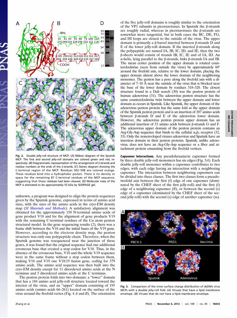
Major Capsid Protein. The structures of the 13 MCPs in the icosa-hedral asymmetric unit were built independently and then re-fined with the program CNS (27). The refined structure had an
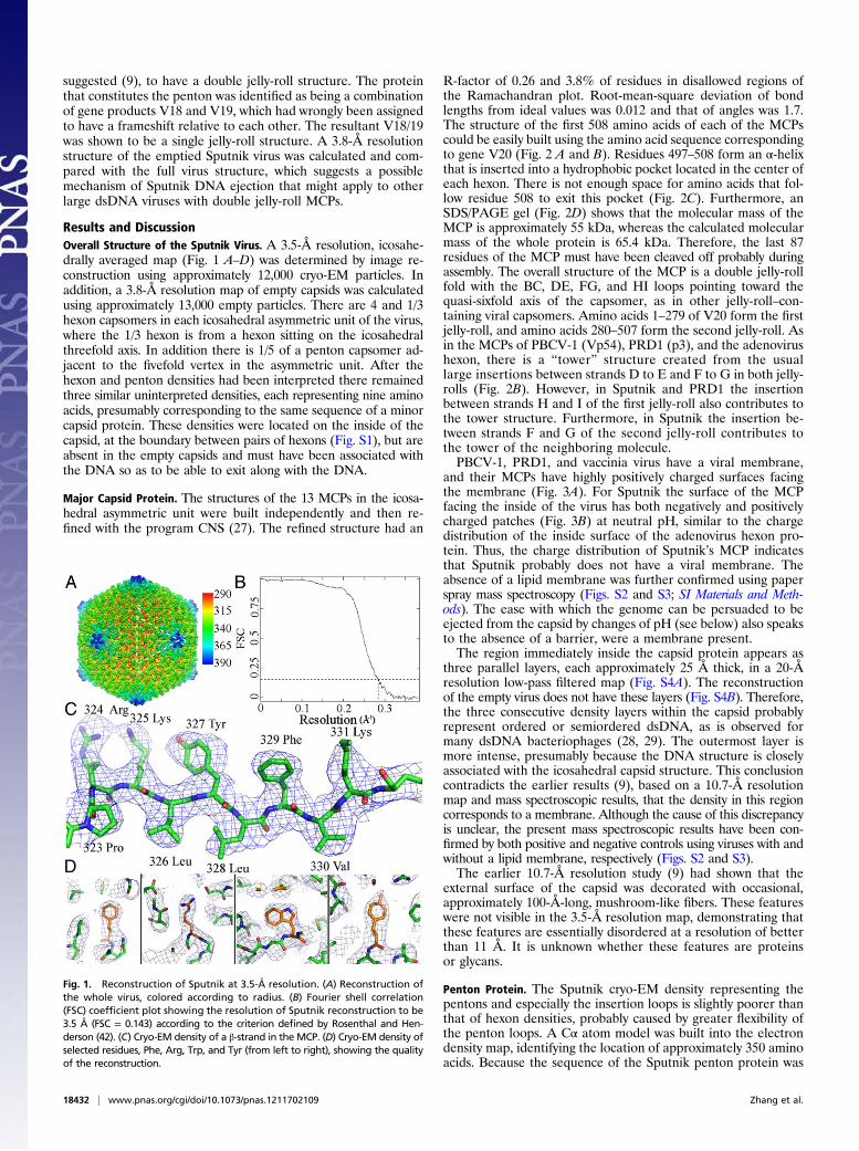
R-factor of 0.26 and 3.8% of residues in disallowed regions ofthe Ramachandran plot. Root-mean-square deviation of bondlengths from ideal values was 0.012 and that of angles was 1.7.The structure of the first 508 amino acids of each of the MCPscould be easily built using the amino acid sequence correspondingto gene V20 (Fig. 2 A and B). Residues 497–508 form an α-helixthat is inserted into a hydrophobic pocket located in the center ofeach hexon. There is not enough space for amino acids that fol-low residue 508 to exit this pocket (Fig. 2C). Furthermore, anSDS/PAGE gel (Fig. 2D) shows that the molecular mass of theMCP is approximately 55 kDa, whereas the calculated molecularmass of the whole protein is 65.4 kDa. Therefore, the last 87residues of the MCP must have been cleaved off probably duringassembly. The overall structure of the MCP is a double jelly-rollfold with the BC, DE, FG, and HI loops pointing toward thequasi-sixfold axis of the capsomer, as in other jelly-roll–con-taining viral capsomers. Amino acids 1–279 of V20 form the firstjelly-roll, and amino acids 280–507 form the second jelly-roll. Asin the MCPs of PBCV-1 (Vp54), PRD1 (p3), and the adenovirushexon, there is a “tower” structure created from the usuallarge insertions between strands D to E and F to G in both jelly-rolls (Fig. 2B). However, in Sputnik and PRD1 the insertionbetween strands H and I of the first jelly-roll also contributes tothe tower structure. Furthermore, in Sputnik the insertion be-tween strands F and G of the second jelly-roll contributes tothe tower of the neighboring molecule.PBCV-1, PRD1, and vaccinia virus have a viral membrane,
and their MCPs have highly positively charged surfaces facingthe membrane (Fig. 3A). For Sputnik the surface of the MCPfacing the inside of the virus has both negatively and positivelycharged patches (Fig. 3B) at neutral pH, similar to the chargedistribution of the inside surface of the adenovirus hexon pro-tein. Thus, the charge distribution of Sputnik’s MCP indicatesthat Sputnik probably does not have a viral membrane. Theabsence of a lipid membrane was further confirmed using paperspray mass spectroscopy (Figs. S2 and S3; SI Materials and Meth-ods). The ease with which the genome can be persuaded to beejected from the capsid by changes of pH (see below) also speaksto the absence of a barrier, were a membrane present.The region immediately inside the capsid protein appears as
three parallel layers, each approximately 25 Å thick, in a 20-Åresolution low-pass filtered map (Fig. S4A). The reconstructionof the empty virus does not have these layers (Fig. S4B). Therefore,the three consecutive density layers within the capsid probablyrepresent ordered or semiordered dsDNA, as is observed formany dsDNA bacteriophages (28, 29). The outermost layer ismore intense, presumably because the DNA structure is closelyassociated with the icosahedral capsid structure. This conclusioncontradicts the earlier results (9), based on a 10.7-Å resolutionmap and mass spectroscopic results, that the density in this regioncorresponds to a membrane. Although the cause of this discrepancyis unclear, the present mass spectroscopic results have been con-firmed by both positive and negative controls using viruses with andwithout a lipid membrane, respectively (Figs. S2 and S3).The earlier 10.7-Å resolution study (9) had shown that the
external surface of the capsid was decorated with occasional,approximately 100-Å-long, mushroom-like fibers. These featureswere not visible in the 3.5-Å resolution map, demonstrating thatthese features are essentially disordered at a resolution of betterthan 11 Å. It is unknown whether these features are proteinsor glycans.
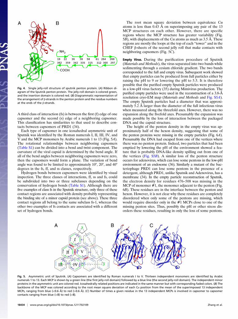
Penton Protein. The Sputnik cryo-EM density representing thepentons and especially the insertion loops is slightly poorer thanthat of hexon densities, probably caused by greater flexibility ofthe penton loops. A Cα atom model was built into the electrondensity map, identifying the location of approximately 350 aminoacids. Because the sequence of the Sputnik penton protein was
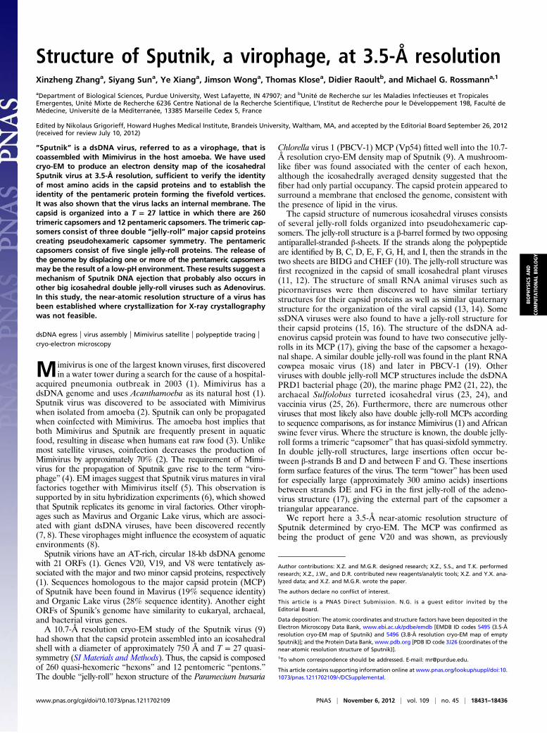
Fig. 1. Reconstruction of Sputnik at 3.5-Å resolution. (A) Reconstruction ofthe whole virus, colored according to radius. (B) Fourier shell correlation(FSC) coefficient plot showing the resolution of Sputnik reconstruction to be3.5 Å (FSC = 0.143) according to the criterion defined by Rosenthal and Hen-derson (42). (C) Cryo-EM density of a β-strand in the MCP. (D) Cryo-EM density ofselected residues, Phe, Arg, Trp, and Tyr (from left to right), showing the qualityof the reconstruction.
18432 | www.pnas.org/cgi/doi/10.1073/pnas.1211702109 Zhang et al.
unknown, a program was designed to align the protein sequencesgiven by the Sputnik genome, expressed in terms of amino acidsizes, with the sizes of the amino acids in the cryo-EM densitymap (SI Materials and Methods). A satisfactory alignment wasobtained for the approximately 150 N-terminal amino acids ofgene product V18 and for the alignment of gene products V19with the remaining C-terminal residues of the Cα atom pentonstructural model. In the gene sequencing results (2), there was aframe shift between the V18 and the initial bases of the V19 gene.However, according to the electron density map, the pentonstructure was only one polypeptide chain. Therefore, when theSputnik genome was resequenced near the junction of thesegenes, it was found that the original sequence had one additionalerroneous base that created a stop codon for V18. Thus, in theabsence of the erroneous base, V18 and the whole V19 sequencewere in the same frame without a stop codon between them,making V18 and V19 one V18/19 fusion gene, coding for 379amino acids. The amino acid sequence was then built into thecryo-EM density except for 11 disordered amino acids at the Nterminus and 5 disordered amino acids at the C terminus.The penton protein folds into two domains: a “lower” domain
that has a 184 amino acid jelly-roll structure located toward theinterior of the virus, and an “upper” domain consisting of 195amino acids (amino acids 66–261) located on the surface of thevirus around the fivefold vertex (Fig. 4 A and B). The orientation
of the five jelly-roll domains is roughly similar to the orientationof the VP1 subunits in picornaviruses. In Sputnik the β-strandsare roughly radial, whereas in picornaviruses the β-strands aresomewhat more tangential, but in both cases the BC, DE, FG,and HI loops are closest to the outside of the virus. The upperdomain is primarily a β-barrel inserted between β-strands D andE of the lower jelly-roll domain. If the inserted β-strands alongthe polypeptide are named IA, IB, IC, ID, and IE, then the twoβ-sheets would consist of strands IB, IC, IE and of IA, ID. Anα-helix, lying parallel to the β-strands, links β-strands IA and IB.The mean center position of the upper domain is rotated coun-terclockwise (seen from outside the virus) by approximately 60°around the fivefold axis, relative to the lower domain, placing theupper domain almost above the lower domain of the neighboringmonomer. The penton has a pore along the fivefold axis with a di-ameter of 7–10 Å near the outside of the virus that is blocked nearthe base of the lower domain by residues 318–320. The closeststructure found in a Dali search (30) was the penton protein ofhuman adenovirus (31). The adenovirus penton structure has thesame counterclockwise twist between the upper domain and lowerdomain as occurs in Sputnik. Like Sputnik, the upper domain of theadenovirus penton protein has the same fold as the upper domainof the Sputnik penton protein and is an insertion of 307 amino acidsbetween β-strands D and E of the adenovirus lower domain.However, the adenovirus penton protein upper domain has anadditional insertion of 53 amino acids between β-strands G and F.The adenovirus upper domain of the penton protein contains anArg-Gly-Asp sequence that binds to the cellular αvβ3 receptor (32,33). Only the nonenveloped viruses adenovirus and Sputnik have aninsertion domain in their penton proteins. Sputnik, unlike adeno-virus, does not have an Arg-Gly-Asp sequence or a fiber and at-tachment protein emanating from the fivefold vertices.
Capsomer Interactions. Any pseudohexameric capsomer formedby three double jelly-roll monomers has six edges (Fig. 5A). Eachdouble jelly-roll monomer within a capsomer contributes to twoedges, with each edge having an interaction with a neighboringcapsomer. The interaction between neighboring capsomers canbe divided into three classes. The first two classes form a pseudo-twofold axis between the first (f) edge of one capsomer (domi-nated by the CHEF sheet of the first jelly-roll) and the first (f)edge of a neighboring capsomer (ff), or between the second (s)edge of a capsomer (dominated by the CHEF sheet of the sec-ond jelly-roll) with the second (s) edge of another capsomer (ss).
Fig. 2. Double jelly-roll structure of MCP. (A) Ribbon diagram of the SputnikMCP. The first and second jelly-roll domains are colored green and red, re-spectively. (B) Diagrammatic representation of the arrangement of β-strands andresidue numbers at the ends of the β-strands. (C) Stereo diagram showing theC-terminal region of the MCP. Residues 502–508 are colored orange.These residues bind into a hydrophobic pocket. There is no density orspace for the remaining 87 C-terminal residues of the MCP sequence,suggesting that these residues had been cleaved. (D) Molecular mass of theMCP is estimated to be approximately 55 kDa by SDS/PAGE gel.
Fig. 3. Comparison of the inner surface charge distribution of dsDNA virusMCPs with a double jelly-roll fold. (A) Viruses that have a lipid membraneenvelope. (B) Viruses that do not have a lipid membrane envelope.
Zhang et al. PNAS | November 6, 2012 | vol. 109 | no. 45 | 18433
BIOPH
YSICSAND
COMPU
TATIONALBIOLO
GY
A third class of interaction (fs) is between the first (f) edge of onecapsomer and the second (s) edge of a neighboring capsomer.This classification has similarities to that used to describe con-tacts between capsomers of PRD1 (34).Each type of capsomer in one icosahedral asymmetric unit of
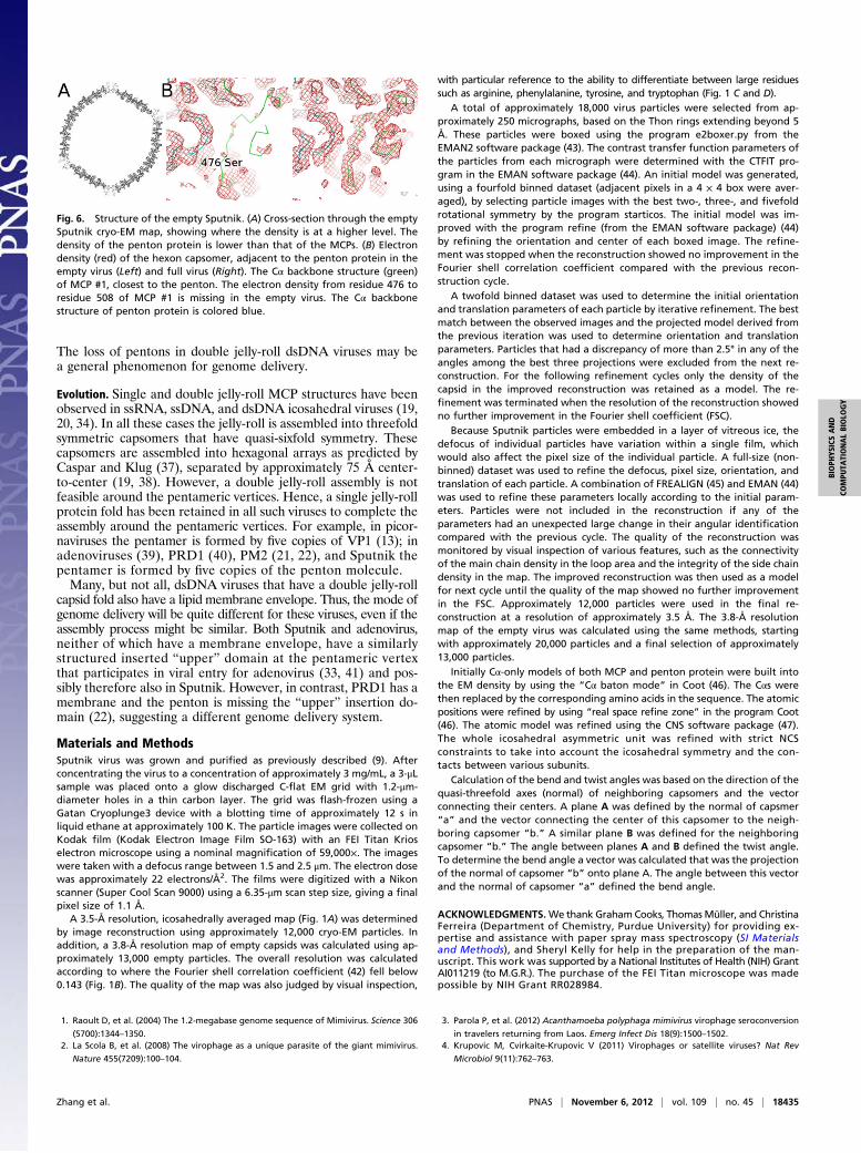
Sputnik was identified by the Roman numerals I, II, III, IV, andV and the MCP monomers by Arabic numerals 1 to 13 (Fig. 5A).The rotational relationships between neighboring capsomers(Table S1) can be divided into a bend and twist component. Thecurvature of the viral capsid is determined by the bend angle. Ifall of the bend angles between neighboring capsomers were zero,then the capsomers would form a plane. The variation of bendangle was found to be limited to approximately 10°, 20°, and 40°degrees in the fs, ff, and ss classes, respectively.Hydrogen bonds between capsomers were identified by visual
inspection. The three classes of interactions, ff, ss and fs, couldbe subdivided into two or three subclasses depending on theconservation of hydrogen bonds (Table S1). Although there arefive examples of class fs in the Sputnik structure, only three of thesecontact regions are associated with density probably representingthe binding site of a minor capsid protein (see above). These threecontact regions all belong to the same subclass fs-1, whereas theother two examples of fs contacts are associated with a differentset of hydrogen bonds.
The root mean square deviation between equivalence Cαatoms is less than 0.43 Å on superimposing any pair of the 13MCP structures on each other. However, there are specificregions where the MCP structure has greater variability (Fig.5B), with displacements of the Cα atoms as much as 3.5 Å. Theseregions are mostly the loops at the top of each “tower” and in theCHEF β-sheets of the second jelly roll that make contacts withneighboring capsomers (Fig. 5C).
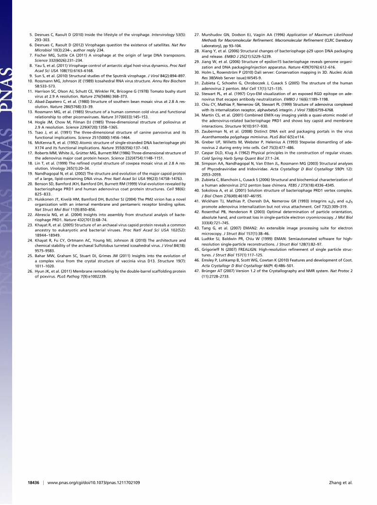
Empty Virus. During the purification procedure of Sputnik(Materials and Methods), the virus separated into two bands whilesedimenting through a cesium chloride gradient. The two bandscorresponded to the full and empty virus. Subsequent work showedthat empty particles can be produced from full particles either byraising the pH to 9 or lowering the pH to 5.5. It is thereforepossible that the purified empty Sputnik particles were producedin a low-pH virus factory (35) during Mimivirus production. Thepurified empty particles were used in the reconstruction of a 3.8-Åresolution cryo-EM map (Materials and Methods and Fig. S5A).The empty Sputnik particles had a diameter that was approxi-mately 5.2 Å larger than the diameter of the full infectious viruswhen measured along the threefold axes. However, there was noexpansion along the fivefold axes. Presumably the expansion wasmade possible by the loss of interaction between the packagedDNA and the capsid structure.The height of the penton density in the empty virus was ap-
proximately half of the hexon density, suggesting that some ofthe penton proteins were missing in the empty particles (Fig. 6A).Presumably the DNA had escaped from one of the vertices wherethere was no penton protein. Indeed, two particles that had beenemptied by lowering the pH of the environment showed a fea-ture that is probably DNA-like density spilling out from one ofthe vertices (Fig. S5B). A similar loss of the penton structureoccurs for adenovirus, which can lose some pentons in the low-pHenvironment of an endosome (36). Similarly a mutant of the bac-teriophage PRD1 can lose some pentons in the presence of adetergent, although PRD1, unlike Sputnik and Adenovirus, has amembrane (34). In the empty particle reconstruction of Sputnik,the electron density for residues 476–508 was missing in theMCP of monomer #1, the monomer adjacent to the penton (Fig.6B). These residues are in the interface between the penton andhexon. However, it is not clear why these residues are completelydisordered when only some of the pentons are missing, whichwould require disorder only in the #1 MCPs close to one of themissing penton sites. Thus, possibly the pH or other stress dis-orders these residues, resulting in only the loss of some pentons.
Fig. 4. Single jelly-roll structure of sputnik penton protein. (A) Ribbon di-agram of the Sputnik penton protein. The jelly-roll domain is colored green,and the insertion domain is colored red. (B) Diagrammatic representation ofthe arrangement of β-strands in the penton protein and the residue numbersat the ends of the β-strands.
Fig. 5. Asymmetric unit of Sputnik. (A) Capsomers are identified by Roman numerals I to V. Thirteen independent monomers are identified by Arabicnumerals 1 to 13. Each MCP is shown by a green line (the first jelly-roll domain) followed by a blue line (the second jelly-roll domain). The independent minorproteins in the asymmetric unit are colored red. Icosahedrally related positions are indicated in the same manner but with corresponding faded colors. (B) Thebackbone of the MCP was colored according to the root mean square deviation of each Cα position from the mean of the superimposed 13 independentMCPs, ranging from blue (<0.6 Å) to red (>0.6 Å). (C) Number of times a given residue in the 13 independent MCPs is involved in capsomer to capsomercontacts ranging from blue (<8) to red (>8).
18434 | www.pnas.org/cgi/doi/10.1073/pnas.1211702109 Zhang et al.
The loss of pentons in double jelly-roll dsDNA viruses may bea general phenomenon for genome delivery.
Evolution. Single and double jelly-roll MCP structures have beenobserved in ssRNA, ssDNA, and dsDNA icosahedral viruses (19,20, 34). In all these cases the jelly-roll is assembled into threefoldsymmetric capsomers that have quasi-sixfold symmetry. Thesecapsomers are assembled into hexagonal arrays as predicted byCaspar and Klug (37), separated by approximately 75 Å center-to-center (19, 38). However, a double jelly-roll assembly is notfeasible around the pentameric vertices. Hence, a single jelly-rollprotein fold has been retained in all such viruses to complete theassembly around the pentameric vertices. For example, in picor-naviruses the pentamer is formed by five copies of VP1 (13); inadenoviruses (39), PRD1 (40), PM2 (21, 22), and Sputnik thepentamer is formed by five copies of the penton molecule.Many, but not all, dsDNA viruses that have a double jelly-roll
capsid fold also have a lipid membrane envelope. Thus, the mode ofgenome delivery will be quite different for these viruses, even if theassembly process might be similar. Both Sputnik and adenovirus,neither of which have a membrane envelope, have a similarlystructured inserted “upper” domain at the pentameric vertexthat participates in viral entry for adenovirus (33, 41) and pos-sibly therefore also in Sputnik. However, in contrast, PRD1 has amembrane and the penton is missing the “upper” insertion do-main (22), suggesting a different genome delivery system.
Materials and MethodsSputnik virus was grown and purified as previously described (9). Afterconcentrating the virus to a concentration of approximately 3 mg/mL, a 3-μLsample was placed onto a glow discharged C-flat EM grid with 1.2-μm-diameter holes in a thin carbon layer. The grid was flash-frozen using aGatan Cryoplunge3 device with a blotting time of approximately 12 s inliquid ethane at approximately 100 K. The particle images were collected onKodak film (Kodak Electron Image Film SO-163) with an FEI Titan Krioselectron microscope using a nominal magnification of 59,000×. The imageswere taken with a defocus range between 1.5 and 2.5 μm. The electron dosewas approximately 22 electrons/Å2. The films were digitized with a Nikonscanner (Super Cool Scan 9000) using a 6.35-μm scan step size, giving a finalpixel size of 1.1 Å.
A 3.5-Å resolution, icosahedrally averaged map (Fig. 1A) was determinedby image reconstruction using approximately 12,000 cryo-EM particles. Inaddition, a 3.8-Å resolution map of empty capsids was calculated using ap-proximately 13,000 empty particles. The overall resolution was calculatedaccording to where the Fourier shell correlation coefficient (42) fell below0.143 (Fig. 1B). The quality of the map was also judged by visual inspection,
with particular reference to the ability to differentiate between large residuessuch as arginine, phenylalanine, tyrosine, and tryptophan (Fig. 1 C and D).
A total of approximately 18,000 virus particles were selected from ap-proximately 250 micrographs, based on the Thon rings extending beyond 5Å. These particles were boxed using the program e2boxer.py from theEMAN2 software package (43). The contrast transfer function parameters ofthe particles from each micrograph were determined with the CTFIT pro-gram in the EMAN software package (44). An initial model was generated,using a fourfold binned dataset (adjacent pixels in a 4 × 4 box were aver-aged), by selecting particle images with the best two-, three-, and fivefoldrotational symmetry by the program starticos. The initial model was im-proved with the program refine (from the EMAN software package) (44)by refining the orientation and center of each boxed image. The refine-ment was stopped when the reconstruction showed no improvement in theFourier shell correlation coefficient compared with the previous recon-struction cycle.
A twofold binned dataset was used to determine the initial orientationand translation parameters of each particle by iterative refinement. The bestmatch between the observed images and the projected model derived fromthe previous iteration was used to determine orientation and translationparameters. Particles that had a discrepancy of more than 2.5° in any of theangles among the best three projections were excluded from the next re-construction. For the following refinement cycles only the density of thecapsid in the improved reconstruction was retained as a model. The re-finement was terminated when the resolution of the reconstruction showedno further improvement in the Fourier shell coefficient (FSC).
Because Sputnik particles were embedded in a layer of vitreous ice, thedefocus of individual particles have variation within a single film, whichwould also affect the pixel size of the individual particle. A full-size (non-binned) dataset was used to refine the defocus, pixel size, orientation, andtranslation of each particle. A combination of FREALIGN (45) and EMAN (44)was used to refine these parameters locally according to the initial param-eters. Particles were not included in the reconstruction if any of theparameters had an unexpected large change in their angular identificationcompared with the previous cycle. The quality of the reconstruction wasmonitored by visual inspection of various features, such as the connectivityof the main chain density in the loop area and the integrity of the side chaindensity in the map. The improved reconstruction was then used as a modelfor next cycle until the quality of the map showed no further improvementin the FSC. Approximately 12,000 particles were used in the final re-construction at a resolution of approximately 3.5 Å. The 3.8-Å resolutionmap of the empty virus was calculated using the same methods, startingwith approximately 20,000 particles and a final selection of approximately13,000 particles.
Initially Cα-only models of both MCP and penton protein were built intothe EM density by using the “Cα baton mode” in Coot (46). The Cαs werethen replaced by the corresponding amino acids in the sequence. The atomicpositions were refined by using “real space refine zone” in the program Coot(46). The atomic model was refined using the CNS software package (47).The whole icosahedral asymmetric unit was refined with strict NCSconstraints to take into account the icosahedral symmetry and the con-tacts between various subunits.
Calculation of the bend and twist angles was based on the direction of thequasi-threefold axes (normal) of neighboring capsomers and the vectorconnecting their centers. A plane A was defined by the normal of capsmer“a” and the vector connecting the center of this capsomer to the neigh-boring capsomer “b.” A similar plane B was defined for the neighboringcapsomer “b.” The angle between planes A and B defined the twist angle.To determine the bend angle a vector was calculated that was the projectionof the normal of capsomer “b” onto plane A. The angle between this vectorand the normal of capsomer “a” defined the bend angle.
ACKNOWLEDGMENTS.We thank GrahamCooks, ThomasMüller, and ChristinaFerreira (Department of Chemistry, Purdue University) for providing ex-pertise and assistance with paper spray mass spectroscopy (SI Materialsand Methods), and Sheryl Kelly for help in the preparation of the man-uscript. This work was supported by a National Institutes of Health (NIH) GrantAI011219 (to M.G.R.). The purchase of the FEI Titan microscope was madepossible by NIH Grant RR028984.
1. Raoult D, et al. (2004) The 1.2-megabase genome sequence of Mimivirus. Science 306
(5700):1344–1350.2. La Scola B, et al. (2008) The virophage as a unique parasite of the giant mimivirus.
Nature 455(7209):100–104.
3. Parola P, et al. (2012) Acanthamoeba polyphaga mimivirus virophage seroconversion
in travelers returning from Laos. Emerg Infect Dis 18(9):1500–1502.4. Krupovic M, Cvirkaite-Krupovic V (2011) Virophages or satellite viruses? Nat Rev
Microbiol 9(11):762–763.
Fig. 6. Structure of the empty Sputnik. (A) Cross-section through the emptySputnik cryo-EM map, showing where the density is at a higher level. Thedensity of the penton protein is lower than that of the MCPs. (B) Electrondensity (red) of the hexon capsomer, adjacent to the penton protein in theempty virus (Left) and full virus (Right). The Cα backbone structure (green)of MCP #1, closest to the penton. The electron density from residue 476 toresidue 508 of MCP #1 is missing in the empty virus. The Cα backbonestructure of penton protein is colored blue.
Zhang et al. PNAS | November 6, 2012 | vol. 109 | no. 45 | 18435
BIOPH
YSICSAND
COMPU
TATIONALBIOLO
GY
5. Desnues C, Raoult D (2010) Inside the lifestyle of the virophage. Intervirology 53(5):293–303.
6. Desnues C, Raoult D (2012) Virophages question the existence of satellites. Nat RevMicrobiol 10(3):234–, author reply 234.
7. Fischer MG, Suttle CA (2011) A virophage at the origin of large DNA transposons.Science 332(6026):231–234.
8. Yau S, et al. (2011) Virophage control of antarctic algal host-virus dynamics. Proc NatlAcad Sci USA 108(15):6163–6168.
9. Sun S, et al. (2010) Structural studies of the Sputnik virophage. J Virol 84(2):894–897.10. Rossmann MG, Johnson JE (1989) Icosahedral RNA virus structure. Annu Rev Biochem
58:533–573.11. Harrison SC, Olson AJ, Schutt CE, Winkler FK, Bricogne G (1978) Tomato bushy stunt
virus at 2.9 A resolution. Nature 276(5686):368–373.12. Abad-Zapatero C, et al. (1980) Structure of southern bean mosaic virus at 2.8 A res-
olution. Nature 286(5768):33–39.13. Rossmann MG, et al. (1985) Structure of a human common cold virus and functional
relationship to other picornaviruses. Nature 317(6033):145–153.14. Hogle JM, Chow M, Filman DJ (1985) Three-dimensional structure of poliovirus at
2.9 A resolution. Science 229(4720):1358–1365.15. Tsao J, et al. (1991) The three-dimensional structure of canine parvovirus and its
functional implications. Science 251(5000):1456–1464.16. McKenna R, et al. (1992) Atomic structure of single-stranded DNA bacteriophage phi
X174 and its functional implications. Nature 355(6356):137–143.17. Roberts MM, White JL, Grütter MG, Burnett RM (1986) Three-dimensional structure of
the adenovirus major coat protein hexon. Science 232(4754):1148–1151.18. Lin T, et al. (1999) The refined crystal structure of cowpea mosaic virus at 2.8 A res-
olution. Virology 265(1):20–34.19. Nandhagopal N, et al. (2002) The structure and evolution of the major capsid protein
of a large, lipid-containing DNA virus. Proc Natl Acad Sci USA 99(23):14758–14763.20. Benson SD, Bamford JKH, Bamford DH, Burnett RM (1999) Viral evolution revealed by
bacteriophage PRD1 and human adenovirus coat protein structures. Cell 98(6):825–833.
21. Huiskonen JT, Kivelä HM, Bamford DH, Butcher SJ (2004) The PM2 virion has a novelorganization with an internal membrane and pentameric receptor binding spikes.Nat Struct Mol Biol 11(9):850–856.
22. Abrescia NG, et al. (2004) Insights into assembly from structural analysis of bacte-riophage PRD1. Nature 432(7013):68–74.
23. Khayat R, et al. (2005) Structure of an archaeal virus capsid protein reveals a commonancestry to eukaryotic and bacterial viruses. Proc Natl Acad Sci USA 102(52):18944–18949.
24. Khayat R, Fu CY, Ortmann AC, Young MJ, Johnson JE (2010) The architecture andchemical stability of the archaeal Sulfolobus turreted icosahedral virus. J Virol 84(18):9575–9583.
25. Bahar MW, Graham SC, Stuart DI, Grimes JM (2011) Insights into the evolution ofa complex virus from the crystal structure of vaccinia virus D13. Structure 19(7):1011–1020.
26. Hyun JK, et al. (2011) Membrane remodeling by the double-barrel scaffolding proteinof poxvirus. PLoS Pathog 7(9):e1002239.
27. Murshudov GN, Dodson EJ, Vagiin AA (1996) Application of Maximum Likelihood
Methods for Macromolecular Refinement. Macromolecular Refinement (CLRC Daresbury
Laboratory), pp 93–104.28. Xiang Y, et al. (2006) Structural changes of bacteriophage ϕ29 upon DNA packaging
and release. EMBO J 25(21):5229–5239.29. Jiang W, et al. (2006) Structure of epsilon15 bacteriophage reveals genome organi-
zation and DNA packaging/injection apparatus. Nature 439(7076):612–616.30. Holm L, Rosenström P (2010) Dali server: Conservation mapping in 3D. Nucleic Acids
Res 38(Web Server issue):W545-9.31. Zubieta C, Schoehn G, Chroboczek J, Cusack S (2005) The structure of the human
adenovirus 2 penton. Mol Cell 17(1):121–135.32. Stewart PL, et al. (1997) Cryo-EM visualization of an exposed RGD epitope on ade-
novirus that escapes antibody neutralization. EMBO J 16(6):1189–1198.33. Chiu CY, Mathias P, Nemerow GR, Stewart PL (1999) Structure of adenovirus complexed
with its internalization receptor, alphavbeta5 integrin. J Virol 73(8):6759–6768.34. Martín CS, et al. (2001) Combined EM/X-ray imaging yields a quasi-atomic model of
the adenovirus-related bacteriophage PRD1 and shows key capsid and membrane
interactions. Structure 9(10):917–930.35. Zauberman N, et al. (2008) Distinct DNA exit and packaging portals in the virus
Acanthamoeba polyphaga mimivirus. PLoS Biol 6(5):e114.36. Greber UF, Willetts M, Webster P, Helenius A (1993) Stepwise dismantling of ade-
novirus 2 during entry into cells. Cell 75(3):477–486.37. Caspar DLD, Klug A (1962) Physical principles in the construction of regular viruses.
Cold Spring Harb Symp Quant Biol 27:1–24.38. Simpson AA, Nandhagopal N, Van Etten JL, Rossmann MG (2003) Structural analyses
of Phycodnaviridae and Iridoviridae. Acta Crystallogr D Biol Crystallogr 59(Pt 12):
2053–2059.39. Zubieta C, Blanchoin L, Cusack S (2006) Structural and biochemical characterization of
a human adenovirus 2/12 penton base chimera. FEBS J 273(18):4336–4345.40. Sokolova A, et al. (2001) Solution structure of bacteriophage PRD1 vertex complex.
J Biol Chem 276(49):46187–46195.41. Wickham TJ, Mathias P, Cheresh DA, Nemerow GR (1993) Integrins αvβ3 and αvβ5
promote adenovirus internalization but not virus attachment. Cell 73(2):309–319.42. Rosenthal PB, Henderson R (2003) Optimal determination of particle orientation,
absolute hand, and contrast loss in single-particle electron cryomicroscopy. J Mol Biol
333(4):721–745.43. Tang G, et al. (2007) EMAN2: An extensible image processing suite for electron
microscopy. J Struct Biol 157(1):38–46.44. Ludtke SJ, Baldwin PR, Chiu W (1999) EMAN: Semiautomated software for high-
resolution single-particle reconstructions. J Struct Biol 128(1):82–97.45. Grigorieff N (2007) FREALIGN: High-resolution refinement of single particle struc-
tures. J Struct Biol 157(1):117–125.46. Emsley P, Lohkamp B, Scott WG, Cowtan K (2010) Features and development of Coot.
Acta Crystallogr D Biol Crystallogr 66(Pt 4):486–501.47. Brünger AT (2007) Version 1.2 of the Crystallography and NMR system. Nat Protoc 2
(11):2728–2733.
18436 | www.pnas.org/cgi/doi/10.1073/pnas.1211702109 Zhang et al.


![Ocean & Coastal Management · 2017-05-01 · effective coastal management [4–6]. Specific, well identified results have been obtained from a number of research studies related](https://static.fdocuments.fr/doc/165x107/5f535aba8fc1d822427a21a7/ocean-coastal-management-2017-05-01-effective-coastal-management-4a6.jpg)