Proteome biology of primary colorectal carcinoma and ...
12
Volume 23 Number 12 Month 2021 pp. 1240–1251 1240 Original Research Proteome biology of primary colorectal carcinoma and corresponding liver metastases Matthias Fahrner 1,2,3 ; Peter Bronsert 1,4,5 ; Stefan Fichtner-Feigl 6,7 ; Andreas Jud 6,9 ; Oliver Schilling 1,7,8 1 Institute for Surgical Pathology, Medical Center – University of Freiburg, Faculty of Medicine, University of Freiburg, Germany 2 Faculty of Biology, Albert-Ludwigs-University Freiburg, Freiburg, Germany 3 Spemann Graduate School of Biology and Medicine (SGBM), University of Freiburg 4 Tumorbank Comprehensive Cancer Center Freiburg, Medical Center - University of Freiburg 5 Core Facility Histopathology and Digital Pathology Freiburg, Medical Center - University of Freiburg 6 Department of General and Visceral Surgery, Medical Center – University of Freiburg, Faculty of Medicine, University of Freiburg, Germany 7 German Cancer Consortium (DKTK) and German Cancer Research Center (DKFZ), Heidelberg, Germany 8 BIOSS Centre for Biological Signaling Studies, University of Freiburg, D-79104 Freiburg, Germany Abstract Colorectal adenocarcinomas (CRC) are one of the most commonly diagnosed tumors worldwide. Colorectal adenocarcinomas primarily metastasize into the liver and (less often) into the peritoneum. Patients suffering from CRC-liver metastasis (CRC-LM) typically present with a dismal overall survival compared to non-metastasized CRC patients. The metastasis process and metastasis- promoting factors in patients with CRC are under intensive debate. However, CRC studies investigating the proteome biology are lacking. Formalin-fixed paraffin-embedded (FFPE) tissue specimens provide a valuable resource for comprehensive proteomic studies of a broad variety of clinical malignancies. The presented pilot study compares the proteome of primary CRC and patient-matched CRC-LM. The applied protocol allows a reproducible and straightforward identification and quantification of over 2,600 proteins within the dissected tumorous tissue. Subsequent unsupervised clustering reveals distinct proteome biologies of the primary CRC and the corresponding CRC-LM. Statistical analysis yields multiple differentially abundant proteins in either primary CRC or their corresponding liver metastases. A more detailed analysis of dysregulated biological processes suggests an active immune response in the liver metastases, including several proteins of the complement system. Proteins with structural roles, e.g. cytoskeleton organization or cell junction assembly appear to be less prominent in liver metastases as compared to primary CRC. Immunohistochemistry corroborates proteomic high expression levels of metabolic proteins in CRC-LM. We further assessed how the in vitro inhibition of two in CRC-LM enriched metabolic proteins affected cell proliferation and chemosensitivity. The presented proteomic investigation in a small clinical cohort promotes a more comprehensive understanding of the distinct proteome biology of primary CRC and their corresponding liver metastases. Neoplasia (2021) 23, 1240–1251 Keywords: Colorectal cancer, liver metastases, proteomics, mass spectrometry 9 To whom correspondence should be addressed: Breisacher Straße 55, D-79106 Freiburg Germany E-mail address: [email protected] (A. Jud). Received 5 October 2021; received in revised form 27 October 2021; accepted 27 October 2021 © 2021 The Authors. Published by Elsevier Inc. This is an open access article under the CC BY license (http://creativecommons.org/licenses/by/4.0/) https://doi.org/10.1016/j.neo.2021.10.005
Transcript of Proteome biology of primary colorectal carcinoma and ...
Volume 23 Number 12 Month 2021 pp. 1240–1251 1240
Original Research
Proteome biology of primary colorectal carcinoma and corresponding liver
metastases
Matthias Fahrner 1 , 2 , 3 ; Peter Bronsert 1 , 4 , 5 ;
Stefan Fichtner-Feigl 6 , 7 ; Andreas Jud
6 , 9 ;
Oliver Schilling
1 , 7 , 8
1 Institute for Surgical Pathology, Medical Center – University of Freiburg, Faculty of Medicine, University of Freiburg, Germany 2 Faculty of Biology, Albert-Ludwigs-University Freiburg, Freiburg, Germany 3 Spemann Graduate School of Biology and Medicine (SGBM), University of Freiburg 4 Tumorbank Comprehensive Cancer Center Freiburg, Medical Center - University of Freiburg 5 Core Facility Histopathology and Digital Pathology Freiburg, Medical Center - University of Freiburg 6 Department of General and Visceral Surgery, Medical Center – University of Freiburg, Faculty of Medicine, University of Freiburg, Germany 7 German Cancer Consortium (DKTK) and German Cancer Research Center (DKFZ), Heidelberg, Germany 8 BIOSS Centre for Biological Signaling Studies, University of Freiburg, D-79104 Freiburg, Germany
Abstract
Colorectal adenocarcinomas (CRC) are one of the most commonly diagnosed tumors worldwide. Colorectal adenocarcinomas primarily metastasize into the liver and (less often) into the peritoneum. Patients suffering from CRC-liver metastasis (CRC-LM) typically present with a dismal overall survival compared to non-metastasized CRC patients. The metastasis process and metastasis- promoting factors in patients with CRC are under intensive debate. However, CRC studies investigating the proteome biology are lacking. Formalin-fixed paraffin-embedded (FFPE) tissue specimens provide a valuable resource for comprehensive proteomic studies of a broad variety of clinical malignancies. The presented pilot study compares the proteome of primary CRC and patient-matched
CRC-LM. The applied protocol allows a reproducible and straightforward identification and quantification of over 2,600 proteins within the dissected tumorous tissue. Subsequent unsupervised clustering reveals distinct proteome biologies of the primary CRC
and the corresponding CRC-LM. Statistical analysis yields multiple differentially abundant proteins in either primary CRC or their corresponding liver metastases. A more detailed analysis of dysregulated biological processes suggests an active immune response in the liver metastases, including several proteins of the complement system. Proteins with structural roles, e.g. cytoskeleton organization
or cell junction assembly appear to be less prominent in liver metastases as compared to primary CRC. Immunohistochemistry corroborates proteomic high expression levels of metabolic proteins in CRC-LM. We further assessed how the in vitro inhibition of two in CRC-LM enriched metabolic proteins affected cell proliferation and chemosensitivity. The presented proteomic investigation
in a small clinical cohort promotes a more comprehensive understanding of the distinct proteome biology of primary CRC and their corresponding liver metastases.
Neoplasia (2021) 23, 1240–1251
Keywords: Colorectal cancer, liver metastases, proteomics, mass spectrometry
9 To whom correspondence should be addressed: Breisacher Straße 55, D-79106 Freiburg Germany
E-mail address: [email protected] (A. Jud). Received 5 October 2021; received in revised form 27 October 2021; accepted 27 October 2021
©T(h
2021 The Authors. Published by Elsevier Inc. his is an open access article under the CC BY license http://creativecommons.org/licenses/by/4.0/ ) ttps://doi.org/10.1016/j.neo.2021.10.005
Neoplasia Vol. 23, No. 12, 2021 Proteome biology of primary colorectal carcinoma and corresponding liver metastases M. Fahrner et al. 1241
tp(
i
cCipu
dm
u
M
E
Mb
P
w
b
Us
b
g
r
Gor
a
"a
i
S
T
m
r
a
tab
w
S
dp
0
T(
i
P
Introduction
Colorectal cancer (CRC) is still one of the most diagnosed cancersworldwide, presenting the third-highest prevalence in men 1 , 2 . Over 1.1million new CRC cases were diagnosed in 2020 with over half a millionCRC-related deaths worldwide, making CRC the second leading cause ofcancer death 2 , 3 . Unhealthy diet, obesity, smoking, lack of physical activity,and genetic predisposition are established risk factors for CRC
4 , 5 . Typically,CRC develops over several years, starting as benign adenomatous polypsbecoming advanced adenoma with high-grade dysplasia, and then progressesto invasive cancer 6 . Multiple consecutive changes on a genetic level arethought to drive the conversion from normal epithelium to malignant tissue7 . Early detection and removal of colonic polyps and advances in primary andadjuvant therapy are paramount improvements declining the mortality andincreasing patients’ 5-year survival in the past decades 8 . Surgical resectionrepresents the preferred therapeutic method of CRC (stage I to III) providinga potentially curative option. Downstaging with neoadjuvant chemotherapyis a possibility for initially unresectable tumor stages 9 , 10 . The postoperativeoutcome depends on the clinical, molecular, and histological features ofthe disease. The strongest prognostic factor is the pathological stage of theresected tumor. Size, presence of distant metastasis, lymph node positivity,and perineural invasion are substantial 11 , 12 .
CRC can spread lymphatic, hematogenous, contiguous, and in a finalstage transperitoneal. The most common metastatic sites are the regionallymph nodes, the liver, and the lungs. Usually, the liver is the first metastaticsite because of the venous drainage via the portal vein system. Up to 25%of all patients carry liver metastases at the time of diagnosis, while over30% develop metastases after resection of primary CRC
4 , 13 . The 5-yearsurvival rate of patients with metastasizing CRC is decreased by eight timescompared to patients with local CRC
13 . Due to the development of refinedsurgical techniques, such as two-stage hepatectomy, preoperative portal veinembolization, and down-sizing chemotherapy, the number and extent ofresections of liver metastasis in CRC increased in the last couple of yearssteadily and showed a positive result towards long-term survival 9 , 14 . On amolecular level, CRC is a heterogeneous disease, as the majority of all CRCsoccur sporadically ( ∼ 70%), caused by somatic mutations 1 , 15 . Microsatelliteinstability, mismatch repair deficiency, APC, RAS, and BRAF mutations arerelevant and determined in the standard histopathological examination. Inthe last couple of years, the growing knowledge of molecular pathogenesisplays an important role to improve targeted treatments 16 , 17 . Multimodaltherapy concepts are well-established with combination chemotherapy andtargeted biologic agents. Subsequently, significant improvements in survivalwere achieved 18 , 19 .
Formalin-fixation and paraffin-embedding (FFPE) represents the mostcommonly used preservation method for clinical tissue specimens worldwide20 . This has led to vast tissue archives, storing tissue specimens of a broadvariety of malignancies. Continuous protocol development and optimizationrendered FFPE tissue readily accessible for proteomic investigations 21–24 .This has prompted an increasing number of clinical proteomic studiesinvestigating a multitude of malignancies, including rare genetic disordersbenefitting from thorough FFPE tissue archives 25–28 . Furthermore, someof the FFPE protocols have been shown to yield comprehensive proteomecoverage, reaching over 8.000 identified proteins in single measurements,even with minor amounts of tissue material 21 , 28 , 29 .
Many malignant tumors have predominantly been studied on the geneticand transcriptomic level, mainly due to the broad availability and enhancedcoverage of those techniques. However, with proteins being the effectorswithin cells and tissues, representing the products of transcriptomic andtranslational processes, the importance of a detailed investigation of theproteome becomes evident. Proteomic profiling of CRC and its metastasesas the functional translation of the genome is challenging but has a greatpotential to identify proteins that are linked to tumor progression 30 . In thisstudy, we compare the primary tumor with patient-matched liver metastasis.
We describe the distinct proteome biology of primary CRC andheir corresponding liver metastases. Further, this project highlights the racticability and feasibility of the previously published direct trypsinization DTR) protocol 5 . An activated immune response in the liver metastasess highlighted by the significant upregulation of several components of theomplement system. The explorative proteome characterization of primary RC and liver metastases is complemented by follow-up experiments
nvestigating the spatially distributed expression of significantly dysregulated roteins using immunohistochemistry. Furthermore, an increase in sensitivity sing tissue macro dissection for the analysis of primary tumors and theiristant metastasis is shown. These characteristics and proteomic profile ay lead to uncovering diagnostic and prognostic markers to improve the
nderlying mechanisms of tumor development and progression.
aterial and Methods
thics statement
The study was approved by the Ethics Committee of the Universityedical Center Freiburg (504/17). Patients gave written informed consent
efore inclusion into the study.
atient cohort
Seven patients diagnosed with colorectal carcinoma (CRC) and CRC-LM
ere included in the study. All patients were operated for CRC and CRC-LMetween 2014 and 2016 at the Department of General and Visceral Surgery,niversity Medical Center Freiburg, Germany. The cohort comprises tissue
pecimens from five male and two female patients with an age rangingetween 48 and 75 years. Further details including tumor localization, tumorrading, and TNM classification are summarized in Table 1 . Patient data,aw LC-MS/MS data, and analysis result files are available at the Europeanenome-phenome Archive for appropriate research use ( https://ega-archive.
rg ; EGAS00001005641). As patient-centric proteomic data is increasingly egarded as sensitive, personal data 31 , EGA requires adherence to a data accessgreement. The data access agreement for this dataset corresponds to theHarmonised Data Access Agreement (hDAA) for Controlled Access Data”s brought forward by the "European standardization framework for datantegration and data-driven in silico models for personalized medicine – EU-TANDS4PM”.
issue Collection, Fixation, and tissue macro dissection
Tissue specimens were harvested during surgical removal of primary andetastatic tumors and put in formalin solution immediately after surgical
emoval. All tissue specimens were gross sectioned, processed, and stainedccording to routine protocols. For proteomic investigation 10 μm thickissue slices were automatically deparaffinized and stained for hematoxylin s previously described 21 , 32 . Macroscopical tumor dissection was performed y an experienced pathologist. Finally, the tumorous tissue of each sampleas transferred into a fresh 1.5 ml microcentrifugation tube.
ample Preparation for LC-MS/MS Analysis and Data Acquisition
For proteomic analysis, the tissue specimens were prepared as previouslyescribed using the direct trypsinization protocol (DTR) 21 . Briefly, rotein extraction was performed by adding 100 μl of buffer containing.1 % Rapigest in 0.1 M HEPES at pH 8.0 to each tissue sample.issue homogenization was performed using sonification in a Bioruptor
Diagenode) (10 cycles with 50/10 sec on/off) followed by the heat-nduced antigen retrieval (HIAR) incubating the samples for 2 h at 95 °C.rotein concentration of the supernatant was measured using the BCA assay
1242 Proteome biology of primary colorectal carcinoma and corresponding liver metastases M. Fahrner et al. Neoplasia Vol. 23, No. 12, 2021
Table 1
Clinical Characteristics of the Patient Cohort
Patient No. Localization of primary tumor Grading TNM Classification
1 ∗ rectosigmoid ACA (G2) ypT3 pN0 (0/25) pM1 (HEP)
2 ascending colon ACA (G2) pT3 pN1a (1/18) pM1 (HEP)
3 rectosigmoid ACA (G2) pT3 pN2b (8/31) pM1 (HEP)
4 sigmoid colon ACA (G2) pT4b pN2b (9/21) pM1 (HEP, PER)
5 sigmoid colon ACA (G2) pT4a pN1b (3/13) pM1 (HEP, PER)
6 ascending colon ACA (G2) pT4b pN0 (0/28) pM1 (HEP, PER)
7 sigmoid colon ACA (G2) pT3 pN0 (0/13) pM1 (HEP)
This study comprised 7 patients with histologically confirmed primary colorectal cancer. All patients
had liver metastasis (HEP) and three had additional peritoneal carcinoma (PER) at primary diagnosis.
All tumors were moderately differentiated (G2). ∗Material after chemotherapy (3 cycles).
Gu4
I
wa(2
rt
ahf
u0hm
C
w1s1
oaC
t7
A
a44
DifbBt
(ThermoScientific) and 100 μg of Protein for each sample was reduced byincubating with 5 mM Dithiothreitol (DTT) for 15 min and alkylated byincubating with 15 mM Iodoacetamide (IAM) for 15 min in the dark.A two-step protein digestion was performed by adding 2 μg of Trypsinand incubating for 2 h at 50 °C followed by adding another 2 μg ofTrypsin and incubation at 37 °C overnight 33 . After digestion, samples wereacidified by adding Trifluoroacetic acid (TFA) to a final concentration of 2% and incubating at 37 °C for 30 min. For peptide clean-up, mixed-phasecolumns (PreOmics) were applied according to the manufacturer’s protocol34 . Following BCA measurement, 4 μg of peptides of each individual samplewere transferred to fresh tubes, vacuum dried, and stored at -80 °C until LC-MS/MS measurement.
LC-MS/MS Data Acquisition and Analysis
For LC-MS/MS analysis 300 ng per sample were measured using anOrbitrap Q-Exactive plus (Thermo Scientific) mass spectrometer coupled toan Easy nanoLC 1000 (Thermo Scientific) with a flow rate of 300 nl/min.Buffer A contained 0.3 % acetic acid in water and buffer B 0.3 % aceticacid in 80 % acetonitrile. Peptides were separated with an increasing gradientof organic solvent (0-60 % acetonitrile in 90 min) on an analytical column(Acclaim PepMap column (Thermo Scientific), 2 μm particle size, 100 Apore size, length 150 mm, inner diameter 50 μm). The MS was operated in adata-dependent mode and each MS scan was followed by a maximum of tenMS/MS scans.
For data analysis, the MaxQuant (V1.6.0.16) software was used witha reviewed human database (retrieved from UniProt, October 6, 2017)containing 20,188 sequences 35 . Additionally, eleven synthetic peptides (iRTpeptides) were added to the database. Decoys for database search weregenerated using the revert function. Precursor, main search, and fragmentmass tolerance were set to be 20, 4.5, and 20 ppm, respectively. Thepeptide search included a fixed modification of carbamidomethyl cysteineas well as the oxidation of methionine and the acetylation of the protein(n-term) as variable modifications. Tryptic cleavage specificity with up totwo missed cleavages was used with a minimum peptide length of sevenamino acids. The false discovery rate (FDR) for peptides and proteinswas set to 0.01. Files obtained by MaxQuant were processed using theopen-source statistical software package R (V4.0.2). Decoy sequences andpotential contaminant entries were removed prior to statistical analysis. Rawintensities were log2 transformed and statistical inference of differentiallyregulated peptides was performed using the limma package (V.3.44.3) 36 .Reported P-values were corrected at a Benjamini-Hochberg FDR of 0.0537 . Further analyses were performed using mixOmics package (V6.12.2)(hierarchical clustering, clustering distance: Euclidean distance, PCA),corrPlot package (V0.84) and EnhancedVolcano package (V1.6.0) 38 , 39 .
ene ontology (GO) and REACTOME enrichment analysis was performed sing the topGO package (V2.40.0) and the ReactomePA package (V1.32.0)
0 .
mmunohistochemistry (IHC)
Immunohistochemical staining of ALDH1A1, ALDOB, and DPP4 as performed as previously described using specific antibodies mouse
nti-human ALDH1A1 (R&D, MAB5869), rabbit anti-human ALDOB
AbCam, ab75751), and mouse anti-human DPP4 (AbCam, ab114033) 6 . Briefly, 2 μm tissue slices were deparaffinized and heat-induced antigen etrieval (HIAR) was performed. Tissue sections were stained by applying he following steps: incubating with 10 % H2O2 for 5 min, with primaryntibody for 30 min, with secondary antibody for 10 min, and with orseradish peroxidase for 20 min, and lastly with 3,3 ′ -diaminobenzidine or 10 min. Between each of the aforementioned steps, the tissue was rinsedsing a washing buffer containing 50 mM Tris-HCl, 150 mM NaCl, and .05% Tween 20. Each tissue slice was counterstained by incubating with ematoxylin for 30 sec and xylene was used as a permanent mounting edium.
ell proliferation assay
CaCo-2 and SW480 cells were cultured in DMEM (Gibco) supplemented ith 10% foetal calf serum (PAN), 1% penicillin/streptomycin (Gibco), % MEM vitamin solution (PAN), 1% MEM non essential amino acid olution (PAN), and 1% L-glutamine (Gibco). Prior to inhibitor treatment × 10 5 cells/well were incubated in 6-well plates for 24 h. For the inhibitionf argininosuccinate synthase (ASS1) cells were incubated for 48 h after dding 5 mM (final conc.) of N-methyl-DL-aspartic acid (MDLA) (Santa ruz), solved in water, or an equal volume of water as a control. For
he inhibition of thymidylate kinase (DTYMK) cells were incubated for 2 h after adding 2 μM (final conc.) of the YMU-1 compound (Sigma-ldrich), solved in 15 % DMSO, or an equal volume of 15 % DMSOs a control. Following the YMU-1 treatment, the cells were incubated for h after adding either the FOLFOX regimen (50 μg/ml 5-fluorouracil, 0μg/ml oxaliplatin, and 10μg/ml folinic acid) or an equal volume of 15 %MSO. Subsequently, the cell culture medium was exchanged and cells were
ncubated in fresh DMEM including supplements for 24 h. Adherent cells or both inhibitor treatments were washed using PBS (Gibco) and harvested y trypsinization. Cell proliferation was determined by counting with Trypan lue staining using EVE
TM Automated Cell Counter (NanoEntek) according o the manufacturer’s protocol.
Neoplasia Vol. 23, No. 12, 2021 Proteome biology of primary colorectal carcinoma and corresponding liver metastases M. Fahrner et al. 1243
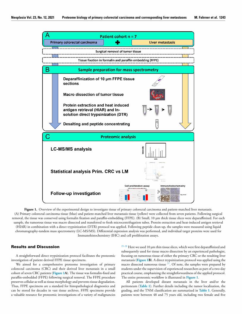
Figure 1. Overview of the experimental design to investigate tissue of primary colorectal carcinoma and patient-matched liver metastasis. (A) Primary colorectal carcinoma tissue (blue) and patient-matched liver metastasis tissue (yellow) were collected from seven patients. Following surgical
removal, the tissue was conserved using formalin fixation and paraffin-embedding (FFPE). (B) Small, 10 μm thick tissue slices were deparaffinized. For each sample, the tumorous tissue was macro dissected and transferred to fresh microcentrifugation tubes. Protein extraction and heat-induced antigen retrieval
(HIAR) in combination with a direct trypsinization (DTR) protocol was applied. Following peptide clean-up, the samples were measured using liquid chromatography-tandem mass spectrometry (LC-MS/MS). Differential expression analysis was performed, and individual target proteins were used for
Immunohistochemistry (IHC) and cell proliferation assays.
2
s
f
m
m
s
pT
p
g
p
Results and Discussion
A straightfor ward direct tr ypsinization protocol facilitates the proteomicinvestigation of patient derived FFPE tissue specimens.
We aimed for a comprehensive proteome investigation of primarycolorectal carcinoma (CRC) and their derived liver metastasis in a smallcohort of seven CRC patients ( Figure 1 A ). The tissue was formalin-fixed andparaffin-embedded (FFPE) following surgical removal. The FFPE procedurepreserves cellular as well as tissue morphology and prevents tissue degradation.Thus, FFPE specimens are a standard for histopathological diagnostics andcan be stored for decades in vast tissue archives. FFPE specimens providea valuable resource for proteomic investigations of a variety of malignancies
5–28 Here we used 10 μm thin tissue slices, which were first deparaffinized and ubsequently used for tissue macro dissection by an experienced pathologist,ocusing on tumorous tissue of either the primary CRC or the resulting liveretastasis ( Figure 1 B ). A direct trypsinization protocol was applied using theacro dissected tumorous tissue 21 . Of note, the samples were prepared by
tudents under the supervision of experienced researchers as part of a two-dayractical course, emphasizing the straightforwardness of the applied protocol. he entire proteomic workflow is illustrated in Figure 1 .
All patients developed distant metastasis in the liver and/or theeritoneum ( Table 1 ). Further details including the tumor localization, therading, and the TNM classification are summarized in Table 1 . Generally,atients were between 48 and 75 years old, including two female and five
1244 Proteome biology of primary colorectal carcinoma and corresponding liver metastases M. Fahrner et al. Neoplasia Vol. 23, No. 12, 2021
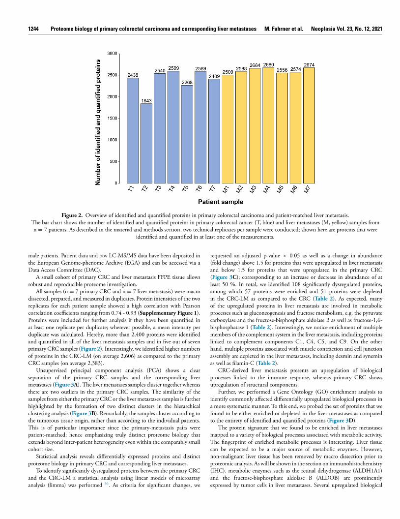
Figure 2. Overview of identified and quantified proteins in primary colorectal carcinoma and patient-matched liver metastasis. The bar chart shows the number of identified and quantified proteins in primary colorectal cancer (T, blue) and liver metastases (M, yellow) samples from
n = 7 patients. As described in the material and methods section, two technical replicates per sample were conducted; shown here are proteins that were identified and quantified in at least one of the measurements.
r
(a(
lai
opcbmlhaa
pu
ia
f
t
mTcnp(ae
male patients. Patient data and raw LC-MS/MS data have been deposited inthe European Genome-phenome Archive (EGA) and can be accessed via aData Access Committee (DAC).
A small cohort of primary CRC and liver metastasis FFPE tissue allowsrobust and reproducible proteome investigation.
All samples (n = 7 primary CRC and n = 7 liver metastasis) were macrodissected, prepared, and measured in duplicates. Protein intensities of the tworeplicates for each patient sample showed a high correlation with Pearsoncorrelation coefficients ranging from 0.74 - 0.93 ( Supplementary Figure 1 ).Proteins were included for further analysis if they have been quantified inat least one replicate per duplicate; wherever possible, a mean intensity perduplicate was calculated. Hereby, more than 2,400 proteins were identifiedand quantified in all of the liver metastasis samples and in five out of sevenprimary CRC samples ( Figure 2 ). Interestingly, we identified higher numbersof proteins in the CRC-LM (on average 2,606) as compared to the primaryCRC samples (on average 2,383).
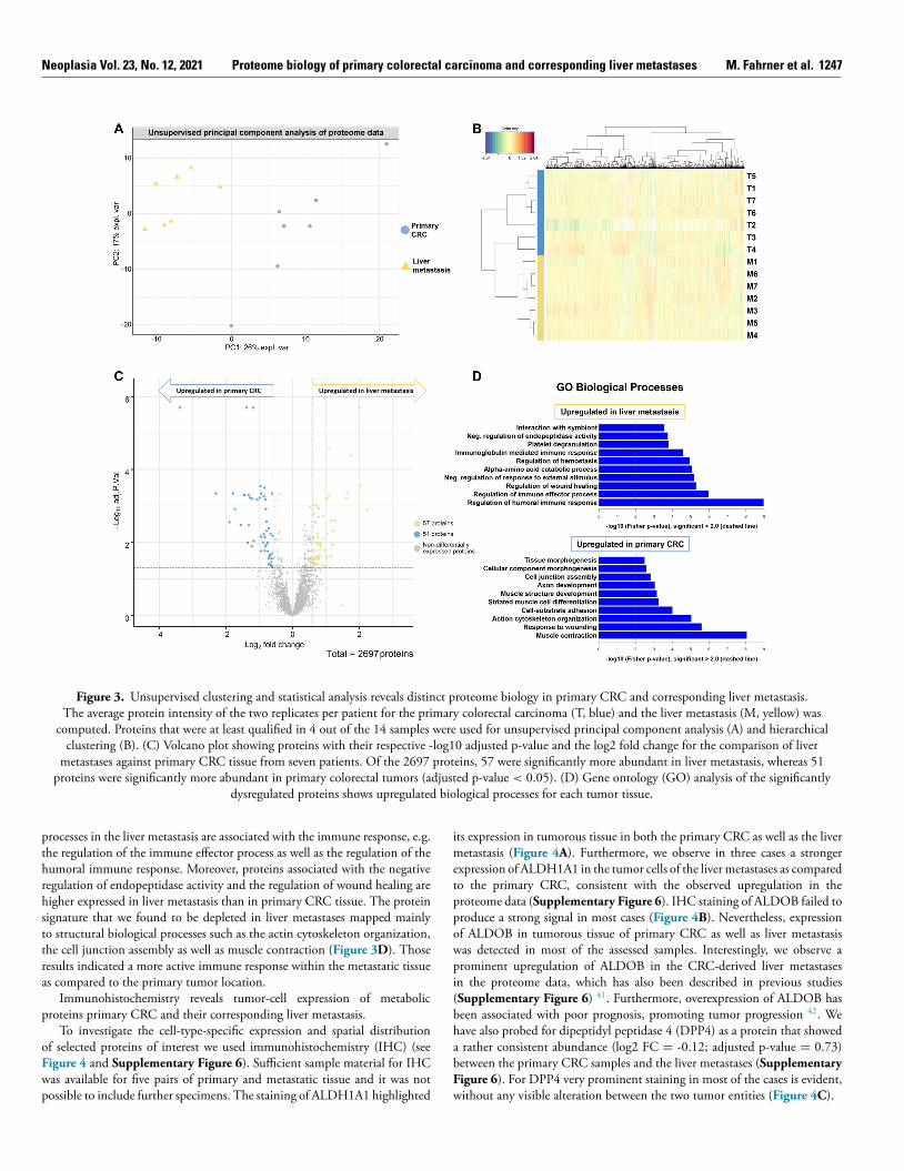
Unsupervised principal component analysis (PCA) shows a clearseparation of the primary CRC samples and the corresponding livermetastases ( Figure 3 A ). The liver metastases samples cluster together whereasthere are two outliers in the primary CRC samples. The similarity of thesamples from either the primary CRC or the liver metastases samples is furtherhighlighted by the formation of two distinct clusters in the hierarchicalclustering analysis ( Figure 3 B ). Remarkably, the samples cluster according tothe tumorous tissue origin, rather than according to the individual patients.This is of particular importance since the primary-metastasis pairs werepatient-matched; hence emphasizing truly distinct proteome biology thatextends beyond inter-patient heterogeneity even within the comparably smallcohort size.
Statistical analysis reveals differentially expressed proteins and distinctproteome biology in primary CRC and corresponding liver metastases.
To identify significantly dysregulated proteins between the primary CRCand the CRC-LM a statistical analysis using linear models of microarrayanalysis (limma) was performed 36 . As criteria for significant changes, we
equested an adjusted p-value < 0.05 as well as a change in abundancefold change) above 1.5 for proteins that were upregulated in liver metastasis nd below 1.5 for proteins that were upregulated in the primary CRC
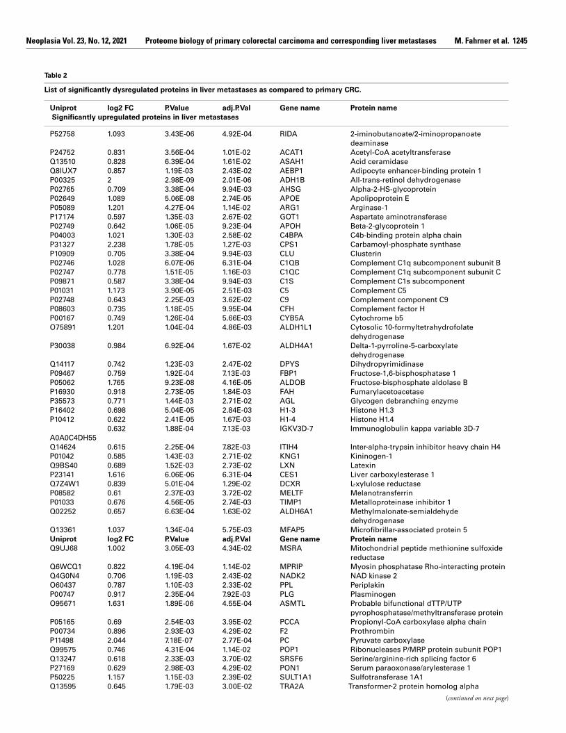
Figure 3 C ); corresponding to an increase or decrease in abundance of ateast 50 %. In total, we identified 108 significantly dysregulated proteins, mong which 57 proteins were enriched and 51 proteins were depleted n the CRC-LM as compared to the CRC ( Table 2 ). As expected, manyf the upregulated proteins in liver metastasis are involved in metabolic rocesses such as gluconeogenesis and fructose metabolism, e.g. the pyruvate arboxylase and the fructose-bisphosphate aldolase B as well as fructose-1,6- isphosphatase 1 ( Table 2 ). Interestingly, we notice enrichment of multiple embers of the complement system in the liver metastasis, including proteins
inked to complement components C1, C4, C5, and C9. On the other and, multiple proteins associated with muscle contraction and cell junction ssembly are depleted in the liver metastases, including desmin and synemin s well as filamin-C ( Table 2 ).
CRC-derived liver metastasis presents an upregulation of biological rocesses linked to the immune response, whereas primary CRC shows pregulation of structural components.
Further, we performed a Gene Ontology (GO) enrichment analysis to dentify commonly affected differentially upregulated biological processes in more systematic manner. To this end, we probed the set of proteins that weound to be either enriched or depleted in the liver metastases as comparedo the entirety of identified and quantified proteins ( Figure 3 D ).
The protein signature that we found to be enriched in liver metastases apped to a variety of biological processes associated with metabolic activity. he fingerprint of enriched metabolic processes is interesting. Liver tissue
an be expected to be a major source of metabolic enzymes. However, on-malignant liver tissue has been removed by macro dissection prior to roteomic analysis. As will be shown in the section on immunohistochemistry IHC), metabolic enzymes such as the retinal dehydrogenase (ALDH1A1) nd the fructose-bisphosphate aldolase B (ALDOB) are prominently xpressed by tumor cells in liver metastases. Several upregulated biological
Neoplasia Vol. 23, No. 12, 2021 Proteome biology of primary colorectal carcinoma and corresponding liver metastases M. Fahrner et al. 1245
Table 2
List of significantly dysregulated proteins in liver metastases as compared to primary CRC.
Uniprot log2 FC P.Value adj.P.Val Gene name Protein name
Significantly upregulated proteins in liver metastases
P52758 1.093 3.43E-06 4.92E-04 RIDA 2-iminobutanoate/2-iminopropanoate
deaminase
P24752 0.831 3.56E-04 1.01E-02 ACAT1 Acetyl-CoA acetyltransferase
Q13510 0.828 6.39E-04 1.61E-02 ASAH1 Acid ceramidase
Q8IUX7 0.857 1.19E-03 2.43E-02 AEBP1 Adipocyte enhancer-binding protein 1
P00325 2 2.98E-09 2.01E-06 ADH1B All-trans-retinol dehydrogenase
P02765 0.709 3.38E-04 9.94E-03 AHSG Alpha-2-HS-glycoprotein
P02649 1.089 5.06E-08 2.74E-05 APOE Apolipoprotein E
P05089 1.201 4.27E-04 1.14E-02 ARG1 Arginase-1
P17174 0.597 1.35E-03 2.67E-02 GOT1 Aspartate aminotransferase
P02749 0.642 1.06E-05 9.23E-04 APOH Beta-2-glycoprotein 1
P04003 1.021 1.30E-03 2.58E-02 C4BPA C4b-binding protein alpha chain
P31327 2.238 1.78E-05 1.27E-03 CPS1 Carbamoyl-phosphate synthase
P10909 0.705 3.38E-04 9.94E-03 CLU Clusterin
P02746 1.028 6.07E-06 6.31E-04 C1QB Complement C1q subcomponent subunit B
P02747 0.778 1.51E-05 1.16E-03 C1QC Complement C1q subcomponent subunit C
P09871 0.587 3.38E-04 9.94E-03 C1S Complement C1s subcomponent
P01031 1.173 3.90E-05 2.51E-03 C5 Complement C5
P02748 0.643 2.25E-03 3.62E-02 C9 Complement component C9
P08603 0.735 1.18E-05 9.95E-04 CFH Complement factor H
P00167 0.749 1.26E-04 5.66E-03 CYB5A Cytochrome b5
O75891 1.201 1.04E-04 4.86E-03 ALDH1L1 Cytosolic 10-formyltetrahydrofolate
dehydrogenase
P30038 0.984 6.92E-04 1.67E-02 ALDH4A1 Delta-1-pyrroline-5-carboxylate
dehydrogenase
Q14117 0.742 1.23E-03 2.47E-02 DPYS Dihydropyrimidinase
P09467 0.759 1.92E-04 7.13E-03 FBP1 Fructose-1,6-bisphosphatase 1
P05062 1.765 9.23E-08 4.16E-05 ALDOB Fructose-bisphosphate aldolase B
P16930 0.918 2.73E-05 1.84E-03 FAH Fumarylacetoacetase
P35573 0.771 1.44E-03 2.71E-02 AGL Glycogen debranching enzyme
P16402 0.698 5.04E-05 2.84E-03 H1-3 Histone H1.3
P10412 0.622 2.41E-05 1.67E-03 H1-4 Histone H1.4
A0A0C4DH55
0.632 1.88E-04 7.13E-03 IGKV3D-7 Immunoglobulin kappa variable 3D-7
Q14624 0.615 2.25E-04 7.82E-03 ITIH4 Inter-alpha-trypsin inhibitor heavy chain H4
P01042 0.585 1.43E-03 2.71E-02 KNG1 Kininogen-1
Q9BS40 0.689 1.52E-03 2.73E-02 LXN Latexin
P23141 1.616 6.06E-06 6.31E-04 CES1 Liver carboxylesterase 1
Q7Z4W1 0.839 5.01E-04 1.29E-02 DCXR L-xylulose reductase
P08582 0.61 2.37E-03 3.72E-02 MELTF Melanotransferrin
P01033 0.676 4.56E-05 2.74E-03 TIMP1 Metalloproteinase inhibitor 1
Q02252 0.657 6.63E-04 1.63E-02 ALDH6A1 Methylmalonate-semialdehyde
dehydrogenase
Q13361 1.037 1.34E-04 5.75E-03 MFAP5 Microfibrillar-associated protein 5
Uniprot log2 FC P.Value adj.P.Val Gene name Protein name
Q9UJ68 1.002 3.05E-03 4.34E-02 MSRA Mitochondrial peptide methionine sulfoxide
reductase
Q6WCQ1 0.822 4.19E-04 1.14E-02 MPRIP Myosin phosphatase Rho-interacting protein
Q4G0N4 0.706 1.19E-03 2.43E-02 NADK2 NAD kinase 2
O60437 0.787 1.10E-03 2.33E-02 PPL Periplakin
P00747 0.917 2.35E-04 7.92E-03 PLG Plasminogen
O95671 1.631 1.89E-06 4.55E-04 ASMTL Probable bifunctional dTTP/UTP
pyrophosphatase/methyltransferase protein
P05165 0.69 2.54E-03 3.95E-02 PCCA Propionyl-CoA carboxylase alpha chain
P00734 0.896 2.93E-03 4.29E-02 F2 Prothrombin
P11498 2.044 7.18E-07 2.77E-04 PC Pyruvate carboxylase
Q99575 0.746 4.31E-04 1.14E-02 POP1 Ribonucleases P/MRP protein subunit POP1
Q13247 0.618 2.33E-03 3.70E-02 SRSF6 Serine/arginine-rich splicing factor 6
P27169 0.629 2.98E-03 4.29E-02 PON1 Serum paraoxonase/arylesterase 1
P50225 1.157 1.15E-03 2.39E-02 SULT1A1 Sulfotransferase 1A1
Q13595 0.645 1.79E-03 3.00E-02 TRA2A Transformer-2 protein homolog alpha
( continued on next page )
1246 Proteome biology of primary colorectal carcinoma and corresponding liver metastases M. Fahrner et al. Neoplasia Vol. 23, No. 12, 2021
Table 2 ( continued )
Uniprot log2 FC P.Value adj.P.Val Gene name Protein name
Significantly upregulated proteins in liver metastases
Q3LXA3 0.784 1.64E-03 2.88E-02 TKFC Triokinase/FMN cyclase
Q9HAW9 1.288 4.08E-05 2.57E-03 UGT1A8 UDP-glucuronosyltransferase 1A8
P02774 0.79 3.06E-05 2.02E-03 GC Vitamin D-binding protein
P04004 0.76 2.59E-03 3.97E-02 VTN Vitronectin
Significantly depleted proteins in liver metastases
P62736 -1.065 6.90E-06 6.60E-04 ACTA2 Actin
P36404 -0.743 3.43E-04 9.97E-03 ARL2 ADP-ribosylation factor-like protein 2
P36405 -0.815 7.08E-06 6.60E-04 ARL3 ADP-ribosylation factor-like protein 3
Q92667 -1.385 2.76E-09 2.01E-06 AKAP1 A-kinase anchor protein 1
O95816 -0.95 7.44E-04 1.73E-02 BAG2 BAG family molecular chaperone regulator 2
P20810 -0.647 2.12E-03 3.47E-02 CAST Calpastatin
P51911 -1.729 1.62E-05 1.21E-03 CNN1 Calponin-1
Q6NZI2 -1.224 5.77E-06 6.31E-04 CAVIN1 Caveolae-associated protein 1
Q969G5 -0.956 1.79E-04 6.92E-03 CAVIN3 Caveolae-associated protein 3
P12277 -1.455 2.99E-06 4.92E-04 CKB Creatine kinase B-type
P21291 -0.715 8.05E-05 4.03E-03 CSRP1 Cysteine and glycine-rich protein 1
P17661 -3.379 1.32E-09 2.01E-06 DES Desmin
Q9NZN4 -1.443 3.46E-06 4.92E-04 EHD2 EH domain-containing protein 2
Q96AC1 -0.661 2.72E-03 4.11E-02 FERMT2 Fermitin family homolog 2
P23142 -0.944 2.33E-06 4.55E-04 FBLN1 Fibulin-1
Q14315 -2.298 2.36E-06 4.55E-04 FLNC Filamin-C
P52735 -0.738 7.58E-04 1.74E-02 VAV2 Guanine nucleotide exchange factor VAV2
P12081 -0.837 1.06E-06 3.18E-04 HARS Histidine–tRNA ligase
Q8IUE6 -0.781 1.02E-04 4.83E-03 HIST2H2AB Histone H2A type 2-B
Q16777 -1.227 4.91E-04 1.29E-02 HIST2H2AC Histone H2A type 2-C
Uniprot log2 FC P.Value adj.P.Val Gene name Protein name
P08648 -0.775 4.11E-04 1.13E-02 ITGA5 Integrin alpha-5
Q13418 -0.72 1.52E-03 2.73E-02 ILK Integrin-linked protein kinase
P55268 -1.166 2.90E-04 9.11E-03 LAMB2 Laminin subunit beta-2
P29536 -0.834 1.93E-04 7.13E-03 LMOD1 Leiomodin-1
Q93052 -0.633 2.84E-03 4.25E-02 LPP Lipoma-preferred partner
P14174 -0.678 1.04E-03 2.21E-02 MIF Macrophage migration inhibitory factor
P15088 -1.327 3.13E-06 4.92E-04 CPA3 Mast cell carboxypeptidase A
Q16853 -1.353 3.55E-04 1.01E-02 AOC3 Membrane primary amine oxidase
Q15746 -0.857 1.45E-04 6.01E-03 MYLK Myosin light chain kinase
P24844 -1.194 1.84E-09 2.01E-06 MYL9 Myosin regulatory light polypeptide 9
P35749 -1.56 6.19E-05 3.31E-03 MYH11 Myosin-11
Q0ZGT2 -0.806 4.98E-04 1.29E-02 NEXN Nexilin
P50479 -0.787 4.36E-05 2.68E-03 PDLIM4 PDZ and LIM domain protein 4
Q9NR12 -0.987 4.85E-06 5.92E-04 PDLIM7 PDZ and LIM domain protein 7
Q9H7Z7 -0.614 9.11E-04 2.00E-02 PTGES2 Prostaglandin E synthase 2
Q8N8S7 -0.595 1.65E-04 6.65E-03 ENAH Protein enabled homolog
P31949 -0.607 3.53E-03 4.71E-02 S100A11 Protein S100-A11
Q7L804 -0.883 3.31E-04 9.94E-03 RAB11FIP2 Rab11 family-interacting protein 2
Q15404 -0.801 1.66E-05 1.21E-03 RSU1 Ras suppressor protein 1
P53814 -1.79 2.23E-06 4.55E-04 SMTN Smoothelin
O76082 -1.278 5.04E-06 5.92E-04 SLC22A5 Solute carrier family 22 member 5
Q9BX66 -1.318 5.91E-05 3.26E-03 SORBS1 Sorbin and SH3 domain-containing protein 1
Q96RF0 -0.627 1.50E-03 2.73E-02 SNX18 Sorting nexin-18
O15061 -1.892 4.86E-05 2.79E-03 SYNM Synemin
P24821 -0.693 1.13E-03 2.37E-02 TNC Tenascin
Q9UGI8 -0.663 1.33E-04 5.75E-03 TES Testin
O43294 -0.862 4.04E-06 5.46E-04 TGFB1I1 Transforming growth factor beta-1-induced
transcript 1 protein
Q01995 -0.965 8.50E-07 2.87E-04 TAGLN Transgelin
P09493 -0.833 7.12E-04 1.69E-02 TPM1 Tropomyosin alpha-1 chain
Q9NRW7 -0.713 2.61E-03 3.98E-02 VPS45 Vacuolar protein sorting-associated protein
45
P18206 -0.61 1.72E-06 4.55E-04 VCL Vinculin
List of proteins that were significantly (adjusted P-value < 0.05 and) upregulated (log2 fold change (FC) > 0.58) and downregulated (log2 fold
change > 0.58) in liver metastases as compared to primary CRC.
Neoplasia Vol. 23, No. 12, 2021 Proteome biology of primary colorectal carcinoma and corresponding liver metastases M. Fahrner et al. 1247
Figure 3. Unsupervised clustering and statistical analysis reveals distinct proteome biology in primary CRC and corresponding liver metastasis. The average protein intensity of the two replicates per patient for the primary colorectal carcinoma (T, blue) and the liver metastasis (M, yellow) was
computed. Proteins that were at least qualified in 4 out of the 14 samples were used for unsupervised principal component analysis (A) and hierarchical clustering (B). (C) Volcano plot showing proteins with their respective -log10 adjusted p-value and the log2 fold change for the comparison of liver
metastases against primary CRC tissue from seven patients. Of the 2697 proteins, 57 were significantly more abundant in liver metastasis, whereas 51 proteins were significantly more abundant in primary colorectal tumors (adjusted p-value < 0.05). (D) Gene ontology (GO) analysis of the significantly
dysregulated proteins shows upregulated biological processes for each tumor tissue.
i
m
e
t
p
p
o
w
pi
(
b
h
a
b
F
w
processes in the liver metastasis are associated with the immune response, e.g.the regulation of the immune effector process as well as the regulation of thehumoral immune response. Moreover, proteins associated with the negativeregulation of endopeptidase activity and the regulation of wound healing arehigher expressed in liver metastasis than in primary CRC tissue. The proteinsignature that we found to be depleted in liver metastases mapped mainlyto structural biological processes such as the actin cytoskeleton organization,the cell junction assembly as well as muscle contraction ( Figure 3 D ). Thoseresults indicated a more active immune response within the metastatic tissueas compared to the primary tumor location.
Immunohistochemistry reveals tumor-cell expression of metabolicproteins primary CRC and their corresponding liver metastasis.
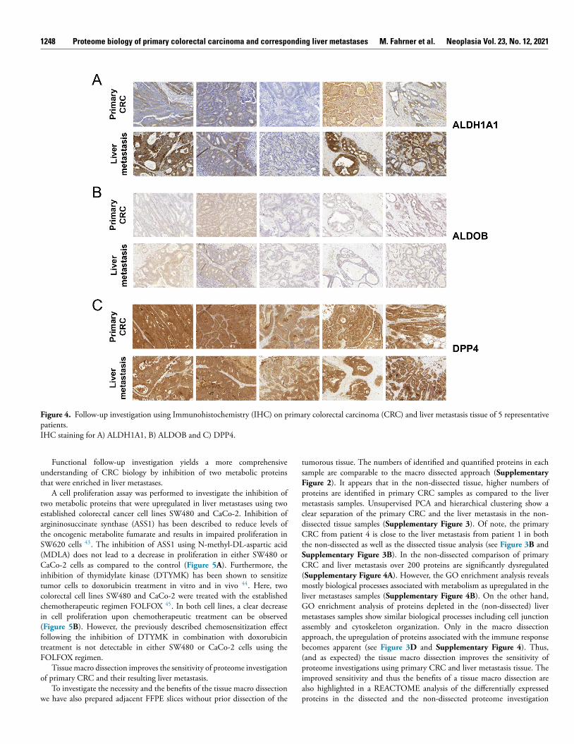
To investigate the cell-type-specific expression and spatial distributionof selected proteins of interest we used immunohistochemistry (IHC) (seeFigure 4 and Supplementary Figure 6 ). Sufficient sample material for IHCwas available for five pairs of primary and metastatic tissue and it was notpossible to include further specimens. The staining of ALDH1A1 highlighted
ts expression in tumorous tissue in both the primary CRC as well as the liveretastasis ( Figure 4 A ). Furthermore, we observe in three cases a stronger
xpression of ALDH1A1 in the tumor cells of the liver metastases as comparedo the primary CRC, consistent with the observed upregulation in theroteome data ( Supplementary Figure 6 ). IHC staining of ALDOB failed toroduce a strong signal in most cases ( Figure 4 B ). Nevertheless, expressionf ALDOB in tumorous tissue of primary CRC as well as liver metastasisas detected in most of the assessed samples. Interestingly, we observe arominent upregulation of ALDOB in the CRC-derived liver metastases
n the proteome data, which has also been described in previous studies Supplementary Figure 6 ) 41 . Furthermore, overexpression of ALDOB haseen associated with poor prognosis, promoting tumor progression 42 . Weave also probed for dipeptidyl peptidase 4 (DPP4) as a protein that showed rather consistent abundance (log2 FC = -0.12; adjusted p-value = 0.73)etween the primary CRC samples and the liver metastases ( Supplementaryigure 6 ). For DPP4 very prominent staining in most of the cases is evident,ithout any visible alteration between the two tumor entities ( Figure 4 C ).
1248 Proteome biology of primary colorectal carcinoma and corresponding liver metastases M. Fahrner et al. Neoplasia Vol. 23, No. 12, 2021
Figure 4. Follow-up investigation using Immunohistochemistry (IHC) on primary colorectal carcinoma (CRC) and liver metastasis tissue of 5 representative patients. IHC staining for A) ALDH1A1, B) ALDOB and C) DPP4.
tsF
pmc
dC
t
SC(mlGmaab(piap
Functional follow-up investigation yields a more comprehensiveunderstanding of CRC biology by inhibition of two metabolic proteinsthat were enriched in liver metastases.
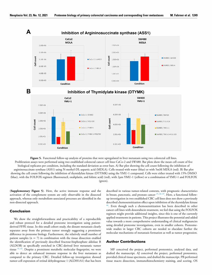
A cell proliferation assay was performed to investigate the inhibition oftwo metabolic proteins that were upregulated in liver metastases using twoestablished colorectal cancer cell lines SW480 and CaCo-2. Inhibition ofargininosuccinate synthase (ASS1) has been described to reduce levels ofthe oncogenic metabolite fumarate and results in impaired proliferation inSW620 cells 43 . The inhibition of ASS1 using N-methyl-DL-aspartic acid(MDLA) does not lead to a decrease in proliferation in either SW480 orCaCo-2 cells as compared to the control ( Figure 5 A ). Furthermore, theinhibition of thymidylate kinase (DTYMK) has been shown to sensitizetumor cells to doxorubicin treatment in vitro and in vivo 44 . Here, twocolorectal cell lines SW480 and CaCo-2 were treated with the establishedchemotherapeutic regimen FOLFOX
45 . In both cell lines, a clear decreasein cell proliferation upon chemotherapeutic treatment can be observed( Figure 5 B ). However, the previously described chemosensitization effectfollowing the inhibition of DTYMK in combination with doxorubicintreatment is not detectable in either SW480 or CaCo-2 cells using theFOLFOX regimen.
Tissue macro dissection improves the sensitivity of proteome investigationof primary CRC and their resulting liver metastasis.
To investigate the necessity and the benefits of the tissue macro dissectionwe have also prepared adjacent FFPE slices without prior dissection of the
umorous tissue. The numbers of identified and quantified proteins in each ample are comparable to the macro dissected approach ( Supplementary igure 2 ). It appears that in the non-dissected tissue, higher numbers ofroteins are identified in primary CRC samples as compared to the liver etastasis samples. Unsupervised PCA and hierarchical clustering show a
lear separation of the primary CRC and the liver metastasis in the non-issected tissue samples ( Supplementary Figure 3 ). Of note, the primary RC from patient 4 is close to the liver metastasis from patient 1 in both
he non-dissected as well as the dissected tissue analysis (see Figure 3 B andupplementary Figure 3B ). In the non-dissected comparison of primary RC and liver metastasis over 200 proteins are significantly dysregulated
Supplementary Figure 4A ). However, the GO enrichment analysis reveals ostly biological processes associated with metabolism as upregulated in the
iver metastases samples ( Supplementary Figure 4B ). On the other hand, O enrichment analysis of proteins depleted in the (non-dissected) liver etastases samples show similar biological processes including cell junction
ssembly and cytoskeleton organization. Only in the macro dissection pproach, the upregulation of proteins associated with the immune response ecomes apparent (see Figure 3 D and Supplementary Figure 4 ). Thus, and as expected) the tissue macro dissection improves the sensitivity of roteome investigations using primary CRC and liver metastasis tissue. The
mproved sensitivity and thus the benefits of a tissue macro dissection are lso highlighted in a REACTOME analysis of the differentially expressed roteins in the dissected and the non-dissected proteome investigation
Neoplasia Vol. 23, No. 12, 2021 Proteome biology of primary colorectal carcinoma and corresponding liver metastases M. Fahrner et al. 1249
Figure 5. Functional follow-up analysis of proteins that were upregulated in liver metastasis using two colorectal cell lines. Proliferation assays were performed using two established colorectal cancer cell lines CaCo-2 and SW480. Bar plots show the mean cell count of five
biological replicates per condition, including the standard deviation as error bars. A) Bar plot showing the cell count following the inhibition of argininosuccinate synthase (ASS1) using N-methyl-DL-aspartic acid (MDLA). Cells treated with water (blue) or with 5mM MDLA (red). B) Bar plot
showing the cell count following the inhibition of thymidylate kinase (DTYMK) using the YMU-1 compound. Cells were either treated with 15% DMSO
(blue), with the FOLFOX regimen (fluorouracil, oxaliplatin, and folinic acid) (red), with 2μm YMU-1 (yellow) or a combination of YMU-1 and FOLFOX
(green).
di
u
d4
c
r
a
vuw
m
A
dp
t
( Supplementary Figure 5 ). Here, the active immune response and theactivation of the complement system are only observable in the dissectedapproach, whereas only metabolism-associated processes are identified in thenon-dissected approach.
Conclusion
We show the straightforwardness and practicability of a reproducibleand robust protocol for a detailed proteome investigation using patient-derived FFPE tissue. In this small cohort study, the distant metastasis clearlyseparates away from the primary tumor strongly suggesting a prominentdifference in proteome biology. Furthermore, the relatively small number ofpatient samples (n = 7) in combination with the tissue dissection enabledthe identification of previously described fructose-bisphosphate aldolase B(ALDOB) as specifically enriched in CRC-derived liver metastatic tumortissue 41 , 42 . Despite a prominent metabolic molecular fingerprint, we wereable to detect an enhanced immune response in the liver metastases ascompared to the primary CRC. Detailed follow-up investigation showedtumor-cell expression of retinal dehydrogenase 1 (ALDH1A1) that has been
escribed in various tumor-related contexts, with prognostic characteristics n breast, pancreatic, and prostate cancer 25 , 46–49 . Here, a functional follow-p investigation in two established CRC cell lines does not show a previouslyescribed chemosensitization effect upon inhibition of the thymidylate kinase
4 . Even though such a chemosensitization has been described in otherancer cell lines with doxorubicin treatment, we feel that using the FOLFOXegimen might provide additional insights, since this is one of the currentlypplied treatments in patients. This project illustrates the potential and addedalue towards a more comprehensive understanding of clinical malignancies sing detailed proteome investigations, even in smaller cohorts. Proteome- ide studies in larger CRC cohorts are needed to elucidate further theolecular mechanisms of metastasis formation as well as tumor progression.
uthor Contributions
MF conceived the project, performed proteomics, analyzed data, and rafted the manuscript. AJ conceived the project, performed proteomics, rovided clinical tissue specimens, and drafted the manuscript. PB performedissue macro dissection, immunohistochemistry staining, and scoring. OS
1250 Proteome biology of primary colorectal carcinoma and corresponding liver metastases M. Fahrner et al. Neoplasia Vol. 23, No. 12, 2021
1
1
1
1
1
1
1
1
2
2
2
2
2
2
2
2
2
2
3
3
3
3
3
conceived the project, supervised the proteomics part, and drafted themanuscript. SF supervised the project. All authors have approved the finalarticle.
Conflict of interest statement
The authors have declared no conflict of interest.
Acknowledgment
OS acknowledges funding by the Deutsche Forschungsgemeinschaft(DFG, SCHI 871/17- 1, NY 90/6-1; SCHI 871/15-1, GR 4553/5-1, PA2807/3-1, project-ID 431984000 – SFB 1453 (project P12); project-ID441891347 - SFB 1479 (project S1); project-ID 423813989/GRK2606(RTG “ProtPath”), INST 39/766-3 (Z1)), the ERA PerMed programs(BMBF, 01KU1916, 01KU1915A), the German-Israel Foundation ( grantno. 1444 ), and the German Consortium for Translational CancerResearch (project Impro-Rec). SF acknowledges funding by the DeutscheForschungsgemeinschaft project-ID 89986987 - SFB 850; project-ID280163318 - FOR 2438. The authors acknowledge the help duringproteomic sample preparation from the students that were part of themolecular and cellular biology course (2018/2019). The authors acknowledgethe help of Lucas Kook during the analysis of the proteome data and ofBettina Wehrle for technical assistance and cell culture experiments. Thearticle processing charge was funded by the Baden-Württemberg Ministryof Science, Research and Art and the University of Freiburg in the fundingprogram Open Access Publishing.
Supplementary materials
Supplementary material associated with this article can be found, in theonline version, at doi:10.1016/j.neo.2021.10.005 .
References
1 De Rosa M , et al. Genetics, diagnosis and management of colorectal cancer(Review). Oncol. Rep. 2015; 34 :1087–96 .
2 Sung H , et al. Global Cancer Statistics 2020: GLOBOCAN Estimates of Incidenceand Mortality Worldwide for 36 Cancers in 185 Countries. CA. Cancer J. Clin.2021; 71 :209–49 .
3 Fitzmaurice C . Global Burden of Disease Cancer Collaboration. Global, regional,and national cancer incidence, mortality, years of life lost, years lived with disability,and disability-adjusted life-years for 29 cancer groups, 2006 to 2016: A systematicanalysis for the Global Burden of Disease study. J. Clin. Oncol. 2018; 36 :1568 .
4 Palaghia M . Metastatic Colorectal Cancer: Review of Diagnosis and TreatmentOptions. Jurnalul Chir 2015; 10 :249–56 .
5 Doubeni CA , et al. Contribution of Behavioral Risk Factors and Obesity toSocioeconomic Differences in Colorectal Cancer Incidence. JNCI J. Natl. CancerInst. 2012; 104 :1353–62 .
6 Vogelstein B , Kinzler KW , Kinzler KW . The Genetic Basis of Human Cancer .McGraw-Hill, Medical Pub. Division; 2002 .
7 Burt MD, RW , DiSario MD, JA , Cannon-Albright PhD , GENETICS OFCOLON CANCER L . Impact of Inheritance on Colon Cancer Risk. Annu. Rev.Med. 1995; 46 :371–9 .
8 Siegel RL , Miller KD , Jemal A . Cancer statistics, 2019. CA. Cancer J. Clin.2019; 69 :7–34 .
9 Al Bandar MH , Kim NK . Current status and future perspectives on treatment ofliver metastasis in colorectal cancer. Oncol. Rep. 2017; 37 :2553–64 .
10 Oki E , et al. Recent advances in treatment for colorectal liver metastasis. Ann.Gastroenterol. Surg. 2018; 2 :167–75 .
11 Edge SB , Compton CC . The American Joint Committee on Cancer: the 7thEdition of the AJCC Cancer Staging Manual and the Future of TNM. Ann. Surg.Oncol. 2010; 17 :1471–4 .
2 Saha S , et al. Tumor size predicts long-term survival in colon cancer: an analysis ofthe National Cancer Data Base. Am. J. Surg. 2015; 209 :570–4 .
3 Kawada K , et al. Molecular mechanisms of liver metastasis. Int. J. Clin. Oncol.2011; 16 :464–72 .
4 Heinrich S , Lang H . Liver metastases from colorectal cancer: Technique of liverresection: Technique of Liver Resection. J. Surg. Oncol. 2013; 107 :579–84 .
5 Pancione M , Remo A , Colantuoni V . Genetic and Epigenetic EventsGenerate Multiple Pathways inColorectal Cancer Progression. Pathol. Res. Int. 2012; 2012 :1–11 .
6 Huang D , et al. Mutations of key driver genes in colorectal cancer progression andmetastasis. Cancer Metastasis Rev 2018; 37 :173–87 .
7 Tsilimigras DI , et al. Clinical significance and prognostic relevance of KRAS, BRAF, PI3K and TP53 genetic mutation analysis for resectable and unresectable colorectal liver metastases: A systematic review of the current evidence. Surg. Oncol. 2018; 27 :280–8 .
8 Cohen R , et al. BRAF-Mutated Colorectal Cancer: What Is the Optimal Strategyfor Treatment? Curr. Treat. Options Oncol. 2017; 18 :9 .
9 Vassos N , Piso P . Metastatic Colorectal Cancer to the Peritoneum: Current Treatment Options. Curr. Treat. Options Oncol. 2018; 19 :49 .
0 Fox CH , Johnson FB , Whiting J , Roller PP . Formaldehyde fixation. J. Histochem.Cytochem. 1985; 33 :845–53 .
1 Föll MC , et al. Reproducible proteomics sample preparation for single FFPE tissue slices using acid-labile surfactant and direct trypsinization. Clin. Proteomics 2018; 15 :11 .
2 Wisniewski JR , Ostasiewicz P , Mann M . High recovery FASP applied to theproteomic analysis of microdissected formalin fixed paraffin embedded cancer tissues retrieves known colon cancer markers. J. Proteome Res. 2011; 10 :3040–9 .
3 Nirmalan NJ , et al. Initial development and validation of a novel extraction methodfor quantitative mining of the formalin-fixed, paraffin-embedded tissue proteome for biomarker investigations. J. Proteome Res. 2011; 10 :896–905 .
4 Shi SR , Key ME , Kalra KL . Antigen retrieval in formalin-fixed, paraffin-embeddedtissues: an enhancement method for immunohistochemical staining based on microwave oven heating of tissue sections. J. Histochem. Cytochem. Off. J. Histochem. Soc. 1991; 39 :741–8 .
5 Oria VO , et al. Proteome Profiling of Primary Pancreatic Ductal Adenocarcinomas Undergoing Additive Chemoradiation Link ALDH1A1 to Early Local Recurrence and Chemoradiation Resistance. Transl. Oncol. 2018; 11 :1307–22 .
6 Müller A-K , et al. Proteomic Characterization of Prostate Cancer to Distinguish Nonmetastasizing and Metastasizing Primary Tumors and Lymph Node Metastases. Neoplasia 2018; 20 :140–51 .
7 Föll MC, et al. Identification of tissue damage, extracellular matrix remodeling and bacterial challenge as common mechanisms associated with high-risk cutaneous squamous cell carcinomas. Matrix Biol 2017. doi: 10.1016/j.matbio.2017.11.004 .
8 Coscia F , et al. Multi-level Proteomics Identifies CT45 as a Chemosensitivity Mediator and Immunotherapy Target in Ovarian Cancer. Cell 2018; 175 :159–70 .e16 .
9 Coscia F , et al. A streamlined mass spectrometry–based proteomics workflow for large-scale FFPE tissue analysis. J. Pathol. 2020; 251 :100–12 .
0 Martínez-Aguilar J , et al. Quantitative mass spectrometry for colorectal cancer proteomics. PROTEOMICS - Clin. Appl. 2013; 7 :42–54 .
1 Bandeira N , Deutsch EW , Kohlbacher O , Martens L , Vizcaíno JA . DataManagement of Sensitive Human Proteomics Data: Current Practices, Recommendations, and Perspectives for the Future. Mol. Cell. Proteomics 2021; 20 :100071 .
2 Bronsert P , et al. Impact of routinely employed procedures for tissue processingon the proteomic analysis of formalin-fixed paraffin-embedded tissue. Proteomics - Clin. Appl. 2014; 8 :796–804 .
3 Yu Y-Q , Gilar M , Lee PJ , Bouvier ESP , Gebler JC . Enzyme-Friendly, MassSpectrometry-Compatible Surfactant for In-Solution Enzymatic Digestion of Proteins. Anal. Chem. 2003; 75 :6023–8 .
4 Kulak NA , Pichler G , Paron I , Nagaraj N , Mann M . Minimal, encapsulatedproteomic-sample processing applied to copy-number estimation in eukaryotic cells. Nat. Methods 2014; 11 :319–24 .
Neoplasia Vol. 23, No. 12, 2021 Proteome biology of primary colorectal carcinoma and corresponding liver metastases M. Fahrner et al. 1251
4
4
4
4
4
4
4
35 Cox J , et al. Accurate Proteome-wide Label-free Quantification by DelayedNormalization and Maximal Peptide Ratio Extraction, Termed MaxLFQ. Mol CellProteomics 2014; 13 :2513–26 .
36 Smyth GK . Linear Models and Empirical Bayes Methods for Assessing DifferentialExpression in Microarray Experiments. Stat. Appl. Genet. Mol. Biol. 2004; 3 :1–25 .
37 Benjamini Y , Hochberg Y . Controlling the False Discovery Rate: A Practicaland Powerful Approach to Multiple Testing. J. R. Stat. Soc. Ser. B Methodol.1995; 57 :289–300 .
38 Lê Cao K-A , González I , Déjean S . integrOmics: an R package to unravelrelationships between two omics datasets. Bioinformatics 2009; 25 :2855–6 .
39 Rohart F , Gautier B , Singh A , Lê Cao K-A . mixOmics: An R package for‘omics feature selection and multiple data integration. PLOS Comput. Biol.2017; 13 :e1005752 .
40 Alexa A , Rahnenfuhrer J , Lengauer T . Improved scoring of functional groupsfrom gene expression data by decorrelating GO graph structure. Bioinformatics2006; 22 :1600–7 .
41 Bu P , et al. Aldolase B-Mediated Fructose Metabolism Drives MetabolicReprogramming of Colon Cancer Liver Metastasis. Cell Metab 2018; 27 :1249–62.e4 .
42 Lia Qingguo , Lia Yaqi , Xub Junyan , Wanga Sheng . Ye Xua, Xinxiang Lia, S. C.Aldolase B Overexpression is Associated with Poor Prognosis and Promotes Tumor
Progression by Epithelial-Mesenchymal Transition in Colorectal Adenocarcinoma. Cell. Physiol. Biochem. 2017; 42 :397–406 .
3 Bateman LA , et al. Argininosuccinate Synthase 1 is a Metabolic Regulator ofColorectal Cancer Pathogenicity. ACS Chem. Biol. 2017; 12 :905–11 .
4 Hu C-MM , et al. Tumor Cells Require Thymidylate Kinase to Prevent dUTPIncorporation during DNA Repair. Cancer Cell 2012; 22 :36–50 .
5 Roh SA , et al. Characterization of biological responses of colorectal cancer cells toanticancer regimens. J. Korean Surg. Soc. 2012; 83 :21–9 .
6 Li T , et al. ALDH1A1 is a marker for malignant prostate stem cells and predictorof prostate cancer patients’ outcome. Lab. Invest. 2010; 90 :234–44 .
7 Althobiti M , et al. The prognostic significance of ALDH1A1 expression in earlyinvasive breast cancer. Histopathology 2020; 77 :437–48 .
8 Charkoftaki G , et al. Integrated multi-omics approach reveals a role ofALDH1A1 in lipid metabolism in human colon cancer cells. Chem. Biol. Interact.2019; 304 :88–96 .
9 Singh S , Arcaroli J , Thompson DC , Messersmith W , Vasiliou V , Vasiliou V ,Zakhari S , Seitz HK , Hoek JB . Acetaldehyde and Retinaldehyde-MetabolizingEnzymes in Colon and Pancreatic Cancers. In: Biological Basis of Alcohol-InducedCancer, 815. Springer International Publishing; 2015. p. 281–94 .


![Guide to Biology - WordPress.com · Significant contribution Nelson Thornes AQA Science [GCSE Biology] 3 Active Transport Two of the main ways in which diffused substances are transported](https://static.fdocuments.fr/doc/165x107/5e8a088f531a680df900aee2/guide-to-biology-significant-contribution-nelson-thornes-aqa-science-gcse-biology.jpg)