Tennis et Epidémiologie Maladie D’ Osgood Schlatter Maladie de Sever
Maladie d’Erdheim-Chester
Julien Haroche
Service de médecine interne 2, Institut E3M DIU Groupe Hospitalier Pitié-Salpêtrière 22 Juin 2018 Paris, France
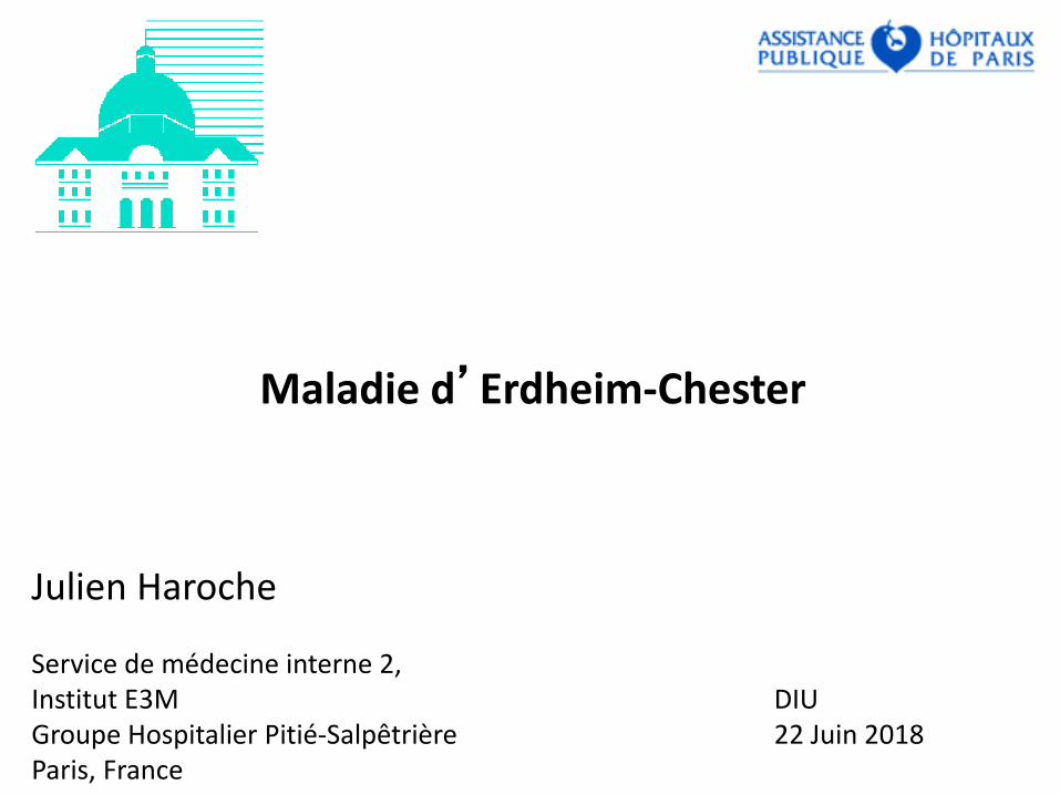
Le macrophage : une cellule caméléon
Hémophagocytose Sidérophage
Cellule de Touton
(xanthogranulome)
Cellule de Virchows
(lèpre) Lipophage
Ç de Langhans
(tuberculose)
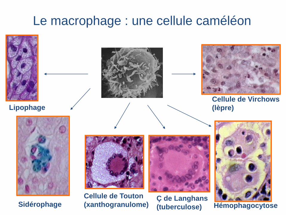
D'une cellule CD34+ à l'histiocyte
Histiocyte = aspect
morphologique d'un
macrophage résident
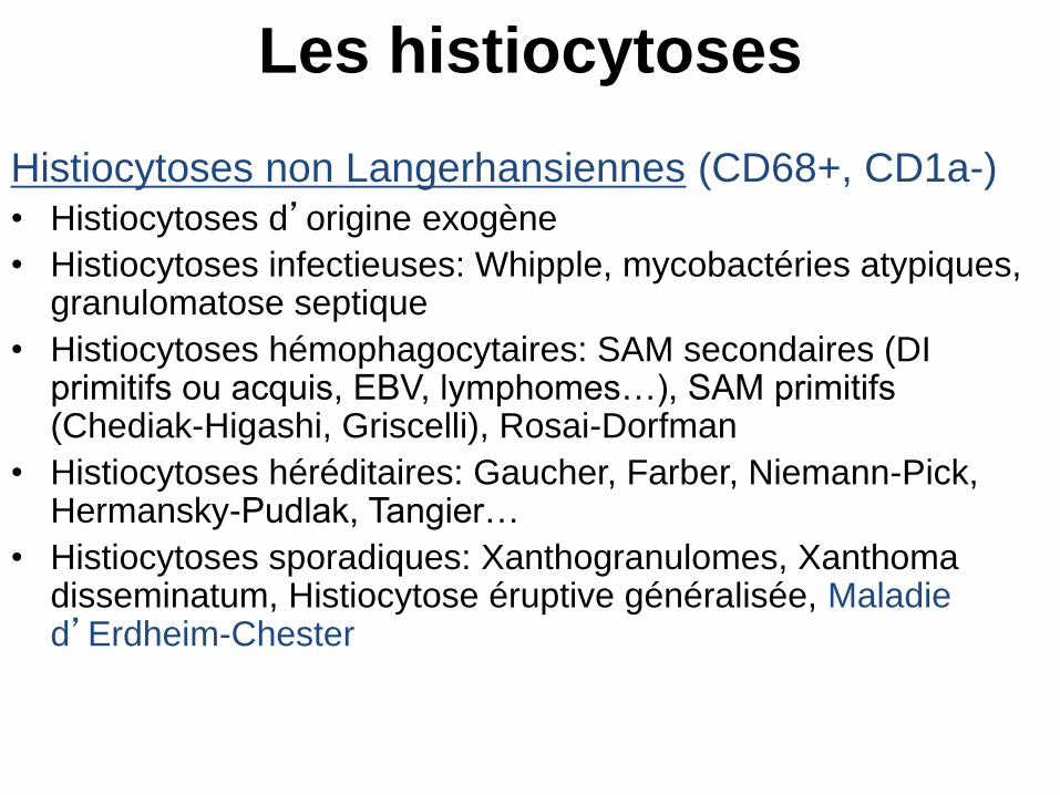
Les histiocytoses
Histiocytoses non Langerhansiennes (CD68+, CD1a-)
• Histiocytoses d’origine exogène
• Histiocytoses infectieuses: Whipple, mycobactéries atypiques, granulomatose septique
• Histiocytoses hémophagocytaires: SAM secondaires (DI primitifs ou acquis, EBV, lymphomes…), SAM primitifs (Chediak-Higashi, Griscelli), Rosai-Dorfman
• Histiocytoses héréditaires: Gaucher, Farber, Niemann-Pick, Hermansky-Pudlak, Tangier…
• Histiocytoses sporadiques: Xanthogranulomes, Xanthoma disseminatum, Histiocytose éruptive généralisée, Maladie d’Erdheim-Chester
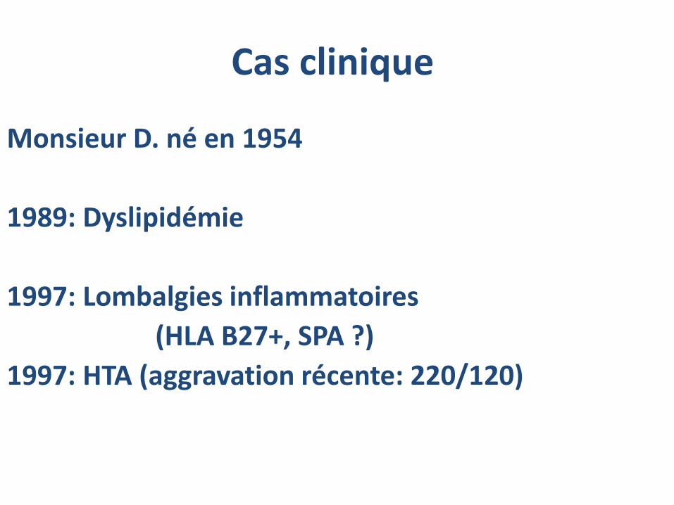
Cas clinique
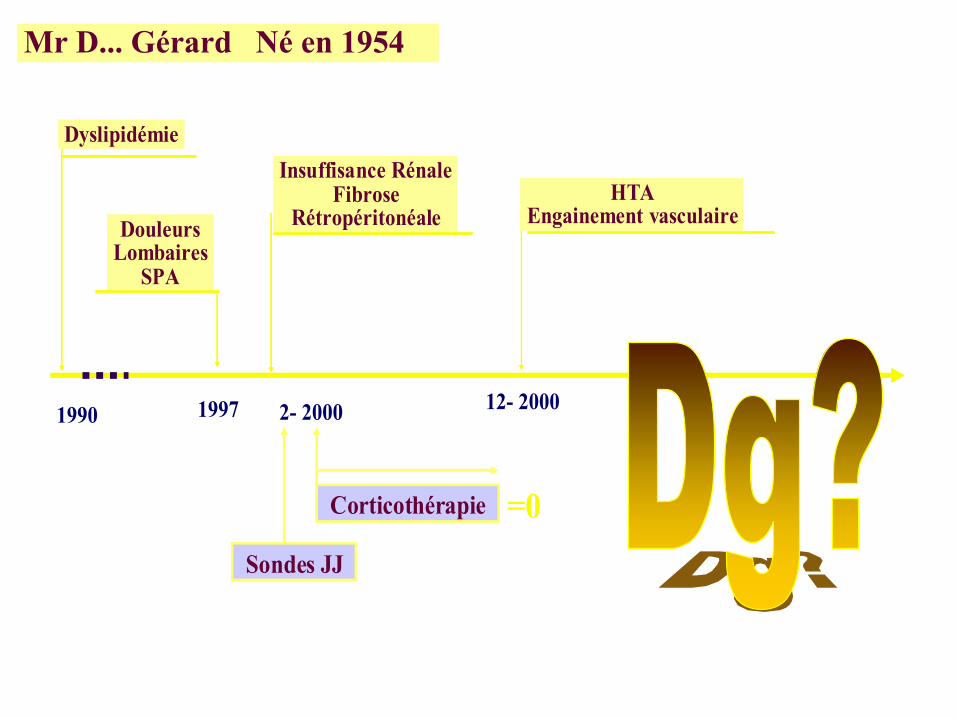
Monsieur D. né en 1954
1989: Dyslipidémie
1997: Lombalgies inflammatoires
(HLA B27+, SPA ?)
1997: HTA (aggravation récente: 220/120)

2-2000 : Découverte d’une insuffisance rénale
créatinine 182 micromoles/l
3-2000 : syndrome inflammatoire
CRP 71 mg/l, VS 121 mm
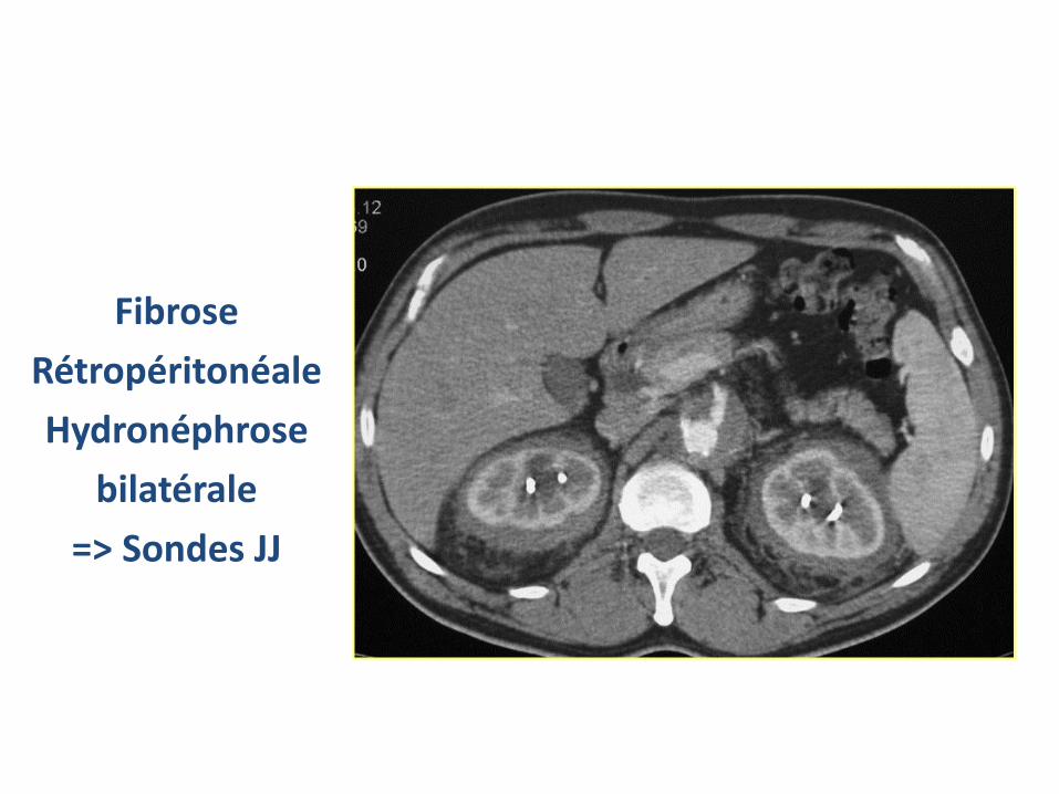
Fibrose
Rétropéritonéale
Hydronéphrose
bilatérale
=> Sondes JJ

Sténose artérielle rénale => stent

L’aorte thoracique est aussi touchée…
reconstruction sagittale
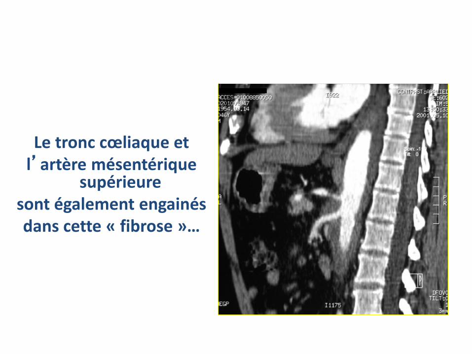
Le tronc cœliaque et l’artère mésentérique
supérieure sont également engainés dans cette « fibrose »…

… visible à l’IRM :
Hyper-signal en T1
de la partie interne
du mur aortique
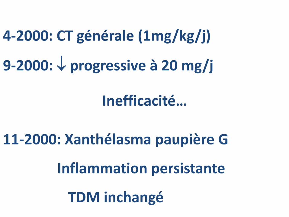
4-2000: CT générale (1mg/kg/j)
9-2000: progressive à 20 mg/j
11-2000: Xanthélasma paupière G
Inflammation persistante
TDM inchangé
Inefficacité…

Janvier 2001
Adressé pour échec thérapeutique
. Raideur rachidienne : indice de Schober à 2 cm
. HTA : 180/110 mmHg sous traitement par Nicardipine Avlocardyl
1997
DouleursLombaires
SPA
1997
DouleursLombaires
SPA
2- 2000
Insuffisance RénaleFibrose
Rétropéritonéale
2- 2000
Insuffisance RénaleFibrose
Rétropéritonéale
1990
Dyslipidémie
1990
Dyslipidémie
HTAEngainement vasculaire
12- 2000
HTAEngainement vasculaire
12- 2000
Corticothérapie
Sondes JJ
=0
Mr D... Gérard Né en 1954
1997
DouleursLombaires
SPA
1997
DouleursLombaires
SPA
2- 2000
Insuffisance RénaleFibrose
Rétropéritonéale
2- 2000
Insuffisance RénaleFibrose
Rétropéritonéale
1990
Dyslipidémie
1990
Dyslipidémie
HTAEngainement vasculaire
12- 2000
HTAEngainement vasculaire
12- 2000
Corticothérapie
Sondes JJ
=0
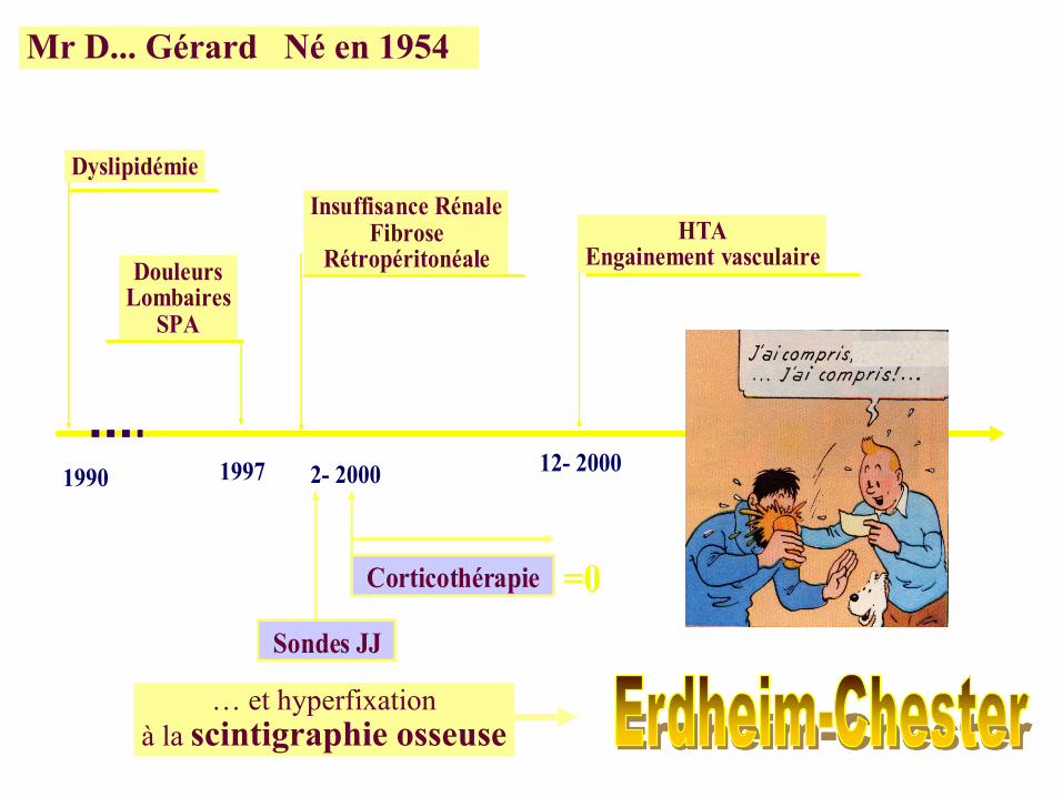
Mr D... Gérard Né en 1954
… et hyperfixation
à la scintigraphie osseuse
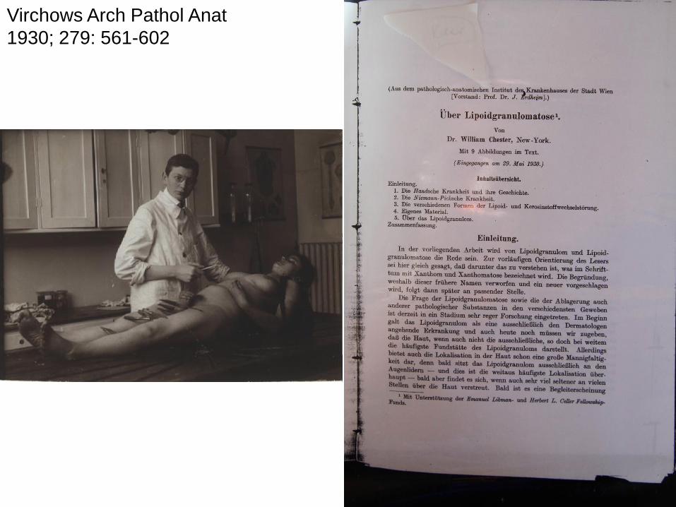
Virchows Arch Pathol Anat
1930; 279: 561-602
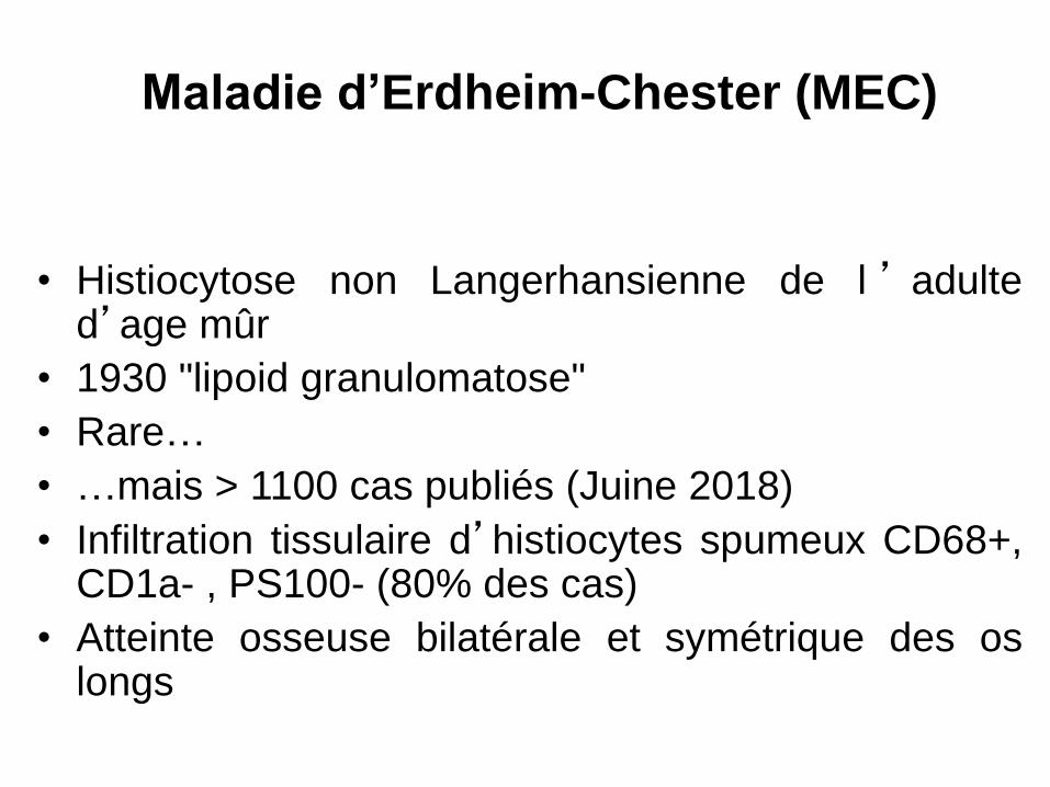
Maladie d’Erdheim-Chester (MEC)
• Histiocytose non Langerhansienne de l ’ adulte d’age mûr
• 1930 "lipoid granulomatose"
• Rare…
• …mais > 1100 cas publiés (Juine 2018)
• Infiltration tissulaire d’histiocytes spumeux CD68+, CD1a- , PS100- (80% des cas)
• Atteinte osseuse bilatérale et symétrique des os longs

Bone scintigraphy (99 Tc)
"Hairy kidney aspect" and peri-renal infiltration
(96%) (≈ 50%)
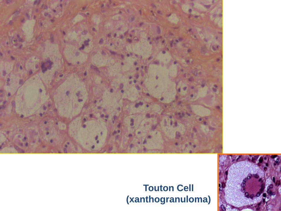
Touton Cell
(xanthogranuloma)
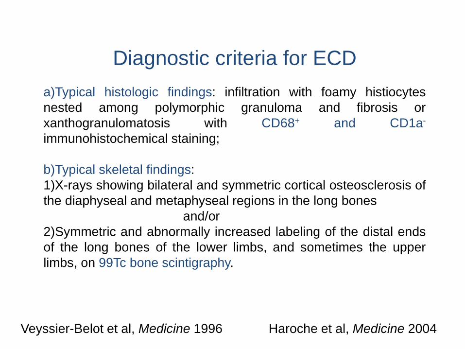
Diagnostic criteria for ECD
a)Typical histologic findings: infiltration with foamy histiocytes
nested among polymorphic granuloma and fibrosis or
xanthogranulomatosis with CD68+ and CD1a-
immunohistochemical staining;
b)Typical skeletal findings:
1)X-rays showing bilateral and symmetric cortical osteosclerosis of
the diaphyseal and metaphyseal regions in the long bones
and/or
2)Symmetric and abnormally increased labeling of the distal ends
of the long bones of the lower limbs, and sometimes the upper
limbs, on 99Tc bone scintigraphy.
Veyssier-Belot et al, Medicine 1996 Haroche et al, Medicine 2004
Natural history unknown
• Asymtomatic forms of the disease
• Multisystemic & life-threatening forms of the disease
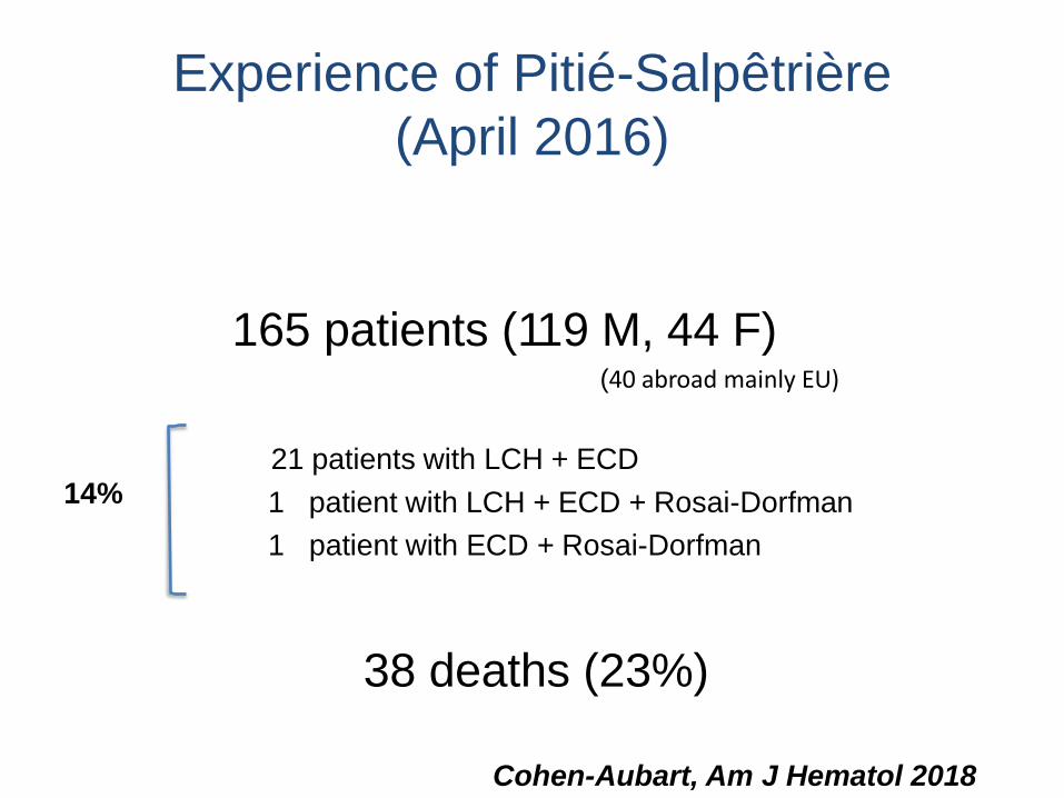
Experience of Pitié-Salpêtrière
(April 2016)
165 patients (119 M, 44 F) (40 abroad mainly EU)
21 patients with LCH + ECD
1 patient with LCH + ECD + Rosai-Dorfman
1 patient with ECD + Rosai-Dorfman
38 deaths (23%)
14%
Cohen-Aubart, Am J Hematol 2018

Extra-osseous involvement ≈ 98% of patients
• Xanthelasma • Exophthalmos • Diabetes insipidus • Central nervous system (CNS) Cerebellar • Pulmonary involvement • Peri-renal (“hairy kidney”) • “Retro-peritoneal fibrosis” /
HN • Coated aorta • Reno-vascular hypertension • Pericardial • Pseudo-atrial tumor
43 pts (26%) 36 pts (22%) 46 pts (28%) 60 pts (37%) 28 pts (17%) 58 pts (36%) 95 pts (58%) 40 pts (25%) 75 pts (46%) 29 pts (18%) 51 pts (31%) 61 pts (37%)

165 patients ECD (1992 - 2016)
• Mean age at diagnosis: 56.1 yr (14.4), range: 5-80
• Mean age at first sign: 51.9 yr (15.8)
• Mean diagnostic delay : 48 mo (up to 372 mo)
• Increased levels of acute inflammatory protein (CRP) : 87 pts (71%)
• 1st symptom extremely variable (bone pain 10%, diabetes insipidus 16%) but also exophthalmos, seizures, sinusitis…

Manifestations rhumatologiques
Douleurs osseuses : 56 pts (38%)
Atteinte osseuse souvent asymptomatique
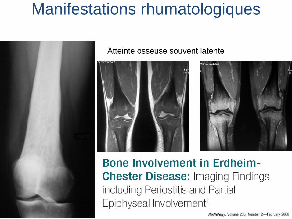
Manifestations rhumatologiques
Atteinte osseuse souvent latente
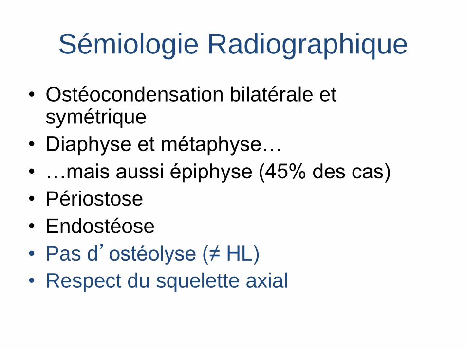
Sémiologie Radiographique
• Ostéocondensation bilatérale et symétrique
• Diaphyse et métaphyse…
• …mais aussi épiphyse (45% des cas)
• Périostose
• Endostéose
• Pas d’ostéolyse (≠ HL)
• Respect du squelette axial
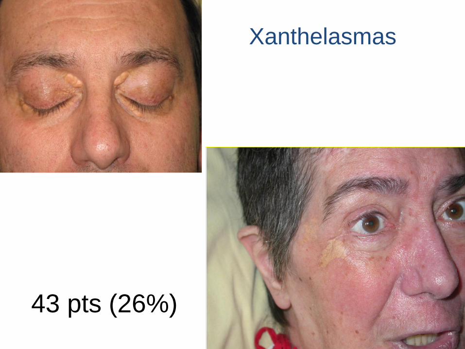
43 pts (26%)
Xanthelasmas

36 pts (22%)
Exophthalmos
Diabetes insipidus
46 pts (28%)

•CNS involvement in 15 to 25 % patients
depending on series.
•Clinical symptoms are proteiform: pyramidal
syndrom, cerebellar syndrom (sometimes
wheelchair), more rarely seizures, headaches,
psychiatric or cognitive impairments.
•Important mortality : 9% (6/66 pts)
In our 165 patients series: 60 pts (37%) have CNS,
28 pts (17%) of which have cerebellar involvement
AX T2
CORO T1 G
TDM
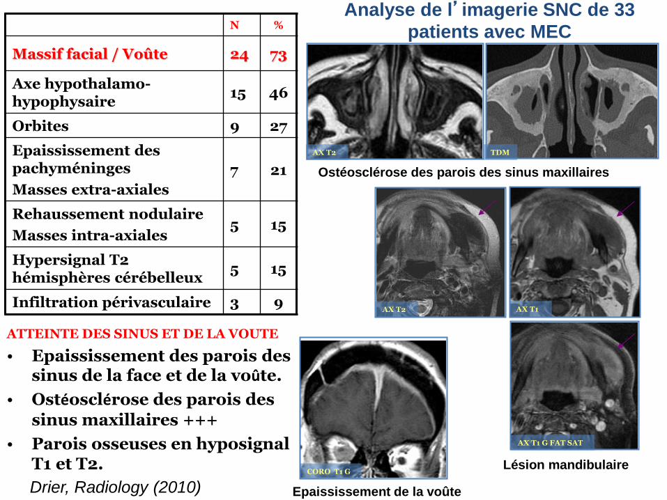
ATTEINTE DES SINUS ET DE LA VOUTE
• Epaississement des parois des sinus de la face et de la voûte.
• Ostéosclérose des parois des
sinus maxillaires +++
• Parois osseuses en hyposignal T1 et T2. Lésion mandibulaire
Ostéosclérose des parois des sinus maxillaires
Epaississement de la voûte
AX T1 G FAT SAT
AX T1 AX T2
N %
Massif facial / Voûte 24 73
Axe hypothalamo-hypophysaire
15 46
Orbites 9 27
Epaississement des pachyméninges
Masses extra-axiales
7 21
Rehaussement nodulaire
Masses intra-axiales 5 15
Hypersignal T2 hémisphères cérébelleux
5 15
Infiltration périvasculaire 3 9
Analyse de l’imagerie SNC de 33
patients avec MEC
Drier, Radiology (2010)
CORO T1 G CORO T2
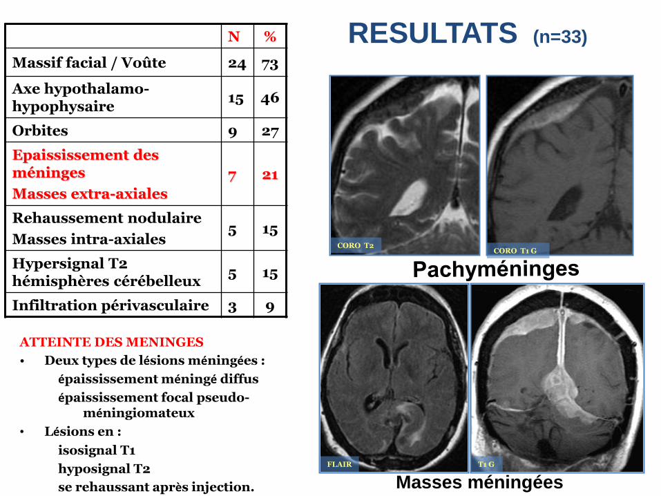
ATTEINTE DES MENINGES
• Deux types de lésions méningées :
épaississement méningé diffus
épaississement focal pseudo-méningiomateux
• Lésions en :
isosignal T1
hyposignal T2
se rehaussant après injection. Masses méningées T1 G FLAIR
RESULTATS (n=33) N %
Massif facial / Voûte 24 73
Axe hypothalamo-hypophysaire
15 46
Orbites 9 27
Epaississement des méninges
Masses extra-axiales
7 21
Rehaussement nodulaire
Masses intra-axiales 5 15
Hypersignal T2 hémisphères cérébelleux
5 15
Infiltration périvasculaire 3 9
AX T1 G AX T2
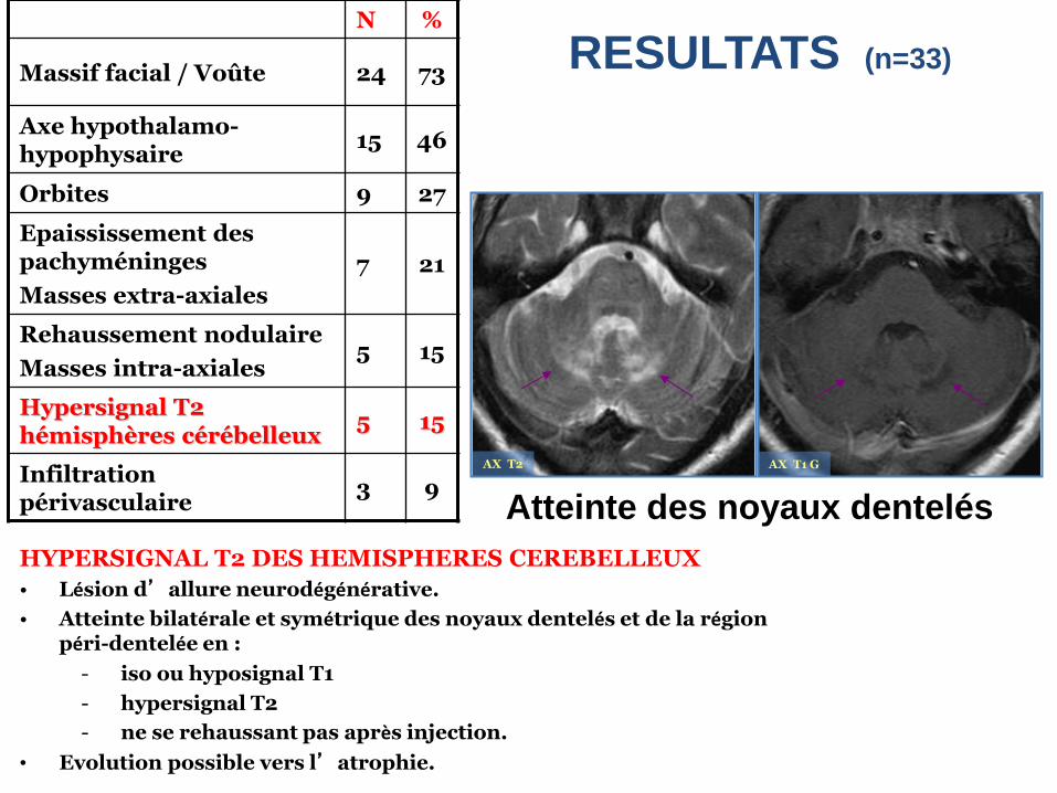
HYPERSIGNAL T2 DES HEMISPHERES CEREBELLEUX
• Lésion d’allure neurodégénérative.
• Atteinte bilatérale et symétrique des noyaux dentelés et de la région péri-dentelée en :
- iso ou hyposignal T1
- hypersignal T2
- ne se rehaussant pas après injection.
• Evolution possible vers l’atrophie.
Atteinte des noyaux dentelés
RESULTATS (n=33)
N %
Massif facial / Voûte 24 73
Axe hypothalamo-hypophysaire
15 46
Orbites 9 27
Epaississement des pachyméninges
Masses extra-axiales
7 21
Rehaussement nodulaire
Masses intra-axiales 5 15
Hypersignal T2 hémisphères cérébelleux
5 15
Infiltration périvasculaire
3 9
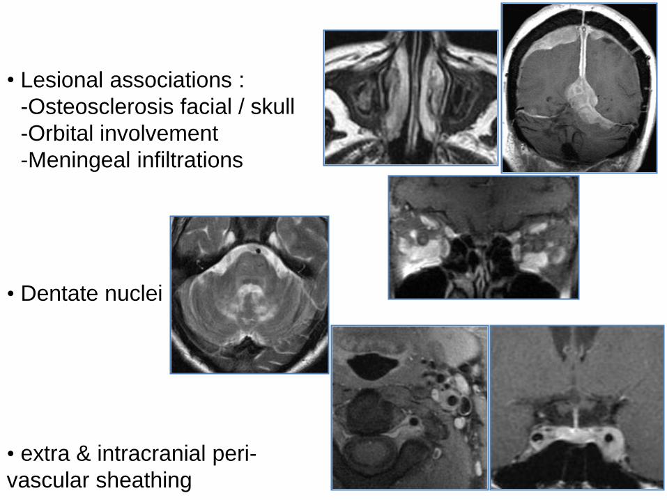
• Lesional associations :
-Osteosclerosis facial / skull
-Orbital involvement
-Meningeal infiltrations
• Dentate nuclei
• extra & intracranial peri-
vascular sheathing
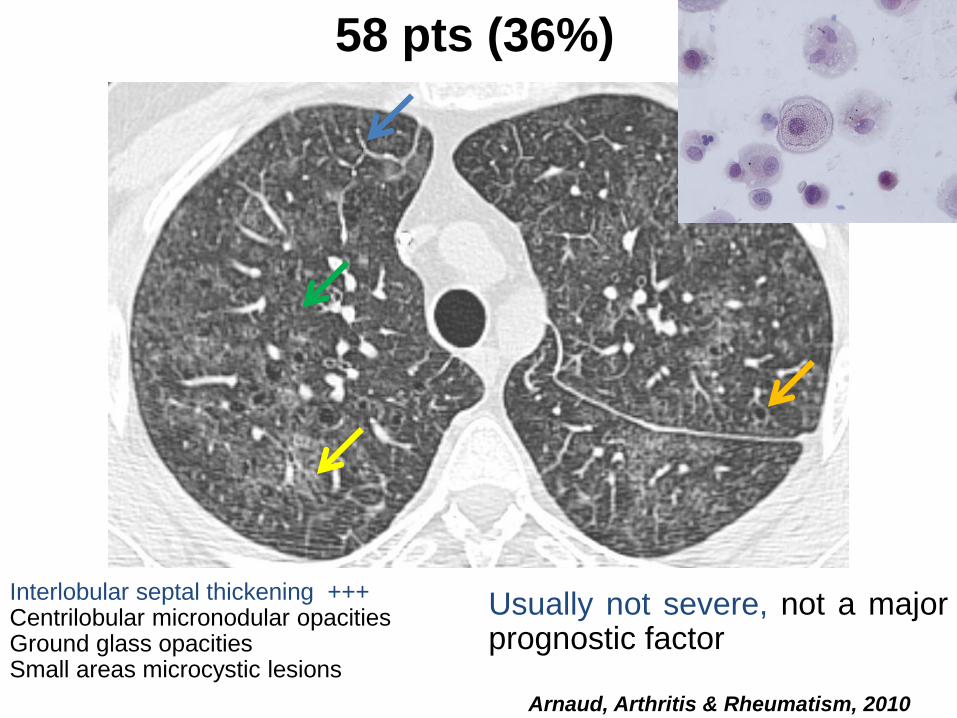
Interlobular septal thickening +++ Centrilobular micronodular opacities Ground glass opacities Small areas microcystic lesions
Arnaud, Arthritis & Rheumatism, 2010
58 pts (36%)
Usually not severe, not a major prognostic factor

« hairy kidney » aspect with infiltration of the perirenal fat and of
the perirenal fascia = 95 pts (58%)


Hydronephrosis
40 pts (25%)
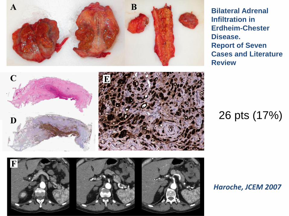
Haroche, JCEM 2007
Bilateral Adrenal
Infiltration in
Erdheim-Chester
Disease.
Report of Seven
Cases and Literature
Review
26 pts (17%)

Atteintes endocriniennes N (%)
Insuffisance somatotrope 22/28 (78.6)
Insuffisance testiculaire 26/49 (53.1)
Hyperprolactinémie 26/59 (44.1)
Diabète insipide 19/57 (33.3)
Insuffisance gonadotrope 14/63 (22.2)
Insuffisance thyréotrope 6/63 (9.5)
Hypothyroïdie périphérique 6/63 (9.5)
Insuffisance corticotrope 2/64 (3.1)
Aucune atteinte endocrinienne 1/61 (1.6)
1 déficit AH = 61%
≥ 2 déficits AH = 30%
Pas de ≠ H/F sauf
insuffisance
gonadique
Pas de corrélation
entre IAH et IPH
Dysfonction AH
Atteinte des axes
dans le même ordre
de fréquence que
HL ou radiothérapie
Diabète insipide
Inaugural 65% cas
Permanent
Courtillot, JCEM 2016
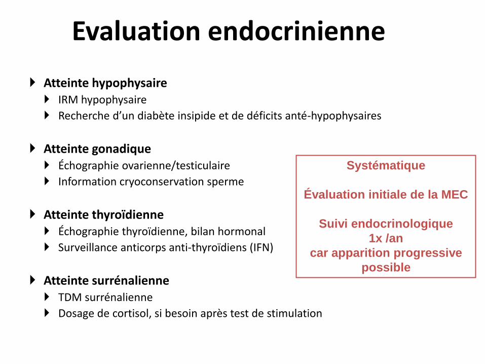
Evaluation endocrinienne
Atteinte hypophysaire IRM hypophysaire
Recherche d’un diabète insipide et de déficits anté-hypophysaires
Atteinte gonadique Échographie ovarienne/testiculaire
Information cryoconservation sperme
Atteinte thyroïdienne Échographie thyroïdienne, bilan hormonal
Surveillance anticorps anti-thyroïdiens (IFN)
Atteinte surrénalienne TDM surrénalienne
Dosage de cortisol, si besoin après test de stimulation
Systématique
Évaluation initiale de la MEC
Suivi endocrinologique
1x /an
car apparition progressive
possible

Décrite depuis longtemps…mais longtemps
méconnue
• Infiltration aorte et branches collatérales
• Péricarde
• Myocarde
...Et « renominée » récemment : « Coated aorta »
Serratrice et al. (2000) J Rheumatol
MEC : atteinte cardio-vasculaire
Medicine, 2004
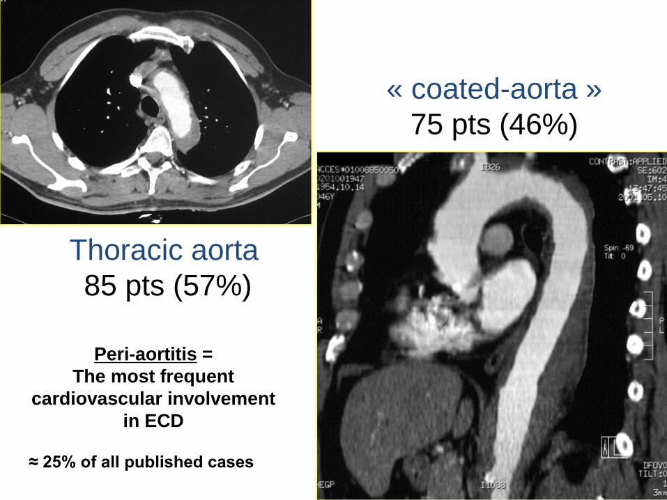
Thoracic aorta
85 pts (57%)
« coated-aorta »
75 pts (46%)
Peri-aortitis =
The most frequent
cardiovascular involvement
in ECD
≈ 25% of all published cases

Abdominal aorta 87 pts (57%)

Frequent involvement of aorta branches
- mesenteric superior artery
- celiac trunk
- left sub-clavian artery
- left common carotid artery
- renal artery (stents when reno-vascular hypertension)
Currently 29 pts (18%) with reno-vascular HT
should be systematically looked for +++
Overall little clinical
consequences
Pericardial involvement :
2nd most frequent
≈ 20% of the published cases
can lead to tamponade (sometimes lethal)
51 pts (31%)
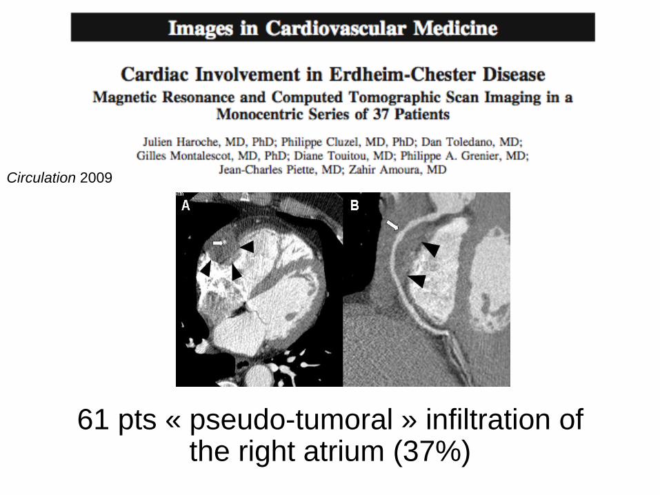
61 pts « pseudo-tumoral » infiltration of the right atrium (37%)
Circulation 2009

Coronary involvement in ECD
Myocardial infarction in more then 20 pts
(13 personal), fatal in at least 3 cases
Preferentially Right coronary artery 35 pts (23%)
27 pts (18%) Left coronary
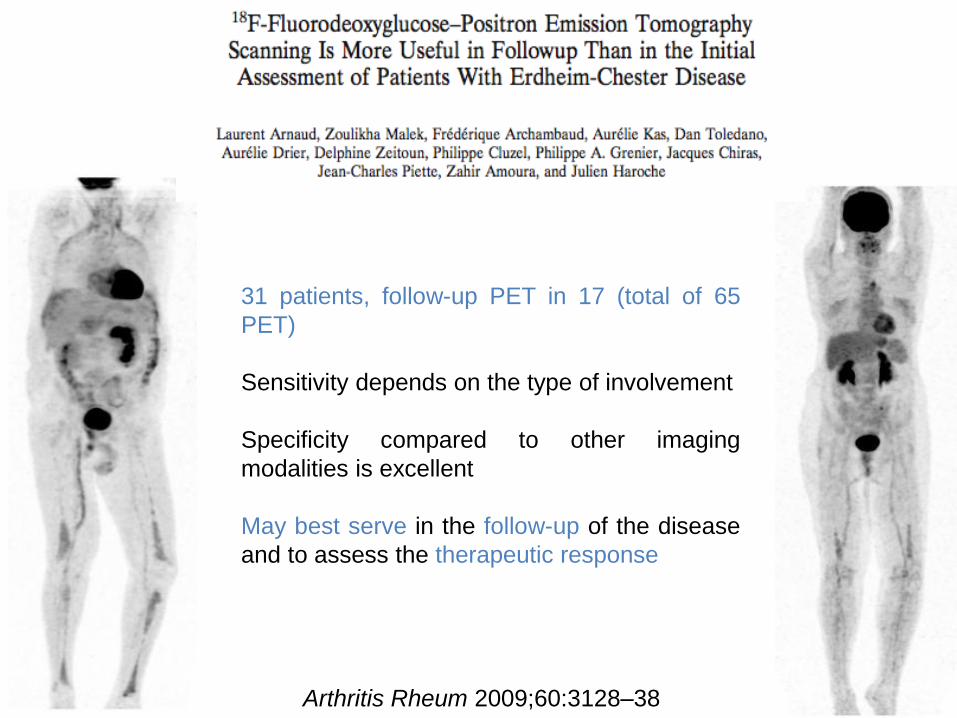
Arthritis Rheum 2009;60:3128–38
31 patients, follow-up PET in 17 (total of 65
PET)
Sensitivity depends on the type of involvement
Specificity compared to other imaging
modalities is excellent
May best serve in the follow-up of the disease
and to assess the therapeutic response

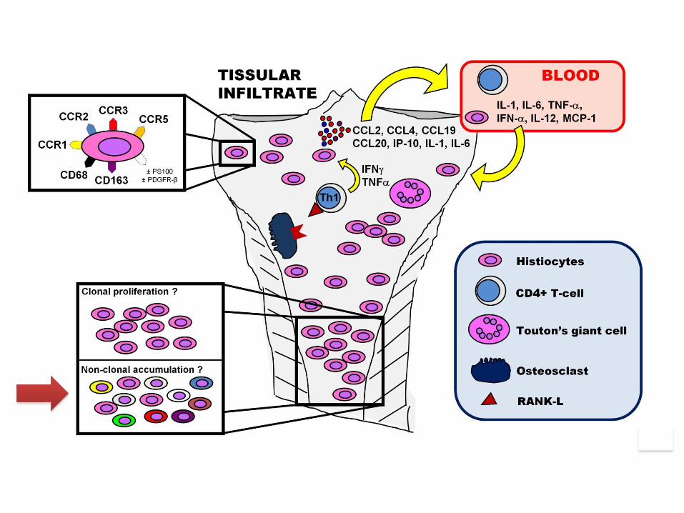
Physiopathologie

Arthritis Rheum 2006
CCR3
CCR7 CCL19
CCR5
IL-6 IFN-g
C-
RANK-L

Impaired Th1/Th2 balance Production of IFN- (source ?)
Histiocyte recruitment via MCP-1 ?
Identification of a cytokine « signature »
Blood 2011
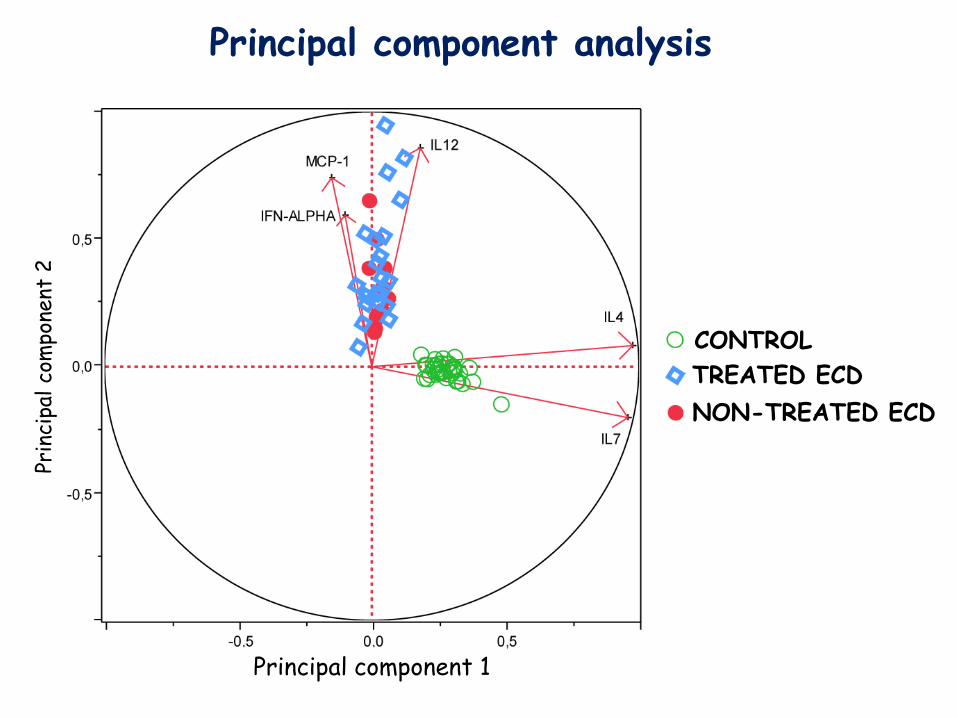
CONTROL
TREATED ECD
NON-TREATED ECD
Principal component 1
Prin
cipa
l co
mpo
nent
2
Principal component analysis


Looking for BRAF V600E mutations
• Histological samples taken from 127 patients with histiocytosis were reviewed
• Detection of BRAFV600 mutations was performed by
pyrosequencing of DNA extracted from paraffin
embedded samples.
• 46 ECD
• 39 LCH
• 23 Rosai-Dorfman
• 12 Juvenile Xanthogranuloma
• 3 Histiocytic Sarcoma
• 2 Xanthoma disseminatum
• 1 interdigitating dendritic cell sarcoma
• 1 necrobiotic xanthogranuloma
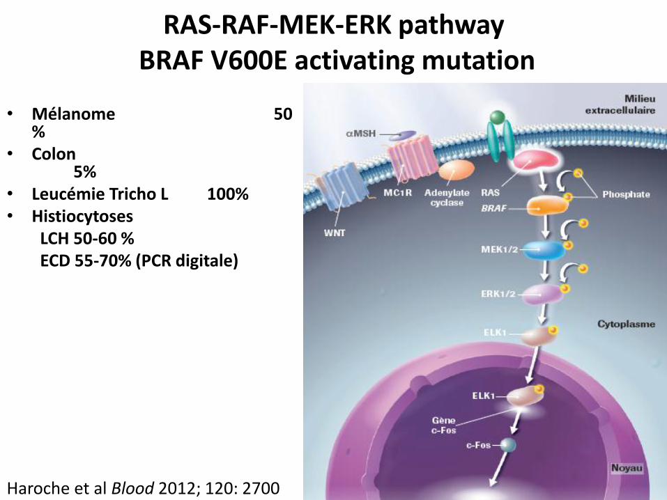
RAS-RAF-MEK-ERK pathway BRAF V600E activating mutation
• Mélanome 50 %
• Colon 5%
• Leucémie Tricho L 100% • Histiocytoses
LCH 50-60 % ECD 55-70% (PCR digitale)
Haroche et al Blood 2012; 120: 2700

Classification taking into account molecular alterations

Démarches Thérapeutiques
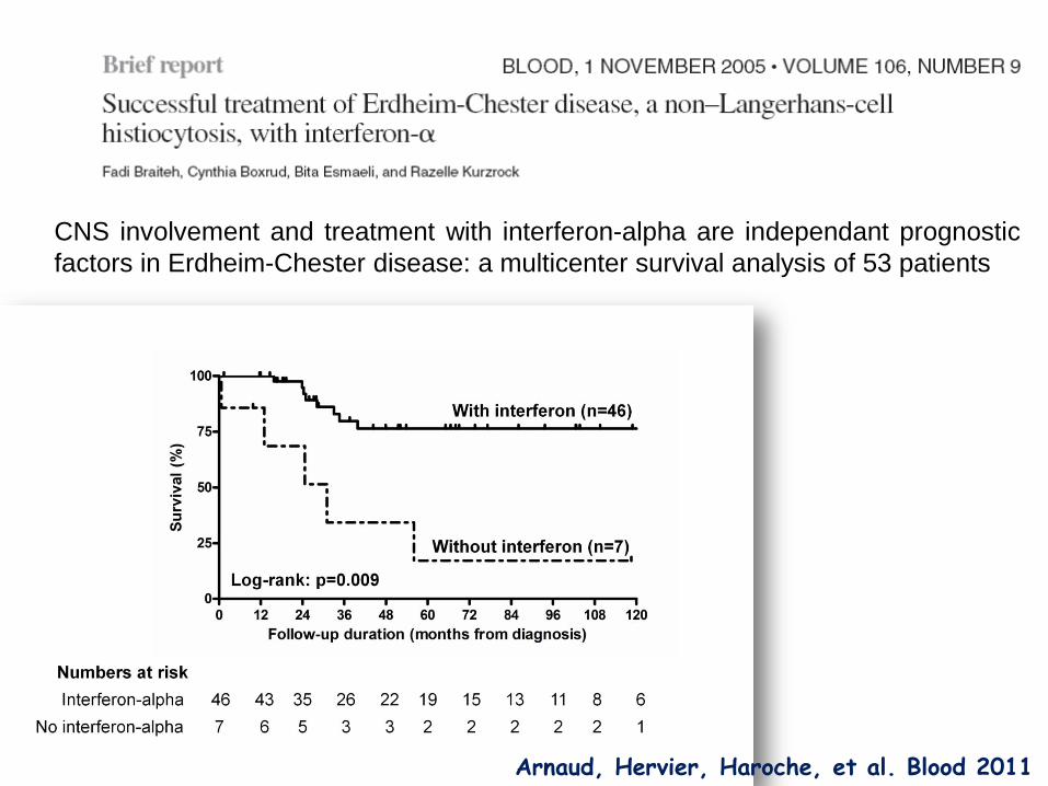
CNS involvement and treatment with interferon-alpha are independant prognostic
factors in Erdheim-Chester disease: a multicenter survival analysis of 53 patients
Arnaud, Hervier, Haroche, et al. Blood 2011

Arnaud, Hervier, Haroche, et al. Blood 2011
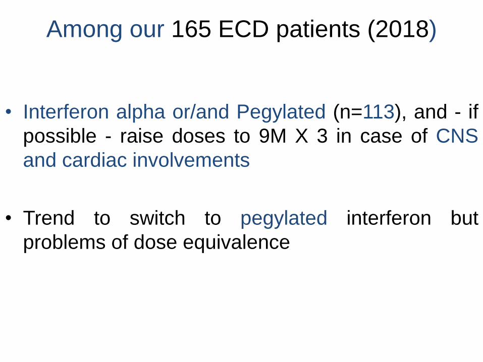
Among our 165 ECD patients (2018)
• Interferon alpha or/and Pegylated (n=113), and - if
possible - raise doses to 9M X 3 in case of CNS
and cardiac involvements
• Trend to switch to pegylated interferon but
problems of dose equivalence

05/2007 04/2008
01/2009
Man, 30-year-old, referred to our institution in october 2006 ECD diagnosed on bone biopsy in september 2002 CNS involvement only with sus and retro-sellar infiltration with diabetes insipidus, hypogonadism and complex partial seizures Major side effects to vinblastine in 2002 IFN alpha 9 M x 3 per week initiated in October 2006
Before IFN
After 3 mo of IFN 9 Mx3
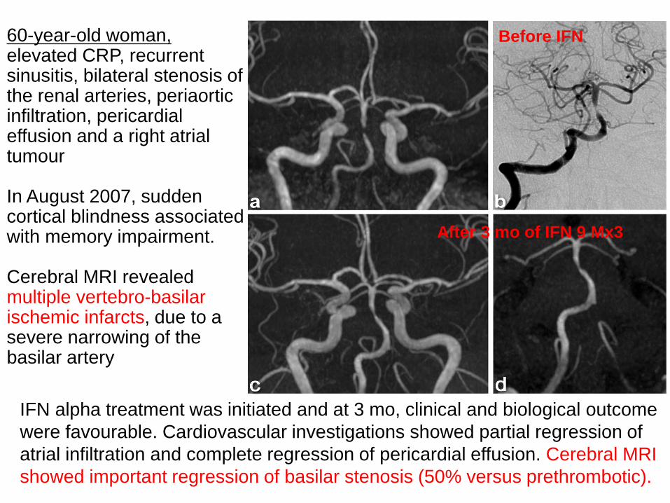
IFN alpha treatment was initiated and at 3 mo, clinical and biological outcome
were favourable. Cardiovascular investigations showed partial regression of
atrial infiltration and complete regression of pericardial effusion. Cerebral MRI
showed important regression of basilar stenosis (50% versus prethrombotic).
60-year-old woman, elevated CRP, recurrent sinusitis, bilateral stenosis of the renal arteries, periaortic infiltration, pericardial effusion and a right atrial tumour In August 2007, sudden cortical blindness associated with memory impairment. Cerebral MRI revealed multiple vertebro-basilar ischemic infarcts, due to a severe narrowing of the basilar artery
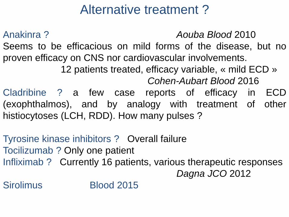
Alternative treatment ?
Anakinra ? Aouba Blood 2010
Seems to be efficacious on mild forms of the disease, but no
proven efficacy on CNS nor cardiovascular involvements.
12 patients treated, efficacy variable, « mild ECD »
Cohen-Aubart Blood 2016
Cladribine ? a few case reports of efficacy in ECD
(exophthalmos), and by analogy with treatment of other
histiocytoses (LCH, RDD). How many pulses ?
Tyrosine kinase inhibitors ? Overall failure
Tocilizumab ? Only one patient
Infliximab ? Currently 16 patients, various therapeutic responses
Dagna JCO 2012
Sirolimus Blood 2015
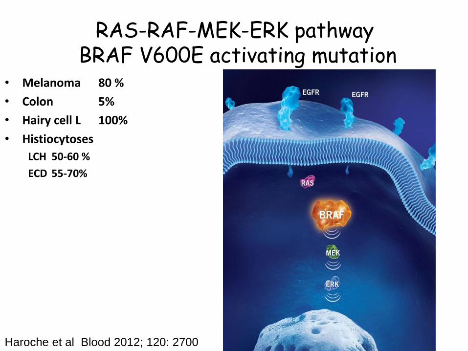
RAS-RAF-MEK-ERK pathway BRAF V600E activating mutation
• Melanoma 80 %
• Colon 5%
• Hairy cell L 100%
• Histiocytoses
LCH 50-60 %
ECD 55-70%
Haroche et al Blood 2012; 120: 2700
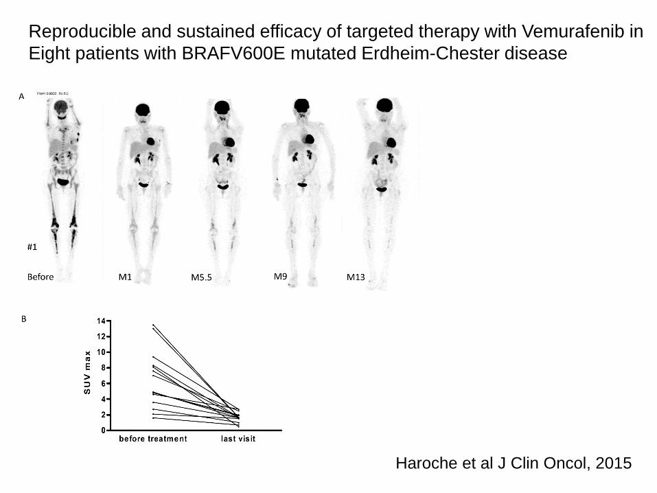
Reproducible and sustained efficacy of targeted therapy with Vemurafenib in
Eight patients with BRAFV600E mutated Erdheim-Chester disease
Haroche et al J Clin Oncol, 2015
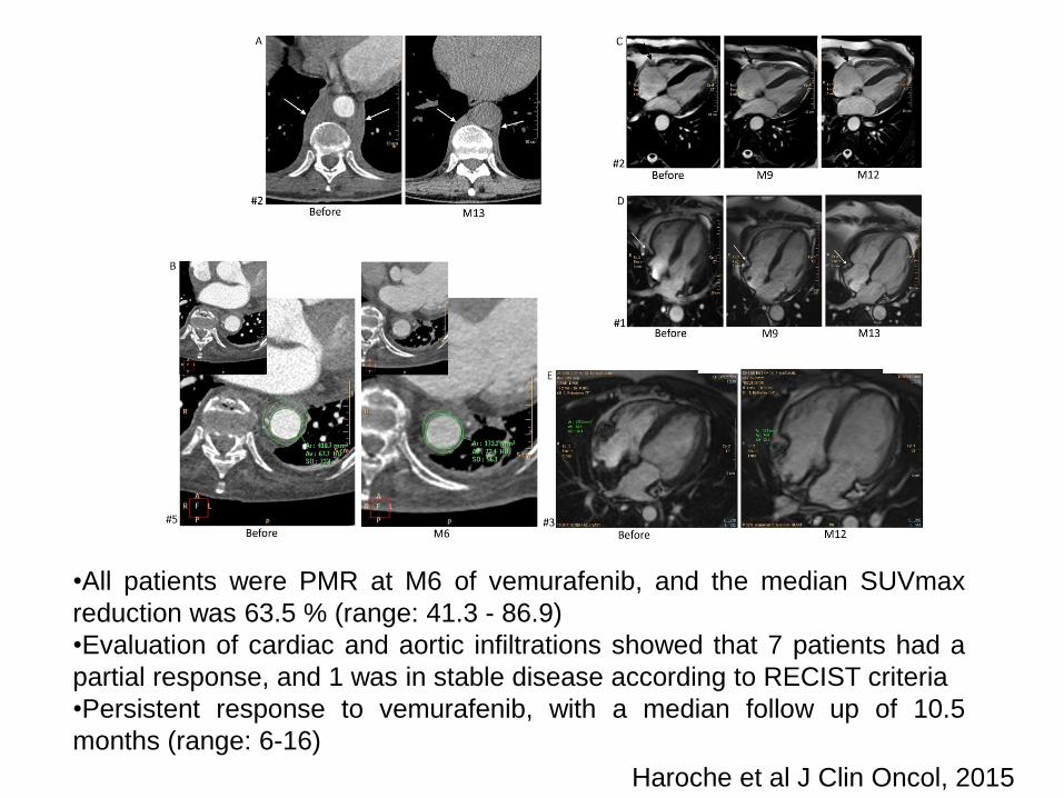
•All patients were PMR at M6 of vemurafenib, and the median SUVmax
reduction was 63.5 % (range: 41.3 - 86.9)
•Evaluation of cardiac and aortic infiltrations showed that 7 patients had a
partial response, and 1 was in stable disease according to RECIST criteria
•Persistent response to vemurafenib, with a median follow up of 10.5
months (range: 6-16)
Haroche et al J Clin Oncol, 2015

Neurologic regression
Haroche J Clin Oncol 2015

A
B
C
D
E F G H
PET at M4 : total disappearance of
suprasellar and lung hypermetabolism
Cohen-Aubart, Neurology 2014
Marked efficacy of vemurafenib in suprasellar Erdheim-Chester disease

Xanthelasma
M0 M12
Haroche J Clin Oncol 2015
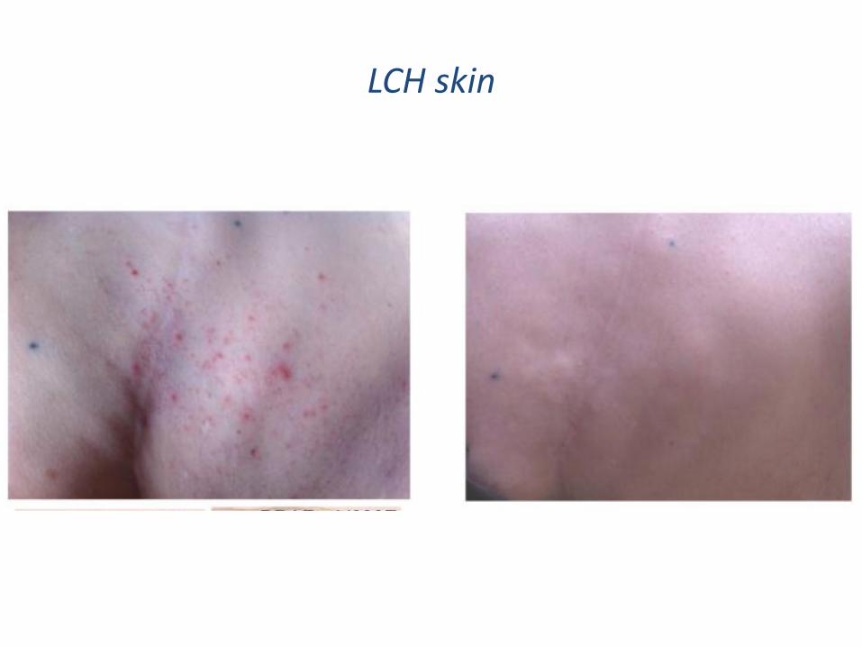
LCH skin

BRAF inhibition 2012-2018 • MSKCC: Basket Trial: 18 histiocytoses (mainly ECD)
published in NEJM august 2015, close to 40 pts currently • Italy: 10 pts treated by Augusto Vaglio (Parma) • Italy: Lorenzo Dagna (Milano) : 10 - 15 pts • Israel: 5 pts treated • Germany at least 1 patient (ICU) • Norway: 2 cases • Sweden: at least one case with CNS • EU group & Jean Donadieu LCH children 50 VMF • Houston Group : 5 – 10 LCH children pts • PITIE-SALPETRIERE : 90 patients (Mai 2018)
> 200 patients worldwide adults &
children with ECD, ECD + LCH and
LCH
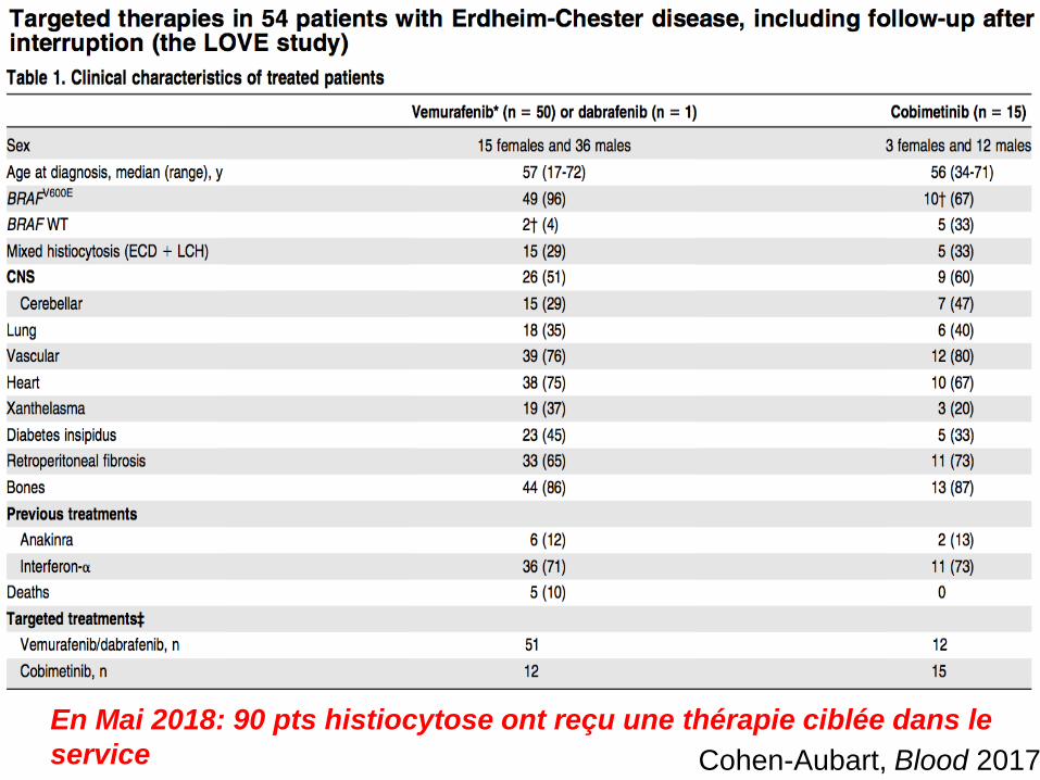
En Mai 2018: 90 pts histiocytose ont reçu une thérapie ciblée dans le
service Cohen-Aubart, Blood 2017

BRAF inhibitors = analysis of 51 treated patients until June 2016
PET evaluation of efficacy (M6 or M3 if M6 was not available)
n= 48 (3 interruption before 3 months because of intolerance)
- 2 worsening (the 2 WT BRAF patients)
- 4 stable
- 35 partial metabolic remission
- 7 complete metabolic remission
Cohen-Aubart, Blood 2017
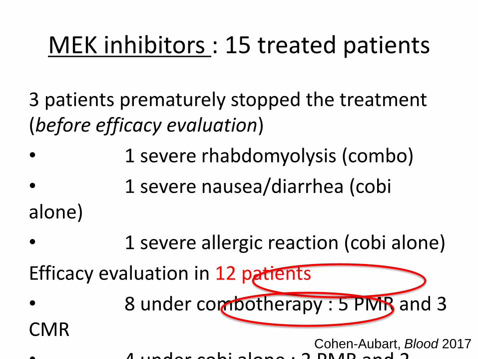
MEK inhibitors : 15 treated patients
3 patients prematurely stopped the treatment (before efficacy evaluation)
• 1 severe rhabdomyolysis (combo)
• 1 severe nausea/diarrhea (cobi alone)
• 1 severe allergic reaction (cobi alone)
Efficacy evaluation in 12 patients
• 8 under combotherapy : 5 PMR and 3 CMR
• 4 under cobi alone : 2 PMR and 2 CMR
Cohen-Aubart, Blood 2017

Cohen-Aubart, Blood 2017 Et depuis 2016: + 3 mélanomes; un adéno K
pancréatique muté KRAS; 2 pancréatites; 2
SMD

Infiltrant squamous cell carcinoma requiring surgery and radiotherapy (treatment discontinuation)
J Clin Oncol 2015
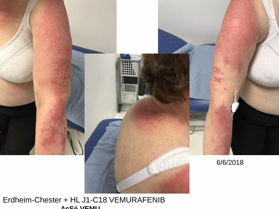
Erdheim-Chester + HL J1-C18 VEMURAFENIB
AcSé VEMU
6/6/2018
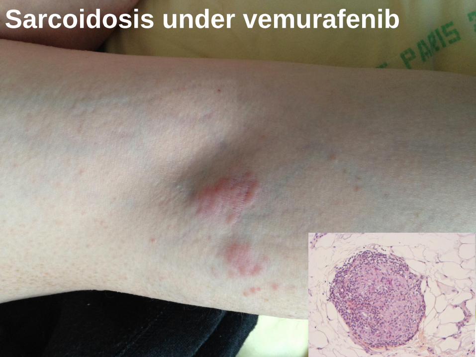
Sarcoidosis under vemurafenib
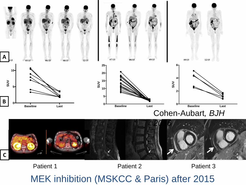
Baseline Last0
5
10
SU
V
A
B
C
Baseline Last0
2
4
6
SU
V
Baseline Last0
5
10
15
20
25
SU
V
Patient 1 Patient 2 Patient 3
Cohen-Aubart, BJH
2016
MEK inhibition (MSKCC & Paris) after 2015

Severe acneic
rash under
cobimetinib
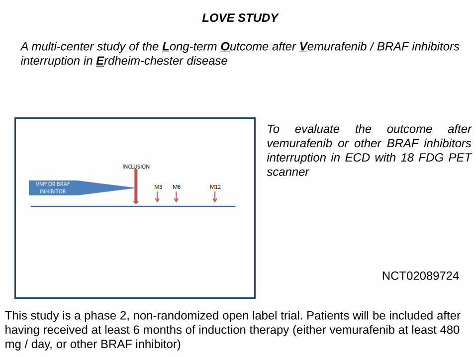
LOVE STUDY
A multi-center study of the Long-term Outcome after Vemurafenib / BRAF inhibitors
interruption in Erdheim-chester disease
This study is a phase 2, non-randomized open label trial. Patients will be included after
having received at least 6 months of induction therapy (either vemurafenib at least 480
mg / day, or other BRAF inhibitor)
To evaluate the outcome after
vemurafenib or other BRAF inhibitors
interruption in ECD with 18 FDG PET
scanner
NCT02089724

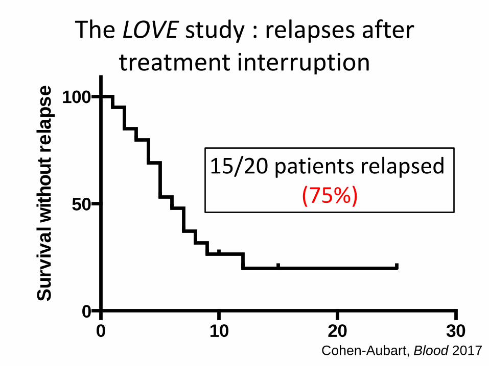
The LOVE study : relapses after treatment interruption
0 10 20 300
50
100
Months
Su
rviv
al w
ith
ou
t re
lap
se
15/20 patients relapsed (75%)
Cohen-Aubart, Blood 2017
The LOVE study : relapses after treatment interruption
0 10 20 300
50
100
Months
Su
rviv
al w
ith
ou
t re
lap
se
15/20 patients relapsed (75%)
Cohen-Aubart, Blood 2017
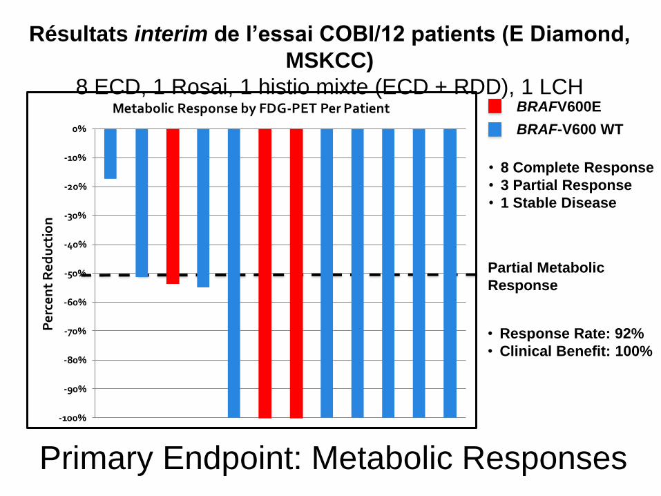
Primary Endpoint: Metabolic Responses
BRAFV600E
BRAF-V600 WT
Partial Metabolic
Response
• 8 Complete Response
• 3 Partial Response
• 1 Stable Disease
• Response Rate: 92%
• Clinical Benefit: 100%
-100%
-90%
-80%
-70%
-60%
-50%
-40%
-30%
-20%
-10%
0%
Pe
rce
nt
Re
du
ctio
n
Metabolic Response by FDG-PET Per Patient
Résultats interim de l’essai COBI/12 patients (E Diamond,
MSKCC)
8 ECD, 1 Rosai, 1 histio mixte (ECD + RDD), 1 LCH
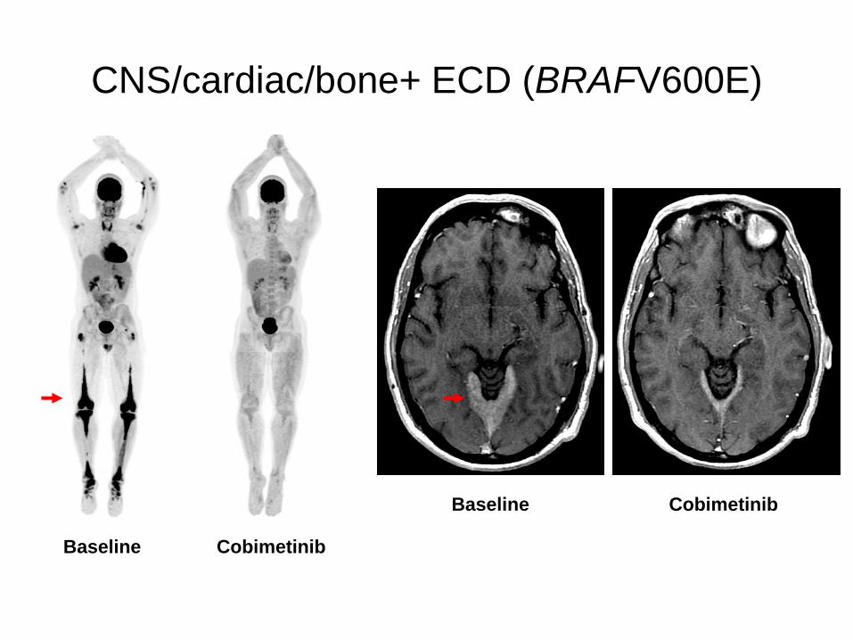
CNS/cardiac/bone+ ECD (BRAFV600E)
Baseline Cobimetinib
Baseline Cobimetinib
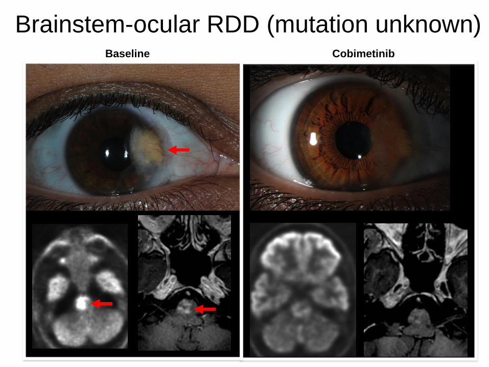
Brainstem-ocular RDD (mutation unknown) Baseline Cobimetinib
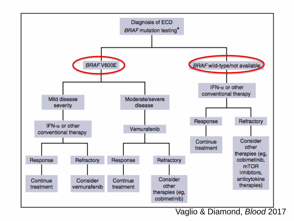
Vaglio & Diamond, Blood 2017
0 100 200 300 4000
50
100
Time
Pe
rce
nt s
urv
iva
l
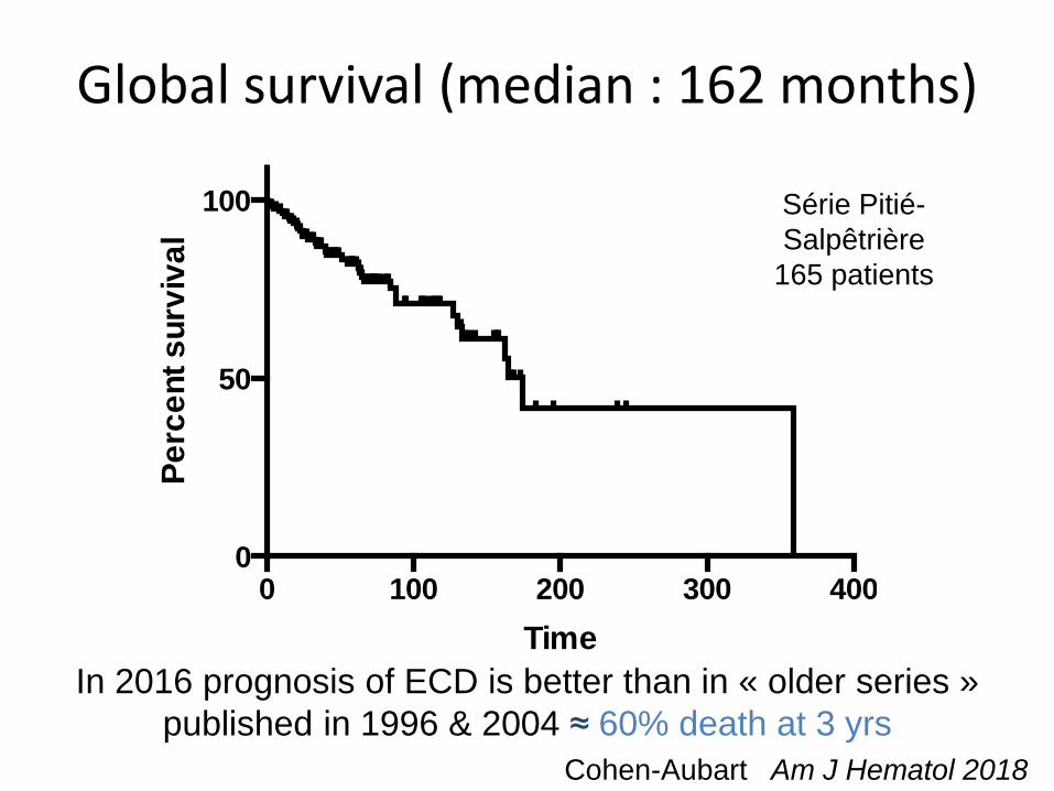
Global survival (median : 162 months)
In 2016 prognosis of ECD is better than in « older series »
published in 1996 & 2004 ≈ 60% death at 3 yrs
Série Pitié-
Salpêtrière
165 patients
Cohen-Aubart Am J Hematol 2018
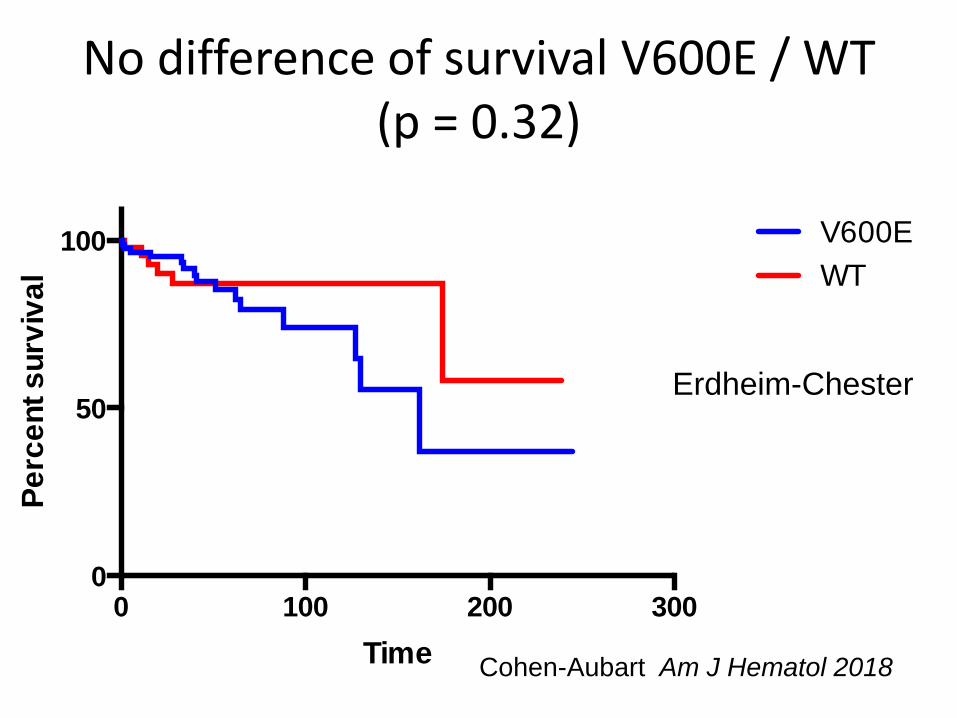
No difference of survival V600E / WT (p = 0.32)
0 100 200 3000
50
100
Time
Pe
rce
nt s
urv
iva
l
V600E
WT
Cohen-Aubart Am J Hematol 2018
Erdheim-Chester

Papo, Blood
2017
23 LCH + ECD
Diagnosis
7 before (4 years
[1-22])
6 simultaneously
6 after (1 year [1-
4])
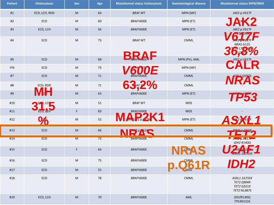
Patient Histiocytosis Sex Age Mutationnal status histiocytosis Haematological disease Mutationnal status MPN/MDS
#1 ECD, LCH, RDD M 64 BRAF WT MPN (MF) JAK2 p.V617F
#2 ECD M 60 BRAFV600E MPN (ET) CALR
#3 ECD, LCH M 56 BRAFV600E MPN (ET) JAK2 p.V617F TET2 K95X
#4 ECD M 73 BRAF WT CMML JAK2 p.V617F NRAS G12S
TET2 L757fsX56 U2AF1
#5 ECD M 68 BRAFV600E MPN (PV), AML JAK2 p.V617F
IFN ECD M 73 BRAF WT MPN (MF) -
#7 ECD M 71 BRAFV600E CMML ASXL1 Q592X
#8 ECD, RDD M 71 BRAF WT CMML -
#9 ECD M 63 BRAFV600E MPN (ET) JAK2 p.V617F
#10 ECD, LCH M 51 BRAF WT MDS -
#11 ECD, LCH F 83 BRAFV600E MDS -
#12 ECD M 52 MAP2K1 p.K57N MPN (ET) JAK2 p.V617F
#13 ECD M 66 NRAS p.Q61R CMML NRAS p.Q61R
#14 ECD M 73 BRAFV600E CMML JAK2 p.V617F IDH2 R140Q
#15 ECD F 64 BRAFV600E CMML NRAS p.G13D ASXL1 1Q733X
#16 ECD M 75 BRAFV600E CMML -
#17 ECD M 55 BRAFV600E MDS -
#18 ECD M 78 BRAFV600E CMML ASXL1 1A733X TET2 Q904X TET2 Q321X TET2 N1387S
#19 ECD, LCH M 70 BRAFV600E
AML IDH2R140Q TP53N131K
MH
31,5
%
BRAF
V600E
63,2%
NRAS
MAP2K1
JAK2
V617F
36,8% CALR
NRAS
ASXL1
TET2
U2AF1
IDH2
TP53
NRAS
p.Q61R

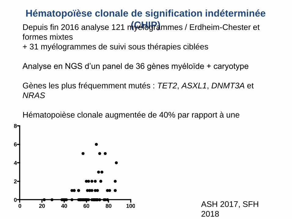
Hématopoïèse clonale de signification indéterminée
(CHIP) Depuis fin 2016 analyse 121 myélogrammes / Erdheim-Chester et
formes mixtes
+ 31 myélogrammes de suivi sous thérapies ciblées
Analyse en NGS d’un panel de 36 gènes myéloïde + caryotype
Gènes les plus fréquemment mutés : TET2, ASXL1, DNMT3A et
NRAS
Hématopoièse clonale augmentée de 40% par rapport à une
population de même age
ASH 2017, SFH
2018 0 20 40 60 80 100
0
2
4
6
8
correlation age/nb de mutations

Au bout du bout

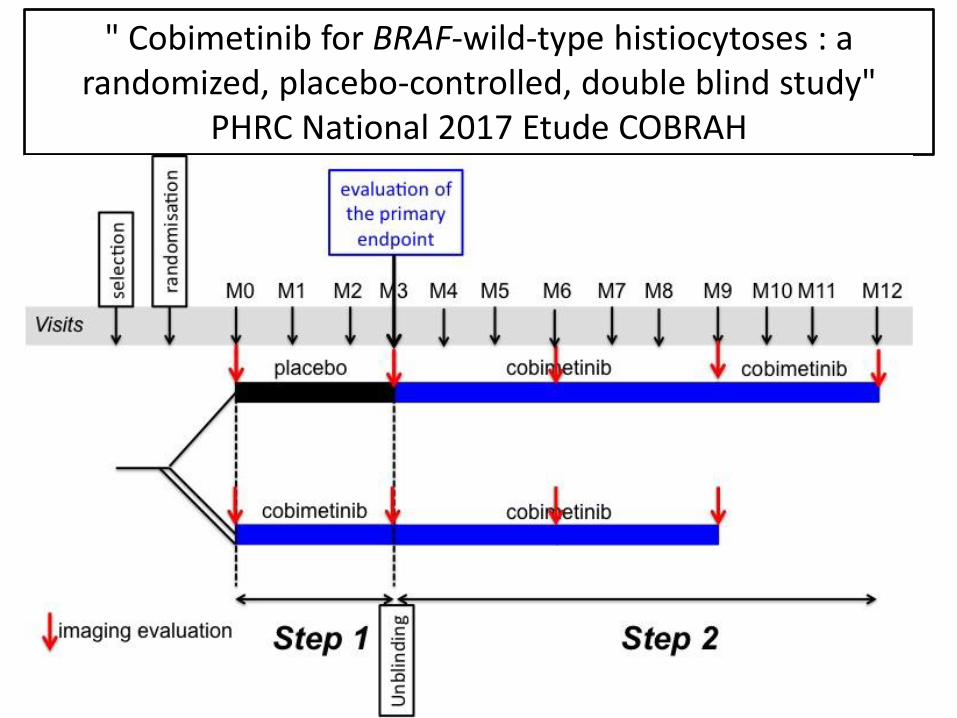
" Cobimetinib for BRAF-wild-type histiocytoses : a randomized, placebo-controlled, double blind study"
PHRC National 2017 Etude COBRAH

Patient (1950) : LCH + RDD + ECD
Dyspnea,
Pl Effusion Skin lesions
LCH
12/08 08/09
pleurodesis
ECD
Pericardial thickening
Aortitis
Retroperitoneal fibrosis
RDD
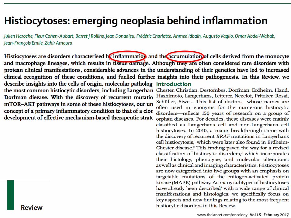
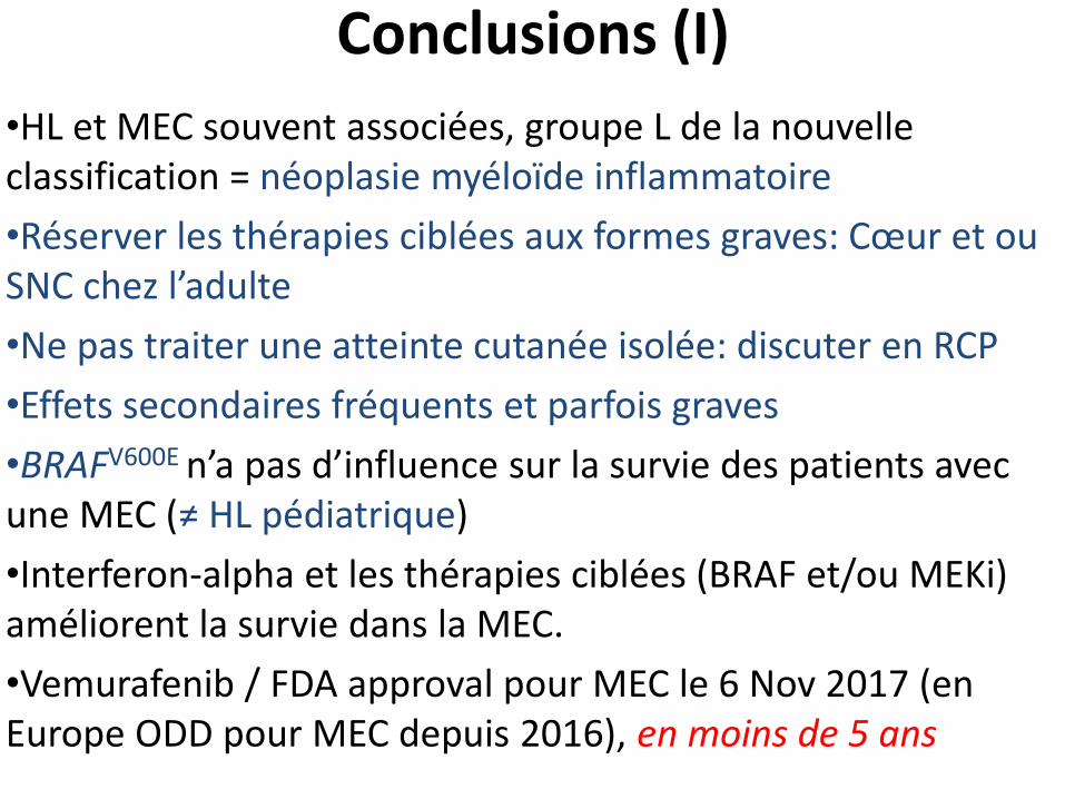
Conclusions (I)
•HL et MEC souvent associées, groupe L de la nouvelle classification = néoplasie myéloïde inflammatoire
•Réserver les thérapies ciblées aux formes graves: Cœur et ou SNC chez l’adulte
•Ne pas traiter une atteinte cutanée isolée: discuter en RCP
•Effets secondaires fréquents et parfois graves
•BRAFV600E n’a pas d’influence sur la survie des patients avec une MEC (≠ HL pédiatrique)
•Interferon-alpha et les thérapies ciblées (BRAF et/ou MEKi) améliorent la survie dans la MEC.
•Vemurafenib / FDA approval pour MEC le 6 Nov 2017 (en Europe ODD pour MEC depuis 2016), en moins de 5 ans
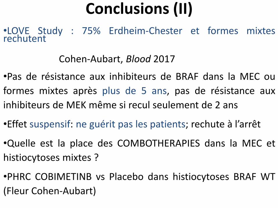
Conclusions (II)
•LOVE Study : 75% Erdheim-Chester et formes mixtes rechutent Cohen-Aubart, Blood 2017
•Pas de résistance aux inhibiteurs de BRAF dans la MEC ou
formes mixtes après plus de 5 ans, pas de résistance aux
inhibiteurs de MEK même si recul seulement de 2 ans
•Effet suspensif: ne guérit pas les patients; rechute à l’arrêt
•Quelle est la place des COMBOTHERAPIES dans la MEC et
histiocytoses mixtes ?
•PHRC COBIMETINB vs Placebo dans histiocytoses BRAF WT
(Fleur Cohen-Aubart)
Merci
Fleur Cohen-Aubart
Zahir Amoura
Tous les internes
Frédéric Charlotte
Jean-François Emile
Jean Donadieu
Abdellatif Tazi
Eli Diamond, Ben Durham, Omar Abdel-Wahab, MSKCC, New
York
Les patients
Kathy Brewer and the ECD global alliance
Histiocyte Society
+33 6 30 04 02 88
r