Genou Pédiatrique Dr Farid BOUSBAH 22/03/2007. Anatomie du genou.

Thèse rédigée sous la direction du Professeur Robin PETER
Evolution des fractures du genou chez l’adulte au Sultanat d’Oman
Revue à long terme de 30 cas
THESE
Présentée à la faculté de Médecine de l’Université de Genève
pour obtenir le grade de Docteur en médecine
par
Ayman EL SHARAWY
Du Caire / Egypte
Thèse n° 10443
Genève
2005

UNIVERSITE DE GENEVE FACULTE DE MEDECINE
Département de Chirurgie Service de Chirurgie Orthopédique et Traumatologie de l’Appareil moteur
_________________________________________________________________________
Thèse rédigée sous la direction du Professeur Robin PETER
Evolution des fractures du genou chez l’adulte au Sultanat d’Oman
Revue à long terme de 30 cas
THESE
Présentée à la faculté de Médecine de l’Université de Genève
pour obtenir le grade de Docteur en médecine
par
Ayman EL SHARAWY
Du Caire / Egypte
Thèse n° 10443
Genève
2005
DOCTORAT EN MEDECINE
Thèse de:
Monsieur Ayman EL SHARAWY originaire du Caire (Egypte)
Intitulée EVOLUTION DES FRACTURES DU GENOU CHEZ L’ADULTE
AU SULTANAT D’OMAN REVUE A LONG TERME DE 30 CAS
La Faculté de médecine, sur le préavis de Monsieur Robin PETER, professeur adjoint au Département de chirurgie, autorise l’impression de la présente thèse, sans prétendre par là émettre d’opinion sur les propositions qui y sont énoncées. Genève, le 7 octobre 2005
Thèse n° 10443 Jean-Louis Carpentier Doyen

ACKNOWLEDGEMENT
I would like to dedicate this thesis to my wife Ammal and my sons Omar & Ali
and to my Parents.
I would like to express my sincere and deep gratitude to Prof. Robin Peter, Chief
of Trauma, in Geneva University for his faithful supervision, constructive
guidance and real interest in the progress of this work.
I thank sincerely Dr. Jean Marc Meyer for his great care, support and full time
encouragement and finally I would like to thank Dr. Wahid Al Kharusi, Chief of
Trauma, Orthopaedic and Rehabilitation in Khoula Hospital in Oman for his
sincere advice, support and guideness through all the process of this work.
I would like as well to thank Geneva University and the orthopaedic department of
Geneva University for giving me the chance to present this work.

CONTENTS
1. Summary In French 5
2. Introduction 9
3. Historical Review 10
4. Anatomy and Classification 15
- Anatomy of distal femur 15
- Mechanism of Injury 17
- Classification of distal femur 18
- Diagnosis of distal femur 21
- Anatomy of Tibial Plateau 23
- Mechanism of Injury 24
- Classification of Tibial Plateau 26
- Diagnosis of Tibial Plateau 28
5. Material and Methods 30
6. Results 34
7. Discussion 39
8. Conclusion 44
9. References 45
10. Statistics 50
5
RESUME
Les fractures du genou chez l’adulte représentent 2% de toutes les fractures, mais en
raison de notre mode de vie moderne et de l’augmentation de la rapidité des moyens de
transport, la fréquence de ces lésions augmente. Les fractures du genou se présentent
souvent comme des lésions complexes susceptibles de nombreuses complications. C’est
la raison pour laquelle la prise en charge de telles lésions est encore sujette à discussion.
Autrefois ces fractures étaient en général prises en charge par des rebouteux, ou par les
chirurgiens généraux. Ils utilisaient divers types d’extension pendant des durées variables
suivis de l’application d’appareils plâtrés et immobilisation de toute sorte. Ces
traitements étaient grevés de nombreuses complications telle que retard de consolidation,
pseudarthrose, déformations, raccourcissement etc. En raison de ces complications de
nombreuses méthodes de fixation interne ont été proposées.
Le but de cette étude est de décrire l’évolution des protocoles de traitement des fractures
du genou survenues ces 30 dernières années à Oman et de présenter la situation actuelle
avec les techniques modernes utilisées aujourd’hui dans ce pays. Nos résultats ont été
obtenus suivant l’application de ces techniques.
Jusqu’en 1970, il n’existait pas de système de santé contrôlé par l’état à Oman. Le
secteur médical était composé de 3 hôpitaux répartis dans le pays et d’un 4e rallié à
l’ « American Missionary Hospital » construit au début des années 50. Les patients
avaient accès à la médecine occidentale à l’hôpital américain situé dans la vieille ville de
Muscat. Pour la médecine traditionnelle et Islamique. Elle consistait pour l’essentiel è
cautériser les zones douloureuses par des pointes de feu « WASAM » et à appliquer de
ventouses « HEGAMA ». Les fractures étaient traitées localement par des rebouteux qui
les manipulaient et les réduisaient puis les contenaient par des mélanges de farine et
d’œufs. La contention par feuilles de bananes était également fréquemment employée.
6
En 1970, sous le règne de Sa majesté le Sultan, le premier hôpital fut crée avec son
service ambulatoire à 300 km. de la capitale. A ce jour le pays compte 14 hôpitaux et de
très nombreux services ambulatoires. Le système de santé a été conçu pour permettre à
chaque omanais de pouvoir atteindre un service de santé dans les 10 minutes.
Du point de vue anatomique, le genou se compose de l’extrémité distale du fémur, de la
rotule et de la tête tibiale. Les pièces osseuses sont reliées entre elles par 5 ligaments
principaux qui stabilisent cette articulation. A Oman, les fractures de la rotule sont rares.
Nous avons réalisé l’ étude de 30 patients victimes de fractures comminutives de
l’extrémité distale intra articulaire du fémur et des plateaux tibiaux présentant un
déplacement supérieur à 5 mm ou avec une angulation dépassant 10 degrés associées à
une ouverture cutanée avec d’importantes lésions des tissus mous.
Tous les patients ont suivi le protocole suivant :
1- Radiographies standard de face et de profil
2- CT Scan pour planning préopératoire
3- Prise en charge chirurgicale avec débridement et parage des lésions cutanées dans
les 5 heures suivant l’admission
4- Ostéosynthèse des fractures chez les polytraumatisés aussitôt que l’état général le
permettait
5- Mobilisation passive pendant les 3 à 5 jours postopératoires
6- Décharge par plâtre cruro-pédieux pendant 3 à 5 semaines dépendant du
psychisme du patient.
7- Charge partielle en fonction des signes cliniques et radiologiques de consolidation
8- La durée des contrôles médicaux était décidée en fonction des observations
cliniques et radiologiques
23 patients étaient de sexe masculin (76,7%) et 7 de sexe féminin (23,3%). 18 patients
présentaient une fracture du fémur distal (60%) et 12 patients présentaient une fracture
des plateaux tibiaux. (40%). 21 patients ont été victimes d’accidents de la circulation

7
(70%), 9 (30%) ont été l’objet de chutes d’une hauteur plus ou moins importante. Le côté
droit a été atteint chez 16 patients (53,3%) et le gauche chez 13 patients (43,3%). Un
patient a présenté une fracture du tibia droit et du fémur gauche (3,4%). La série
comporte 8 polytraumatisés (26,7%). Une fracture du rachis a été observée chez 2 sujets
(6,6%) et un traumatisme cranio-cérébral dans 3 cas (10%).
Les 30 patients ont été contrôlés cliniquement et radiologiquement dans une moyenne de
30 mois après l’accident (18 à 51 mois). La consolidation avait été obtenue chez tous les
patients au moment du contrôle. Le matériel a été retiré chez 6 d’entre eux entre le 24e et
le 36e mois après l’accident.
La moyenne d’âge était de 39 ans (21 à 63 ans) et la durée moyenne du séjour hospitalier
a été de 21 jours (3 à 82 jours). Un patient a présenté un démontage de l’ostéosynthèse
suite à une charge prématurée et a du être réopéré. En général nous avons constaté que les
fractures des plateaux tibiaux présentaient des résultats bons ou excellents malgré des
images radiologiques et une réduction moyennes.
Dans le cas de fractures inter condyliennes, les résultats n’ont pas été aussi satisfaisants
même avec une image bonne radiologique et une réduction opératoire anatomique de la
fracture. 23 patients n’ont pas présenté de complications post opératoires immédiates
(76,7%). Une infection s’est développée chez 3 patients (10%). Une neurapraxie a été
observée dans un cas (3,3%). 3 patients ont présenté une TVP (10%).
Nous avons utilisé plusieurs types d’implants. Pour les plateaux tibiaux notre choix s’est
porté sur un vissage percutané par vis d’Asnis chez 2 patients (16,7%) dans le cas de
fractures peu déplacées, ne disposant pas de vis perforées. De même dans des fractures
intra articulaires uni condyliennes du fémur distal, 2 patients ont bénéficié du même
traitement, les vis ayant été insérées perpendiculairement au trait de fracture dans un but
de neutralisation (11,1%). Des plaques en L ou en T ont été utilisées chez 10 patients (
83,3%) en cas de fractures des plateaux tibiaux. Pour le plateau tibial externe, nous

8
avons recouru à une voie antéro-extene para rotulienne et pour le plateau tibial interne le
choix s’est porté sur la voie antéro-interne para rotulienne . En cas de fracture bi
condylienne la voie d’abord a été antérieure longitudinale ou comme dans 3 cas l’incision
en coupe de champagne (25%) sans complication liée au lambeau cutané.
Bien que les fractures du genou soient très difficiles et représentent un défi pour le
chirurgien, avec une bonne planification opératoire et une bonne technique, le résultat du
traitement chirurgical reste nettement supérieur à tout autre type de traitement avec de
bons ou excellents résultats et des complications dans une moindre mesure.
Grâce a l’utilisation de nouvelles technique développées par le groupe AO il est
désormais acquis qu’une amélioration globale des résultats peut être obtenue. Il reste
quelques incertitudes quant au meilleur type de fixation interne ou externe. Le type de
fixation, le moment de l’opération et l’emploi de greffes est encore en discussion et fait
l’objet de recherches.
Dans notre étude, le mode de vie des Omanais a un excellent effet sut la récupération de
l’amplitude articulaire résultant de l’habitude de s’asseoir sur les talons et de la position
du genou en flexion maximale pendant la prière. La tolérance à la douleur est excellente
chez les patients de l’ancienne génération en raison de leur mode de vie. Récemment des
techniques plus biologiques telles que la plaque de Liss et l’enclouage fémoral distal ont
amélioré les résultats. A Oman nous sommes entrain d’adapter ces nouvelles techniques
dans le but d’ améliorer l’évolution de nos patients.
Nos résultats ont étés rassemblés et analysés dans un but de contrôle de qualité et
d’enseignement. En conclusion la mise en œuvre des techniques modernes de réduction
chirurgicale et fixation interne des fractures du genou dans le cadre du système de santé
omanais ont amélioré les résultats de ces graves lésions parmi les habitants de ce pays.

9
INTRODUCTION
Fractures around the knee in adults account for only 2% of all the fractures. But
because of our modern life styles and high velocity means of transportation,
these injuries are being seen with increasing frequency. Fractures around the
knee are often complex injuries that present to the surgeon with numerous
potential complications, the management of these fractures remains
controversial.
In the past, this injury was treated by the General Surgeons who used to apply
skeletal traction for variable duration, followed by some form of cast or brace
immobilization. Complications associated with closed management of those
fractures have led to the proposal of a number of alternative methods of
internal fixation.
Although early attempts of internal fixation for distal femur fractures
frequently gave unacceptably high rates of malunion, nonunion and infection,
improved techniques of internal fixation have yielded results far superior to
those achieved with nonsurgical management. Meticulous internal fixation has
been shown to yield good to excellent results in 60% to 80% of cases and
allows immediate mobilization of the patient and the extremity, minimizing the
cardiopulmonary and other multisystem sequelae of long-term immobility (50).
Our goal of this study is to give a description of the tremendous evolution in
the treatment protocols for fractures around the knee, which occurred during
the past 30 years in Oman, and to describe the state of the art with modern
techniques which are now days used in Oman and our results achieved using
these modern techniques.

10
HISTORICAL
Up to 1970 there was no updated system of the government in Oman.
Everything was through the palace, the medical sector was formed of 3 palace
clinics around the whole country and one American Missionary Hospital built
in the early 50’ s. For western type of medicine, people used to come to the old
city for the American Hospital. Otherwise Local Omani medicine and Islamic
medicine in the form of “Wasam” cautery by hot iron rod and “Hegama”
negative suction by hot cups were used.
Fractures were treated by the local osteopath who used to manipulate the
fractures and fix them with local ingredients as flour and eggs using banana
leaves. Till 1970, osteopath, except in the American Hospital, treated all the
fractures. From 1970 the utilization of osteopath slowly eliminated and starting
from 1982, more hospitals were opened and the people of Oman came to know
and accept modern type of medicine. By then, most trauma cases were treated
in the main referral hospitals.
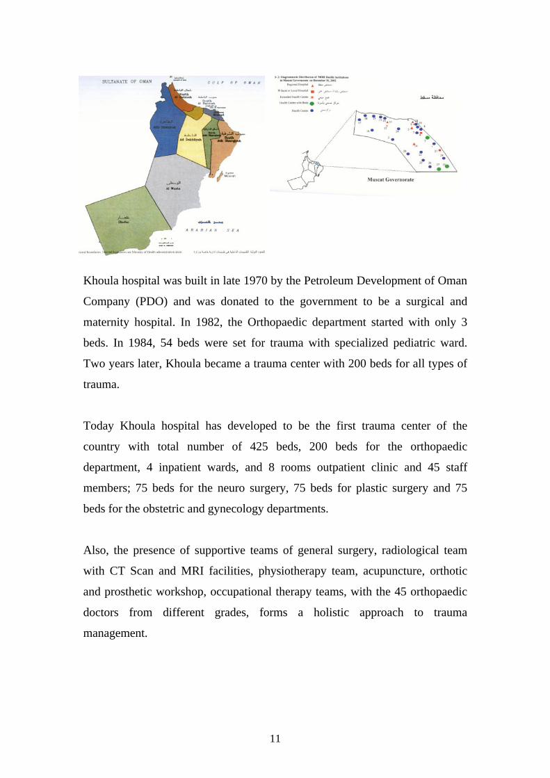
In 1970 under the instructions of His Majesty The Sultan, the first ever
governmental hospital, 300 km from the capital, Muscat, together with the
clinics were launched. In 1974, 4 hospitals were built, now we have 14 regional
hospitals and many health centers. The health system was founded to allow any
patient in Oman to reach a health institute within 10 minutes.
Healed Wasam Fresh Wasam
11
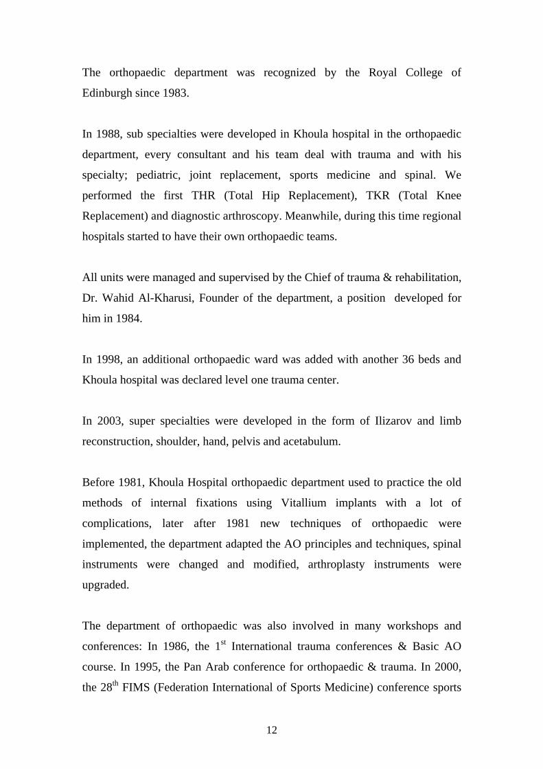
Khoula hospital was built in late 1970 by the Petroleum Development of Oman
Company (PDO) and was donated to the government to be a surgical and
maternity hospital. In 1982, the Orthopaedic department started with only 3
beds. In 1984, 54 beds were set for trauma with specialized pediatric ward.
Two years later, Khoula became a trauma center with 200 beds for all types of
trauma.
Today Khoula hospital has developed to be the first trauma center of the
country with total number of 425 beds, 200 beds for the orthopaedic
department, 4 inpatient wards, and 8 rooms outpatient clinic and 45 staff
members; 75 beds for the neuro surgery, 75 beds for plastic surgery and 75
beds for the obstetric and gynecology departments.
Also, the presence of supportive teams of general surgery, radiological team
with CT Scan and MRI facilities, physiotherapy team, acupuncture, orthotic
and prosthetic workshop, occupational therapy teams, with the 45 orthopaedic
doctors from different grades, forms a holistic approach to trauma
management.
12
The orthopaedic department was recognized by the Royal College of
Edinburgh since 1983.
In 1988, sub specialties were developed in Khoula hospital in the orthopaedic
department, every consultant and his team deal with trauma and with his
specialty; pediatric, joint replacement, sports medicine and spinal. We
performed the first THR (Total Hip Replacement), TKR (Total Knee
Replacement) and diagnostic arthroscopy. Meanwhile, during this time regional
hospitals started to have their own orthopaedic teams.
All units were managed and supervised by the Chief of trauma & rehabilitation,
Dr. Wahid Al-Kharusi, Founder of the department, a position developed for
him in 1984.
In 1998, an additional orthopaedic ward was added with another 36 beds and
Khoula hospital was declared level one trauma center.
In 2003, super specialties were developed in the form of Ilizarov and limb
reconstruction, shoulder, hand, pelvis and acetabulum.
Before 1981, Khoula Hospital orthopaedic department used to practice the old
methods of internal fixations using Vitallium implants with a lot of
complications, later after 1981 new techniques of orthopaedic were
implemented, the department adapted the AO principles and techniques, spinal
instruments were changed and modified, arthroplasty instruments were
upgraded.
The department of orthopaedic was also involved in many workshops and
conferences: In 1986, the 1st International trauma conferences & Basic AO
course. In 1995, the Pan Arab conference for orthopaedic & trauma. In 2000,
the 28th FIMS (Federation International of Sports Medicine) conference sports
13
for all. In 2004,the Celebrate of the 75th Anniversary of FIMS, first time
outside Europe & America.
The transport of patients from the scene of accidents to the hospitals was
usually done by either the relatives, Royal Oman Police, and only few
ambulances were used to transport patients between the hospitals. However on
the 7th of April 2004 , EMS services were inaugurated with well-equipped and
well-trained paramedics to cover Muscat area as first stage, then all over the
Sultanate of Oman.
The department of orthopaedic has taken the initiatives to show the impact of
RTA (Road Traffic Accident) by being involved in the national programm of
injury prevention safety promotion and internationally by taking the matter to
the United Nations (UN) and within 16 months managed to get 3 UN
resolutions.
World wide the Supracondylar and Intracondylar fractures of the distal femur
have historically been difficult to treat. These fractures often are unstable and
comminuted and tend to occur in either elderly or multiply injured patients.
Regaining of full knee motion and function may be difficult. The incidences of
malunion, non union and infection are relatively high in many reported series.
In the decade of the 1960s, non operative treatment methods, such as traction
and cast bracing produced better results than operative treatment because of the
lack of adequate internal fixation devices. In 1966 Stewart and Sisk (64) from
Campell clinic, retrospectively reviewed 213 cases, results were satisfactory;
67% with non operative treatment and 54% of those treated operatively.
Delayed union of non union occurred in 9.7% in fractures treated by closed
methods and 29% of fractures treated operatively. Two-pin traction was
recommended as the treatment of choice. In 1967, Neer (47), Grantham and
Shelton compared operative with non operative treatment and they found 84%
satisfactory results with operative treatment.
14
With the development of improved internal fixation devices by the AO group,
treatment recommendations began to change. In 1972, Olerud (48) reported
93% satisfactory results in 16 patients treated with blade plates. In 1979
Schatzker and Lamert found that 71% of their patients treated with blade plate
had good to excellent results.
In 1989 Mize reported good to excellent results in 76% of his cases with the
AO techniques. Also in 1989 Aswell Siliski (63), Mahring and Hofer reported
81% good to excellent results.
15
ANATOMY AND CLASSIFICATIONS
Relevant Anatomy of the Distal Femur:
The distal femur traditionally encompasses the lower third of this bone. This
zone in the literature varies greatly, from the distal 7.6 cm to the distal 15 cm of
the femur.
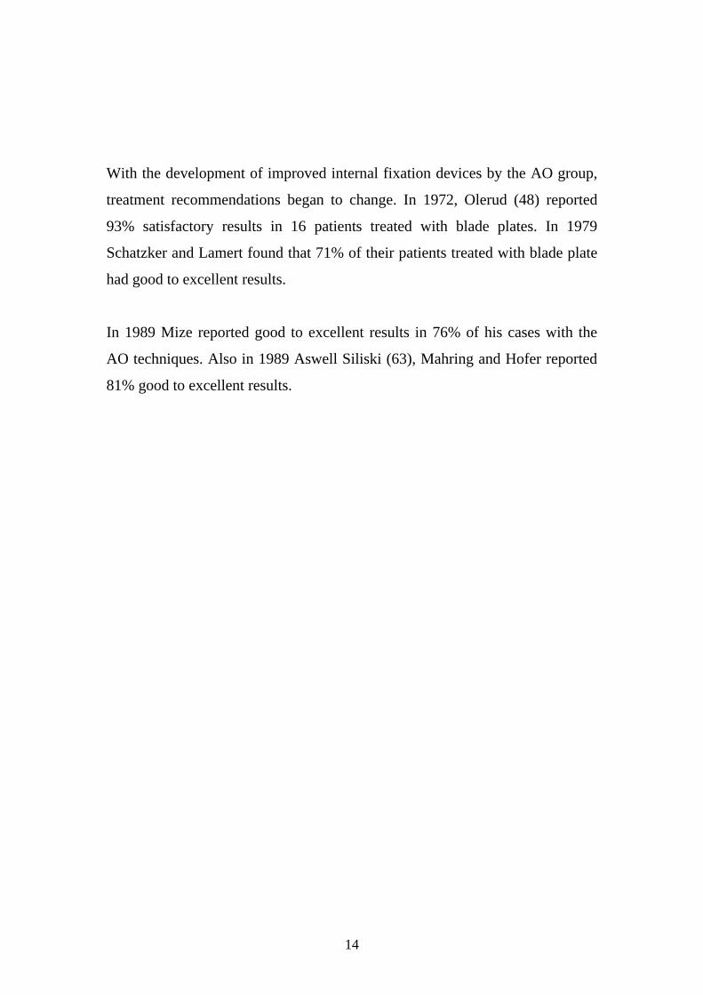
Bone: The supracondylar ( metaphyseal ) area of the distal femur is the transition
zone between the distal diaphysis and the femoral articular condyles. At the
diaphyseal-metaphyseal junction, the metaphysis flares, especially on the
medial side, to provide a platform for the broad condylar weight-bearing
surface of the knee joint. Anteriorly between these two condyles is a smooth
articular depression for the patella, the trochlear groove. Posteriorly between
the two condyles is the intercondylar notch. Medially, a readily identifiable
landmark is the adductor tubercule at the maximum point of the flare of the
metaphysis. Both condyles have epicondyles on their outer surfaces. (Gray’s,
1980)
Of surgical importance, the shaft of the femur in the sagittal view is aligned to
the anterior half of the condyles, leaving the posterior half of both condyles in
the posterior position relative to the proximal femoral shaft. Also the condyles
16
are wider posteriorly than anteriorly. A transverse cut through the condyles
shows a trapezoid with a 25° decrease in the width, from posterior to anterior,
on the medial side (last 1973).
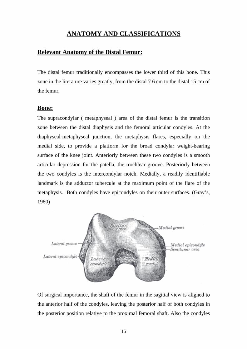
Anteriorly, the extensor compartment contains the quadriceps femoris, the
single largest muscle in the body. It consists of four heads: the rectus femoris,
more superficially and in the deeper layer from lateral to medial; the vastus
lateralis, vastus intermedius and the vastus medialis (grey’s 1980).
The anterior extensor compartment is separated from the posterior
compartment by the lateral and medial intermuscular septa. These provide
important landmarks for both the lateral and medial approaches to the knee
joint. Of major significance on the medial side is the superficial femoral artery,
which runs down the thigh between the extensor and the adductor
compartments. The artery passes into the popliteal fossa approximately 10cm
above the knee joint by passing through the adductor magnus muscle. It
obviously must be identified and avoided in the medial approaches to the distal
femur.
The powerful muscles of the distal thigh produce characteristic bony
deformities with fractures. The muscle pull of the quadriceps and the posterior
hamstrings produce shortening of the femur .As the shaft overrides anteriorly
and the gastrocnemius muscles pull posteriorly, the condyles are displaced and
17
angulated posteriorly. When the condyles are separated by the fracture,
rotational malalignments are the common, because of the unrestrained pull of
the gastrocnemius muscles and the anterior overriding of the shaft.
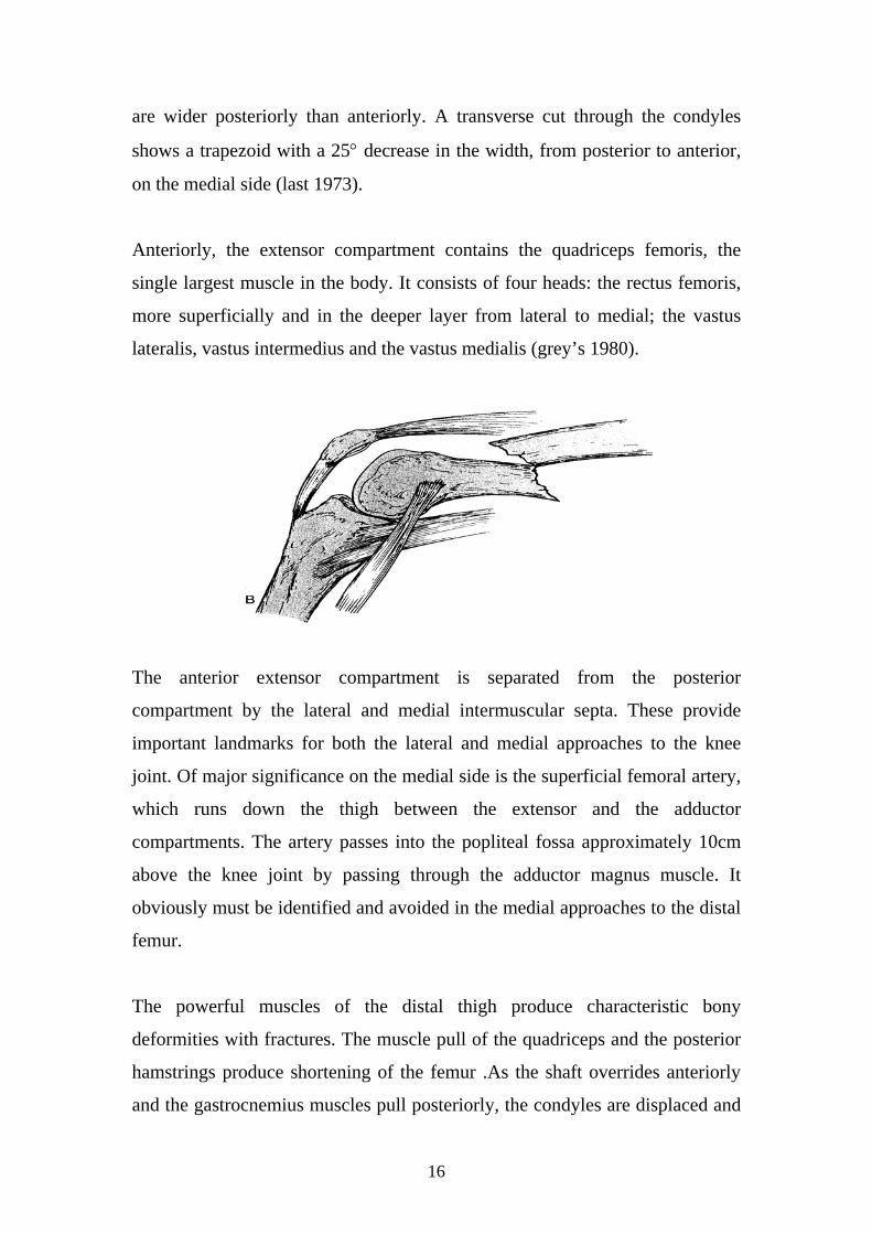
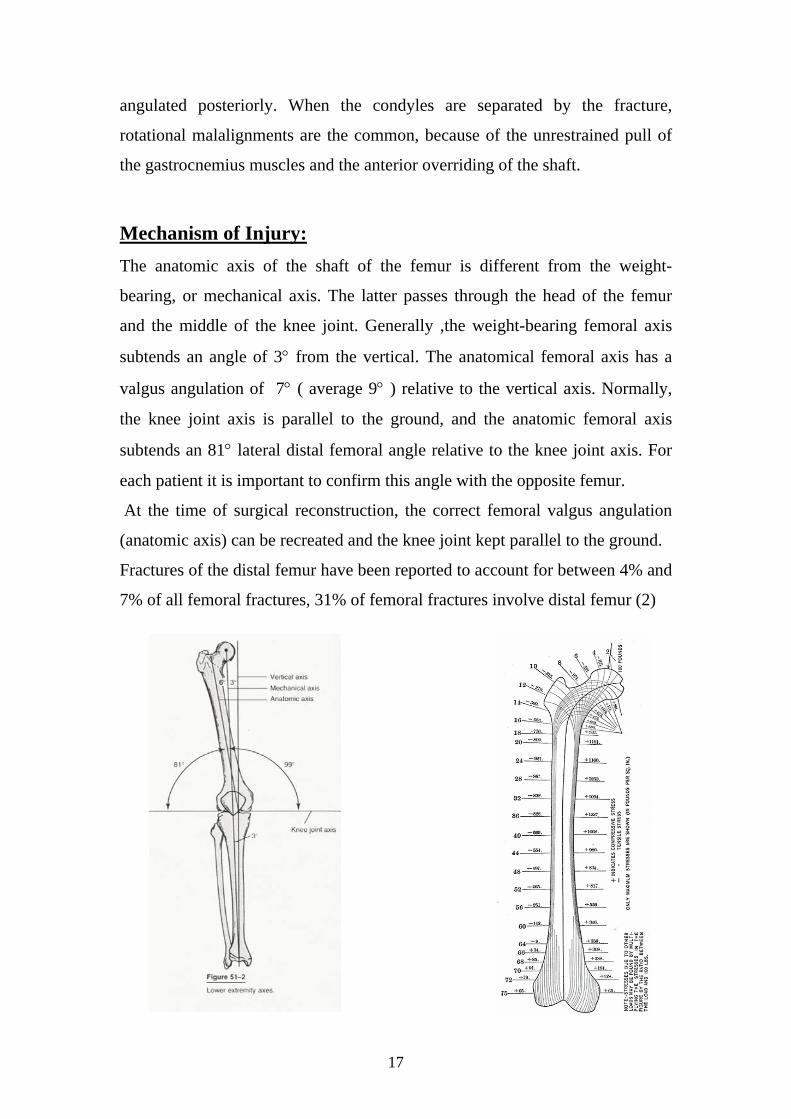
Mechanism of Injury: The anatomic axis of the shaft of the femur is different from the weight-
bearing, or mechanical axis. The latter passes through the head of the femur
and the middle of the knee joint. Generally ,the weight-bearing femoral axis
subtends an angle of 3° from the vertical. The anatomical femoral axis has a
valgus angulation of 7° ( average 9° ) relative to the vertical axis. Normally,
the knee joint axis is parallel to the ground, and the anatomic femoral axis
subtends an 81° lateral distal femoral angle relative to the knee joint axis. For
each patient it is important to confirm this angle with the opposite femur.
At the time of surgical reconstruction, the correct femoral valgus angulation
(anatomic axis) can be recreated and the knee joint kept parallel to the ground. Fractures of the distal femur have been reported to account for between 4% and
7% of all femoral fractures, 31% of femoral fractures involve distal femur (2)

18
The most common mechanism for distal femur fracture is direct trauma to the
flexed knee, typically impact against the dashboard of a moving vehicle (high-
energy). Common associated injuries are concomitant acetabular fractures, hip
dislocation, femoral neck fractures, and associated femoral shaft fractures.
Significant soft tissue injuries of the knee are often associated with distal
femoral fractures, Ligamentous disruptions of the knee joint have been
reported in approximately 20% of these fractures (68), usually discovered after
stabilization of the distal femoral fractures but can be diagnosed with MRI .
Associated tibial plateau or tibial shaft fractures with high-energy trauma
(floating knee) is a good indication for early operative treatment to reduce the
morbidity. The popliteal artery is at great risk of injury with associated
ligamentous disruption of the knee and with floating knee.
Classification of Distal Femur Fractures: For the classification system to have a clinical significance it must be able to do
the following:
1) Allow for adequate documentation of all fractures so that a common
language is possible when discussing these injuries.
2) Be simple enough that it is “ user friendly”.
3) Help the surgeon in his clinical decision making, so that the correct
treatment option can be selected for a particular fracture.
4) Provide prognostic information detailing the result that can be expected
for a particular fracture depending on treatment option selected.
Unfortunately, anatomical fracture classifications fail to address the conditions
commonly associated with supracondylar femur fractures, which often
influence treatment or outcome.
These factors, which play a dynamic role in management, determine the
"personality" of a fracture. Among these are (1) amount of fracture
displacement, (2) degree of comminution, (3) extent of soft-tissue injury, (4)
associated neurovascular injuries, (5) magnitude of joint involvement, (6)

19
degree of osteoporosis, (7) presence of multiple trauma, and (8) complex
ipsilateral injuries (ie, patella or plateau fracture) (17, 28, 34)
One of the original and more simple classification was that of Neer and
associates (47), which classify the fractures according to the amount of
displacement.
Seinsheimer (62) found that all patients with type I and type II had preexisting
pathologic osteoprosis before their injuries.
20
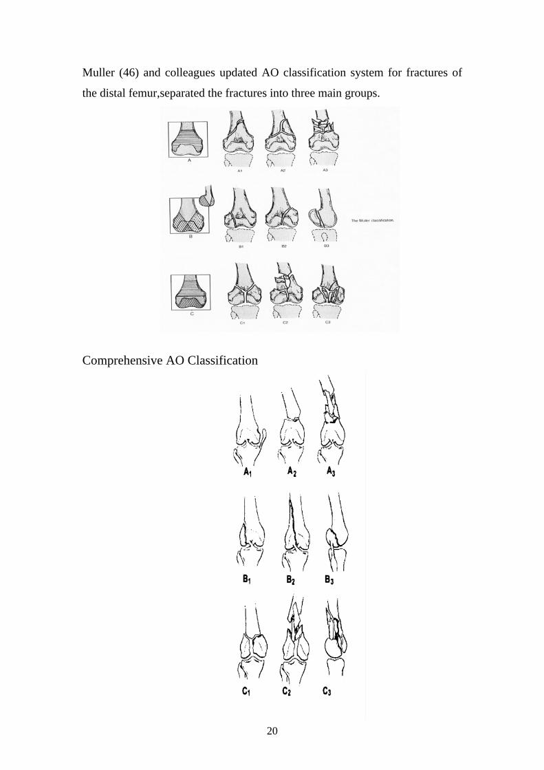
Muller (46) and colleagues updated AO classification system for fractures of
the distal femur,separated the fractures into three main groups.
Comprehensive AO Classification

21
Diagnosis:
History and Physical Examination:
A careful evaluation of the whole patient and also of the involved lower
extremity is mandatory, especially in the polytraumatised patient. This must
include careful detailed examination of the hip joint above the fracture and of
the knee and leg below it. If there is any concern about the vascularity to the
lower extremity, Doppler pulse pressures can be obtained. If there is still
concern after these procedures, an urgent arteriogram may be indicated.
Rarely, if there is tense swelling of the thigh, the presence of an undetected
thigh compartment syndrome also must be ruled out by compartment pressure
monitoring.
Grossly, open and contaminated wounds are easily identifiable. However,
when the injury results from direct trauma there are often skin abrasions that
must be differentiated from open fracture wounds of the soft tissues. The
examination usually reveals swelling of the knee and supracondylar area, often
obvious deformity and marked tenderness on palpation. Manipulation of the
extremity, if tolerated by the patient, demonstrates motion and crepitance at the
fracture site. However, such manipulation is cruel and unnecessary if
immediate radiographs are available.
Radiographic Evaluation:
Routine antero posterior (AP) and lateral radiographs of the knee and
supracondylar region are standard. When the fractures are comminuted or
displaced an exact classification of the fracture is often difficult to make. AP
and lateral radiographs, both with manual traction applied to the lower
extremity, often demonstrate more clearly the fracture morphology. These
studies can be done in the emergency department or the operating room. If
there is intercondylar involvement, 45º oblique radiographs also help delineate
the extent of the injury, especially if comminution or additional tibial plateau
22
injuries are present. Stress radiographs to identify ligamentous disruptions of
the knee or associated tibial plateau fractures usually are not indicated until the
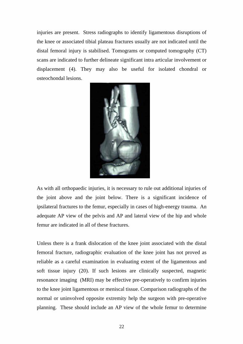
distal femoral injury is stabilised. Tomograms or computed tomography (CT)
scans are indicated to further delineate significant intra articular involvement or
displacement (4). They may also be useful for isolated chondral or
osteochondal lesions.
As with all orthopaedic injuries, it is necessary to rule out additional injuries of
the joint above and the joint below. There is a significant incidence of
ipsilateral fractures to the femur, especially in cases of high-energy trauma. An
adequate AP view of the pelvis and AP and lateral view of the hip and whole
femur are indicated in all of these fractures.
Unless there is a frank dislocation of the knee joint associated with the distal
femoral fracture, radiographic evaluation of the knee joint has not proved as
reliable as a careful examination in evaluating extent of the ligamentous and
soft tissue injury (20). If such lesions are clinically suspected, magnetic
resonance imaging (MRI) may be effective pre-operatively to confirm injuries
to the knee joint ligamentous or meniscal tissue. Comparison radiographs of the
normal or uninvolved opposite extremity help the surgeon with pre-operative
planning. These should include an AP view of the whole femur to determine
23
the valgus alignment and AP and lateral views of the distal femur to allow
superimposition of the fracture fragments on the normal template .
Arteriography is indicated when there is an associated frank dislocation of the
knee joint, because there is a reported 40% incidence of arterial injuries with
knee dislocations (28, 68). An absent or diminished pulse (determined
clinically or by Doppler assessment in the emergency room) when compared
with the normal lower extremity, is also an indication for immediate
arteriography or vascular exploration.
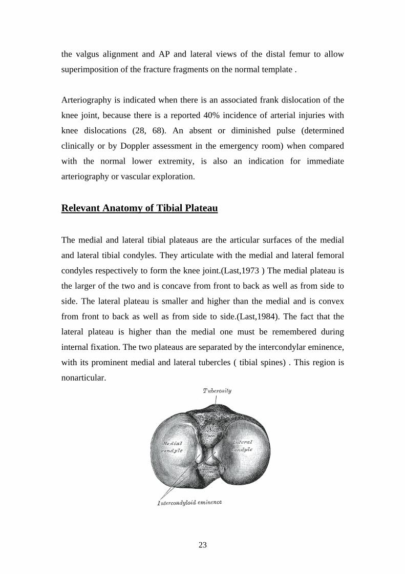
Relevant Anatomy of Tibial Plateau
The medial and lateral tibial plateaus are the articular surfaces of the medial
and lateral tibial condyles. They articulate with the medial and lateral femoral
condyles respectively to form the knee joint.(Last,1973 ) The medial plateau is
the larger of the two and is concave from front to back as well as from side to
side. The lateral plateau is smaller and higher than the medial and is convex
from front to back as well as from side to side.(Last,1984). The fact that the
lateral plateau is higher than the medial one must be remembered during
internal fixation. The two plateaus are separated by the intercondylar eminence,
with its prominent medial and lateral tubercles ( tibial spines) . This region is
nonarticular.

24
The tibial attachment of the anterior cruciate ligament (ACL) is just anterior to
the medial intercondylar tubercle. The posterior cruciate ligament’s attachment
is in the posterior intercondylar area, extending into the posterior surface of the
metaphysis.(Insal,1984)
The outer portion of each plateau is covered by semilunar fibrocartilaginous
meniscus. The lateral meniscus covers a much larger portion of the articular
surface than does the medial. The medial articular surface and its supporting
medial condyle are stronger than their lateral counterparts.(Last,1984)
As a result, fractures of the lateral plateau are more common. When fractures
of the medial plateau occur, they are invariably associated with more violent
injuries and more commonly have associated soft tissue injuries , such as
disruptions of the lateral collateral ligament complex, lesions of the lateral
peroneal nerve, or damage to the popliteal vessels.
Mechanism of Injury:
Injuries to the plateaus occur as a result of :
1) Force directed either medially(valgus deformity,the classic bumper
fracture)or laterally(varus deformity)
2) An axial compressive force
3) Both an axial force and a force from the side.
The respective femoral condyle in this mechanism of injury exerts both
shearing and compressive forces into the underlaying tibial plateau. The
resulting fracture is therefore most commonly a split fracture or a depression
fracture,or both . Pure split fractures are more common in younger patients, in
whom the strong bone of the tibial condyle is able to withstand the compressive
force of the overlaying femoral condyle. With age; the dense cancellous bone
of the young tibial condyle becomes osteopenic, with diminished compressive
forces as well. As a result,split- depression fractures become common in
patient after their fifth decade of life.These typically result from low-energy
injuries.(5)
25
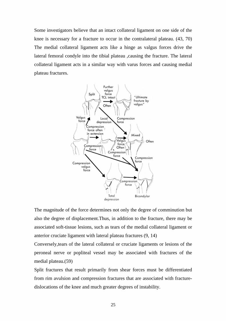
Some investigators believe that an intact collateral ligament on one side of the
knee is necessary for a fracture to occur in the contralateral plateau. (43, 70)
The medial collateral ligament acts like a hinge as valgus forces drive the
lateral femoral condyle into the tibial plateau ,causing the fracture. The lateral
collateral ligament acts in a similar way with varus forces and causing medial
plateau fractures.
The magnitude of the force determines not only the degree of comminution but
also the degree of displacement.Thus, in addition to the fracture, there may be
associated soft-tissue lesions, such as tears of the medial collateral ligament or
anterior cruciate ligament with lateral plateau fractures (9, 14)
Conversely,tears of the lateral collateral or cruciate ligaments or lesions of the
peroneal nerve or popliteal vessel may be associated with fractures of the
medial plateau.(59)
Split fractures that result primarily from shear forces must be differentiated
from rim avulsion and compression fractures that are associated with fracture-
dislocations of the knee and much greater degrees of instability.
26
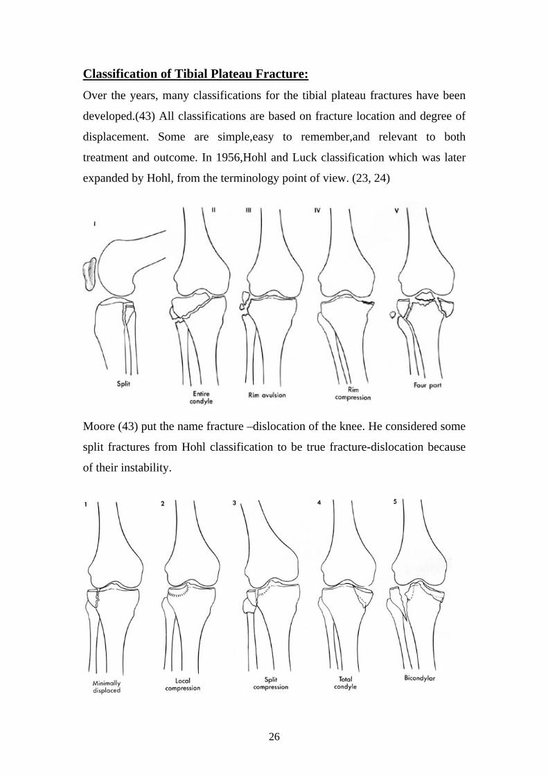
Classification of Tibial Plateau Fracture:
Over the years, many classifications for the tibial plateau fractures have been
developed.(43) All classifications are based on fracture location and degree of
displacement. Some are simple,easy to remember,and relevant to both
treatment and outcome. In 1956,Hohl and Luck classification which was later
expanded by Hohl, from the terminology point of view. (23, 24)
Moore (43) put the name fracture –dislocation of the knee. He considered some
split fractures from Hohl classification to be true fracture-dislocation because
of their instability.

27
Classification of tibial plateau fractures as described by Hohl and Moore: type
1, minimally displaced; type 2, local compression; type 3, split compression;
type 4, total condyle; and type 5, bicondylar. (Redrawn from Hohl M, Moore
TM: Articular fractures of the proximal tibia.
The Association for the study of Internal Fixation (ASIF-AO) IN 1990
published the comprehensive classification of fractures of long bones.
Perhaps the most widely used classification of tibial plateau fractures in North
America is the one proposed by Schatzker (59) with six types.

28
Type I, pure cleavage fracture. B, Type II, cleavage combined with depression.
Reduction requires elevation of fragments with bone grafting of resultant hole
in metaphysis. Lateral wedge is lagged on lateral cortex protected with buttress
plate. C, Type III, pure central depression. There is no lateral wedge.
Depression also can be anterior, posterior, or involve whole plateau. After
elevation of depression and bone grafting, lateral cortex is best protected with
buttress plate. D, Type IV. Medial condyle is either split off as wedge (type A)
as illustrated, or it can be crumbled and depressed (type B), which is
characteristic of older patients with osteoporosis (not illustrated). E, Type V.
Continuity of metaphysis and diaphysis should be considered. In internal
fixation both sides must be protected with buttress plates. F, Type VI. Essence
of this fracture is fracture line that dissociates metaphysis from diaphysis.
Fracture pattern of condyles is variable and all types can occur. If both
condyles are involved, proximal tibia should be buttressed on both sides (59).
Diagnosis of Tibial Plateau Fracture: Patient with tibial plateau fractures usually presents with a painful swollen
knee and is unable to bear full weight on the affected extremity. The patient is
able usually to describe the mechanism of injury. Most commonly, these are
valgus injury such as bumper injuries to the knee, football or soccer accident,
or fall from a height. The history of the injury is important to show the surgeon
whether the injury was caused by high or low energy forces. This is important
because associated injuries such as fracture blisters, compartment syndromes,
ligamentous disruption, and neurovascular injuries are common with high
energy forces.
Anteroposterior and lateral x-rays of the knee will usually show a plateau
fracture. If a fracture is suspected ,but is not observed in these views, 40o
internal and external oblique views should be obtained. The internal oblique
view profiles the lateral plateau while the external oblique view project the
medial condyle and plateau (25).
29
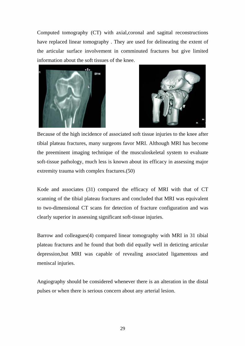
Computed tomography (CT) with axial,coronal and sagittal reconstructions
have replaced linear tomography . They are used for delineating the extent of
the articular surface involvement in comminuted fractures but give limited
information about the soft tissues of the knee.
Because of the high incidence of associated soft tissue injuries to the knee after
tibial plateau fractures, many surgeons favor MRI. Although MRI has become
the preeminent imaging technique of the musculoskeletal system to evaluate
soft-tissue pathology, much less is known about its efficacy in assessing major
extremity trauma with complex fractures.(50)
Kode and associates (31) compared the efficacy of MRI with that of CT
scanning of the tibial plateau fractures and concluded that MRI was equivalent
to two-dimensional CT scans for detection of fracture configuration and was
clearly superior in assessing significant soft-tissue injuries.
Barrow and colleagues(4) compared linear tomography with MRI in 31 tibial
plateau fractures and he found that both did equally well in deticting articular
depression,but MRI was capable of revealing associated ligamentous and
meniscal injuries.
Angiography should be considered whenever there is an alteration in the distal
pulses or when there is serious concern about any arterial lesion.

30
MATERIALS AND METHODS
We studied, retrospectively, 30 patients with complex fractures of inter-
articular distal femur (AO classification B,C) and tibial plateau fractures
displaced more than 5mm of the articular surface or more than 10 degrees
angulation, associated with either an open wound or severe soft tissue injuries.
Exclusion criteria included patients who were not fully mobile before their
injury and patients who did not continue the follow up in Khoula Hospital. All
the patients were treated between January 1999 and December 2003 with at
least one year of follow up. All the patients were operated and followed up by
the same doctor.
All the patients were treated according to a protocol which consisted of:
1. Standard Antero posterior and lateral plain X-ray.
2. Pre Operative CT Scan for pre operative planning.
3. Open wounds were taken to theatre for wound debridements within 5
hours of admission.
4. Poly trauma patients were fixed as soon as their general condition
allowed for surgery.
5. CPM (Continous Passive Movement) for 3-5 days immediately post
operative.
6. Patient discharge with above knee cast for 3-5 weeks depending on the
patient understanding.
7. Partial weight bearing with early signs clinically and radiologically of
union.
8. Patient follow up depended on the clinical examination as well as the x
ray findings.

31
METHODS OF FIXATIONS
The goal of treatment of fractures around the knee is to achieve anatomical
reduction and stable fixation. The patient can have early mobilization and
function with functional union and to achieve this goal, we must follow these
principles:
1. Good pre-operative planning.
2. Gentle handling of soft tissues.
3. Accurate anatomical reduction of the fracture.
4. Rigid, stable fixation.
5. Bone grafting of any major defects if needed.
6. Early and active rehabilitation of the limb and the patient.
In our study we used different types of implants. For the tibial plateau we used
percutaneous Asnis screws in 2 patients (16.7%) in minimally displaced
fractures because we didn't have canulated screws. We used the percutaneous
Asnis screws as well with intraarticular unicondylar fractures of the distal
femur, 2 patients (11.1%). and the screws were usually perpendicular to the
fracture line for built of buttressing effect. We used L-shape and T-shape plates
in 10 patients (83.3%) with fractures of the tibial plateau; for lateral tibial
plateau we used the anterolateral parapatellar approach, and for the medial
tibial plateau we used the anteromedial parapatellar approach, for the
bicondylar fractures we used either the central midline approach or we used
Tri-radiate skin incision in 3 cases (25%) without any skin complications due
to full skin flap technique.
In intercondylar fractures of the distal femur with displacement, we used the
appoach discribed by Mize, Busholz and Grogn 1982, (40) for perfect
visualisation of the fracture. We used AO 950 Angled Blade Plate in 2 patients
32
(11.1%) and because of its one piece construction and broad flat blade, it
provides stable fixation for most fracture types but technically it is difficult to
use because it needs precision and correct alignment in all three planes.
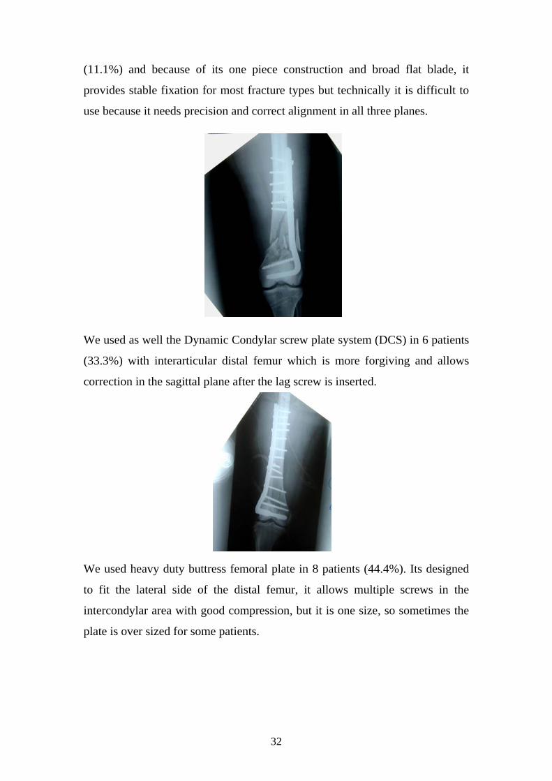
We used as well the Dynamic Condylar screw plate system (DCS) in 6 patients
(33.3%) with interarticular distal femur which is more forgiving and allows
correction in the sagittal plane after the lag screw is inserted.
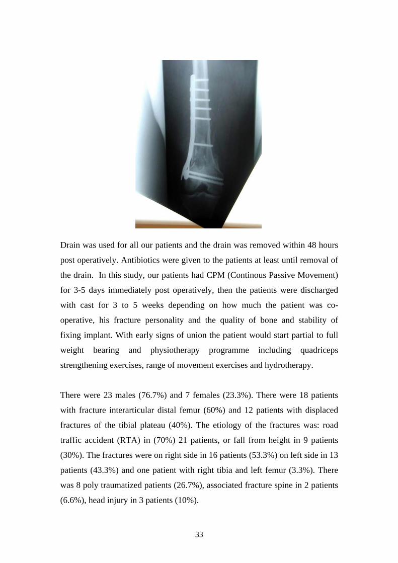
We used heavy duty buttress femoral plate in 8 patients (44.4%). Its designed
to fit the lateral side of the distal femur, it allows multiple screws in the
intercondylar area with good compression, but it is one size, so sometimes the
plate is over sized for some patients.
33
Drain was used for all our patients and the drain was removed within 48 hours
post operatively. Antibiotics were given to the patients at least until removal of
the drain. In this study, our patients had CPM (Continous Passive Movement)
for 3-5 days immediately post operatively, then the patients were discharged
with cast for 3 to 5 weeks depending on how much the patient was co-
operative, his fracture personality and the quality of bone and stability of
fixing implant. With early signs of union the patient would start partial to full
weight bearing and physiotherapy programme including quadriceps
strengthening exercises, range of movement exercises and hydrotherapy.
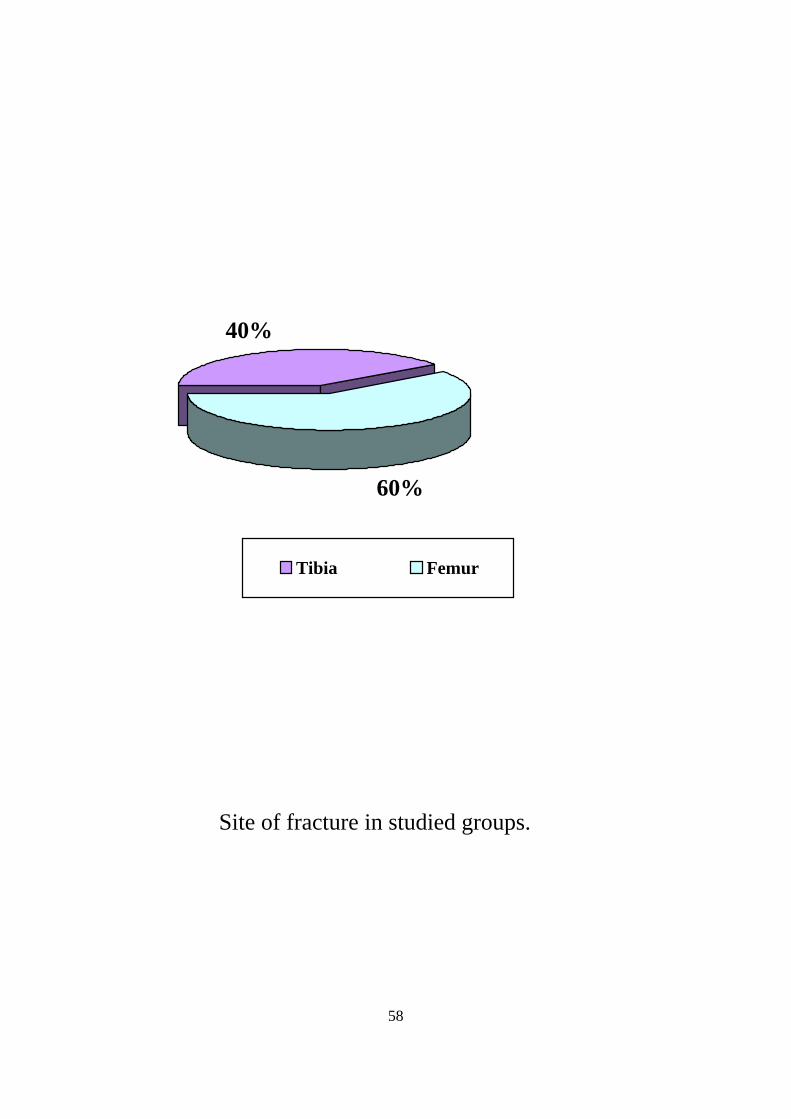
There were 23 males (76.7%) and 7 females (23.3%). There were 18 patients
with fracture interarticular distal femur (60%) and 12 patients with displaced
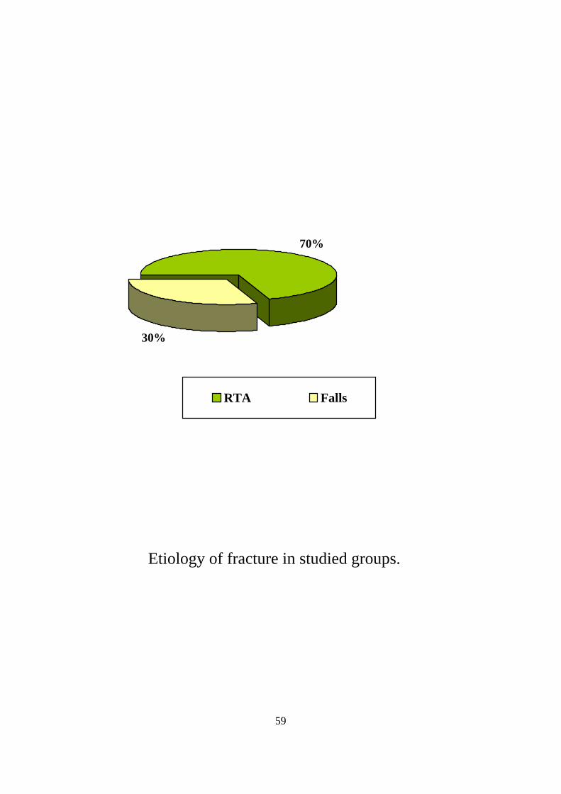
fractures of the tibial plateau (40%). The etiology of the fractures was: road
traffic accident (RTA) in (70%) 21 patients, or fall from height in 9 patients
(30%). The fractures were on right side in 16 patients (53.3%) on left side in 13
patients (43.3%) and one patient with right tibia and left femur (3.3%). There
was 8 poly traumatized patients (26.7%), associated fracture spine in 2 patients
(6.6%), head injury in 3 patients (10%).

34
RESULTS
Statistical methods: SPSS (Statistical Package for Social Sciences) version 10.0 was used
for data analysis. Mean and standard deviation are descriptive values for
quantitative data with median and range for non-normally distributed data.
Non parametric t test (Mann Whitney test) was used for comparing means of
two independent groups. Spearman Rho correlation measured the association
between quantitative variables (age and healing time). Chi-square – Fisher
exact test were the tests for proportion independence. P value is significant at
0.05 level.
The 30 patients were assessed clinically and radiologically with mean follow
up of 30 months (18 to 51 months). All achieved bony union at the end of
follow up. While 6 patients had implants removal after 24 – 36 months from
the injury. The mean age was 39 years (21 to 63 years) and the mean length of
hospital stay is 21 days (3 to 82 days). One patient had implant failure after
five weeks from surgery due to premature weight bearing and was reoperated.
Pain: Our observation suggests that the pain tolerance was much higher in the
older age group compared to younger age group regardless of the x-ray picture
or the clinical findings.
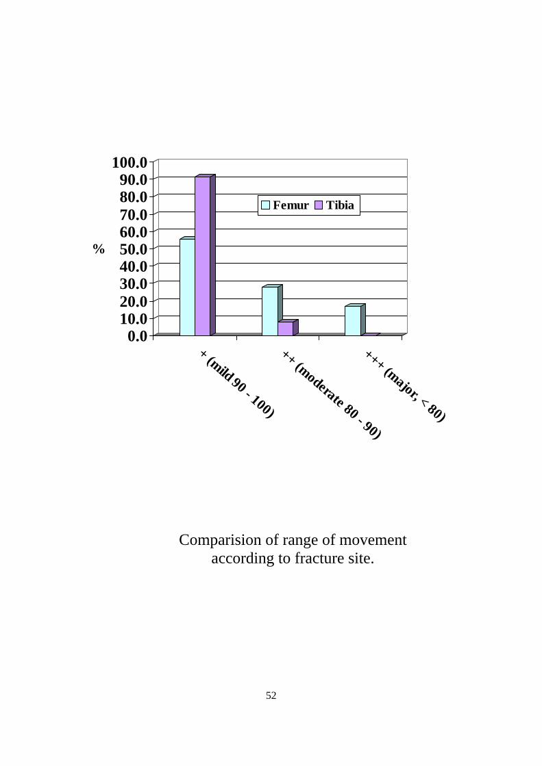
Range of Movements: Our observation suggests that the range of movement
in patients with fractures of tibial plateau is much better than the femoral one
regardless of the amount of comminution and the quality of reduction and
fixation.
35
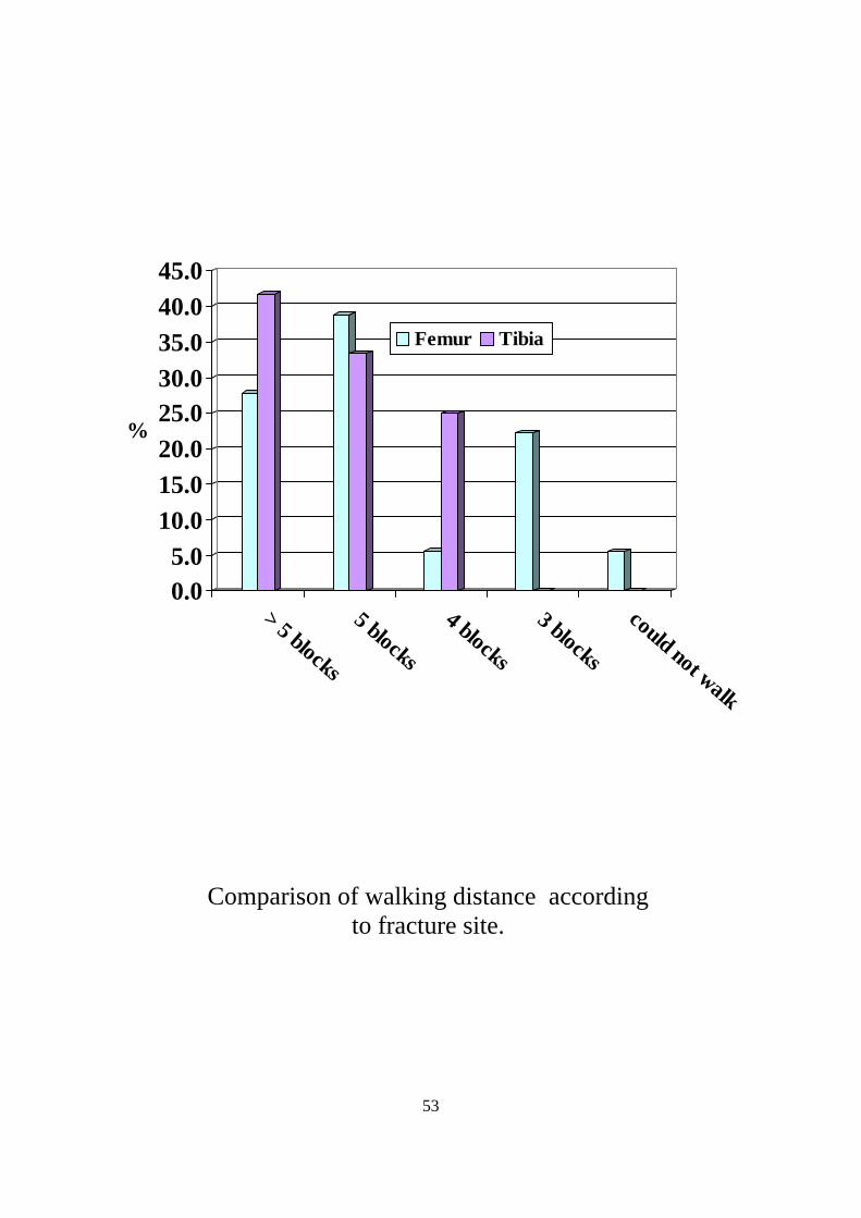
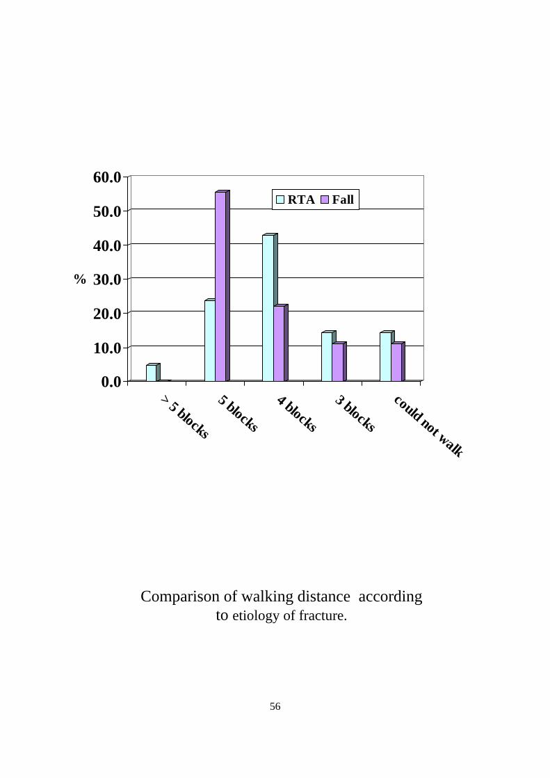
Walking Distance: 74% of tibial fracture patients were able to walk 5 blocks
or more and 65% of the femoral fracture patients were able to walk 5 blocks or
more, only one patient with fracture femur was unable to move.
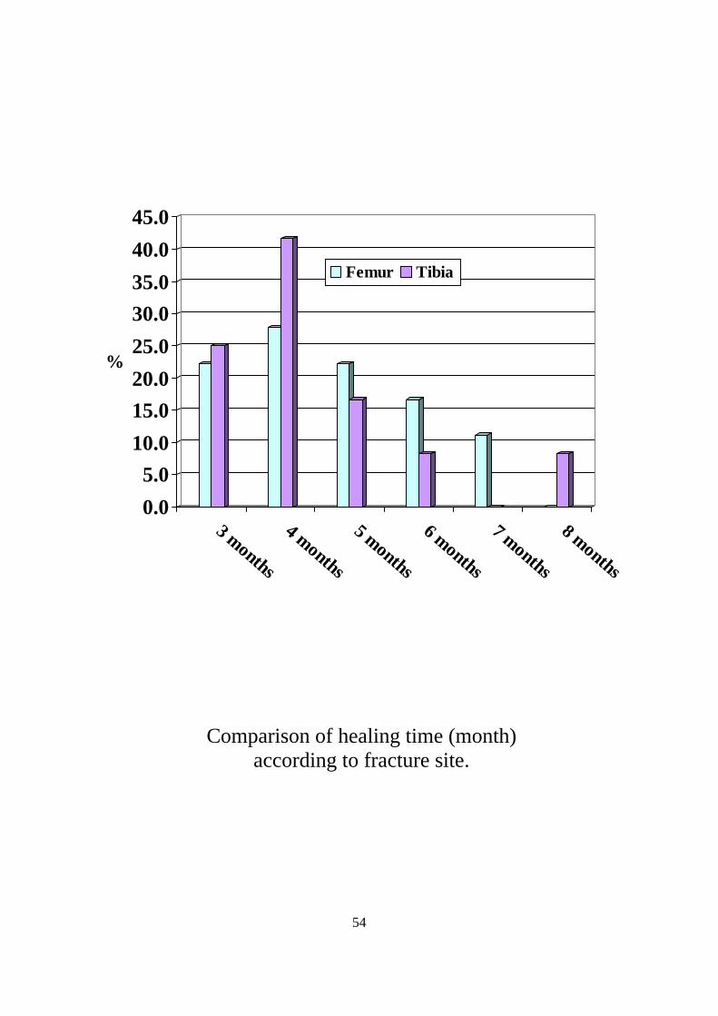
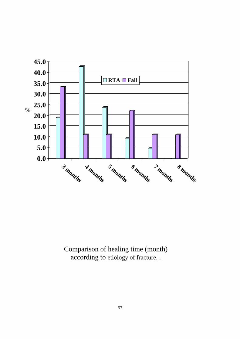
Healing time: Our results showed that the median healing time for the tibia
was 4 months (3 – 8 months) and the median healing time for the femur was
4.5 months (3 – 7 months).
• 6 patients with comminuted fractures needed bone grafts after 8-12
weeks and all our patients fractures healed.
• 8 patients needed double plating.
• 6 tibias and 2 femurs, the femur patients second plate was done as
second stage when post operatively we discovered that the fracture
fixation is unstable usually during bone grafting.
One of our patients with buttress femoral plate had implant failure and needed
re-operation and fixation with DCS and larger plate because the patient was not
co-operative and walked full weight bearing without crutches after five weeks
of the trauma.
In general, our observations in fractures of the tibial plateau, the clinical
outcome was very good to excellent, inspite of the X-ray picture and the quality
of the reduction.
In fractures of intercondylar femur, the results were not as satisfactory, even
with good X-ray and intraoperative reduction of the fracture.
We could not use any of the known knee scores ( Lowa and Rasmussen)
because our criteria was not fitting completely with any of them.
36
Immediate post operative complications
There was no complications in 23 patients (76.7%), wound infections in 3
patients (10.0%), neuropraxia in one patient (3.3%), DVT (Deep Venous
Thrombosis) in 3 patients (10.0%).
Examples :
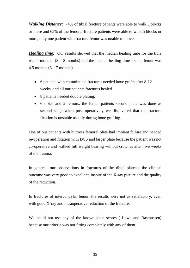
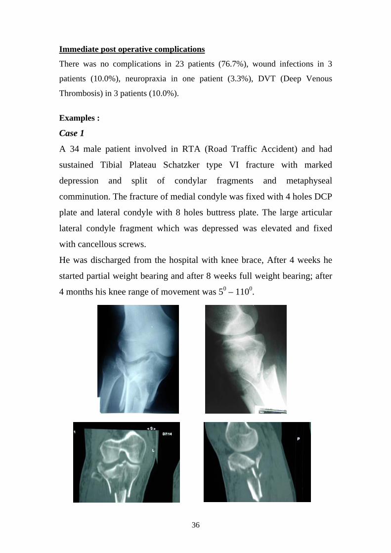
Case 1
A 34 male patient involved in RTA (Road Traffic Accident) and had
sustained Tibial Plateau Schatzker type VI fracture with marked
depression and split of condylar fragments and metaphyseal
comminution. The fracture of medial condyle was fixed with 4 holes DCP
plate and lateral condyle with 8 holes buttress plate. The large articular
lateral condyle fragment which was depressed was elevated and fixed
with cancellous screws.
He was discharged from the hospital with knee brace, After 4 weeks he
started partial weight bearing and after 8 weeks full weight bearing; after
4 months his knee range of movement was 50 – 1100.
37
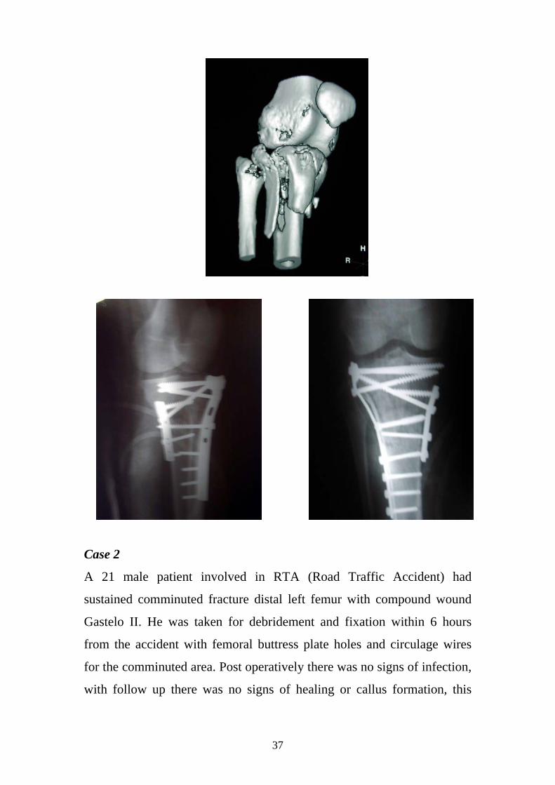
Case 2
A 21 male patient involved in RTA (Road Traffic Accident) had
sustained comminuted fracture distal left femur with compound wound
Gastelo II. He was taken for debridement and fixation within 6 hours
from the accident with femoral buttress plate holes and circulage wires
for the comminuted area. Post operatively there was no signs of infection,
with follow up there was no signs of healing or callus formation, this
38
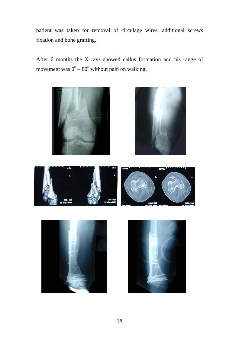
patient was taken for removal of circulage wires, additional screws
fixation and bone grafting.
After 6 months the X rays showed callus formation and his range of
movement was 00 – 800 without pain on walking.
39
DISCUSSION
The management of an adult patient with interarticular fractures around
the knee continues to pose a challenge. Specially for Omani patients
when good range of movement around the knee is needed for daily social
activities, sitting on the ground and praying.
Before 1982 almost all fractures around the knee in Oman was treated by
general surgeons and usually through non operative treatment,
unfortunately we don’t have any documents for this period but we used to
review elderly patients with post traumatic deformities or severely
arthritic knee as long term result of old injuries to the knee and they were
usually treated by TKR (Total Knee Replacement) as final management.
In our study, patients continue to improve with time after treatment and
the healing of bone and soft tissue injuries, their range of movement
slowly improved and their pain tolerance gradually improved.
In our study as well, so many patients could not participate in the study
due to the fact that they were expatriates working in Oman and they left
the country after their accidents. We didn’t use any of the knee scores as
Lowa, Hariss or SF-36 questionnaire because our data were not fitting as
timing of union or patient going back to work.
If we go to literature we will find that in fracture supra condylar and
intracondylar of distal femur, some authors achieved good to excellent
results with closed methods in about 54% of their patients (Sf wart, Sisk
and Wallace 1966 (64), Neer 1967 (47), Mooney 1970 (41) and 1/3 of the
patients had non union. However, Neer had satisfactory result in 84%, his
study was retrospective, non randomized and he used different measures
40
from those used by Schatker 1979 (59) and he used an earlier method of
internal fixation.
After 1982 we started in Oman to adapt AO Philosophy and techniques
of open reduction and internal fixation, various devices have been used
for internal fixation. For supracondylar fracture femur, the literature
including angled blade plates [Sheatizker 1979] (59), Rush pins
[Shelbourne & Brueckman 1982] (8), Enders nails [Kolmert, Egund &
Persson 1983] (34) and purpose designed nails [Zickel, Hobeika &
Robbins 1986], Pryor & Doran 1988, Marks, Isbister & Porter 1994, the
dynamic candylar screw (DCS) has been shown to give satisfactory
results [Sanders 1989 (56).
Recently liss plates and retrograde femoral nail show satisfactory results
but there is no studies with enough follow up period to give accurate
results [Rademakers 2004] (49).

41
Table 1 (Implant Options) Type of Implant
Relative Indications
Relative Contraindications
Advantages Disadvantages
Blade Plate Comminuted Supracondylar fracture Low fracture
Intracondylar comminution
Strong able to maintain varus / valgus and antecurvatum or retrocurvatum alignment Most stable fixation
Technically demanding Can comminute unrecognized intracondylar fractures
Compression Screw
Comminuted supracondylar fractures associated with simple intracondylar splits
Intracondylar comminution Very low fractures Coronal fractures
Technically easier to use than blade plate Compresses simple intracondylar spilts Able to maintain varus / valgus alignment
More difficult to maintain recurvatum / antecurvatum alignment in low fractures Occupies a large bone volume in the intracondylar region Requires additional screw fixation in disal fragment for stability
Condylar Plate
Simple supracondylar fracture in association with intracondylar comminution
Comminuted supracondylar fracture
Can be contoured to achieve anatomic reduction of a simple fracture Multiple screw insertion can help reduction of intracondylar comminution
Poor resistance to varus / valgus moments, so requires reconstruction of medical cortical continuity
Dual Plate Supracondylar and intracondylar comminution
Should be reserved for situation in which no other device will work
Allows multiple-screw fixation of intracondylar comminution Dual plates provide strength for supracondylar comminution
Massive dissection with resultant stiffness Potential for “dead bone sandwich”
Antegrade Nail
Extensive supracondylar comminution, especially proximal
Low fractures Intracondylar extension
Minimal dissection and injury of the soft tissue envelope Strong fixation automatic grafting (reamings)
Can “blow apart” unrecognized intracondylar fractures Can be difficult to achieve anatomic alignment
Retrograde Nail
Osteoporosis Supracondylar periprosthetic fracture
“High-demand” patient Low Fracture
Minimal dissection and injury to soft-tissue envelope Some grafting of the fracture site by reaming
Low strength device Residual fracture instability may necessitate caution when initiating postoperative motion
42
Treatment of complex high-energy fractures of the tibial plateau remains
difficult. The goals of treatment of these injuries are the restoration of
joint congruity, normal alignment, joint stability, and a functional range
of knee motion. For markedly displaced bicondylar fractures of the tibial
plateau and those associated with joint instability, conventional open
reduction and internal fixation through a single anterior approach has
been the standard way of care. For many bicondylar fractures of the tibial
plateau [Schatzker types V and VI] (58), fixation with two plates may be
necessary to prevent axial collapse.
However, the soft-tissue stripping in these injuries, together with the
surgical dissection needed to apply large plates, has been associated with
a high rate of complications particularly infection and wound breakdown.
In an effort to improve the outcome of the repair of high-energy fractures
of the tibial plateau, less invasive methods of treatment (54) have been
introduced with the use of either tensioned circular wire, hybrid, or large-
pin monolateral external fixators (37) or internal fixation through two
incisions and use of small-fragment specialized plates.
In the last decade, treatment strategies for high-energy fractures of the
tibial plateau have changed, resulting in numerous recent results of
treatment, unfortunately, there was no results for the functional status of
the patients. Most patients show acceptable outcome after sustaining a
high-energy fractures of the tibial plateau despite of injury to the articular
surface, imperfect reduction, and associated meniscal and ligament
injuries, most patients reported that they were functioning well and were
able to pursue recreational activities and their occupations with few
limitations which was supported by our results.

43
In our study,fracture of the tibial plateau, 85% of our patients with closed
fractures had good to excellent function regardless of the X- ray findings,
this is more obvious with lateral tibial plateau fractures.
For minimally displaced fractures of tibial plateau,we treated our patients
with percutaneous canulated screws, which is acceptable in most of the
studies, some articles discussed the role of external fixator either mono
frame or circular frame. In displaced fractures uni or bicondyle of tibial
plateau reduction and fixation with one plate or double plates either
through one midline tri-radiate incision or through separate two incisions.
There is little information in the literature regarding the outcomes of total
knee arthroplasty following open reduction and internal fixation of tibial
plateau fractures. Saleh et al (55) reviewed fifteen patients after a
minimum duration of follow up of five years. They concluded that total
knee arthroplasty after open reduction and internal fixation of the tibial
plateau fractures decreased pain and improved knee function but the
procedure is technically demanding and is associated with a high failure
rate.
44
CONCLUSION Although interarticular fractures around the knee are very difficult and are
challenging to the surgeon but with good preoperative planning and using good
operative techniques the results of operative treatment is very superior to the
other types of treatment, with good to excellent results and less complications.
Using the new techniques of internal fixation provided by the AO group,
improvement of the quality of care and the final outcome is now well
established. Some controversy about the perfect type of fixation internal or
external. The type, the time and the need for bone graft or not, still need more
research.
In our study the life style of the Omani people has shown good effect on the
final range of movement due to sitting on the ground and prayers. Pain
tolerance was excellent in older generation due to their life style. Recently,
more biological techniques show better progress like Liss plate and retrograde
distal femoral nail. In Oman we are in the process of adapting these new
techniques to improve the outcome of our patients.
We have collected our data and analysed our results for auditing and teaching
purposes. This shows that the adoption of these modern techniques of open
reduction and internal fixation within the Omani Health Care system have
significantly improved the outcome of these severe injuries in the Omani
population.
45
REFERENCES
1. Apley, A. : Fractures of the Lateral Tibial Condyle Treated by Skeletal Traction and Early Mobilization, J. Bone Jount Surg., 38B:699, 1956.
2. Arneson, T.J.; Melton, L.J. III; Lewallen, D.G.; and O’Fallon, W.M. :
Epidermiology of Diaphyseal and Distal Femoral Fractures in Rochester, Minnesota, 1965-1984. Clin. Orthop 234:188-194, 1988
3. Barei D.P.; Nork, S.E.; Mills, W.J.; Henley, M.B.; and Benirschke S.K.:
Complications Associated with Internal Fixation of High-energy Bicondylar Tibial Plateau Fractures Utilizing a Two-incision Technique., J. Orthop Trauma. 18(10): 649-57, Nov-Dec. 2004.
4. Barrow, B.A.; Fajman, W.A.; Parker, L.M.; et ail. : Tibial Plateau Fractures;
Evaluation with MR Imaging. Radiographics 14:553-559, 1994.
5. Bennett, W.F.; and Browner, B. : Tibial Plateau Fractures; A Study of Associated Soft Tissue Injuries. J. Orthop Trauma 8:183-188, 1994.
6. Blokker, C.P.; Rorabeck; C.H.; and Bourne, R.B. : Tibial Plateau Fractures
and Analysis of Treatment in 60 patients. Clin. Orthop., 182:193, 1984.
7. Brooker W.L.: Distal Femoral Fractures: Comparison of Open and Closed Methods of Treatment. Clin. Orthop., 174; 166-71, 1983.
8. Brueckmann FR.: Rush Pin Fixation of the Supracondylar and Intercondylar
Fractures of the Femur. J. Bone Joint Surg. [Am] 64-A:161-9, 1982.
9. Burri, C.; Bartzke, G.; Coldeway, J., and Muggler, E.: Fractures of the Tibial Plateau. Clin. Orthop., 138:84, 1979.
10. Catagni, M.: Fractures of the Leg (Tibia). In Maioccki, A.B., and Aronson, J.
(eds.): Operative Principles of Ilizarov, p. 91. Baltimore, Williams & Wilkins, 1991.
11. Chapman, M.W.; and Mahoney, M.: The Role of Early Internal Fixation in the
Management of Open Fractures. Clin. Orthop., 138:120, 1979.
12. Chiron, H.S.; Tremoulet, J.; Casey, P.; and Muller, M: Fractures of the Distal Third of the Femur Treated by Internal Fixation. Clin. Orthop., 100:160-170, 1974.
13. Decoster, T.A.; and Nepola, J.V.: Cast Brace Treatment of Proximal Tibial
Plateau Fractures Ten Years Follow-up Study. Clin. Orthop., 231:196, 1988.
14. Dias, J.J.; Stirling, A.M.; Finlay, D.B.; and Gregg, R.J.: Computerised Axial Tomography for Tibial Plateau Fractures. J. Bone Joint Surg., 69B:84, 1987.
46
15. Drennan, D.B.; Locher, F.G.; and Maylahn, D.J.: Fractures of the Tibial Plateau: Treatment by Closed Reduction and Spica Cast. J. Bone Joint Surg., 61A:989, 1979.
16. Duweilus, P.J.; and Connolly, J.F.: Closed Reduction of Tibial Plateau
Fractures: A Comparison of Functional and Roentgenographic End Results. Clin. Orthop., 230:116, 1988.
17. Frankel, V.H.; Green, S.A.; Paley, D., et al. : Symposium: Current
Applications of the Ilizarov Technique. Contemp Orthop., 28:51, 1994.
18. Giles, J.B.; DeLee, J.C.; Heckman, J.D.; and Keever, J.E.: Supracondylar-Intercondylar Fractures of the Femur Treated with a Supracondylar Plate and Lag Screw. J. Bone Joint Surg., 64A:864-870, 1982.
19. Gossling, H.R.; and Peterson, C.A.: A New Surgical Approach in the
Treatment of Depressed Lateral Condylar Fractures of the Tibia. Clin. Orthop., 140:96, 1979.
20. Green, N.E.; and Allen, F.L.: Vascular Injuries Associated with Dislocation of
the Knee. J. Bone Joint Surg. 59A:236-239,1977.
21. Gustilo, R.B.: Fractures of the Tibial Plateau. In Gustilo, R.B., Kyle, R., and Templeman, D. (eds.): Fractures and Dislocations, p. 945. St. Louis, C.V. Mosby, 1993.
22. Healy, W.L.; and Brooker, A.F.: Distal Femoral Fractures: Comparison of
Open and Closed Methods of Treatment. Clin. Orthop., 174:166-171, 1983.
23. Hohl, M.: Tibial Condylar Fractures. Instruct Course Lect 33:206-217, 1963.
24. Hohl, M.: Tibial Condylar Fractures. J. Bone Joint Surg., 49A:1455, 1967.
25. Hohl, M.: Part I: Fractures of the Proximal Tibia and Fibula. In Rockwood, C., Green, D., and Bucholz, R. (eds.): Fractures in Adults, 3rd ed. Philadelphia, J.B. Lippincott, 1991.
26. Hohl, M.; and Luck, J.V.: Fractures of the Tibial Condyle. J. Bone Joint Surg.,
38A:1001, 1956.
27. Honkonen, S.E.: Indications for Surgical Treatment of Tibial Condyle Fractures. Clin. Orthop., 302:199-205, 1994.
28. Johnson, K.D.; and Hicken, G.: Distal Femoral Fractures. Orthop. Clin. North
Am., 18:115-132, 1987.
29. Karunakar M.A.; and Egol K.A.: Split Depression Tibial Plateau Fractures., J. Orthop. Trauma. 16:172-7, 2002.

47
30. Kennedy, J.C.: Complete Dislocation of the Knee Joint. J. Bone Joint Surg. 45A:889-904, 1963.
31. Kode, L.; Lieberman, J.M.; Motta, A.O.; Wilber, J.H.; Vasen, A.; and Yagan,
R.: Evaluation of Tibial Plateau Fractures: Efficacy of MR Imaging Compared with CT.A.J.R., 163:141, 1994.
32. Koechlin, P.; Nael, J.F.; Bonnet, J.C.; D’Ythurbide, B.; and Apoil, A.:
Ligamentous Lesions Associated with Fractures of the Tibial Plateau. Acta Orthop. Belg., 49:751, 1983.
33. Kolmert, L.; Egund, N.; and Persson, B.D.: Internal Fixation of Supracondylar
and Bicondylar Fractures using a New Semi-Elastic Device. Clin. Orthop., 181:204-219, 1983.
34. Kolmert, L.; and Wulff, K.: Epidemiology and Treatment of Distal Femoral in
Adults. Acta Orthop. Scand 53:957-962, 1982.
35. Koval, K.J.; Sanders, R.; Borrelli, J.; Helfet, D.; DiPasquale, T.; and Mast, J.W.: Indirect Reduction and Percutaneous Screw Fixation of Displaced Tibial Plateau Fractures. J. Orthop. Trauma, 6:340, 1992.
36. Lansinger, O.; Bergman, B.; Komer, L.; and Anderssonn, G.M.J.: Tibial
Condylar Fractures: A twenty-year follow-up. J. Bone Joint Surg., 68A:13, 1986.
37. Mallik, A.R.; Covall, D.J.; and Whitelaw, G.P.: Internal Versus External
Fixation of Bicondylar Tibial Plateau Fractures. Orthop. Rev., 21:1433, 1992.
38. Mast, J.; Jakob, R.; and Ganz, R., eds.: Planning and Reduction Technique. In Fracture Surgery, p. 100-114. New York, Springer-Verlag, 1989.
39. Mast, J.; Jakob, R.; and Ganz, R.: Planning and Reduction Technique in
Fracture Surgery, Berlin, Springer, 1989.
40. Mize, R.D.; Bucholz, R.W.; and Grogan, D.P.: Surgical Treatment of Displaced, Comminuted Fractures of the Distal End of the Femur. J. Bone Joint Surg., 64A:871-879, 1982.
41. Mooney,V;Nickel,VL;Harvey,JP; and Snelson,R:Cast-Brace Treatment for
Fractures of the Distal Part of the Femur.J.Bone Joint Surg.,52-A:1563,1970.
42. Moore, T.M.; Meyers, M.H.; and Harbey, J.P., Jr.: Collateral Ligament Laxity of the Knee Long-term Comparison Between Plateau Fractures and Normal J. Bone Joint Surg., 58A:594, 1976.
43. Moore, T.M.; Patzakis M.G.; and Harvey, J.B.: Tibial Plateau Fractures
Definition, Demographics, Treatment Rationale, and Long-term results of Closed Traction Management or Operative Reduction. J. Orthop Trauma 1:97-119, 1987.
48
44. Muller, M.E.; Allgower, M.; Schneider, R.; and Willenegger, H.: Manual of
Internal Fixation. New York, Springer-Verlag, 1979.
45. Muller, M.E.; Allgower. M.; Schneider, R.; and Willenegger, H.: Manual of Internal Fixation, 2nd ed. New York, Springer-Verlag. 1979.
46. Muller, M.E.; Nazarian, S.; and Koch, P.: Classification AO Des Fractures.
New York, Springer-Verlag, 1987.
47. Neer, C.S.; II, Grantham, S.A.; and Shelton, M.L.: Supracondylar Fracture of the Adult Femur. J. Bone Joint Surg. 49A:591-613, 1967.
48. Olerud, S.: Operative Treatment of Supracondylar-Condylar Fractures of the
Femur. J. Bone Joint Surg., 54A:1015-1032, 1972.
49. Rademakers, M.V.; Kerkhoffs, G.M.; Sierevelt, I.N.; Raaymakers, E.L.; and Marti, R.K.: Intra-articular Fractures of the Distal Femur: A Long-term Follow-up Study of Surgically Treated Patients., J. Orthop Trauma. 18(4):
213-9, Apr. 2004. 50. Rafii, M.; Lamont, J.G.; and Firooznia, H.: Tibial Plateau Fractures: CT
Evaluation and Classification. Crit. Rev. Diagn. Imaging. 27:91, 1987. 51. Radford, PJ; and Howell, CJ;The AO Dynamic Condylar Screw for Fractures
of the Femur:Injury.,Br.J.Acc.Surg.,23:89,1992.
52. Regazzoni, P.; Leutenegger, A.; Ruedi, T.; and Staehelin, F.: Erste Erfahrungen mit der dynamischen kondylenschraube (DCS) bei distalen femurfrakturen. Helv Chir Acta 53:61-64, 1986.
53. Regazzoni, P.; Ruedi, T.; and Allgower, M.: The Dynamic Condylar Screw
Implant System for Fractures of the Distal Femur. AO/ASIF Dialogue, 1:8-9, 1986.
54. Ricci, W.M.; Rudzki, J.R.; and Borrelli J. Jr.: Treatment of Complex Proximal
Tibia Fractures with the Less Invasive Skeletal Stabilization System., J. Orthop Trauma. 18(8):521-7, Sep. 2004.
55. Saleh K.J.; and Sherman P: Total Knee Arthoplasty after Open Reduction and Internal Fixation of Fractures Tibial Plateau., J. Bone Joint Surg. Am. 83:1144-48, 2001.
56. Sanders, R.; Regazzoni, P.; and Ruedi, T.P.: Treatment of Supracondylar-
Intercondylar Fractures of the Femur Using the Dynamic Condylar Screw. J. Orthop. Trauma, 3:214-222, 1989.
57. Schatzker, J.: Fractures of the Tibial Plateau. In Schatzker, J., Tile, M. (eds.): Rationale of Operative Fracture Care, p. 279. New York: Springer, 1987.

49
58. Schatzker, J.: Tibial Plateau Fractures. In Browner, Jupiter, Levine, and Trafton (eds.): Skeletal Trauma, p. 1745. Philadelphia, W.B. Saunders, 1993.
59. Schatzker, J.; and Lambert, D.C.: Supracondylar Fractures of the Femur. Clin.
Orthop., 138:77-83, 1979.
60. Schatzker, J.; McBroom, R.; and Bruce, D.: Tibial Plateau Fractures: The Toronto Experience 1968-1975. Clin. Orthop., 138:94, 1979.
61. Segal, D.; Mallik, A.R.; Wetzler, M.J.; Franchi, A.V.; and Whitelaw, G.P.:
Early Weight Bearing of Lateral Tibial Plateau Fractures. Clin. Orthop., 294:232, 1993.
62. Seinsheimer, F.: Fractures of the Distal Femur. Clin. Orthop., 153:169-179,
1980.
63. Siliski, J.M.; Mahring, M.; and Hofer, H.P.: Supracondylar-Intercondylar Fractures of the Femur. J. Bone Joint Surg., 71A:95-104, 1989.
64. Sisk T.D.; and Wallace S.L.: Fractures of the Distal Third of the Femur:
A Comparison of Methods of Treatment. J. Bone Joint. Surg. [AM] 48-A:784- 2807, 1966.
65. Spiegel, P.G.; and Shybut, G.T.: Tibial Plateau Fractures. Editorial. Clin. Orthop. 183:12-16, 1979.
66. Stannard J.P.; Wilson, T.C.; Volgas, D.A.; and Alonso, J.E.: The Less
Invasive Stabilization System in the Treatment of Complex Fractures of the Tibial Plateau: short-term results., J. Orthop Trauma. 18(8):528-35, Sep. 2004.
67. Stevens D.G.; and Beharry R.: The Long-term Functional Outcome of
Operatively Treated Tibial Plateau Fractures., J. Orthop. Trauma. 15:312-20, 2001.
68. Walling, A.K.; Seradge, H.; and Spiegel, P.G.: Injuries to the Knee Ligaments
with Fractures of the Femur. J. Bone Joint Surg. 64A:1324-1327, 1982.
69. Watson, J.T.: High Energy Fractures of the Tibial Plateau. Orthop. Clin. North Am., 25:723, 1994.
70. Wilppula, E.; and Bakalim, G.: Ligamentous Tear Concomitant with Tibial
Condylar Fracture. Acta Orthop. Scand., 43:292, 1972.
71. Wiss,D.A:What’s New in Orthopaedic Trauma.J.Bone Joint Surg., 84A:2111-9,2002.
50
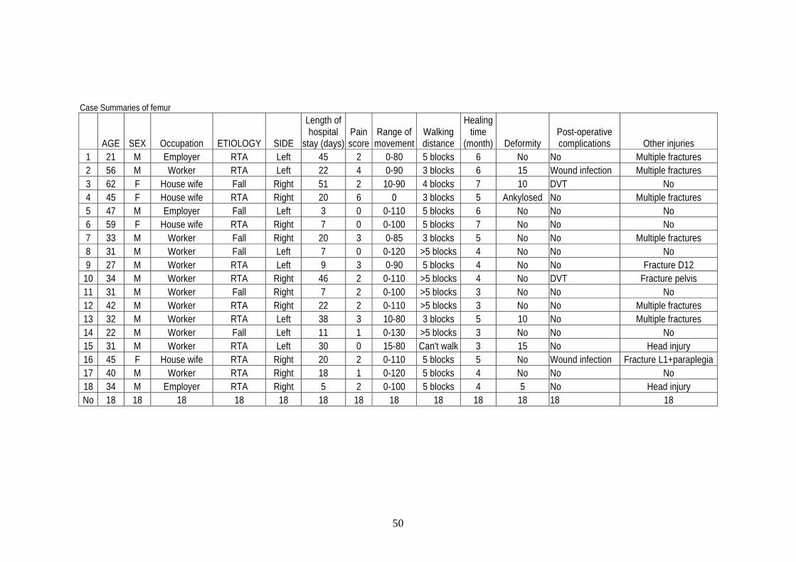
Case Summaries of femur
AGE SEX Occupation ETIOLOGY SIDE
Length of hospital
stay (days)Pain score
Range of movement
Walking distance
Healing time
(month) Deformity Post-operative complications Other injuries
1 21 M Employer RTA Left 45 2 0-80 5 blocks 6 No No Multiple fractures 2 56 M Worker RTA Left 22 4 0-90 3 blocks 6 15 Wound infection Multiple fractures 3 62 F House wife Fall Right 51 2 10-90 4 blocks 7 10 DVT No 4 45 F House wife RTA Right 20 6 0 3 blocks 5 Ankylosed No Multiple fractures 5 47 M Employer Fall Left 3 0 0-110 5 blocks 6 No No No 6 59 F House wife RTA Right 7 0 0-100 5 blocks 7 No No No 7 33 M Worker Fall Right 20 3 0-85 3 blocks 5 No No Multiple fractures 8 31 M Worker Fall Left 7 0 0-120 >5 blocks 4 No No No 9 27 M Worker RTA Left 9 3 0-90 5 blocks 4 No No Fracture D12
10 34 M Worker RTA Right 46 2 0-110 >5 blocks 4 No DVT Fracture pelvis 11 31 M Worker Fall Right 7 2 0-100 >5 blocks 3 No No No 12 42 M Worker RTA Right 22 2 0-110 >5 blocks 3 No No Multiple fractures 13 32 M Worker RTA Left 38 3 10-80 3 blocks 5 10 No Multiple fractures 14 22 M Worker Fall Left 11 1 0-130 >5 blocks 3 No No No 15 31 M Worker RTA Left 30 0 15-80 Can't walk 3 15 No Head injury 16 45 F House wife RTA Right 20 2 0-110 5 blocks 5 No Wound infection Fracture L1+paraplegia 17 40 M Worker RTA Right 18 1 0-120 5 blocks 4 No No No 18 34 M Employer RTA Right 5 2 0-100 5 blocks 4 5 No Head injury No 18 18 18 18 18 18 18 18 18 18 18 18 18
51
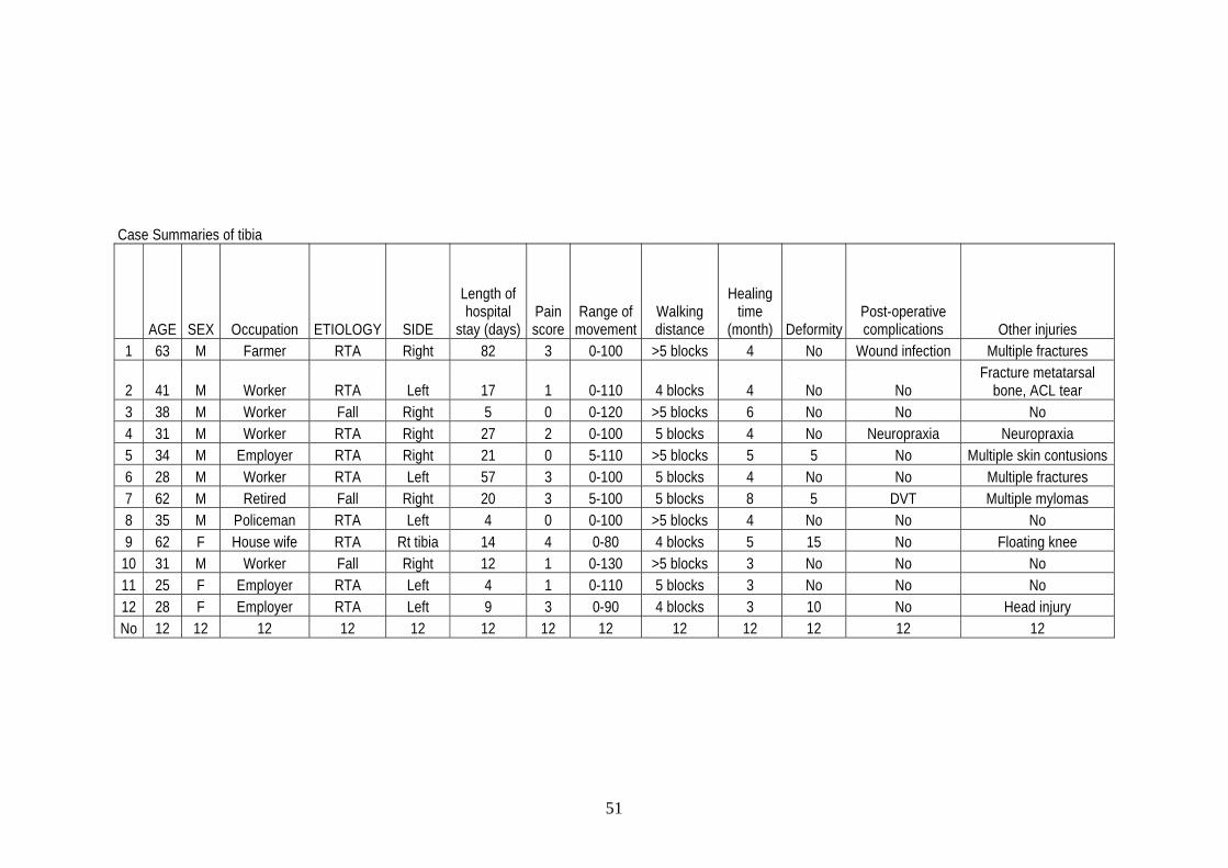
Case Summaries of tibia
AGE SEX Occupation ETIOLOGY SIDE
Length of hospital
stay (days)Pain score
Range of movement
Walking distance
Healing time
(month) DeformityPost-operative complications Other injuries
1 63 M Farmer RTA Right 82 3 0-100 >5 blocks 4 No Wound infection Multiple fractures
2 41 M Worker RTA Left 17 1 0-110 4 blocks 4 No No Fracture metatarsal
bone, ACL tear 3 38 M Worker Fall Right 5 0 0-120 >5 blocks 6 No No No 4 31 M Worker RTA Right 27 2 0-100 5 blocks 4 No Neuropraxia Neuropraxia 5 34 M Employer RTA Right 21 0 5-110 >5 blocks 5 5 No Multiple skin contusions 6 28 M Worker RTA Left 57 3 0-100 5 blocks 4 No No Multiple fractures 7 62 M Retired Fall Right 20 3 5-100 5 blocks 8 5 DVT Multiple mylomas 8 35 M Policeman RTA Left 4 0 0-100 >5 blocks 4 No No No 9 62 F House wife RTA Rt tibia 14 4 0-80 4 blocks 5 15 No Floating knee
10 31 M Worker Fall Right 12 1 0-130 >5 blocks 3 No No No 11 25 F Employer RTA Left 4 1 0-110 5 blocks 3 No No No 12 28 F Employer RTA Left 9 3 0-90 4 blocks 3 10 No Head injury No 12 12 12 12 12 12 12 12 12 12 12 12 12
52
Comparision of range of movement according to fracture site.
0.010.020.030.040.050.060.070.080.090.0
100.0
%
+ (mild 90 - 100)
++ (moderate 80 - 90)
+++ (major, < 80)
Femur Tibia
53
Comparison of walking distance according to fracture site.
0.05.0
10.015.020.025.030.035.040.045.0
%
> 5 blocks
5 blocks
4 blocks
3 blocks
could not walk
Femur Tibia
54
Comparison of healing time (month) according to fracture site.
0.05.0
10.015.020.025.030.035.040.045.0
%
3 months
4 months
5 months
6 months
7 months
8 months
Femur Tibia

55
Comparison of range of movement according to etiology of fracture.
0.010.020.030.040.050.060.070.080.0
%
+ ( mild 90 - 100)
++ (moderate
80 - 90)
+++ (major, <
80)
RTA Fall
56
Comparison of walking distance according to etiology of fracture.
0.0
10.0
20.0
30.0
40.0
50.0
60.0
%
> 5 blocks
5 blocks
4 blocks
3 blocks
could not walk
RTA Fall
57
0.05.0
10.015.020.025.030.035.040.045.0
%
3 months
4 months
5 months
6 months
7 months
8 months
RTA Fall
Comparison of healing time (month) according to etiology of fracture. .
58
Site of fracture in studied groups.
40%
60%
Tibia Femur
59
Etiology of fracture in studied groups.
70%
30%
RTA Falls

60
Post-operative complications among the studied groups.
10%10%3%
77%
DVT
Wound infection
Neuropraxia
No
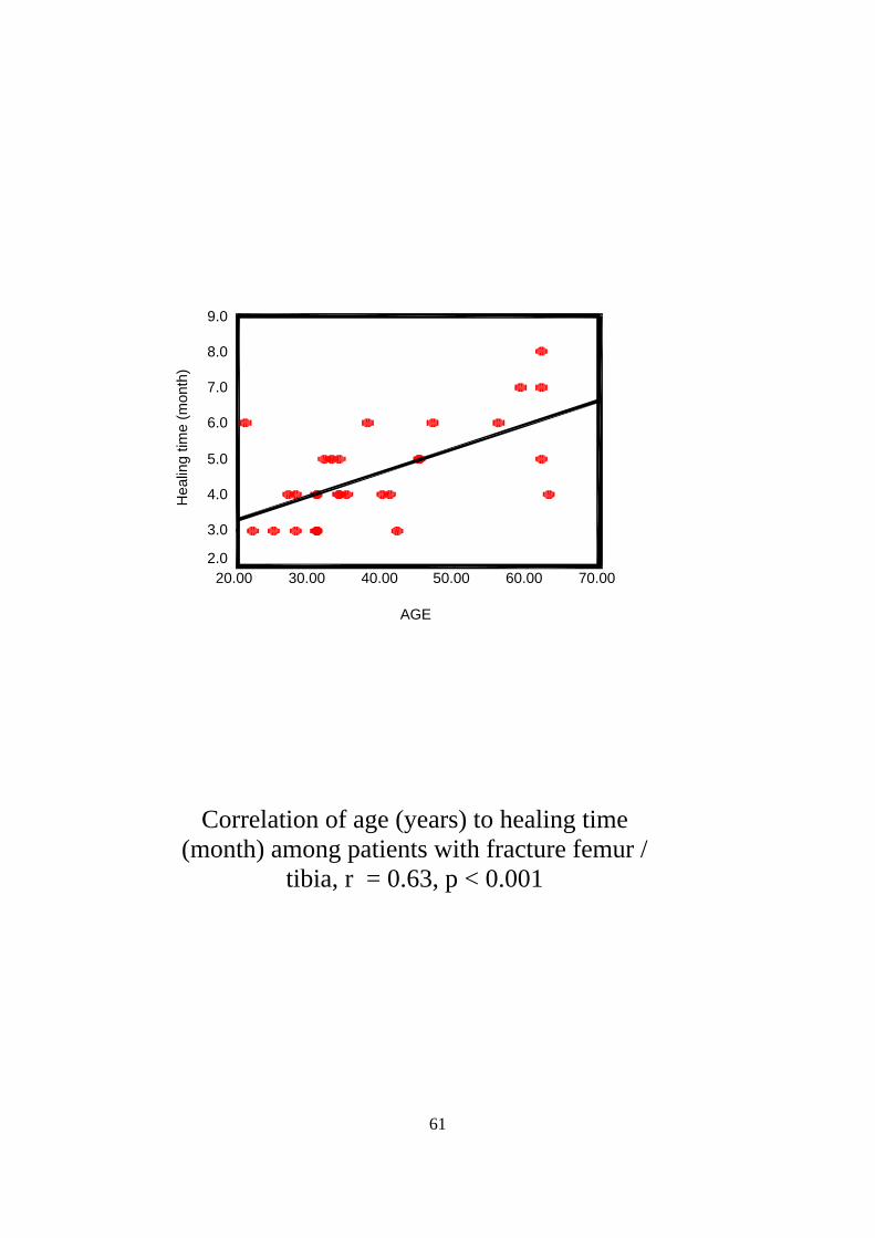
61
Correlation of age (years) to healing time (month) among patients with fracture femur /
tibia, r = 0.63, p < 0.001
AGE
70.0060.0050.0040.0030.0020.00
Hea
ling
time
(mon
th)
9.0
8.0
7.0
6.0
5.0
4.0
3.0
2.0