Antifungal Activity of Amphotericin B Conjugated to Carbon Nanotubes
10
Antifungal Activity of Amphotericin B Conjugated to Carbon Nanotubes Monica Benincasa, † Sabrina Pacor, † Wei Wu, ‡, Maurizio Prato, §, * Alberto Bianco, ‡, * and Renato Gennaro †, * † Department of Life Sciences, University of Trieste, Italy, ‡ CNRS, Institut de Biologie Mole ´culaire et Cellulaire, Laboratoire d’Immunologie et Chimie The ´rapeutiques, Strasbourg, France, and § Department of Pharmaceutical Sciences, University of Trieste, Italy. Current address: Laboratory of Mesoscopic Chemistry and Department of Polymer Science & Engineering, College of Chemistry & Chemical Engineering, Nanjing University, People’s Republic of China. O pportunistic fungal infections are a major problem for public health. The increased number of immunocompromised patients associated with the advent of AIDS, with the increase in the frequency of solid organ and hemato- poietic stem cell transplants and with more aggressive chemotherapy has led to a dra- matic rise in the incidence of systemic my- coses. Amphotericin B (AMB) is considered the first-line therapy for systemic fungal in- fections because of its broad-spectrum anti- fungal activity. For over 50 years, AMB deox- ycholate (AMBD), the conventional colloidal dispersion formulation known as Fungi- zone, has been the treatment of choice for these infections, despite its association with significantly high adverse effects, 1,2 notably severe nephrotoxicity. 3 One reason for this toxicity is the formation of aggregates as a result of its low water solubility. 4 Improved treatment options for inva- sive fungal infections have been devel- oped during the last 15 years. These in- volve both new antifungal agents, such as triazoles and the echinocandins, and less toxic lipid formulations of AMB, which have been added to the arsenal available against fungal infections. Some of the new drugs can now replace AMBD as primary therapy, for example, caspofungin for candidiasis 5 and voriconazole for aspergillosis, 610 while others offer therapeutic options for difficult- to-treat infections, such as posaconazole for zygomycosis, 1113 fusariosis 14,15 and chromoblastomycosis. 16 However, the ex- tensive use of the newer antifungal agents, such as fluconazole, while decreasing the mortality attributed to candidiasis, is result- ing in the selection of drug-resistant strains. 17,18 As for AMB, in the last 1015 years, new formulations have been developed that incorporate the drug into small unila- mellar liposome carriers (AmBisome) to overcome its toxic effects. 19,20 We have re- cently exploited the conjugation of AMB to carbon nanotubes (CNTs), to improve its solubility and decrease its toxic effects, also benefiting the antifungal activity. 21 Investi- gations on the potential of CNTs for bio- medical applications are rapidly expanding, since functionalized CNTs were found to be biocompatible and nontoxic at the cellu- lar level. 2226 In particular, the use of CNTs as carriers for biologically active molecules holds great promises, 27,28 both as an innova- tive drug delivery system 29,30 and for engi- neering new carriers for genes or siRNAs. 31,32 *Address correspondence to [email protected], [email protected], rgennaro@ units.it. Received for review September 9, 2010 and accepted December 01, 2010. Published online December 9, 2010. 10.1021/nn1023522 © 2011 American Chemical Society ABSTRACT Amphotericin B (AMB) has long been considered the most effective drug in the treatment of serious invasive fungal infections. There are, however, major limitations to its use, due to several adverse effects, including acute infusional reactions and, most relevant, a dose-dependent nephrotoxicity. At least some of these effects are attributed to the aggregation of AMB as a result of its poor water solubility. To overcome this problem, reformulated versions of the drug have been developed, including a micellar dispersion of AMB with sodium deoxycholate (AMBD), its encapsulation into liposomes, or its incorporation into lipidic complexes. The development of nanobiotechnologies provides novel potential drug delivery systems that make use of nanomaterials such as functionalized carbon nanotubes (f-CNTs), which are emerging as an innovative and efficient tool for the transport and cellular translocation of therapeutic molecules. In this study, we prepared two conjugates between f-CNTs and AMB. The antifungal activity of these conjugates was tested against a collection of reference and clinical fungal strains, in comparison to that of AMB alone or AMBD. Measured minimum inhibition concentration (MIC) values for f-CNTAMB conjugates were either comparable to or better than those displayed by AMB and AMBD. Furthermore, AMBD-resistant Candida strains were found to be susceptible to f- CNTAMB 1. Additional studies, aimed at understanding the mechanism of action of the conjugates, suggest a nonlytic mechanism, since the compounds show a major permeabilizing effect on the tested fungal strains only after extended incubation. Interestingly, the f-CNTAMB 1 does not show any significant toxic effect on Jurkat cells at antifungal concentrations. KEYWORDS: carbon nanotubes · functionalized carbon nanotubes · amphotericin B · antifungal activity · Candida spp. · cytotoxicity ARTICLE www.acsnano.org VOL. 5 ▪ NO. 1 ▪ 199–208 ▪ 2011 199
Transcript of Antifungal Activity of Amphotericin B Conjugated to Carbon Nanotubes
Antifungal Activity of Amphotericin BConjugated to Carbon NanotubesMonica Benincasa,† Sabrina Pacor,† Wei Wu,‡,� Maurizio Prato,§,* Alberto Bianco,‡,* and Renato Gennaro†,*†Department of Life Sciences, University of Trieste, Italy, ‡CNRS, Institut de Biologie Moleculaire et Cellulaire, Laboratoire d’Immunologie et Chimie Therapeutiques,Strasbourg, France, and §Department of Pharmaceutical Sciences, University of Trieste, Italy. �Current address: Laboratory of Mesoscopic Chemistry and Department ofPolymer Science & Engineering, College of Chemistry & Chemical Engineering, Nanjing University, People’s Republic of China.
Opportunistic fungal infections area major problem for publichealth. The increased number of
immunocompromised patients associated
with the advent of AIDS, with the increase
in the frequency of solid organ and hemato-
poietic stem cell transplants and with more
aggressive chemotherapy has led to a dra-
matic rise in the incidence of systemic my-
coses. Amphotericin B (AMB) is considered
the first-line therapy for systemic fungal in-
fections because of its broad-spectrum anti-
fungal activity. For over 50 years, AMB deox-
ycholate (AMBD), the conventional colloidal
dispersion formulation known as Fungi-
zone, has been the treatment of choice for
these infections, despite its association with
significantly high adverse effects,1,2 notably
severe nephrotoxicity.3 One reason for this
toxicity is the formation of aggregates as a
result of its low water solubility.4
Improved treatment options for inva-
sive fungal infections have been devel-
oped during the last 15 years. These in-
volve both new antifungal agents, such as
triazoles and the echinocandins, and less
toxic lipid formulations of AMB, which have
been added to the arsenal available against
fungal infections. Some of the new drugs
can now replace AMBD as primary therapy,
for example, caspofungin for candidiasis5
and voriconazole for aspergillosis,6�10 while
others offer therapeutic options for difficult-
to-treat infections, such as posaconazole
for zygomycosis,11�13 fusariosis14,15 and
chromoblastomycosis.16 However, the ex-
tensive use of the newer antifungal agents,
such as fluconazole, while decreasing the
mortality attributed to candidiasis, is result-
ing in the selection of drug-resistant
strains.17,18
As for AMB, in the last 10�15 years,new formulations have been developedthat incorporate the drug into small unila-mellar liposome carriers (AmBisome) toovercome its toxic effects.19,20 We have re-cently exploited the conjugation of AMB tocarbon nanotubes (CNTs), to improve itssolubility and decrease its toxic effects, alsobenefiting the antifungal activity.21 Investi-gations on the potential of CNTs for bio-medical applications are rapidly expanding,since functionalized CNTs were found tobe biocompatible and nontoxic at the cellu-lar level.22�26 In particular, the use of CNTsas carriers for biologically active moleculesholds great promises,27,28 both as an innova-tive drug delivery system29,30 and for engi-neering new carriers for genes or siRNAs.31,32
*Address correspondence [email protected],[email protected], rgennaro@ units.it.
Received for review September 9, 2010and accepted December 01, 2010.
Published online December 9, 2010.10.1021/nn1023522
© 2011 American Chemical Society
ABSTRACT Amphotericin B (AMB) has long been considered the most effective drug in the treatment of
serious invasive fungal infections. There are, however, major limitations to its use, due to several adverse effects,
including acute infusional reactions and, most relevant, a dose-dependent nephrotoxicity. At least some of these
effects are attributed to the aggregation of AMB as a result of its poor water solubility. To overcome this problem,
reformulated versions of the drug have been developed, including a micellar dispersion of AMB with sodium
deoxycholate (AMBD), its encapsulation into liposomes, or its incorporation into lipidic complexes. The
development of nanobiotechnologies provides novel potential drug delivery systems that make use of
nanomaterials such as functionalized carbon nanotubes (f-CNTs), which are emerging as an innovative and
efficient tool for the transport and cellular translocation of therapeutic molecules. In this study, we prepared
two conjugates between f-CNTs and AMB. The antifungal activity of these conjugates was tested against a
collection of reference and clinical fungal strains, in comparison to that of AMB alone or AMBD. Measured minimum
inhibition concentration (MIC) values for f-CNT�AMB conjugates were either comparable to or better than those
displayed by AMB and AMBD. Furthermore, AMBD-resistant Candida strains were found to be susceptible to f-
CNT�AMB 1. Additional studies, aimed at understanding the mechanism of action of the conjugates, suggest a
nonlytic mechanism, since the compounds show a major permeabilizing effect on the tested fungal strains only
after extended incubation. Interestingly, the f-CNT�AMB 1 does not show any significant toxic effect on Jurkat
cells at antifungal concentrations.
KEYWORDS: carbon nanotubes · functionalized carbon nanotubes · amphotericinB · antifungal activity · Candida spp. · cytotoxicity
ARTIC
LE
www.acsnano.org VOL. 5 ▪ NO. 1 ▪ 199–208 ▪ 2011 199
In addition, the modulation of molecular functions is
another emerging domain in which f-CNTs might find
potential use.33 In the field of antimicrobial applications,
CNTs have been investigated as supports to induce
pathogen aggregation following an appropriate func-
tionalization with sugar-based ligands recognized by
receptors on the surface of the microorganisms.34�37
This approach is particularly interesting for the design
of new tools for biodefense purposes based on selec-
tive interaction of the nanoscale conjugates with tar-
geted pathogens. Alternatively, the conjugation of anti-
microbials, such as AMB, to CNTs could lead to several
advantages: (i) increased solubility of the molecule,
thereby avoiding aggregation, (ii) a more favorable se-
lectivity for target versus host cells, leading to an im-
proved therapeutic index, and (iii) improved efficacy,
due to a clustering effect and/or to better cellular inter-
nalization capacity of the CNTs.
In this study, we have tested the antifungal activity
of CNT�AMB-conjugates against a number of fungal
reference strains and clinical isolates and compared it
to that of AMB alone or AMBD. A relevant outcome of
this screening is the fact that f-CNT�AMB 1 is active
against Candida strains that are highly resistant to AMB
and AMBD. In addition, the effect of f-CNT�AMB 1and of AMB on the trans-membrane potential and
membrane integrity of selected C. neoformans and C. al-
bicans strains were compared with the aim of identify-
ing possible differences in the mechanism of action.
Finally, we have evaluated the cytotoxicity of the conju-
gate, used at antifungal concentrations, toward Jurkat
cells, a human T-cell leukemia cell line.
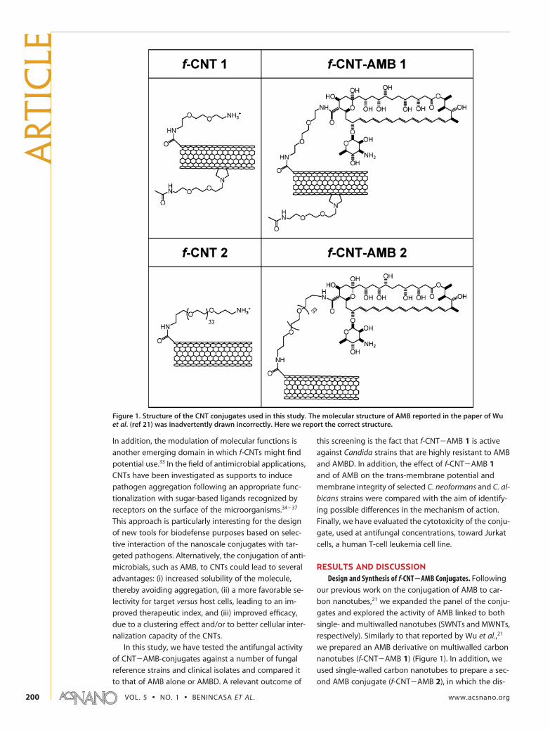
RESULTS AND DISCUSSIONDesign and Synthesis of f-CNT�AMB Conjugates. Following
our previous work on the conjugation of AMB to car-
bon nanotubes,21 we expanded the panel of the conju-
gates and explored the activity of AMB linked to both
single- and multiwalled nanotubes (SWNTs and MWNTs,
respectively). Similarly to that reported by Wu et al.,21
we prepared an AMB derivative on multiwalled carbon
nanotubes (f-CNT�AMB 1) (Figure 1). In addition, we
used single-walled carbon nanotubes to prepare a sec-
ond AMB conjugate (f-CNT�AMB 2), in which the dis-
Figure 1. Structure of the CNT conjugates used in this study. The molecular structure of AMB reported in the paper of Wuet al. (ref 21) was inadvertently drawn incorrectly. Here we report the correct structure.
ART
ICLE
VOL. 5 ▪ NO. 1 ▪ BENINCASA ET AL. www.acsnano.org200
persibility of CNTs was improved by inserting polyethyl-
ene glycol chains. The conjugates 1 and 2 contained,
respectively, 25% and 10% of AMB by weight. f-CNTs
devoid of AMB were used as a control.
Antifungal Activity of the f-CNT�AMB Conjugates. The anti-
fungal activity of the two f-CNT�AMB conjugates and
of their respective controls (structures shown in Figure
1) was first evaluated against 10 strains of Candida spp.
(8 clinical isolates and 2 ATCC reference strains) and 4
strains of C. neoformans (1 clinical isolate and 3 ATCC
reference strains). The activity was compared to that of
AMB alone and of AMBD, the colloidal dispersion for-
mulation in clinical use (Table 1). Antifungal susceptibil-
ity testing was then extended to other fungal species
by including Pichia, Rhodotorula, and Saccharomyces
strains. Several of the tested strains were resistant to
commonly used antifungal drugs including AMB, itra-
conazole, and fluconazole, as indicated in Table 1.
The f-CNT�AMB 1 and 2 conjugates were both
found to display a broad-spectrum antifungal activity,
although with a variable potency against the strains
tested. f-CNT�AMB 1 globally displayed the most po-
tent and broad-spectrum activity, with MIC values lower
than 10 �g/mL against most of the microorganisms
tested. The only exception was the C. famata SA550
strain, which showed a MIC value of 20 �g/mL. The sec-ond conjugate (f-CNT�AMB 2) exhibited a reasonablygood antifungal activity with MIC values ranging from5 to 20 �g/mL for most of the tested strains. Interest-ingly, the two acapsular strains of C. neoformans arefrom 2 to 8 fold more susceptible to the conjugatesthan the parental capsulated strain, indicating that thepolysaccharidic capsule likely constitutes an effectivebarrier for the drug. A similarly increased susceptibilityof the acapsular strain versus the capsulated strain isalso shown by AMB and AMBD (Table 1). No antifungalactivity (MIC �80 �g/mL) was shown by the functional-ized CNTs (f-CNT 1 and 2) devoid of AMB, which wereused as a control (data not shown). The activity of theconjugates is thus not ascribable to a direct action ofthe f-CNTs. In addition, the antifungal activity shown byf-CNT�AMB 1 against some drug-resistant strains sug-gests that the conjugation of AMB to MWNTs favors insome way the activity of this antifungal drug. The ma-jority of the AMB-resistant strains characterized so farhave quantitative or qualitative alterations in the sterolcomposition of their cell membranes;38 it is thus likelythat the AMB conjugated to the nanotubes favors inter-action with residual membrane sterols, in addition tohaving an improved solubility.
Taking into account that the AMB content by weightin the two conjugates is 25% and 10% for f-CNT�AMB1 and 2, respectively, it is clearly evident that the conju-gates are in general significantly more active than AMBand somewhat more active than AMBD (Table 1). In par-ticular, f-CNT�AMB 2 displays MIC values that are fre-quently better than those of AMB alone and compa-rable to those of AMBD, with the notable exception ofC. lusitaniae, C. guillermondii, C. famata, Rhodotorula,and Pichia strains (Table 1). The lower activity off-CNT�AMB 2 against these strains has no explanationat present, although it may depend on a lower drugdensity on the surface of the f-CNTs. The highest activ-ity is displayed by f-CNT�AMB 1, which shows MIC val-ues that are generally better than those of AMBD. Ofnote is the potent activity of this conjugate againststrains resistant to both AMB and AMBD, such as C. albi-cans ATCC 90029 and L21, and C. famata SA550 (Table1).
Killing Kinetics of Fungal Strains by f-CNT�AMB Conjugates.The antifungal activity of f-CNT�AMB 1 and 2 was fur-ther investigated by analyzing their killing kineticsagainst two representative clinical isolates: C. neofor-mans L1 and C. albicans L21. The latter strain was nottested with the f-CNT�AMB 2 and with AMBD as it wasresistant to these compounds (MIC �80 �g/mL). Thef-CNT�AMB 2 did not cause any decrease in the num-ber of viable C. neoformans cells when added at its MICvalue, and caused a reduction of over 90% only at a con-centration 4 times the MIC at 4 h incubation (Figure2A). The f-CNT�AMB 1 conjugate did not cause anycell viability reduction at 2 times its MIC value (2.5 �g/
TABLE 1. Antifungal Activity of f-CNT�AMB ConjugatesCompared to AMB and AMBD
minimum inhibitory concentration (MIC)a (�g/mL)
fungal strain f-CNT�AMB 1 f-CNT�AMB 2 AMB AMBDb
C. neoformans ATCC 90112 2.5 (0.6) 10 (1) 5 1.25C. neoformans ATCC 52816c 1.25 (0.3) 5 (0.5) 1.25 0.3C. neoformans ATCC 52817c 0.3 (0.075) 2.5 (0.25) 0.3 0.3C. neoformans L1 1.25 (0.3) 10 (1) 1.25 0.6C. albicans ATCC 90029 10 (2.5) �80 (�8) �80 �80C. albicans L21d 10 (2.5) �80 (�8) �80 �80C. parapsilosis ATCC 90118 2.5 (0.6) 10 (1) 5 1.25C. parapsilosis L51 2.5 (0.6) 20 (2) 5 1.25C. dubliniensis L70 2.5 (0.6) 20 (2) 1.25 1.25C. tropicalis L42 1.25 (0.3) 20 (2) 2.5 0.6C. lusitaniae 1557VC2 2.5 (0.6) 80 (8) 2.5 1.25C.guillermondii EMAT S 2.5 (0.6) 80 (8) 2.5 0.6C. famata M100d,e 2.5 (0.6) �80 (�8) 10 1.25C. famata SA550d,f 20 (5) �80 (�8) �80 �80R. rubra L8 1.25 (0.3) 80 (8) 2.5 0.6R. rubra ROMA 2.5 (0.6) �80 (�8) 10 1.25P. etchelsii L40 2.5 (0.6) �80 (�8) 5 1.25P. etchelsii L41 2.5 (0.6) �80 (�8) 5 1.25S. cerevisiae 1557VC1 2.5 (0.6) 10 (1) 5 1.25
aThe MIC corresponds to the lowest concentration of compound that inhibited vis-ible growth of fungal cells. Results given are mean values of at least two indepen-dent determinations performed in duplicate. In this table, the MIC values forf-CNT�AMB 1 and 2 refer to the total weight of conjugate (respective AMB con-tent of 25% and 10% by weight). Data in brackets are the MIC values normalizedwith respect to AMB content. bAMBD: AMB deoxycholate. cAcapsular mutants.dResistant to amphotericin B. eResistant to itraconazole. fSusceptible-dose depend-ent to itraconazole and fluconazole Susceptibility of these selected strains to AMBwas also determined using the Etest (AB Biodisk, Solna, Sweden) according to themanufacturer’s instructions. Interpretive breakpoints were reported in ref 41.
ARTIC
LE
www.acsnano.org VOL. 5 ▪ NO. 1 ▪ 199–208 ▪ 2011 201
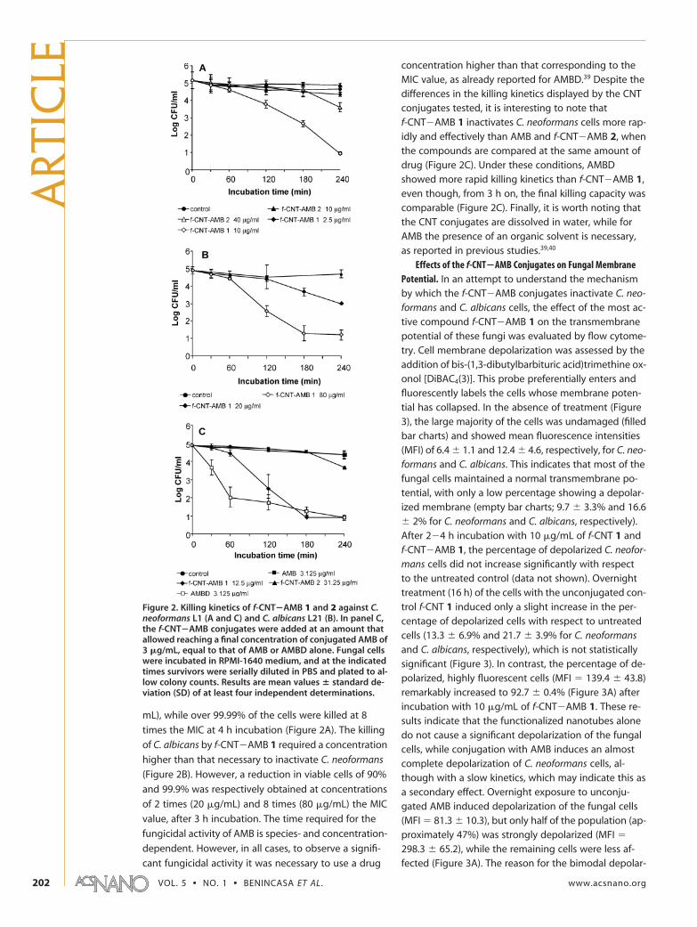
mL), while over 99.99% of the cells were killed at 8
times the MIC at 4 h incubation (Figure 2A). The killing
of C. albicans by f-CNT�AMB 1 required a concentration
higher than that necessary to inactivate C. neoformans
(Figure 2B). However, a reduction in viable cells of 90%
and 99.9% was respectively obtained at concentrations
of 2 times (20 �g/mL) and 8 times (80 �g/mL) the MIC
value, after 3 h incubation. The time required for the
fungicidal activity of AMB is species- and concentration-
dependent. However, in all cases, to observe a signifi-
cant fungicidal activity it was necessary to use a drug
concentration higher than that corresponding to theMIC value, as already reported for AMBD.39 Despite thedifferences in the killing kinetics displayed by the CNTconjugates tested, it is interesting to note thatf-CNT�AMB 1 inactivates C. neoformans cells more rap-idly and effectively than AMB and f-CNT�AMB 2, whenthe compounds are compared at the same amount ofdrug (Figure 2C). Under these conditions, AMBDshowed more rapid killing kinetics than f-CNT�AMB 1,even though, from 3 h on, the final killing capacity wascomparable (Figure 2C). Finally, it is worth noting thatthe CNT conjugates are dissolved in water, while forAMB the presence of an organic solvent is necessary,as reported in previous studies.39,40
Effects of the f-CNT�AMB Conjugates on Fungal MembranePotential. In an attempt to understand the mechanismby which the f-CNT�AMB conjugates inactivate C. neo-formans and C. albicans cells, the effect of the most ac-tive compound f-CNT�AMB 1 on the transmembranepotential of these fungi was evaluated by flow cytome-try. Cell membrane depolarization was assessed by theaddition of bis-(1,3-dibutylbarbituric acid)trimethine ox-onol [DiBAC4(3)]. This probe preferentially enters andfluorescently labels the cells whose membrane poten-tial has collapsed. In the absence of treatment (Figure3), the large majority of the cells was undamaged (filledbar charts) and showed mean fluorescence intensities(MFI) of 6.4 � 1.1 and 12.4 � 4.6, respectively, for C. neo-formans and C. albicans. This indicates that most of thefungal cells maintained a normal transmembrane po-tential, with only a low percentage showing a depolar-ized membrane (empty bar charts; 9.7 � 3.3% and 16.6� 2% for C. neoformans and C. albicans, respectively).After 2�4 h incubation with 10 �g/mL of f-CNT 1 andf-CNT�AMB 1, the percentage of depolarized C. neofor-mans cells did not increase significantly with respectto the untreated control (data not shown). Overnighttreatment (16 h) of the cells with the unconjugated con-trol f-CNT 1 induced only a slight increase in the per-centage of depolarized cells with respect to untreatedcells (13.3 � 6.9% and 21.7 � 3.9% for C. neoformansand C. albicans, respectively), which is not statisticallysignificant (Figure 3). In contrast, the percentage of de-polarized, highly fluorescent cells (MFI � 139.4 � 43.8)remarkably increased to 92.7 � 0.4% (Figure 3A) afterincubation with 10 �g/mL of f-CNT�AMB 1. These re-sults indicate that the functionalized nanotubes alonedo not cause a significant depolarization of the fungalcells, while conjugation with AMB induces an almostcomplete depolarization of C. neoformans cells, al-though with a slow kinetics, which may indicate this asa secondary effect. Overnight exposure to unconju-gated AMB induced depolarization of the fungal cells(MFI � 81.3 � 10.3), but only half of the population (ap-proximately 47%) was strongly depolarized (MFI �
298.3 � 65.2), while the remaining cells were less af-fected (Figure 3A). The reason for the bimodal depolar-
Figure 2. Killing kinetics of f-CNT�AMB 1 and 2 against C.neoformans L1 (A and C) and C. albicans L21 (B). In panel C,the f-CNT�AMB conjugates were added at an amount thatallowed reaching a final concentration of conjugated AMB of3 �g/mL, equal to that of AMB or AMBD alone. Fungal cellswere incubated in RPMI-1640 medium, and at the indicatedtimes survivors were serially diluted in PBS and plated to al-low colony counts. Results are mean values � standard de-viation (SD) of at least four independent determinations.
ART
ICLE
VOL. 5 ▪ NO. 1 ▪ BENINCASA ET AL. www.acsnano.org202
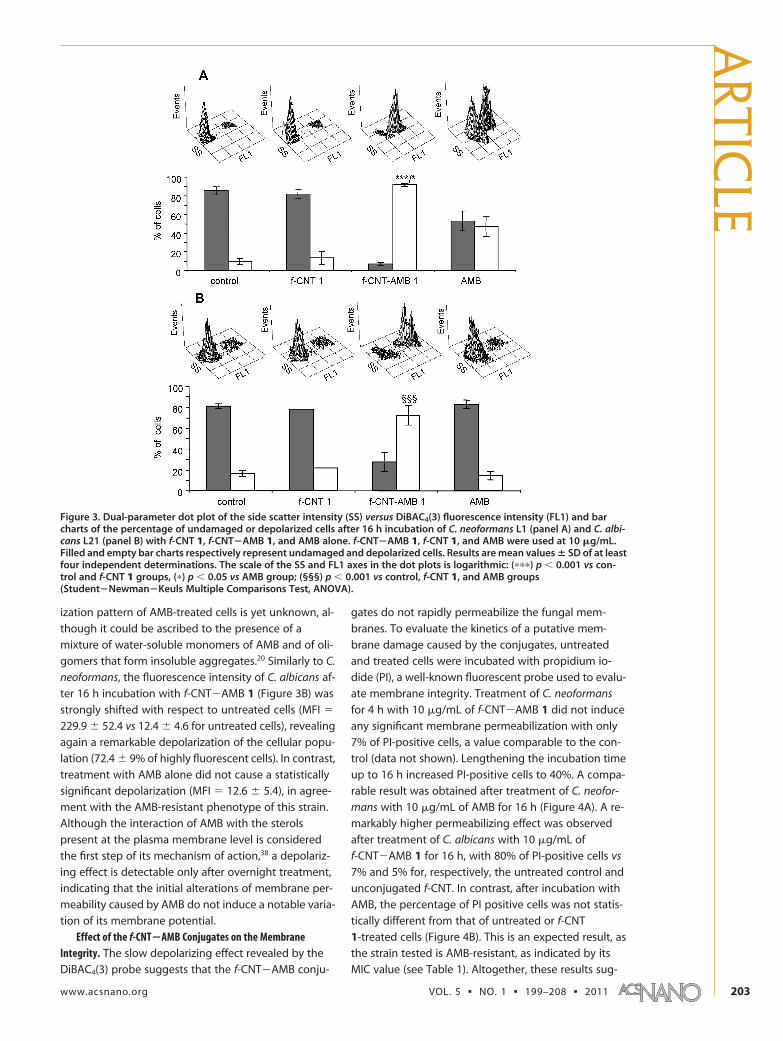
ization pattern of AMB-treated cells is yet unknown, al-
though it could be ascribed to the presence of a
mixture of water-soluble monomers of AMB and of oli-
gomers that form insoluble aggregates.20 Similarly to C.
neoformans, the fluorescence intensity of C. albicans af-
ter 16 h incubation with f-CNT�AMB 1 (Figure 3B) was
strongly shifted with respect to untreated cells (MFI �
229.9 � 52.4 vs 12.4 � 4.6 for untreated cells), revealing
again a remarkable depolarization of the cellular popu-
lation (72.4 � 9% of highly fluorescent cells). In contrast,
treatment with AMB alone did not cause a statistically
significant depolarization (MFI � 12.6 � 5.4), in agree-
ment with the AMB-resistant phenotype of this strain.
Although the interaction of AMB with the sterols
present at the plasma membrane level is considered
the first step of its mechanism of action,38 a depolariz-
ing effect is detectable only after overnight treatment,
indicating that the initial alterations of membrane per-
meability caused by AMB do not induce a notable varia-
tion of its membrane potential.
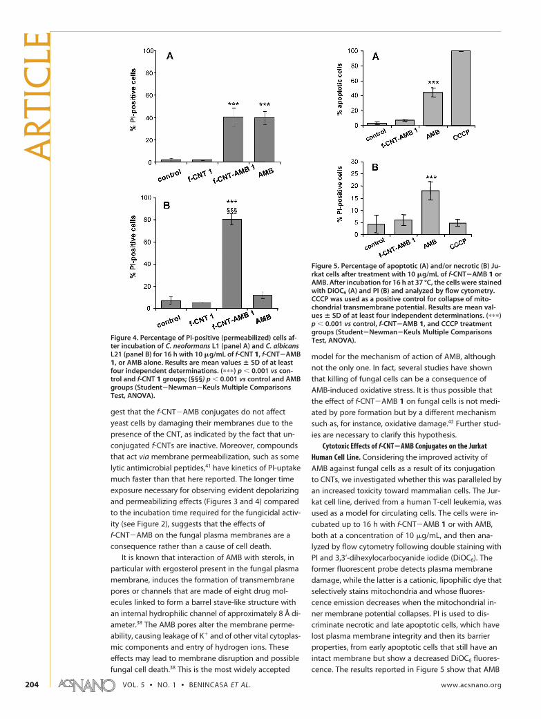
Effect of the f-CNT�AMB Conjugates on the MembraneIntegrity. The slow depolarizing effect revealed by the
DiBAC4(3) probe suggests that the f-CNT�AMB conju-
gates do not rapidly permeabilize the fungal mem-
branes. To evaluate the kinetics of a putative mem-
brane damage caused by the conjugates, untreated
and treated cells were incubated with propidium io-
dide (PI), a well-known fluorescent probe used to evalu-
ate membrane integrity. Treatment of C. neoformans
for 4 h with 10 �g/mL of f-CNT�AMB 1 did not induce
any significant membrane permeabilization with only
7% of PI-positive cells, a value comparable to the con-
trol (data not shown). Lengthening the incubation time
up to 16 h increased PI-positive cells to 40%. A compa-
rable result was obtained after treatment of C. neofor-
mans with 10 �g/mL of AMB for 16 h (Figure 4A). A re-
markably higher permeabilizing effect was observed
after treatment of C. albicans with 10 �g/mL of
f-CNT�AMB 1 for 16 h, with 80% of PI-positive cells vs
7% and 5% for, respectively, the untreated control and
unconjugated f-CNT. In contrast, after incubation with
AMB, the percentage of PI positive cells was not statis-
tically different from that of untreated or f-CNT
1-treated cells (Figure 4B). This is an expected result, as
the strain tested is AMB-resistant, as indicated by its
MIC value (see Table 1). Altogether, these results sug-
Figure 3. Dual-parameter dot plot of the side scatter intensity (SS) versus DiBAC4(3) fluorescence intensity (FL1) and barcharts of the percentage of undamaged or depolarized cells after 16 h incubation of C. neoformans L1 (panel A) and C. albi-cans L21 (panel B) with f-CNT 1, f-CNT�AMB 1, and AMB alone. f-CNT�AMB 1, f-CNT 1, and AMB were used at 10 �g/mL.Filled and empty bar charts respectively represent undamaged and depolarized cells. Results are mean values � SD of at leastfour independent determinations. The scale of the SS and FL1 axes in the dot plots is logarithmic: (���) p � 0.001 vs con-trol and f-CNT 1 groups, (�) p � 0.05 vs AMB group; (§§§) p � 0.001 vs control, f-CNT 1, and AMB groups(Student�Newman�Keuls Multiple Comparisons Test, ANOVA).
ARTIC
LE
www.acsnano.org VOL. 5 ▪ NO. 1 ▪ 199–208 ▪ 2011 203
gest that the f-CNT�AMB conjugates do not affect
yeast cells by damaging their membranes due to the
presence of the CNT, as indicated by the fact that un-
conjugated f-CNTs are inactive. Moreover, compounds
that act via membrane permeabilization, such as some
lytic antimicrobial peptides,41 have kinetics of PI-uptake
much faster than that here reported. The longer time
exposure necessary for observing evident depolarizing
and permeabilizing effects (Figures 3 and 4) compared
to the incubation time required for the fungicidal activ-
ity (see Figure 2), suggests that the effects of
f-CNT�AMB on the fungal plasma membranes are a
consequence rather than a cause of cell death.
It is known that interaction of AMB with sterols, in
particular with ergosterol present in the fungal plasma
membrane, induces the formation of transmembrane
pores or channels that are made of eight drug mol-
ecules linked to form a barrel stave-like structure with
an internal hydrophilic channel of approximately 8 Å di-
ameter.38 The AMB pores alter the membrane perme-
ability, causing leakage of K� and of other vital cytoplas-
mic components and entry of hydrogen ions. These
effects may lead to membrane disruption and possible
fungal cell death.38 This is the most widely accepted
model for the mechanism of action of AMB, although
not the only one. In fact, several studies have shown
that killing of fungal cells can be a consequence of
AMB-induced oxidative stress. It is thus possible that
the effect of f-CNT�AMB 1 on fungal cells is not medi-
ated by pore formation but by a different mechanism
such as, for instance, oxidative damage.42 Further stud-
ies are necessary to clarify this hypothesis.
Cytotoxic Effects of f-CNT�AMB Conjugates on the JurkatHuman Cell Line. Considering the improved activity of
AMB against fungal cells as a result of its conjugation
to CNTs, we investigated whether this was paralleled by
an increased toxicity toward mammalian cells. The Jur-
kat cell line, derived from a human T-cell leukemia, was
used as a model for circulating cells. The cells were in-
cubated up to 16 h with f-CNT�AMB 1 or with AMB,
both at a concentration of 10 �g/mL, and then ana-
lyzed by flow cytometry following double staining with
PI and 3,3’-dihexylocarbocyanide iodide (DiOC6). The
former fluorescent probe detects plasma membranedamage, while the latter is a cationic, lipophilic dye thatselectively stains mitochondria and whose fluores-cence emission decreases when the mitochondrial in-ner membrane potential collapses. PI is used to dis-criminate necrotic and late apoptotic cells, which havelost plasma membrane integrity and then its barrierproperties, from early apoptotic cells that still have anintact membrane but show a decreased DiOC6 fluores-cence. The results reported in Figure 5 show that AMB
Figure 4. Percentage of PI-positive (permeabilized) cells af-ter incubation of C. neoformans L1 (panel A) and C. albicansL21 (panel B) for 16 h with 10 �g/mL of f-CNT 1, f-CNT�AMB1, or AMB alone. Results are mean values � SD of at leastfour independent determinations. (���) p � 0.001 vs con-trol and f-CNT 1 groups; (§§§) p � 0.001 vs control and AMBgroups (Student�Newman�Keuls Multiple ComparisonsTest, ANOVA).
Figure 5. Percentage of apoptotic (A) and/or necrotic (B) Ju-rkat cells after treatment with 10 �g/mL of f-CNT�AMB 1 orAMB. After incubation for 16 h at 37 °C, the cells were stainedwith DiOC6 (A) and PI (B) and analyzed by flow cytometry.CCCP was used as a positive control for collapse of mito-chondrial transmembrane potential. Results are mean val-ues � SD of at least four independent determinations. (���)p � 0.001 vs control, f-CNT�AMB 1, and CCCP treatmentgroups (Student�Newman�Keuls Multiple ComparisonsTest, ANOVA).
ART
ICLE
VOL. 5 ▪ NO. 1 ▪ BENINCASA ET AL. www.acsnano.org204
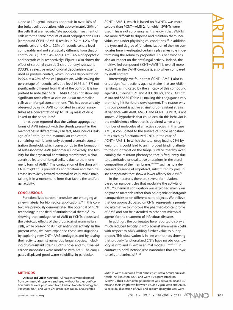
alone at 10 �g/mL induces apoptosis in over 40% ofthe Jurkat cell population, with approximately 20% ofthe cells that are necrotic/late apoptotic. Treatment ofcells with the same amount of AMB conjugated to CNTs(compound f-CNT�AMB 1) results in 7.2 � 1.2% of ap-optotic cells and 6.0 � 2.3% of necrotic cells, a levelcomparable and not statistically different from that ofcontrol cells (3.2 � 1.4% and 4.3 � 3.65% of apoptoticand necrotic cells, respectively). Figure 5 also shows theeffect of carbonyl cyanide 3-chlorophenylhydrazone(CCCP), a selective mitochondrial depolarizing agentused as positive control, which induces depolarizationin 99.6 � 0.28% of the cell population, while leaving thepercentage of necrotic cells at a level (4.74 � 1.37) notsignificantly different from that of the control. It is im-portant to note that f-CNT�AMB 1 does not show anysignificant toxic effect in vitro on Jurkat mammaliancells at antifungal concentrations. This has been alreadyobserved by using AMB conjugated to carbon nano-tubes at a concentration up to 10 �g mass of druglinked to the nanotubes.21
It has been reported that the various aggregationforms of AMB interact with the sterols present in themembranes in different ways. In fact, AMB induces leak-age of K� through the mammalian cholesterol-containing membranes only beyond a certain concen-tration threshold, which corresponds to the formationof self-associated AMB (oligomers). Conversely, the tox-icity for the ergosterol-containing membranes, a char-acteristic feature of fungal cells, is due to the mono-meric form of AMB.20 The conjugation of the drug withCNTs might thus prevent its aggregation and then de-crease its toxicity toward mammalian cells, while main-taining it in a monomeric form that favors the antifun-gal activity.
CONCLUSIONSFunctionalized carbon nanotubes are emerging as
a new material for biomedical applications.43 In this con-text, we previously demonstrated the potential of f-CNTtechnology in the field of antimicrobial therapy21 byshowing that conjugation of AMB to f-CNTs decreasedthe cytotoxic effects of the drug against mammaliancells, while preserving its high antifungal activity. In thepresent work, we have expanded those investigationsby exploring new CNT�AMB conjugates and by testingtheir activity against numerous fungal species, includ-ing drug-resistant strains. Both single- and multiwalledcarbon nanotubes were modified with AMB. The conju-gates displayed good water solubility. In particular,
f-CNT�AMB 1, which is based on MWNTs, was moresoluble than f-CNT�AMB 2, for which SWNTs wereused. This is not surprising, as it is known that SWNTsare more difficult to disperse and maintain them indi-vidualized under physiological conditions.44 In addition,the type and degree of functionalization of the two con-jugates here investigated certainly play a key role in de-termining the solubility properties. This behavior hasalso an impact on the antifungal activity. Indeed, themultiwalled compound f-CNT�AMB 1 is overall moreactive than the SWNT conjugate, also when comparedby AMB content.
Interestingly, we found that f-CNT�AMB 1 also ex-erts a significant activity against strains that are AMB-resistant, as indicated by the efficacy of this compoundagainst C. albicans L21 and ATCC 90029, and C. famataM100 and SA550 (Table 1), making this conjugate a verypromising hit for future development. The reason whythis compound is active against drug-resistant strains,at variance with AMB, AMBD, and f-CNT�AMB 2, is notknown. A hypothesis that could explain this behavior isthe multivalence effect that is obtained when a highnumber of molecules of an active species, in this caseAMB, is conjugated to the surface of single nanostruc-tures such as functionalized CNTs. In the case off-CNT�AMB 1, in which the total drug load is 25% byweight, this could lead to an improved binding affinityto the drug target on the fungal surface, thereby over-coming the resistant phenotype that is frequently dueto quantitative or qualitative alterations in the sterolcomposition of the membrane,38,45,46 such as to a de-creased presence of ergosterol, substituted by precur-sor compounds that show a lower affinity for AMB.47
In the literature, there are several formulationsbased on nanoparticles that modulate the activity ofAMB.48 Chemical conjugation was exploited mainly onpolymeric materials rather than on organic or inorganicnanoparticles or on different nano-objects. We believethat our approach, based on CNTs, represents a promis-ing alternative to improve the pharmacological profileof AMB and can be extended to other antimicrobialagents for the treatment of infectious diseases.
In addition, the conjugates here reported display amuch reduced toxicity in vitro against mammalian cellswith respect to AMB, adding further value to our ap-proach. This observation is in line with others showingthat properly functionalized CNTs have no obvious tox-icity in vitro and in vivo in animal models,21,44,49�52 incontrast to nonfunctionalized nanotubes that are toxicto cells and animals.53�56
METHODSChemicals and Carbon Nanotubes. All reagents were obtained
from commercial suppliers and used without further purifica-tion. SWNTs were purchased from Carbon Nanotechnology Inc.(Houston, USA) and were CNI grade (Lot No. R0496). Purified
MWNTs were purchased from Nanostructured & Amorphous Ma-terials Inc. (Houston, USA) and were 95% pure (stock no.1240XH). Their outer average diameter was between 20 and 30nm and their length was between 0.5 and 2 �m. AMB and AMBD(a colloidal dispersion of AMB and sodium deoxycholate) were
ARTIC
LE
www.acsnano.org VOL. 5 ▪ NO. 1 ▪ 199–208 ▪ 2011 205
purchased from Sigma-Aldrich. AMB was freshly dissolved in10% (v/v) of DMSO and AMBD was dissolved in Milli-Q water.The final concentration of DMSO in the antifungal assays neverexceeded 0.2% (v/v).
Preparation of f-CNT�AMB Conjugates. The MWNTs and the SWNTswere functionalized by slightly modifying a previously describedprocedure21,44 to obtain f-CNT 1 and f-CNT 2, respectively (Fig-ure 1) (see Supporting Information for experimental details).These two precursors were then reacted as reported by Wuet al.21 with a solution of Fmoc�AMB�OH to obtain the respec-tive conjugates f-CNT�AMB 1 and f-CNT�AMB 2 (Figure 1). Thetwo derivatives differ by the type of nanotube support (MWNTsand SWNTs, respectively, for f-CNT 1 and f-CNT 2), the length ofthe PEG chain linking AMB to the CNTs, and the degree of sub-stitution with the drug. The conjugate 1 has a TEG linker and anAMB content of 25% by weight. The conjugate 2 contains aPEG linker, with a repeat of 33 monomers of oxyethylene, andan AMB content of 10% by weight. In addition, f-CNT�AMB 1contains acetylated TEG groups to increase water solubility (2%by weight). Both CNT conjugates were dissolved in Milli-Q waterand stored at �20 °C. The compounds were characterized bytransmission electron microscopy (TEM) performed on a HitachiH600 microscope, working at different accelerating voltage andmagnification.
Fungal Strains and Growth Conditions. Several different clinical iso-lates and ATCC reference fungal strains were used in this study.The former included Candida spp., Cryptococcus neoformans,Rhodotorula rubra, Saccharomyces cerevisiae, and Pichia etchellsii,and were mostly collected from immuno-compromised pa-tients. The latter comprised the capsulated ATCC 90112 strainof Cryptococcus neoformans as well as the acapsular mutantsATCC 52816 and ATCC 52817, Candida albicans ATCC 90029, andCandida parapsilosis ATCC 90018. Fungi were grown on Sab-ouraud agar plates at 30 °C for 48 h. Inocula were prepared bypicking and suspending five colonies in 5 mL of sterile bufferedsaline solution containing 10 mM sodium phosphate and 145mM NaCl, pH 7.4 (phosphate-buffered saline, PBS). The turbidityof the fungal suspensions was measured at 600 nm and was ad-justed to obtain the appropriate inoculum according to previ-ously derived curves relating the number of colony forming units(CFUs) with absorbance.
Antifungal Activity. The antifungal activity of f-CNT�AMB con-jugates, their controls devoid of AMB, AMB alone, and AMBD(see Table 1) was evaluated by the broth microdilution suscepti-bility test performed according to the guidelines of the Clinicaland Laboratory Standards Institute (CLSI), formerly NationalCommittee for Clinical Laboratory Standards, to determine theminimum inhibitory concentration (MIC) values. 2-Fold serial di-lutions of each compound were prepared in 96-well polypropy-lene microtiter plates (Sarstedt, Germany) in RPMI-1640 medium(Sigma-Aldrich) to a final volume of 50 �L. Each series includeda well without addition of any compound, as a growth control. Atotal of 50 �L of the adjusted inoculum, diluted in RPMI-1640medium, was then added to each well to obtain a final concen-tration of approximately 5 � 104 CFUs/mL. Samples were incu-bated at 30 °C for 48 h. The MIC value was taken as the lowestconcentration of antifungal agent resulting in the complete inhi-bition of visible fungal growth after 48 h incubation. Data arethe mean of up to six independent determinations performedin duplicate with values differing by one dilution at the most.
Killing Kinetics Assays. Killing kinetics were determined usingcultures of C. albicans L21 and C. neoformans L1 diluted in freshRPMI-1640 medium to give approximately 5 � 104 CFUs/mL.f-CNT�AMB conjugates were added at different concentrationsand the suspensions were then incubated in a shaking waterbath at 30 °C. At the indicated times, an aliquot of each samplewas removed, serially diluted with PBS, and plated in duplicateon Sabouraud agar. The number of colonies was counted afterincubation of the plates for 48 h at 30 °C. Data are the mean of atleast four independent determinations with comparable results.
Evaluation of Alteration of the Fungal Membrane by Flow CytometricAssays. Specific flow cytometric assays were used to evaluate thetransmembrane potential and the membrane permeabilizationof fungal cells treated with the f-CNT�AMB conjugates. Forthese analyses, C. albicans L21 and C. neoformans L1, subcul-
tured on Sabouraud agar, were diluted in RPMI-1640 mediumto 1 � 105 CFUs/mL. Aliquots of the fungal suspension were thenincubated with or without the f-CNT-conjugates for 2�16 h at30 °C. At the end of the incubation time, the fungal suspensionswere incubated in the dark for 60 min at 30 °C with DiBAC4(3) (In-vitrogen Co., Carlsbad, CA) at a final concentration of 1 �M, toevaluate modifications of the transmembrane potential. Tomonitor alterations in membrane integrity following treatmentwith the CNT-derivatives, a filtered solution of PI (Sigma-Aldrich),at a final concentration of 10 �g/mL, was incubated for 60 minat 30 °C with each sample. An untreated sample of fungal cells,pelleted and resuspended in cold absolute ethanol for 30 min at�20 °C was used as a positive control of permeabilization. Aftercentrifugation at 1000g for 10 min, the ethanol was removed byaspiration, the pellet was suspended in RPMI-1640 medium,and the PI was added as reported above. The fluorescence inten-sity was detected with a Cytomics FC 500 instrument (Beckman-Coulter, Inc., Fullerton, CA) equipped with an argon laser (488nm, 5 mW) and using a photomultiplier tube fluorescence detec-tor for green (525 nm) or red (610 nm) filtered light. The detec-tors were set on logarithmic amplification. Optical and electronicnoise were eliminated by setting an electronic gating thresholdon the forward scattering detector, while the flow rate was keptat a data rate below 200 events/second to avoid cell coincidence.For each sample, at least 10000 events were acquired and storedas list mode files.
All the experiments with the fluorescent probes were con-ducted in triplicate and the data analyses were performed withthe WinMDI software (Dr J. Trotter, Scripps Research Institute, LaJolla, CA, USA). Data are expressed as mean � SD.
Cell Culture and Cytotoxicity Assay. The Jurkat human T-lymphomacell line was cultured in RPMI-1640 (Cambrex Bioscience) supple-mented with gentamicin (10 �g/mL) and 10% of heat-inactivated fetal bovine serum. Cells were grown in suspensionat 37 °C in a humidified atmosphere in the presence of 5% CO2.For the tests, cell suspensions were prepared at a final density of5 � 105 cells/mL in 2 mL of medium containing f-CNT�AMB 1or AMB at a final concentration of 10 �g/mL. After addition ofAMB, the final percentage of DMSO in the cell cultures was al-ways lower than 0.1% (v/v). The cells were then incubated at 37°C up to 16 h. To evaluate apoptosis induction, at the end of theincubation time, the cells were stained in the dark at 37 °C for15 min with 50 nM of DiOC6 (FluoProbes, Interchim, MontluconCedex, France), a fluorescent probe used to measure the mito-chondrial transmembrane potential in intact cells. A positive con-trol for collapse of mitochondrial transmembrane potential wasobtained by incubating a cell suspension at 37 °C with 50 �M ofthe uncoupler CCCP for 15 min. After treatment with DiOC6, totest cell viability, the samples were washed twice with 2 mL ofPBS and then stained in the dark at room temperature with 10�g/mL PI for 10 min. Cells were analyzed by flow cytometry asdescribed above, acquiring at least 10000 events for each sampleand storing the results as list mode files.
Statistical Analysis. Data are expressed as mean � SD. Signifi-cance of differences among groups was assessed by using theprogram Instat (GraphPad Software Inc., San Diego, CA) and per-formed by an analysis of variance between groups (ANOVA) fol-lowed by the Student�Newman�Keuls post test. Values of p �0.05 were considered statistically significant.
Acknowledgment. This work was supported by the CNRS, theUniversity of Trieste, INSTM, MIUR (Cofin Prot. 20085M27SS andFirb RBIN04HC3S) and Regione Friuli Venezia-Giulia. W.W. isgrateful to French Ministry for Research and New Technologiesfor a postdoctoral fellowship. TEM images were recorded at theRIO Microscopy Facility Platform of Esplanade (Strasbourg,France). We wish to thank Cecilia Menard-Moyon for her helpwith TEM.
Supporting Information Available: Detailed experimental pro-cedure for the preparation of f-CNT�AmB conjugates. This ma-terial is available free of charge via the Internet at http://pubs.acs.org.
ART
ICLE
VOL. 5 ▪ NO. 1 ▪ BENINCASA ET AL. www.acsnano.org206
REFERENCES AND NOTES1. Petrikkos, G.; Skiada, A. Recent Advances in Antifungal
Chemotherapy. Int. J. Antimicrob. Agents. 2007, 30,108–117.
2. Burke, D.; Lal, R.; Finkel, K. W.; Samuels, J.; Foringer, J. R.Acute Amphotericin B Overdose. Ann. Pharmacother.2006, 40, 2254–2259.
3. Slavin, M. A.; Szer, J.; Grigg, A. P.; Roberts, A. W.; Seymour,J. F.; Sasadeusz, J.; Thursky, K.; Chen, S. C.; Morrissey, C. O.;Heath, C. H.; et al. Guidelines for the Use of AntifungalAgents in the Treatment of Invasive Candida and MouldInfections. Intern. Med. J. 2004, 34, 192–200.
4. Charvalos, E.; Tzatzarakis, M. N.; Van Bambeke, F.; Tulkens,P. M.; Tsatsakis, A. M.; Tzanakakis, G. N.; Mingeot-Leclercq,M. P. Water-Soluble Amphotericin B�PolyvinylpyrrolidoneComplexes with Maintained Antifungal Activity againstCandida spp. and Aspergillus spp. and Reduced Haemolyticand Cytotoxic Effects. J. Antimicrob. Chemother. 2006, 57,236–244.
5. Falagas, M. E.; Ntziora, F.; Betsi, G. I.; Samonis, G.Caspofungin for the Treatment of Fungal Infections: ASystematic Review of Randomized Controlled Trials. Int. J.Antimicrob. Agents. 2007, 29, 136–143.
6. Jain, L. R.; Denning, D. W. The Efficacy and Tolerability ofVoriconazole in the Treatment of Chronic CavitaryPulmonary Aspergillosis. J. Infect. 2006, 52, 133–137.
7. Kirkpatrick, W. R.; Coco, B. J.; Patterson, T. F. Sequential orCombination Antifungal Therapy with Voriconazole andLiposomal Amphotericin B in a Guinea Pig Model ofInvasive Aspergillosis. Antimicrob. Agents Chemother. 2006,50, 1567–1569.
8. Klein, K. C.; Blackwood, R. A. Topical Voriconazole Solutionfor Cutaneous Aspergillosis in a Pediatric Patient afterBone Marrow Transplant. Pediatrics 2006, 118, 506–508.
9. Sambatakou, H.; Dupont, B.; Lode, H.; Denning, D. W.Voriconazole Treatment for Subacute Invasive and ChronicPulmonary Aspergillosis. Am. J. Med. 2006, 119, 517–524.
10. Singh, N.; Limaye, A. P.; Forrest, G.; Safdar, N.; Munoz, P.;Pursell, K.; Houston, S.; Rosso, F.; Montoya, J. G.; Patton, P.;et al. Combination of Voriconazole and Caspofungin asPrimary Therapy for Invasive Aspergillosis in Solid OrganTransplant Recipients: A Prospective, Multicenter,Observational Study. Transplantation 2006, 81, 320–326.
11. Barchiesi, F.; Spreghini, E.; Santinelli, A.; Fothergill, A. W.;Pisa, E.; Giannini, D.; Rinaldi, M. G.; Scalise, G. PosaconazoleProphylaxis in Experimental Systemic Zygomycosis.Antimicrob. Agents Chemother. 2007, 51, 73–77.
12. Greenberg, R. N.; Mullane, K.; van Burik, J. A.; Raad, I.;Abzug, M. J.; Anstead, G.; Herbrecht, R.; Langston, A.; Marr,K. A.; Schiller, G.; et al. Posaconazole as Salvage Therapyfor Zygomycosis. Antimicrob. Agents Chemother. 2006, 50,126–133.
13. van Burik, J. A.; Hare, R. S.; Solomon, H. F.; Corrado, M. L.;Kontoyiannis, D. P. Posaconazole Is Effective as SalvageTherapy in Zygomycosis: A Retrospective Summary of 91Cases. Clin. Infect. Dis. 2006, 42, 61–65.
14. Gupta, S.; Almyroudis, N. G.; Battiwalla, M.; Bambach, B. J.;McCarthy, P. L.; Proefrock, A. D.; Ball, D.; Paplham, P.;Varma, A.; Kwon-Chung, J.; et al. Successful Treatment ofDisseminated Fusariosis with Posaconazole duringNeutropenia and Subsequent Allogeneic HematopoieticStem Cell Transplantation. Transplant Infect. Dis. 2007, 9,156–160.
15. Raad, I. I.; Hachem, R. Y.; Herbrecht, R.; Graybill, J. R.; Hare,R.; Corcoran, G.; Kontoyiannis, D. P. Posaconazole asSalvage Treatment for Invasive Fusariosis in Patients withUnderlying Hematologic Malignancy and OtherConditions. Clin. Infect. Dis. 2006, 42, 1398–1403.
16. Negroni, R.; Tobon, A.; Bustamante, B.; Shikanai-Yasuda,M. A.; Patino, H.; Restrepo, A. Posaconazole Treatment ofRefractory Eumycetoma and Chromoblastomycosis. Rev.Inst. Med. Trop. Sao Paulo. 2005, 47, 339–346.
17. Andes, D.; Forrest, A.; Lepak, A.; Nett, J.; Marchillo, K.;Lincoln, L. Impact of Antimicrobial Dosing Regimen onEvolution of Drug Resistance In Vivo: Fluconazole and
Candida albicans. Antimicrob. Agents Chemother. 2006, 50,2374–2383.
18. Andes, D.; Lepak, A.; Nett, J.; Lincoln, L.; Marchillo, K. InVivo Fluconazole Pharmacodynamics and ResistanceDevelopment in a Previously Susceptible Candida albicansPopulation Examined by Microbiologic and TranscriptionalProfiling. Antimicrob. Agents Chemother. 2006, 50,2384–2394.
19. Adler-Moore, J.; Proffitt, R. T. AmBisome: LiposomalFormulation, Structure, Mechanism of Action and Pre-Clinical Experience. J. Antimicrob. Chemother. 2002, 49, 21–30.
20. Torrado, J. J.; Espada, R.; Ballesteros, M. P.; Torrado-Santiago, S. Amphotericin B Formulations and DrugTargeting. J. Pharm. Sci. 2008, 97, 2405–2425.
21. Wu, W.; Wieckowski, S.; Pastorin, G.; Benincasa, M.;Klumpp, C.; Briand, J. P.; Gennaro, R.; Prato, M.; Bianco, A.Targeted Delivery of Amphotericin B to Cells by UsingFunctionalized Carbon Nanotubes. Angew. Chem., Int. Ed.2005, 44, 6358–6362.
22. Lacerda, L.; Bianco, A.; Prato, M.; Kostarelos, K. CarbonNanotubes as Nanomedicines: From Toxicology toPharmacology. Adv. Drug Delivery Rev. 2006, 58,1460–1470.
23. Bianco, A.; Kostarelos, K.; Prato, M. Applications of CarbonNanotubes in Drug Delivery. Curr. Opin. Chem. Biol. 2005,9, 674–679.
24. Bianco, A.; Kostarelos, K.; Partidos, C. D.; Prato, M.Biomedical Applications of Functionalised CarbonNanotubes. Chem. Commun. (Cambridge). 2005, 571–577.
25. Lin, Y.; Taylor, S.; Li, H.; Fernando, K. A. S.; Qu, L.; Wang, W.;Gu, L.; Zhou, B.; Sun, Y.-P. Advances towardBioapplications of Carbon Nanotubes. J. Mater. Chem.2004, 14, 527–541.
26. Kam, N. W.; Liu, Z.; Dai, H. Carbon Nanotubes asIntracellular Transporters for Proteins and DNA: AnInvestigation of the Uptake Mechanism and Pathway.Angew. Chem., Int. Ed. 2006, 45, 577–581.
27. Kostarelos, K.; Bianco, A.; Prato, M. Promises, Facts andChallenges for Carbon Nanotubes in Imaging andTherapeutics. Nat. Nanotechnol. 2009, 4, 627–633.
28. Menard-Moyon, C., V. E.; Fabbro, C.; Samorı, C.; Da Ros, T.;Kostarelos, K.; Prato, M.; Bianco, A. The Alluring Potentialof Functionalized Carbon Nanotubes in Drug Discovery.Expert Opin. Drug Delivery 2010, 5, 691–707.
29. Prato, M.; Kostarelos, K.; Bianco, A. Functionalized CarbonNanotubes in Drug Design and Discovery. Acc. Chem. Res.2008, 41, 60–68.
30. Ilbasmis-Tamer, S.; Yilmaz, S.; Banoglu, E.; Degim, I. T.Carbon Nanotubes to Deliver Drug Molecules. J. Biomed.Nanotechnol. 2010, 6, 20–27.
31. Pantarotto, D.; Singh, R.; McCarthy, D.; Erhardt, M.; Briand,J. P.; Prato, M.; Kostarelos, K.; Bianco, A. FunctionalizedCarbon Nanotubes for Plasmid DNA Gene Delivery.Angew. Chem. Int. Ed. 2004, 43, 5242–5246.
32. Herrero, M. A.; Toma, F. M.; Al-Jamal, K. T.; Kostarelos, K.;Bianco, A.; Da Ros, T.; Bano, F.; Casalis, L.; Scoles, G.; Prato,M. Synthesis and Characterization of a Carbon Nanotube�Dendron Series for Efficient siRNA Delivery. J. Am. Chem.Soc. 2009, 131, 9843–9848.
33. Menard-Moyon, C.; Kostarelos, K.; Prato, M.; Bianco, A.Functionalized Carbon Nanotubes for Probing andModulating Molecular Functions. Chem. Biol. 2010, 17,107–115.
34. Luo, P. G.; Wang, H.; Gu, L.; Lu, F.; Lin, Y.; Christensen, K. A.;Yang, S. T.; Sun, Y. P. Selective Interactions of Sugar-Functionalized Single-Walled Carbon Nanotubes withBacillus Spores. ACS Nano 2009, 3, 3909–3916.
35. Wang, H.; Gu, L.; Lin, Y.; Lu, F.; Meziani, M. J.; Luo, P. G.;Wang, W.; Cao, L.; Sun, Y. P. Unique Aggregation ofAnthrax (Bacillus anthracis) Spores by Sugar-CoatedSingle-Walled Carbon Nanotubes. J. Am. Chem. Soc. 2006,128, 13364–13365.
ARTIC
LE
www.acsnano.org VOL. 5 ▪ NO. 1 ▪ 199–208 ▪ 2011 207
36. Elkin, T.; Jiang, X.; Taylor, S.; Lin, Y.; Gu, L.; Yang, H.; Brown,J.; Collins, S.; Sun, Y. P. Immuno-Carbon Nanotubes andRecognition of Pathogens. Chembiochem. 2005, 6,640–643.
37. Gu, L.; Elkin, T.; Jiang, X.; Li, H.; Lin, Y.; Qu, L.; Tzeng, T. R.;Joseph, R.; Sun, Y. P. Single-Walled Carbon NanotubesDisplaying Multivalent Ligands for Capturing Pathogens.Chem. Commun. (Cambridge) 2005, 874–876.
38. O’Shaughnessy, E. M.; Lyman, C. A.; Walsh, T. J.Amphotericin B: Polyene Resistance Mechanisms. InAntimicrobial Drug Resistance; Georgiev, V. S., Ed.; HumanaPress: Totowa, NJ, 2009; pp 295�305.
39. Canton, E.; Peman, J.; Gobernado, M.; Viudes, A.; Espinel-Ingroff, A. Patterns of Amphotericin B Killing Kineticsagainst Seven Candida Species. Antimicrob. AgentsChemother. 2004, 48, 2477–2482.
40. Canton, E.; Peman, J.; Sastre, M.; Romero, M.; Espinel-Ingroff, A. Killing Kinetics of Caspofungin, Micafungin, andAmphotericin B against Candida guilliermondii. Antimicrob.Agents Chemother. 2006, 50, 2829–2832.
41. Benincasa, M.; Scocchi, M.; Pacor, S.; Tossi, A.; Nobili, D.;Basaglia, G.; Busetti, M.; Gennaro, R. Fungicidal Activity ofFive Cathelicidin Peptides against Clinically IsolatedYeasts. J. Antimicrob. Chemother. 2006, 58, 950–959.
42. Sokol-Anderson, M. L.; Brajtburg, J.; Medoff, G.Amphotericin B-Induced Oxidative Damage and Killing ofCandida albicans. J. Infect. Dis. 1986, 154, 76–83.
43. Escorcia, F. E.; McDevitt, M. R.; Villa, C. H.; Scheinberg, D. A.Targeted Nanomaterials for Radiotherapy. Nanomedicine2007, 2, 805–815.
44. Dumortier, H.; Lacotte, S.; Pastorin, G.; Marega, R.; Wu, W.;Bonifazi, D.; Briand, J. P.; Prato, M.; Muller, S.; Bianco, A.Functionalized Carbon Nanotubes Are Non-cytotoxic andPreserve the Functionality of Primary Immune Cells. NanoLett. 2006, 6, 1522–1528.
45. Hitchcock, C. A.; Barrett-Bee, K. J.; Russell, N. J. The LipidComposition and Permeability to Azole of an Azole- andPolyene-Resistant Mutant of Candida albicans. J. Med. Vet.Mycol. 1987, 25, 29–37.
46. Subden, R. E.; Safe, L.; Morris, D. C.; Brown, R. G.; Safe, S.Eburicol, Lichesterol, Ergosterol, and Obtusifoliol fromPolyene Antibiotic-Resistant Mutants of Candida albicans.Can. J. Microbiol. 1977, 23, 751–754.
47. Ghannoum, M. A.; Rice, L. B. Antifungal Agents: Mode ofAction, Mechanisms of Resistance, and Correlation ofThese Mechanisms with Bacterial Resistance. Clin.Microbiol. Rev. 1999, 12, 501–517.
48. Vyas, S. P.; Gupta, S. Optimizing Efficacy of Amphotericin Bthrough Nanomodification. Int. J. Nanomedicine. 2006, 1,417–432.
49. Liu, Z.; Tabakman, S.; Welsher, K.; Dai, H. CarbonNanotubes in Biology and Medicine: In Vitro and in VivoDetection, Imaging, and Drug Delivery. Nano Res. 2009, 2,85–120.
50. Liu, Z.; Davis, C.; Cai, W.; He, L.; Chen, X.; Dai, H. Circulationand Long-Term Fate of Functionalized, BiocompatibleSingle-Walled Carbon Nanotubes in Mice Probed byRaman Spectroscopy. Proc. Natl. Acad. Sci. U.S.A. 2008, 105,1410–1415.
51. Schipper, M. L.; Nakayama-Ratchford, N.; Davis, C. R.; Kam,N. W.; Chu, P.; Liu, Z.; Sun, X.; Dai, H.; Gambhir, S. S. A PilotToxicology Study of Single-Walled Carbon Nanotubes in aSmall Sample of Mice. Nat. Nanotechnol. 2008, 3, 216–221.
52. Sayes, C. M.; Liang, F.; Hudson, J. L.; Mendez, J.; Guo, W.;Beach, J. M.; Moore, V. C.; Doyle, C. D.; West, J. L.; Billups,W. E.; et al. Functionalization Density Dependence ofSingle-Walled Carbon Nanotubes Cytotoxicity. In Vitro.Toxicol. Lett. 2006, 161, 135–142.
53. Cui, D.; Tian, F.; Ozkan, C. S.; Wang, M.; Gao, H. Effect ofSingle Wall Carbon Nanotubes on Human HEK293 Cells.Toxicol. Lett. 2005, 155, 73–85.
54. Lam, C. W.; James, J. T.; McCluskey, R.; Hunter, R. L.Pulmonary Toxicity of Single-Wall Carbon Nanotubes inMice 7 and 90 Days after Intratracheal Instillation. Toxicol.Sci. 2004, 77, 126–134.
55. Warheit, D. B.; Laurence, B. R.; Reed, K. L.; Roach, D. H.;Reynolds, G. A.; Webb, T. R. Comparative PulmonaryToxicity Assessment of Single-Wall Carbon Nanotubes inRats. Toxicol. Sci. 2004, 77, 117–125.
56. Poland, C. A.; Duffin, R.; Kinloch, I.; Maynard, A.; Wallace,W. A.; Seaton, A.; Stone, V.; Brown, S.; Macnee, W.;Donaldson, K. Carbon Nanotubes Introduced into theAbdominal Cavity of Mice Show Asbestos-likePathogenicity in a Pilot Study. Nat. Nanotechnol. 2008, 3,423–428.
ART
ICLE
VOL. 5 ▪ NO. 1 ▪ BENINCASA ET AL. www.acsnano.org208
![Pharmacologically active boranes*old.iupac.org/publications/pac/2006/pdf/7807x1425.pdf · 2017. 7. 24. · teresting pharmacological activity such as antifungal [21], peptidomimetic](https://static.fdocuments.fr/doc/165x107/60d89ee659f0b108862ebb5e/pharmacologically-active-boranesoldiupacorgpublicationspac2006pdf-2017.jpg)