ANTIFUNGAL ACTIVITY AND MOLECULAR IDENTIFICATION OF …
16
133 ANTIFUNGAL ACTIVITY AND MOLECULAR IDENTIFICATION OF NATIVE STRAINS OF Bacillus subtilis ACTIVIDAD ANTIFUNGICA E IDENTIFICACION MOLECULAR DE CEPAS NATIVAS DE Bacillus subtilis Esaú Ruiz-Sánchez 1 , Miguel Ángel. Mejía-Bautista 1 , Alejandra Serrato-Díaz 2 , Arturo Reyes-Ramírez 1 , Yokiushirdhilgilmara Estrada-Girón 3 , Alberto J. Valencia-Botín 4* * Autor responsable v Author for correspondence. Recibido: septiembre, 2014. Aprobado: noviembre, 2015. Publicado como ARTÍCULO en Agrociencia 50: 133-148. 2016. 1 División de Estudios de Postgrado e Investigación, Instituto Tecnológico de Conkal, Conkal, Yucatán, México. 2 Universidad Autónoma Metropolitana Iztapalapa, Laboratorio Divisional de Biología Molecular, División de Ciencias Biológicas y de la Salud, Rafael Atlixco 186, Colonia Vicentina, México. 3 Universidad de Guadalajara, Centro Universitario de Ciencias Exactas e Ingenierías, Bulevard Marcelino García Barragán 1421, Colonia Olímpica, Guadalajara, Jalisco, 44430 México. 4 Universidad de Guadalajara, Centro Universitario de la Ciénega. Avenida Universidad 1115, Colonia Lindavista, Ocotlán, Jalisco, 47810, México. ([email protected]). ABSTRACT e bioprospection for antagonistic bacteria, as biological control agents to manage plant pathogenic fungi, is a preponderant activity to reduce the use of chemical fungicides. e bacterium Bacillus subtilis has shown an excellent capacity to inhibit the growth and sporulation of a wide variety of plant pathogenic fungi. e phenotypic and biochemical characteristics to differentiate among subspecies of B. subtilis vary at the regional level. In this research, Bacillus strains were isolated from the Yucatan Peninsula in Mexico and their antagonistic activities were assessed by direct confrontation in a completely randomized design. To identify the most active strains, molecular characterization was performed. For an accurate identification, partial 16S rRNA, gyrA and rpoB genes were sequenced and compared with those deposited in the GenBank. e most active strains CBCC2, CBMT2, CBMN22, CBRF24, CBCK36, CBCK47, and CBMT51 caused 32 to 78 % inhibition of mycelial growth in Alternaria alternata, Helminthosporium rostratum and Curvularia lunata in direct confrontation tests; likewise the cell-free culture filtrates of these Bacillus strains showed significant differences (p£0.05) in the degree of inhibition of conidial germination of the plant pathogenic fungi. e amplification, sequencing and comparison of 16S rRNA, gyrA and rpoB genes showed that the strains had 95 to 100 % identity with Bacillus subtilis subsp. inaquosorum and B. subtilis subsp. subtilis. e gyrA gene showed the RESUMEN La bioprospección de bacteria antagónicas, como agentes de control biológico para el manejo de hongos fitopatógenos, es una actividad preponderante para reducir el uso de fun- gicidas químicos. La bacteria Bacillus subtilis ha mostrado una excelente capacidad para inhibir el crecimiento y espo- rulación de una gama amplia de hongos fitopatógenos. Las características bioquímicas para diferenciar entre subespecies de B. subtilis varían a nivel regional. En esta investigación se aislaron cepas de Bacillus de la Península de Yucatán en México y se evaluaron sus actividades antagónicas por con- frontación directa en un diseño completamente al azar. Para identificar las cepas más activas se realizó una caracterización molecular. Para una identificación precisa se secuenciaron parte de los genes 16S rRNA, gyrA y rpoB y se compararon con los depositados en el GenBank. Las cepas más activas CBCC2, CBMT2, CBMN22, CBRF24, CBCK36, CBCK47 y CBMT51 causaron la inhibición del crecimiento micelial de 32 a 78 % en Alternaria alternata, Helminthosporium rostratum y Curvularia lunata en pruebas de confrontación directa; asimismo, los filtrados de cultivos libres de células de estas cepas Bacillus mostraron diferencias significativas (p£0.05) en el grado de inhibición de germinación conidial de los hongos fitopatógenos. La amplificación, secuenciación y comparación de los genes 16S rRNA, gyrA y rpoB mostra- ron que las cepas tenían una identidad de 95 a 100 % con Ba- cillus subtilis subsp. inaquosorum y B. subtilis subsp. subtilis. El gen gyrA mostró la tasa evolutiva más alta al usar el análisis de la relación filogenética con las cepas estudiadas.
Transcript of ANTIFUNGAL ACTIVITY AND MOLECULAR IDENTIFICATION OF …

133
ANTIFUNGAL ACTIVITY AND MOLECULAR IDENTIFICATION OF NATIVE STRAINS OF Bacillus subtilis
ACTIVIDAD ANTIFUNGICA E IDENTIFICACION MOLECULAR DE CEPAS NATIVAS DE Bacillus subtilis
Esaú Ruiz-Sánchez1, Miguel Ángel. Mejía-Bautista1, Alejandra Serrato-Díaz2, Arturo Reyes-Ramírez1, Yokiushirdhilgilmara Estrada-Girón3, Alberto J. Valencia-Botín4*
* Autor responsable v Author for correspondence.Recibido: septiembre, 2014. Aprobado: noviembre, 2015.Publicado como ARTÍCULO en Agrociencia 50: 133-148. 2016.
1División de Estudios de Postgrado e Investigación, Instituto Tecnológico de Conkal, Conkal, Yucatán, México. 2Universidad Autónoma Metropolitana Iztapalapa, Laboratorio Divisional de Biología Molecular, División de Ciencias Biológicas y de la Salud, Rafael Atlixco 186, Colonia Vicentina, México. 3Universidad de Guadalajara, Centro Universitario de Ciencias Exactas e Ingenierías, Bulevard Marcelino García Barragán 1421, Colonia Olímpica, Guadalajara, Jalisco, 44430 México. 4Universidad de Guadalajara, Centro Universitario de la Ciénega. Avenida Universidad 1115, Colonia Lindavista, Ocotlán, Jalisco, 47810, México. ([email protected]).
AbstrAct
The bioprospection for antagonistic bacteria, as biological control agents to manage plant pathogenic fungi, is a preponderant activity to reduce the use of chemical fungicides. The bacterium Bacillus subtilis has shown an excellent capacity to inhibit the growth and sporulation of a wide variety of plant pathogenic fungi. The phenotypic and biochemical characteristics to differentiate among subspecies of B. subtilis vary at the regional level. In this research, Bacillus strains were isolated from the Yucatan Peninsula in Mexico and their antagonistic activities were assessed by direct confrontation in a completely randomized design. To identify the most active strains, molecular characterization was performed. For an accurate identification, partial 16S rRNA, gyrA and rpoB genes were sequenced and compared with those deposited in the GenBank. The most active strains CBCC2, CBMT2, CBMN22, CBRF24, CBCK36, CBCK47, and CBMT51 caused 32 to 78 % inhibition of mycelial growth in Alternaria alternata, Helminthosporium rostratum and Curvularia lunata in direct confrontation tests; likewise the cell-free culture filtrates of these Bacillus strains showed significant differences (p£0.05) in the degree of inhibition of conidial germination of the plant pathogenic fungi. The amplification, sequencing and comparison of 16S rRNA, gyrA and rpoB genes showed that the strains had 95 to 100 % identity with Bacillus subtilis subsp. inaquosorum and B. subtilis subsp. subtilis. The gyrA gene showed the
resumen
La bioprospección de bacteria antagónicas, como agentes de control biológico para el manejo de hongos fitopatógenos, es una actividad preponderante para reducir el uso de fun-gicidas químicos. La bacteria Bacillus subtilis ha mostrado una excelente capacidad para inhibir el crecimiento y espo-rulación de una gama amplia de hongos fitopatógenos. Las características bioquímicas para diferenciar entre subespecies de B. subtilis varían a nivel regional. En esta investigación se aislaron cepas de Bacillus de la Península de Yucatán en México y se evaluaron sus actividades antagónicas por con-frontación directa en un diseño completamente al azar. Para identificar las cepas más activas se realizó una caracterización molecular. Para una identificación precisa se secuenciaron parte de los genes 16S rRNA, gyrA y rpoB y se compararon con los depositados en el GenBank. Las cepas más activas CBCC2, CBMT2, CBMN22, CBRF24, CBCK36, CBCK47 y CBMT51 causaron la inhibición del crecimiento micelial de 32 a 78 % en Alternaria alternata, Helminthosporium rostratum y Curvularia lunata en pruebas de confrontación directa; asimismo, los filtrados de cultivos libres de células de estas cepas Bacillus mostraron diferencias significativas (p£0.05) en el grado de inhibición de germinación conidial de los hongos fitopatógenos. La amplificación, secuenciación y comparación de los genes 16S rRNA, gyrA y rpoB mostra-ron que las cepas tenían una identidad de 95 a 100 % con Ba-cillus subtilis subsp. inaquosorum y B. subtilis subsp. subtilis. El gen gyrA mostró la tasa evolutiva más alta al usar el análisis de la relación filogenética con las cepas estudiadas.
AGROCIENCIA, 16 de febrero - 31 de marzo, 2016
VOLUMEN 50, NÚMERO 2134
highest evolutionary rate using the analysis of phylogenetic relationship within the strains studied.
Key words: Bacillus subtilis, biocontrol, gene-protein sequence, DNA gyrase, RNA polymerase.
IntroductIon
Fungi form a large eukaryotic group of organisms characterized by their chitinous cell walls and also by their lack of photosynthetic
pigment. Although the majority of fungal species are saprophytes, a number of them are plant parasites, damaging different organs in cultivated and wild plants (Agrios, 2005; González-Fernández et al., 2010). Chemical fungicides are essential for the effective control of plant diseases (Knight et al., 1997). In the last two decades new classes of fungicides with novel modes of action were developed and new molecules will be identified for the foreseeable future (Knight et al., 1997; De Ward et al., 1993). The extensive and irrational uses of synthetic fungicides caused damage to the environment and to human health due particularly to occupational exposure and to their residues in food products. In addition, resistant populations of fungi in high artificially managed agro-ecosystems are increasingly detected (Blasco et al., 2002; Waard et al., 2006). To cope with the risk associated to the use of chemical fungicides, antagonistic microorganisms represent a feasible alternative to manage plant pathogenic fungi. In this context, the bacterium Bacillus subtilis has shown an excellent capacity to inhibit in vitro and in vivo the growth and sporulation of a wide variety of plant pathogenic fungi (Korsten and Jager, 1995; Podile and Laxmi, 1998; Schisler et al., 2004). The antagonistic effects of B. subtilis are associated at least to five mechanisms: 1) direct parasitism, where bacteria move to germinating spore and attach to the surface via polarity (Korsten and Jager, 1995); 2) production of extracellular antibiotics, like bacilomycin, iturin, mycosubtilin and zwittermicin (Pal and Gardener, 2006; Fickers, 2012); 3) production of lytic enzymes, like chitinase, protease, b-1,3 glucanase and cellulose (Kumar et al., 2012); 4) competition for nutrients on the host, which causes nutrient stress and starvation in the germinating fungus (Janiciewickz et al., 2003); and 5) stimulation of host defenses through induced systemic resistance via jasmonic acid pathway (Van Loon, 2007).
Palabras clave: Bacillus subtilis, biocontrol, secuencia gen-pro-teína, DNA girasa, RNA polimerasa.
IntroduccIon
Los hongos forman un gran grupo eucarióti-co de organismos que se caracterizan por sus paredes celulares quitinosas y también por su
falta de pigmento fotosintético. Si bien la mayoría de las especies fúngicas son saprófitas, un número de ellas son parásitas de plantas y dañan diferentes órganos en plantas silvestres y sembradas (Agrios, 2005; González-Fernández et al., 2010). Los fungi-cidas químicos son esenciales para el control efectivo de las enfermedades en plantas (Knight et al., 1997). En las dos décadas recientes se desarrollaron nuevas clases de fungicidas con nuevos modos de acción, y se identificarán nuevas moléculas en el futuro previsible (Knight et al., 1997; De Ward et al., 1993). El uso extensivo e irracional de fungicidas sintéticos causó daños ambientales y a la salud humana, debido, sobre todo, a la exposición ocupacional y a sus residuos en productos alimenticios. Además, se detectan cada vez más poblaciones resistentes de hongos en agroeco-sistemas controlados artificialmente (Blasco et al., 2002; Waard et al., 2006). Para tratar con el riesgo relaciona-do al uso de fungicidas químicos, los microorganismos antagónicos representan una alternativa factible para el control de hongos fitopatógenos. En este contexto, la bacteria Bacillus subtilis ha mostrado una excelente ca-pacidad de inhibir in vitro e in vivo el crecimiento y la esporulación de una amplia variedad de hongos fitopa-tógenos (Korsten y Jager, 1995; Podile y Laxmi, 1998; Schisler et al., 2004). Los efectos antagónicos de B. subtilis se relacio-nan con al menos cinco mecanismos: 1) parasitismo directo, donde las bacterias se mueven hacia las es-poras en germinación y se adhieren a la superficie por medio de la polaridad (Korsten y Jager, 1995); 2) producción de antibióticos extracelulares como bacilomicina, iturina, micosubtilina y zwittermicina (Pal y Gardener, 2006; Fickers, 2012); 3) producción de enzimas líticas como quitinasa, proteasa, b-1,3 glucanasa y celulosa (Kumar et al., 2012); 4) com-petencia por los nutrientes en el hospedante, que causa estrés por nutrientes e inanición en el hongo en germinación (Janiciewickz et al., 2003; y 5) esti-mulación de defensas del hospedante a través de la resistencia sistémica inducida por medio de la ruta del ácido jasmónico (Van Loon, 2007).

135RUIZ-SÁNCHEZ et al.
ANTIFUNGAL ACTIVITY AND MOLECULAR IDENTIFICATION OF NATIVE STRAINS OF Bacillus subtilis
Bacillus subtilis has also shown other beneficial applications in agricultural production, environmental engineering and industrial processes. For example, some strains can promote plant growth in horticultural, field and fruit crops (Adesemoye et al., 2008; Lugtenberg and Kamilova, 2009). Bacillus subtilis can also be used for bioremediation of crude-oil and heavy metal polluted soils, as well as for bioremediation of textile industrial effluents (Augusto-Costa and Pereira-Duta, 2001; Cubitto et al., 2004; Ajao et al., 2011). There are well-documented cases of success using Bacillus subtilis for production of antibiotic, surfactant and enzymes (Harwood, 1992; Price et al., 2007). Bacillus subtilis is a species commonly found in the environment, its taxonomy and that of related species has been extensively studied in the last two decades (Rooney et al., 2009). Bacillus subtilis and related species have high levels of similarity in their 16S rRNA gene sequence (99 % or higher), therefore their identity has to be aided by other genes that give more precision to the species level. Moreover, there are few phenotypic and biochemical characteristics to differentiate among subspecies of B. subtilis, which make the scenario more difficult to identify species among the B. subtilis complex, as well as to resolve the evolutionary relationship of such species. In this respect, the use of amplification, sequencing and comparison of genes gyrA, that codify for the subunit A of DNA gyrase, and rpoB, that codifies for the subunit b of the RNA polymerase, may be integrated to the analysis of 16S rRNA to identify B. subtilis strains (Chun and Bae, 2000; Geetha et al., 2007; Geetha and Manonmani, 2008; Geetha and Manonmani, 2010). This study was carried out to evaluate the antagonistic activity of native strains of Bacillus isolated from the Yucatan Peninsula, Mexico, and to identify the most active strains by molecular characterization using the genes 16S rRNA, gyrA and rpoB. The hypothesis was that the knowledge of the particular characteristics of endemic Bacillus strains will contribute to the enrichment of the genetic records in the region, with the possibility of future applications of such resources for the control of plant fungi.
Bacillus subtilis también ha mostrado otras apli-caciones benéficas en la producción agrícola, la in-geniería ambiental y procesos industriales. Por ejem-plo, algunas cepas pueden estimular el crecimiento de plantas en cultivos hortícolas, de campo y frutales (Adesemoye et al., 2008; Lugtenberg y Kamilova, 2009). Bacillus subtilis también puede usarse para la biorremediación de crudo y tierras contaminadas con metales pesados, y para la biorremediación de efluen-tes de las industrias textiles (Augusto-Costa y Pereira-Duta, 2001; Cubitto et al., 2004; Ajao et al., 2011). Hay casos de éxito bien documentados de usos de B. subtilis para la producción de antibióticos, surfac-tantes y enzimas (Harwood, 1992; Price et al., 2007). Bacillus subtilis es una especie común en el ambiente; su taxonomía y la de especies relacionadas se estudió de manera extensa en las dos últimas décadas (Roo-ney et al., 2009). B. subtilis y las especies relacionadas tienen altos grados de semejanza en sus secuencias génicas 16S rRNA (99 % o más), por lo que su iden-tidad debe ser auxiliada por otros genes que dan ma-yor precisión al nivel de especie. Asimismo, hay pocas características fenotípicas y bioquímicas para diferenciar entre subespecies de B. subtilis, lo que dificulta la identificación de espe-cies dentro del complejo B. subtilis, así como para resolver la relación evolutiva de dicha especie. En este sentido, el uso de la amplificación, secuenciación y comparación de genes gyrA que codifican para la subunidad A de la ADN girasa, y rpoB, que codifica para la subunidad b de la ARN polimerasa, puede integrarse al análisis de 16S rRNA para identificar cepas de B. subtilis (Chun y Bae, 2000; Geetha et al., 2007; Geetha y Manonmani, 2008; Geetha y Ma-nonmani, 2010). El presente estudio se realizó para evaluar la actividad antagónica de cepas nativas de Bacillus aisladas de la Península de Yucatán, México, y para identificar las cepas más activas por caracteriza-ción molecular usando los genes 16S rRNA, gyrA y rpoB. La hipótesis fue que conocer las caracterís-ticas particulares de las cepas endémicas de Bacillus contribuirá a enriquecer los registros genéticos de la región, con la posibilidad de aplicaciones futu-ras de dichos recursos para el control de hongos fitopatógenos.

AGROCIENCIA, 16 de febrero - 31 de marzo, 2016
VOLUMEN 50, NÚMERO 2136
mAterIAls And methods
Isolation of Bacillus strains The strains were isolated from soil samples collected in the Yucatan Peninsula in Mexico. Soil samples were taken from natural vegetated areas by scrapping off surface material with a sterile spatula and about 200 g samples were obtained from 10 to 20 cm depth. All samples were placed in sterile plastic bags and stored at 4 °C until processed. Isolation was carried out using the methodology described by Basurto-Cadena et al. (2012). Briefly, 1 g of each sample was suspended in 10 mL sterile distilled water and pasteurized at 80 °C for 20 min. Subsequently, 50 mL of the suspension was placed in microfuge tubes with 500 mL of distilled water. Aliquots of 50 mL of the diluted suspensions were spread in duplicate on a thin layer of nutrient agar (NA) (Merck, Germany) in Petri dishes. The incubation was carried out under aerobic conditions at 30 oC for 48 h. After incubation the Bacillus-like colonies, roughly identified based on their morphology, were transferred and incubated in new Petri dishes for one week at the above conditions, before storing at 4 °C. The colonies were counted and the results expressed as bacteria count per milliliter (CFU mL-1). Prior to use, all isolates were reactivated in NA for five days.
Evaluation of antagonistic activity Plant pathogenic fungi were kindly donated by Dr. Jairo Cristobal from the Laboratory of Plant Pathology at the Instituto Tecnológico de Conkal. Alternaria alternata and Curvularia lunata were originally isolated from Chamaedorea elegans, and in turn, Helminthosporium rostratum was isolated from Heliconia wagneriana. Antagonistic activity was evaluated by the inhibition of fungal colony growth in direct confrontation and by inhibition of conidial germination using the Bacillus culture filtrates. Bioassays of direct confrontation were carried out in potato-dextrose-agar (PDA) medium. Briefly, discs of PDA (5 cm diameter) from axenic fungal culture were inoculated in the center of a 15 cm diameter Petri dish with PDA. Spore suspensions (1x10⁷ CFU mL-1) of Bacillus strains were obtained in sterilized distilled water by scrapping with sterile scalpel blade 5-day-old bacterial cultures grown in NA. Bacterial spore suspensions were inoculated 2 cm from the margin of PDA discs that contained the fungal culture. All control fungal colonies were not confronted with the antagonists. Petri dishes with fungal cultures were incubated at 30 °C. The inhibition of radial growth of fungal colony was daily evaluated and the percentage of mycelial growth inhibition was calculated with respect to the
mAterIAles y metodos
Aislamiento de cepas de Bacillus
Las cepas se aislaron de muestras de suelos obtenidas en la Península de Yucatán en México. Las muestras de suelos se reco-lectaron en áreas con vegetación natural al raspar material de su-perficie con una espátula esterilizada y se obtuvieron muestras de unos 200 g de una profundidad de 10 a 20 cm. Todas las mues-tras se colocaron en bolsas de plástico esterilizadas y almacenadas a 4 °C hasta ser procesadas. El aislamiento se realizó usando la metodología descrita por Basurto-Cadena et al. (2012). Breve-mente, 1 g de cada muestra se suspendió en 10 mL agua destilada estéril y pasteurizó a 80 °C por 20 min. Luego, 50 mL de la sus-pensión se colocaron en tubos de microcentrífuga con 500 mL de agua destilada. Alícuotas de 50 mL de las suspensiones diluidas fueron dis-persadas por duplicado sobre una capa delgada de agar nutri-tivo (AN) (Merck, Alemania) en cajas de Petri. La incubación se realizó en condiciones aeróbicas a 30 °C por 48 h. Después de la incubación, las colonias semejantes a Bacillus, identificadas de manera aproximada según su morfología, se transfirieron e incubaron en cajas nuevas de Petri por una semana, bajo las con-diciones ya mencionadas, antes de ser almacenadas a 4 °C. Las colonias se contaron y los resultados se expresaron como conteo de bacterias por mililitro (CFU mL-1). Antes de usarse, todos los aislamientos se reactivaron en AN por cinco días.
Evaluación de actividad antagónica
Los hongos fitopatógenos fueron donados amablemente por el Dr. Jairo Cristóbal del Laboratorio de Fitopatología del Insti-tuto Tecnológico de Conkal. Alternaria alternata y Curvularia lunata fueron aislados originalmente de Chamaedorea elegans, y a su vez, Helminthosporium rostratum fue aislado de Heliconia wag-neriana. La actividad antagónica fue evaluada por la inhibición del crecimiento de la colonia fúngica en confrontación directa y por la inhibición de la germinación conidial usando las filtracio-nes de cultivo de Bacillus. Bioensayos de confrontación directa se realizaron en un me-dio de papa-dextrosa-agar (PDA). Brevemente, discos de PDA (diámetro de 5 cm) de cultivos axenicos fúngicos se inocularon en el centro de una caja de Petri de 15 cm de diámetro con PDA. Suspensiones de esporas (1x10⁷ CFU mL-1) se obtuvieron de cepas de Bacillus en agua destilada estéril al raspar con una hoja de bisturí esterilizada cultivos bacteriales de 5 d de cultivo en AN. Las suspensiones de esporas bacteriales se inocularon a 2 cm del margen de los discos de PDA que contenían el cultivo fúngi-co. Ninguna de las colonias fúngicas testigo se confrontaron con

137RUIZ-SÁNCHEZ et al.
ANTIFUNGAL ACTIVITY AND MOLECULAR IDENTIFICATION OF NATIVE STRAINS OF Bacillus subtilis
control colony according to Korsten and Jager (1995) as follows: GI(%)=((RC-RE)/RC)x100, where GI (%) is the percentage of mycelial growth inhibition, RC is the radius of the fungal colony in the control Petri dish, and RE is the radius of the fungal colony that was confronted with the antagonistic Bacillus. For the bioassays of conidial germination, Bacillus strains were cultured in agar broth (BD, USA) for 72 h in an orbital shaker at 150 rpm and 30 °C. Cultures were centrifuged at 7000 g for 10 min and the precipitate (vegetative cells and spores) was discarded. A previous study carried out in our laboratory showed that autoclaved culture filtrates from Bacillus spp. revealed antifungal activity on Colletotrichum gloeosporioides. Thus, in this study, filtered free-cell culture were autoclaved at 120 °C for 15 min and cooled down before using. Culture filtrates were mixed with conidial suspensions of fungi (1:1 v/v) obtained from colony grown in PDA. Mixture of culture filtrate and conidial suspensions were then inoculated in a PDA plate and observed for 12 h for conidial germination using a light microscope (LEICA DM500®, USA) at 100x magnification. Conidia were counted as germinated when the germ tube had at least the length of the germinated conidia, according to Mahadtanapuk et al. (2007).
Extraction of DNA of Bacillus strains All Bacillus strains were cultured in Brain Hearth Infusion Broth (BHI) (BIOXON, USA) in 10 mL tubes at 30 °C for 48 h. Cultures were centrifuged at 2500 g for 5 min and washed with saline solution (0.85 %). Pellet was collected and supernatant discarded. DNA extraction was carried out with the kit Master Pure® Gram Positive DNA purification (Epicentre Biotechnologies, USA). Briefly, pellet was suspended in 150 mL TE buffer (Tris-EDTA), then added with 1 mL of lisozime ready lyse and incubated at 37 °C for 30 min. Subsequently, 159 ml of proteinase K was added and incubated at 65 °C for 15 min; tubes were agitated every 5 min. Then, tubes were maintained for 2 min at 37 °C, followed by 5 min in ice, to cool down. Samples were precipitated in a 175 mL of MPC (protein precipitation reagent) solution, agitated vigorously and centrifuged at 10 000 g for 10 min. The supernatant was separated and mixed with 1 mL RNase in a new tube. Samples were incubated at 37 °C for 30 min and 500 mL isopropanol was added. Tubes were centrifuged at 10000 x g for 10 min, the supernatant eliminated and the pellet remixed to suspension in 500 mL of ethanol (ETOH 70 %). Tubes were again centrifuged at 13000 x g for 1 min, supernatant eliminated and the pellet remixed to suspension in 35 mL of TE buffer (10 mM Tris-HCl and 1 mM EDTA Na2, pH 8.0; PROMEGA Corp., USA). The DNA was kept in a freezer at -20 °C. DNA quality was confirmed by agarose gel electrophoresis 1.2 % (87 V for 60 min). Gel was
los antagonistas. Las cajas de Petri con cultivos fúngicos se incu-baron a 30 °C. La inhibición de crecimiento radial del cultivo fúngico se evaluó cada día y el porcentaje de crecimiento micelial se calculó con respecto al cultivo testigo, según lo indicado por Korsten y Jager (1995), de la siguiente forma: GI(%)=((RC-RE)/RC)x100, donde GI (%) es el porcentaje de inhibición de crecimiento micelial, RC es el radio del cultivo fúngico en la pla-ca de Petri de control, y RE es el radio del cultivo fúngico que fue confrontado con el antagónico Bacillus. Para las biopruebas de germinación conidial, se cultivaron cepas de Bacillus en caldo agar (BD, USA) 72 h en un agita-dor orbital a 150 rpm y 30 °C. Los cultivos se centrifugaron a 7000 g por 10 min y el precipitado (células vegetales y esporas) se descartó. Un estudio en nuestro laboratorio mostró que las filtraciones de cultivo esterilizado de Bacillus spp. revelaban ac-tividad antifúngica en Colletotrichum gloeosporioides. Por ello, en este estudio se esterilizó por autoclave y se filtró a 120 °C por 15 min y se dejaron enfriar antes de usarse. Las filtracio-nes de cultivos fueron mezcladas con suspensiones conidiales de hongos (1:1 v/v) obtenidos de cultivos en PDA. La mezcla de filtraciones de cultivos y suspensiones conidiales se inocula-ron después en una caja con PDA y observada por 12 h para germinación conidial usando un microscopio óptico (LEICA DM500®, USA) a una magnificación de 100x. Los conidios se contaron como germinados cuando el tubo de germinación al-canzaba, por lo menos, el largo de los conidios germinados, según Mahadtanapuk et al. (2007).
Extracción de ADN de cepas de Bacillus
Todas las cepas de Bacillus fueron cultivadas en un Caldo In-fusión Cerebro Corazón (BHI) (BIOXON, USA) en tubos de 10 mL a 30 °C por 48 h. Los cultivos se centrifugaron a 2500 g por 5 min y lavados con una solución salina (0.85 %). El agregado se juntó y se descartó el flotante. La extracción de DNA se realizó con el kit Master Pure® Gram Positive DNA purification (Epi-centre Biotechnologies, USA). Brevemente, se suspendió agreda-do en 150 mL amortiguador TE (Tris-EDTA) y se adicionó 1 mL de lisozima ready lyse e incubado a 37 °C por 30 min. Después se agregó 159 mL de proteinasa K y se incubó a 65 °C por 15 min; los tubos se agitaron cada 5 min. Luego los tubos se mantuvieron 2 min a 37 °C, seguido por 5 min en hielo para enfriarlos. Las muestras fueron precipitadas en una solución de 175 mL de MPC (reactivo de precipitación de proteína), agitadas vigorosamente y centrifugadas a 10 000 x g por 10 min. El flotante se separó y se mezcló con 1 mL RNasa en un tubo nuevo. Las muestras se incubaron a 37 °C por 30 min y se agregaron 500 mL de iso-propanol. Los tubos se centrifugaron a 10000 x g por 10 min, se eliminó el flotante y el agregado se suspendió con 500 mL de etanol (ETOH 70 %). Los tubos se volvieron a centrifugar
AGROCIENCIA, 16 de febrero - 31 de marzo, 2016
VOLUMEN 50, NÚMERO 2138
stained with ethidium bromide (0.5 mg mL-1) for 30 min. Image was taken using a photo documentation system (Canon-Biotop, USA).
Polymerase Chain Reaction (PCR) analysis for molecular identification
For molecular identification of the strains, gen amplification by polymerase chain reaction (PCR) was carried out using the partial genes 16S rRNA, species-specific rpoB and gyrA. The methodology for each case was as follows: For PCR analysis of 16S rRNA gene, specific primers for the internal portion of the “B. subtilis group” 16S rRNA, Bsub5F (5’-AAG TCG AGC GGA CAG ATG G-3’) and Bsub3R (5’-CCA GTT CCA ATG ACC CTC CCC-3’) were used (Wattiau et al., 2001; Geetha et al., 2007). The reaction mixtures (25 mL) consisted of 12.5 mL master mix 1X (PROMEGA Corp., USA), 1 mL of the primer (0.4 mM) (IDT Technologies, USA), 9.5 mL DNAse-free water and 1 mL genomic DNA. The PCR program consisted of denaturation at 94 °C for 12 min, followed by 30 cycles of denaturation at 95 °C for 30 s, annealing at 65 °C for 2 min, extension at 72 °C for 2 min and, a final extension at 72 °C for 7 min. For PCR analysis of rpoB gene specific primers corresponding to B. subtilis position 6-585; rpoB-f (5’-AGG TCA ACT AGT TCA GTA TGG AC-3’) and rpoB-r (5’-AAG AAC CGT AAC CGG CAA CTT-3’) were used (De Clerck et al., 2004; Geetha and Manonmani, 2010). The reaction mix (25 mL) consisted of 12.5 mL master mix 1X (PROMEGA Corp., USA), 0.5 mL primer (0.2 mM) (IDT Technologies, USA), 10.5 mL DNAase-free water and 1 mL genomic DNA. The PCR program consisted of denaturation at 94 °C for 2 min, followed by 40 cycles of denaturation at 94 °C for 30 s, alignments at 51 °C for 45 sec, extension at 68 °C for 50 min and, final extension at 68 °C for 90 sec. For PCR analysis gyrA gene, specific primers corresponding to B. subtilis, position 43-1070; gryA-f (5’-CAG TCA GGA AAT GCG TAC GTC CTT-3’) and gyrA-r (5’- CAA GGT AAT GCT CCA GGC ATT GCT-3’) were used (De Clerck et al., 2004; Geetha and Manonmani, 2008). The PCR reaction mixture (25 mL) consisted of 12.5 mL master mix 1X (PROMEGA, Corp., USA), 0.5 mL primer (10 mM) (IDT Technologies, USA), 10.5 mL DNAse-free water and 1 mL genomic DNA. The PCR program consisted of denaturation at 94 °C for 2 min, 40 cycles denaturation at 94 °C for 30 s, alignment at 51 °C for 45 s, extension at 68 °C for 50 min and, a final extension at 68 °C for 10 min. The resulted amplicons were visualized in agarose gel 1.2 %, purified with the kit Qiagen QIAquick (Qiagen Corp., USA) and sequenced in both directions using the corresponding primers (Bioscience, San Diego, CA. USA).
a 13000 x g por 1 min, se eliminó el flotante y el agregado se volvió a mezclar y se suspendió con 35 mL de amortiguador TE (10 mM Tris-HCl y 1 mM EDTA Na2, pH 8.0; PROMEGA Corp., USA). El ADN se mantuvo en un congelador a -20_°C. La calidad del se confirmó por electroforesis de gel de agarosa 1.2 % (87 V por 60 min). El gel se tiño con bromuro de etidio (0.5 mg mL-1) por 30 min. Una imagen se tomó con un sistema de fotodocumentación (Canon-Biotop, USA).
Análisis de reacción en cadena de la polimerasa (PCR) para la identificación molecular
Para la identificación molecular de las cepas, se usaron los ge-nes parciales 16S rRNA, y los rpoB y gyrA, específico de especies. La metodología para cada caso fue la siguiente: Para el análisis de PCR del gen 16S rRNA se usaron ini-ciadores específicos para la región interna del “grupo B. subti-lis” 16S rRNA, Bsub5F (5’-AAG TCG AGC GGA CAG ATG G-3’) y Bsub3R (5’-CCA GTT CCA ATG ACC CTC CCC-3’) (Wattiau et al., 2001; Geetha et al., 2007). Las mezclas de reacción (25 mL) consistieron de 12.5 mL mezcla maestra 1X (PROMEGA Corp., USA), 1 mL del iniciador (0.4 mM) (IDT Technologies, USA), 9.5_mL agua libre de ADNasa y 1 mL ADN genómico. El programa de PCR consistió de desnaturalización a 94_°C por 12 min, seguido de 30 ciclos de desnaturalización a 95 °C por 30 s, alineamiento a 65_°C por 2 min, extensión a 72 °C por 2 min y una extensión final a 72 °C por 7 min. Para el análisis de PCR del gen rpoB, se usaron iniciadores específicos correspondientes a B. subtilis posición 6-585; rpoB-f (5’-AGG TCA ACT AGT TCA GTA TGG AC-3’) y rpoB-r (5’-AAG AAC CGT AAC CGG CAA CTT-3’) (De Clerck et al., 2004; Geetha y Manonmani, 2010). La mezcla de reacción (25 mL) consistió de 12.5 mL mezcla maestra 1X (PROMEGA Corp., USA), 0.5 mL iniciador (0.2_mM) (IDT Technologies, USA), 10.5 mL agua libre de ADNasa y 1 mL de ADN genómico. El programa de PCR consistió de desnaturalización a 94 °C por 12 min, seguido de 40 ciclos de desnaturalización a 94 °C por 30 s, alineamiento a 51 °C por 45 seg, extensión a 68 °C por 50 min y extensión final a 68 °C por 90 seg. Para el análisis de PCR del gen gyrA, se usaron iniciadores es-pecíficos correspondientes a B. subtilis, posición 43-1070; gryA-f (5’-CAG TCA GGA AAT GCG TAC GTC CTT-3’) y gyrA-r (5’- CAA GGT AAT GCT CCA GGC ATT GCT-3´) (De Clerck et al., 2004; Geetha y Manonmani, 2008). La mezcla de reacción de PCR (25 mL) consistió en 12.5 mL de mezcla maes-tra 1X (PROMEGA, Corp., USA), 0.5_mL iniciador (10 mM) (IDT Technologies, USA), 10.5 mL agua libre de ADNasa y 1 mL ADN genómico. El programa de PCR consistió en la desnatura-lización a 94 °C por 2 min, 40 ciclos de desnaturalización a 94 °C por 30 s, alineamiento a 51 °C por 45 s, extensión a 68 °C por 50 min y una extensión final a 68 °C por 10 min.

139RUIZ-SÁNCHEZ et al.
ANTIFUNGAL ACTIVITY AND MOLECULAR IDENTIFICATION OF NATIVE STRAINS OF Bacillus subtilis
Phylogenetic analysis
Gene sequences were edited and aligned with the software BioEdit version 7.1.3.0 (Hall, 1999). To obtain the identity, the sequences were compared with those of the database from GenBank (National Center for Biotechnology Information), using the program BLASTN (Nucleotid Basic Local Alignment Search Tool). All sequences were deposited in the GenBank. To study the relationship of the isolates with other members of the genus Bacillus, sequences of the regions used for each gene were obtained from GenBank and multiple alignments were carried out in the program CLUSTALW (Larkin et al., 2007). The evolutionary model for each gene was estimated and the analyses of Neighbor-Joining with Bootstrap of 1000 replicates were carried out in the program MEGA (Molecular Evolutionary Genetics Analysis) 5 (Tamura et al., 2011). A matrix of partitions, with the sequences of the three analyzed regions, was built. The homogeneity test suggested that the three regions show a similar phylogenetic signal; therefore, a combined of such partitions may be built. Bacillus sonorensis as external group was used. The analysis of Neighbor-Joining was carried out with analysis Bootstrap of 1000 replicates. A matrix of evolutionary distances was built using the program MEGA 5 to determine the highest evolutionary rate of the analyzed region.
Data analysis
Experimental data were statistically analyzed using ANOVA with a completely randomized design. The Tukey test was used to determine significant differences among means (p£0.05), using SAS 9.1. Unless otherwise indicated, all measurements were carried out in triplicate. Prior to run, data in percent were transformed to arcsin [y=arcsin(sqrt x/100)].
RESULTS AND DISCUSSION
Antagonistic activity of the Bacillus strains on three plant pathogenic fungi
The antagonistic activity of the strains by direct confrontation and by the use of the cell-free culture filtrate was evaluated in three plant pathogenic fungi: Alternaria alternata, Helminthosporium rostratum and Curvularia lunata. The strains used in this were molecularly identified as B. subtilis subsp. inaquosorum and B. subtilis subsp. subtilis. The results showed that the percentage of mycelial growth inhibition ranged from 32 to 78 % (Table 1). Moreover, the strain CBK36 presented the
Los amplicones resultantes fueron visualizados en gel de agarosa 1.2 %, purificados con el kit Qiagen QIAquick (Qiagen Corp., USA) y secuenciados en ambas direcciones usando los ini-ciadores correspondientes (Bioscience, San Diego, CA. USA).
Análisis filogenético
Las secuencias génicas se editaron y alinearon con el software BioEdit versión 7.1.3.0 (Hall, 1999). Para obtener la identidad, las secuencias se compararon con las de la base de datos del Gen-Bank (Centro Nacional para la Información de Biotecnología), usando el programa BLASTN (Herramienta Búsqueda Básica de Alineamiento de Nucleótidos). Todas las secuencias se deposita-ron en el GenBank. Para estudiar la relación de los aislamientos con otros miem-bros del género Bacillus, se obtuvieron secuencias de las regiones usadas para cada gen en el GenBank y se realizaron múltiples alineamientos en el programa CLUSTALW (Larkin et al., 2007). El modelo evolutivo para cada gen fue estimado y los análisis de Neighbor-Joining con Bootstrap de 1000 repeticiones se realiza-ron en el programa MEGA (Molecular Evolutionary Genetics Analysis) 5 (Tamura et al., 2011). Una matriz de particiones se construyó con las secuencias de las tres regiones analizadas. La prueba de homogeneidad sugirió que las tres regiones muestran una señal filogenética similar; por lo tanto, una combinación de tales particiones se puede cons-truir. Bacillus sonorensis se usó como grupo externo. El análisis Neighbor-Joining se realizó con un análisis Bootstrap de 1000 repeticiones. Una matriz de distancias evolutivas se construyó usando el programa MEGA 5 para determinar la tasa evolutiva más alta según la región analizada.
Análisis de datos
Los datos experimentales se analizaron estadísticamente usando ANDEVA con un diseño completamente al azar. La prueba de Tukey se utilizó para determinar diferencias significa-tivas entre promedios (p£0.05), usando SAS 9.1. A menos que se indique lo contrario, todas las medidas se realizaron por tripli-cado. Antes del análisis, los datos en porcentajes fueron transfor-mados en arcoseno [y=arcoseno (sqrt x/100)].
RESULTADOS Y DISCUSIÓN
Actividad antagónica de cepas de Bacillus cepas sobre tres hongos fitopatógenos
La actividad antagónica de las cepas por confron-tación directa y por el uso de los filtrados de cultivos

AGROCIENCIA, 16 de febrero - 31 de marzo, 2016
VOLUMEN 50, NÚMERO 2140
highest activity, causing more than 60 % inhibition of mycelial growth among the three evaluated fungi. In contrast, the strain CBMT51 showed the lowest effect, less than 40 % inhibition of colony growth in two of the tested fungi (Table 1). The antagonistic activity of B. subtilis on plant pathogenic fungi is reported in literature. In vitro effects, specifically on mycelial growth and conidial germination are reported. In vivo effects are also reported, particularly on incidence and severity of foliar spots and blights, postharvest fruit rots and root rots (Chaurasia et al., 2005; Guillén-Cruz et al., 2006; Zongzheng et al., 2009; Basurto-Cardena et al., 2012). The effect of culture filtrates on conidial germination of the fungi showed significant differences (p£0.05). Culture filtrate of the strains CBCK36, CBMN22 and CBRF24 caused up to 37 % inhibition of conidial germination in at least two of the evaluated fungi. The culture filtrates of the strains CBCC2 and CBCK47 caused up to 46 % inhibition of conidial germination in A. alternata, but their effects on the other two fungi were lower (Table 2). Moreover, the culture filtrate of this strain showed also one of the highest effects on conidial germination of the evaluated fungi, which suggest that part of its activity is mediated by the production of antifungal compounds. Culture filtrates of B. subtilis have suppressed the mycelial growth as well
libres de células se evaluó en tres hongos fitopatóge-nos: Alternaria alternata, Helminthosporium rostratum y Curvularia lunata. Las cepas usadas en este estudio se identificaron molecularmente como B. subtilis subsp. inaquosorum y B. subtilis subsp. subtilis. Los re-sultados mostraron que el porcentaje de inhibición de crecimiento micelial varió de 32 a 78 % (Cuadro 1). Además, la cepa CBK36 presentó la mayor actividad, causando más de 60 % de inhibición de crecimiento micelial entre los tres hongos evaluados. En contras-te, la cepa CBMT51 presentó el efecto más bajo, me-nos de 40 % de la inhibición de crecimiento colonial en dos de los hongos examinados (Cuadro1).
La actividad antagónica de B. subtilis sobre los hongos fitopatógenos está reportada en la literatura. Efectos in vitro, están documentados específicamente en crecimiento micelial y germinación de conidios. También hay efectos in vivo, en particular en la inci-dencia y severidad de manchas foliares y mildiú, pu-drición poscosecha de frutas y raíces (Chaurasia et al., 2005; Guillén-Cruz et al., 2006; Zongzheng et al., 2009; Basurto-Cardena et al., 2012).
El efecto de filtrados de cultivos sobre la germi-nación de conidios de los hongos mostró diferencias significativas (p£0.05). Los filtrados de las cepas CBCK36, CBMN22 y CBRF24 causaron hasta 37 % de la inhibición de la germinación de conidios en al menos dos de los hongos evaluados. Los filtrados de las cepas CBCC2 y CBCK47 causaron hasta 46 % de la inhibición de la germinación de conidios en
Table 1. Effects of Bacillus strains on the percentage of mycelial growth inhibition of three plant pathogenic fungi.Cuadro 1. Efectos de cepas de Bacillus sobre el porcentaje de inhibición del crecimiento micelial en tres hongos fitopatógenos.
Bacillus strainsInhibition of conidial germination (%)
Alternaria alternata Curvularia lunata Helminthosporium rostratum
CBCK36 71.9 ± 1.3 a 64.4 ± 5.3 ab 78.3 ± 3.5 aCBCC2 65.1 ± 1.0 a 70.8 ± 4.1 a 50.0 ± 3.7 bCBCK47 64.1 ± 1.6 b 75.8 ± 0.0 ab 56.6 ± 3.1 abCBMN22 63.0 ± 1.0 b 73.7 ± 3.6 ab 48.3 ± 4.0 bCBRF24 53.3 ± 2.2 c 78.3 ± 3.4 a 32.1 ± 0.1 bCBMT2 48.7 ± 1.6 c 68.3 ± 1.8 ab 33.3 ± 6.4 bCBMT51 38.2 ± 1.1 d 59.5 ± 5.4 b 36.9 ± 3.0 bCV 5.7 13.3 29.3HSD 6.6 18.7 28.2
Mean values (± standard errors) in a column followed by different letters are significantly different (Tukey; p£0.05). CV(%): Coefficient of variation, HSD: Tukey’s Honest Significant Differences v Valores promedio (±errores estándar) en una columna seguidos de diferentes letras son estadísticamente diferentes (Tukey; p£0.05). CV (%): Coeficiente de variación; DSH: Diferencia Significativa Honesta de Tukey.
141RUIZ-SÁNCHEZ et al.
ANTIFUNGAL ACTIVITY AND MOLECULAR IDENTIFICATION OF NATIVE STRAINS OF Bacillus subtilis
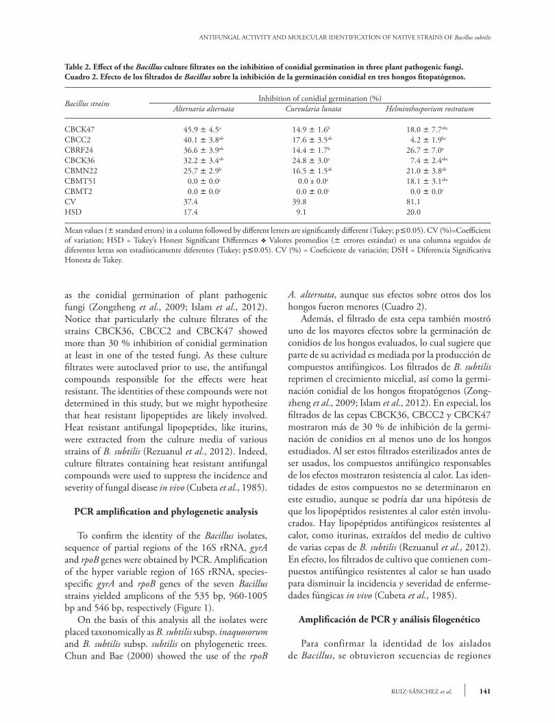
as the conidial germination of plant pathogenic fungi (Zongzheng et al., 2009; Islam et al., 2012). Notice that particularly the culture filtrates of the strains CBCK36, CBCC2 and CBCK47 showed more than 30 % inhibition of conidial germination at least in one of the tested fungi. As these culture filtrates were autoclaved prior to use, the antifungal compounds responsible for the effects were heat resistant. The identities of these compounds were not determined in this study, but we might hypothesize that heat resistant lipopeptides are likely involved. Heat resistant antifungal lipopeptides, like iturins, were extracted from the culture media of various strains of B. subtilis (Rezuanul et al., 2012). Indeed, culture filtrates containing heat resistant antifungal compounds were used to suppress the incidence and severity of fungal disease in vivo (Cubeta et al., 1985).
PCR amplification and phylogenetic analysis
To confirm the identity of the Bacillus isolates, sequence of partial regions of the 16S rRNA, gyrA and rpoB genes were obtained by PCR. Amplification of the hyper variable region of 16S rRNA, species-specific gyrA and rpoB genes of the seven Bacillus strains yielded amplicons of the 535 bp, 960-1005 bp and 546 bp, respectively (Figure 1). On the basis of this analysis all the isolates were placed taxonomically as B. subtilis subsp. inaquosorum and B. subtilis subsp. subtilis on phylogenetic trees. Chun and Bae (2000) showed the use of the rpoB
Table 2. Effect of the Bacillus culture filtrates on the inhibition of conidial germination in three plant pathogenic fungi.Cuadro 2. Efecto de los filtrados de Bacillus sobre la inhibición de la germinación conidial en tres hongos fitopatógenos.
Bacillus strainsInhibition of conidial germination (%)
Alternaria alternata Curvularia lunata Helminthosporium rostratum
CBCK47 45.9 ± 4.5a 14.9 ± 1.6b 18.0 ± 7.7abc
CBCC2 40.1 ± 3.8ab 17.6 ± 3.5ab 4.2 ± 1.9bc
CBRF24 36.6 ± 3.9ab 14.4 ± 1.7b 26.7 ± 7.0a
CBCK36 32.2 ± 3.4ab 24.8 ± 3.0a 7.4 ± 2.4abc
CBMN22 25.7 ± 2.9b 16.5 ± 1.5ab 21.0 ± 3.8ab
CBMT51 0.0 ± 0.0c 0.0 ± 0.0c 18.1 ± 3.1abc
CBMT2 0.0 ± 0.0c 0.0 ± 0.0c 0.0 ± 0.0c
CV 37.4 39.8 81.1HSD 17.4 9.1 20.0
Mean values (± standard errors) in a column followed by different letters are significantly different (Tukey; p£0.05). CV (%)=Coefficient of variation; HSD = Tukey’s Honest Significant Differences v Valores promedios (± errores estándar) es una columna seguidos de diferentes letras son estadísticamente diferentes (Tukey; p£0.05). CV (%) = Coeficiente de variación; DSH = Diferencia Significativa Honesta de Tukey.
A. alternata, aunque sus efectos sobre otros dos los hongos fueron menores (Cuadro 2). Además, el filtrado de esta cepa también mostró uno de los mayores efectos sobre la germinación de conidios de los hongos evaluados, lo cual sugiere que parte de su actividad es mediada por la producción de compuestos antifúngicos. Los filtrados de B. subtilis reprimen el crecimiento micelial, así como la germi-nación conidial de los hongos fitopatógenos (Zong-zheng et al., 2009; Islam et al., 2012). En especial, los filtrados de las cepas CBCK36, CBCC2 y CBCK47 mostraron más de 30 % de inhibición de la germi-nación de conidios en al menos uno de los hongos estudiados. Al ser estos filtrados esterilizados antes de ser usados, los compuestos antifúngico responsables de los efectos mostraron resistencia al calor. Las iden-tidades de estos compuestos no se determinaron en este estudio, aunque se podría dar una hipótesis de que los lipopéptidos resistentes al calor estén involu-crados. Hay lipopéptidos antifúngicos resistentes al calor, como iturinas, extraídos del medio de cultivo de varias cepas de B. subtilis (Rezuanul et al., 2012). En efecto, los filtrados de cultivo que contienen com-puestos antifúngico resistentes al calor se han usado para disminuir la incidencia y severidad de enferme-dades fúngicas in vivo (Cubeta et al., 1985).
Amplificación de PCR y análisis filogenético
Para confirmar la identidad de los aislados de Bacillus, se obtuvieron secuencias de regiones

AGROCIENCIA, 16 de febrero - 31 de marzo, 2016
VOLUMEN 50, NÚMERO 2142
gene sequences in Bacillus. Later, Geetha and Manonmani (2010) also showed the utility of this gene sequence to identify the isolate B. subtilis subsp. subtilis VCRC B471. The protein rpoB was also used to infer relationships among archaea orders and Gram positive and Gram-negative bacteria (Matte-Tailliez et al., 2002; Morse et al., 2002). The highest BLASTN identity of each of the strain was confirmed and also the genus identity of Bacillus subtilis group (Table 3). The gene sequences 16S rRNA, rpoB and gyrA–based PCR had values in a range of 95 to 100 %, similar to those deposited in Genbank (Table 3). The amplicons obtained for regions 16S rRNA, rpoB and gyrA were 535, 546 and 953 bp, respectively. The Neighbor-Joining analyses of each of the regions (Figures 2, 3 and 4) and the total evidence (Figure 5) showed that the isolates were separated into two clades. In all analyses, the isolates CBRF24 and CBMT2 were grouped in a different clade. Using the 16S rRNA region, the strains CBMT2, CBMN22, CBCK36, CBMT51 and CBCK47 formed a clade (Figure 2), with sequences of B. subtilis taken from GenBank. These isolates formed a clade with stand of 100 with B. subtilis subsp. subtilis; while with the analysis performed using the regions gyrA and rpoB, the mentioned strains formed a clade with B. subtilis subsp. inaquosorum. The analysis of matrix partitions separated the isolates into two distinct clades, forming a clade with
parciales de los genes 16S rRNA, gyrA y rpoB por PCR. La amplificación de la región hipervariable de 16S rRNA, genes especie específicos gyrA y rpoB de las siete cepas de Bacillus produjeron amplicones de 535 pb, 960-1005 pb y 546 bp, respectivamente (Figura 1). De acuerdo con este análisis, todos los aislados fueron colocados taxonómicamente como B. subti-lis subsp. inaquosorum y B. subtilis subsp. subtilis en árboles filogenéticos. Chun y Bae (2000) mostraron el uso de las secuencias genéticas de rpoB en Baci-llus. Luego, Geetha y Manonmani (2010) también mostraron la utilidad de esta secuencia genética para identificar el aislado B. subtilis subsp. subtilis VCRC B471. La proteína rpoB también se usó para inferir relaciones entre órdenes arquea y bacterias Gram po-sitiva y Gram negativa (Matte-Tailliez et al., 2002; Morse et al., 2002). La máxima identidad BLASTN de cada cepa y del género del grupo Bacillus subtilis fue confirmada (Cuadro 3). Las secuencias de PCR basados en los genes 16S rRNA, rpoB y gyrA tenían valores en el rango de 95 a 100 %, similares a los depositados en Genbank (Cuadro 3). Los amplicones obtenidos para las regiones 16S rRNA, rpoB y gyrA fueron 535, 546 y 953 bp, respec-tivamente. Los análisis Neighbor-Joining de cada una de las regiones (Figuras 2, 3 y 4) y la evidencia total (Figura 5) mostraron que los aislados fueron separa-dos en dos clados. En todos los análisis, los aislados
Figure 1. Agarose gel (1.2 %) electrophoresis of the PCR amplification of rpoB DNA, 16S rRNA and gyrA DNA genes of seven native Bacillus subtilis. Lane M: 1 kb ladder Promega, 1: CBCC2, 2: CBMT2, 3: CBMN22, 4: CBRF24, 5: CBCK36, 6: CBCK47 and 7: CBMT51.
Figura 1. Gel de agarosa (1.2 %) electroforesis de la amplificación por PCR de los genes rpoB DNA, 16S rRNA y gyrA DNA de siete Bacillus subtilis nativas. Carriles M: marcador Promega 1 kb, 1: CBCC2, 2: CBMT2, 3: CBMN22, 4: CBRF24, 5: CBCK36, 6: CBCK47 and 7: CBMT51.
rpoB gene 16S rRNA gene gyrA gene
M 1 2 3 4 5 6 7 1 2 3 4 5 6 7 M 1 2 3 4 5 6 7
0.5 kb0.5 kb
1 kb

143RUIZ-SÁNCHEZ et al.
ANTIFUNGAL ACTIVITY AND MOLECULAR IDENTIFICATION OF NATIVE STRAINS OF Bacillus subtilis
CBRF24 and CBMT2, completely independent of the other strains (Figure 5). Amplification, sequencing and sequence analysis of 16S rRNA, rpoB and gyrA genes for molecular identification of Bacillus species are reported only in separate studies (Geetha et al., 2007; Geetha and Manonmani, 2008; Geetha and Manonmani, 2010). In our research, however, the first sequence analysis of 16S rRNA, gyrA and rpoB genes to locate taxonomically seven native strains of B. subtilis was successfully integrated. This molecular technique allowed us to infer the species of Bacillus and the existence of two new subspecies. Although 16S rRNA gene sequence was very useful for the determination of bacterial species, in the genus Bacillus some problems have arisen (Wattiau et al., 2001; Janda et al., 2007).
Evolutionary distance matrices
The region that showed lower evolutionary distances was the 16S rRNA (from zero to 0.002) followed by the rpoB gene (up to 0.004). In contrast,
Table 3. Maximum identity values (%) of identified Bacillus strains based on the analysis of 16S rRNA, gyrA and rpoB genes.Cuadro 3. Valores máximos de identidad (%) de cepas de Bacillus identificadas con base en el análisis de los genes 16S rRNA,
gyrA y rpoB.
Gene Strain Accession no. Identity Maximum identity (%)
16S rRNA CBCC2 KC223570 Bacillus subtilis subsp. inaquosorum (=B. inaquosus) 100
CBMT2 KC223571 Bacillus subtilis 100CBMN22 KC223572 Bacillus subtilis 100CBRF24 KC223573 Bacillus subtilis subsp. inaquosorum 100CBCK36 KC223574 Bacillus subtilis 100CBCK47 KC223575 Bacillus subtilis 100CBMT51 KC223576 Bacillus subtilis 100
gyrA CBCC2 KC253388 Bacillus subtilis subsp. inaquosorum 96CBMT2 KC253389 Bacillus subtilis 96CBMN22 KC253390 Bacillus subtilis 99CBRF24 KC253391 Bacillus subtilis subsp. inaquosorum 99CBCK36 KC253392 Bacillus subtilis subsp. inaquosorum 95CBCK47 KC253393 Bacillus subtilis 99CBMT51 KC253394 Bacillus subtilis 99
rpoB CBCC2 KC253395 Bacillus subtilis subsp. subtilis 99CBMT2 KC253396 Bacillus subtilis subsp. subtilis 99CBMN22 KC253397 Bacillus subtilis subsp. subtilis 100CBRF24 KC253398 Bacillus subtilis subsp. subtilis 99CBCK36 KC253399 Bacillus subtilis subsp. subtilis 99CBCK47 KC253400 Bacillus subtilis subsp. subtilis 99CBMT51 KC253401 Bacillus subtilis subsp. subtilis 99
CBRF24 y CBMT2 se agruparon en un clado dife-rente. Al usar la región 16S rRNA, las cepas CBMT2, CBMN22, CBCK36, CBMT51 y CBCK47 forma-ron un clado (Figura 2), con secuencias de B. subtilis tomadas de GenBank. Estos aislados formaron un clado con valor de 100 con B. subtilis subsp. subtilis; mientras que con el análisis realizado usando las re-giones gyrA y rpoB, las cepas mencionadas formaron un clado con B. subtilis subsp. inaquosorum. El análisis de particiones de matrices separaron los aislados en dos clados distintos, formando un clado con CBRF24 y CBMT2, completamente indepen-diente de las otras cepas (Figura 5). La amplificación, secuenciación y el análisis de secuencias de los genes 16S rRNA, rpoB y gyrA para la identificación mo-lecular de especies de Bacillus sólo están reportados en estudios separados (Geetha et al., 2007; Geetha y Manonmani, 2008; Geetha y Manonmani, 2010). Sin embargo, en nuestra investigación, el primer análisis de secuencias de los genes 16S rRNA, gyrA y rpoB para ubicar taxonómicamente a siete cepas na-tivas de B. subtilis se intregó de manera exitosa. Esta

AGROCIENCIA, 16 de febrero - 31 de marzo, 2016
VOLUMEN 50, NÚMERO 2144
Figure 2. Neighbor-joining tree with bootstrap values based on partial sequences of the 16S rRNA gene. Analysis made with a nucleotide substitution model of Jukes-Cantor.
Figura 2. Árbol Neighbor-joining con valores bootstrap basados en secuencias parciales del gen 16S rRNA. Análisis realizado con un modelo de sustitución nucleotídica Jukes-Cantor.
Figure 3. Neighbor-Joining tree with bootstrap values based on partial sequences of the gyrA gene. Analysis made with a nucleotide substitution model of Tamura-Nei with discrete gamma distribution.
Figura 3. Árbol Neighbor-joining con valores bootstrap basados en secuencias parciales del gen gyrA. Análisis realizado con un modelo de sustitución nucleotídica Tamura-Nei con distribución gamma discreta.
65
gi 83286902 Bacillus subtilisgi 83286903 Bacullus subtilisCBMT51CBCK47CBCK36CBMN22CBMT2
CBCC2CBRF24gi 375271204 Bacillus subtilis subsp. inaquosorumgi 350935486 Bacillus subtilis subsp. inaquosorum
gi 332691808 Bacillus mojavensisgi 332650763 Bacillus amyloliquefaciens
gi 219856870 Bacillus atrophaeus
9476
gi 383478265 Bacillus tequlensisgi 219856877 Bacillus vallismortis
99gi 382947310 Bacillus sonorensis
gi 384071194 Bacillus licheniformis
0.002
gi 13810774 Bacillus subtilis subsp. subtilis
gi 157644497 Bacillus subtilis subsp. subtilis83
61
CBMN22
CBCK4740
100CBMT51
CBMT2
CBCK3644
gi 319826933 Bacillus subtilis subsp. subtilis
gi 160905565 Bacillus vallismortis
90
79
100 10071
gi 160905575 Bacillus subtilis subsp. spizizenii
gi 160905581 Bacillus inaquosus
gi 2936627260 Bacillus subtilis subsp. inaquosorum
CBCC2
CBRF24100
gi 160905585 Bacillus sonorensis
gi 160905665 Bacillus atrophaeus
0.01

145RUIZ-SÁNCHEZ et al.
ANTIFUNGAL ACTIVITY AND MOLECULAR IDENTIFICATION OF NATIVE STRAINS OF Bacillus subtilis
Figure 4. Neighbor-Joining tree with bootstrap values based on partial sequences rpoB gene. Analysis made with a nucleotide substitution model of Tamura-3 parameter using discrete gamma distribution.
Figura 4. Árbol Neighbor-joining con valores bootstrap basados en secuencias parciales del gen rpoB. Análisis realizado con un modelo de sustitución nucleotídica Tamura-3 parámetro usando una distribución gamma discreta.
Figure 5. Neighbor-Joining tree with bootstrap values based on partial sequences of the 16S rRNA, gyrA and rpoB genes. Analysis made with a nucleotide substitution model of Tamura-3 parameter using discrete gamma distribution.
Figura 5. Árbol Neighbor-joining con valores bootstrap basados en secuencias parciales de los genes 16S rRNA, gyrA y rpoB. Anály-sis realizado con un modelo de sustitución nucleotídica Tamura-3 parámetro usando una distribución gamma discreta.
CBMT2gi 284154767 Bacillus subtilis subsp. inaquosorum
CBCK47gi 284154807 Bacillus subtilis subtilis. inaquosorum41CBMN22gi 284154885 Bacillus subtilis subsp. inaquosorum
CBCK3656
CBMT51CBCC282
65 CBRF24
66gi 158602608 Bacillus subtilis subsp. subtilis
gi 116563649 Bacillus subtilis subsp. subtilis
gi 116563585 Bacillus mojavensisgi 116563669 Bacillus vallismortis
gi 161345230 Bacillus amyloliquefaciens
gi 116563671 Bacillus sonorensis
gi 1561181153 Bacillus licheniformis
100
47
99
0.002
CBMT2
CBCK36
CBMT51
100
68
100
CBMN22
CBCK4767
CBCC2
CBRF24100
gi 382947310 Bacillus sonorensis
0.02

AGROCIENCIA, 16 de febrero - 31 de marzo, 2016
VOLUMEN 50, NÚMERO 2146
the gyrA gene showed the highest evolutionary rate (up to 0.09), being even higher than that obtained from the matrix partitions, which includes information from the three regions analyzed (0.034 maximum). The greatest distances were observed between the isolates CBRF24 and CBMT2. Thus, for the 16S rRNA, the proper determination of the B. subtilis subspecies could be difficult. In contrast, for the gyrA gene, its higher evolutionary rate allowed to allocate B. subtilis at the subspecies level. In particular, the comparative analysis of gyrA sequences proved to be robust for rapid classification and identification of B. subtilis and related taxa (Chun and Bae, 2000), as shown in our study. The characterization of all strains at the subspecies level was not possible with the data obtained. The strains CBMT2 and CBRF24 might eventually form new subspecies, since the rest of the strains were grouped into clades with different subspecies.
conclusIons The native Bacillus strains from the Yucatan Peninsula, Mexico, were identified as Bacillus subtilis subsp. inaquosorum and Bacillus subtilis subsp. subtilis by the analysis of gene: 16S rRNA, gyrA and rpoB. Seven strains showed antagonistic activity on mycelial growth inhibition of the plant pathogenic fungi Alternaria alternata, Helminthosporium rostratum and Curvularia lunata. The cell-free culture filtrates of CBCK36, CBCC2 and CBCK47 strains showed inhibition of mycelial growth and caused different degree of inhibition of conidial germination in the evaluated plant pathogenic fungi. The strain CBCK36 showed the highest activity on inhibition of fungal mycelial growth.
lIterAture cIted
Adesemoye, A. O., M. Obini, and E. O. Ugoji. 2008. Comparison of plant growth promotion with Pseudomonas aeruginosa and Bacillus subtilis in three vegetables. Braz. J. Microbiol. 36: 423-426.
Agrios, G. N. 2005. Plant Pathology. Elsevier Academic Press, San Diego, California, USA. 903 p.
Ajao, A. T., G. B. Adebayo, and S. E. Yakubu. 2011. Bioremediation of Textile Industrial Effluent using mixed culture of Pseudomonas aeruginosa and Bacillus subtilis immobilized on agar-agar in a Bioreactor. J. Microbiol. Biotech. Res. 1: 50-56.
técnica molecular nos permitió inferir las especies de Bacillus y la existencia de dos nuevas subespecies. Aunque la secuencia genética16S rRNA fue muy útil para determinar las especies bateriales, en el género Bacillus han surgido algunos problemas (Wattiau et al., 2001; Janda et al., 2007).
Matrices de distancia evolutiva
La región que presentó las distancias evolutivas menores fue el 16S rRNA (de cero a 0.002), seguida del gen rpoB (hasta 0.004). En contraste, el gen gyrA mostró la tasa evolutiva mayor (hasta 0.09), resultan-do incluso más alta que la obtenida por las particio-nes de matrices, que incluye información de las tres regiones analizadas (0.034 máximo). Las distancias mayores observadas fueron entre los aislados CBRF24 y CBMT2. Así, para el 16S rRNA, una determinación adecuada de la subespecie B. subtilis podría resultar difícil. En contraste, para el gen gyrA, su tasa evolutiva más alta permitió colocar a B. subtilis en el nivel de subespecie. En particular, el análisis comparativo de secuencias de gyrA probó ser robusto para una clasificación e identificación rápida B. subtilis y taxones relacionados (Chun y Bae, 2000), como lo muestra nuestro estudio. La caracterización de todas las cepas a nivel de subespecie no fue po-sible con los datos obtenidos. Las cepas CBMT2 y CBRF24 podrían, a la larga, formar nuevas subespe-cies, porque las demás cepas se agruparon en clados con diferentes subespecies.
conclusIones
Las cepas de Bacillus nativas de la Península de Yucatán, México, fueron identificadas como Bacillus subtilis subsp. inaquosorum y Bacillus subtilis subsp. subtilis por el análisis de los genes 16S rRNA, gyrA y rpoB. Siete cepas presentaron actividad antagónica en la inhibición de crecimiento micelial en los hon-gos fitopatógenos Alternaria alternata, Helminthos-porium rostratum y Curvularia lunata. Los filtrados libres de células de CBCK36, CBCC2 y CBCK47 presentaron inhibición de crecimiento micelial y cau-saron diferentes grados de inhibición de germinación de conidios en los hongos fitopatógenos evaluados. La cepa CBCK36 presentó la actividad más alta de inhibición de crecimiento fúngico micelial.

147RUIZ-SÁNCHEZ et al.
ANTIFUNGAL ACTIVITY AND MOLECULAR IDENTIFICATION OF NATIVE STRAINS OF Bacillus subtilis
Augusto-Costa, A. C., and F. Pereira-Duta. 2001. Bioaccumulation of copper, zinc, cadmium and lead by Bacillus SP., Bacillus cereus, Bacillus speaerecus and Bacillus subtillus. Braz. J. Microbiol. 32:1-5.
Basurto-Cadena, M. G. L., M. Vázquez-Arista, J. García-Jiménez, R. Salcedo Hernández, D. K. Bideshi, and J. E. Barboza-Corona. 2012. Isolation of a new Mexican strain of Bacillus subtilis with antifungal and antibacterial activities. Scientific World J. doi:10.1100/2012/384978, 7 p.
Blasco, C., Y. Picó., J. Mañes, and G. Font. 2002. Determination of fungicide residues in fruits and vegetables by liquid chromatography-atmospheric pressure chemical ionization mass spectrometry. J. Chomatogr. 947: 227-235.
Chaurasia, B., A. Pandey., L. M. S. Palni., P. Trivedi., B. Kumar, and N. Colvin. 2005. Diffusible and volatile compounds produced by an antagonistic Bacillus subtilis strain cause structural deformations in pathogenic fungi in vitro. Microbiol. Res. 160: 75-81.
Cubeta, M. A., G. L. Hartman., and J. B. Sinclair. 1985. Interaction between Bacillus subtilis and fungi associated with soybeans seeds. Plant Dis. 69:506-509.
Cubitto, M. A., A. C. Morán., M. Commendatore, M. N. Chiarello., M. D. Baldini, and F. Siñeriz. 2004. Effects of Bacillus subtilis O9 biosurfactant on the bioremediation of crude oil-polluted soils. Biodegradation 15: 281-287.
Chun, J., and K. S. Bae. 2000. Phylogenetic analysis of Bacillus subtilis and related taxa based on partial gyrA gene sequences. Anton. Leeuw. 78: 123-127.
De Clerck, E., T. Vanhouette., T. Hebb., J. Geerinck, and P. De Vos. 2004. Isolation, characterization and identification of bacterial contaminants in semifinal gelatin extracts. Appl. Environ. Microb. 70: 3664-3672.
De Waard, M. A., S. G. Georgopoulas, D. W., Hollomon, H. Ishii, P. Leroux, N. M. Ragsdale, and F. J. Schwinn. 1993. Chemical control of plant diseases: problems and prospects. Annu. Rev. Phytopathol. 31: 403-421.
Fickers, P. 2012. Antibiotic compounds from Bacillus: Why are they so amazing? Am. J. Biochem. Biotechnol. 8: 40-46.
Geetha, I., G. Prabakaran., K. P. Paily, and A. M. Manonmani. 2007. Characterisation of three mosquitocidal Bacillus strains isolated from mangrove forest. Biol. Control. 42: 34-40.
Geetha, I., and A. M. Manonmani. 2008. Mosquito pupicidal toxin production by Bacillus subtilis subsp. subtilis. Biol. Control. 44: 242–247.
Geetha, I., and A. M. Manonmani. 2010. Surfactin: a novel mosquitocidal biosurfactant produced by Bacillus subtilis ssp. subtilis (VCRC B471) and influence of abiotic factors on its pupicidal efficacy. Lett. Appl. Microbiol. 51: 406-412.
González-Fernández, R., E. Prats, and J. Jorrín-Novo. 2010. Proteomics of plant pathogenic fungi. Journal of Biomedicine and Biotechnology. doi:10.1155/2010/932527, 36 p.
Guillén-Cruz, R., F. D. Hernández-Castillo., G. Gallegos-Morales., R. Rodríguez-Herrera., C. N. Aguilar-González., E. Y. Padrón-Corral, y M. H. Reyes-Valdés. 2006. Bacillus spp. como biocontrol en un suelo infestado con Fusarium spp., Rhizoctonia solani Kühn y Phytophthora capsici Leonian y su efecto en el desarrollo y rendimiento1 del cultivo de chile (Capsicum annuum L.). Rev. Mex. Fitopatol. 24: 105-114.
Hall, T. A. 1999. BioEdit: a user-friendly biological sequence alignment editor and analysis program for Windows 95/98/NT. Nucl. Acid. S. 41: 95–98.
Harwood, C. R. 1992. Bacillus subtilis and its relatives: molecular biological and industrial workhorses. Trends Biotechnol. 10: 247-56.
Islam, M. R., Y. T. Jeong., Y. S. Lee, and C. H. Song. 2012. Isolation and identification of antifungal compounds from Bacillus subtilis C9 inhibiting the growth of plant pathogenic fungi. Mycobiology 40: 59-66.
Janda, J. M., L. Sharon, and Abbott, S. L. 2007. 16S rRNA Gene Sequencing for Bacterial Identification in the Diagnostic Laboratory: Pluses, Perils, and Pitfalls. J. Clin. Microbiol. 45: 2761-2764.
Janiciewickz, W.J., Leverentz, B., Conway, W.S., Saftner, R.A., Reed, A.N., and Camp, M.J. 2003. Control of bitter rot and blue mold of apples integrating heat and antagonistic treatments on 1-MCP treated fruits stored under controlled atmosphere conditions. Postharvest Biology and Technology 29: 129-143.
Knight, S. C., V. M. Anthony., A. M. Brady., A. J. Greenland., S. P. Heaney., D. C. Murray., K. A. Powell., M. A. Schulz., C. A. Spinks., P. A. Worthington, and D. Youle. 1997. Rationale and perspectives on the development of fungicides. Annu. Rev. Phytopathol. 35: 349-372.
Korsten, L., and E. E. Jager. 1995. Mode of action of Bacillus subtilis for control of avocado postharvest pathogens. SAAGA Yearbook 18: 124-130.
Kumar D. J. M., C. L. Poovai., P. Kumar, Y. S. Saroja., A. Manimaran, and P. T. Kalaichelvan. 2012. Optimization of Bacillus cereus MRK1 cellulase production and its Biostoning activity. Der Pharmacia Lettre 4: 881-888.
Larkin, M. A., G. Blackshields., N. P. Brown, R. Chenna., P. A. Mcgettigan., H. Mcwilliam., F. Valentin., I. M. Wallace., A. Wilm., R. Lopez., J. D. Thompson., T. J. Gibson, and D. G. Higgins. 2007. ClustalW and ClustalX version 2. Bioinformatics 23: 2947-2948.
Lugtenberg, B., and F. Kamilova. 2009. Plant-growth-promoting rhizobacteria. Annu. Rev. Microbiol. 63: 541-556.
Mahadtanapuk, S., M. Sanguansermsri., R. W. Cutler., V. Sardsud, and S. Anuntalabhochai. 2007. Control of Anthracnose caused by Colletotrichum musae on Curcuma alismatifolia Gagnep. using antagonistic Bacillus spp. ARPN. 2: 54-61.
Matte-Tailliez, O., C. Brochier., P. Forterre, and H. Philippe. 2002. Archaeal phylogeny based on ribosomal proteins. Mol. Biol. Evol. 19: 631-639.
Morse, R., K. O’hanlon, and M. D. Collins. 2002 .Phylogenetic, amino acid content and indel analyses of the beta subunit of DNA-dependent RNA polymerase of grampositive and gram-negative bacteria. IJSEM 52: 1477–1484.
Pal, K. K., and B. M. Gardener. 2006. Biological control of plant pathogens. The plant health instructor. Biological control. DOI: 10.1094/PHI-A-2006-1117-02.
Podile, A. R., and V. D. V. Laxmi. 1998. Seed bacterization with Bacillus subtilis AF1 increases phenylalanine ammonia lyase and reduces the incidence of fusarial wilt in pigeonpea. J. Phytopathol. 146: 255-259.
Price, N. P. J., A. P. Rooney., J. L. Swezey., E. Perry, and F. M. Cohan. 2007. Mass spectroscopic analysis of lipopeptide for
AGROCIENCIA, 16 de febrero - 31 de marzo, 2016
VOLUMEN 50, NÚMERO 2148
Bacillus strains isolated from diverse geographical locations. FEMS Microbiol. Lett. 271: 83-89.
Rezuanul, I., Y. T. Jeong., Y. S. Lee, and C. H. Song. 2012. Isolation and Identification of Antifungal Compounds from Bacillus subtilis C9 Inhibiting the Growth of Plant Pathogenic Fungi. Mycobiology 40: 59-66.
Rooney, A. P., N. P. J. Price., C. Ehrhardt., J. L. Swezey, and J. D. Bannan. 2009. Phylogeny and molecular taxonomy of the Bacillus subtilis complex and description of Bacillus subtilis subsp. inaquosorum subsp. nov. Int. J. Syst. Evol. Micr. 59: 2429-2436.
Schisler, D. A., P. J. Slininger., R. W. Behle, and M. A. Jackson. 2004. Formulation of Bacillus spp. for biological control of plant disease. Phytopathology 94: 1267-1271.
Tamura, K., D. Peterson., N. Peterson., G. Stecher., M. Nei, and S. Kumar. 2011. MEGA5: Molecular Evolutionary
Genetics Analysis using Maximum Likelihood, Evolutionary Distance, and Maximum Parsimony Methods. Mol. Biol. Evol. 28: 2731-2739.
Van Loon L. C. 2007. Plant responses to plant growth-promoting rhizobacteria. Eur. J. Plant Pathol. 119: 243-254.
Wattiau, P., M. E. Renard., P. Ledent., V. Debois., G. Blackman, and S. N. Agathos. 2001. A PCR test to identify Bacillus subtilis and closely related species and its application to the monitoring of wastewater biotreatment. Appl. Microbiol. Biotechnol. 56: 816-819.
Waard, M. A., A. C. Andrade., K. Hayashi., H. J. Schoonbeek., I. Stergiopoulos, and L. H. Zwiers. 2006. Impact of fungal drug transporters on fungicide sensitivity, multidrug resistance and virulence. Pest Manag. Sci. 62: 195-207.
Zongzheng, Y., L. Xin., L. Zhong., P. Jinzhao., Q. Jin, and Y. Wenyan. 2009. Effect of Bacillus Subtilis SY1 on antifungal activity and plant growth. IJABE 2: 55-61.
![Pharmacologically active boranes*old.iupac.org/publications/pac/2006/pdf/7807x1425.pdf · 2017. 7. 24. · teresting pharmacological activity such as antifungal [21], peptidomimetic](https://static.fdocuments.fr/doc/165x107/60d89ee659f0b108862ebb5e/pharmacologically-active-boranesoldiupacorgpublicationspac2006pdf-2017.jpg)