Angiographie coronaire par tomodensitométrie 64-détecteurs - KCE
118
Angiographie coronaire par tomodensitométrie 64-détecteurs chez les patients suspects de maladie coronarienne KCE reports 82 B Federaal Kenniscentrum voor de Gezondheidszorg Centre fédéral d’expertise des soins de santé 2008
Transcript of Angiographie coronaire par tomodensitométrie 64-détecteurs - KCE

Angiographie coronaire par tomodensitométrie 64-détecteurs chez
les patients suspects de maladie coronarienne
KCE reports 82 B
Federaal Kenniscentrum voor de Gezondheidszorg Centre fédéral d’expertise des soins de santé
2008

Le Centre fédéral d’expertise des soins de santé
Présentation : Le Centre fédéral d’expertise des soins de santé est un parastatal, créé le 24 décembre 2002 par la loi-programme (articles 262 à 266), sous tutelle du Ministre de la Santé publique et des Affaires sociales, qui est chargé de réaliser des études éclairant la décision politique dans le domaine des soins de santé et de l’assurance maladie.
Conseil d’administration
Membres effectifs : Gillet Pierre (Président), Cuypers Dirk (Vice-Président), Avontroodt Yolande, De Cock Jo (Vice-Président), De Meyere Frank, De Ridder Henri, Gillet Jean-Bernard, Godin Jean-Noël, Goyens Floris, Maes Jef, Mertens Pascal, Mertens Raf, Moens Marc, Perl François, Van Massenhove Frank, Vandermeeren Philippe, Verertbruggen Patrick, Vermeyen Karel.
Membres suppléants : Annemans Lieven, Bertels Jan, Collin Benoît, Cuypers Rita, Decoster Christiaan, Dercq Jean-Paul, Désir Daniel, Laasman Jean-Marc, Lemye Roland, Morel Amanda, Palsterman Paul, Ponce Annick, Remacle Anne, Schrooten Renaat, Vanderstappen Anne..
Commissaire du gouvernement : Roger Yves
Direction
Directeur général : Dirk Ramaekers
Directeur général adjoint : Jean-Pierre Closon
Contact
Centre fédéral d’expertise des soins de santé (KCE). Rue de la Loi 62 B-1040 Bruxelles Belgium
Tel: +32 [0]2 287 33 88 Fax: +32 [0]2 287 33 85
Email : [email protected] Web : http://www.kce.fgov.be
Angiographie coronaire par tomodensitométrie
64-détecteurs chez les patients suspects
de maladie coronarienne
KCE reports 82B
HANS VAN BRABANDT, CECILE CAMBERLIN, IRINA CLEEMPUT
Federaal Kenniscentrum voor de gezondheidszorg Centre fédéral d’expertise des soins de santé
2008

KCE Reports 82 B
Titre : Angiographie coronaire par tomodensitométrie 64-détecteurs chez les patients suspects de maladie coronarienne
Auteurs : Hans Van Brabandt, Cécile Camberlin, Irina Cleemput
Experts externes : Lieven Annemans (UZ Gent), Victor Legrand (CHU Sart-Tilman Liège), Piet Vanhoenacker (OLV Ziekenhuis Aalst), Jean-Louis Vanoverschelde (Clinques Universitaires St Luc, Bruxelles)
Validateurs : Jan Bogaert (KU Leuven); Frank Rademakers (KU Leuven), Frans Rutten (Erasmus Universiteit Rotterdam)
Conflict of interest : Victor Legrand, Piet Vanhoenacker, Jean-Louis Vanoverschelde ont déclaré qu'il n'y avait pas de conflit d'intérêts. Lieven Annemans a déclaré avoir reçu des honoraires de Cordis & StJude
Disclaimer: Les experts externes ont collaboré au rapport scientifique qui a ensuite été soumis aux validateurs. La validation du rapport résulte d’un consensus ou d’un vote majoritaire entre les validateurs. Le KCE reste seul responsable des erreurs ou omissions qui pourraient subsister de même que des recommandations faites aux autorités publiques.
Mise en Page : Wim Van Moer
Bruxelles, 7 juillet 2008
Etude n° 2007-06
Domaine : Health Technology Assessment
MeSH : Tomography, X-ray Computed; Coronary Disease; Technology Assessment, Biomedical;
Belgium
NLM classification : WG 141
Langage : français, anglais
Format : Adobe® PDF™ (A4)
Dépôt légal : D/2008/10.273/41
La reproduction partielle de ce document est autorisée à condition que la source soit mentionnée. Ce document est disponible en téléchargement sur le site Web du Centre fédéral d’expertise des soins de santé.
Comment citer ce rapport ?
Van Brabandt H., Camberlin C., Cleemput I. Angiographie coronaire par tomodensitométrie 64-détecteurs chez les patients suspects de maladie coronarienne. Health Technology Assessment (HTA). Bruxelles: Centre fédéral d’expertise des soins de santé (KCE); 2008. KCE Reports 82 B (D/2008/10.273/41)
KCE reports 82 B Multislice CT in Coronary Heart Disease iii
PRÉFACE L’intérêt croissant pour les techniques d’imagerie médicale, tant diagnostiques que thérapeutiques, contribue à leur perpétuel perfectionnement. Ainsi, dans le domaine de la radiologie, la tomographie computée ou CT-scan a évolué depuis l’imagerie d’organes statiques, tels que le cerveau, jusqu’à la visualisation de structures beaucoup plus mobiles telles que le cœur et les artères coronaires. C’est l’avènement du CT multi-détecteurs ou MSCT (multi-slice CT) qui a permis d’aboutir à ce stade de perfectionnement. En quelques secondes, ce scanner permet de prendre un grand nombre de clichés radiograhiques qui sont ensuite traités par ordinateur afin de reconstruire une image tridimensionnelle du cœur.
Jusqu’il y a peu, la coronarographie invasive était le seul moyen de visualiser les artères coronaires. Or, il s’agit d’un examen complexe, qui n’est pas sans danger. Pour ce faire, un petit tuyau est introduit dans une artère et acheminé jusqu’aux artères coronaires où un produit de contraste y est alors directement injecté. Ce produit rend les artères coronaires opaques aux rayons X, ce qui permet de les enregistrer sur film radiographique. L’intérêt potentiel de la MSCT des artères coronaires réside dans le caractère moins invasif de l’examen puisque, pour obtenir une visualisation similaire, le produit de contraste est administré par une simple injection intraveineuse. L’utilisation de la MSCT permettrait ainsi d’éviter un certain nombre d’examens invasifs et donc de réduire l’inconfort et les risques encourus par le patient. La MSCT est également moins coûteuse puisqu’elle peut être faite en ambulatoire alors que la coronarographie classique requiert généralement une hospitalisation.
Ce rapport a pour but de déterminer les utilisations diagnostiques scientifiquement fondées de la MSCT des artères coronaires, les indications qui justifient la dose de radiation élevée qu’elle entraîne ainsi que les implications de cette technique sur le plan financier. En d’autres termes, l’objectif du rapport est d’établir si la MSCT est prête à faire partie de la pratique clinique courante.
Jean Pierre Closon Dirk Ramaekers
Directeur Général Adjoint Directeur Général


iv Multislice CT in Coronary Heart Disease KCE Reports 82 B
Sommaire
CHAMP D'APPLICATION Le présent rapport d’évaluation des technologies de la santé (HTA) résume les données probantes actuelles qui soutiennent l'utilisation de l'angiographie CT multi-détecteurs (multi-slice computed tomography – MSCT) en tant qu'aide au diagnostic chez les patients susceptibles de souffrir d'une athéromatose coronaire (CAD – coronary artery disease). Ce rapport s'intéresse au premier chef à l'usage diagnostique de la MSCT en tant que technique d'imagerie des artères coronaires natives, excluant ainsi les greffes de pontage coronarien et les stents intra-coronariens.
CONTEXTE On entend par athéromatose coronaire (CAD) toute affection cardiaque se caractérisant par une entrave au débit sanguin et un déficit d'oxygénation du myocarde qui sont essentiellement induits par une sténose athéromateuse d'une ou plusieurs des artères coronaires. Une CAD peut se manifester par de l'angine de poitrine, un infarctus du myocarde ou la mort subite. On s'accorde généralement à reconnaître qu'une plaque athéromateuse doit réduire le diamètre interne d'un vaisseau d'au minimum 50 % pour diminuer, à l'effort, le débit sanguin dans cette artère et provoquer une ischémie et une angine de poitrine. D'autre part, une crise cardiaque aiguë résulte d'un blocage thrombotique brutal du débit sanguin coronarien, suite à la rupture d’une plaque athéromateuse vulnérable sans nécessairement impliquer de sténoses inhibitrices du débit sanguin.
Souvent, il est possible de poser le diagnostic d'une CAD grâce aux seuls antécédents du patient, en se fondant sur les caractéristiques de la douleur et en tenant compte du profil de risque cardiovasculaire du patient. On peut également procéder à des tests diagnostiques non invasifs du type électrocardiogramme au repos, scintigraphie de perfusion du myocarde et échocardiographie de stress sous dobutamine. De tels examens peuvent aboutir à une meilleure estimation de la probabilité d'une CAD et du risque d'épisodes graves à l'avenir (IM et décès). Certains patients, notamment ceux chez qui le traitement médicamenteux ne suffit pas à juguler les symptômes, bénéficient d'une revascularisation myocardique. Il s'agit de rétablir le débit sanguin déficient grâce à une intervention chirurgicale ou une angioplastie percutanée par ballonnet. Dans de tels cas, une imagerie préalable des artères coronaires est nécessaire afin de confirmer le diagnostic et d'orienter la stratégie de revascularisation. Ce volet du diagnostic implique un cathétérisme cardiaque et une coronarographie, une procédure invasive nécessitant l'injection d'un agent de contraste dans les coronaires qui sont ensuite visualisées par radiographie. C'est dans ce contexte que peut éventuellement intervenir la MSCT. Cette technique a été préconisée, car elle permettrait d'éviter le recours à la coronographie invasive chez les patients dont il s'avère qu'ils souffrent de CAD non obstructive.
LA TOMODENSITOMETRIE COMPUTEE MULTI-DETECTEURS
TECHNOLOGIE La tomographie assistée par ordinateur conventionnelle (CT) est une technique radiologique qui produit une image en trois dimensions d'un objet à partir d'une série importante de clichés radiographiques en deux dimensions pris autour d'un axe de rotation unique. Les mouvements continus du cœur rendent inappropriée la réalisation d'une CT cardiaque en raison de la résolution temporelle médiocre. En outre, les artères coronaires sont de petites structures qui nécessitant une résolution spatiale élevée. La tomodensitométrie computée multi-coupes (MSCT), introduite en 1998, a en partie apporté une solution à ces contraintes. Par rapport à la tomographie assistée par ordinateur conventionnelle, la MSCT fournit des éléments d'information de plus petite taille et couvre plus rapidement une zone plus vaste. L'ensemble du cœur est scanné en une seule apnée après l'administration intraveineuse d'un produit de contraste iodé. Les progrès engrangés au niveau des logiciels et du matériel ont abouti à une MSCT de pointe qui génère davantage d'images en moins de temps. Les CT Scans 64 détecteurs sont entrés dans la pratique clinique en 2004. Les artéfacts dus au
KCE reports 82 B Multislice CT in Coronary Heart Disease v
mouvement restant un problème, en raison des limites au niveau de la résolution temporelle le CT Scan à double source a été mis au point. Cet appareil permet de réduire davantage le temps effectif d'acquisition. L'année 2007, a connu l'avènement des scanners dotés de 256 et 320 détecteurs qui permettent d'acquérir une imagerie des artères coronaires pendant un ou deux battements de cœur.
Les trois principaux axes de préoccupations relatifs à la MSCT sont : les artéfacts dus au mouvement en cas de rythme cardiaque rapide ou irrégulier, les artéfacts provoqués par le calcium coronaire et la dose d'irradiation importante. On peut remédier en partie aux artéfacts dus au mouvement en cas de rythme cardiaque rapide grâce à l'administration de bêtabloquants avant l'examen. En revanche, les artéfacts provoqués par le calcium coronaire restent une limitation majeure à l'utilisation de la MSCT. La charge calcique d'un patient donné peut être évaluée radiologiquement préalablement à l’utilisation de la MSCT, et s'exprime par le score d'Agatston. Chez les patients présentant un score d'Agatston supérieur à 400, la MSCT n’a pas lieu en raison de la probabilité d’obtenir des images non fiables. Les risques d'irradiation associés à la CT n'ont été pleinement reconnus que tout récemment. Les scanners et les protocoles de la toute dernière génération permettent de réduire la dose de rayons chez des patients triés sur le volet, mais un compromis s'impose entre la réduction de la dose et la qualité diagnostique des images.
SECURITE La dose d'irradiation élevée reste l'inconvénient le plus important en termes de sécurité. La dose d'irradiation moyenne estimée par patient dans les essais cliniques était de 15 et 20 mSv et, en cas de protocoles modulés, de 7 et 14 mSv respectivement pour les hommes et les femmes. Ce qui équivaut à 500 radios thorax et est nettement supérieur à la dose d'une CCA (coronarographie conventionnelle) qui est d'environ 2-7 mSv. Le risque de cancer au cours de la vie pour une MSCT standard est fonction de l'âge et du sexe. Dans une étude de simulation, il était de 1 sur 143 pour une femme âgée de 20 ans et de 1 sur 3261 pour un homme de 80 ans.
Comme dans le cas de la CCA, la MSCT exige l'administration intraveineuse d'un produit de contraste. Ce dernier peut provoquer des réactions allergiques et une insuffisance rénale. À l'heure actuelle, la plupart des patients reçoivent un bêtabloquant avant la MSCT afin d'améliorer la qualité de l'image, et ce, même si le besoin semble moins pressant lorsque de l’utilisation de CT Scan 64-détecteurs à double source. L'administration de bêtabloquants dans le département de radiologie est susceptible d’entraîner un risque supplémentaire pour les patients.
PERFORMANCES DIAGNOSTIQUES La plupart des essais cliniques publiés s'intéressent à l’exactitude diagnostique du CT Scan 64-détecteurs en tant qu'outil d'imagerie, avec pour test de référence, la CCA. Une sténose coronarienne qui réduit de 50% au moins le diamètre interne d'un vaisseau à la coronarographie conventionnelle est considérée comme étant obstructive dans la plupart des essais. Dans toutes les études publiées sur le scan CT 64-détecteurs, la sensibilité du test est bonne et est comprise entre 95 et 100% dans des populations présentant une probabilité pré-test intermédiaire à élevée de CAD obstructive. Un pourcentage qui indique une très bonne valeur prédictive négative. En revanche, la spécificité du test est moins bonne. Dans une méta-analyse des résultats d'essais publiés entre 2005 et 2007, la spécificité totalisée (pooled specificity) était de 91% (87.5%-94%) et dans notre méta-analyse des études récentes, elle était de 83.5% (79.8%-86.8%). Les performances de l'examen chez les sujets des deux sexes ont été comparées dans un essai de grande envergure,. Si la sensibilité est apparue excellente chez les deux sexes (93-100), la spécificité était acceptable chez les hommes (90%; 81%-95%) mais médiocre chez les femmes (75%; 62%-85%). Une CCA invasive était déjà programmée chez pratiquement tous les patients enrôlés dans les essais cliniques. Ce constat remet en question la validité externe des observations. Il convient encore d'évaluer la reproductibilité des performances de la MSCT chez des patients ayant fait l'objet d'une sélection moins sévère et présentant une prévalence moindre de CAD. Pour obtenir des images de bonne qualité, il faut que le rythme sinusal des patients soit stable, ils ne doivent pas être en surpoids trop important et leurs artères coronaires ne doivent pas être calcifiées.
A ce jour, il n'y a qu'un seul essai randomisé de petite envergure publié qui s'est penché sur l'effet de la MSCT sur les résultats cliniques pour le patient. Dans cet essai, les patients référés au départ pour une MSCT ont subi davantage de procédures radiotoxiques que ceux randomisés pour l'imagerie nucléaire. Par ailleurs, on a dénombré une augmentation des revascularisations sans aucun effet sur les

vi Multislice CT in Coronary Heart Disease KCE Reports 82 B
résultats cliniques à 6 mois, y compris le décès, l'infarctus du myocarde, les réadmissions et les consultations ultérieures.
QUESTIONS RELATIVES AUX PATIENTS Hormis les implications techniques de la MSCT, à savoir l'exposition à des radiations ionisantes et l'administration d'un produit de contraste par voie intraveineuse, la MSCT peut également avoir un impact sur les patients en raison des incertitudes associées à ses performances diagnostiques. Ainsi, non seulement les faux négatifs et les faux positifs sont indésirables, mais l'identification correcte d'une sténose significative d'une artère coronaire ou la découverte fortuite d'une anomalie extracardiaque peuvent induire des effets indésirables, par exemple, en suscitant davantage d'examens et de traitements en aval.
Les valeurs prédictives positive et négative de la tomodensitométrie computée 64-détecteurs pour le diagnostic de la CAD dans la pratique clinique au quotidien sont inconnues. Jusqu'à présent, on ne dispose d'aucune donnée probante issue d'essais cliniques attestant un effet bénéfique de la MSCT sur les résultats pour le patient, notamment la maîtrise des symptômes, la prévention de l'infarctus du myocarde ou l’allongement de la durée de vie.
RAPPORT COUT- EFFICACITE Pour effectuer une évaluation économique complète de la MSCT, il faut disposer de davantage de données sur l'efficacité clinique de cette technique de diagnostic dans la prévention de la morbidité et de la mortalité. Pour l'heure, il reste impossible de conclure si la MSCT est coût-efficace en comparaison avec les protocoles de diagnostic standards chez les patients présentant une probabilité pré-test faible à intermédiaire.
QUESTIONS DE NATURE ORGANISATIONNELLE L'investissement initial pour un scanner MSCT va de 850.000 euros (CT Scan 64-détecteurs) à 2 millions d'euros (scanner doté d'un nombre plus élevé de détecteurs). En outre, le coût du logiciel nécessaire pour l'examen des artères coronaires représente 20% du prix de l'appareil. Quant au logiciel de post-traitement, il coûte 100.000 euros et sa mise à jour se monte à 20.000 euros par an. Enfin, les frais de maintenance annuels sont de 100.000 euros.
Des 240 tomographes en service en Belgique, 75% sont des scanners MSCT, dont 45% sont dotés de plus de 16 détecteurs (en 2005). Environ 20% des hôpitaux belges pratiquent la tomodensitométrie cardiaque. A l'heure actuelle, la coronarographie par MSCT est remboursée au radiologue au titre d'une CT thoracique ou abdominale conventionnelle, les honoraires médicaux à charge de l'INAMI/RIZIV se montant à 121.4 euros.
Comme en Belgique, la coronarographie par MSCT est facturée sous un code générique au Québec, en Angleterre, aux Pays-Bas, en Allemagne et en France. Les États-Unis sont le seul pays étudié où il existe un remboursement spécifique. En Australie, le remboursement spécifique est en cours de préparation.

KCE reports 82 B Multislice CT in Coronary Heart Disease vii
CONCLUSIONS
EFFICACITE TECHNIQUE Il est établi que le CT Scan 64-détecteurs fournit des images des artères coronaires natives d'une qualité acceptable dans des populations de patients triés sur le volet. Afin d'obtenir des images MSCT de qualité élevée, le rythme sinusal des patients doit être stable, ils ne doivent pas être trop obèses, être capables de coopérer et ne pas présenter de calcification des artères coronaires.
La lourde charge de radiations ionisantes induite par la MSCT reste un obstacle majeur. Pour l'instant, nul ne sait clairement si les progrès techniques futurs permettront de réduire la dose de rayonnement tout en conservant des performances diagnostiques adéquates.
EXACTITUDE DIAGNOSTIQUE L’exactitude diagnostique de la MSCT dans la CAD a été testée de manière approfondie essentiellement chez des patients à haut risque pour lesquels il avait déjà été décidé de procéder à une coronarographie conventionnelle. Dans ces populations, la MSCT est pratiquement aussi performante que la CCA pour détecter les vrais positifs. En revanche, la MSCT est moins performante dans la détection des vrais négatifs, ce qui pose donc un risque potentiel de nombre important de faux positifs. La validité externe des résultats obtenus dans les essais cliniques reste incertaine.
REFLEXION DIAGNOSTIQUE On ne dispose que de données limitées qui soutiennent l'utilisation de la MSCT par rapport à son rôle dans les algorithmes de soins aux patients. L'examen est le plus performant chez les patients présentant des artères coronaires normales, mais il reste encore à vérifier si ces patients (normaux) n'auraient pas pu être identifiés de manière non invasive, plus sûre et plus coût-efficace.
IMPACT THERAPEUTIQUE Si les performances de la MSCT sont aussi bonnes dans la réalité que dans les essais cliniques, elle peut être considérée comme un examen utile pour exclure une CAD significative. Le fait de révéler la CAD obstructive par MSCT ne présente qu'une valeur limitée, car la gestion et le pronostic du patient sont fonction de l'impact fonctionnel de la sténose coronaire que la MSCT à elle seule ne permet pas d'évaluer. De plus, dans les cas où une revascularisation est considérée comme appropriée, une CCA invasive est inévitable.
RESULTATS CLINIQUES POUR LE PATIENT On ne dispose que de données limitées sur la valeur pronostique de la MSCT et il n'existe pas la moindre donnée probante selon laquelle la MSCT améliore la qualité de la vie, prévient des crises cardiaques ou sauve des vies.
COUT-EFFICACITE Les données relatives à l'efficacité clinique de la MSCT dans la prévention de la morbidité et de la mortalité faisant défaut, il est pour l'instant impossible de conclure si cette technique est coût-efficace par rapport aux protocoles de diagnostic standards chez les patients présentant une probabilité pré-test intermédiaire à élevé.

viii Multislice CT in Coronary Heart Disease KCE Reports 82 B
RECOMMANDATIONS Les données probantes manquent quant à l’efficacité clinique de la MSCT dans le diagnostic de la maladie coronarienne (CAD) chez les patients appartenant à la population générale. Elles font également défaut quant au rapport coût-efficacité de cette technique par rapport aux autres examens diagnostiques.
Pourtant, cette technologie est déjà largement répandue à travers le pays. Au moins 20 hôpitaux pratiquent actuellement l’angiographie coronaire par tomodensitométrie computée et beaucoup d’autres établissements prévoient de la pratiquer. En outre, le gouvernement a décidé de consacrer un budget de 1 260 000 € à cet examen diagnostique pour l’année 2008.
Dans le souci d’orienter l’utilisation de la MSCT vers les indications les plus prometteuses, d’empêcher une diffusion inappropriée de cette technologie et en même temps de pouvoir utiliser les données relatives à la prescription des futurs examens, les stratégies de remboursement suivantes pourraient être envisagées :
L’élaboration d’un code de remboursement spécifique à l’angiographie coronaire par MSCT comprenant des règles restrictives:
1. A l’égard des patients: l’angiographie coronaire par MSCT devrait être réservée aux patients souffrant de douleur thoracique atypique chez lesquels les autres modalités diagnostiques non invasives sont impraticables ou non concluantes. Les patients présentant un score Agatston supérieur à 400 ne devraient pas être soumis à un examen par MSCT. L’examen ne devrait pas être pratiqué chez les patients asymptomatiques ou comme outil de dépistage.
2. A l’égard des médecins: les radiologues pratiquant l’angiographie coronaire par MSCT devraient recevoir une formation spécialisée dans cette technique. Seuls les cardiologues et les futurs internistes urgentistes devraient pouvoir adresser leurs patients aux radiologues pour une angiographie coronaire par MSCT.
3. En raison de l’indisponibilité d’études portant sur les résultats cliniques des patients, il devrait être envisagé de lier le remboursement de l’angiographie coronaire par MSCT à l’enrôlement des patients dans une étude nationale randomisée portant sur les résultats cliniques et financée par l’INAMI. Un registre formel, comprenant des informations cliniques et de suivi au sujet des patients examinés par MSCT, devrait au moins être tenu à disposition pour une évaluation par les pairs. Le régistre et son évaluation pourraient être conduits par les organisations professionnelles belges et controlés par l’INAMI.


KCE Reports 82 Multislice CT in Coronary Heart Disease 1
Scientific summary 1 SCOPE............................................................................................................................... 4 2 BACKGROUND............................................................................................................... 5 2.1 CORONARY HEART DISEASE ................................................................................................................ 5
2.1.1 Pathophysiology. ............................................................................................................................... 5 2.1.2 Definitions.......................................................................................................................................... 5
2.2 DIAGNOSIS OF CAD IN NON-ACUTE CONDITIONS.................................................................. 7 2.2.1 Baseline clinical investigations........................................................................................................ 7 2.2.2 Noninvasive testing.......................................................................................................................... 8 2.2.3 Invasive testing: conventional coronary angiography (CCA) ................................................12
2.3 DIAGNOSIS OF CAD IN ACUTE CONDITIONS ............................................................................13 2.4 MULTISLICE CT CORONARY ANGIOGRAPHY..............................................................................14
2.4.1 Technique ........................................................................................................................................14 2.4.2 MSCT of coronary arteries in the diagnostic arena................................................................15
2.5 TREATMENT OPTIONS IN CHD .........................................................................................................17 2.5.1 Treatment of stable angina...........................................................................................................17 2.5.2 Treatment options in ACS ...........................................................................................................18 2.5.3 Treatment of asymptomatic CAD..............................................................................................18
2.6 PROGNOSIS OF STABLE ANGINA AND NON-ACUTE CHEST PAIN.....................................19 2.7 PROGNOSIS OF ACUTE CORONARY SYNDROME .....................................................................19 3 CLINICAL EFFECTIVENESS OF MSCT .................................................................... 21 3.1 LITERATURE SEARCH..............................................................................................................................21
3.1.1 Search strategy and study eligibility............................................................................................21 3.1.2 Data extraction...............................................................................................................................23
3.2 LITERATURE REVIEW ..............................................................................................................................23 3.2.1 Health Technology Assessments ................................................................................................23 3.2.2 Systematic reviews.........................................................................................................................24 3.2.3 Primary diagnostic trials................................................................................................................26 3.2.4 Outcome studies ............................................................................................................................30
3.3 LITERATURE REVIEW SUMMARY ........................................................................................................30 3.4 SUGGESTIONS FOR FUTURE RESEARCH.........................................................................................36 4 SAFETY OF MSCT ........................................................................................................ 38 4.1 RADIATION................................................................................................................................................38 4.2 CONTRAST MEDIUM ADMINISTRATION........................................................................................39 4.3 BETA-BLOCKADE.....................................................................................................................................39 4.4 EXTRACARDIAC FINDINGS .................................................................................................................40 5 COST-EFFECTIVENESS OF MSCT COMPARED TO OTHER DIAGNOSTIC
MODALITIES ................................................................................................................. 41 5.1 ECONOMIC LITERATURE REVIEW..................................................................................................... 41
5.1.1 Methodology ...................................................................................................................................41 5.1.2 Results ..............................................................................................................................................41
5.2 CHALLENGING THE “ECONOMIC EVALUATION” OF GOLDSTEIN ET AL5 .......................47 6 ORGANISATIONAL ISSUES....................................................................................... 49 6.1 MULTI-SLICE CT ANGIOGRAPHY MARKET ....................................................................................49
6.1.1 Multislice Cardiac CT abroad ......................................................................................................49

2 Multislice CT in Coronary Heart Disease KCE Reports 82
6.1.2 Multislice Cardiac CT in Belgium................................................................................................49 6.2 REGULATORY ISSUES..............................................................................................................................51
6.2.1 Authorization ..................................................................................................................................51 6.2.2 Planning.............................................................................................................................................51 6.2.3 Financing...........................................................................................................................................51 6.2.4 Patients referral for CT examinations .......................................................................................51
6.3 COVERAGE OF MULTI-SLICE CT ANGIOGRAPHY .......................................................................52 6.3.1 United States of America..............................................................................................................52 6.3.2 Canada..............................................................................................................................................54 6.3.3 Australia ...........................................................................................................................................54 6.3.4 The Netherlands.............................................................................................................................55 6.3.5 France ...............................................................................................................................................55 6.3.6 Germany...........................................................................................................................................55 6.3.7 England..............................................................................................................................................56 6.3.8 Belgium .............................................................................................................................................57
6.4 DISCUSSION...............................................................................................................................................57 7 PATIENT ISSUES.......................................................................................................... 59 7.1 TRUE NEGATIVE TEST RESULT............................................................................................................59 7.2 FALSE NEGATIVE TEST RESULT...........................................................................................................59 7.3 TRUE POSITIVE TEST RESULT...............................................................................................................59 7.4 FALSE POSITIVE TEST RESULT..............................................................................................................60 8 GENERAL DISCUSSION.............................................................................................. 61 8.1 TECHNICAL EFFICACY...........................................................................................................................61 8.2 DIAGNOSTIC ACCURACY....................................................................................................................62 8.3 DIAGNOSTIC THINKING ......................................................................................................................62 8.4 THERAPEUTIC IMPACT ..........................................................................................................................62 8.5 PATIENT OUTCOMES.............................................................................................................................63 8.6 COST-EFFECTIVENESS ............................................................................................................................63 9 CONCLUSIONS ............................................................................................................ 64 9.1 TECHNICAL EFFICACY...........................................................................................................................64 9.2 DIAGNOSTIC ACCURACY....................................................................................................................64 9.3 DIAGNOSTIC THINKING ......................................................................................................................64 9.4 THERAPEUTIC IMPACT ..........................................................................................................................64 9.5 PATIENT OUTCOMES.............................................................................................................................64 9.6 COST-EFFECTIVENESS ............................................................................................................................64 10 APPENDICES................................................................................................................. 65 REFERENCES ........................................................................................................................... 92

KCE Reports 82 Multislice CT in Coronary Heart Disease 3
GLOSSARY
ACC American College of Cardiology
ACS Acute Coronary Syndrome
AHA American Heart Association
AMI Acute Myocardial Infarction
AR Absolute Risk
ARR Absolute Risk Reduction
b.p.m. beats per minute
CABG Coronary Artery Bypass Grafting
CAC Coronary Artery Calcium
CAD Coronary Artery Disease
CCA Conventional coronary angiography
CHD Coronary Heart Disease
CPU Chest Pain Unit
CVD Cardiovascular Disease
DSE Dobutamine Stress Echocardiogram
EBCT Electron Beam Computed Tomography
ECG Electrocardiogram
ED Emergency Department
EF Ejection Fraction
ER Emergency Room
ESC European Society of Cardiology
FN False negative
FP False Positive
HF Heart Failure
HR Hazard Rate
HTA Health Technology Assessment
ICER Incremental Cost-Effectiveness Ratio
IHD Ischemic Heart Disease
LR Likelihood Ratio
LVEF Left Ventricular Ejection Fraction
MI Myocardial Infarction
MPI Myocardial Perfusion Imaging
MPS Myocardial Perfusion Scintigraphy
MRI Magnetic Resonance Imaging
MSCT Multislice computed tomography (of coronary arteries)
NHSEED National Health Service Economic Evaluation Database
NNT Number Needed to Treat
NUR Nationale Unie der Radiologen
NYHA New York Heart Association
PCI Percutaneous Coronary Intervention
QALY Quality Adjusted Life Year
RCT Randomized Controlled Trial
RR Relative Risk
RRR Relative Risk Reduction
SR Systematic Review
STEMI ST-Elevation Myocardial Infarction
TN True Negative
TP True Positive
UNR Union Nationale des Radiologues
x-SCT x-slice computed tomography (of coronary arteries): e.g. 64-SCT

4 Multislice CT in Coronary Heart Disease KCE Reports 82
1 SCOPE This Health Technology Assessment (HTA) report summarises current evidence supporting the use of multi slice computed tomography (MSCT) as a diagnostic aid in patients suspected for coronary artery disease (CAD).
The technique has been available since 1998 but underwent substantial technical improvements during the last few years. Originally, MSCT systems were capable of acquiring only 4 sections of the heart simultaneously but in 2004, 64-slice devices were introduced on the market and have been studied in several diagnostic trials since. In 2006, the first trials using dual-source 64-SCT scanners were published and in 2007, 256- and 320-slice devices became available. Because of an increasing penetration of recent generation scanners into the radiological realm, and several trials being completed with them, this report will focus on the performance of 64 (or more) slices CT scanners. Computed tomography in evaluating CAD can be used (1) for risk stratification by assessing calcification of coronary arteries and (2) if coupled with intravenous contrast administration, as a diagnostic imaging technique to obtain a noninvasive coronary angiogram. This report does not address the use of MSCT for risk profiling based on calcium scoring, but is primarily concerned with the diagnostic use of MSCT as an imaging technique for native coronary arteries, by which coronary bypass grafts and intracoronary stents are excluded. Our major interest lies in the diagnosis of CAD in a population with no known heart disease, where an increase of the use of MSCT in the years to come is expected to be high. MSCT for screening in asymptomatic populations does not fall into the scope of the current report. No assessment was done of the diagnostic performance of MSCT in chest pain originating from extra-cardiac disease, such as pulmonary embolism, dissecting aneurysm of the aorta, or pleural effusion.
Key point
• This review is primarily concerned with the use of 64-SCT as an imaging technique for the diagnosis of obstructive CAD in native coronary arteries.

KCE Reports 82 Multislice CT in Coronary Heart Disease 5
2 BACKGROUND
2.1 CORONARY HEART DISEASE
2.1.1 Pathophysiology
Coronary heart disease (CHD) or coronary artery disease (CAD) refers to any cardiac disease caused by an impaired blood flow and deficient oxygen supply to the myocardium, due to atheromatous narrowing of the coronary arteries. It is one of the main causes of mortality and morbidity in Western countries. It can be manifested by stable angina pectoris, acute coronary syndromes (ACS) - including myocardial infarction (MI) and unstable angina -, or sudden death. Loss of myocardial tissue due to MI can lead to heart failure and it can constitute the anatomical basis for arrhythmias, leading to “sudden death”. Cardiac disease may also be related to high blood pressure, valvular dysfunction, congenital abnormalities, primary cardiac muscle problems, or other rarer conditions. These are not part of the disease spectrum of CHD.
Two separate arteries carry oxygenated blood to the heart muscle: the right and the left coronary artery. The first part of the left coronary artery, known as the “left main stem”, shortly after its origin divides into two branches: the circumflex artery (Cx) and the left anterior descending artery (LAD). Because the two branches of the left coronary artery are generally considered separately in clinical practice, it is common to refer to three coronary arteries instead of the anatomically more correct “two”. Depending on whether one, two or three coronary arteries are significantly involved in the atheromatous proces, the labels single, double, or triple vessel disease are attributed. Due to its prognostic significance, if the left main stem is involved in the atheromatous process in a given patient, it is stipulated as such.
The underlying mechanism of CAD is a gradual build-up of fatty material into the coronary vessel wall that leads to the formation of atheromatous plaques. The pathophysiological mechanisms leading to stable angina pectoris or an ACS are different. It is traditionally accepted that a plaque has to reduce the internal diameter if a vessel by at least 50% (or >75% reduction in cross sectional area), in order to reduce blood flow through the coronary artery during exertion and provoke ischemia and angina pectoris. ACSs on the other hand result from a sudden blockage of coronary blood flow, due to rupture of a vulnerable atheromatous plaque, not necessarily involving flow-limiting stenoses.1-3
The main risk factors for CAD development are tobacco use, high blood pressure, raised blood cholesterol, and diabetes mellitus. Several interventions aiming to prevent CAD have been well documented, ranging from lifestyle changes to a daily and lifelong intake of drugs. The best documented are smoking cessation, blood pressure lowering, anti-platelet aggregation therapy (low-dose aspirin) and pharmaceutical lipid management (statins).
2.1.2 Definitions
Symptomatic CAD can be manifested either by stable angina pectoris, as an ACS or as sudden death. Loss of a substantial part of myocardial tissue can lead to heart failure, cardiogenic shock and death. Heart failure is a distinct clinical syndrome characterised by symptoms such as breathlessness and fatigue and signs such as fluid retention. The clinical spectrum of CAD is displayed in Table 1.
Table 1: Clinical spectrum of CAD.
AMI
Other Sudden death, heart failure, …
Asymptomatic CAD
Manifestations
ACS unstable angina Symptomatic CAD
Pathologystable angina
6 Multislice CT in Coronary Heart Disease KCE Reports 82
2.1.2.1 Typical stable angina
Typical angina has three characteristics: (1) discomfort in the chest, jaw, shoulder, back or arms, that is (2) provoked by exertion or emotional stress and (3) relieved by rest or nitroglycerin.4, 5 In most cases, it is caused by a temporary imbalance of the blood supply to the heart muscle combined with the increased demand induced by exercise or emotion.
A grading system of angina pectoris has been proposed by the Canadian Cardiovascular Society and is generally adopted.6 It attributes a higher, i.e. more severe class of angina, depending on the intensity of exercise that elicits chest pain:
• Class I: Ordinary physical activity does not cause angina. Angina occurs with strenuous work.
• Class II: Slight limitation of ordinary activity. Angina occurs on walking or climbing stairs rapidly, walking uphill, …
• Class III: Marked limitations of ordinary physical activity.
• Class IV: Inability to carry on any physical activity without discomfort. Angina symptoms may be present at rest.
Angina is “stable” when the symptoms remain unchanged, i.e. there is no change in the usual pattern of the discomfort, such as an alteration in its frequency or the occurrence with less exertion or at rest. “Unstable” angina is discussed under the heading “acute coronary syndromes”.
2.1.2.2 Atypical angina
Atypical angina has only two of the three characteristics of typical angina. Very often, these patients have significant CAD7 and sometimes, it is referred to as “probable angina” in contrast to “typical angina”.8 The term “atypical angina” is not commonly used in Belgian cardiological practice where the epithet “atypical” most often is applied in combination with “chest pain” suggesting a noncardiac origin of the complaints as discussed below.
2.1.2.3 Atypical chest pain
Atypical or nonanginal chest pain is diagnosed in patients with only one or none of the characteristics of typical angina.9 Such as the other types of chest pain, it is a descriptive term resulting from clinical history taking and is sometimes referred to as nonanginal, atypical or noncardiac chest pain. By assuming this diagnosis, the physician involved indicates his belief in a noncardiac origin of the patient’s chest pain.
2.1.2.4 Non-acute vs. acute chest pain
Non-acute chest pain typically refers to stable angina or chest pain that exists since several weeks or more and that is not experienced as severely discomforting, thus excluding ACS. Acute chest pain refers to pain for which the patient is admitted to an emergency department.
2.1.2.5 Myocardial infarction
A myocardial infarction is a condition in which myocardial tissue is damaged and lost because of prolonged ischeamia induced by an abrupt occlusion (mostly due to thrombus formation) of a coronary vessel. Whereas traditionally a substantial amount of myocardial tissue had to be destroyed before the diagnosis of MI could be made, recent developments in the detection of small quantities of myocardial necrosis using serum biomarker levels, such as cardiac troponin, have lead to a more sensitive diagnosis of MI. A universal definition of MI has been proposed to be used whenever there is evidence of myocardial necrosis in a clinical setting consistent with myocardial ischemia.10 Chest pain is a major symptom of acute myocardial infarction (AMI), mostly occuring at rest and usually lasting at least 20 min.10

KCE Reports 82 Multislice CT in Coronary Heart Disease 7
2.1.2.6 Acute coronary syndromes
Acute coronary syndromes (ACS) encompass a heterogeneous spectrum of acute ischemic heart diseases, extending from acute MI, through minimal myocardial injury to unstable angina. In MI, per definition, there is loss of myocardial tissue. Unstable angina refers to a syndrome of cardiac ischemia clinically manifestating itself as prolonged chest pain, in which no myocardial necrosis can be documented. As opposed to stable angina, unstable angina is also diagnosed when the chest pain started recently, when it becomes more easily provoked or when it occurs with increased frequency, severity or duration.5, 9 Patients with an ACS may have chest discomfort that has all the qualities of typical angina except that the episodes are more severe and prolonged, may occur at rest, or may be precipitated by less exertion than in the past.11
2.1.2.7 Obstructive CAD
Obstructive CAD in this report is defined as CAD in which at least one coronary stenosis exceeding 50% in luminal diameter is present, mostly as documented by invasive coronary angiography.
Key points
• The underlying mechanism of CAD is a gradual build-up of fatty material into the coronary vessel wall, leading to the formation of atheromatous plaques. These may cause narrowing of the coronary arteries leading to angina pectoris, or they may suddenly rupture and induce thrombosis of the vessel giving rise to an acute MI.
• Chest pain can be induced by several non-cardiac conditions as well, originating from the lungs, other intrathoracic structures or the chest wall. It may also be psychosomatic in origin, e.g. caused by anxiety.
2.2 DIAGNOSIS OF CAD IN NON-ACUTE CONDITIONS
2.2.1 Baseline clinical investigations
Diagnosis of CAD can often be made by history taking alone, based on the pain characteristics, taking into account the patient’s age, gender and cardiovascular risk profile. If other risk factors exist, such as smoking, hypertension, family history, hypercholesterolaemia, diabetes, the probability of CAD increases.5 Physical examination can further increase the likelihood of CAD when signs of peripheral atheromatosis or heart failure are found. Very often however, especially in younger patients with angina pectoris, the physical examination is normal. Sometimes, other causes of chest pain may become apparent (pericarditis, pleuritis, orthopaedic disease, …).
In a much-referred to paper, Diamond and Forrester describe how the probability of CAD can be estimated in a given patient from information readily obtainable by clinical evaluation.7 In 4952 patients with different types of chest pain (as defined earlier), the prevalence of angiographic CAD was 90% in patients with typical angina, 50% in patients with atypical angina and 16% in patients with nonanginal chest pain. By combining data from different patient subgroups with disease likelihoods from autopsy studies, probability estimates for angiographic CAD for a set of combinations of age, sex and symptoms were calculated as shown in Table 2.
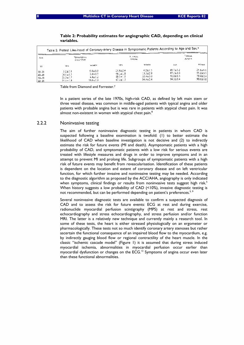
8 Multislice CT in Coronary Heart Disease KCE Reports 82
Table 2: Probability estimates for angiographic CAD, depending on clinical variables.
Table from Diamond and Forrester.7
In a patient series of the late 1970s, high-risk CAD, as defined by left main stem or three vessel disease, was common in middle-aged patients with typical angina and older patients with probable angina but is was rare in patients with atypical chest pain. It was almost non-existent in women with atypical chest pain.8
2.2.2 Noninvasive testing
The aim of further noninvasive diagnostic testing in patients in whom CAD is suspected following a baseline examination is twofold: (1) to better estimate the likelihood of CAD when baseline investigation is not decisive and (2) to indirectly estimate the risk for future events (MI and death). Asymptomatic patients with a high probability of CAD, and symptomatic patients with a low risk for serious events are treated with lifestyle measures and drugs in order to improve symptoms and in an attempt to prevent MI and prolong life. Subgroups of symptomatic patients with a high risk of future events may benefit from revascularisation. Identification of these patients is dependent on the location and extent of coronary disease and on left ventricular function, for which further invasive and noninvasive testing may be needed. According to the diagnostic algorithm as proposed by the ACC/AHA, angiography is only indicated when symptoms, clinical findings or results from noninvasive tests suggest high risk.5 When history suggests a low probability of CAD (<10%), invasive diagnostic testing is not recommended, but can be performed depending on patient’s preferences.5, 9
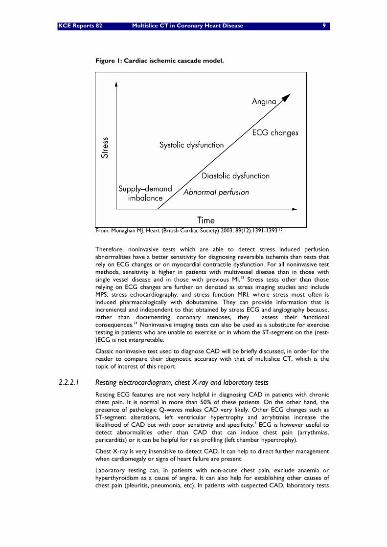
Several noninvasive diagnostic tests are available to confirm a suspected diagnosis of CAD and to assess the risk for future events: ECG at rest and during exercise, radionuclide myocardial perfusion scintigraphy (MPS) at rest and stress, rest echocardiography and stress echocardiography, and stress perfusion and/or function MRI. The latter is a relatively new technique and currently mainly a research tool. In some of these tests, the heart is either stressed physiologically on an ergometer or pharmacologically. These tests not so much identify coronary artery stenoses but rather ascertain the functional consequence of an impaired blood flow to the myocardium, e.g. by indirectly gauging blood flow or regional contractility of the heart muscle. In the classic “ischemic cascade model” (Figure 1) it is assumed that during stress induced myocardial ischemia, abnormalities in myocardial perfusion occur earlier than myocardial dysfunction or changes on the ECG.12 Symptoms of angina occur even later than these functional abnormalities.
KCE Reports 82 Multislice CT in Coronary Heart Disease 9
Figure 1: Cardiac ischemic cascade model.
From: Monaghan MJ. Heart (British Cardiac Society) 2003; 89(12):1391-1393.12
Therefore, noninvasive tests which are able to detect stress induced perfusion abnormalities have a better sensitivity for diagnosing reversible ischemia than tests that rely on ECG changes or on myocardial contractile dysfunction. For all noninvasive test methods, sensitivity is higher in patients with multivessel disease than in those with single vessel disease and in those with previous MI.13 Stress tests other than those relying on ECG changes are further on denoted as stress imaging studies and include MPS, stress echocardiography, and stress function MRI, where stress most often is induced pharmacologically with dobutamine. They can provide information that is incremental and independent to that obtained by stress ECG and angiography because, rather than documenting coronary stenoses, they assess their functional consequences.14 Noninvasive imaging tests can also be used as a substitute for exercise testing in patients who are unable to exercise or in whom the ST-segment on the (rest-)ECG is not interpretable.
Classic noninvasive test used to diagnose CAD will be briefly discussed, in order for the reader to compare their diagnostic accuracy with that of multislice CT, which is the topic of interest of this report.
2.2.2.1 Resting electrocardiogram, chest X-ray and laboratory tests
Resting ECG features are not very helpful in diagnosing CAD in patients with chronic chest pain. It is normal in more than 50% of these patients. On the other hand, the presence of pathologic Q-waves makes CAD very likely. Other ECG changes such as ST-segment alterations, left ventricular hypertrophy and arryhtmias increase the likelihood of CAD but with poor sensitivity and specificity.5 ECG is however useful to detect abnormalities other than CAD that can induce chest pain (arrythmias, pericarditis) or it can be helpful for risk profiling (left chamber hypertrophy).
Chest X-ray is very insensitive to detect CAD. It can help to direct further management when cardiomegaly or signs of heart failure are present.
Laboratory testing can, in patients with non-acute chest pain, exclude anaemia or hyperthyroidism as a cause of angina. It can also help for establishing other causes of chest pain (pleuritis, pneumonia, etc). In patients with suspected CAD, laboratory tests
10 Multislice CT in Coronary Heart Disease KCE Reports 82
most often are used to establish cardiovascular risk factors (glucose, lipids, renal function, etc).
2.2.2.2 Exercise ECG test
In exercise ECG testing, the effect of exercise (in Belgium mostly by cyclo-ergometry) on the electrocardiogram is evaluated. In patients with obstructive CAD, exercise induced ischemia may lead to alterations (depression) of the ST-segment of the ECG which represents the best studied and most often used parameter in this kind of testing. The diagnostic accuracy of the test is dependent on the extent of the ST-segment depression: the more the ST segment becomes depressed during exercise, the higher the likelihood of obstructive CAD. For example, in a 60 year old male with atypical chest pain, the likelihood of angiographic significant CAD is 6% when there is a less than 0.5 mm ST-segment depression whereas it is more than 90% if a more than 2.5 mm ST-segment depression is induced by exercise.7 The electrocardiographic data obtained during exercise testing can be supplemented by additional information that improves the diagnostic capability of the test: age and gender, exercise capacity, anginal symptoms, blood pressure during exercise, heart rate and arrhythmias.
In patients where the resting ECG is abnormal because of left bundle branch block, cardiac pacing, left ventricular hypertrophy or drug effects, electrocardiographic changes induced by exercise are of no help. In these patients, MPS or DSE may be used to further evaluate chest pain. These noninvasive tests can also be considered in patients that are unable to exercise due to orthopaedic, pneumologic or other reasons.
IN ASYMPTOMATIC PATIENTS
Exercise testing is often performed in asymptomatic patients in order to detect CAD, despite the fact that hard evidence on its clinical value in this context is absent. In these patients, ECG exercise testing performs poorly, relating to the fact that in low-risk populations the positive predictive value of a test is low because of a high number of false positives, the latter giving rise to unnecessary further testing, overtreatment and labeling.15 Conversely, because many acute coronary events occur because of plaque rupture involving minor stenoses, a negative stress test in these patients does not preclude the occurrence of subsequent MI.16
Some authors argue that exercise testing in asymptomatic individuals may be reasonable in order to decide whether to start agressive medical therapy to correct risk factors. This indication has been attributed a class IIa recommendation in the most recent ACC/AHA joint guideline, indicating that the weight of evidence/opinion is in favor of usefulness/efficacy although hard data supporting this position are lacking. Routine screening of asymptomatic men or women received a class III recommendation, indicating that it is not useful/effective and may even be harmful.16
IN PATIENTS PRESENTING WITH NON-ACUTE CHEST PAIN
From meta-analyses of diagnostic studies that excluded patients with prior MI and excluded studies showing workup bias (i.e. studies in which patient selection depended on test results), the approximate sensitivity and specificity of 1.0 mm horizontal or downsloping ST segment depression were 50% and 90% respectively.16 A meta-analysis published in 2004, calculated median sensitivities and specificities of stress ECGs from studies excluding patients with previous MI as 0.66 (0.42-0.85) and 0.77 (0.58-0.88).14 These authors calculated an overall estimate of postitive likelihood ratio (LR) of 1.83 (95%CI 1.48-2.26) and a negative LR of 0.51 (95%CI 0.39-0.67) but a significant heterogeneity was evident among included studies. Another systematic review found LRs of 2.79 and 0.44 for a 1 mm ST depression cut-off and 3.85 and 0.72 for a 2 mm cut-off respectively.17
The true diagnostic value of exercise ECG testing lies in its relatively high specificity, indicating that symptomatic patients with a positive test are likely to have obstructive coronary disease. The modest sensitivity is generally less than the sensitivity of imaging tests but taking into consideration scores other than mere ECG-changes such as age, gender, heart rate, maximum work load, and inducible symptoms, “appears to make the

KCE Reports 82 Multislice CT in Coronary Heart Disease 11
tests comparable”.16 Because of these diagnostic capabilities and because exercise testing is safe and relative cheap, it is the first test in the diagnostic evaluation of patients with chest pain suspected of cardiac origin, provided the test is technical feasible and the ECG is deemed interpretable.9, 16
2.2.2.3 Nuclear perfusion imaging
The underlying principle of nuclear perfusion scintigraphy (MPS, often also referred to as SPECT – cf. infra) is that the uptake of a radioactive tracer by the heart is less than normal in poorly perfused or diseased myocardium. To obtain an image of the heart, a cardiac specific radiopharmaceutical such as thallium (201Tl) or technecium-sestamibi (99mTc-sestamibi) is administered intravenously. Imaging by using a gamma camera may be accomplished either by planar or SPECT (Single Photon Emission Computed Tomography) techniques, the latter being most often used. There is general agreement that Tl and Tc-sestamibi have similar diagnostic accuracy in CAD.5, 18 Besides the examination at rest, the heart can be stressed by exercise or pharmacologically with vasodilators (dipyridamole, adenosine) or dobutamine. The images following stress and at rest are compared to assess whether defects are reversible (ischemia) or fixed (infarction).14
Diagnostic accuracy results widely vary between different studies, depending on the technique used, the patient population studied and work-up bias. Without correction, vasodilator stress SPECT has a high sensitivity (90%) and an acceptable specificity (75%). After adjustment for referral bias, sensitivities are somewhat lower.5, 18 A meta-analysis published in 2004, calculated median sensitivities and specificities of SPECT from studies excluding patients with previous MI as 0.92 (0.76-0.93) and 0.74 (0.54-0.90).14 These authors calculated an overall estimate of positive LR of 2.29 (95%CI 1.68-3.12) and a negative LR of 0.25 (95%CI 0.17-0.37) but a significant heterogeneity was evident among included studies.
MPS provides information on coronary disease that is incremental and independent to that obtained by stress ECG or coronary angiography because, rather than merely documenting coronary stenoses, it assesses their functional consequences.14 MPS can also be of substantial prognostic use: patients with stable chest pain syndromes and normal stress SPECT images have a risk of death or nonfatal MI that is as low as in the general population.19, 20 Stress MPI has been shown superior to coronary angiographic variables for predicting outcome across many patient subsets.21
MPS exposes patients to ionizing radiation. Radiation exposure from a 1-day stress/rest MPS study with Tc-99m-tetrofosmin is higher than that from a conventional X-ray coronary angiogram (2–6 mSv) but comparable to that from a multislice CT coronary angiography (6–15 mSv).22
Severe side effects are rare with dipyridamole but this drug may cause bronchospasm in patients with asthma or reactive airway disease; therefore the drug is contraindicated in these patients.23
2.2.2.4 Stress echocardiography
In stress echocardiography segmental left ventricular wall motion and thickening during stress is compared to baseline, using echography. Image quality can be improved by administering intravenous echo contrast or by tissue doppler imaging. As in MPS, stress can be induced by exercise or pharmacologically with vasodilators (dipyridamole, adenosine) or dobutamine, the latter being most often used. Further on in this report, it is being referred to as DSE (dobutamine stress echo). The technique implies a substantial level of skill, which lead some authors to suggest the technique being preferentially used in patients who have a contraindication to MPS.24 This can e.g. be the case in patients with asthma in whom dipyridamole and adenosine may cause severe bronchospasm.18 Approximately 5% of patients have an inadequate acoustic window (due to chest or lung structure) needed to perform an echocardiographic examination.
On the basis of a total number of 2,246 patients, reported in 28 studies, the sensitivity and specificity of the test for the detection of CAD were 80% and 84% respectively.25

12 Multislice CT in Coronary Heart Disease KCE Reports 82
Comparable figures are reported in the most recently published ACC/AHA guidelines for the clinical application of echocardiography.13 From these data, we (crudely) calculated positive and negative LRs of 5.0 and 0.24 respectively. These figures correspond closely to those reported in more recent literature.26, 27
2.2.2.5 Summary of diagnostic accuracy of noninvasive diagnostic tests
The diagnostic performance of noninvasive tests is summarised in Table 3. One should however be cautious to mutually compare them, because MPS and especially stress-ECG have been more thoroughly studied in larger and less selected populations than MSCT. Moreover the diagnostic value of ECG-stress-tests in clinical practice may be better than suggested in diagnostic studies, because information additional to the mere ECG data (chest pain during the test, maximal workload, blood pressure response) are mostly not taken into account in studies on the diagnostic performance of ECG stress testing. This is confirmed in the AHA/ACC guidelines on exercise testing which state that, taking into consideration age, gender, heart rate, maximum work load, and inducible symptoms, “appears to make exercise ECG and imaging procedures comparable”.16
Table 3: Diagnostic accuracy of noninvasive tests
SENS SPEC pos LR neg LR ref 95%CI 95%CI 95%CI 95%CI
stress ECG 0,66 0,42-0,85 0,77 0,58-0,88
1,83 1,48-2,26
0,51 0,39-0,67
14
dipyridamole MPS
0,92 0,76-0,93 0,74 0,54-0,90
2,29 1,68-3,12
0,25 0,17-0,37
14
dobutamine stress ECHO
0,8 NA 0,84 NA 5 3,16-7,92
0,24 0,16-0,36
Calculated from 25
Sens: sensitivity, spec; specificity; pos and neg LR: positive and negative likelihood ratio
Key points
• Diagnosis of CAD can often be made by history taking alone, based on the pain characteristics and taking into account the patient’s age, gender and cardiovascular risk profile.
• The aim of the additional noninvasive diagnostic tests discussed so far is twofold: (1) to better estimate the likelihood of CAD when baseline investigation is not decisive and (2) to indirectly estimate the risk for future events.
• These tests not so much identify coronary artery stenoses by directly imaging the coronary tree, but rather assess the functional consequence of an impaired blood flow to the myocardium. In this way, they provide information that is additional to pure imaging techniques like coronary angiography and multislice CT.
2.2.3 Invasive testing: conventional coronary angiography (CCA)
The only absolute way to anatomically document obstructive CAD is by means of cardiac catheterisation and coronary angiography by which contrast material is injected into the coronary arteries that are subsequently radiologicaly visualised. The invasive diagnostic examination can, if deemed necessary, be further extended by a therapeutic intervention during which the culprit coronary stenosis is dilated by means of a balloon (mostly combined with the insertion of a supporting stent) mounted on a catheter, i.e. the percutaneous coronary intervention or PCI. If PCI is not feasible, patients are referred for coronary artery bypass grafting (CABG).
Although conventional coronary angiography (CCA) is considered the gold standard for assessing coronary stenosis, it is not a reliable indicator of the functional significance of a coronary stenosis and it is ineffective in determinating which plaques are likely to lead

KCE Reports 82 Multislice CT in Coronary Heart Disease 13
to an acute coronary event.28 Therefore, and owing to the high cost and the potential complications, routine use of CCA without prior noninvasive testing is not advisable.14 If noninvasive functional testing is not feasable, functional testing can be done invasively by means of pressure-derived fractional flow reserve (FFR) measurements.28 This can be done immediately after the imaging procedure by intravascular pressure recordings through the catheter that was used for contrast injection into the coronary arteries.
CCA is an invasive procedure, carrying a certain risk that is related to radiation exposure, the direct access of the heart and vascular structures and the administration of contrast media. The most serious complications of CCA are death (0.1–0.2%), non-fatal MI (0.1%) and cerebrovascular accidents (0.1%).24 Allergic contrast reactions and renal failure may result from contrast medium exposure. Bleeding from vascular access sites (groin) may result in substantial bleeding, requiring transfusion and sometimes, vascular surgery is needed to repair the damage to the femoral artery. In addition, patients are temporarily subjected to bed rest, often staying overnight in hospital and delayed in returning to work. The composite rate of major complications associated with routine diagnostic catheterisation is between 1 and 2%.4
It has been a matter of concern that in some series up to 50% of CCAs reveal normal coronary arteries or do not lead to revascularisation. Consequently, in order to try to avoid these “unnecessary” invasive procedures, there has been increasing interest in noninvasive imaging techniques.
Some of the inconveniences and complications of CCA, related to the intravascular access by means of a catheter, can be avoided by using CT scanning for coronary artery imaging, which is the topic of further discussion in this report.
Key points
• Conventional coronary angiography (CCA) is considered the gold standard for assessing coronary anatomy.
• It is however not a reliable indicator of the functional significance of a coronary stenosis indicating that the results of a functional test are necessary before proceeding to revascularisation.
• Another limitation is that it carries risks related to radiation exposure, the direct access of the heart and the administration of contrast media. The most serious complications of CCA are death (0.1–0.2%), non-fatal MI (0.1%) and cerebrovascular accidents (0.1%)
2.3 DIAGNOSIS OF CAD IN ACUTE CONDITIONS
Based on history taking and an electrocardiogram (ECG), a qualified physician must be able to assign a diagnosis of “ACS” or “highly unlikely ACS” within 10 minutes after the first medical contact.29 In patients with an atypical history, negative clinical findings and a non-evolutive ECG, serum biomarkers are useful in diagnosing the cardiac origin of the patient’s complaints and in assessing prognosis.
Troponins are the best biomarkers to predict short and long-term outcome (beyond 1 year) with respect to MI and death.29 Even minor myocardial damage can be excluded based on two repetitive troponin measurements, one on admission and a second between 6 and12 hours later. Patients fulfilling the following criteria may be considered at low risk for future events and should not be submitted to early invasive evaluation: no recurrence of chest pain, no heart failure, no abnormalities on the first and a subsequent ECG and no elevation of troponins (at arrival and after 6 to12 hours). Patients who cannot be excluded by the above criteria should go on to cardiac catheterisation.

14 Multislice CT in Coronary Heart Disease KCE Reports 82
Key points
• In patients with acute chest pain, the main clinical interest lies in assessing the risk for the occurrence of serious events in the (near) future. This is essentially accomplished by baseline examination, repetitive ECGs, and serial determination of biomarker levels.
2.4 MULTISLICE CT CORONARY ANGIOGRAPHY
2.4.1 Technique
Computed tomography (CT) is a radiological technique that generates a 3-dimensional picture of an object from a large series of 2-dimensional X-ray images taken around a single axis of rotation. Continuous cardiac motion makes conventional CT examination of the heart unsuitable. Moreover, coronary arteries are small structures (a few millimeters wide) requiring high spatial resolution. Multislice computed tomography (MSCT), a.k.a. multidetector computed tomography (MDCT) has been introduced in 1998 and has partly overcome these limitations. The whole heart is covered within one single breath hold after intravenous administration of a iodinated contrast medium. Besides assessment of the coronary arteries, right and left ventricular function and valve morphology can be assessed. MSCT also allows to detect and quantify coronary artery calcification (CAC), often reflected in the “Agatston-score” which has been advocated for use as a screening tool for identifying patients at increased risk for developing cardiac events.30 Such a CAC score can also be obtained by another CT modality: ultra-fast or electron beam CT but nowadays, it is routinely performed before a planned MSCT and can be done without administration of contrast medium.
The market of CT is dominated by four different manufacturers: General Electric, Philips, Siemens and Toshiba. The technical performance of their respective 64-SCT devices has been assessed recently by the ECRI Institute (GE LightSpeed VCT, Philips Brilliance 64, Siemens Sensation 64 and Toshiba Aquillion 64). They all reportedly met or exceeded the criteria proposed by ECRI Institute.31
Compared to conventional CT scanning, MSCT provides smaller pieces of information and cover a larger area faster. Initially, it produced 4 slices of 5 mm thickness, requiring a patient’s breath holding during 35 sec. Gradually, improvements in hardware and software lead to advanced MSCT technology that can produce more images in less time: 16-slice CT (16 sec), 64 slices (9 sec). 64-SCT scanners have been introduced in clinical practice in 2004. Since motion artefacts due to limitations in the temporal resolution remained a problem, even in 64-SCT scanners, dual-source CT has been introduced which allowed for a further shortening of effective scan time.32 The improvements in spatial and temporal resolution however, were paralleled by an increase in the radiation dose.33 In 2007, scanners with 256 and 320 slices became available. These enabled imaging of the coronary arteries during one or two heartbeats. Although the spatial resolution is comparable between older and newer CT scanners, the newer generation scans enable to obtain evaluable scans in a higher proportion of patients: while with 16-slice scans, 4,4% of patients had nonevaluable scans, this was 1.9% with 64-slice CT. Whereas invasive CCA provides a resolution of 0.1 mm, with 64-SCT a spatial resolution of 0.4 mm is obtained. To differentiate a 10 from a 20% coronary stenosis, a resolution of 0.3 mm is required.34 In contrast with CCA, MSCT offers semi-quantitative estimates of coronary stenoses and only vessels with a diameter >1.5 mm can be reliably assessed. The available evidence suggests that the ability of MSCT to accurately assess the degree of luminal narrowing is modest. Studies with 64-SCT indicate that quantitative estimates of stenosis severity by MSCT correlate only modestly with quantitative coronary angiography.35, 36
The three main areas of concern for MSCT include (1) motion artifacts from rapid or irregular heart rhythm, (2) artifacts from coronary artery calcium or intracoronary stents and (3) radiation dose. With an increase of the number of slices within a shorter timeframe with newer devices, heart rate and irregularities in the heart beat have become less disturbing to obtain good quality images and the need for beta-blockade

KCE Reports 82 Multislice CT in Coronary Heart Disease 15
became less compelling, but a hearbeat between 50 and 60 is preferable to obtain optimal images and most patients are still pre-treated with a beta-blocker.
So-called “blooming” artifacts occur due to the presence of highly attenuating objects in the coronary vessel, such as calcium and stents. These artefacts make such objects appear larger on CT image than their actual size, leading to an overestimation of luminal narrowing. Although 64-SCT is associated with a lesser degree of blooming artifacts than with 16-SCT, the problem remains.37 Because the presence of calcium in the wall of the coronary vessels increases with age, this can compromise the ability to perform technically adequate MSCTs in the elderly.38 The quantification of coronary calcium prior to imaging, may thus play an important role in identifying optimal candidates for MSCT imaging. Some centres have adopted the policy of routine CAC scoring before MSCT to minimize uninterpretable studies. In patients with a CAC Agatston score above 400 U, MSCT scanning is not performed because unreliable images are to be expected in these cases. Interestingly, one state Medicare authority has refused to reimburse MSCT studies in patients with significant CAC levels.36
The radiation hazards of CT have only recently been fully recognized and the dose delivered by MSCT is higher or at best comparable to that of CCA. Newer generation scanners and newer scanning protocols (prospective ECG gating, “step-and-shoot mode”) induce less radiation in selected patients, but there is some degree of trade-off between dose reduction and the diagnostic quality of the images.31 The high radiation dose currently remains the most important safety issue of MSCT and it will be further discussed later on in this report (“Safety of MSCT”).
The diagnostic performance of MSCT in detecting one or more coronary stenoses within the coronary tree can be expressed on a per-segment and a per-patient level. Reporting on a per-segment level as in earlier studies, may be misleading because the prevalence of CAD based on per-segment compared with per-patient analysis is much lower since most of the coronary segments will not be narrowed. In a patient with several coronary stenoses, detecting one of these will be sufficient to decide to proceed to CCA while in a patient without any stenosis, one false positive will inevitable lead to further investigations. Diagnostic performance on a patient-level is considered more clinically relevant and therefore is focused on in this report.39, 40
In recent years, several randomised trials have been performed comparing new generation MSCTs with CCA in the detection of CAD in different populations. Its diagnostic accuracy together with clinical and cost-effectiveness will be further reviewed. Hybrid technology, combining MSCT with positron emission tomography (PET/CT) and with nuclear imaging (SPECT/CT) is currently under investigation but so far no major trials have been published using these techniques.36 They will not be further considered.
2.4.2 MSCT of coronary arteries in the diagnostic arena
The positioning of MSCT in the diagnostic arena of CAD is yet not clear. It has been propagated as a screening tool in asymptomatic subjects although currently there is global consensus that it should not be used for this purpose, both because of safety reasons and lack of diagnostic accuracy in this population.41 It has also been proposed as a noninvasive alternative to CCA and as a new noninvasive diagnostic test that can be used instead of or in addition to other existing noninvasive tests.
2.4.2.1 MSCT for screening
MSCT for screening has been extensively studied in two recent HTA reports. The HTA report commissioned by the NHS HTA programme and published in October 2006, predominantly studied the use of CAC as a screening tool.42 It concluded that CT screening for heart disease in asymptomatic populations cannot be justified at present. A Canadian HTA report on coronary artery imaging for screening was published in May 2007.41 It also concluded that screening was not justifiable because WHO criteria for screening were not met. No evidence was found for the impact of screening on patient management. Moreover it was stated that, if population-based screening were

16 Multislice CT in Coronary Heart Disease KCE Reports 82
implemented, a high rate of false positives would result in increased downstream costs and interventions.
The radiation dose associated with MSCT represents one of the major reasons to preclude its use as a screening tool for asymptomatic patients.43 Even in the year 2008, this advice remains: “For the time being, MSCT continues not to be recommended as a screening tool, and the low radiation dose of the step-and-shoot mode in selected patients should not be taken as a justification for using this indication.”44
2.4.2.2 MSCT as an alternative for invasive coronary angiography
As compared to CCA, MSCT has the advantage of avoiding some of the inconveniences and of the morbidity associated with CCA. Nevertheless, the exposure to ionising radiation and the need for contrast medium injection remains a matter of concern. On the other hand, if revascularisation is indicated, an invasive procedure with a second exposure to radiation remains necessary. MSCT seems especially useful when the result shows normal coronary arteries but then, one might question if the same conclusion could not have been obtained by other noninvasive techniques in a more efficient way. Moreover, false postive examinations will lead to further invasive tests and may annihilate the alleged advantages of the noninvasive angiography.
2.4.2.3 MSCT as an additional noninvasive test
The diagnostic accuracy of MSCT has to be compared with that of other tests such as ECG stress testing, MPS and DSE. The latter tests however have the advantage that they provide information on myocardial perfusion, additional to the mere documentation of coronary stenoses. MSCT can only visualise coronary lesions without assessing the functional impact of them. It might be possible that future generation scanners will be able to assess the nature of coronary plaques and give information that thus far is not obtainable by any other noninvasive test. This is a matter of current research.
2.4.2.4 MSCT for the evaluation of coronary artery stents and bypass grafts
Owing to the artifacts caused by metal, visualization of the coronary lumen within stents by MSCT is more challenging than evaluation of the native coronary arteries. Clinical studies published so far, show a consistently low sensitivity to identify in-stent restenosis. The limited spatial resolution of MSCT, the type of stent, and stent diameter all contribute to limited clinical results. 36, 45
Visualisation of bypass grafts with MSCT on the other hand, is generally less problematic because they are larger than native vessels and less subject to motion artifacts. The presence of metal clips on mammary artery grafts can be problematic due to blooming artifacts. Despite the high degree of accuracy to detect lesions within grafts, MSCT has limited value after CABG, because an assessment of the native coronary arteries is also required, which tend to be more challenging because native vessels often are heavily calcified in postoperative patients.36
The role of MSCT in patients after CABG or PCI will not be further discussed in this report, that focuses on native coronary arteries.

KCE Reports 82 Multislice CT in Coronary Heart Disease 17
Key points
• The feasibility of anatomic imaging of the continuously moving coronary arteries by CT became possible by the introduction of spiral scanning and multislice CT scanning, which provide smaller pieces of information and cover a larger area faster than conventional CT.
• The main areas of concern for MSCT are (1) motion artifacts from rapid or irregular heart rhythm, (2) artifacts from coronary artery calcium, and (3) high radiation dose.
• The radiation dose delivered by MSCT is higher than that of CCA although newer generation scanners and newer scanning protocols induce less radiation in selected patients. However, there is a trade-off between dose reduction and the diagnostic efficacy of the images.
2.5 TREATMENT OPTIONS IN CHD
Treatment of CAD aims at two different objectives: (1) to alleviate symptoms or (2) to improve prognosis by preventing MI and death. This can be achieved by medical treatment and by myocardial revascularisation, the latter referring to a restoration of the impaired blood flow surgically (coronary artery bypass grafting – CABG) or percutaneously (percutaneous coronary intervention – PCI). There is a large international variation in the proportion of patients that undergo revascularisation, both in acute and non-acute ischemic syndromes.46
Apart from the management of ischemia, treatment is further supplemented with secondary preventive measures, including life style changes and drug treatment, in an attempt to prevent recurrent events and improve life expectancy.
2.5.1 Treatment of stable angina
In patients with stable angina, symptomatic treatment can be implemented by medical treatment (nitrates, beta-blockers, calcium-blockers, antiplatelets), by lifestyle changes (smoking cessation, weight reduction, physical activities), or through myocardial revascularization. Except for patients with left main stem disease who are generally excluded from randomized trials, there is no robust evidence that revascularisation improves survival.46, 47 Although guidelines advocate an initial approach with pharmacological treatment, PCI became common practice in the initial management strategy of patients with stable CAD.4, 48 Very recently, the results of the COURAGE trial in 2287 patients comparing optimal medical therapy with or without PCI for stable CAD were published.2 The primary outcome of the study was death from any cause and nonfatal MI during a median follow-up period of 4.6 years. Nearly 70% of patients had multi-vessel disease and in more than 30% the proximal left anterior descending artery (LAD) was involved. The 4.6-year cumulative primary-event rates were 19.0% in the PCI group and 18.5% in the medical therapy group (hazard ratio for the PCI group, 1.05; 95% CI 0.87-1.27). There were no significant differences between the PCI group and the medical therapy group in the composite of death, myocardial infarction and stroke. PCI resulted in a better symptomatic outcome of patients. Nearly 33% of patients crossed from medical therapy to revascularisation during the 4.6 year period, but since there was no increased risk of death or MI and no significant difference in hospitalization for ACS, the conclusion of the trialists that PCI can be safely deferred in patients with stable angina stood firm, provided optimal medical therapy is instituted and maintained. When these results were added to a previously published meta-analysis, calculations reinforced the absence of a difference between PCI and medical therapy in patients with stable coronary artery disease, with no difference in outcomes in terms of MI or death from any cause.49 Another trial (MASS-II), also published in 2007, compared medical therapy, PCI and CABG in 611 patients with stable angina, multi-vessel disease and
18 Multislice CT in Coronary Heart Disease KCE Reports 82
preserved left ventricular function. The three treatment regimens yielded comparable, relatively low rates of death. Medical therapy was associated with an incidence of long-term events and rate of additional revascularization similar to those for PCI. CABG was superior to medical therapy in terms of the primary end points.48
In 2004, of 23 426 PCIs performed in Belgium, nearly half were done for non-ACS indications.50
2.5.2 Treatment options in ACS
2.5.2.1 STEMI
In patients with an ACS, early treatment is primarily directed at treating complications and improving prognosis by limiting loss of myocardial tissue by means of drugs and/or revascularisation. Limiting infarct size in ST-elevation MI (STEMI) can be obtained by early reperfusion of the infarct related artery. The thrombus inside the blood vessel can be resolved chemically or removed mechanically resulting in a recanalization of the vessel and subsequent reperfusion of the jeopardised myocardium. For patients with STEMI, immediate PCI (“primary PCI”) is the treatment of choice in patients who are admitted early after the onset of symptoms, to a hospital with PCI facilities and an experienced team. The superiority of primary PCI over (chemical) thrombolysis seems to be especially clinically relevant for the time interval between 3 and 12 hours after the onset of symptoms. When the patient is being admitted to a hospital without a cath-lab, immediate (or pre-hospital) thrombolysis is generally the preferred treatment.51 In patients in whom angiography after the acute phase of a MI shows the infarct related artery to be completely occluded, percutaneous opening of this vessel later on (i.e. 3 to 28 days after the acute event) does not clearly affect prognosis. In 2166 stable high-risk patients, the 4-year cumulative primary event rate was 17.2% in the PCI group and 15.6% in the medical therapy group (hazard ratio for death, reinfarction, or heart failure in the PCI group as compared with the medical therapy group, 1.16; 95% CI, 0.92 -1.45).52
2.5.2.2 Unstable angina and non-STEMI
In patients with unstable angina or a non-STEMI, a thrombus does not completely block blood flow through that vessel and in these instances, thrombolysis has shown to offer no benefit. A clear benefit from early angiography (<48 hours) and, when needed PCI or CABG has only been reported in the high-risk subgroups, i.e. in patients with ongoing ischemia or with hemodynamic problems.53
2.5.3 Treatment of asymptomatic CAD
Asymptomatic patients in whom cardiovascular disease has been documented by accident or because it is known because of previous events, are treated by means of lifestyle interventions and/or medical therapy in order to reduce their risk for future events. The effectiveness of invasive treatment of asymptomatic disease, i.e. correcting vascular lesions that have been documented by accident in an attempt to prevent future events, is still a matter of debate. For some vascular problems, such as aneurysm of the abdominal aorta, preventive surgery has shown to be effective in subgroups with large aneurysms. In CAD, so far, no benefit of intervention in asymptomatic subjects has been documented. As mentioned earlier, even in patients with stable angina pectoris, revascularisation generally does not affect survival. Nevertheless, in everyday practice, coronary interventions in asymptomatic patients are not uncommon. In Belgium, in 2004 of 23 426 PCIs, 6% were performed because of “silent ischemia”.50 This strategy is inspired by the observation that half of acute MI patients never had suggestive symptoms before42 and one is hoping that restoration of an optimal bloodflow to the heart might improve prognosis. Moreover, with the advent of PCI, the threshold to proceed to revascularisation has substantially been lowered, due to simplicity of the procedure. Unfortunately, re-opening of stenosed lesions does not necessarily lead to a better prognosis for the patient.

KCE Reports 82 Multislice CT in Coronary Heart Disease 19
In years to come, one can expect an increasing number of asymptomatic people emerging into the cardiologic arena because of abnormal routine tests. Multislice CT of the coronary arteries is one of the tests that are increasingly being used, which may lead to the identification of coronary stenoses by accident, inducing downstream examinations and interventions that may be inappropriate.
2.6 PROGNOSIS OF STABLE ANGINA AND NON-ACUTE CHEST PAIN
The likelihood of CAD in symptomatic patients has been estimated by Diamond and Forrester in an often referred to study. Depending on age and gender, in patients with atypical chest pain, the prevalence of CAD was estimated to be between 0.8 and 28.1%. In case of symptoms of typical angina, these numbers were much higher, from 25.8 to 94.3%, depending on age and gender.7 Data from these authors are depicted in Table 2 and Table 19.
The Coronary Artery Surgery Study that correlated clinical and angiographic findings in 20 391 patients who underwent CCA during the 1970s, showed some interesting results. In patients with atypical chest pain, high-risk CAD, defined as left main or three vessel disease, was rare in men younger than 70 years of age and almost nonexistent in women of any age.8 Besides, contrary to many patients’ (and physicians’) belief, patients with documented CAD may have an excellent prognosis. In a series of 2170 patients with isolated stable angina and a median age of 65 years, it was found that during a mean follow-up of 4.9 years, 147 of these died (1.4/100 patient-years).54 Based on 16 routinely available clinical variables, in the same study, patients with stable angina could be classified according to their 5-year risk of a composite of death, MI or disabling stroke, in risk categories ranging from 4% for patients in the lowest decile to 35% for patients in the highest risk decile.55 The risk score used combines the following clinical variables, in order of decreasing contribution: age, left ventricular ejection fraction, smoking, white blood cell count, diabetes, casual blood glucose concentration, creatinine concentration, previous stroke, at least one angina attack a week, coronary angiographic findings (if available), lipid lowering treatment, QT interval, systolic blood pressure ≥155 mm Hg, number of drugs used for angina, previous myocardial infarction, and sex. Estimates of annual mortality from modern clinical trials range from 0.9% to 1.7%, with a higher mortality in populations with more severe symptoms. Reported annual incidences of non-fatal MI range from 1.1% to 1.5%.56
2.7 PROGNOSIS OF ACUTE CORONARY SYNDROME
Patients with ST-elevation MI (STEMI) and high risk patients with a non-ST-elevation ACS are candidates for early reperfusion and revasculariation therapy, and will mostly be diagnosed invasively by CCA and treated with PCI. In lower risk patients, a major objective is to further stratify them in order to decide whether hospital admission is necessary or whether the patient can be safely sent home, and be further examined on an ambulatory basis, if appropriate.
Patients with no recurrence of chest pain, no heart failure, no abnormalities on the first and a subsequent ECG and no elevation of troponins (at arrival and after 6-12 hours) may be considered at low risk for future events and should not be submitted to early invasive evaluation.29 These patients may be further considered for stress testing, before discharge or as an outpatient.11 Some authors have questioned the incremental value over clinical assessment of doing a stress test in the ED. It has been shown that simply meeting the clinical criteria for having a stress test identifies individuals as being low-risk.57

20 Multislice CT in Coronary Heart Disease KCE Reports 82
Key points
• Treatment of CAD aims at alleviating chest pain symptoms or improving prognosis (i.e. preventing MI). This can be achieved by medical treatment, by myocardial revascularisation or both.
• In stable angina, meta-analyses show no difference in outcomes between PCI and medical therapy, in terms of MI or death from any cause.
• In ACS, early revascularisation is beneficial in certain high risk populations.

KCE Reports 82 Multislice CT in Coronary Heart Disease 21
3 CLINICAL EFFECTIVENESS OF MSCT
3.1 LITERATURE SEARCH
3.1.1 Search strategy and study eligibility
3.1.1.1 HTAs
In order to find previously published HTA reports we started our search on Nov, 26 2007 by consulting the database of CRD, making use of two MeSH terms: (1) “coronary angiography” and (2) “tomography, X-ray computed”. This resulted in 31 and 55 hits respectively of which 17 and 16 reports were selected based on title. From these, we eventually selected 8 HTA reports. One of the reports was strictly limited to screening of CAD, one was related to safety issues only (OHTAC, June, 2006. Not shown in table). Further hand searching revealed three extra HTAs (Table 4).
Table 4: HTAs on MSCT retrieved from published literature.
HTA SEARCH RESULTS Publication Source Content x-SCT (max)
1 May 2004 ICSI58 Electron Beam and Helical CT for Coronary Artery Disease
4-SCT
2 April 2005 OHTAC59 MSCT for Coronary Artery Disease
16-SCT
3 May 2005 Tec BlueCBS60 MSCT for Coronary Artery Evaluation
16-SCT
4 Feb 2006 AETSA61 MSCT Coronary Angiography
one 64-SCT trial included
5 March 2006 ANZHSN62 MSCT for the detection of coronary heart disease
two 64-SCT trials included
6 August 2006 Tec BlueCBS63 MSCT in the diagnosis of CAD or for evaluation of acute chest pain
six 64-SCT trials included
7 Oct 2006 AHRQ64 Noninvasive imaging for CAD
six 64-SCT trials included
8 Oct 2006 NHS HTA42
The effectiveness and CE of CT screening for coronary artery disease: systematic review.
9 Oct 2006 Harvard Pilgrim65 MSCT for CAD one 64-SCT trial included
10 May 2007 OHTAC41 MSCT for CAD screening in asymptomatic populations
x-SCT (max) indicates the highest MSCT level appearing in the corresponding HTA, and the number of retrieved primary studies that made use of it.
3.1.1.2 Primary studies and SRs
On Dec, 7, 2007 a comprehensive systematic review on the use of 64-SCT was published.66 Its literature search time frame ended by April 2007. This SR was of acceptable quality according to the checklist issued by the Dutch Cochrane Centre
22 Multislice CT in Coronary Heart Disease KCE Reports 82
(appendix).i We decided not to repeat this search but instead to extend it by searching Medline (through PubMed) and EMBASE from Jan, 1, 2007 through March, 10, 2008 for trials and reviews on 64(or higher)-SCT in native coronary vessels (i.e. not in bypass grafts or coronary stents). SUMSearch (“multislice CT”) was also consulted. On April, 7, 2008 these searches were repeated. Diagnostic studies were considered eligible if they enrolled at least 30 patients with proven or suspected CAD, using 64-(or higher)SCT compared with CCA as the reference to identify significant stenosis and if they provided per-patient data on native coronary arteries. Significant coronary luminal stenosis was defined as >50% reduction in diameter of the vessel by quantitative CCA or visual estimation of CCA.
Trials assessing patient outcomes based on MSCT results were also retrieved. It turned out that identification of those trials was not straightforward, presumably because of variable and less strict methodological nature of this kind of studies and their inferior indexation in major literature databases. This part of our literature search consequently is less stringent and connot be regarded as a formal systematic review. We could only identify one randomised controlled outcome trial.67
Systematic reviews that reported a meta-analysis of the diagnostic accuracy of MSCT used within the defined scope of this report, were eligible.
The following search string was used in PubMed:
("Tomography, X-Ray Computed"[Mesh] AND "Coronary Artery Disease"[Mesh]) AND (sensitiv*[Title/Abstract] OR sensitivity and specificity[MeSH Terms] OR diagnos*[Title/Abstract] OR diagnosis[MeSH:noexp] OR diagnostic *[MeSH:noexp] OR diagnosis,differential[MeSH:noexp] OR diagnosis[Subheading:noexp]) AND (("2007/01/01"[PDat]:"3000"[PDat]) AND (Humans[Mesh]) AND (English[lang]))
From the resulting 170 hits, based on title and abstract, and after excluding articles that were included in Abdulla’s SR,66 4 diagnostic studies,68-71 1 outcome study,72 and 2 SRs39,
73 were selected.
A second search string was tried within PubMed, more specifically addressing outcome studies, but no additional articles were identified by it:
("Tomography, X-ray Computed" [Mesh] AND "Coronary Artery Disease" [Mesh]) AND (incidence[MeSH:noexp] OR mortality[MeSH Terms] OR follow up studies[MeSH:noexp] OR prognos*[Text Word] OR predict*[Text Word] OR course*[Text Word]) AND (("2007/01/01"[PDat]:"3000"[PDat]) AND (Humans[Mesh]) AND (English[lang]))
On April, 7, 2008 a third search was performed in PubMed, simply by using the unlimited entry “dual-source [ti]”. This resulted in 80 hits. Four were related to dual-source CT scanning of the coronary arteries and matched our eligibility criteria and were not detected by previous searches. Of these, three were originating from the same group of researches and were duplicates (with increasing numbers of included patients).33, 74, 75 This search eventually resulted in two additional diagnostic trials.33, 76
In EMBASE, we used the following search string:
'multidetector computed tomography'/exp AND 'ischemic heart disease'/exp AND [english]/lim AND [humans]/lim AND [embase]/lim AND [2007-2008]/py
This resulted in 202 hits. Based on title and abstract, and after excluding articles that were included in Abdulla’s SR,66 or were identified by PubMed, three diagnostic studies,40, 77, 78 two outcome studies,79, 80 and one SR81 were identified.
Two additional articles were found via SUMSearch by using the entry “multislice CT”: one SR82 and one single center trial.83 Handsearching revealed one extra outcome study.67
i http://www.cochrane.nl/index.html

KCE Reports 82 Multislice CT in Coronary Heart Disease 23
In conclusion, in addition to the trials selected by Abdulla,66 we retrieved 10 additional primary diagnostic papers that were published in 2007 or 2008. Moreover, we selected 4 prognostic studies from 2 different research groups and 4 meta-analyses Table 5.
Table 5: Number of trials identified via different search engines and number of trials qualified as eligible.
N identified N eligible diagnostic outcome SR
PubMed 250 2 1 2
EMBASE (excl PubMed) 202 3 2 1
SUMSearch + handsearching 104 1 1 1
TOTAL 10 4 4
The quality of systematic reviews was assessed using the Dutch Cochrane Centre checklist (http://www.cochrane.nl/index.html) (cf Figure 4 in the appendix to this report). The quality of primary diagnostic papers was assessed by means of the QUADAS tool as shown in Figure 5 and Table 18 in the appendix.84
3.1.2 Data extraction
Demographic, methodological (clinical context, exclusion criteria) and technical data, numbers of patients, use of beta-blocking agents, radiation exposure, numbers of true positives, false positives, true negatives and false negatives were extracted from each study. The results at a patient-level and the prevalence of disease (defined as at least one >50% coronary artery narrowing by CCA) were also extracted.
Key points
• This report predominantly focuses on the diagnostic performance of 64-SCT in CAD as studied in trials that were published in the year 2007 and the beginning of 2008.
• Data are added to those from a systematic review on 64-SCT that searched literature until April 2007 and was published in December 2007.
• We selected two relevant HTA reports, 4 systematic reviews and 10 primary diagnostic trials.
3.2 LITERATURE REVIEW
3.2.1 Health Technology Assessments
64-SCT devices were only released in the fall of 2004 and the Andalusian Agency’s report61 published in February 2006 was the first in the series of HTA-reports that we retrieved that included at least one 64-SCT study.85 Also the report from Harvard Pilgrim included only one primary trial that made use of 64-slice technology.35 A rapid assessment was published by the National Horizon Scanning Unit of the Adelaide HTA86 and incorporated two such studies.87, 88 The most comprehensive HTA reports dealt with MSCT for screening in asymptomatic populations, which is beyond the scope of the current report.41, 42
The Tec Blue Cross Blue Shield63 report of August 2006 and the AHRQ64 technology assessment released in October 2006, each included six 64-SCT trials. In October 2007, the California Technology Assessment Forum issued a report (which we did not consider a full HTA) that can be regarded as an update of the Tec BCBC report of August 2006 and to which it added one additional 64-SCT trial.70

24 Multislice CT in Coronary Heart Disease KCE Reports 82
These recent reports ended up with similar conclusions: current studies are inadequate to determine the effect of MSCT on health outcomes for the diagnosis of CAD in patients referred for angiography or for evaluation of acute chest pain in the ED. The available evidence is also inadequate to determine whether MSCT is as effective as established alternatives in these patient subsets. MSCT is therefore not recommended as a substitute for CCA for the diagnosis of CAD or in the evaluation of acute chest pain in the ED.
Key point
• In recent international HTA reports, MSCT is not recommended as a substitute for CCA for the diagnosis of CAD or in the evaluation of acute chest pain in the ED.
3.2.2 Systematic reviews
3.2.2.1 Abdulla et al66
This SR has been published in December 2007 and performed a literature search until April 2007. Its search was limited to primary studies that made use of 64-SCT and used CCA as the comparator. Twenty-seven studies including 1740 patients were retrieved. Of these, 19 studies examined native coronary arteries (i.e. not bypass grafts or stents) in 1251 patients of which per-patient analysis was available in 875 patients. The prevalence of CAD was 57.5%. Accuracy tests with 95% CI comparing 64-SCTA vs. CCA showed that sensitivity, specificity, positive predictive and negative predictive values for native coronary arteries were 86(85–87), 96(95.5–96.5), 83 and 96.5% by per-segment analysis; 97.5(96–99), 91(87.5–94), 93 and 96.5% by per-patient analysis. The pooled positive likelihood ratio (LR) estimate was 7.3 (95%CI 4.4-12.2) and the pooled negative LR estimate was 0.05 (95%CI 0.03-0.08). A summary of the results are displayed in Table 6 and Table 7. The per-patient analyses showed significant heterogeneity for specificity and positive likelihood ratio. Heterogeneity however seemed to be less as compared to that found in previously published meta-analyses that included lower-level MSCTs (i.e. <64 slices). The number of unassessable coronary segments was found to represent the most reasonable source of heterogeneity between the different studies.
The authors conclude that 64-SCT can be used to rule out or detect the presence of CAD in carefully selected populations suspected for CAD. They stress that it is likely that their results are biased by the fact that the included populations were of small sizes, enrolled selected patients usually at high risk for CAD, and the investigators obviously had a better experience compared with the real-life centres which examine larger and more broad-spectrum populations and may have less experience with the technique.
3.2.2.2 Sun et al.82
This SR was published in August 2007 and performed a literature search until March 2007. Fifteen studies with at least 10 patients comparing 64-SCT with CCA in the detection of CAD were included. It included one study that was not incorporated by Abdulla, and which did not provide 2 x 2 tables and did not clearly provide a per-patient analysis of results.89 Pooled sensitivity, specificity, PPV and NPV were 97% (94-99%), 88% (79-97%), 94% (91-97%), and 95% (90-99%) for patient-based assessment.
3.2.2.3 Hamon et al.39
This SR was published in December 2007 and performed a literature search until October 2006. It aimed to compare the diagnostic performance of 16- versus 64-SCT for the diagnosis of CAD in native vessels. The patient based analysis included pooled data from 16 studies corresponding to 1292 patients who underwent 16-SCT and 12 studies on 695 patients who underwent 64-SCT. Respectively, the results for 16-SCT versus 64-SCT were 95 (93- 96) versus 97 (95-98) for sensitivity, 69 (66-73) versus 90 (86- 93) for specificity, 79 (76- 82) versus 93 (91- 96) for positive predictive value, 92
KCE Reports 82 Multislice CT in Coronary Heart Disease 25
(88- 94) versus 96 (92-98) for negative predictive value. The diagnostic accuracy estimate in the 64-SCT population is shown in Table 6. Heterogeneity was less pronounced in 16-SCT versus 64-SCT studies although with 64-SCT, significant heterogeneity remained significant for specificity, PPV and positive LR.
The authors conclude that 64-SCT has significantly higher specificity and PPV on a per-patient basis compared with 16-SCT for the detection of greater than 50% stenosis of coronary arteries. Furthermore they stress limitations to this study, due to the fact that results were obtained in patients drawn from populations with a high prevalence of CAD.
3.2.2.4 Vanhoenacker et al.73
This meta-analysis was published in August 2007 and searched the literature until May 2006 for studies on 4-, 16- and 64-SCTs. Six studies with 64-SCT angiography were retrieved. These were all included in the SRs of Abdulla and Hamon, as discussed earlier. The pooled sensitivity and specificity for detecting a greater than 50% stenosis in a per patient analysis obtained in 64-SCT trials (n=6) were 99% (97-100) and 93% (89-98). Results of regression analysis indicated that the diagnostic performance improved with the newer generations of MSCT scanners (64- and 16-SCT versus 4-SCT units). The nonassessable proportion of segments significantly decreased with the newer generations of MSCT scanners, adjusted for heart rate, prevalence of significant disease, and mean age.
3.2.2.5 Vanhoenacker et al.81
This meta-analysis was different in nature than those that are described above, in that it specifically evaluated trials in patients admitted to an ED because of acute chest pain. Literature search extended through June 2007. Besides the comparison with CCA (2 studies), clinical diagnoses were also accepted as comparator (7 studies). According to the authors, their paper represents the first ever published review on the diagnostic performance of MSCT in ACS. Nine studies totalling 566 patients, were included in the meta-analysis: one randomised trial67 and eight prospective cohort studies. Five studies on 64-SCT and 4 studies on MSCT with less than 64 detectors were included. A positive diagnosis of ACS was accepted when CCA showed at least one coronary stenosis >50% or when clinical diagnosis was made, mostly based on repetitive troponin levels. The pooled sensitivity and specificity were 0.95 (0.90–0.98) and 0.90 (0.87–0.93). The pooled negative and positive LR were 0.12 (0.06–0.21) and 8.60 (5.03–14.69). Interesting in this SR is that it included studies that looked at outcome in a broader sense than CCA only, in contrast to most other systematic reviews published so far. On the other hand this characteristic represents an inconvenience in its own right, since putting biomarkers and the presence of obstructive CAD on a similar level as an outcome of positive diagnosis, is an oversimplification. Not each of these patients with obstructive CAD will have an ACS and patients with negative biomarkers might have been identified as low-risk without the need of an imaging study.
3.2.2.6 SRs summary
Table 6 summarizes the diagnostic accuracy results obtainded in SRs of trials that compared MSCT with CCA, and the primary studies included in each of them.

26 Multislice CT in Coronary Heart Disease KCE Reports 82
Table 6: Recent SRs of 64-SCT diagnostic imaging studies of CAD in native coronary arteries.
First author ref Abdulla66
(search until April 2007)
Sun82 (search
until March 2007)
Hamon39 (search until
October 2006)
Vanhoenacker73 (search until May
2006)
Ehara 90 * * *
Fine 89 *
Ghostine 91 *
Leber 35 * * * *
Leschka 85 * * * *
Meijboom pre-op 92 * *
Meijboom ACS 93 * *
Mollet 87 * * *
Muhlenbruch 94 * * *
Nikolaou 95 * * *
Oncel 96 * *
Ong 97 * *
Plass 98 * * *
Pugliese 99 * * * *
Raff 88 * * * *
Ropers 100 * * * *
Scheffel 75 * *
Schuijf 101 * * *
Sheth 102 *
Schlosser 103 *
Pooled prevalence % (angiographically significant stenosis)
57.5 53
(range: 17-88)
59 (range: 25.7-
89.6) 67
Pooled sensitivity (patient level) 97.5 (96-99) 97.0 (94-
99) 97 (95-98) 99 (97-100)
Pooled specificity (patient level) 91 (87.5-94) 88 (79-
97) 90 (86-93) 93 (89-98)
In 2008 another systematic review was published that searched the literature until March 2006 and that included five 64-SCT studies, all of them included in Abdulla’s SR.104
Key points
• Recently published meta-analyses on the diagnostic performance of 64-SCT in the diagnosis of CAD revealed a good sensitivity and an acceptable specificity, obtained from selected intermediate to high pre-test likelihood populations.
• All the trials included in these meta-analyses selected patients that were already scheduled for CCA, questioning the external validity of the findings.
3.2.3 Primary diagnostic trials
Trials are ordered alphabetically. In six clinical trials a standard 64-SCT device was used. In four other studies, patients were scanned on the newer generation dual-source 64-

KCE Reports 82 Multislice CT in Coronary Heart Disease 27
SCT. Coronary artery stenoses were quantified either visually or digitally. In all studies, lesions were classified as significant if the luminal diameter reduction on CCA was ≥50%. Some trials additionally assessed MSCT performance in detecting stenoses ≥70%40 or >75%68. Radiation burden and kappa values for interobserver variation in the detection of significant coronary stenoses were reported by some but not all authors.
3.2.3.1 Alkadhi et al.33
Hundred and fifty patients that were referred for CCA for clinical reasons, underwent dual-source 64-SCT without heart rate control. Inclusion criteria were a clinical assessed intermediate pre-test risk for CAD. Exclusion criteria were known CAD and renal failure (crea>130µmol/L). Patients were divided into subgroups depending on their BMI, Agatston score, and heart rate. Not-evaluative segments at MSCT were considered as false-positive. CAD prevalence by CCA turned out to be 39.3%. Overall per-patient sensitivity, specificity, positive, and negative predictive value were 96.6%, 86.8%, 82.6%, and 97.5%, respectively. High heart rate did not deteriorate diagnostic accuracy of MSCT. High BMI and Agatston score were associated with a decrease in per-patient specificity to 84.1% and 77.8%, respectively, while sensitivity and negative predictive value remained high. The diagnostic perfomance thus identified resembles that obtained by standard 64-SCT (cf lower), but in this study using dual-source 64-SCT, no additional heart rate control was implemented before scanning. 31% of patients were on prior beta-blockade for clinical reasons.
3.2.3.2 Hausleiter et al.77
243 patients with an intermediate pre-test probability for having CAD were studied by 16- or 64-slice CT angiography before a planned CCA. Per-artery and per-segment based analyses obtained from 16-SCT (129) and 64-SCT (114) were reported separately. The intermediate pre-test probability for having CAD was rather poorly defined and included both patients with chest pain in the absence of a positive stress test or with an equivocal stress test as well as asymptomatic patients with a positive stress test. Exclusion criteria included absence of sinus rhythm, patients with known CAD or with previous myocardial infarction, patients with previous revascularization procedures, patients at risk for iodinated contrast agents (dye allergy, elevated serum creatinine >1.8 mg/dL or reduced thyroid stimulating hormone <0.36 mU/L. In patients with a heart rate of >60/min (in 68.7%) up to four doses of 5 mg of metoprolol were administered intravenously to lower heart rate at the time of the CT study. Coronary artery segments with a diameter of <2.0 mm at their origin were excluded from analysis. In the total group of 243 patients, 102 had at least one significant lesion detected by CCA, resulting in a prevalence of CAD of 42%. The overall sensitivity, NPV, and specificity for CAD detection by MSCT were 99% (94%–99%), 99% (94–99%), and 75% (67–82%), respectively. On a per-segment basis, the use of 64-slice CT was associated with significantly less inconclusive segments (7.4 vs. 11.3%). The kappa-value for interobserver varaiation in the detection of significant coronary stenoses was 0.84 and 0.76 on a per-artery and on a per-segment basis, respectively. A comparison of radiation dose estimates for MSCT and CCA was provided in 119 patients. The radiation dose of MSCT was significantly higher than that of CCA (7.7+2.8 mSv for MSCT vs. 4.6+2.4 mSv for CCA).
3.2.3.3 Herzog et al.40
Fifty-five symptomatic patients, presenting with atypical chest pain and scheduled to undergo CCA were included. Exclusion criteria were previous revascularisation and renal failure (creatinine level > 2.0 mg/dl). Irregular heart rate and marked coronary calcifications were not considered exclusion criteria. Patients with average heart rates greater than 65 b.p.m. and no contraindications to the use of beta-blockers received up to three intravenous injections of 5 mg (up to 15 mg total) of metoprolol immediately prior to the examination. There was no lower diameter of vessels to be included in the analysis. In 7.6% of segments, image quality was compromised either by misregistration (16%), motion artifacts (30%), or small vessel size, i.e. <1.5 mm (54%). Stenoses of 50% or greater were detected with accuracy, sensitivity, and specificity of 89% (49 of 55),
28 Multislice CT in Coronary Heart Disease KCE Reports 82
100% (19 of 19), and 83% (30 of 36) on a per-patient basis. Stenoses of 70% or greater were detected with accuracy, sensitivity, and specificity of 95% (52 of 55), 100% (13 of 13), and 93% (39 of 42) on a per-patient basis.
3.2.3.4 Leber et al.68
These authors studied a newer generation 64-SCT device with a dual-source 64-SCT, which was expected to provide better diagnostic accuracy because of a better temporal resolution. Ninety patients with an intermediate likelihood for CAD who were referred for CCA were enrolled. Appropriate diagnostic image quality was obtained in 88 patients. Inclusion criteria were negative or equivocal stress tests, no prior known CAD, intermediate pretest probability for CAD. Exclusion criteria were renal insufficiency, known allergy to iodinated contrast material, unstable clinical condition, clear evidence for ischemia in any stress test. No additional beta-blocker was administred prior to the scan. Significant narrowing of at least one coronary artery was present in 21 patients, indicating a prevalence of significant CAD of 24%. The mean calculated radiation dose was 9.6 mSv (range 7.1–12.3 mSv). Twenty out of the 21 patients with at least one stenosis >50% (sensitivity 95%) were correctly identified by MSCT. In 60 out of 67 patients, a lesion >50% was correctly excluded (specificity 90%; positive predictive value 74%). The accuracy of dual-source MSCT to detect patients with coronary stenoses >50% (sensitivity 92% vs. 100%; specificity 88% vs. 91%) was not significantly different among patients with HR >65 b.p.m. (n=46) and <65 b.p.m. (n=44). Patient-based sensitivity and specificity to detect >75% stenoses was 100% and 96% respectively. The concordance of dual-source CT-derived stenosis quantification showed good correlation (r=0.76) to quantitative CCA with a slight trend to overestimate the stenosis degree.
3.2.3.5 Meijboom et al.’s prevalence trial83
In this study, 254 patients presenting with typical angina pectoris, atypical angina and non-anginal chest pain who were referred for CCA were included. Exclusion criteria were previous revascularisation, previous MI, impaired renal function (serum creatinin >120 µmol/l) and persistent arrhythmias. Patients with a heart rate exceeding 65 b.p.m. received additional beta-blockers (50/100 mg metoprolol) 1 h before the CT examination. The estimated pretest probability for obstructive CAD was estimated using the Duke Clinical Score, which includes type of chest discomfort, age, gender, and traditional risk factors. Patients were categorized into a low (1% to 30%), intermediate (31% to 70%), or high (71% to 99%) estimated pretest probability group of having significant CAD. The estimated pretest probability of CAD in the high (n = 105), intermediate (n = 83), and low (n = 66) groups was 87%, 53%, and 13% respectively. On a per-patient based analysis, overall sensitivity was 98 (94-100) and sensitivity was 86 (78-91). The diagnostic performance of 64-SCT was different in the 3 subgroups. The estimated post-test probability of the presence of significant CAD after a negative CT scan was 17%, 0%, and 0% and after a positive CT scan was 96%, 88%, and 68%, respectively.
The intra- and interobserver variability for the detection of significant stenosis by MSCT was acceptable. The kappa-value for intraobserver variability at the segment level was 0.72. For interobserver variability kappa was 0.84 for the patient-based analysis, 0.71 for the vessel-based analysis and 0.64 for the segment-based analysis. The estimated radiation exposure using prospective X-ray tube modulation for the calcium score in women and men was 1.8 and 1.4 mSv respectively. The estimated radiation exposure for the contrast-enhanced scan without prospective X-ray tube modulation was 17.0 mSv in women and 13.4 mSv in men.
3.2.3.6 Meijboom et al.’s gender trial69
402 patients with acute or stable chest pain symptoms who were referred for CCA were included in the study. No patients with a history of PCI or CABG, impaired renal function (serum creatinine >0.120 µmol/L), persistent arrhythmias, or known intolerance to iodinated contrast material were included. It can be inferred from the

KCE Reports 82 Multislice CT in Coronary Heart Disease 29
article, though it is not explicitly stated, that this trial incorporates the patients enrolled in the previously mentioned trial,83 the most prominent difference being that in the present trial patients presenting with an ACS could also be included. Otherwise, inclusion and exclusion criteria were the same. The reported radiation doses are exactly the same as well.
The aim of this study was to compare the diagnostic accuracy of 64-SCT in men and women. It represents the largest primary study we identified in our search. The sensitivity and negative predictive value to detect significant CAD was very good, both for women and men (100% vs 99%; 100% vs 98%), whereas specificity (75% vs 90%), and positive predictive value (81% vs 95%) were lower in women.
3.2.3.7 Ropers et al.76
100 patients were studied by dual-source 64-SCT. Inclusion criteria were patients scheduled for CCA because of suspected stable CAD. Exclusion criteria were previously known CAD, a history of revascularisation (PCI or CABG), atrial fibrillation and impaired renal function (creatinine >1.5mg/dl). Significant narrowing of at least one coronary artery was present in 41 patients (prevalence 41%). 34% were on beta-blocking medication but no additional beta-blockade was given prior to the CT scanning. Coronary lesions with a reference diameter <1.5 mm were excluded from the analysis. The mean effective radiation dose was 15.3±3.7 mSv for patients with a heart rate <65 b.p.m. and 15.9±3.11 mSv for patients with a heart rate ≥65 b.p.m.. By classifying all unevaluable patients as positive, analysis of all 100 patients yielded an overall sensitivity of 98% (41 of 42) with a specificity of 81% (47 of 58).
3.2.3.8 Shabestari et al.70
This study was conducted in a group of 143 patients with presentations suggestive of CAD, including those with unstable angina pectoris, who underwent both 64-SCT and CCA. Atrial fibrillation, frequent extrasystoles or impaired renal function (serum creatinin >1.5 mg/dl) were considered as exclusion criteria. In patients with a heart rate above 70/min, a beta-blocker was administered orally (100 to 150 mg metoprolol). Minimal vessel diameter to be assessed was 1.5 mm. Disease prevalence was 76%. In the per-patient assessment, the calculated sensitivity, specificity, positive predictive value, and negative predictive value of 64-SCT were 96%, 67%, 91%, and 83%, respectively.
3.2.3.9 Shapiro et al.78
Thirty-seven patients referred for CCA underwent 64-SCT within 4 weeks. If heart rate was >60/min, 5 mg of the beta-blocker metoprolol was administred intravenously before the MSCT. There was no lower vessel diameter limit. Disease prevalence was 78%. Out of 29 patients with significant coronary narrowing on CCA, 28 were correctly classified by MSCT (sensitivity 97%; 80-100). Overall, 13% of coronary segments (70 of 546) were not assessable using MSCT (heavy calcium in 48 segments). Out of 8 patients without obstructive CAD on CCA, 5 were correctly assessed by MSCT if unevaluable segments were regarded as “positive” (specificity 63%; 20-93). PPV was 96% when unevaluable segments were excluded from analysis but decreased to 60% when these segments were included. Interobserver agreement for the detection of stenosis per segment by MSCT and CCA was 0.83 and 0.88 respectively.
3.2.3.10 Weustink et al.71
This is another study stemming from the Rotterdam group, though it is different than previous studies, in that it uses a dual-source 64-SCT. 100 symptomatic patients with stable or unstable chest pain that were prescheduled for CCA, were included in the trial. Exclusion criteria were previous revascularisation, impaired renal function (serum creatinine >120µmol/l) and persistent arrhythmias. No oral or intravenous prescan beta-blocker were administered before the scan although most (71%) patients were on longterm beta-blocker medication. Disease prevalence was 77%. Sensitivity, specificity, and PPV and NPV of 64-SCT for the detection of significant lesions on a patient-based
30 Multislice CT in Coronary Heart Disease KCE Reports 82
analysis were 99% (92-100), 87% (65-97), 96% (89-99), and 95% (74-100), respectively. The overall radiation exposure for MSCT was estimated as 11.1 to 14.4 (men to women) mSv.
3.2.4 Outcome studies
3.2.4.1 Rubinshtein et al.72, 79, 80
This research group from Haifa, Israel, performed two outcome studies on 64-SCT. In one trial, patients with chronic chest pain and in the other, patients admitted to the ED because of a suspected ACS were studied.
In a retrospective trial, 103 patients with chest pain suspected to be ischemic in origin and with a negative or nondiagnostic exercise treadmill test, underwent 64-SCT.80 Scans with sufficient diagnostic quality were obtained in 100 of the 103 patients. Quality was “severely suboptimal” in 3 (and these were excluded) and moderate in 17. In 26 out of 29 in whom MSCT indicated obstructive CAD, this was confirmed by CCA. In the 71 patients without obstructive CAD on MSCT, CCA was nevertheless performed in 20 of them (“clinically driven”) and detected CAD in 3 patients during a 12-month follow-up period. In the remaining 51 patients, there were no major adverse clinical events during follow-up.
The ED study was prospective in nature and enrolled 58 patients with chest pain possibly ischemic in origin but with high risk ACS being excluded (no new ECG changes, no elevated biomarkers).72, 79 MSCT showed normal coronary vessels (no or trivial atheroma) in 15 patients, nonobstructive plaque in 20 patients, and obstructive coronary disease (>50% luminal narrowing) in 23. By further investigation (new elevation of cardiac biomarkers, abnormal myocardial perfusion scintigraphy and/or invasive angiography), ACS was diagnosed in 20 of the 23 MSCT-positive patients. During a 15-month follow-up period, no deaths or myocardial infarctions occurred in the 35 patients discharged from the ED after initial triage and MSCT findings. One patient underwent late PCI. MSCT reportedly led to a change in the planned management strategy in 43% of the patient cohort.
3.2.4.2 Goldstein et al.67
This study represents the one and only RCT that studied the effect of MSCT on patients’ outcome. It enrolled 197 patients with chest pain, admitted to the ED and estimated at low risk for serious future events. 99 patients were randomised to further testing with MSCT and 98 with myocardial perfusion scintigraphy (MPS). If MSCT or MPS indicated severe CAD, the patient was sent for CCA. If MSCT was inconclusive, patients were sent for MPS and subsequently for CCA if deemed necessary. MSCT alone immediately excluded or identified CAD disease as the source of chest pain in 75% of patients, including 67 with normal coronary arteries and 8 with severe disease referred for CCA. The remaining 25% of patients required stress testing, owing to intermediate severity lesions or non-diagnostic MSCTs. MSCT evaluation reduced diagnostic time compared with the MPS arm, and lowered costs as far as the decision to the need of CCA was concerned. It should be noticed that patients initially referred to MSCT underwent 30% more (139 vs. 106) radiotoxic procedures than those randomised to MPS, and had a sixfold increase in revascularisations (in 6 patients vs. 1) without any effect on 6-month outcomes, incorporating death, ACS, readmissions and late office visits. This study will be further discussed in the chapter on economic evaluation.
3.3 LITERATURE REVIEW SUMMARY
The diagnostic performance of 64-SCT in native coronary arteries as reported in recently (i.e. in 2007/2008) published primary diagnostic trials is summarised in Table 7. Trials are grouped according to whether a standard or a dual-source 64-SCT device was studied. Table 8 lists the absolute numbers of positively and negatively MSCT results by per-patient analysis from these trials. In this table, all inconclusive

KCE Reports 82 Multislice CT in Coronary Heart Disease 31
examinations are counted as false positives since, in real world circumstances, a positive MSCT would obviously lead to the performance of a CCA.
64-SCT is almost as good as CCA in terms of detecting true positives: from Table 8 it can be calculated that of 608 patients with obstructive CAD on CCA, 596 were correctly identified by 64-SCT. 64-SCT performs less well in detecting true negatives: of 469 patients with no significant stenoses on CCA, 83 were false positive by 64-SCT. In order to obtain a pooled estimate of the diagnostic performance of 64-SCT, we executed a meta-analysis of the results using software package Meta-DiSc version 1.4 (Unit of clinical biostatistics, the Ramo y Cajal Hospital, Madrid, Spain).105 The data from Hausleiter et al.77 were not included in this meta-analysis because these authors did not provide per-patient data for 16- and 64-SCT examinations separately. The results from Meijboom’s prevalence study83 were also not incorporated because they allegedly were included in the same authors’ gender study, as discussed earlier. The pooled estimates, resulting from this meta-analysis, are added to the data from the original papers in Table 7. Comprehensive calculations are shown in the appendix to this report.

32 Multislice CT in Coronary Heart Disease KCE Reports 82
Table 7: Diagnostic performance of 2007/2008 diagnostic studies on 64-SCT in native coronary arteries and meta-analyses.
n CAD prevalence
SENSITIVITY SPECIFICITY pos LR neg LR
ABDULLA's SR Abdulla66 1251 57,50% 97,5 (96-99) 91 (87,5-94) 7,3 (4,4-12,2) 0,05 (0,03-0,08)
64-SCT Hausleiter77 114 42% 99 (94-99) 75 (67-82) - - Herzog40 55 35% 100 (85,4-100) 83,3 (67,1-93,6) 5,5 (2,8-11,2) 0,03 (0,00-0,47) Meijboom prevalence83 254 50% 98 (94-100) 86 (78-91) 7,00 0,02
51% F 100 (93-100) 75 (62-85) 3,9 (2,5-6,0) 0,01 (0,00-0,17) Meijboom gender69 402
68% M 99 (96-100) 90 (81-95) 9,8 (5,3-18,2) 0,01 (0,00-0,05) Shabestari70 143 76% 96 (91-99) 67 (47-83) 2,2 (1,5-3,3) 0,07 (0,02-0,18) Shapiro78 37 78% 97 (80-100) 63 (20-93) 2,6 (1,1-6,3) 0,06 (0,01-0,41)
DUAL-SOURCE 64-SCT Alkadhi33 150 39,3 96,6 (87,2-99,9) 86,8 (77,2-93,9) 7,3 (4,3-12,4) 0,04 (0,01-0,15) Leber68 90 24% 95 (76-99) 90 (80-95) 7,3 (3,9-13,5) 0,06 (0,01-0,37) Ropers76 100 41% 98 (88-100) 81 (69-89) 5,1 (3,0-8,8) 0,03 (0,00-0,21) Weustink71 100 77% 99 (92-100) 87 (65-97) 7,6 (2,6-21,7) 0,02 (0,00-0,11)
KCE META-ANALYSES 2007/2008 studies 1077 56.5% 98 (96,6-99,0) 82,3 (78,5-85,7) 5,0 (3,5-7,4) 0,03 (0,02-0,06) DUAL-SOURCE 64-SCT 440 45.2% 97,5 (94,2-99,2) 85,5 (80,4-89,7) 6,5 (4,8-8,9) 0,03 (0,01-0,08)
CAD prevalence referring to the presence of at least one ≥50% stenosis on CCA. Sensitivity and specificity data as reported in original papers. Results from meta-analyses are shaded grey. In LR-calculations and KCE meta-analyses, inconclusive MSCT results are counted as false positives which made results slightly different as compared to original data in 68 and 78. Hausleiter and Meijboom-prevalence data not incorporated in meta-analysis. Pooled estimates form Abdulla66 are shown for comparison. Standard “64-SCT” refers to studies with the original single source 64-SCT devices. “KCE-report” refers to the meta-analyses discussed in this report, obtained by pooling (1) all recent trials and (2) in a second calculation the dual-source 64-SCT trials separately (cf. text and appendix).

KCE Reports 82 Multislice CT in Coronary Heart Disease 33
Table 8: Absolute numbers of positive and negative MSCTs in recent primary diagnostic trials.
TP FP FN TN Total
STANDARD 64-CT
Herzog 19 6 0 30 55
Meijboom female 63 15 0 45 123
Meijboom male 188 9 2 80 279
Shabestari 104 15 4 20 143
Shapiro 28 3 1 5 37 DUAL-SOURCE 64-CT
Alkahdi 57 12 2 79 150
Leber 20 9 1 60 90
Ropers 41 11 1 47 100
Weustink 76 3 1 20 100
Total 596 83 12 386 1077
TP: true positives (defined as the presence of at least one ≥50% stenosis on CCA), FP: false positives, FN: false negatives, TN: true negatives. For references, see text and Table 7.
64-SCT vs. CCA showed a sensitivity and specificity (with confidence intervals) for diagnosing obstructive CAD in native coronary arteries of 98.0 (96.6-99.0) and 82.3 (78.5-85.7). Positive and negative likelihood ratios were 5.0 (3.5-7.4) and 0.03 (0.02-0.06). We also ran these analyses separately in studies that used dual-source 64-SCT. Sensitivity and specificity for native coronary arteries were 97.5 (94.2-99.2) and 85.5 (80.4-89.7) by per-patient analysis. Positive and negative likelihood ratios were 6.5 (4.8-8.9) and 0.03 (0.01-0.08). It can be inferred from Figure 2 that variability of specificity among trials is pronounced.

34 Multislice CT in Coronary Heart Disease KCE Reports 82
Figure 2: Sensitivity and specificity of 64-SCT in recent trials and pooled estimates.
Sensitivity and specificity calculations do not completely correspond with these reported in Table 7 because in our own calculations (Table 8 and Figure 2), all inconclusive examinations are counted als false positives, leading to slightly different figures for the trials 68 and 78. Cfr. Table 7 and text for references.
In Table 7 the results from the meta-analyses published by Abdulla66 are shown in combination with our own calculations for the sake of comparison. It can be inferred from the table that in 2007/2008 studies, the very good sensitivity of 64-SCT for diagnosing obstructive CAD in native vessels as calculated by Abdulla is confirmed. Although a direct comparison of historic and more recent studies may not be valid, the estimated specificity from recent studies seems to be weaker. This may be due to a lower disease prevalence in the more recent trials, to a less strict heart rate control (in order to avoid beta-blockade), and to the inclusion of patients with a higher coronary calcium load, leading to more inconclusive and/or false positive results. In dual-source SCT studies, inclusion criteria were definitely less strict than in former trials, and most patients did not receive additional beta-blockade prior to the examination. In some of the trials, atrial fibrillation was not an exclusion criterium for enrollment.
The technical performance of MSCT remains restricted by motion artifacts from rapid or irregular heart rhythm, artifacts from coronary artery calcium, and to a lesser degree by obesity. With standard 64-SCT, the temporal resolution is not high enough to compensate for motion artifacts with higher heart rates. Studies found up to 13% non-evaluative coronary segments.66 Consequently, beta-receptor blockers were administered in most studies in patients with heart rates above 65-70/min (Table 9) Dual-source 64-SCT on the other hand provides a better temporal resolution, leading
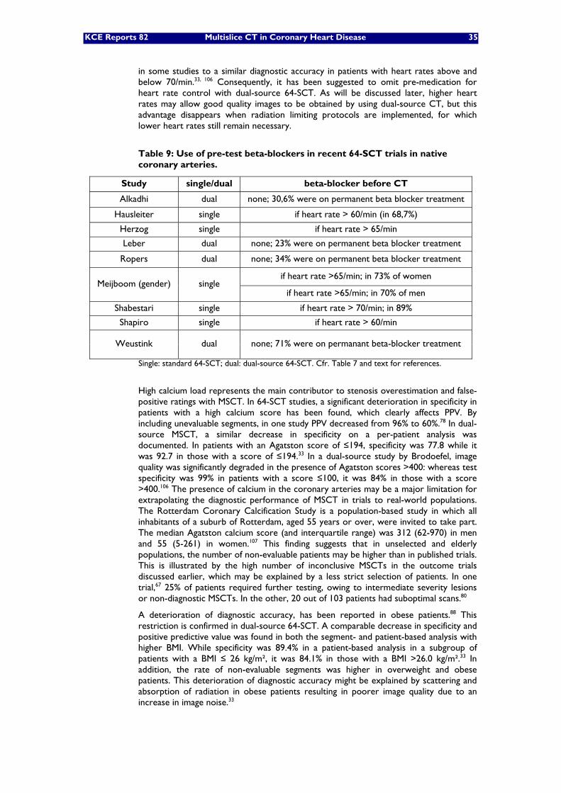
KCE Reports 82 Multislice CT in Coronary Heart Disease 35
in some studies to a similar diagnostic accuracy in patients with heart rates above and below 70/min.33, 106 Consequently, it has been suggested to omit pre-medication for heart rate control with dual-source 64-SCT. As will be discussed later, higher heart rates may allow good quality images to be obtained by using dual-source CT, but this advantage disappears when radiation limiting protocols are implemented, for which lower heart rates still remain necessary.
Table 9: Use of pre-test beta-blockers in recent 64-SCT trials in native coronary arteries.
Study single/dual beta-blocker before CT
Alkadhi dual none; 30,6% were on permanent beta blocker treatment
Hausleiter single if heart rate > 60/min (in 68,7%)
Herzog single if heart rate > 65/min
Leber dual none; 23% were on permanent beta blocker treatment
Ropers dual none; 34% were on permanent beta blocker treatment
if heart rate >65/min; in 73% of women Meijboom (gender) single
if heart rate >65/min; in 70% of men
Shabestari single if heart rate > 70/min; in 89%
Shapiro single if heart rate > 60/min
Weustink dual none; 71% were on permanant beta-blocker treatment
Single: standard 64-SCT; dual: dual-source 64-SCT. Cfr. Table 7 and text for references.
High calcium load represents the main contributor to stenosis overestimation and false-positive ratings with MSCT. In 64-SCT studies, a significant deterioration in specificity in patients with a high calcium score has been found, which clearly affects PPV. By including unevaluable segments, in one study PPV decreased from 96% to 60%.78 In dual-source MSCT, a similar decrease in specificity on a per-patient analysis was documented. In patients with an Agatston score of ≤194, specificity was 77.8 while it was 92.7 in those with a score of ≤194.33 In a dual-source study by Brodoefel, image quality was significantly degraded in the presence of Agatston scores >400: whereas test specificity was 99% in patients with a score ≤100, it was 84% in those with a score >400.106 The presence of calcium in the coronary arteries may be a major limitation for extrapolating the diagnostic performance of MSCT in trials to real-world populations. The Rotterdam Coronary Calcification Study is a population-based study in which all inhabitants of a suburb of Rotterdam, aged 55 years or over, were invited to take part. The median Agatston calcium score (and interquartile range) was 312 (62-970) in men and 55 (5-261) in women.107 This finding suggests that in unselected and elderly populations, the number of non-evaluable patients may be higher than in published trials. This is illustrated by the high number of inconclusive MSCTs in the outcome trials discussed earlier, which may be explained by a less strict selection of patients. In one trial,67 25% of patients required further testing, owing to intermediate severity lesions or non-diagnostic MSCTs. In the other, 20 out of 103 patients had suboptimal scans.80
A deterioration of diagnostic accuracy, has been reported in obese patients.88 This restriction is confirmed in dual-source 64-SCT. A comparable decrease in specificity and positive predictive value was found in both the segment- and patient-based analysis with higher BMI. While specificity was 89.4% in a patient-based analysis in a subgroup of patients with a BMI ≤ 26 kg/m², it was 84.1% in those with a BMI >26.0 kg/m².33 In addition, the rate of non-evaluable segments was higher in overweight and obese patients. This deterioration of diagnostic accuracy might be explained by scattering and absorption of radiation in obese patients resulting in poorer image quality due to an increase in image noise.33

36 Multislice CT in Coronary Heart Disease KCE Reports 82
In most studies, coronary segments with a diameter <1.5 to 2.0 mm have been excluded from analysis. Although it is correct that these stenoses are less well amenable to revascularisation, their documentation is not without diagnostic value. From a prognostic point of view, a completely normal coronary angiogram (i.e. without any sign of atheromatous disease) is different from an angiogram with signs of atheromatosis in smaller vessel segments. Furthermore, in MSCT the assessment of vessel stenosis is semiquantitative, and most often dichotomous, merely reporting a luminal narrowing being less or more than 50%. This is an oversimplification: in many CCA trials, a stenosis has to exceed 70% narrowing to be considered obstructive, and coronary lesions <50% do not exclude future severe events. Acute coronary syndromes result from a sudden blockage of coronary blood flow, due to rupture of a vulnerable atheromatous plaque, very often not involving flow-limiting stenoses.1-3
Based on the aforementioned trial results, it has been concluded by several authors and by professional organisations that 64-SCT is useful to rule out CAD in carefully selected patient populations.66, 108, 109 Both the ESC109 and the ACC43, 108 endorse the suitability of MSCT as a noninvasive tool to rule out the presence of obstructive coronary artery lesions. The technique is particularly advocated in patients with atypical chest pain, patients with equivocal stress test results and patients admitted to the ED with acute chest pain in the absence of ECG changes or biomarker elevations. It should be noticed, that this hypothesis has not yet been tested in clinical trials.
Published trials have been focusing on the accuracy of MSCT in imaging coronary arteries, but that is not what patients are asking for. What they want is their symptoms to be alleviated or their survival to be improved. From a societal perspective, these goals should be achieved at a reasonable cost. These issues will be discussed in further sections.
Key points
• The diagnostic performance of MSCT has been tested in trials that enrolled patients selected for CCA with a high pre-test likelihood of CAD. A negative LR < 0.1 indicates that in these populations MSCT performs very well to rule out obstructive CAD.
• It has not been shown that the available trial results can be extrapolated to populations in which the use of MSCT is currently advocated.
• Coronary artery calcium load, body mass index, and high or irregular heart rates impose restrictions on the diagnostic performance of MSCT, even with dual-source 64-SCT.
3.4 SUGGESTIONS FOR FUTURE RESEARCH
The need for outcome trials, in order to define the position of MSCT in clinical practice, has been stressed by many authors.21, 110-112 So far, the diagnostic performance of 64-SCT has been demonstrated only in patients in whom the decision to proceed to invasive CCA was already taken. Published trials included a variety of symptomatic and asymptomatic patients with known CAD, with positive noninvasive tests, with inconclusive noninvasive tests or with no information on whether noninvasive testing had been performed. These populations are clearly different from the target populations in which the use of MSCT is supported by current guidelines.43, 108, 109 Figure 3 represents the patient populations that have been studied in clinical trials (light grey shade) in contrast to those for whom MSCT is currently advocated by international guidelines (dark grey shade) but in whom no evidence from clinical trials is available (“terra incognita”).

KCE Reports 82 Multislice CT in Coronary Heart Disease 37
Figure 3: Clinical path of patients studied in trials and those deemed appropriate for MSCT by current guidelines
Shaded area: patients studied in clinical trials so far. Dark grey area: patients in whom MSCT is
currently advocated in international guidelines.108, 109 Bottom area: terra icognita. MPS: myocardial perfusion scintigraphy. DSE: dobutamine stress echocardiogram. CCA: conventional (invasive) coronary angiography.
The most decisive evidence for judging the effectiveness of MSCT should come from randomised controlled trials. MSCT can affect patient outcome when the information obtained from it is used to guide decisions to start, withhold, modify or stop treatment.113 Only patients in whom coronary imaging is deemed appropriate but the likelihood for revascularisation is low, should be enrolled in such a trial. If the potential need for revascularisation is high, invasive CCA is a more efficient first step because it allows to proceed to the therapeutic intervention (PCI) within the same procedure. Noninvasive imaging by MSCT can e.g. be envisaged for reassurance of a patient (or his/her cardiologist) or for making an early decision for discharge of a patient admitted with acute chest pain from the emergency department. Randomisation of such patients in a trial can take place at different decision points in the clinical path (after stress testing, MPS, or DSE) and against several alternative diagnostic options (MPS, DSE, CCA, sequential biomarkers). The outcome of such a trial should not focus on the correctness of the anatomical diagnosis but on endpoints that are relevant to patients, such as symptom control, prevention of MI, and prolongation of survival. From a societal perspective, long term downstream costs differences between different pathways should be obtained.
Key point
• There is an urgent need for evidence on (1) the diagnostic performance of MSCT in real world clinical practice, (2) its effect on patient outcomes (QoL, prevention of infarction, prolongation of life) and (3) its cost-effectiveness as compared to diagnostic pathways in which MSCT is not embedded.
STANDARD CARE. CCA if APPROPRIATE.
CHEST PAIN ANGINASTANDARD CARE.
CCA if APPROPRIATE.
ATYPICAL CHEST PAIN
NEGATIVE MPS/DSE POSITIVE
NEGATIVE ECG STRESS TEST POSITIVE
STANDARD CARE. CCA if APPROPRIATE.
INCONCLUSIVE
APPROPRIATE CANDIDATE FOR MSCT ?
INCONCLUSIVE
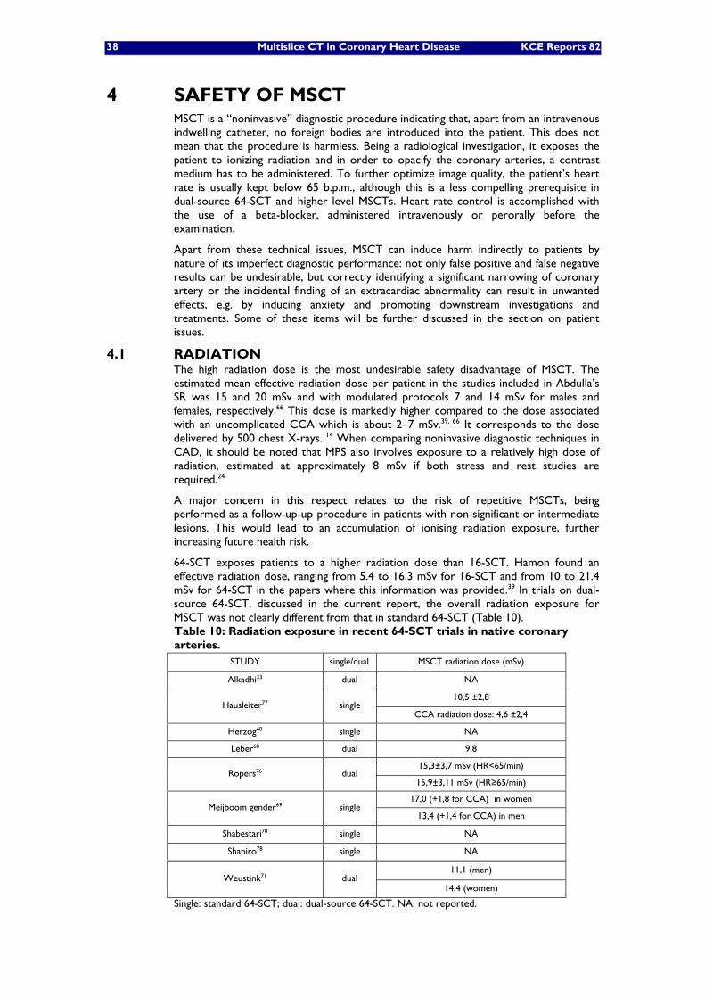
38 Multislice CT in Coronary Heart Disease KCE Reports 82
4 SAFETY OF MSCT MSCT is a “noninvasive” diagnostic procedure indicating that, apart from an intravenous indwelling catheter, no foreign bodies are introduced into the patient. This does not mean that the procedure is harmless. Being a radiological investigation, it exposes the patient to ionizing radiation and in order to opacify the coronary arteries, a contrast medium has to be administered. To further optimize image quality, the patient’s heart rate is usually kept below 65 b.p.m., although this is a less compelling prerequisite in dual-source 64-SCT and higher level MSCTs. Heart rate control is accomplished with the use of a beta-blocker, administered intravenously or perorally before the examination.
Apart from these technical issues, MSCT can induce harm indirectly to patients by nature of its imperfect diagnostic performance: not only false positive and false negative results can be undesirable, but correctly identifying a significant narrowing of coronary artery or the incidental finding of an extracardiac abnormality can result in unwanted effects, e.g. by inducing anxiety and promoting downstream investigations and treatments. Some of these items will be further discussed in the section on patient issues.
4.1 RADIATION The high radiation dose is the most undesirable safety disadvantage of MSCT. The estimated mean effective radiation dose per patient in the studies included in Abdulla’s SR was 15 and 20 mSv and with modulated protocols 7 and 14 mSv for males and females, respectively.66 This dose is markedly higher compared to the dose associated with an uncomplicated CCA which is about 2–7 mSv.39, 66 It corresponds to the dose delivered by 500 chest X-rays.114 When comparing noninvasive diagnostic techniques in CAD, it should be noted that MPS also involves exposure to a relatively high dose of radiation, estimated at approximately 8 mSv if both stress and rest studies are required.24
A major concern in this respect relates to the risk of repetitive MSCTs, being performed as a follow-up-up procedure in patients with non-significant or intermediate lesions. This would lead to an accumulation of ionising radiation exposure, further increasing future health risk.
64-SCT exposes patients to a higher radiation dose than 16-SCT. Hamon found an effective radiation dose, ranging from 5.4 to 16.3 mSv for 16-SCT and from 10 to 21.4 mSv for 64-SCT in the papers where this information was provided.39 In trials on dual-source 64-SCT, discussed in the current report, the overall radiation exposure for MSCT was not clearly different from that in standard 64-SCT (Table 10). Table 10: Radiation exposure in recent 64-SCT trials in native coronary arteries.
STUDY single/dual MSCT radiation dose (mSv)
Alkadhi33 dual NA
10,5 ±2,8 Hausleiter77 single
CCA radiation dose: 4,6 ±2,4
Herzog40 single NA
Leber68 dual 9,8
15,3±3,7 mSv (HR<65/min) Ropers76 dual
15,9±3,11 mSv (HR≥65/min)
17,0 (+1,8 for CCA) in women Meijboom gender69 single
13,4 (+1,4 for CCA) in men
Shabestari70 single NA
Shapiro78 single NA
11,1 (men) Weustink71 dual
14,4 (women)
Single: standard 64-SCT; dual: dual-source 64-SCT. NA: not reported.
KCE Reports 82 Multislice CT in Coronary Heart Disease 39
By means of prospective ECG-gating (the “step-and-shoot mode”) where the X-ray beam is turned on only during late diastole, ionizing radiation exposure can be reduced. In a small series, it was brought down to 2.1 mSv when heart rates were lower than 63 b.p.m.115 In this feasibility study, heart rates were substantially lower than in previous reports because a higher beta-blocker dose was used. Successful MSCT scanning with low-dose radiation clearly requires careful patient selection. According to a recent study, succesful step-and-shoot mode dual-source 64-SCT could be obtained in patients younger than 65 years of age, with a BMI <30 kg/m², a heart rate <70 b.p.m., a <10 b.p.m. heart rate variability, and a CAC score <400 U. When these predictors where used, succesful imaging was attained in 90.5% of patients.116 Very recently, radiation exposure with 320-SCT was reported to be 6.8±1.4 mSv (n = 25; 120 kV, 400 mA, prospective ECG-gating, 60-100% phase window, 16 cm craniocaudal coverage, single heartbeat).117 There seems to be a trend towards lowering radiation exposure of patients by MSCT, but this may occur at the cost of lowering diagnostic perfomance because of stricter heart rate prerequisites and less images being available for post-processing. No diagnostic studies have been published where prospective ECG-gating has been compared with CCA. On the oher hand, the emergence of hybrid techniques combining MSCT with MPS will inevitably lead to an increase of radiation exposure.115
In a simulation study, equivalent doses to individual organs from MSCT were determined, and life time cancer risks from these doses were calculated using the approach of the BEIR II (National Academies’ Biological Effects of Ionizing Radiation 7th report).118 Lifetime cancer risk estimates for standard MSCT varied from 1 in 143 for a 20-year-old woman to 1 in 3261 for an 80-year-old man. Use of simulated ECG controlled current modulation decreased these risk estimates to 1 in 219 and 1 in 5017, respectively. The highest organ lifetime attributable risks were for lung cancer and, in younger women, breast cancer. In December 2006, the Belgian Hoge Gezondheidsraad formulated its concerns on the increasing use of CT scanning in Belgium. 119
4.2 CONTRAST MEDIUM ADMINISTRATION
MSCT necessitates the intravenous administration of a contrast medium. This can give rise to allergic reactions and to renal failure. Contrast-induced nephropathy is an important cause of iatrogenic acute renal impairment and it represents the third leading cause of new acute renal failure in hospitalized patients.120 Whether a patient develops clinically significant acute renal failure, depends on the presence or absence of certain risk factors. A multivariate analysis of prospective trials has shown that baseline renal impairment, diabetes mellitus, congestive heart failure, and higher doses of contrast media increase the risk of contrast nephropathy.120 Patients with these risk factors most often have been excluded from trials on the diagnostic accuracy of MSCT. Prospective studies have produced varied estimates of the incidence of contrast nephropathy, due to differences in the definition of renal failure as well as differences in patient comorbidity and the presence of other potential causes of acute renal failure. A epidemiologic study reported a rate of 14.5% in a series of approximately 1800 consecutive patients undergoing invasive cardiac procedures.121 Patients without any significant risk factors have a much lower risk, averaging about 3% in prospective studies.122 In most cases renal impairment reverses within a week, taking care to avoid further nephrotoxic insults and careful control of fluid and electrolyte balance. In more severe cases, temporary dialysis may be necessary.
Adverse events of MSCT imaging, induced by a contrast medium, such as allergic reactions and renal insufficiency are shared by CCA in which these agents are used in comparable doses.
4.3 BETA-BLOCKADE
In order to reduce heart rate and improve image quality, most patients who are prepared for MSCT will receive a beta-blocker if their heart rate is above a certain threshold, typically 60 or 65 b.p.m. Potential adverse effects of beta-blockade are hypotension, extreme bradycardia and bronchospasm, indicating that careful clinical monitoring of patients is necessary before, during and after the procedure. The need for

40 Multislice CT in Coronary Heart Disease KCE Reports 82
pre-test administration of beta-blocking agents is less compelling in dual-source 64-SCT, although this advantage may be disappear when prospective ECG-gating is used.
4.4 EXTRACARDIAC FINDINGS
A broad spectrum of extracardiac incidental findings have been described in patients that underwent MSCT. In 5 to 56% of patients these findings were considered to make follow-up mandatory.123 Commercially available software programs for pulmonary nodules have reasonable sensitivity, but they are limited by poor specificity and a high rate of false positive findings.37 Unexpected extracardiac findings may lead to further, and sometimes inapproppriate testing and therapeutic acts.124 In a recent poll among Belgian radiologists, 24 out of 31 reported identifying extra-cardiac pathologies in 2 to 50% (mean=15%) of patients. Seven respondents did not report finding significant extra-cardiac abnormalities. More information on this national poll is discussed in the chapter on organisational issues.
Key points
• The high radiation dose is the most undesirable safety disadvantage of 64-SCT. The estimated mean effective radiation dose per patient is 15 and 20 mSv and with modulated protocols 7 and 14 mSV for males and females, respectively.
• It is unclear whether radiation saving algorithms that are currently under investigation will preserve the diagnostic performance of the test.
• Cancer risk induced by MSCT can be substantial, and depends on age and gender. In elderly men, the excess risk of fatal cancer is estimated at 1/5000, whereas in younger women, it can be as high as 1/200.
• Safey issues are also related to contrast medium administration and the need for beta-blocker pre-medication in many patients.
KCE Reports 82 Multislice CT in Coronary Heart Disease 41
5 COST-EFFECTIVENESS OF MSCT COMPARED TO OTHER DIAGNOSTIC MODALITIES
5.1 ECONOMIC LITERATURE REVIEW
The interest in the costs and cost-effectiveness of multi-slice CT angiography comes from the alleged savings induced by MSCT from avoiding unnecessary invasive procedures. If MSCT is used for screening purposes, its aim is to detect subclinical atheromatosis, the idea being that by treating such manifestations in an early phase, future expensive interventions may be avoided and the quality and quantity of life may increase. However, the sword might cut at two sides. These beneficial effects on costs and health effects remain to be proven while limitations and potential harms have to be considered. Concerns are the administration of beta-blockers, the injection of iodinated contrast, the exposure to high-dose radiation and the potential of misdiagnosis (false positives).
5.1.1 Methodology
For the review of the economic literature on MSCT angiography, we searched Medline, Premedline, Embase, Econlit, HTA database and NHSEED. The search was performed between November 30th and December 6th, 2007. Search strategies for each database are presented in appendix.
The search strategy resulted in 290 unique references across all databases. Two researchers independently selected relevant titles and abstract. For references selected by one researcher but not the other a consensus was sought.
Inclusion criteria were:
• population: low to medium risk chest pain
• intervention: multi-slice CT angiography
• outcome: avoided invasive procedures (intermediary outcome), quality adjusted life years or simply life years gained (final outcome)
• design: full or partial economic evaluation, cost-outcome description
Exclusion criteria were focus on EBCT or MSCT of less than 64 slices, absence of economic information, MSCT in high-risk population. Letters and editorials were also excluded.
After a first selection round, 53 titles and abstracts were selected for full text retrieval. Scanning the full text led to the 50 exclusions. Two of these were excluded for reasons of language (Chinese and Hebrew). Three additional references were found manually.
None of the six studies that were retained for the literature review used final outcome parameters in the analysis. Moreover, no single full economic evaluation, including an incremental cost-effectiveness analysis, was found in literature. We discuss briefly the six economic studies of MSCT found in literature.
The resulting schematic tree can be found in appendix.
5.1.2 Results
The study by Dewey et al. (2007) was a decision analytic model, comparing 6 alternative strategies to diagnose coronary artery disease (CAD) in different hypothetical cohorts of patients, defined according to their pre-test likelihood ranging from 10% to 100%.125 No other characteristics than pre-test likelihood of CAD were given about the cohorts.

42 Multislice CT in Coronary Heart Disease KCE Reports 82
The six alternatives were:
• multi-slice CT angiography,
• EBCT calcium scoring,
• dobutamine stress MRI,
• exercise ECG,
• dobutamine stress echocardiography and
• immediate conventional coronarography (CCA).
In the 5 first strategies, a CCA was done if the diagnostic test was inconclusive or positive. Accuracy tests characteristics with their 95% confidence-interval and rate of complications were drawn from the literature. MSCT sensitivity and specificity were assumed to be 95.6% (93.5%-97.2%) and 78.8% (73.9%-83.2%) respectively. Significant CAD meant that at least one coronary vessel with at least one stenosis with a minimum diameter reduction of 50% was identified by CCA.
The primary outcome was the number of correctly CAD diagnosed patients. Costs of strategies were based on the German outpatient reimbursement scheme and purchasing prices of drugs. Possible treatment costs subsequent to diagnosis were not included. Myocardial infarction was assumed as a typical serious complication and considered to cost €11 742 (including hospitalisation, rehabilitation and loss of productivity). A probability of 25% AMI over 10 years was assumed in case of a false negative test.
The authors concluded that MSCT was the most cost-effective diagnostic technique up to a pre-test likelihood of CAD of 50%, being replaced by CCA in populations with a higher pre-test likelihood of CAD. Between 10% and 50% likelihood, MSCT costs per correctly diagnosed patients ranged between €4 435 and €1 469. At 60% MSCT and CCA cost about the same, i.e. around €1 345. Between 70% and 100% likelihood, the cost of CCA was between €1 153 and €807. Actually, all strategies’ costs per correctly diagnosed CAD patient decreased when pre-test likelihood increased. This seems intuitively correct, as the number of true positives increases as the likelihood of CAD increases.
Sensitivity analyses on specificities, sensitivities, reimbursement rates and complications of CCA (varying from 0.5% to 2%) did not change much of the conclusions, the 50% limit sliding to 80% when the reimbursement rate was maximized for CCA. Even the so-called order of cost-effective tests stayed roughly the same (the rest of the tests never outrunning MSCT or CCA).
While the authors note that the omission of the costs of therapeutic management after diagnosis of CAD is a strength of the model, this can be disputed. What policy makers are really interested in is the final outcome of MSCT angiography in terms of life-years gained or QALYs gained and the amount of resources needed to obtain this final outcome. Obviously, the treatment following the diagnosis is part of the clinical path and should be taken into account when calculating the relative value for money of MSCT angiography compared to its alternatives.
Another weakness of the study is the exclusion of the false positives cases and their associated costs and outcomes from the calculation, especially in the light of the MSCT specificity of 78.8% (CI95%: 73.9%–83.2%). Beyond the distress and suffering, false positive tested patients underwent unnecessary invasive procedures; they may have been prescribed drugs inducing a supplementary costs and possible secondary effects, etc. Hence the rate of false positives would increase downstream costs, especially if prevalence of CAD is low.
The costs of false negatives, on the other hand, were included, implying a cost of an AMI in the next ten years in one out of four missed diagnoses. One could argue that a patient re-experiencing chest pain will seek further medical consultations and will not wait until AMI occurs in the next 10 years. Moreover, it has not been shown that the presence or absence of a >50% coronary stenosis allows to predict the future occurrence of a MI. Indeed, AMIs often occur in lesions <50%, induced by plaque
KCE Reports 82 Multislice CT in Coronary Heart Disease 43
rupture in non-obstructive lesions. Morbidity and mortality per diagnostic test were introduced into the model but no further details on the results were given, because these rates were considered as cost drivers rather than outcomes of the intervention.
This example leads to the next point: symptomatic patients suffering from chest pain were not distinguished from asymptomatic patients within the same cohort, while the care process may be totally different. The model cannot fit the whole range of population it intends to cover. For example, an immediate conventional coronarography is very unlikely to be a diagnostic tool for a low prevalence population. Next, it seems more plausible that different alternative tests would be combined into one diagnostic strategy instead of substituting one for another. This might especially be the case if the first test is inconclusive. For example, it might be useful to do another noninvasive test first if the MSCT angiography is inconclusive rather than proceeding immediately to CCA.
The costs were expressed per correctly CAD diagnosed patient instead of per patient. Surprisingly, Dewey et al. state that “correct diagnosis of absence of disease was not considered a direct criterion of effectiveness”.125 However, the negative predictive value of a test is the crucial point when testing a 10% pre-test likelihood population. Again, modelling decisions might differ in function of the pre-test CAD likelihood of the cohort.
In order to assess the feasibility of a new chest pain unit (CPU) protocol in the Carmel Medical Center based at Haifa (Israel), Rubinshtein et al. 126 followed 124 patients presenting at the emergency room, including 90 patients with chest pain (of whom 42 with atypical pain and/or uncertain aetiology). After a first triage by the emergency room team, 14 patients were directly referred for early CCA. Patients with neither clear-cut ACS, nor clear non-cardiac diagnosis were subsequently assessed by the CPU protocol managed by two cardiologists (including ECG every 4-6 hours, cardiac troponin T test and TIMI score assessment). Patients with negative tests were referred to noninvasive testing. According to physician preference, 29 patients were referred to 64-SCT coronary angiography (other possible choices included exercise tress test, MPS, echocardiography and CT scans of other organs than heart or coronary vessels). Finally 40 out of 124 patients (32%) were discharged. No myocardial infarctions or deaths were observed in these patients at 30 days follow-up. Thirty percent (30/101) of the patients designated for hospitalisation by the ER team were finally discharged after the CPU evaluation while 56% (13/23) of the candidates for discharge were finally hospitalised after CPU evaluation. Seven out of those 13 redirected patients underwent a revascularisation.
The length of stay in ER (and CPU) before discharge or hospitalisation was reported to be 13.6 hours (SD =10.3 hours). Use of noninvasive testing was significantly higher in discharged patients than in hospitalised patients (85% vs 15%). Authors concluded to a potential saving in hospitalization days, work-up and consultations and to a prevention of unwarranted discharges from the emergency room.
Unfortunately, as stated by the authors themselves, the precise role and accuracy of 64-SCT as such was not evaluated in the study.
In the cost study published by Cole et al. 127 in 2007, 206 patients with no “high-risk” markers and with mildly abnormal, equivocal or un-interpretable MPS were referred for CT coronary angiography on a 64-SCT unit, in a (physician’s) desire to avoid invasive catheterization. This number was more or less a selection of 40% of the 6% of patients undergoing MPS with unclear results. Sixty-six out of the 206 patients (32%) had potentially obstructive plaque and were therefore sent to CCA (including 10% patients with un-interpretable CT studies). Among the remaining patients, 61 had normal studies (29.6%) and 76 studies showed atherosclerosis but no evidence of potential obstruction on study (38.3%).
In order to compare costs, this management strategy (catheterization only in patients with potentially obstructive plaque detected on MSCT coronary angiography or with inconclusive MSCT coronary angiography) was opposed to a second strategy: direct CCA for all 206 patients. Only direct costs that would be involved by the procedures
44 Multislice CT in Coronary Heart Disease KCE Reports 82
and outpatient hospital costs were calculated. Cost estimates consisted of patient co-payments and hospital reimbursement amounts from insurers and Medicare. The cost in case of the immediate catheterization strategy for 206 patients was simulated by multiplying the unit cost of catheterization by 206. This cost was compared with the cost of 206 MSCT coronary angiographies followed by 66 catheterizations.
The results show that the cost of the direct CCA strategy is $1486 per patient higher than the strategy with MSCT. This is due to catheterisation being more expensive than MSCT coronary angiography ($2940 versus $544). According to a one-way sensitivity analysis, MSCT gate-keeping is no longer cost saving if more than 81.5% (instead of 32%) of the patients are sent to CCA after MSCT. In other words, MSCT coronary angiography cannot be cost saving in a practice where physician’s referral to coronary angiography is high, i.e. more than 81.5%.
This study did not report patient outcomes. Six patients had a negative CCA after they had been sent to CCA following an un-interpretable MSCT. The morbidity or mortality associated with CCA was not included in the model. The original population from which the 206 patients with unclear MPS were selected is not described, nor the reason for encounter, even if it may be (unqualified) chest pain. The paper thus illustrates possible cost-savings under the 2005 Alabama particular reimbursement scheme for this hospital case-mix.
Last year, Otero et al. (2007) published a study aiming at determining the maximum budget neutral reimbursement rate for MSCT if this technique was to become the method of choice in acute chest pain imaging in the emergency setting.128 This decision modelling from the Medicare perspective compared 3 alternatives strategies of CAD diagnosis:
1. MSCT
2. Stress echocardiography (DSE)
3. SPECT (MPS)
Medicare costs and patients’ outcomes were simulated for a cohort of 10 000 patients without changes on ECG and without cardiac enzyme abnormality. While the authors state that this are patients at intermediate risk, they should actually be considered at low risk for future cardiovascular events (see chapter 2). The prevalence of CAD in this population was assumed to be 20% (19% of the patients presenting annually to the emergency room for chest pain actually have CAD). Three percent of the CAD patients would have an AMI or angina during the index hospital admission. The mortality in those CAD patients after AMI is 7.5% if the AMI occurs in hospital and 25% if the AMI occurs outside the hospital.
In both alternatives to MSCT, the initial emergency test was followed by CCA when positive, by discharge home when negative and by a 24 hour observation period when inconclusive in order to decide on doing a CCA or discharging the patient. In the MSCT strategy, inconclusive test results on MSCT were followed by stress echocardiography. Observation after inconclusive MSCT was not an option. Test characteristics were based on English language literature on 64-SCT and American College of Cardiology/American Heart Association expert consensus. MSCT sensitivity and specificity were assumed to be 95% and 90% respectively. Costs included only the actual national Medicare average reimbursements for diagnostics and observation unit fees.
Rates of complications from noninvasive tests were considered negligible and therefore not included. Outcomes studied were deaths, intra- and extra-hospital myocardial infarction, number of tests performed and observation time needed.
To make the costs of the MSCT strategy equal to those of the strategy with stress echo, the maximum reimbursement for MSCT should amount to $433. To equal the costs of the MSCT strategy with the costs of the MPS strategy, the maximum amount should be $990. Three deaths and 19 (in- and out-hospital) AMI were reported as results of the stress echo algorithm, one death and 14 AMI for the MPS algorithm and one death and 8 AMI in the case of the MSCT algorithm. As for the numbers of negative CCAs, they were respectively 2 352 (stress Echo), 1 060 (MPS) and 266 (MSCT).

KCE Reports 82 Multislice CT in Coronary Heart Disease 45
The results of this study are not easily reproducible as the incidence of AMI in (missed) CAD patients was not given. Moreover, next to the AMI mortality, the mortality associated with CCA was apparently not included in the model. For example, according to the assumed mortality risk associated with CCA of 0.1%, 3 deaths are expected in the MPS arm, where the number of CCA after a positive MPS was almost 3 000. However, the results report only one death. The same applies to CCA morbidity, where the authors mentioned a morbidity rate of less than 1% but do not include this in their model.
Finally, Goldstein et al. (2007) performed a randomized controlled trial of MSCT for the evaluation of acute chest pain.67 One hundred ninety-seven (197) patients aged 25 years or older, at low risk for coronary events, no history of CAD and presenting at the emergency department with acute chest pain were randomized to “Standard of Care” or MSCT. Their ECG at time=0 and time=4 hours were normal as well as their serum biomarkers. The standard of care diagnostic protocol to rule out myocardial infarction included serial ECG and cardiac enzymes, followed by rest-stress MPS before referring home or to catheterization laboratory. The MSCT strategy included calcium scoring and angiography, followed by CCA when positive, discharge home when normal and by nuclear stress testing when MSCT results are intermediate or inconclusive. Outcomes included number of tests complications, major adverse cardiovascular events (death, AMI, unstable angina), number of correctly diagnosed patients and time to diagnosis. A diagnosis was judged correct based on the results of a catheterization or the presence or absence of major adverse cardiovascular events during the index admission or the 6-month follow-up period. Costs were calculated based on data from the hospital billing department and based on the emergency department’s cost-to-charge ratio. Although not clearly stated in the methods section of the article, the cost-to-charge ratio seems to be a cost per hour of use of the emergency department. The authors were contacted to obtain more details about the ratio used, but did not respond to our e-mail message. Costs of the procedure were included (MSCT $507 and nuclear imaging $538).
Among the 99 patients following the MSCT arm, 96 (95%) were correctly diagnosed: 88 without CAD (including 1 readmission for a negative CCA) and 8 with a positive CCA. Twenty-four (24.2%) had to have a nuclear stress imaging due to non-diagnostic MSCT and 4 patients had a CCA that turned to be negative. In the emergency department setting, MSCT was able to immediately identify or exclude CAD in 75% of cases. No test complications or major cardiovascular events were noticed in both arms in the 6-month follow-up period. Eight patients in each group required a late office or emergency department visit for recurrent chest pain. Fewer patients required additional noninvasive evaluations (the protocol was not described) in the MSCT than in the standard of care arm (2% versus 7%; p=0.10). The median time to diagnosis was 3.4 hours in the MSCT arm (25th percentile: 2.3 hours, 75th percentile 14.8 hours) versus 15 hours in the “standard of care”-arm (25th percentile 7.3 hours; 75th percentile 20.2 hours). As a result from reduced time in the emergency department, costs were significantly lower for MSCT patients amounting to $1 586 (25th percentile $1 413; 75th percentile to $2 059) against $1 872 for the standard of care arm (25th percentile $1 727; 75th percentile $2 069).
The authors conclude that MSCT is safe and highly effective to give a correct diagnosis. However, MSCT still has limitations in terms of being able to determine the physiological significance of intermediate coronary lesions. They warn against a possible oculostenotic reflex, caused by the inability of MSCT to provide coronary blood flow data. Further studies are recommended to determine the optimal use of MSCT.
Just like the other studies described, this study does not strictly satisfy the criteria of a full economic evaluation. No incremental calculations were made, no incremental cost-effectiveness ratio was calculated and costs were incompletely taken into account (e.g. costs of CCA, PCI, CABG, repeat evaluations during follow-up).
While the authors are very enthusiastic about MSCT for the evaluation of acute chest pain, it should be noted that the number of invasive procedures (CCA, PCI and CABG) is higher in the MSCT-arm than in the “standard of care”-arm, while the outcomes in
46 Multislice CT in Coronary Heart Disease KCE Reports 82
terms of mortality and morbidity up to 6 months are not any different between the arms.
Moreover, despite the apparent safety of both strategies (absence of adverse complications), 10% of the patients in the MSCT arm had to undergo a radiotoxic test twice (MSCT+nuclear testing) and 4% even three times (MSCT+nuclear testing+CCA). Iodinated contrast also presents a potential harm in MSCT evaluation. Although 8 CCAs out of 12 were positive in the MSCT-arm against only 1 out of 7 in the ”standard of care”-arm, this does not necessarily mean anything for the prognosis of the patients with a positive CCA.
The numbers of patients were too small to evaluate the true incidence of false positive cases and false negative cases, especially in a population with a low prevalence.
Recently the Andalusian HTA agency 129 published a report on MSCT coronary angiography including a meta-analysis of 16-SCT or more coronary angiography and an economic model based on a decision tree. The input parameters were drawn from the meta-analysis of studies using “patients” as units of analysis. The model was populated with patients with a suspected coronary stenosis (>50% vessel diameter). The three following strategies were compared:
• direct CCA,
• 16-SCT coronary angiography followed by CCA when positive,
• 64-SCT coronary angiography followed by CCA when positive
The prevalence of significant coronary stenosis was assumed to be 40%. The sensitivity and specificity of 16-SCT were 94% and 77.9% respectively. Those of 64-SCT were 98.4% and 93.7% respectively. Complications due to CCA occurred in 2.2% of the procedures and lead to death in 5% of the complicated procedures, AMI in 45% and urgent surgery in the remaining 50%.
The perspective was that of the Andalusian public health system. Costs included direct costs of equipment, consumables (including pharmaceuticals), procedures (including ECG and blood tests in the three arms), and costs of personnel. Procedure costs and costs of complications were obtained from the public tariffs of the SSPA (Sistema Sanitario Público Andaluz). For the equipment cost, an average per patient was calculated based on the purchasing price, the throughput and the lifetime of the equipment. Costs of personnel were calculated as the legal hourly wage cost per professional qualification multiplied by the time per test, based on a 2004 Spanish paper comparing CCA to 16-SCT coronary angiography. Costs drawn from the literature were validated by a radiologist and a nurse of the radiology department of two Andalusian hospitals. The time spent by the radiologist, the technician and the nursing auxiliary in the case of a 16-SCT coronary angiography was 45 minutes against 12 minutes for a 64-SCT coronary angiography. Total costs per patient of following the three paths were respectively €203.96 for the 64-SCT path, €259.06 for the 16-SCT path and €307.85 for direct CCA.
The ICER was calculated comparing CCA and 16-SCT to 64-SCT. Two denominators, i.e. effectiveness parameters, were used: (1) number of cases correctly diagnosed with stenosis (true positives) and (2) number of effective cases, defined as the number of true positives minus the number of false negatives. In both methods, the 16-SCT strategy was dominated by the 64-SCT strategy. In the CCA arm more patients were correctly diagnosed (0.64%) than in the 64-SCT arm, but CCA was also more expensive (€103.89 more). The ICER of CCA relative to 64-SCT was €16 596 per correctly diagnosed case and €8 206 per ‘effective case’.

KCE Reports 82 Multislice CT in Coronary Heart Disease 47
Table 11: AETSA cost-effectiveness study baseline results applied to 100 patients 129
Strategy Cost per patient
Incremental costs
True positives
False negatives
ICER1* ICER2**
64-SCT (+CCA if positive)
€203.96 38.5% 0.64%
16-SCT(+CCA if positive)
€259.06 €55.1
36.8% 0.4% <0 <0
Direct CCA €307.85 €103.89 39.1% 0% €16 596 €8 206
* incremental cost per correctly diagnosed patient
** incremental cost per effective case diagnosed
The impact of the assumed prevalence on the results was tested in a one-way sensitivity analysis. When the prevalence was respectively 25%, 50% and 75%, the ICER of CCA versus 64-SCT was €37 425, €9 653 and €396 per correctly diagnosed stenosis and €18 505, €4 773 and €196 per ‘effective’ case. The 16-SCT strategy remained dominated by the 64-SCT strategy in all scenarios. Based on these figures, the reason is not documented why the authors conclude in their executive summary that 64-SCT is most cost-effective when the prevalence of obstructive CAD is 56% while CCA is most cost-effective in a 70% prevalence population.
The AETSA economic evaluation shares three essential problems with the study by Dewey cited above.125 First, mortality and complications after CCA were only considered for the calculation of the costs while they were considered irrelevant for the evaluation of the outcomes of the diagnostic path. Second, the number of correctly diagnosed cases with stenosis is only a surrogate measure of the effectiveness of a diagnostic test. Authors themselves acknowledged as limitation the fact that treatment and treatment outcomes were not considered in their model. Third, tests accuracy characteristics are considered fixed estimates, independent from the prevalence of stenoses. As explained in the chapter 3, this is an invalid assumption.
5.2 CHALLENGING THE “ECONOMIC EVALUATION” OF GOLDSTEIN ET AL67
Despite the lack of evidence about the impact of 64-SCT on patient outcomes, the described economic evaluations of MSCT compared to an alternative diagnostic procedure for CAD are frequently cited to demonstrate the cost-effectiveness of MSCT. Each of these studies, however, was limited by the major gap in knowledge about the clinical effectiveness of MSCT. Therefore, assumptions about sensitivity and specificity of MSCT in low to intermediate risk populations were made and intermediate outcome parameters were used such as “number of correctly diagnosed cases”. No single economic evaluation attempted to model effectiveness in terms of life-years gained or QALYs gained, which is legitimate given the already important uncertainties about the clinical relevance of MSCT in populations who are at low to intermediate risk for coronary events.
The most frequently cited study to claim cost-effectiveness of MSCT compared to standard of care is the “economic evaluation” by Goldstein et al.67. This study did not, however, include an outcome parameter in its economic assessment but merely calculated to median cost of the length of stay in the MSCT arm and the standard of care arm. However, the cost-effectiveness of MSCT depends not only on the costs and effects of the diagnostic strategy and initial hospital stay, but also the costs and effects of its sequelae, i.e. the changes in therapeutic behaviour and the consequent impact on patient outcomes. Therefore, it is insufficient to consider only the technique’s diagnostic accuracy (sensitivity and specificity) in an economic evaluation. An economic evaluation should also incorporate the technique’s effect on patients’ outcomes (life years gained or quality-adjusted life years gained).
48 Multislice CT in Coronary Heart Disease KCE Reports 82
We challenged the conclusions drawn by Goldstein et al. about the cost-effectiveness of MSCT compared to standard of care by fully exploring all information provided by their RCT, including the costs and quality of life effects of invasive angiography, revascularisations and complications in each diagnostic arm up to 6 months after initial admission to the emergency department for acute chest pain. We supplemented the data of the study with data on the quality of life in case of interventional procedures and applied Belgian health care costs to each of the procedures included in the study. The full details of this exercise are presented in appendix.
The results showed that, given the limitations of this exercise, the MSCT diagnostic strategy is on average €479,56 more expensive than the standard of care strategy from the perspective of the health care payer. Moreover, it leads to a higher loss in QALYs: 0.0016 QALYs are lost in the MSCT arm as compared to 0.00056 QALYs in the standard of care arm. This is equivalent to about 6 hours of life in perfect health more lost in the MSCT arm than in the CCA arm. If we neglect the costs of revascularisations and invasive angiography -as did Goldstein et al.67- the costs of the MSCT strategy are lower than the costs of the standard of care strategy. In that case, we reach the same conclusion as the authors.
The figures resulting from this exercise should be treated with caution, as the evaluation was based on data from only one RCT. The patient numbers in each health state were too small to reliably estimate transition probabilities and make the model more generic. For instance, none of the patients in the “standard of care”-arm who underwent a late CCA were revascularised. This might be a coincidence due to the small number of patients undergoing a late CCA. The RCT was not powered to detect such potential relevant differences. In real life, with very large patient numbers, the situation might be different, and some patients might undergo revascularisation if late CCA is positive. To increase the generalizability of the results, more data on the long term consequences of both diagnostic interventions would be needed (need for revascularisation, AMI, death). Data from larger data sets would allow us to define transition probabilities and hence built a more generic model.
A full economic evaluation would require evidence on the effectiveness of MSCT in real world in low- to intermediate risk patients. Evidence on diagnostic accuracy in well-defined patient populations is being built up, meanwhile leaving the assessment of the impact of MSCT on patient outcomes unevaluated.
Key points
• Published economic evaluations of MSCT to detect CAD in low to intermediate risk populations are all limited by the gap in evidence about the clinical effectiveness of MSCT in these populations.
• None of the studies related costs to treatment effects or patient outcomes. Nevertheless, they are frequently cited to demonstrate the cost-effectiveness of MSCT relative to the standard of care.
• A basic economic evaluation, based on data from one RCT, showed that taking treatment or patient outcomes into account might change the conclusions with respect to the cost-effectiveness of MSCT.
• However, given the small number of patients in the RCT, firm conclusions about cost-effectiveness cannot be drawn from this exercise.
• More trials, sufficiently powered to study differences in relevant economic and outcome variables, are needed.

KCE Reports 82 Multislice CT in Coronary Heart Disease 49
6 ORGANISATIONAL ISSUES
6.1 MULTI-SLICE CT ANGIOGRAPHY MARKET
Currencies were converted to euros based on the mid-market rates at 21th April 2008 (1CA$=€0.63, 1AUS$=€0.59, 1£ =€1.25, 1 US$=€0.63).
6.1.1 Multislice Cardiac CT abroad
In the United States, the cost for the equipment of a MSCT scanner runs from $1 million to $2 million (€627 000 to €1.25 mio), workstation and software included. The main producers of 64-slice hardware equipment units are Toshiba America Medical Systems, Siemens Medical Systems, Philips Medical Systems and General Electric. They also offer software and workstations but these can also be bought from third-party manufacturers such as TeraRecon or Vital Images. UK prices range from £600 000 to £1 000 000 for a 64-SCT scanner (€747 000 to €1.25 mio). 130. A new 64-SCT scanner with cardiac capabilities would cost approximately AUS$1.25-1.35 million (around €740 000 to €800 000) or existing 64-SCT scanners may be upgraded with the purchase of the appropriate cardiac softwares and hardware at a cost of approximately $100 000 (€63 000).62
The AquilionONE™ dynamic volume CT, a 320-SCT scanner will most probably be commercialized by Toshiba in the summer of 2008. Its main feature is to scan an organ such as the heart in only one rotation in order to reduce examination time, as well as radiation and contrast agent dose.
6.1.2 Multislice Cardiac CT in Belgium
According to the survey conducted by the Belgian College of Medical Imaging that had a response rate of 94% among Belgian hospitals, there were 240 CT units in 2005, spread among 115 hospitals (excepted 6 units in private surgeries). Approximately 75% were multi-slice units from which 45% with more than 16 detectors. But 40- and 64-SCT units are gaining ground. No other details were available on MSCT angiography in Belgium.
In order to acquire insight in the current local use of MSCT in cardiac applications in Belgium, a meeting with industry representatives was held on December, 7, 2007. These were convened via Unamec, an organisation representing Belgian manufacturers, importers and distributors of medical devices. Representatives from Siemens, Philips and Toshiba were present at this meeting. General Electric, the fourth player on the Belgian market, was contacted via e-mail and telephone. The information thus obtained was completed with data retrieved from manufacturers’ websites and is summarised in this chapter.
Cardiac CT has been used since the late 1970s for left ventricular imaging, but the first MSCT coronary angiography was introduced in 1998. Before, electron beam CT (EBCT - Imatron®) had been developed for coronary calcium scoring (CAC) but this device has never been used in clinical practice in Belgium. This was due to the very high acquisition cost (120 mio BEF) and the fact that the device could not be used for other imaging applications. This lead to the industry’s decision to stop producing this type of scanner, further focusing on the development of newer generation MSCT-scanners that were able to combine calcium scoring and coronary artery imaging.
According to data from Siemens 131, the device cost of a 64-SCT (or higher) scanner is as follows:
• CT hardware system: 64-slice €850 000 and above 64-slice €1.2 to 2.0 million . These devices are also used for scanning of organs other than the heart. Additional software is needed for MSCT of the coronary arteries and makes up 20% of the cost of the device.

50 Multislice CT in Coronary Heart Disease KCE Reports 82
• Post-processing software: €100 000 with a yearly upgrading costing €20 000.
Maintenance: €100 000 yearly.
According to Philips, currently about 240 CT scanners are installed in Belgium (22.7/mio inhabitants); 45% of these have 16 or more detectors. It is estimated that 20% of Belgian hospitals are doing cardiac CT. An unknown but probable high number is contemplating implementation of cardiology applications in the near future.
Approximate market share of CT devices of different manufacturers in Belgium:
1. Siemens: 50-54%
2. General Electric: 25-28%
3. Toshiba: 15%
4. Philips: 8-10%
The type of scanner and its use for cardiac applications for Siemens devices is as follows in Belgium:
• 16 slice: 51 scanners of which 10% are used for cardiac applications in 1 to 15 patients per week.
• 64 slice: 19 scanners of which 30% are used for cardiac applications in 5 patients per week.
• dual 64 slice: 8 scanners, all of which are used for cardiac applications. They examine 5 to 15 patients per week.
Of the 7 university hospitals, 5 are currently performing cardio-CT whereas two of them (UZ Gent, ULB Brussels) do not.
6.1.2.1 NUR/UNR 2008 Survey
The Belgian professional association NUR/UNR (Nationale Unie der Radiologen/Union Nationale des Radiologues) launched a survey around MSCT coronary angiography. The results were transmitted by the association to the KCE, and will become available in the press and on the association website (http://www.nur-unr.be/). Thirty-seven hospitals sent their answers, representing more or less one third of the hospitals in Belgium. Their mean number of MSCT coronary angiographies was around 7 per week, with some hospitals performing up to 30-40 examinations a week. There may be a reporting bias in the sense that radiologist performing this type of examinations might have been more prone to participate in the poll. The lowest reported number was one examination a week, not speaking of three hospitals that did not perform the examination, including one hospital that has ordered a 64-SCT unit and another that did not use its 16-SCT unit for cardiac purposes. The reported waiting time (excluding emergency cases) ranged from 0 days to more than 3 weeks and averaged 6 days per hospital. In most hospitals, there is a good collaboration between radiologists and cardiologists while there is no interest from the cardiologists for MSCT coronary angiography in 6 centres and a refusal to refer patient for MSCT coronary angiographies according to radiologists in 2 hospitals. Most radiologists performing MSCT coronary angiography followed a specific education in cardiac radiology. In one hospital, cardiologists received this education. A cardiac catheterization laboratory was available in the majority of the hospitals (55 %) using MSCT coronary angiography. Some of the remaining respondents work in collaboration with a neighbouring cardiac catheterization laboratory. Almost half of the hospitals performed coronary interventions or worked with a nearby hospital offering it. When they reported the number of coronary interventions, the number ranged from 800 to more than 5 000 (with a mean of 2000 interventions). Ten (corresponding to an approximately a third) out of the responding hospitals using MSCT coronary angiography also offered a service of cardiac surgery, reporting from 270 to 1 200 operations (mean=575 interventions). Every hospital performing MSCT coronary angiography declared itself ready to welcome a higher number of cardiac patients from its region for MSCT coronary angiography.

KCE Reports 82 Multislice CT in Coronary Heart Disease 51
Only one hospital answered positively to the required presence of a cardiologist to interpret a cardiac CT, two others considering it in case of beta-blockers injection and possible contra-indications to it. About 15% of the respondents found a CT coronary angiography more difficult to interpret than other CT examinations, the other 85% did not find it more difficult. Some mentioned the importance of an adequate education. Concerning incidentally found extra-cardiac pathologies, most radiologists reported identifying them in 2 to 50% (mean=15%) of patients, leaving seven out of 31 respondents who did not report significant extra-cardiac abnormalities. Finally, three radiologists mentioned discussion with physicians specialized in nuclear medicine, only one respondent reported to discuss results with other specialists (non-radiologists such as oncologists, vascular surgeons), all other respondents did not need to discuss with another specialist for the performing or interpretation of a CT examination.
6.2 REGULATORY ISSUES
6.2.1 Authorization
MSCT scanners have been granted Class III licenses for use in Canada. Class III encompasses diagnostic and therapeutic devices “potentially hazardous or representing an immediate danger if they fail” 132 Sixty-four slice scanners received US FDA clearance in early 2004 and are CE marked in the EU. The 320-SCT Toshiba scanner received FDA clearance in November 2007.
6.2.2 Planning
There is no formal limitation on the number of CT- or MSCT-units in Belgium (unlike MRI units).
6.2.3 Financing
Unlike in the case of MRI, the hospital budget does not include a part to cover neither investment and depreciation CT costs, nor CT operating costs. Hence, CT direct and indirect costs have to be financed from the CT nomenclature fee-for-services. Contrast material is covered separately and beta-blockers when administered are billed as pharmaceutical products. More details are to be found in the KCE report on Magnetic Resonance, 133.
6.2.4 Patients referral for CT examinations
In the strict sense of the word, self-referral means that the requesting physician and the providing physician are the same. In the USA, cardiologists may perform their own cardiac radiographic studies. In Belgium, this is not possible as cardiologists refer their patients to radiologists for CT imaging. Accordingly, the fees-for-service are reserved to the radiologists only.
In the United States, possible overuse due to non radiologist self-referrals especially among cardiologists is no recent debate. Between 2000 and 2005, US Medicare payments for outpatient medical imaging almost doubled from $6 billion to $11 billion (€3.8 billion to €6.9 billion) 134 New sophisticated and costly technologies such as CT and MR imaging may explain a part of this rise: CT performed by radiologists increased 69% in five years to 500 scans per 1000 beneficiaries while MR rose 82% to 140 scans per 1000. But while in other specialties increases stayed similar around 24% in five years, the rise among cardiologists was 65%, from 400 to nearly 700 scans per 1000 beneficiaries in 2005. Papers published by Hillman in the beginning of the 1990s revealed that nonradiologist self-referral could multiply the number of imaging studies by a factor from 2 to 8 per episode of illness. Eighty percent of self-referred radiology would be unnecessary. 135. In a study based on the 1991 Pennsylvania Blue Shield (PBS) claims data, Levin et al. found 70% of the private office radiologic examinations were performed by non-radiologists (48% of chest radiographs) 135. Next to the inappropriate costs, the quality of self-referred imaging would be disturbingly insufficient. A blind audit of the PBS claims of 1000 radiographic studies (re-assessed by a single board-certified

52 Multislice CT in Coronary Heart Disease KCE Reports 82
radiologist) showed that the rate of unacceptable imaging quality performed by radiologists would amount to 12%. This rate would rise to 41% when imaging is performed by internists, to 45% when imaging is performed by general practitioners and to 53% by pulmonary disease specialists. 135.
In a broader sense, self-referral means that a cardiologist refers his/her patient for imaging to the institution where he has a practice. In the United States the law prohibits this broader form of self-referral since 1995. According to the so-called Stark regulation, a physician referring patients to an entity for services, including radiology, including magnetic resonance imaging, computerized axial tomography scans and ultrasound, cannot hold an ownership interest in this entity. Congress provided for a number of exceptions to this prohibition and gave the Centers for Medicare and Medicaid Services (CMS) the authority to create additional exceptions such as referrals by a physician to the academic centre that employs him and where he teaches 136 Recently, CMS published a proposed rule for 2008 which contained a number of restrictions and clarifications on the Stark Regulation aiming at preventing overuse of services and program abuse. 137
Radiologist activity depends on referrals from other specialists. To answer this challenge, some of them pleaded for an accreditation of imaging facilities, for local reimbursement policies subordinated to quality performance standards as well as for more radiology research by radiologists to be published into other non radiology journals. 135 138 Such initiatives have been implemented in the United States since 2005. As a matter of fact, some health insurers apply conditional reimbursement in function of the equipment (see section 6.3.1). Most US health insurers require a training certification, a continual medication education and a minimum number of cases per year from radiologists and cardiologists. The American College of Radiology issued its cardiac CT practice guideline in 2006 139. According to this guideline, physician performing a cardiac CT should have followed an approved programme and have performed at least 50 cardiac CT scans in the last 6 months. Curriculum ameliorations and reinforcement of physics and engineering principles in the curriculum have also been advocated. 140. In Europe, the rapid evolution in cardiac imaging technologies is also a challenging issue and the main European and national radiological societies are developing programmes for Master in cardiovascular imaging. 141
6.3 COVERAGE OF MULTI-SLICE CT ANGIOGRAPHY
6.3.1 United States of America
Today, Medicare does not refer to MSCT technology in its National Coverage Determination on CT (“NCD for Computerized Tomography (220.1)”). 142 MSCT coronary angiography is not nationally covered but local Medicare contractors may have local determination policies, which is the case for every state by now. Local health insurers generally rely on the AHA 2006 consensus on cardiac computed tomography and the 2007 consensus on CT calcium scoring to make their coverage policy 143. Accordingly, some of the indications that received a class IIb recommendation (conflicting evidence on their usefulness and efficacy) are covered, but the coverage varies with the health insurer. Some other health insurers consider MSCT coronary angiography investigational and do not cover it at all.
According to the Technology Assessment Policy of Harvard Pilgrim HealthCare (Massachusetts, New Hampshire, Maine), MSCT coronary angiography for CAD is a new and promising technology but remains investigational, unproven, and experimental. It is therefore only covered on a case by case basis after review of the patient file. For Unicare, MSCT coronary angiography is considered medically necessary for the evaluation of suspected congenital anomalous coronary arteries when conventional coronary angiography has been unsuccessful or has provided equivocal results and the results will impact treatment. It is considered not medically necessary when used in screening asymptomatic patients, for the detection of coronary artery calcium or for the evaluation of cardiac function.
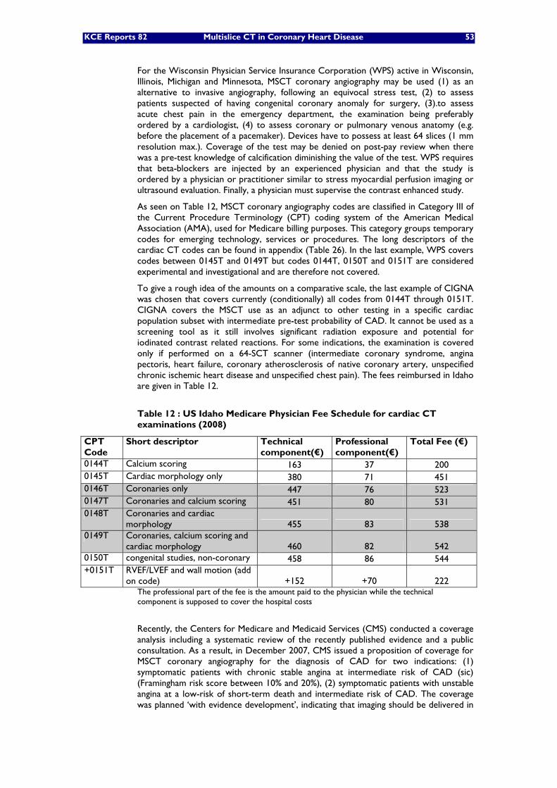
KCE Reports 82 Multislice CT in Coronary Heart Disease 53
For the Wisconsin Physician Service Insurance Corporation (WPS) active in Wisconsin, Illinois, Michigan and Minnesota, MSCT coronary angiography may be used (1) as an alternative to invasive angiography, following an equivocal stress test, (2) to assess patients suspected of having congenital coronary anomaly for surgery, (3).to assess acute chest pain in the emergency department, the examination being preferably ordered by a cardiologist, (4) to assess coronary or pulmonary venous anatomy (e.g. before the placement of a pacemaker). Devices have to possess at least 64 slices (1 mm resolution max.). Coverage of the test may be denied on post-pay review when there was a pre-test knowledge of calcification diminishing the value of the test. WPS requires that beta-blockers are injected by an experienced physician and that the study is ordered by a physician or practitioner similar to stress myocardial perfusion imaging or ultrasound evaluation. Finally, a physician must supervise the contrast enhanced study.
As seen on Table 12, MSCT coronary angiography codes are classified in Category III of the Current Procedure Terminology (CPT) coding system of the American Medical Association (AMA), used for Medicare billing purposes. This category groups temporary codes for emerging technology, services or procedures. The long descriptors of the cardiac CT codes can be found in appendix (Table 26). In the last example, WPS covers codes between 0145T and 0149T but codes 0144T, 0150T and 0151T are considered experimental and investigational and are therefore not covered.
To give a rough idea of the amounts on a comparative scale, the last example of CIGNA was chosen that covers currently (conditionally) all codes from 0144T through 0151T. CIGNA covers the MSCT use as an adjunct to other testing in a specific cardiac population subset with intermediate pre-test probability of CAD. It cannot be used as a screening tool as it still involves significant radiation exposure and potential for iodinated contrast related reactions. For some indications, the examination is covered only if performed on a 64-SCT scanner (intermediate coronary syndrome, angina pectoris, heart failure, coronary atherosclerosis of native coronary artery, unspecified chronic ischemic heart disease and unspecified chest pain). The fees reimbursed in Idaho are given in Table 12.
Table 12 : US Idaho Medicare Physician Fee Schedule for cardiac CT examinations (2008)
CPT Code
Short descriptor Technical component(€)
Professional component(€)
Total Fee (€)
0144T Calcium scoring 163 37 200 0145T Cardiac morphology only 380 71 451 0146T Coronaries only 447 76 523 0147T Coronaries and calcium scoring 451 80 531 0148T Coronaries and cardiac
morphology 455 83 538 0149T Coronaries, calcium scoring and
cardiac morphology 460 82 542 0150T congenital studies, non-coronary 458 86 544 +0151T RVEF/LVEF and wall motion (add
on code) +152 +70 222 The professional part of the fee is the amount paid to the physician while the technical component is supposed to cover the hospital costs
Recently, the Centers for Medicare and Medicaid Services (CMS) conducted a coverage analysis including a systematic review of the recently published evidence and a public consultation. As a result, in December 2007, CMS issued a proposition of coverage for MSCT coronary angiography for the diagnosis of CAD for two indications: (1) symptomatic patients with chronic stable angina at intermediate risk of CAD (sic) (Framingham risk score between 10% and 20%), (2) symptomatic patients with unstable angina at a low-risk of short-term death and intermediate risk of CAD. The coverage was planned ‘with evidence development’, indicating that imaging should be delivered in

54 Multislice CT in Coronary Heart Disease KCE Reports 82
a particular research setting with prospective data collection and analysis plan. In this case, a clinical study would be required following specified conditions. MSCT angiography would have to be performed with 32- or more slices CT machines. Finally, the coronary disease screening as well as other uses of cardiac MSCT would stay explicitly outside Medicare scope.
After the 30-day public comment period, Medicare did not introduce this proposed coverage determination deciding that “no national coverage determination on the use of cardiac computed tomography angiography for coronary artery disease is appropriate at this time and that coverage should be determined by local contractors through the local coverage determination process or case-by-case adjudication”. 144 If the proposed Memo had become definitive, this would have replaced the current local policies by a much more restrictive national one. The requirements in terms of evidence development, the list of indications excluded from the proposed coverage and the definition of population eligible for coverage would have limited the access to MSCT coronary angiography and meant an end of the coverage for a majority of Medicare Beneficiaries.
6.3.2 Canada
Canada has a national health program composed of 13 interlocking provincial and territorial health insurance plans, all of which share certain common features and basic standards of coverage.
In Ontario for example, CT for coronary vessels scanning is not covered by the Ontario Health Insurance Program unlike (multi-slice) CT for thorax and for other anatomic sites. 41
In Québec private hospitals, the patient is charged the MSCT coronary angiography while in public hospitals, the examination is covered under the thorax CT fee-for-service with no extra out-of-pocket payment. The thorax CT medical fee-for-service, independently from the technology involved, amounts to CAD 55.10 (€34) without contrast product injection or CAD 63.60 (€40) with contrast product injection (tariffs at March 1, 2008) . 145
A specific code for cardiac CT is currently under examination (personal communication from Dr. Noël Bernard, Hôpital Laval, Institut de cardiologie de Québec).
6.3.3 Australia
Until now (May 2008), non-coronary CT angiography was covered by the Australian national Medicare Benefit Schedule but coronary angiography was not covered. The computed tomography coronary angiography not yet being assessed by the Medical Services Advisory Committee, nor the CT angiography items neither the chest CT items could be used for a MSCT coronary angiography. Details on non-coronary CT angiography items are presented in appendix (Table 27).
In 2006, MSCT coronary angiography was submitted to be assessed by the Medical Services Advisory Committee (MSAC) of Australia. The report has just been finalized by the Adelaide Health Technology Assessment Agency (AHTA) and should be published on the MSAC website around June, 2008. 146
Based on this report, the MSAC considered MSCT coronary angiography safer than CCA and as effective as CCA in ruling out significant CAD in patients with symptoms consistent with coronary ischemia, with a high negative predictive value allowing CCA to be avoided if MSCT reveals no significant disease. The AHTA report included a decision analytic model in order to determinate the post-test probability of CAD based on the results of their own meta-analysis on the diagnostic accuracy of 64-SCT coronary angiography on a per-patient basis. (Personal communication from Tracy Merlin, Manager AHTA, University of Adelaide, May 2008) MSCT coronary angiography was considered to be cost-effective only in patients presenting a low to intermediate pre-test likelihood of CAD.
Therefore, MSAC recommended a public funding for MSCT coronary angiography on specialist referral of patients with stable symptoms consistent with coronary ischaemia,
KCE Reports 82 Multislice CT in Coronary Heart Disease 55
having a low to intermediate risk of CAD and being considered for CCA. The public funding was also recommended for the exclusion of coronary anomaly or fistula. Nevertheless, MSAC recommended no public funding of MSCT coronary angiography in the evaluation of coronary arteries in patients with cardiomyopathy or in ruling out coronary artery disease in patients prior to non coronary cardiac surgery.
The MSAC recommendations were accepted by the Minister for Health and Ageing on 11 April 2008. Hence, the Medicare Benefit Schedule will include a new item soon when it is next updated in order to cover MSCT coronary angiography.
6.3.4 The Netherlands
Since 2005, the Dutch hospital and specialist inpatient and outpatient activity is financed by a case mix financing system based on the DBCs (diagnosis treatment combinations). Each DBC is a set of interventions and activities. Next to those is a category of distinct procedures that have their own fixed national reimbursement. This category “Ondersteunende en overige producten” (ancillary and other products) include CT examinations and other procedures done in patients referred by a physician. The code 85042 covers the CT examination of the thorax, the heart and the great vessels, including the injection of contrast product. There is no specific code for the MSCT coronary angiography. The reimbursement by the National health insurance amounts to €237.5 consisting of €173.5 for the costs incurred by the hospital and €64 as specialist honorarium fee.
6.3.5 France
First, the hospital CT operating, maintenance and investment cost CT examinations are financed by the national health insurance through a technical amount (“forfait technique”) per scan as presented in the appendix (Table 28). The amount is reduced when the equipment is written off or when the annual activity exceeds a reference activity threshold that depends on the equipment class and the region.
Second, there is no specific code for the MSCT coronary angiography examination. The CT angiography of heart or thorax vessels could be billed under the item ECHQ10 of the CCAM nomenclature (Classification des Actes Médicaux) at €25 and added to the injection of the contrast agent and the archival storage fee.
Table 13: French CT angiography of heart and/or thorax vessels and conventional CT examinations (January 2008 - CCAM version 11)
Code Name of procedure Fee (€) ECQH010 Angiography of heart and/or thorax vessels 25.27 YYYY2011 Numeric archival storage (facultative)
4.00
YYYY4671 Contrast injection 4.00 Total 33.27
In private ambulatory centres (that do no have the pharmacy status), the patient has to buy the contrast product from a public pharmacy and bring it to the centre on the day of the examination.
6.3.6 Germany
In the public outpatient sector, there is no reimbursement for outpatient multi-slice cardiac CT examinations as such from the German statutory health insurance, and there is no specific code in the outpatient EBM fee schedule (‘Einheitlicher Bewertungsmaßstab’ or Uniform standard of valuation). Reimbursement will be rather done under a CT thorax code 125. Table 14 presents an average of the fees that vary by region.

56 Multislice CT in Coronary Heart Disease KCE Reports 82
Table 14: German public CT fees-for-service in case of a MSCT coronary angiography (based on EBM 2008)
Code Name of procedure Fee ranges (€) 34330 CT thorax 74.6 34345 Injection of contrast agent 25.8
24211 - 24212 Consultation and interpretation (between 6 and 59 years - above 60 years) 5-6
TOTAL 105.4-106.4
Conversely, most private health insurances cover cardiac CT. In that case, the medical fee schedule (Gebührenordnung für Ärzte, GOÄ) fixes a tariff range per procedure for private-insured patients. A multi-slice coronary CT would be assimilated to a CT Neck and/or Thorax item and may be reimbursed between €134 and €241. Next to the medical fee, some ancillary costs may be added such as a counselling fee, a physical examination fee; an intravenous injection of contrast media may also be billed in the case of a MSCT coronary angiography. Table 15 presents the total costs that may be billed for a MSCT coronary angiography in a private outpatient clinic, amounting between €208 and €350. These amounts would be the same if the private-insured patient was hospitalized.
Table 15: German private CT fees-for-service in case of a CT angiography (based on GOÄ)
Code Name of procedure Fee ranges (€) 1 Counseling 4.66 – 10.72 5 Physical examination, according to symptoms 4.66 – 10.72
5371 CT Neck and/or Thorax 134.06 – 241.31 346 Intravenous injection of contrast media 17.49 – 40.23
5377 Surplus charge for computerized analysis and 3D reconstruction 46.63 – 46.63
Total 207.5 – 349.61
For public inpatient care, Germany has a case-mix financing system which makes the reimbursement process of a specific imaging technique more complex to delineate, as for all systems including a case-mix funding.). Radiology (including computer tomography) is one of the twelve cost centres considered into the German DRG costs calculation. Beside the DRGs, the NUB, Neue Untersuchungs- und Behandlungsmethoden or “New diagnostic or therapeutic methods” cover a list of new costly procedures that are not compensated (yet) within DRGs. This list is made at hospitals individual request. One hospital requested to negotiate an additional financing for CT coronarography in 2008, in vain. Thus, so far, inpatient CT is not covered by the statutory health insurance in Germany. 147
6.3.7 England
According to the English Payment by Results (PbR) policy programme, healthcare providers’ payment is linked to activity and adjusted for casemix, regardless of setting (inpatient, daycases and outpatient). Treatments are grouped in Healthcare Resource Groups (HRGs) based on diagnoses and procedures. From January 1st, 2008, the system includes a new unbundled HRG for the financing of Diagnostic Imaging procedures, meaning that it is paid in addition to the core HRG for the episode of care, each time a diagnostic imaging is done in the patient. 148 149
The indicative tariffs which apply for CT imaging in 2008, thus also cardiac CT, are presented in
Table 16 (these tariffs are indicative because they represent a possible starting point for local negotiation).
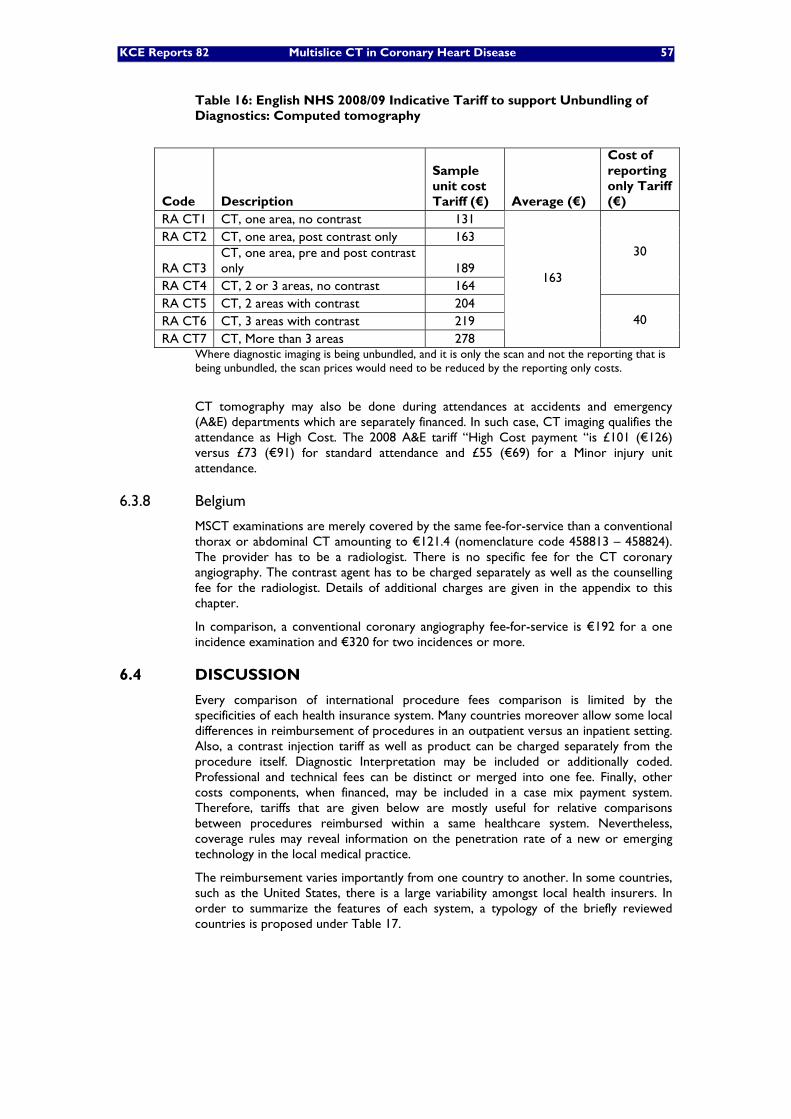
KCE Reports 82 Multislice CT in Coronary Heart Disease 57
Table 16: English NHS 2008/09 Indicative Tariff to support Unbundling of Diagnostics: Computed tomography
Code Description
Sample unit cost Tariff (€) Average (€)
Cost of reporting only Tariff (€)
RA CT1 CT, one area, no contrast 131 RA CT2 CT, one area, post contrast only 163
RA CT3 CT, one area, pre and post contrast only 189
RA CT4 CT, 2 or 3 areas, no contrast 164
30
RA CT5 CT, 2 areas with contrast 204 RA CT6 CT, 3 areas with contrast 219 RA CT7 CT, More than 3 areas 278
163
40
Where diagnostic imaging is being unbundled, and it is only the scan and not the reporting that is being unbundled, the scan prices would need to be reduced by the reporting only costs.
CT tomography may also be done during attendances at accidents and emergency (A&E) departments which are separately financed. In such case, CT imaging qualifies the attendance as High Cost. The 2008 A&E tariff “High Cost payment “is £101 (€126) versus £73 (€91) for standard attendance and £55 (€69) for a Minor injury unit attendance.
6.3.8 Belgium
MSCT examinations are merely covered by the same fee-for-service than a conventional thorax or abdominal CT amounting to €121.4 (nomenclature code 458813 – 458824). The provider has to be a radiologist. There is no specific fee for the CT coronary angiography. The contrast agent has to be charged separately as well as the counselling fee for the radiologist. Details of additional charges are given in the appendix to this chapter.
In comparison, a conventional coronary angiography fee-for-service is €192 for a one incidence examination and €320 for two incidences or more.
6.4 DISCUSSION
Every comparison of international procedure fees comparison is limited by the specificities of each health insurance system. Many countries moreover allow some local differences in reimbursement of procedures in an outpatient versus an inpatient setting. Also, a contrast injection tariff as well as product can be charged separately from the procedure itself. Diagnostic Interpretation may be included or additionally coded. Professional and technical fees can be distinct or merged into one fee. Finally, other costs components, when financed, may be included in a case mix payment system. Therefore, tariffs that are given below are mostly useful for relative comparisons between procedures reimbursed within a same healthcare system. Nevertheless, coverage rules may reveal information on the penetration rate of a new or emerging technology in the local medical practice.
The reimbursement varies importantly from one country to another. In some countries, such as the United States, there is a large variability amongst local health insurers. In order to summarize the features of each system, a typology of the briefly reviewed countries is proposed under Table 17.
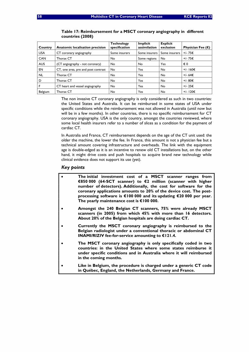
58 Multislice CT in Coronary Heart Disease KCE Reports 82
Table 17: Reimbursement for a MSCT coronary angiography in different countries (2008)
Country Anatomic localisation precision Technology specification
Implicit assimilation
Explicit exclusion Physician Fee (€)
USA CT coronary angiography Some insurers Some insurers Some insurers +/- 75€
CAN Thorax CT No Some regions No +/- 75€
AUS (CT angiography - not coronary) No No Yes € 0
EN CT, one area, pre and post contrast No Yes No +/- 160€
NL Thorax CT No Yes No +/- 64€
D Thorax CT No Yes No +/- 80€
F CT heart and vessel angiography No Yes No +/- 25€
Belgium Thorax CT No Yes No +/- 120€
The non invasive CT coronary angiography is only considered as such in two countries: the United States and Australia. It can be reimbursed in some states of USA under specific conditions while the reimbursement was not allowed in Australia (until now but will be in a few months). In other countries, there is no specific reimbursement for CT coronary angiography. USA is the only country, amongst the countries reviewed, where some local health insurers refer to a number of slices as a condition for the payment of cardiac CT.
In Australia and France, CT reimbursement depends on the age of the CT unit used: the older the machine, the lower the fee. In France, this amount is not a physician fee but a technical amount covering infrastructure and overheads. The link with the equipment age is double-edged as it is an incentive to renew old CT installations but, on the other hand, it might drive costs and push hospitals to acquire brand new technology while clinical evidence does not support its use (yet).
Key points
• The initial investment cost of a MSCT scanner ranges from €850 000 (64-SCT scanner) to €2 million (scanner with higher number of detectors). Additionally, the cost for software for the coronary applications amounts to 20% of the device cost. The post-processing software is €100 000 and its updating €20 000 per year. The yearly maintenance cost is €100 000.
• Amongst the 240 Belgian CT scanners, 75% were already MSCT scanners (in 2005) from which 45% with more than 16 detectors. About 20% of the Belgian hospitals are doing cardiac CT.
• Currently the MSCT coronary angiography is reimbursed to the Belgian radiologist under a conventional thoracic or abdominal CT INAMI/RIZIV fee-for-service amounting to €121.4.
• The MSCT coronary angiography is only specifically coded in two countries: in the United States where some states reimburse it under specific conditions and in Australia where it will reimbursed in the coming months.
• Like in Belgium, the procedure is charged under a generic CT code in Québec, England, the Netherlands, Germany and France.

KCE Reports 82 Multislice CT in Coronary Heart Disease 59
7 PATIENT ISSUES
7.1 TRUE NEGATIVE TEST RESULT
A true-negative test result (TN) will benefit the patient if it can reassure him or her concerning a pre-existing worry in a way that is less demanding than other tests. A potential for MSCT to reassure patients with chest pain has been anticipated, thus contributing to an improvement in QoL. According to estimates by Diamond and Forrester, patients with atypical chest pain have a low probability for angiographic significant CAD.7 (appendix) Depending on age and gender, the estimate varies between 0.8 and 28.1%. With a normal exercise test, the probability for angiographic significant CAD in this population is between 0.2 and 8.2%.7 The diagnostic accuracy of MSCT in patients with this low pre-test probability of CAD has not been studied so far.150 Moreover, according to Bayesian reasoning, the usefulness of further testing in these low probability patients is limited by the large number of false postive results. If we (unrealistically) would extrapolate the diagnostic performance of MSCT from intermediate and high risk populations66 to a population with a pre-test probability of 5%, of 100 patients tested, 13 would have a positive result of which 9 would be false positives. Instead of reassuring 100 patients, MSCT would induce anxiety in 13 patients out of 100, without any proven benefit in terms of clinical outcomes. Patients with chest pain that is probably not cardiac in origin, should decide for themselves what level of diagnostic certainty they want, at what cost in monetary terms and in terms of discomfort and risk incurred by additional downstream procedure(s), i.e. radiation exposure, risk of false positives, risk of useless revascularisation, etc. They should moreover be aware that a zero-risk will be never attainable.
A TN can have an adverse effect when it leads the patient to be less cautious when symptoms appear or when generally accepted lifestyle measures become neglected by it,151 e.g. a patient may wish not to quit smoking because he or she currently tests negative for CAD or lung cancer.
7.2 FALSE NEGATIVE TEST RESULT
A false-negative test result (FN) would give the individual false reassurance, and he or she may ignore signs of early disease which would cause a delay in diagnosis and treatment.151
Coronary artery imaging can also be misleading by its semiquantitative nature, merely reporting a luminal narrowing being less or more than 50%. This is an oversimplification because coronary lesions <50% may not lead to symptoms (stable angina) but their presence does not exclude future severe events. Low grade stenoses may be prone to plaque rupture and may lead to serious clinical events, yet they may be regarded as innocent.2, 3, 152
7.3 TRUE POSITIVE TEST RESULT
A true positive test result (TP) will benefit a patient only if it leads to a correct diagnosis in an easier way than other tests do, and if this leads to a better treatment or to a better outcome than would have been the case if that particular test were not used. In studies examining whether knowledge of CAC scores would affect compliance with lifestyle measures, perception of risk was affected, but it did not improve smoking cessation rates, although it did increase anxiety.42 The presence of obstructive CAD does not necessarily indicate a bad prognosis. The risk for the development of serious future events can be estimated by standard cardiologic examination and noninvasive testing. When the annual cardiovascular mortality rate estimated by noninvasive testing is low (<1%), the use of CCA to identify patients whose prognosis can be improved by revascularisation, is likely to be inappropriate.4 These patients may have stable angina or may be free of symptoms after an old uncomplicated MI. They can be reassured that invasive testing and revascularisation is not needed as a first step. This has been confirmed in recent trials enrolling patients with single and multivessel disease.2, 48 A

60 Multislice CT in Coronary Heart Disease KCE Reports 82
number of patients with obstructive CAD have a higher risk for future events as estimated by baseline examination and noninvasive tests and they may benefit from invasive testing and revascularisation. In those patients, directly proceeding to CCA is the logic next step because it enables revascularisation during the same procedure and an intermediate diagnostic step with MSCT seems futile.
7.4 FALSE POSITIVE TEST RESULT
A false positive test result (FP) may induce anxiety and lead to further diagnostic and unnnecassary therapeutic acts. In the case of MSCT, an inconclusive test result will be qualify as FP because it inevitably will lead to CCA. The low specificity of MSCT documented in diagnostic trials currently available is due to motion artifacts and intramural coronary calcifications. The resulting high number of false positives, especially in low risk populations, remains a major limitation to the clinical usefulness of the technique.
The detection of an obstructive coronary lesion by MSCT may be anatomically significant but clinical irrelevant if the patient’s symptoms have no relation to the coronary stenosis thus detected. The same reasoning holds true for CCA where it has been documented that angiographically significant lesions very often are hemodynamically insignificant.28
It has been discussed in previous chapters that at this moment, the performance of 64-SCT in clinical practice is not yet clearly defined, because no diagnostic trials have been performed in real-world conditions and no results of outcome trials that focused on issues relevant ot patients, are available. Therefore, positive and negative predictive values of 64-SCT for diagnosing clinical meaningful CAD in everyday clinical practice are unknown.
Key points
• Diagnostic tests may affect patients in many different ways. Independent of being true or false, both positive and negative test results may induce adverse effects that should be taken into account when considering the submission of a patient to MSCT of the coronary arteries.
• Positive and negative likelihood ratios of 64-SCT for the diagnosis of obstructive CAD in everyday practice are unknown.
KCE Reports 82 Multislice CT in Coronary Heart Disease 61
8 GENERAL DISCUSSION For the general discussion of the performance of 64-SCT in the diagnosis of CAD, we follow the hierarchy of diagnostic efficacy as described by Fryback and Thornbury (appendix).153, 154 Once a new diagnostic modality has been developed, its diagnostic accuracy has to be established as the next step. However, making diagnoses is not what patients are expecting from their physician: they want their symptoms to be alleviated and/or their life to be prolonged. In order to be worthwile in clinical practice, a diagnostic modality should alter diagnostic thinking of the attending physician, leading to a better patient management and improving patient outcomes. From the perspective of society, these goals should be achieved at a reasonable cost.
8.1 TECHNICAL EFFICACY
The feasibility of anatomic imaging of the coronary arteries by computed tomography became possible by the introduction of spiral scanning and multislice CT scanning, which provide smaller pieces of information and cover a larger area faster than conventional CT. Especially, 64-SCT and dual-source 64-SCT enable imaging of coronary arteries with acceptable quality, at least in selected patient populations. Some technical shortcomings remain a matter of concern. Image quality is less adequate in patients with fast or irregular heart rates, a problem that might be partly overcome by the administation of a beta-blocker prior to the examination, but nevertheless; patients with atrial fibrillation have been excluded from most clinical trials. The most bothersome problem is the presence of coronary artery calcifications that may preclude imaging of the calcified segments of the coronary tree, due to image blurring (the so-called “blooming”) that leads to an overestimation of the underlying stenosis or makes stenosis appraisal impossible altogether. Older age, diabetes and a high Agatston score (>400) are among the main predictors of poor diagnostic quality, all parameters related to severe coronary artery calcification.150, 155 Therefore, prior to contrast enhanced MSCT, patients are first evaluated for their calcification burden, which is quantified as the Agatston score. For patients with a score of more than 400, most authors agree that MSCT is futile.36, 150 Some authors have suggested that novel technical developments, such as subtraction techniques, requiring many years will be needed to resolve the problems associated with coronary calcification.45
In less selected populations, the number of inconclusive MSCTs can be rather high. For example in the RCT by Goldstein, 24/99 (24%) of the MSCTs were considered intermediate or non-diagnostic, necessitating additional testing by MPS and/or CCA.67 Inconclusive results mostly are due to coronary calcifications or motion artifacts although morbid obesity in some patients precludes CT scanning. Because of inherent spatial resolution limits, small calibre vessels (<1.5mm) are less well evaluable.
In 2006, an appropriateness review for MSCT has been issued under the auspices of the American College of Cardiology Foundation (ACCF) together with key specialty and subspecialty societies.108 The long list of patient characteristics assumed not to be present, illustrate the limitations that still are to be considered before proceeding to MSCT: irregular rhythm (e.g., atrial fibrillation/flutter), extreme obesity, renal failure (creatinine >1.8 mg/dl), heart rate greater than 70 b.p.m. refractory to heart-rate-lowering agents, metallic interference (e.g., surgical clips, pacemaker, and/or defibrillator wires). Moreover, patients must be able to hold still, to follow breathing instruction (breath holding during 10 to 20 sec), take nitroglycerin, take iodine in spite of steroid preparation for contrast allergy and lift both arms above the shoulders.
There is a non-negligible cancer risk associated with CT. It is estimated that currently 1.5 to 2.0% of all cancers in the United States may be attributable to the radiation from (all) CT studies.156 The 10 to 20 mSv exposure dose used in MSCT reportedly corresponds on average to 1 new (fatal or nonfatal) cancer for every 1000-2000 scans.157
62 Multislice CT in Coronary Heart Disease KCE Reports 82
8.2 DIAGNOSTIC ACCURACY
Most published clinical trials, are dealing with the diagnostic accuracy of 64-SCT as an imaging tool (for coronary arteries), referring to CCA as the gold standard and considering a coronary stenosis ≥50% on CCA as clinically relevant. In all published 64-SCT studies, in populations at intermediate or high pre-test probability of obstructive CAD (mostly >35% and up to 100%), test sensitivity is good and ranges between 95 and 100%, indicating a very good negative predictive value. At present, the main value of MSCT seems to be its use to rule out obstructiuve CAD.
Test specificity on the other hand is less good. In Abdulla’s SR, it was 91% (87.5-94),66 and in our meta-analysis it was 82.3% (78.5-85.7) in recent studies and 85.5% (80.4-89.7) in dual-source 64-SCT studies. Positive and negative likelihood ratios in recent studies were 5.0 (3.5-7.4) and 0.03 (0.02-0.06) respectively. In the large Meijboom gender trial (n=402), test performance was compared in women vs. men: whereas sensitivity was excellent in both sexes (93-100), specificity was acceptable in men (90%; 81-95) but poor in women (75%; 95% CI: 62-85).69
All studies enrolled patients with a high probability of CAD that were selected based on several additional parameters, including regular and controlled heart rate, preserved renal function, breath-hold capacity, and hemodynamic status. Virtually all patients were already scheduled for an invasive CCA before entry in the study. Whether the performance of MSCT can be reproduced in less selected patients at lower prevalence of CAD remains to be assessed, questioning the external validity of the results.158 Moreover, selection bias may have played a role in published trials, by exclusion of patients in whom calcified coronary arteries were expected (elderly, diabetes, non-cardiac atheromatous disease, renal failure) or in whom calcified coronary arteries had been documented by prior CCA. Finally, results of studies may also be biased by the fact that investigators had a better experience compared with the real-life centres which usually examine larger and more broad-spectrum populations and may be less experienced.66
8.3 DIAGNOSTIC THINKING
This level of diagnostic efficacy is concerned with the assessment of the effect of a test result on diagnostic reasoning and disease categorization, or in other words its role in clinical decision making. Few empirical evidence is available on this subject.
In a patient with a negative MSCT, i.e. in which no >50% stenosis is detected, a physician will decide that the chest pain symptoms for which the test was performed, most probably were not provoked by CAD. The question remains to what extent this test result changes the initial diagnosis put forward by the attending physician (i.e. pre-test likelihood). In a patient population such as that described by Goldstein,67 the pre-test probability of ACS was very low because of a profound pre-selection of patients. In these cases, further testing becomes irrelevant because diagnosis is almost certain, and additional testing will predominantly lead to false positives. In the study by Rubinshtein et al., MSCT reportedly was useful in the diagnostic work-up of patients with chest pain and an inconclusive stress test.80 However, in a substantial proportion of patients (20 out of 71, 28%) that tested negative with MSCT, treating physicians later on still proceeded to CCA, “because of clinical reasons” (sic), despite previous trials indicating a high negative predictive value of MSCT. In this trial, MSCT clearly did not alter the diagnostic path followed by the physician.
The “2006 appropriateness review for MSCT”, issued under the auspices of the American College of Cardiology Foundation, confirms that limited data are available supporting the use of MSCT coronary angiography within patient care algorithms.108
8.4 THERAPEUTIC IMPACT
MSCT of the coronary arteries up to now does nothing more than providing an anatomical image of the coronary tree. In several trials it has been shown that MSCT reliably can be used to rule out the presence of CAD in subsets of patients. In contrast to other imaging techniques such as MPS and DSE, MSCT does however not provide

KCE Reports 82 Multislice CT in Coronary Heart Disease 63
insight in the functional importance of the coronary lesion thus documented. Some studies indicated that less than half of the significant lesions on MSCT have hemodynamic consequences.111, 159 The lack of functional information being provided by both CCA and MSCT is a major limitation. Ideally, any patient with (atypical) chest pain in whom diagnostic imaging is deemed appropriate (i.e. a patient in whom one expects - if CAD is present - that revascularisation will improve symptoms or prognosis), should undergo a functional test before proceeding to an anatomic imaging test.160 If the functional examination leads to the decision that revascularisation is appropriate, one should go for invasive CCA, enabling immediate therapeutic intervention, and obviating the need for MSCT. If the functional exam leads to a decision that revascularisation is not appropriate, no further diagnostic imaging steps are needed. If noninvasive functional tests are impossible to be performed or inconclusive, MSCT may be efficacious, but these populations have not been studied so far. Yet, invasive functional evaluation (functional flow reserve) will often be needed.28
In summary, because MSCT has limited ability to define myocardium jeopardized by ischemia, its potential for predicting benefit from revascularisation is limited.111
8.5 PATIENT OUTCOMES
The real issue of diagnosing CAD is not to correctly identify coronary artery stenoses but to help in predicting and improving patients’ outcome. In this respect, coronary artery imaging may be misleading: significant though prognostic benign lesions may be identified and lead to inappropriate interventions, because they do not affect blood supply to the myocardium or they can be left untreated (i.e. not revascularised). In patients with stable angina pectoris, PCI as the first therapeutic option does not reduce the risk of death, myocardial infarction or other major cardiovascular events when added to optimal medical therapy.2 This means that myocardial revascularisation (and hence both invasive or noninvasive angiography) can be safely deferred in these patients and can be restricted to those in whom medical therapy does not lead to symptom control. In the latter event, cardiologists will immediately proceed to CCA, enabling them to intervene during the same procedure, obviating the need for a preliminary MSCT.
Coronary artery imaging can also be misleading because low grade stenoses may be prone to plaque rupture and may lead to serious clinical events, yet they may be regarded as innocent.2, 152
8.6 COST-EFFECTIVENESS
Economic evaluations of interventions with unproven clinical effectiveness are not very useful. An intervention should prove clinically effective first before it can be considered cost-effective. Several researchers, however, have attempted to build an economic model to assess the cost-effectiveness of MSCT relative to a suitable comparator. All of these models suffer from lack of evidence about the relevance of MSCT for improving patient outcomes. Therefore, they are necessarily limited to an assessment of the cost-per-case detected. And even these results can be questioned, as sensitivity and specificity of MSCT has not yet been tested in real-world populations where the prevalence of clinically significant CAD is low.
Nevertheless, a cost-outcome description by Goldstein et al. is frequently cited to demonstrate the technology’s cost-effectiveness. While this study did include clinical parameters about therapeutic impact, these were not taken into account in the cost-effectiveness analysis. A simple exercise, extending the cost and outcome (QALY) calculation to include treatments following the diagnosis of CAD showed that from hospitalisation up to 6 months of follow-up, a diagnostic strategy with MSCT was more costly and led to a higher loss in QALYs than a standard diagnostic strategy without MSCT. The results need to be interpreted with caution, as this RCT was actually underpowered to draw full economic conclusions.
Further studies on the diagnostic efficacy of MSCT are needed. Until then, results of cost-effectiveness analyses remain inconclusive.
64 Multislice CT in Coronary Heart Disease KCE Reports 82
9 CONCLUSIONS
9.1 TECHNICAL EFFICACY
64-SCT has shown to be able to image native coronary arteries with acceptable quality in selected patient populations. Patients should be in a stable sinus rhythm, they shoud be not too obese, they should be able to cooperate and they should have non-calcified coronary arteries. The high burden of ionizing radiation remains a major obstacle. It is currently not clear whether future technical improvements will lead to less radiation yet preserve adequate diagnostic performance.
9.2 DIAGNOSTIC ACCURACY
The diagnostic accuracy of MSCT in CAD has been thoroughly tested predominantly in patients at high-risk in whom it had already been decided to proceed to CCA, or in whom the results of CCA were already available. In these populations the technique can very well document normal coronary arteries and can adequately rule out obstructive CAD. The test’s specificity is less than optimal, leading to false positives, especially in lower prevalence populations.
The diagnostic performance of MSCT in real world clinical practice is not known.
9.3 DIAGNOSTIC THINKING
Only limited data are available supporting the use of MSCT with regard to its role within patient care algorithms. The test performs best in patients with normal coronary arteries, but it has yet to be ascertained whether these (normal) patients could not have been identified otherwise in a safer and more cost-effective way.
9.4 THERAPEUTIC IMPACT
If MSCT performs in real world as good as in clinical trials, it might be a useful test to exclude significant CAD. Documenting obstructive CAD by MSCT is of rather limited value, because patient management and prognosis depend on the functional impact of the coronary stenosis which cannot be assessed by MSCT alone. Moreover, in case revascularisation is deemed appropriate, invasive CCA is inevitable.
9.5 PATIENT OUTCOMES
There is limited data on the prognostic value of MSCT and there is no evidence whatsoever that the use of MSCT improves quality of life, prevents heart attacks or saves lives.
9.6 COST-EFFECTIVENESS
A full economic evaluation of MSCT requires more data on the clinical effectiveness of this diagnostic technique in preventing morbidity and mortality. It is yet impossible to conclude whether MSCT is cost-effective compared to the standard diagnostic protocols in low to intermediate risk patients.

KCE Reports 82 Multislice CT in Coronary Heart Disease 65
10 APPENDICES
QUALITY ASSESSMENT OF SYSTEMATIC REVIEWS
Figure 4: Checklist of the Dutch Cochrane Centre, items 1 through 7. (http://www.cochrane.nl/index.html)
The SR by Abdulla66 and by Vanhoenacker73 did not provide a quality score of the included studies. Hamon39 and Vanhoenacker81 made use of the QUADAS tool for quality assessment. Other items were scored similarly by the four SR that were reported on.
The quality of the meta-analysis methodology could not be adequately assessed.
Items 9 through 12 of the checklist from the Dutch Cochrane Centre (not shown in Figure 4) could not be rated because it was concluded that the diagnostic performance, resulting from these meta-analyses, lacked external validity because of the highly selected populations that were included in the different studies.

66 Multislice CT in Coronary Heart Disease KCE Reports 82
QUALITY ASSESSMENT OF PRIMARY DIAGNOSTIC PAPERS
Figure 5: QUADAS tool.84
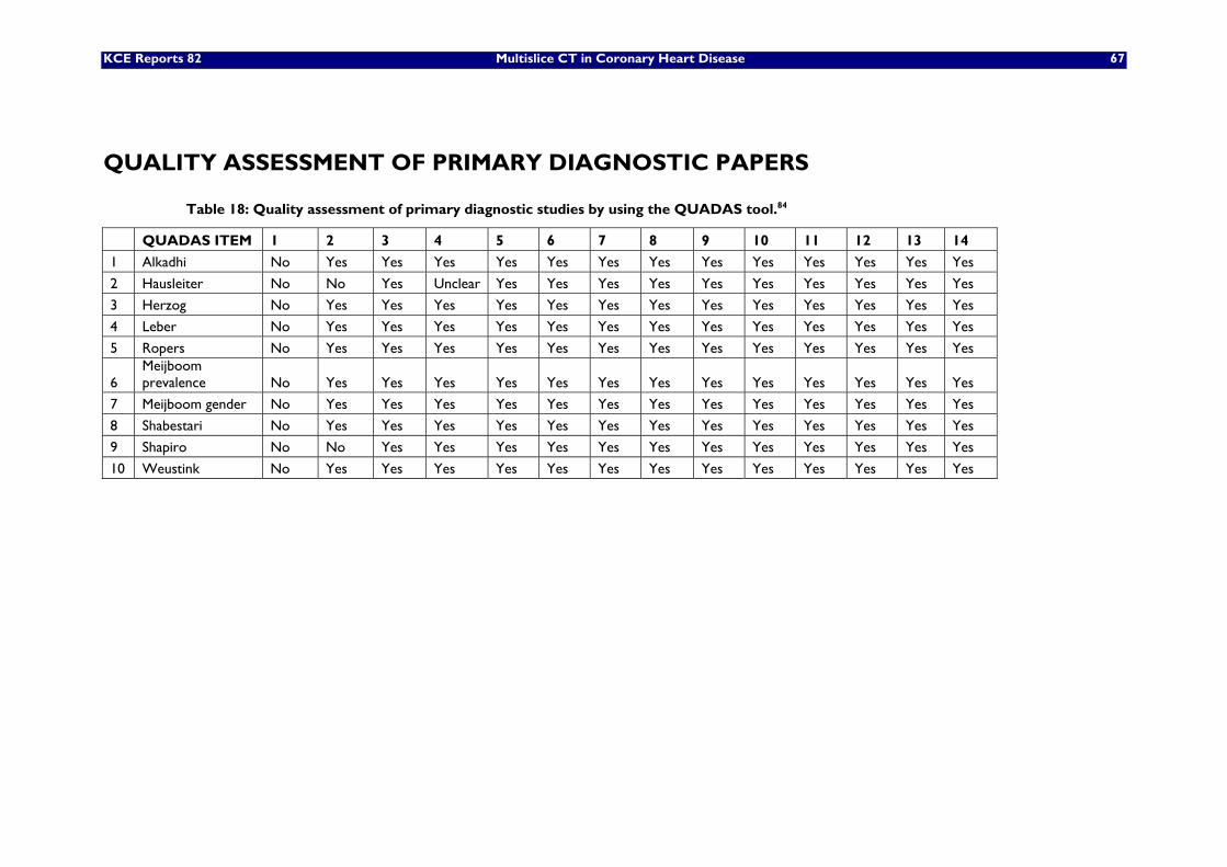
KCE Reports 82 Multislice CT in Coronary Heart Disease 67
QUALITY ASSESSMENT OF PRIMARY DIAGNOSTIC PAPERS
Table 18: Quality assessment of primary diagnostic studies by using the QUADAS tool.84
QUADAS ITEM 1 2 3 4 5 6 7 8 9 10 11 12 13 14
1 Alkadhi No Yes Yes Yes Yes Yes Yes Yes Yes Yes Yes Yes Yes Yes
2 Hausleiter No No Yes Unclear Yes Yes Yes Yes Yes Yes Yes Yes Yes Yes
3 Herzog No Yes Yes Yes Yes Yes Yes Yes Yes Yes Yes Yes Yes Yes
4 Leber No Yes Yes Yes Yes Yes Yes Yes Yes Yes Yes Yes Yes Yes
5 Ropers No Yes Yes Yes Yes Yes Yes Yes Yes Yes Yes Yes Yes Yes
6 Meijboom prevalence No Yes Yes Yes Yes Yes Yes Yes Yes Yes Yes Yes Yes Yes
7 Meijboom gender No Yes Yes Yes Yes Yes Yes Yes Yes Yes Yes Yes Yes Yes
8 Shabestari No Yes Yes Yes Yes Yes Yes Yes Yes Yes Yes Yes Yes Yes
9 Shapiro No No Yes Yes Yes Yes Yes Yes Yes Yes Yes Yes Yes Yes
10 Weustink No Yes Yes Yes Yes Yes Yes Yes Yes Yes Yes Yes Yes Yes

68 Multislice CT in Coronary Heart Disease KCE Reports 82
META-ANALYSIS OF 2007/2008 STUDIES COMPARING 64-SCT VS. CONVENTIONAL CORONARY ANGIOGRAPHY (≥50% STENOSIS) IN NATIVE CAD Summary Sensitivity Study | Sen [95% Conf. Iterval.] TP/(TP+FN)
TN/(TN+FP) --------------------------------------------------------------------------------------
------ Herzog | 1,000 0,824 - 1,000 19/19 30/36 Meijboom female | 1,000 0,943 - 1,000 63/63 45/60 Meijboom male | 0,989 0,962 - 0,999 188/190 80/89 Shabestari | 0,963 0,908 - 0,990 104/108 20/35 Shapiro | 0,966 0,822 - 0,999 28/29 5/8 Alkadhi | 0,966 0,883 - 0,996 57/59 79/91 Leber | 0,952 0,762 - 0,999 20/21 60/69 Ropers | 0,976 0,874 - 0,999 41/42 47/58 Weustink | 0,987 0,930 - 1,000 76/77 20/23 --------------------------------------------------------------------------------------
------ Pooled Sen | 0,980 0,966 - 0,990 --------------------------------------------------------------------------------------
------ Heterogeneity chi-squared = 7,22 (d.f.= 8) p = 0,513 Inconsistency (I-square) = 0,0 % No. studies = 9. Filter OFF Add 1/2 to all cells of the studies with zero Summary Specificity Study | Spe [95% Conf. Iterval.] TP/(TP+FN)
TN/(TN+FP) --------------------------------------------------------------------------------------
------ Herzog | 0,833 0,672 - 0,936 19/19 30/36 Meijboom female | 0,750 0,621 - 0,853 63/63 45/60 Meijboom male | 0,899 0,817 - 0,953 188/190 80/89 Shabestari | 0,571 0,394 - 0,737 104/108 20/35 Shapiro | 0,625 0,245 - 0,915 28/29 5/8 Alkadhi | 0,868 0,781 - 0,930 57/59 79/91 Leber | 0,870 0,767 - 0,939 20/21 60/69 Ropers | 0,810 0,686 - 0,901 41/42 47/58 Weustink | 0,870 0,664 - 0,972 76/77 20/23 --------------------------------------------------------------------------------------
------ Pooled Spe | 0,823 0,785 - 0,857 --------------------------------------------------------------------------------------
------ Heterogeneity chi-squared = 22,66 (d.f.= 8) p = 0,004 Inconsistency (I-square) = 64,7 % No. studies = 9. Filter OFF Add 1/2 to all cells of the studies with zero

KCE Reports 82 Multislice CT in Coronary Heart Disease 69
Summary Positive Likelihood Ratio (Random effects model) Study | LR+ [95% Conf. Iterval.] % Weight --------------------------------------------------------------------------------------
------ Herzog | 5,550 2,752 - 11,193 10,35 Meijboom female | 3,905 2,539 - 6,006 13,33 Meijboom male | 9,785 5,266 - 18,182 11,24 Shabestari | 2,247 1,530 - 3,300 13,82 Shapiro | 2,575 1,050 - 6,315 8,43 Alkadhi | 7,326 4,315 - 12,439 12,24 Leber | 7,302 3,941 - 13,528 11,27 Ropers | 5,147 3,017 - 8,780 12,19 Weustink | 7,567 2,633 - 21,744 7,12 --------------------------------------------------------------------------------------
------ (REM) pooled LR+ | 5,047 3,463 - 7,355 --------------------------------------------------------------------------------------
------ Heterogeneity chi-squared = 29,49 (d.f.= 8) p = 0,000 Inconsistency (I-square) = 72,9 % Estimate of between-study variance (Tau-squared) = 0,2288 No. studies = 9. Filter OFF Add 1/2 to all cells of the studies with zero Summary Negative Likelihood Ratio (Random effects model) Study | LR- [95% Conf. Iterval.] % Weight --------------------------------------------------------------------------------------
------ Herzog | 0,030 0,002 - 0,470 3,95 Meijboom female | 0,010 0,001 - 0,166 3,88 Meijboom male | 0,012 0,003 - 0,047 15,58 Shabestari | 0,065 0,024 - 0,177 29,47 Shapiro | 0,055 0,007 - 0,407 7,43 Alkadhi | 0,039 0,010 - 0,153 15,94 Leber | 0,055 0,008 - 0,372 8,09 Ropers | 0,029 0,004 - 0,205 7,88 Weustink | 0,015 0,002 - 0,105 7,78 --------------------------------------------------------------------------------------
------ (REM) pooled LR- | 0,034 0,020 - 0,058 --------------------------------------------------------------------------------------
------ Heterogeneity chi-squared = 6,31 (d.f.= 8) p = 0,612 Inconsistency (I-square) = 0,0 % Estimate of between-study variance (Tau-squared) = 0,0000 No. studies = 9. Filter OFF Add 1/2 to all cells of the studies with zero Summary Diagnostic Odds Ratio (Random effects model) Study | DOR [95% Conf. Iterval.] % Weight --------------------------------------------------------------------------------------
------ Herzog | 183,00 9,752 - 3434,2 6,43 Meijboom female | 372,81 21,743 - 6392,3 6,75 Meijboom male | 835,56 176,57 - 3953,9 14,83 Shabestari | 34,667 10,418 - 115,36 18,67 Shapiro | 46,667 4,007 - 543,55 8,41 Alkadhi | 187,63 40,416 - 871,02 15,02 Leber | 133,33 15,892 - 1118,7 10,26 Ropers | 175,18 21,678 - 1415,7 10,50 Weustink | 506,67 49,984 - 5135,9 9,13 --------------------------------------------------------------------------------------
------ (REM) pooled DOR | 166,83 72,228 - 385,36 --------------------------------------------------------------------------------------
------ Heterogeneity chi-squared = 13,02 (d.f.= 8) p = 0,111 Inconsistency (I-square) = 38,6 % Estimate of between-study variance (Tau-squared) = 0,6010 No. studies = 9. Filter OFF Add 1/2 to all cells of the studies with zero

70 Multislice CT in Coronary Heart Disease KCE Reports 82
META-ANALYSIS OF DUAL-SOURCE 64-MSCT VS. CONVENTIONAL CORONARY ANGIOGRAPHY (≥50% STENOSIS) IN NATIVE CAD Summary Sensitivity Study | Sen [95% Conf. Iterval.] TP/(TP+FN)
TN/(TN+FP) --------------------------------------------------------------------------------------
------ Alkadhi | 0,966 0,883 - 0,996 57/59 79/91 Leber | 0,952 0,762 - 0,999 20/21 60/69 Ropers | 0,976 0,874 - 0,999 41/42 47/58 Weustink | 0,987 0,930 - 1,000 76/77 20/23 --------------------------------------------------------------------------------------
------ Pooled Sen | 0,975 0,942 - 0,992 --------------------------------------------------------------------------------------
------ Heterogeneity chi-squared = 1,08 (d.f.= 3) p = 0,783 Inconsistency (I-square) = 0,0 % No. studies = 4. Filter OFF Add 1/2 to all cells of the studies with zero Summary Specificity Study | Spe [95% Conf. Iterval.] TP/(TP+FN)
TN/(TN+FP) --------------------------------------------------------------------------------------
------ Alkadhi | 0,868 0,781 - 0,930 57/59 79/91 Leber | 0,870 0,767 - 0,939 20/21 60/69 Ropers | 0,810 0,686 - 0,901 41/42 47/58 Weustink | 0,870 0,664 - 0,972 76/77 20/23 --------------------------------------------------------------------------------------
------ Pooled Spe | 0,855 0,804 - 0,897 --------------------------------------------------------------------------------------
------ Heterogeneity chi-squared = 1,16 (d.f.= 3) p = 0,764 Inconsistency (I-square) = 0,0 % No. studies = 4. Filter OFF Add 1/2 to all cells of the studies with zero Summary Positive Likelihood Ratio (Random effects model) Study | LR+ [95% Conf. Iterval.] % Weight --------------------------------------------------------------------------------------
------ Alkadhi | 7,326 4,315 - 12,439 33,66 Leber | 7,302 3,941 - 13,528 24,80 Ropers | 5,147 3,017 - 8,780 33,07 Weustink | 7,567 2,633 - 21,744 8,47 --------------------------------------------------------------------------------------
------ (REM) pooled LR+ | 6,531 4,804 - 8,880 --------------------------------------------------------------------------------------
------ Heterogeneity chi-squared = 1,15 (d.f.= 3) p = 0,764 Inconsistency (I-square) = 0,0 % Estimate of between-study variance (Tau-squared) = 0,0000 No. studies = 4. Filter OFF Add 1/2 to all cells of the studies with zero

KCE Reports 82 Multislice CT in Coronary Heart Disease 71
Summary Negative Likelihood Ratio (Random effects model) Study | LR- [95% Conf. Iterval.] % Weight --------------------------------------------------------------------------------------
------ Alkadhi | 0,039 0,010 - 0,153 40,16 Leber | 0,055 0,008 - 0,372 20,39 Ropers | 0,029 0,004 - 0,205 19,86 Weustink | 0,015 0,002 - 0,105 19,59 --------------------------------------------------------------------------------------
------ (REM) pooled LR- | 0,033 0,014 - 0,078 --------------------------------------------------------------------------------------
------ Heterogeneity chi-squared = 0,99 (d.f.= 3) p = 0,803 Inconsistency (I-square) = 0,0 % Estimate of between-study variance (Tau-squared) = 0,0000 No. studies = 4. Filter OFF Add 1/2 to all cells of the studies with zero
Summary Diagnostic Odds Ratio (Random effects model) Study | DOR [95% Conf. Iterval.] % Weight --------------------------------------------------------------------------------------
------ Alkadhi | 187,63 40,416 - 871,02 40,00 Leber | 133,33 15,892 - 1118,7 20,84 Ropers | 175,18 21,678 - 1415,7 21,59 Weustink | 506,67 49,984 - 5135,9 17,57 --------------------------------------------------------------------------------------
------ (REM) pooled DOR | 205,00 77,640 - 541,30 --------------------------------------------------------------------------------------
------ Heterogeneity chi-squared = 0,79 (d.f.= 3) p = 0,852 Inconsistency (I-square) = 0,0 % Estimate of between-study variance (Tau-squared) = 0,0000 No. studies = 4. Filter OFF Add 1/2 to all cells of the studies with zero

72 Multislice CT in Coronary Heart Disease KCE Reports 82
POST-TEST LIKELIHOOD OF CAD AFTER AN ECG STRESS TEST.
Table 19: Post-test likelihood of CAD after an ECG stress test according to age, gender, symptom and ST-segment depression.7
AGE ASYMPTOMATIC NON SPECIFIC PAIN ATYPICAL ANGINA TYPICAL ANGINA
ST-segement
depression on
exercise testing
men women men women men women men women
30-39 43 10,5 68,1 23,9 91,8 63,1 98,9 93,1
40-49 69,4 28,3 86,5 52,9 97,1 85,7 99,6 98
50-59 80,7 56,3 91,4 78,1 98,2 94,9 99,8 99,3 ≥2,5
60-69 84,5 76 93,8 89,9 98,8 97,9 99,8 99,7
30-39 17,7 3,2 37,8 8,2 76 32,7 96,2 79,4
40-49 39,2 10,1 64,5 74,2 90,5 63 98,7 93,2
50-59 54,3 26,8 75,2 50,4 94,1 84,2 99,2 97,7 2,0-2,5
60-69 60,9 47,3 81,2 71,7 95,8 93 99,5 99,1
30-39 7,5 1,2 18,7 3,3 54,5 15,5 90,6 59,3
40-49 19,6 4,1 40,8 10,8 78,2 39,1 96,6 83,8
50-59 31 12,2 53,4 27,8 85,7 66,8 98 94,2 1,5-2,0
60-69 37 25,4 62,1 48,9 89,5 83,3 98,6 97,6
30-39 3,9 0,6 10,4 1,7 37,7 8,5 83 42,4
40-49 11 2,1 25,8 5,8 64,4 24,5 93,6 72,3
50-59 18,5 6,5 36,7 16,3 75,2 50,4 96,1 89,1 1,0-1,5
60-69 22,9 14,7 45,3 32,6 81,2 71,6 97,2 95,3
30-39 1,7 0,3 4,8 0,7 20,7 3,9 67,8 24,2
40-49 5,1 0,9 13,1 2,6 43,9 12,3 96,3 53
50-59 9 2,9 20,1 7,8 56,8 30,5 91,3 77,9 0,5-1,0
60-69 11,4 6,9 26,4 17,3 65,1 52,2 93,8 89,8
30-39 0,4 0,1 1,2 0,2 6,1 1 24,5 7,4
40-49 1,3 0,2 3,6 0,7 16,4 3,4 61,1 22
50-59 2,4 0,8 5,9 2,1 24,7 9,9 72,5 46,9
0-0,5
60-69 3,1 1,8 8,2 5 31,8 21,4 79,1 68,8

KCE Reports 82 Multislice CT in Coronary Heart Disease 73
SEARCH STRATEGY FOR ECONOMIC LITERATURE REVIEW
Embase
Date of search: 06.12.2007
Number of hits: 51 (for entire strategy) No. Query Hits 1 2 3 4 5 6 7 8 9 10 11 12 13 14 15 16 18 19 20 21 22 23 24 25 26 27 28 29
'computer assisted tomography'/exp 'multidetector computed tomography'/exp 'angiocardiography'/exp coronary AND ('angiography'/exp OR 'angiography') 'computed tomographic angiography'/exp computed AND tomographic AND ('angiography'/exp OR 'angiography') coronary AND ('artery'/exp OR arter*) coronary AND ('vessel'/exp OR vessel*) mdct* OR msct* OR multi*row* OR multi*detect* OR multi*spiral* OR multi*slice* ((#3 AND (#7 OR #8)) OR #4) (#2 OR ((#1 OR #5 OR #6) AND #9)) #10 AND #11 'coronary artery bypass graft'/exp OR cabg OR (coron* AND by*pass) 'calcium'/exp OR 'artery calcification'/exp OR 'calcinosis'/exp OR calci* 'coronary stent'/exp OR 'drug eluting stent'/exp OR stent* 'ischemic heart disease'/exp OR (ischemi* OR myocar* OR arteriosclero* OR ('angina'/exp AND pectoris) OR ((('chest'/exp OR thora*) AND 'pain'/exp) OR 'thorax pain'/exp) OR atherom* OR coronar* OR steno*) OR 'coronary artery disease'/exp #13 OR #14 OR #15 OR #17 #12 AND #18 'clinical trial'/exp OR 'clinical trial' OR random* OR rct* OR cohort* OR 'cohort analysis'/exp 'diagnostic accuracy'/exp OR 'sensitivity and specificity'/exp #22 AND #23 #19 AND #24 #25 AND [humans]/lim AND [2000-2007]/py (((fiscal:ab,ti,de OR financial:ab,ti,de OR finance:ab,ti,de OR funding:ab,ti,de) OR ((variable*:ab,ti,de OR unit*:ab,ti,de OR estimate*:ab,ti,de) AND cost*:ab,ti,de) OR ('socioeconomics'/ OR 'cost benefit analysis'/ OR 'cost effectiveness analysis'/ OR 'cost of illness'/ OR 'cost control'/ OR 'economic aspect'/ OR 'financial management'/ OR 'health care cost'/ OR 'health care financing'/ OR 'health economics'/ OR 'hospital cost'/ OR 'cost minimization analysis'/)) OR ('economic evaluation'/ OR 'cost'/ OR 'reimbursement'/ OR 'cost utility analysis'/ OR 'drug cost'/ OR 'energy cost'/ OR 'hospital cost'/ OR 'hospital running cost'/ OR 'biomedical technology assessment'/)) #26 AND #27 #28 AND [embase]/lim AND [2000-2007]/py AND [2000-2007]/py #30. #19 AND #27 #31. #30 AND [humans]/lim AND [embase]/lim AND [2000-2007]/py
278,698 2,507 48,033 52,937 3,011 5,125 249,531 47,812 7,439 52,937 5,779 1,111 56,110 556,705 50,257 859,250 1,383,870 1,111 1,052,965 185,276 26,805 86 85 608,245 8 8 59 51

74 Multislice CT in Coronary Heart Disease KCE Reports 82
CRD: HTA(13), NHS-EED(7), DARE (6)
Date of search: 06.12.2007
Number of hits: 26 (for entire strategy) HTA(13), NHS-EED(7), DARE (6) No. Query Hits 1 2 3 4 5 6
MSCTA OR MSCT OR MDCT OR MDCTA CT OR "compute tomograph*" OR CTA multi*detector* OR multi*row* OR multi*slice* OR multi*spiral* #2 AND #3 #4 OR #1 #4 OR #1 RESTRICT YR 2000 2007
14 843 24 24 31 26
Econlit(Ovid)
Date of search: 06.12.2007
Coverage period database: 1969 to November 2007
Number of hits: 15 (for entire strategy)
No. Query Hits
1 (MSCT$ or MDCT$ or CTA or multi$slice$ or multi$detector$ or multi$row$ or multi$spiral$).mp. [mp=heading words, abstract, title, country as subject]
15
OVID MEDLINE(R)
Date of search: 30.11.2007
Coverage period database: 1950-November, week 2, 2007.
Number of hits: 215 (for entire strategy)
1 exp tomography, x-ray computed/ or multi-slice computed tomography.mp. (191800)
2 (mdct or msct or ((multi$row$ or multi$detect$ or multi$spiral$ or multi$slice$) and ((compute$ and
tomograph) or ct))).mp. (3695)
3 1 or 2 (192051)
4 exp coronary angiography/ (32102)
5 (coronar$ and angiograp$).mp. (49714)
6 exp coronary vessels/ (38297)
7 (coronar$ and (vessel$ or arter$)).mp. (173752)
8 exp myocardial ischemia/ (273633)
9 (myocard$ or ischemi$ or arteriosclero$ or angina pectoris or chest pain or atherom$).mp. (579065)
10 (calcium or calcinos$ or calcification$).mp. (412807)
11 exp stents/ or stent$.mp. (37840)
12 exp coronary artery bypass/ or cabg.mp. or by$pass.mp. (86191)
13 exp coronary stenosis/ or stenos$.mp. (115427)
14 or/3-6 (269182)
15 or/7-12 (1109590)
16 3 and 14 and 15 (20105)
17 limit 16 to humans (19529)
18 limit 17 to yr="2000 - 2007" (9908)
19 clinical trial$.mp. or clinical trial.pt. or random$.mp. or RCT.mp. or exp *cohort studies/ (858660)
20 exp *"sensitivity and specificity"/ (1496)

KCE Reports 82 Multislice CT in Coronary Heart Disease 75
21 18 and 19 and 20 (0)
22 (price$ or pricing$).mp. (14212)
23 ec.fs. (235528)
24 cost$.tw. (200524)
25 exp "Costs and Cost Analysis"/ (133793)
26 22 or 23 or 24 or 25 (396432)
27 21 and 26 (0)
28 18 and 26 (215)
OVID MEDLINE(R) IN-PROCESS & OTHER NON-INDEXED CITATIONS
Date of search: 3.12.2007
Coverage period database: November 30, 2007
Number of hits: 0/4/59
1 (MDCT or MSCT or ((multi$row$ or multi$detect$ or multi$spiral$ or multi$slice$) and
((compute$ and tomograph$) or CT))).tw. (416)
2 (coronar$ and angiograp$).tw. (758)
3 (coronar$ and (vessel$ or arter$)).tw. (3097)
4 2 or 3 (3246)
5 (myocard$ or ischemi$ or arteriosclero$ or angina pectoris or chest pain or atherom$).tw. (7296)
6 (calcium or calcinos$ or calcification$).tw. (5231)
7 stent$.tw. (1453)
8 (CABG or by$pass).tw. (1923)
9 stenos$.tw. (1728)
10 5 or 6 or 7 or 8 or 9 (15777)
11 1 and 4 and 10 (59)
12 (clinical trial$ or random$ or RCT or cohort).tw. (25267)
13 11 and 12 (4)
14 (price$ or pricing).tw. (543)
15 econom$.tw. (3696)
16 cost$.tw. (7752)
17 14 or 15 or 16 (10872)
18 11 and 17 (4)

76 Multislice CT in Coronary Heart Disease KCE Reports 82
FLOW CHART
Economic evaluationsPotentially relevant citations identified: 290
Based on title and abstract evaluation, citations excluded: 238Reasons:
High-Risk population 1EBCT, MSCT<64 33Out-of-scope 147Letters / editorials / no original studies 0No economical info
Studies retrieved for more detailed evaluation: 52
Based on full text evaluation, studies excluded: 50Reasons:
High-Risk population 0EBCT, MSCT<64 3Out-of-scope 7Letters / editorials / no original studies 9No economical info 22Unretrieved 9
Relevant studies: 2
Inclusion of one relevant economic evaluation Handsearched economic evaluations 3performed in a HTA report: 1
Economic evaluations selected: 6
KCE Reports 82 Multislice CT in Coronary Heart Disease 77
CHALLENGING THE ECONOMIC CONCLUSIONS OF GOLDSTEIN ET AL.
Several international articles refer to the paper by Goldstein et al.67 to claim cost-effectiveness of MSCT. In this appendix, we challenge the economic conclusions of the authors, by fully using their findings to assess the economic benefit of the technology.
This analysis was a simple exercise to evaluate the validity of the economic conclusions drawn from the Goldstein study. Not too much weight should be given to the precise figures resulting from this exercise, as the initial clinical trial was not set up to do an economic evaluation. Patient numbers were too small for instance to draw firm conclusions about the therapeutic impact of MSCT or the standard of care as defined in the study. In the study, the number of invasive treatments was higher in the MSCT arm than in the standard of care arm (6 versus 1). This might be due to coincidence. However, in the absence of better data, applying these crude figures in the economic model has an important impact on the costs and effects of the initial diagnostic strategy.
Because of the limited value of the precise figures resulting from this evaluation, the evaluation has been put in appendix. It substantiates, however, the argument that erroneous conclusions about the economic benefit of MSCT might be drawn if based only on an RCT that was not initially set up to assess economic benefit of this technology.
METHODOLOGY The principles of the methodological guidelines for pharmacoeconomic evaluations in Belgium were applied in this excercise.161
Design
Three outcome studies were identified in the clinical literature review, one being a randomised controlled trial comparing a diagnostic strategy with MSCT with standard of care (serial ECGs + cardiac biomarkers + MPS).67 The study was a cost-outcome description, drawing conclusions about the economic benefits, defined as the difference between median costs of MSCT and standard of care.
For the evaluation of the incremental costs and effects of a diagnostic strategy with MSCT and a standard diagnostic strategy in patients with chest pain, we used the data from this RCT. In contrast to the authors of the study, we decided to extrapolate the economic results to include the costs of invasive angiography, revascularisations and complications up to 6 months after initial admission to the emergency department for acute chest pain.
The basic idea is that the cost-effectiveness of MSCT depends not only on the costs and effects of the diagnostic strategy, but also the costs and effects of its sequelae, i.e. the changes in therapeutic behaviour and the consequent impact on patient outcomes. Therefore, it is insufficient to consider only the technique’s diagnostic accuracy (sensitivity and specificity) in an economic evaluation. An economic evaluation should also incorporate the technique’s effect on patients’ outcomes (life years gained or quality-adjusted life years gained).
The design of our economic evaluation is a piggy-back economic evaluation, based on a data from one RCT. A decision tree was constructed based on the observed movements of patients in that RCT. In that sense, the decision tree is a limited representation of the expected reality, as the number of patients in the RCT was limited and not all branches of a more realistic model could be filled with data from the trial. However, with the limited data available in literature, it was unfortunately unrealistic to fill a decision tree that included all possible real-life scenarios.
The economic evaluation was performed in Microsoft® Excel 2002, using @RISK 4.5.5 for the bootstrapping.

78 Multislice CT in Coronary Heart Disease KCE Reports 82
Structure of the decision tree
A simple decision tree was constructed, where the numbers of patients moving from one intervention to another were derived directly from the RCT. The structure of the decision tree is presented in Figure 6.
Figure 6: Structure of the decision tree for the economic model
59discharge
6immediate discharge
67
re-admission: ECG+blood alive revascularisation PCI
8 1 1 1
CCA1
deathfurther testing 0
2
SPECT DischargeMSCT 1 1
99
PCI3
revascularisation5
alive CABG8 2
no revascularisation3
CCAChest pain 8
death0
Further testing alive no revascularisation32 3 3
CCA3
deathSPECT 0
24
Discharge21
87 dischargeimmediate discharge 1
95 alive no revascularisation4 4
re-admission: SoC8 CCA
4death
Further testing 0Standard of care 7
98
SPECT Discharge3 3
revascularisation PCI1 1
alive3
CCA no revascularisation3 2
death0
no re-admission (within 6 months)
no re-admission (within 6 months)
Analytic technique
Because outcome in terms of mortality is not different between the two diagnostic arms in the study, an analysis of the “cost-per-life year gained” based on these data would ultimately boil down to a cost-minimisation analysis. However, invasive angiography, PCI and CABG have a demonstrated impact on the quality of life of patients undergoing this procedure. Therefore, it is worth looking at the QALY gains or losses of the two diagnostic work-up paths being compared. A cost-utility approach is therefore performed, calculating the incremental cost-per-QALY gained associated with MSCT as compared to standard of care.

KCE Reports 82 Multislice CT in Coronary Heart Disease 79
Perspective
The perspective taken is that of the Belgian health care payer, including both the National Institute for Health and Disability Insurance (RIZIV/INAMI) and the patients. For the calculation of the costs of the two diagnostic work-up arms, we calculate the total reimbursement by the RIZIV/INAMI and add, if applicable, the patients’ out-of-pocket expenses.
Target population
The target population of our model is as in the RCT: adult patients with acute chest pain who are deemed at low risk for coronary events after and initial work-up in the emergency department (ECG, biomarkers).
Population characteristics in both groups are presented in Table 20:
Table 20: Patient characteristics in the two diagnostic groups
MSCT Group N=99
Standard of Care group N=98
P-value
Age, mean in yrs 47 50 0.08 Male, % 43 57 0.05 Body Mass Index 28 28 0.78 Hypertension, % 39 38 0.88 Diabetes, % 8.2 12.2 0.35 Family history of early coronary disease, %
40 44 0.56
Current smoker, % 15 20 0.35 Goldman Riley criteria, % 0 very low risk 1 low risk 2 moderate risk
100
0 0
99 1 0
1
Source: Goldstein et al.67
Comparator
The comparator to MSCT angiography is ”standard of care” as defined by Goldstein et al. (2007). This includes noninvasive coronary tests, i.e. serial electrocardiograms and cardiac biomarkers at 0, at 4 and at 8 hours and rest-stress MPS. Common procedures to both diagnostic arms were the electrocardiograms and cardiac biomarkers at 0 and at 4 hours. Patients were randomised if both of these were normal. Therefore, the difference in primary diagnostic protocol between the ”intervention”, i.e. MSCT, and the comparator, i.e. ”standard of care”, is one cardiac biomarker and SPECT as part of the initial diagnostic strategy in the ”standard of care” group.
Costs
According to the Belgian pharmacoeconomic guidelines only included direct health care costs should be included in the base-case cost analysis. Indirect costs of productivity losses were not included.
Initial diagnostic strategy
The costs of the initial diagnostic strategy were calculated on the basis of the prevailing reimbursement tariffs and out-of-pocket expenses of the procedures associated with the strategy.
For MSCT angiography no reimbursement tariff exists (yet). Therefore, we used the reimbursement and patients’ out-of-pocket expenses for „chest CT“, which are the tariffs actually applied for MSCT. Usually other costs are associated with procedures than the costs of the procedure itself. For example, when a patient enters an emergency department and gets a MSCT angiography after which he is immediately discharged, the hospital can charge other costs to the RIZIV/INAMI such as a physician’s fee.

80 Multislice CT in Coronary Heart Disease KCE Reports 82
To identify the resource use, and especially the lump sums a hospital can charge if a patient is either discharged the same day after MSCT or “standard of care” or is hospitalised for a CCA that is eventually not followed by an invasive procedure, we presented different scenarios to the accounting service of a hospital who then retrieved the actual bill of a patient fitting into the respective scenarios to identify what can be charged in each of the cases. The scenarios presented to the hospitals were the following:
• a patient enters the emergency department for chest pain, undergoes a standard diagnostic work-up and subsequently a conventional coronarography (CCA) which turns out to be negative.
• a patient enters the emergency department for chest pain, undergoes a diagnostic work-up including a chest CT (used as a proxy for MSCT) and a nuclear stress test and is then discharged home.
• a patient enters the emergency department for chest pain, undergoes a chest CT and is immediately discharged home.
• a patient enters the emergency department for chest pain, undergoes the standard of care, including nuclear stress test and is then discharged home
If no bill could be retrieved for an actual patient fitting in one of these scenarios, medical experts and accounting services simulated what would be charged in these cases. This was the case for the scenarios where a nuclear stress test is performed at the emergency department, on the basis of which it would be decided to send the patient home. According to the medical administration, no single patient fitted in this scenario according to their register. This would mean that the scenario presented in the study by Goldstein might not be realistic in Belgium. Because we nevertheless had to calculate a cost for this scenario, as we had to stick to the diagnostic protocols suggested in the trial, we simply added the procedure cost of a MPS to the cost of a patient satisfying the other criteria of the scenario where MPS was included.
For CCA that is not followed by revascularisation, we obtained a patient bill from one hospital, on the basis of which we identified the procedures that are charged in such a case.
Types of resources and volumes of resource use for the resources for which no real observational data were available are presented in Table 21.
Table 21: Resource for MSCT, standard of care and conventional coronarography (RIZIV/INAMI code or pseudo-code)
Both strategies* MSCT diagnostic path Standard diagnostic path (standard tests+MPS)
Conventional coronarography
Procedure fees 2 blood tests (cardiac biomarkers - 542356)
MSCT procedure (458813) 1 additional blood test (cardiac biomarkers - 542356)
Coronarography (464144 or 453143)
2 ECG MPS procedure fee (442396)
additional blood tests
Products Iodinated contrast (699112) Radio-isotope (1/6 kit sestamibi) (699112)
Iodinated contrast (699112)
Physician fees Radiologist’s fee (460795) Radiologist’s fee (460795) Radiologist’s fee Cardiologist’s fee in ED
(590531) Cardiologist’s fee in ED (590531)
Cardiologist’s fee (212026+212041)
Surveillance fee per hospitalisation day (598706)
Lump sums 1 “mini lump sum” (if MSCT scan is not followed by hospitalisation)
1 “mini lump sum” (if MPS is not followed by hospitalisation)
2 hospitalisation days – per diem price
Lump sum medical imaging (461016)
Lump sum clinical biology per day
Lump sum medical imaging Lump sum for hospital admission
* these costs are not taken into account as they are equal between the two strategies. Only incremental costs are calculated.

KCE Reports 82 Multislice CT in Coronary Heart Disease 81
The corresponding cost figures for these items are presented in Table 22. The distributions mentioned are the ones used for the sensitivity analysis. Distributions are only defined for cost items that are variable across hospitals. Other amounts, e.g. those defined in the Belgian “nomenclature”, are deterministic and have hence no distribution.
Table 22: Cost items included in the analyses
Cost item Mean (RIZIV + Patient)
Standard deviation
Distribution Lower limit
Upper limit
Procedures Cardiac biomarkers 10.33 (7.75+2.58) - MSCT procedure fee 121.35 (118.87+2.48) - SPECT procedure fee 318.11
(280.93+37.18) -
Coronarography procedure fee
484.56 (484.56+0) -
Additional blood tests in case of CCA
25.69 (25.69+0)
-
Products Contrast agent MSCT 44.68 (44.68+0) - Radio-isotope SPECT 37.18 (37.18+0) - Physician’s fees
Radiologist’s fee 25.96 (18.52+7.44)
-
Radiologist’s fee (sum for all procedures in case of CCA)
39.87 (33.33+6.54)
-
Cardiologist’s fee in ED 35.25 (31.18+4.07)
-
Surveillance honorarium per hospitalisation day
24.84 (19.88+4.96) -
Lump sums Hospital per diem price 20.83 3.61 normal 15.77 42.67 “Mini” lump sum 56,39 12,798 normal 41,27 124 Lump sum clinical
biology (per hospital admission) 143,92 30,88 normal 94,03 233,17
Lump sum clinical biology (per day)
21,89 5,82 normal 11,84 45,48
Lump sum medical imaging (per day)
50,72 12,07 normal 22,13 88,74
Revascularisation: PCI and CABG
The costs of PCI and CABG were derived from a Belgian HTA on drug eluting stents.50 These cost data were based on actually observed cost data of all patients having received a bare metal stent (BMS) or drug eluting stent (DES) in 2004 in Belgium. A distinction was made between the costs associated with PCI with bare metal stents and PCI with DES and between treatment with one or another stent-type in diabetic and non-diabetic patients. Account was taken of the distribution of the costs for a hospitalisation episode due to PCI and CABG in the sensitivity analysis.
Point estimates, along with their normal distribution used in the sensitivity analysis, are presented in Table 23 (all data expressed in € for the year 2004):
Table 23: Costs of PCI and CABG included in the economic analysis
Costs Mean
Standard deviation of mean distribution
lower bound
upper bound source
PCI with stent (BMS) in non-diabetic patients 6298 255 2018 24221 KCE report 66A; www.kce.fgov.bePCI with stent (DES) in non-diabetic patients 7000 1541 3371 68450 KCE report 66A; www.kce.fgov.bePCI with stent (BMS) in diabetic patients 7190 773 2118 17444 KCE report 66A; www.kce.fgov.bePCI with stent (DES) in diabetic patients 7732 770 1836 51591 KCE report 66A; www.kce.fgov.beCABG in non-diabetic patients 15319 804 7650 56287 KCE report 66A; www.kce.fgov.beCABG in diabetic patients 17439 2459 8742 52521 KCE report 66A; www.kce.fgov.be
From the same HTA, we derived the distribution of BMS and DES across diabetic patients and non-diabetic patients respectively.50 The data are as follows:
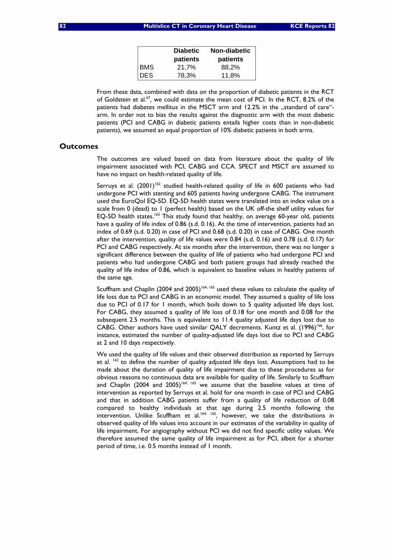
82 Multislice CT in Coronary Heart Disease KCE Reports 82
Diabetic patients
Non-diabetic patients
BMS 21,7% 88,2%DES 78,3% 11,8%
From these data, combined with data on the proportion of diabetic patients in the RCT of Goldstein et al.67, we could estimate the mean cost of PCI. In the RCT, 8.2% of the patients had diabetes mellitus in the MSCT arm and 12.2% in the „standard of care“-arm. In order not to bias the results against the diagnostic arm with the most diabetic patients (PCI and CABG in diabetic patients entails higher costs than in non-diabetic patients), we assumed an equal proportion of 10% diabetic patients in both arms.
Outcomes
The outcomes are valued based on data from literature about the quality of life impairment associated with PCI, CABG and CCA. SPECT and MSCT are assumed to have no impact on health-related quality of life.
Serruys et al. (2001)162 studied health-related quality of life in 600 patients who had undergone PCI with stenting and 605 patients having undergone CABG. The instrument used the EuroQol EQ-5D. EQ-5D health states were translated into an index value on a scale from 0 (dead) to 1 (perfect health) based on the UK off-the shelf utility values for EQ-5D health states.163 This study found that healthy, on average 60-year old, patients have a quality of life index of 0.86 (s.d. 0.16). At the time of intervention, patients had an index of 0.69 (s.d. 0.20) in case of PCI and 0.68 (s.d. 0.20) in case of CABG. One month after the intervention, quality of life values were 0.84 (s.d. 0.16) and 0.78 (s.d. 0.17) for PCI and CABG respectively. At six months after the intervention, there was no longer a significant difference between the quality of life of patients who had undergone PCI and patients who had undergone CABG and both patient groups had already reached the quality of life index of 0.86, which is equivalent to baseline values in healthy patients of the same age.
Scuffham and Chaplin (2004 and 2005)164, 165 used these values to calculate the quality of life loss due to PCI and CABG in an economic model. They assumed a quality of life loss due to PCI of 0.17 for 1 month, which boils down to 5 quality adjusted life days lost. For CABG, they assumed a quality of life loss of 0.18 for one month and 0.08 for the subsequent 2.5 months. This is equivalent to 11.4 quality adjusted life days lost due to CABG. Other authors have used similar QALY decrements. Kuntz et al. (1996)166, for instance, estimated the number of quality-adjusted life days lost due to PCI and CABG at 2 and 10 days respectively.
We used the quality of life values and their observed distribution as reported by Serruys et al. 162 to define the number of quality adjusted life days lost. Assumptions had to be made about the duration of quality of life impairment due to these procedures as for obvious reasons no continuous data are available for quality of life. Similarly to Scuffham and Chaplin (2004 and 2005)164, 165 we assume that the baseline values at time of intervention as reported by Serruys et al. hold for one month in case of PCI and CABG and that in addition CABG patients suffer from a quality of life reduction of 0.08 compared to healthy individuals at that age during 2.5 months following the intervention. Unlike Scuffham et al.164, 165, however, we take the distributions in observed quality of life values into account in our estimates of the variability in quality of life impairment. For angiography without PCI we did not find specific utility values. We therefore assumed the same quality of life impairment as for PCI, albeit for a shorter period of time, i.e. 0.5 months instead of 1 month.

KCE Reports 82 Multislice CT in Coronary Heart Disease 83
The quality of life values and their distributions for each of the states, on the basis of which the QALY decrements are calculated are presented in Table 24.
Table 24: Distributions of quality of life index values used to calculate the number of quality adjusted life days lost
Health state
Duration of state
Quality of life index values, mean
Standard deviation
Distribution Lower bound
Upper bound
Baseline 0,86 0,16 Normal 0.2 1 PCI procedure
1 month 0,69 0,2 Normal 0.25 1
CABG procedure
1 month 0,68 0,2 Normal 0.25 1
CABG follow-up
2.5 months
0,78 0,17 Normal 0.5 1
The impact of symptom relief from revascularisation on health-related quality of life was not taken into account in our economic evaluation because it is questionable whether the revascularisations performed in the study by Goldstein et al. actually induced pain relief. They presumably were rather meant for diagnostic reasons, i.e. the prevention of major cardiac events or death. While more revascularisations were performed during the index hospitalisation in the MSCT-arm compared to the ”standard of care”-arm (5% versus 1%), an equal number of patients were re-admitted for recurrent chest pain during the 6 months follow-up period in both arms.4 Hence, we can reasonable conclude that the higher number of revascularisations did not reduce the risk of recurrent chest pain.
Time horizon
The time horizon used in the economic model is the time horizon for which data are available from the RCT, i.e. from admission to the emergency department up to 6 months follow-up. We assume that longer time horizons would not change the results of the economic analysis, because it is uncertain whether the immediate CCAs and consequent revascularisations performed in the 8 patients showing severe stenosis on MSCT (over 70%) were clinically meaningful. As no MPS has been performed in these patients it is impossible to draw conclusions about the clinical relevance of these CCAs and revascularisations.
As for the outcomes, we assume that only invasive coronary procedures (CCA, PCI and CABG) have an impact on the number QALYs. The absolute difference between the number of QALYs in both procedures remains therefore de facto the same in extended time periods if the difference in the number of invasive procedures remains the same. Obviously, the relative impact of the quality of life loss due to the procedures decreases if the time horizon increases.
Sensitivity analysis
Bootstrapping was performed to obtain confidence intervals around the cost and outcome estimates in the economic evaluation. 1000 bootstrap samples were drawn from the defined distributions. The distributions used in the bootstrapping for cost and outcome variables are presented in the paragraphs where the sources and assumptions with respect to the cost and outcome variables are discussed.
We verified the conclusions of Goldstein et al. by calculating the costs of both diagnostic strategies up to the point where the decision to perform CCA is taken. Costs of CCA or revascularisation were not included.67 On the basis of this analysis, Goldstein et al. concluded that the MSCT procedure is less costly than the standard of care procedure. Outcomes, however, were not measured in terms of QALYs but in terms of ”time to diagnosis”, which is, as explained earlier, not relevant for resource

84 Multislice CT in Coronary Heart Disease KCE Reports 82
allocation decisions in health care. Health care decision makers are interested in obtaining the highest improvement in health with a given amount of resources.
Discounting
Because the time horizon of the evaluation is less than 1 year, there is no need to discount costs and outcomes.
RESULTS
Costs and outcomes of a diagnostic strategy with MSCT angiography and a standard diagnostic strategy for coronary artery disease
Base-case analysis
The results of the base-case analysis are presented in Table 25.
Table 25: Results of the base-case economic analysis
Diagnostic path Cost per patient, mean (95% C.I.)
QALYs lost per patient, mean (95% C.I.)
MSCT, index hospitalisation
914,92 (875;955) 0,0014 (0;0.004)
MSCT follow-up 88,55 (83.15;94.49) 0,00014 (0;0.0046) Total 1003,48 (959;1047) 0,0016 (0;0.0045) Standard of care, index hospitalisation
461,38 (444;484) 0,0003 (0;0.0009)
Standard of care, follow-up
62,53 (58.63;66.49) 0,00028 (0;0.0009)
Total 523,91 (505.12;548.03) 0,00056 (0;0.0018)
According to these results the MSCT diagnostic strategy is on average €479,56 more expensive than the standard of care strategy. Moreover, it leads to a higher loss in QALYs: 0.0016 QALYs are lost in the MSCT arm as compared to 0.00056 QALYs in the standard of care arm. This is equivalent to about 6 hours of life in perfect health more lost in the MSCT arm than in the CCA arm.
With its lower cost and better outcomes, the standard of care diagnostic strategy dominates the MSCT diagnostic strategy.
Sensitivity analysis
We tested the primary results of Goldstein et al.67 and found that both the costs of the MSCT and the „standard of care“ diagnostic strategy are lower if the costs of CCA during the index hospitalisation and the costs of late diagnostic testing and revascularisations are not included in the cost estimates. The corresponding costs per patient for both diagnostic arms in Belgium are as follows:
Cost per patient (€) QALYs per patient MSCT 347,71 (331;370) 0 Standard of care 383,26 (366;406) 0
In this case, costs of the strategy with MSCT are indeed lower than the costs of the standard of care strategy. Because in this scenario the model stops right before the decision to do a CCA is taken and because no quality of life loss is assumed due to nuclear stress testing or MSCT, the number of QALYs is the same in both diagnostic arms.

KCE Reports 82 Multislice CT in Coronary Heart Disease 85
DISCUSSION
The economic evaluation presented in this paragraph is limited to an application of Belgian cost data to one RCT and extending the economic assessment presented by Goldstein et al. to include health outcomes. The objective was modest, being to challenge the conclusion drawn by Goldstein et al. and frequently cited about the cost-effectiveness of MSCT relative to standard of care. A full economic evaluation would require evidence on the effectiveness of MSCT in real world in low- to intermediate risk patients. Evidence on diagnostic accuracy in well-defined patient populations is being built up, meanwhile leaving the assessment of the impact of MSCT on patient outcomes unevaluated. An option is to model final outcomes such as life-years gained based on evidence about the relationship between intermediate outcomes parameters (e.g. detected CAD) and final outcome parameters (e.g. life-years gained). This means that sufficient information must be available about the clinical significance of the CAD detected by MSCT or its comparator in the target population. Unfortunately this information is rarely available for patients at low- to intermediate risk for coronary events, as most studies stop whenever CAD is diagnosed.
Our economic assessment, based on observed data from one RCT, showed that the total costs of MSCT angiography in patients admitted to the emergency department because of chest pain and deemed at low risk for future events are higher than that of the standard of care, defined as 3 cardiac biomarker tests (at 0, 4 and 8 hours), 2 ECGs and nuclear stress testing. The outcomes of the diagnostic strategy with MSCT as a filter for nuclear stress testing, i.e. only patients with intermediate or inconclusive MSCT test results undergo a nuclear stress test, are worse than the outcomes of the standard of care strategy. Because more patients in the MSCT arm undergo revascularisation, and revascularisation impacts on health-related quality of life, this result is not surprising. If the observed trend in the RCT of more revascularisations in the MSCT arm continues in longer follow-up periods, the difference between the costs and outcomes of both diagnostic strategies will only increase. The RCT is, however, underpowered to allow such hypothesis.
Goldstein et al. did not reach the same conclusion, mainly because they stopped their costing procedure when the decision to do an invasive angiography was taken.67 Their endpoint, therefore, was an intermediate one. The relevance of it can be questioned, in general but especially in this patient population. The general argument against the use of intermediate endpoints in economic evaluation is that they are not relevant for the policy maker or the patient. The policy maker is interested in how he can obtain the highest health benefit at a given cost. The patient is interested in how he can obtain the highest health benefit at reasonable out-of-pocket expenses. The fact of reaching more or less quickly a decision to do a CCA is not relevant if eventually this has no impact on final outcomes such as life years gained or quality-adjusted life years gained.
The results of our economic evaluation only pertain to the diagnostic and treatment path followed by actual patients observed in the trial and to the period of observation in the trial. The advantage of this approach is that no assumptions have to be made about the future events and interventions, thereby reducing the uncertainty of the results. The disadvantage of the approach, however, is that it also introduces a level of uncertainty in the sense that it is uncertain to what extent the results would hold if larger patient populations are treated. The patient numbers in each health state were too small to reliably estimate transition probabilities and make the model more generic. For instance, none of the patients in the “standard of care”-arm who underwent a late CCA were revascularised. This might be a coincidence due to the small number of patients undergoing a late CCA. The RCT was not powered to detect such potential relevant differences. In real life, with very large patient numbers, the situation might be different, and some patients might undergo revascularisation if late CCA is positive. To increase the generalizability of the results, more data on the long term consequences of both diagnostic interventions would be needed (need for revascularisation, AMI, death). Data from larger data sets would allow us to define transition probabilities and hence built a more generic model.

86 Multislice CT in Coronary Heart Disease KCE Reports 82
As far as the limited duration of the trial, and consequently the economic model, is concerned, we know from the clinical literature review that the prognosis of patients who present with atypical chest pain is generally good. Early intervention in patients with CAD but no documented ischemia diagnosed by MSCT angiography does not necessarily improve long-term outcomes in these patients.
The cost estimates in our base-case analysis only included direct health care costs. Indirect costs from productivity losses were not valued. In the RCT, more patients in the MSCT arm underwent revascularisation than in the standard of care arm. Revascularisation requires hospitalisation for, on average, 3 to 7 (PCI) or 13 to 18 (CABG) days in Belgium.(https://tct.fgov.be/etct/anonymous?lang=nl; visited on April 10, 2008) This would imply higher indirect costs associated with MSCT. As it was already clear from the direct cost calculation that MSCT is more expensive than standard of care, indirect costs would only add to the cost difference between MSCT and standard of care. This is a qualitative conclusion that can be drawn, without having to quantify the precise impact on productivity.
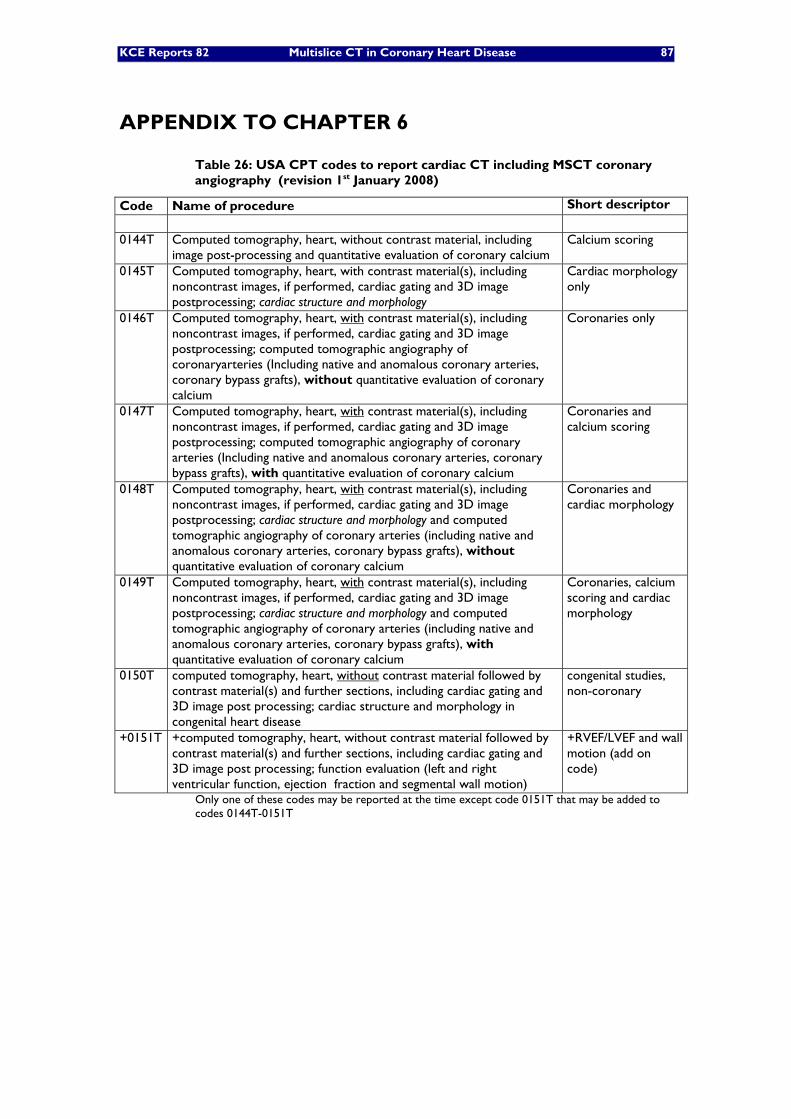
KCE Reports 82 Multislice CT in Coronary Heart Disease 87
APPENDIX TO CHAPTER 6
Table 26: USA CPT codes to report cardiac CT including MSCT coronary angiography (revision 1st January 2008)
Code Name of procedure Short descriptor
0144T Computed tomography, heart, without contrast material, including
image post-processing and quantitative evaluation of coronary calcium Calcium scoring
0145T Computed tomography, heart, with contrast material(s), including noncontrast images, if performed, cardiac gating and 3D image postprocessing; cardiac structure and morphology
Cardiac morphology only
0146T Computed tomography, heart, with contrast material(s), including noncontrast images, if performed, cardiac gating and 3D image postprocessing; computed tomographic angiography of coronaryarteries (Including native and anomalous coronary arteries, coronary bypass grafts), without quantitative evaluation of coronary calcium
Coronaries only
0147T Computed tomography, heart, with contrast material(s), including noncontrast images, if performed, cardiac gating and 3D image postprocessing; computed tomographic angiography of coronary arteries (Including native and anomalous coronary arteries, coronary bypass grafts), with quantitative evaluation of coronary calcium
Coronaries and calcium scoring
0148T Computed tomography, heart, with contrast material(s), including noncontrast images, if performed, cardiac gating and 3D image postprocessing; cardiac structure and morphology and computed tomographic angiography of coronary arteries (including native and anomalous coronary arteries, coronary bypass grafts), without quantitative evaluation of coronary calcium
Coronaries and cardiac morphology
0149T Computed tomography, heart, with contrast material(s), including noncontrast images, if performed, cardiac gating and 3D image postprocessing; cardiac structure and morphology and computed tomographic angiography of coronary arteries (including native and anomalous coronary arteries, coronary bypass grafts), with quantitative evaluation of coronary calcium
Coronaries, calcium scoring and cardiac morphology
0150T computed tomography, heart, without contrast material followed by contrast material(s) and further sections, including cardiac gating and 3D image post processing; cardiac structure and morphology in congenital heart disease
congenital studies, non-coronary
+0151T +computed tomography, heart, without contrast material followed by contrast material(s) and further sections, including cardiac gating and 3D image post processing; function evaluation (left and right ventricular function, ejection fraction and segmental wall motion)
+RVEF/LVEF and wall motion (add on code)
Only one of these codes may be reported at the time except code 0151T that may be added to codes 0144T-0151T

88 Multislice CT in Coronary Heart Disease KCE Reports 82
Table 27: Australia Medicare Benefit Schedule: MSCT except coronary angiography (valid 1st November 2007 – next update May 2008)
Code Name of procedure (adaptated from Australia MBS) Benefit (€)
COMPUTED TOMOGRAPHY - spiral angiography with intravenous contrast medium, including any scans performed before intravenous contrast injection - 1 or more spiral data acquisitions, including image editing, and maximum intensity projections or 3 dimensional surface shaded display, with hardcopy recording of multiple projections, where the service is not a study performed to image the coronary arteries
57350 the service is performed for the exclusion of arterial stenosis, occlusion, aneurysm or embolism; and the service has not been performed on the same patient within the previous 12 months;
226 – 263
57351 the service is performed for the exclusion of acute or recurrent pulmonary embolism; acute symptomatic arterial occlusion; post operative complication of arterial surgery; acute ruptured aneurysm; or acute dissection of the aorta, carotid or vertebral artery; and the services to which 57350 or 57355 apply have been performed on the same patient within the previous 12 months;
226 – 263
57355 Same as 57350, performed on 10 years or older equipment 133 –156 57356 Same as 57350, performed on 10 years or older equipment 133 –156
The Australian Medicare Benefit Schedule covers a spiral angiography between AUD 264 and AUD 510 (€156 and €302) (MBS item 57350-57356) against between AUD 384 and AUD 1152 (€227 and €682) for conventional coronary angiography (MBS item numbers 38215-38246
Table 28: French CT scanner ‘forfaits technique’ 167
Equipment Full tariff Reduced tariff
≤ reference activity threshold (**) > reference activity threshold (**)
≤ 11 000 scans
> 11 000 and ≤ 13 000 scans > 13 000 scans
Written off (*) €71.38 Not written off yet €100.51
€59.72 €42.88 €30.63
(*) installed since more than 7 years on the 1st January of the year.
(**) The threshold for multislice scanners varies between 6 000 (province), 6 350 (around Paris) and 6 700 scans (Paris).

KCE Reports 82 Multislice CT in Coronary Heart Disease 89
EVIDENCE LEVELS OF DIAGNOSTIC STUDIES Fryback and Thornbury described a hierarchy of diagnostic efficacy, which is used as the basis of this report.153, 154 The model is characterized by a change in perceived goals. It is hierarchical: on one extreme are endpoints describing only the technical performance of the test, on the other extreme are endpoints pertaining to the value of the diagnostic technology to society. If a test performs poorly at one level, it is unlikely to perform well at a higher level. The reverse, however, is not true: increases in the technical performance of a test will not necessarily guarantee improvement at a higher level, for example effect on patient outcome. A diagnostic test does not necessarily have to demonstrate effectiveness at each level before it can be used in clinical practice, but the possible gain and remaining uncertainty on the test’s efficacy is clearly presented by this approach.
Level 1: technical efficacy
The technical efficacy of a test refers to the ability to produce usable information. The test’s feasibility and operator dependence refer to in what circumstances and by whom the test can be performed. The analytical sensitivity is the ability to detect small quantities of the measured component. This should be distinguished from the diagnostic sensitivity, the ability of a test to detect disease. The precision or reproducibility of results is the ability to obtain the same test results on repeated testing or observations. It is influenced by analytical variability and observer interpretation. Analytical variability consists of inaccuracy and imprecision. Inaccuracy implies systematic error, such as calibration error. Imprecision implies random error. Agreement between two continuous test methods can be expressed in a regression analysis or Bland & Altman plots. A correlation coefficient does not provide information on agreement. The agreement between two observers (interobserver) or the same observer on different occasions (intraobserver) can be expressed with a kappa statistic. It is often assumed that the technical efficacy does no longer need to be evaluated once a test is being used in clinical practice.
Level 2: diagnostic accuracy
This level refers to the test’s ability to detect or exclude disease in patients compared with a criterion standard or reference test. Test characteristics are sensitivity, specificity, predictive values, likelihood ratios and ROC curves. Sensitivity and specificity are the most widely used outcome measures, but are sensitive to spectrum bias. Spectrum bias may occur when the study population has a different clinical spectrum (more advanced cases, for instance) than the population in whom the test is to be applied. If sensitivity is determined in seriously diseased subjects and specificity in clearly healthy subjects, both will be grossly overestimated relative to practical situations where diseased and healthy subjects cannot be clinically distinguished in advance. This design has been called “inappropriate case-control design” in the pilot assessments. Predictive values, with the positive predictive value being the proportion of patients with a positive test result that actually has the disease and the negative predictive value the proportion of patients with a negative test result that does not have the disease, are dependent on disease prevalence in the study sample. For example, in a situation where disease prevalence is very low, say 1%, the negative predictive value of the test will be easily over 95% as already 99% of the population do not have the disease. Prevalence and the setting in which patients were recruited should be noted to reflect on this. The likelihood ratios show how a test result alters the pre-test probability into a posttest probability, using Bayesian reasoning. The pre-test probability depends on the prevalence of the target condition and the results of previous tests, for example history, clinical examination, imaging or laboratory tests. Another outcome measure which is sometimes used, is the number needed to diagnose, analogous to the number needed to treat in intervention studies. However, using this measure it is assumed that diagnostic testing is always done to rule in a target condition, to diagnose the target condition, while in clinical practice tests are also used to rule out a target condition. Finally, test accuracy can be illustrated using an ROC curve. The ROC curve graphs test sensitivity
90 Multislice CT in Coronary Heart Disease KCE Reports 82
versus 1-specificity for various cut-off points. The area under the curve provides a summary measure of the test performance. It also allows comparison of two different tests by testing the two areas under the curve or by testing partial areas under the curve in which the test is most useful. Clearly, the first level of diagnostic efficacy, technical efficacy, contributes to the diagnostic accuracy. But it also becomes apparent that there may be a point beyond which improvement in technical performance no longer improves diagnostic accuracy. Assuming therefore that diagnostic accuracy can be estimated on the basis of technical accuracy studies is not correct.
Level 3: diagnostic thinking
This level of diagnostic efficacy is concerned with assessment of the effect of test information on diagnostic reasoning and disease categorization. Studies on diagnostic thinking serve as a proxy for estimating the effect of a test on patient care. Patients outcome can not be influenced by the diagnostic technology unless the physician is led to do something different than would have been done without the test information. Using the likelihood ratio and calculating the post-test probability, this change in diagnostic thinking can be computed. However, the pre-test probability of a disease is not always available in clinical practice and depends not only on setting, but also on patient characteristics and other selection processes, such as referral and the results or previous tests. Clinicians who wish to apply the Bayesian properties of diagnostic tests require accurate estimates of the pre-test probability of target disorders in their area and setting. These estimates can come from five sources: (1) personal experience, (2) population prevalence figures, (3) practice databases, (4) the publication that described the test or (5) one of a growing number of primary studies of pretest probability in different settings. An alternative are studies that empirically test the change in the physician’s subjective assessment on the probability of disease. In these studies, physicians are asked to estimate the probability of disease before knowing the test result, and estimating it again after the test result has been disclosed. Efficacious tests are those that significantly increase or lower pre-test probabilities assumed by the physician or computed by likelihood ratios using Bayesian reasoning. One major difficulty with this level of diagnostic efficacy is that it is not always known what post-test probability of disease should be used as a threshold. Which probability of disease is low enough to exclude disease, which is high enough to treat the patient? These thresholds will differ according to the target condition and the treatments that are available.
Level 4: therapeutic impact
The most efficacious tests at this level are those that lead to the institution of a new management strategy. Studies can assess this empirically by comparing the intended management before the test result is known with that after the test result has been disclosed. In what proportion of patients did the information change the intended management? In some cases, management changes are considered not only in the patient himself, but also in other persons, for example prophylactic measures in case of an infectious outbreak. These prospective case-series, however, can be subject to bias such as selection bias. The lack of a concurrent control group may lead to confounding, as there is no information on those patients not enrolled in the study and therefore not receiving the new technology. These considerations underscore the need for randomized controlled trials. But, in the absence of RCTs they do play an important role as an intermediate.
Level 5: patient outcome
The ultimate goal of health care is to improve patient outcome. For diagnostic tests that are expensive, dangerous or widely used, knowledge about patient outcome efficacy seems particularly important. It is at this level that expected harm, such as burden, pain, risk, can be weighed directly against its expected benefit, such as improving life expectancy, quality ofleast prone to bias to estimate these risks and benefit. However, it is not always feasible to perform an RCT for ethical, financial or other reasons. In those cases, case-series collected before and after the introduction of a new test technology
KCE Reports 82 Multislice CT in Coronary Heart Disease 91
or case-control studies may provide some of the answers. A methodological difficulty with this level is that the independent contribution of test technology to patient outcomes may be small in the context of all the other influences and therefore very large sample sizes may be required. But, in spite of these difficulties, RCTs on diagnostic tests are feasible. Various designs are possible, according to the specific research question. Some tests, however, will never be able to prove a change in „objective” patient outcomes such as mortality or morbidity, simply because there is no treatment available at this moment that has an impact on these outcomes. This is the case in for example dementia or Amyotrophic Lateral Sclerosis (ALS). A diagnostic test will therefore never produce a difference in mortality, but may improve quality of life measures by giving the patient (and the carer) an affirmative diagnosis and providing an explanation for the signs and symptoms the patient experiences.
Level 6: cost-effectiveness analysis
This level goes beyond the individual risks and benefits, but assesses whether the cost for use of a given test is acceptable for society. Is the price for the positive effect on patient outcome worthwhile? Resources can not be allocated twice; money spent on one technology can not be spent on another. Cost-effectiveness studies compute a cost per unit of output. Any of the measures of the previous levels can be used as input, for example cost per surgery avoided, cost per appropriately treated patient, cost per life year gained or cost per quality adjusted life year gained. Final outcomes, such as life years gained or QALYs gained, are preferred over intermediate outcomes in economic evaluations, as they allow comparisons across a broader range of health interventions, e.g. diagnostic and therapeutic interventions. Because data on these outcomes and costs of the diagnostic and subsequent therapeutic paths are not routinely available from observations, modelling becomes inevitable to examine the cost-effectiveness of diagnostic tests. The validity of the model input parameters is crucial for the credibility of the model. The values of all input variables must be based on solid evidence obtained from literature or observations. Sensitivity analyses can illustrate the robustness of the conclusions, by demonstrating the sensitivity of the results to changes in the values of remaining uncertain input parameters. Costeffectiveness models can only upgrade the level of evidence if level 5 evidence was available on the outcomes used in the model (be it life years gained or procedures avoided) and if this evidence was actually used in the model.

92 Multislice CT in Coronary Heart Disease KCE Reports 82
REFERENCES 1. Yusuf S, Fallen E, Harrington RA, Guyton RA. Clinical decisions. Management of stable
coronary disease. N Engl J Med. 2007;357(17):1762-6. 2. Boden WE, O'Rourke RA, Teo KK, Hartigan PM, Maron DJ, Kostuk WJ, et al. Optimal
medical therapy with or without PCI for stable coronary disease. N Engl J Med. 2007;356(15):1503-16.
3. Ambrose JA, Fuster V. The risk of coronary occlusion is not proportional to the prior severity of coronary stenoses. Heart. 1998;79(1):3-4.
4. Fox K, Garcia MAA, Ardissino D, Buszman P, Camici PG, Crea F, et al. Guidelines on the management of stable angina pectoris: executive summary: the Task Force on the Management of Stable Angina Pectoris of the European Society of Cardiology. Eur Heart J. 2006;27(11):1341-81.
5. Gibbons RJ, Abrams J, Chatterjee K, Daley J, Deedwania PC, Douglas JS, et al. ACC/AHA 2002 guideline update for the management of patients with chronic stable angina--summary article: a report of the American College of Cardiology/American Heart Association Task Force on Practice Guidelines (Committee on the Management of Patients With Chronic Stable Angina). Circulation. 2003;107(1):149-58.
6. Campeau L. The Canadian Cardiovascular Society grading of angina pectoris revisited 30 years later. Can J Cardiol. 2002;18(4):371-9.
7. Diamond GA, Forrester JS. Analysis of probability as an aid in the clinical diagnosis of coronary-artery disease. N Engl J Med. 1979;300(24):1350-8.
8. Chaitman BR, Bourassa MG, Davis K, Rogers WJ, Tyras DH, Berger R, et al. Angiographic prevalence of high-risk coronary artery disease in patient subsets (CASS). Circulation. 1981;64(2):360-7.
9. Williams SV, Fihn SD, Gibbons RJ, American College of C, American Heart A, American College of Physicians-American Society of Internal M. Guidelines for the management of patients with chronic stable angina: diagnosis and risk stratification. Ann Intern Med. 2001;135(7):530-47.
10. Thygesen K, Alpert JS, White HD, Joint ESCAAHAWHFTFftRoMI. Universal definition of myocardial infarction. Eur Heart J. 2007;28(20):2525-38.
11. Anderson JL, Adams CD, Antman EM, Bridges CR, Califf RM, Casey DE, Jr., et al. ACC/AHA 2007 guidelines for the management of patients with unstable angina/non-ST-Elevation myocardial infarction: a report of the American College of Cardiology/American Heart Association Task Force on Practice Guidelines (Writing Committee to Revise the 2002 Guidelines for the Management of Patients With Unstable Angina/Non-ST-Elevation Myocardial Infarction) developed in collaboration with the American College of Emergency Physicians, the Society for Cardiovascular Angiography and Interventions, and the Society of Thoracic Surgeons endorsed by the American Association of Cardiovascular and Pulmonary Rehabilitation and the Society for Academic Emergency Medicine. J Am Coll Cardiol. 2007;50(7):e1-e157.
12. Monaghan MJ. Stress myocardial contrast echocardiography. Heart. 2003;89(12):1391-3. 13. Cheitlin MD, Armstrong WF, Aurigemma GP, Beller GA, Bierman FZ, Davis JL, et al.
ACC/AHA/ASE 2003 guideline update for the clinical application of echocardiography--summary article: a report of the American College of Cardiology/American Heart Association Task Force on Practice Guidelines (ACC/AHA/ASE Committee to Update the 1997 Guidelines for the Clinical Application of Echocardiography). J Am Coll Cardiol. 2003;42(5):954-70.
14. Mowatt G, Vale L, Brazzelli M, Hernandez R, Murray A, Scott N, et al. Systematic review of the effectiveness and cost-effectiveness, and economic evaluation, of myocardial perfusion scintigraphy for the diagnosis and management of angina and myocardial infarction. Health Technol Assess. 2004;8(30):iii-iv, 1-207.
15. Lauer M, Froelicher ES, Williams M, Kligfield P, American Heart Association Council on Clinical Cardiology SoECRaP. Exercise testing in asymptomatic adults: a statement for professionals from the American Heart Association Council on Clinical Cardiology, Subcommittee on Exercise, Cardiac Rehabilitation, and Prevention. Circulation. 2005;112(5):771-6.
16. Gibbons RJ, Balady GJ, Bricker JT, Chaitman BR, Fletcher GF, Froelicher VF, et al. ACC/AHA 2002 guideline update for exercise testing: summary article. A report of the American

KCE Reports 82 Multislice CT in Coronary Heart Disease 93
College of Cardiology/American Heart Association Task Force on Practice Guidelines (Committee to Update the 1997 Exercise Testing Guidelines). J Am Coll Cardiol. 2002;40(8):1531-40.
17. Mant J, McManus RJ, Oakes RAL, Delaney BC, Barton PM, Deeks JJ, et al. Systematic review and modelling of the investigation of acute and chronic chest pain presenting in primary care. Health Technol Assess. 2004;8(2):iii, 1-158.
18. Klocke FJ, Baird MG, Lorell BH, Bateman TM, Messer JV, Berman DS, et al. ACC/AHA/ASNC guidelines for the clinical use of cardiac radionuclide imaging--executive summary: a report of the American College of Cardiology/American Heart Association Task Force on Practice Guidelines (ACC/AHA/ASNC Committee to Revise the 1995 Guidelines for the Clinical Use of Cardiac Radionuclide Imaging). Circulation. 2003;108(11):1404-18.
19. Iskander S, Iskandrian AE. Risk assessment using single-photon emission computed tomographic technetium-99m sestamibi imaging. J Am Coll Cardiol. 1998;32(1):57-62.
20. Reyes E, Underwood SR. Myocardial perfusion scintigraphy: an important step between clinical assessment and coronary angiography in patients with stable chest pain. Eur Heart J. 2006;27(1):3-4.
21. Mahmarian JJ. Computed tomography coronary angiography as an anatomic basis for risk stratification: deja vu or something new? J Am Coll Cardiol. 2007;50(12):1171-3.
22. Marcassa. Clinical value, cost-effectiveness, and safety of myocardial perfusion scintigraphy: a position statement. Eur Heart J. 2008;29(4):557-63.
23. Klocke FJ, Baird MG, Lorell BH, Bateman TM, Messer JV, Berman DS, et al. ACC/AHA/ASNC guidelines for the clinical use of cardiac radionuclide imaging--executive summary: a report of the American College of Cardiology/American Heart Association Task Force on Practice Guidelines (ACC/AHA/ASNC Committee to Revise the 1995 Guidelines for the Clinical Use of Cardiac Radionuclide Imaging). J Am Coll Cardiol. 2003;42(7):1318-33.
24. Sharples L, Hughes V, Crean A, Dyer M, Buxton M, Goldsmith K, et al. Cost-effectiveness of functional cardiac testing in the diagnosis and management of coronary artery disease: a randomised controlled trial. The CECaT trial. Health Technol Assess. 2007;11(49):1-136.
25. Geleijnse ML, Fioretti PM, Roelandt JR. Methodology, feasibility, safety and diagnostic accuracy of dobutamine stress echocardiography. J Am Coll Cardiol. 1997;30(3):595-606.
26. Nixdorff U. Head-to-Head Comparison of Dobutamine Stress Echocardiography and Cardiac Computed Tomography for the Detection of Significant Coronary Artery Disease. Cardiology. 2007;110(2):81-6.
27. Geleijnse ML, Krenning BJ, Nemes A, Soliman OII, Galema TW, ten Cate FJ. Diagnostic value of dobutamine stress echocardiography in patients with normal wall motion at rest. Echocardiography. 2007;24(5):553-7.
28. Wijns W, De Bruyne B, Vanhoenacker PK. What does the clinical cardiologist need from noninvasive cardiac imaging: is it time to adjust practices to meet evolving demands? J Nucl Cardiol. 2007;14(3):366-70.
29. Task Force for Diagnosis and Treatment of Non STSEACSoESoC, Bassand J-P, Hamm CW, Ardissino D, Boersma E, Budaj A, et al. Guidelines for the diagnosis and treatment of non-ST-segment elevation acute coronary syndromes. Eur Heart J. 2007;28(13):1598-660.
30. Erbel R, Mohlenkamp S, Kerkhoff G, Budde T, Schmermund A. Non-invasive screening for coronary artery disease: calcium scoring. Heart. 2007;93(12):1620-9.
31. ECRIInstitute. 64-slice computed tomography systems. Health Devices. 2007;36(12):377-402. 32. Achenbach S. Dual-source cardiac computed tomography: image quality and dose
considerations. Eur Radiol. 2008. 33. Alkadhi H, Scheffel H, Desbiolles L, Gaemperli O, Stolzmann P, Plass A, et al. Dual-source
computed tomography coronary angiography: influence of obesity, calcium load, and heart rate on diagnostic accuracy. Eur Heart J. 2008;29(6):766-76.
34. Kolnes K, Velle OH, Hareide S, Hegbom K, Wiseth R. Multislice computed tomography coronary angiography at a local hospital: Pitfalls and potential. Acta Radiol. 2006;47(7):680-6.
35. Leber AW, Knez A, von Ziegler F, Becker A, Nikolaou K, Paul S, et al. Quantification of obstructive and nonobstructive coronary lesions by 64-slice computed tomography: a comparative study with quantitative coronary angiography and intravascular ultrasound. J Am Coll Cardiol. 2005;46(1):147-54.
36. Di Carli MF, Hachamovitch R. New technology for noninvasive evaluation of coronary artery disease. Circulation. 2007;115(11):1464-80.

94 Multislice CT in Coronary Heart Disease KCE Reports 82
37. Kalra MK, Brady TJ. Current status and future directions in technical developments of cardiac computed tomography. Journal of Cardiovascular Computed Tomography. 2008;1(2):71–80.
38. Berman DS, Hachamovitch R, Shaw LJ, Friedman JD, Hayes SW, Thomson LEJ, et al. Roles of nuclear cardiology, cardiac computed tomography, and cardiac magnetic resonance: Noninvasive risk stratification and a conceptual framework for the selection of noninvasive imaging tests in patients with known or suspected coronary artery disease. J Nucl Med. 2006;47(7):1107-18.
39. Hamon M, Morello R, Riddell JW, Hamon M. Coronary arteries: diagnostic performance of 16- versus 64-section spiral CT compared with invasive coronary angiography--meta-analysis. Radiology. 2007;245(3):720-31.
40. Herzog C, Zwerner PL, Doll JR, Nielsen CD, Nguyen SA, Savino G, et al. Significant coronary artery stenosis: comparison on per-patient and per-vessel or per-segment basis at 64-section CT angiography. Radiology. 2007;244(1):112-20.
41. OHTAC. Multidetector Computed Tomography for Coronary Artery Disease Screening in Asymptomatic populations. 2007.
42. Waugh N, Black C, Walker S, McIntyre L, Cummins E, Hillis G. The effectiveness and cost-effectiveness of computed tomography screening for coronary artery disease: systematic review. Systematic review. The National Coordinating Centre for Health Technology Assessment (NCCHTA); 2006. Available from: http://www.hta.ac.uk/execsumm/summ1039.htm
43. Budoff MJ, Achenbach S, Blumenthal RS, Carr JJ, Goldin JG, Greenland P, et al. Assessment of coronary artery disease by cardiac computed tomography: a scientific statement from the American Heart Association Committee on Cardiovascular Imaging and Intervention, Council on Cardiovascular Radiology and Intervention, and Committee on Cardiac Imaging, Council on Clinical Cardiology. Circulation. 2006;114(16):1761-91.
44. Alkadhi H. The revival of step-and-shoot computed tomography coronary angiography: benefits and open questions. Journal of Cardiovascular Computed Tomography. 2008(2):91-2.
45. De Feyter PJ, Meijboom WB, Weustink A, Van Mieghem C, Mollet NRA, Vourvouri E, et al. Spiral multislice computed tomography coronary angiography: a current status report. Clin Cardiol. 2007;30(9):437-42.
46. Timmis AD, Feder G, Hemingway H. Prognosis of stable angina pectoris: why we need larger population studies with higher endpoint resolution. Heart. 2007;93(7):786-91.
47. SIGN. Management of stable angina. SIGN; 2007 February 2007. Available from: http://www.sign.ac.uk/pdf/sign96.pdf
48. Hueb W, Lopes NH, Gersh BJ, Soares P, Machado LAC, Jatene FB, et al. Five-year follow-up of the Medicine, Angioplasty, or Surgery Study (MASS II): a randomized controlled clinical trial of 3 therapeutic strategies for multivessel coronary artery disease. Circulation. 2007;115(9):1082-9.
49. Katritsis DG, Ioannidis JPA. PCI for stable coronary disease. N Engl J Med. 2007;357(4):414-5; author reply 7-8.
50. Neyt M, Van Brabandt H, Devriese S, Mahieu J, De Ridder A, De Graeve D. Drug Eluting Stents in Belgium:a Health Technology Assessment. Brussel: Federaal Kenniscentrum voor de Gezondheidszorg (KCE); 2007. KCE reports 66A (D/2007/10.273/47) Available from: http://www.kce.fgov.be/index_nl.aspx?ID=0&SGREF=5260&CREF=10069
51. Van Brabandt H, Camberlin C, Vrijens F, Parmentier Y, Ramaekers D, Bonneux L. Variaties in de ziekenhuispraktijk bij acuut myocardinfarct in België. KCE; 2005. ( reports 14A) Available from: http://kce.fgov.be/index_nl.aspx?ID=0&SGREF=5270&CREF=5355
52. Hochman JS, Lamas GA, Buller CE, Dzavik V, Reynolds HR, Abramsky SJ, et al. Coronary intervention for persistent occlusion after myocardial infarction. N Engl J Med. 2006;355(23):2395-407.
53. Braunwald E, Antman EM, Beasley JW, Califf RM, Cheitlin MD, Hochman JS, et al. ACC/AHA 2002 guideline update for the management of patients with unstable angina and non-ST-segment elevation myocardial infarction--summary article: a report of the American College of Cardiology/American Heart Association task force on practice guidelines (Committee on the Management of Patients With Unstable Angina). J Am Coll Cardiol. 2002;40(7):1366-74.
54. Poole-Wilson PA, Voko Z, Kirwan B-A, de Brouwer S, Dunselman PHJM, Lubsen J, et al. Clinical course of isolated stable angina due to coronary heart disease. Eur Heart J. 2007;28(16):1928-35.
KCE Reports 82 Multislice CT in Coronary Heart Disease 95
55. Clayton TC, Lubsen J, Pocock SJ, Voko Z, Kirwan B-A, Fox KAA, et al. Risk score for predicting death, myocardial infarction, and stroke in patients with stable angina, based on a large randomised trial cohort of patients. BMJ. 2005;331(7521):869.
56. Daly CA, De Stavola B, Sendon JLL, Tavazzi L, Boersma E, Clemens F, et al. Predicting prognosis in stable angina--results from the Euro heart survey of stable angina: prospective observational study. BMJ. 2006;332(7536):262-7.
57. Cheitlin MD. Evaluation of the low-risk patient with chest pain: is there incremental value over the clinical assessment of the patient with chest pain to doing a stress test in the emergency department? Cardiol Rev. 1999;7(1):27-8.
58. Institute for Clinical Systems Improvement. Electron-beam and helical computed tomography for coronary artery disease. Review. Institute for Clinical Systems Improvement (ICSI); 2004. Technology Assessment Report Available from: http://www.icsi.org/knowledge/detail.asp?catID=107&itemID=280
59. Ontario Ministry of Health and Long-Term Care. Multi-detector computed tomography angiography for coronary artery disease. Systematic review. Medical Advisory Secretariat Ontario Ministry of Health and Long-Term Care (MAS); 2005. Available from: http://www.health.gov.on.ca/english/providers/program/mas/tech/techlist_mn.html
60. Blue Cross Blue Shield A. Contrast-enhanced cardiac computed tomographic angiography for coronary artery evaluation. Chicago IL: Blue Cross Blue Shield Association (BCBS). 2005:45.
61. Andalusian Agency for Health Technology Assessment. Multi-slice computerised tomography coronary angiography. Report. Andalusian Agency for Health Technology Assessment (AETSA); 2006. Available from: http://www.juntadeandalucia.es/salud/aetsa
62. Australia and New Zealand Horizon Scanning Network. Computed tomography coronary angiography for the detection of coronary artery disease. Report. Adelaide: Australia and New Zealand Horizon Scanning Network (ANZHSN); 2006 March, 2006. Horizon scanning prioritising summary 000184 (Volume 12, Number 4) Available from: http://www.horizonscanning.gov.au/internet/horizon/publishing.nsf/Content/8F86C8B4D8513895CA25715C0005D972/$File/Computed%20tomography%20coronary%20angiography%20for%20the%20detection%20of%20coronary%20artery%20disease%20March2006.pdf
63. Blue Cross Blue Shield A. Contrast-enhanced cardiac computed tomographic angiography in the diagnosis of coronary artery stenosis or for evaluation of acute chest pain. Chicago IL: Blue Cross Blue Shield Association (BCBS). 2006;TEC Assessment 21(5):18.
64. AHRQ. Noninvasive Imaging for Coronary Artery Disease. Rockville, MD: 2006 October 3, 2006.
65. HarvardPilgrimHealthCare. Computed Tomography Angiography for Coronary Artery Disease. 2006 Ocotber 2006. Technology Assessment Policy
66. Abdulla J, Abildstrom SZ, Gotzsche O, Christensen E, Kober L, Torp-Pedersen C. 64-multislice detector computed tomography coronary angiography as potential alternative to conventional coronary angiography: a systematic review and meta-analysis. Eur Heart J. 2007;28(24):3042-50.
67. Goldstein JA, Gallagher MJ, O'Neill WW, Ross MA, O'Neil BJ, Raff GL. A randomized controlled trial of multi-slice coronary computed tomography for evaluation of acute chest pain. J Am Coll Cardiol. 2007;49(8):863-71.
68. Leber AW, Johnson T, Becker A, von Ziegler F, Tittus J, Nikolaou K, et al. Diagnostic accuracy of dual-source multi-slice CT-coronary angiography in patients with an intermediate pretest likelihood for coronary artery disease. Eur Heart J. 2007;28(19):2354-60.
69. Meijboom WB, Weustink AC, Pugliese F, van Mieghem CAG, Mollet NR, van Pelt N, et al. Comparison of diagnostic accuracy of 64-slice computed tomography coronary angiography in women versus men with angina pectoris. Am J Cardiol. 2007;100(10):1532-7.
70. Shabestari AA, Abdi S, Akhlaghpoor S, Azadi M, Baharjoo H, Pajouh MD, et al. Diagnostic performance of 64-channel multislice computed tomography in assessment of significant coronary artery disease in symptomatic subjects. Am J Cardiol. 2007;99(12):1656-61.
71. Weustink AC, Meijboom WB, Mollet NR, Otsuka M, Pugliese F, van Mieghem C, et al. Reliable high-speed coronary computed tomography in symptomatic patients. J Am Coll Cardiol. 2007;50(8):786-94.
72. Rubinshtein R, Halon DA, Gaspar T, Jaffe R, Goldstein J, Karkabi B, et al. Impact of 64-slice cardiac computed tomographic angiography on clinical decision-making in emergency department patients with chest pain of possible myocardial ischemic origin. Am J Cardiol. 2007;100(10):1522-6.

96 Multislice CT in Coronary Heart Disease KCE Reports 82
73. Vanhoenacker PK, Heijenbrok-Kal MH, Van Heste R, Decramer I, Van Hoe LR, Wijns W, et al. Diagnostic performance of multidetector CT angiography for assessment of coronary artery disease: meta-analysis. Radiology. 2007;244(2):419-28.
74. Leschka S. Combining Dual-Source Computed Tomography Coronary Angiography and Calcium Scoring: Added Value for the Assessment of Coronary Artery Disease. Heart. 2007.
75. Scheffel H, Alkadhi H, Plass A, Vachenauer R, Desbiolles L, Gaemperli O, et al. Accuracy of dual-source CT coronary angiography: First experience in a high pre-test probability population without heart rate control. Eur Radiol. 2006;16(12):2739-47.
76. Ropers U, Ropers D, Pflederer T, Anders K, Kuettner A, Stilianakis NI, et al. Influence of heart rate on the diagnostic accuracy of dual-source computed tomography coronary angiography. J Am Coll Cardiol. 2007;50(25):2393-8.
77. Hausleiter J, Meyer T, Hadamitzky M, Zankl M, Gerein P, Dorrler K, et al. Non-invasive coronary computed tomographic angiography for patients with suspected coronary artery disease: the Coronary Angiography by Computed Tomography with the Use of a Submillimeter resolution (CACTUS) trial. Eur Heart J. 2007;28(24):3034-41.
78. Shapiro MD, Butler J, Rieber J, Sheth TN, Cury RC, Ferencik M, et al. Analytic approaches to establish the diagnostic accuracy of coronary computed tomography angiography as a tool for clinical decision making. Am J Cardiol. 2007;99(8):1122-7.
79. Rubinshtein R, Halon DA, Gaspar T, Jaffe R, Karkabi B, Flugelman MY, et al. Usefulness of 64-slice cardiac computed tomographic angiography for diagnosing acute coronary syndromes and predicting clinical outcome in emergency department patients with chest pain of uncertain origin. Circulation. 2007;115(13):1762-8.
80. Rubinshtein R, Halon DA, Gaspar T, Schliamser JE, Yaniv N, Ammar R, et al. Usefulness of 64-slice multidetector computed tomography in diagnostic triage of patients with chest pain and negative or nondiagnostic exercise treadmill test result. Am J Cardiol. 2007;99(7):925-9.
81. Vanhoenacker PK, Decramer I, Bladt O, Sarno G, Bevernage C, Wijns W. Detection of non-ST-elevation myocardial infarction and unstable angina in the acute setting: meta-analysis of diagnostic performance of multi-detector computed tomographic angiography. BMC Cardiovasc Disord. 2007;7:39.
82. Sun. Diagnostic value of 64-slice CT angiography in coronary artery disease: A systematic review. Eur J Radiol. 2007.
83. Meijboom WB, van Mieghem CAG, Mollet NR, Pugliese F, Weustink AC, van Pelt N, et al. 64-slice computed tomography coronary angiography in patients with high, intermediate, or low pretest probability of significant coronary artery disease. J Am Coll Cardiol. 2007;50(15):1469-75.
84. Whiting P, Rutjes AWS, Reitsma JB, Bossuyt PMM, Kleijnen J. The development of QUADAS: a tool for the quality assessment of studies of diagnostic accuracy included in systematic reviews. BMC Med Res Methodol. 2003;3:25.
85. Leschka S, Alkadhi H, Plass A, Desbiolles L, Grunenfelder J, Marincek B, et al. Accuracy of MSCT coronary angiography with 64-slice technology: first experience. Eur Heart J. 2005;26(15):1482-7.
86. Mundy L HJ, Merlin T. Computed Tomography coronary angiography for detection of coronary artery disease. ANZHSN; 2006 March 2006. Volume 12, Number 4 Available from: http://www.horizonscanning.gov.au
87. Mollet NR, Cademartiri F, van Mieghem CAG, Runza G, McFadden EP, Baks T, et al. High-resolution spiral computed tomography coronary angiography in patients referred for diagnostic conventional coronary angiography. Circulation. 2005;112(15):2318-23.
88. Raff GL, Gallagher MJ, O'Neill WW, Goldstein JA. Diagnostic accuracy of noninvasive coronary angiography using 64-slice spiral computed tomography. J Am Coll Cardiol. 2005;46(3):552-7.
89. Fine JJ, Hopkins CB, Ruff N, Newton FC. Comparison of accuracy of 64-slice cardiovascular computed tomography with coronary angiography in patients with suspected coronary artery disease. Am J Cardiol. 2006;97(2):173-4.
90. Ehara M, Surmely J-F, Kawai M, Katoh O, Matsubara T, Terashima M, et al. Diagnostic accuracy of 64-slice computed tomography for detecting angiographically significant coronary artery stenosis in an unselected consecutive patient population: comparison with conventional invasive angiography. Circ J. 2006;70(5):564-71.
91. Ghostine S, Caussin C, Daoud B, Habis M, Perrier E, Pesenti-Rossi D, et al. Non-invasive detection of coronary artery disease in patients with left bundle branch block using 64-slice computed tomography. J Am Coll Cardiol. 2006;48(10):1929-34.

KCE Reports 82 Multislice CT in Coronary Heart Disease 97
92. Meijboom WB, Mollet NR, Van Mieghem CAG, Kluin J, Weustink AC, Pugliese F, et al. Pre-operative computed tomography coronary angiography to detect significant coronary artery disease in patients referred for cardiac valve surgery. J Am Coll Cardiol. 2006;48(8):1658-65.
93. Meijboom WB, Mollet NR, Van Mieghem CA, Weustink AC, Pugliese F, van Pelt N, et al. 64-Slice CT coronary angiography in patients with non-ST elevation acute coronary syndrome. Heart. 2007;93(11):1386-92.
94. Muhlenbruch G, Seyfarth T, Soo CS, Pregalathan N, Mahnken AH. Diagnostic value of 64-slice multi-detector row cardiac CTA in symptomatic patients. Eur Radiol. 2007;17(3):603-9.
95. Nikolaou K, Knez A, Rist C, Wintersperger BJ, Leber A, Johnson T, et al. Accuracy of 64-MDCT in the diagnosis of ischemic heart disease. AJR Am J Roentgenol. 2006;187(1):111-7.
96. Oncel D, Oncel G, Tastan A, Tamci B. Detection of significant coronary artery stenosis with 64-section MDCT angiography. Eur J Radiol. 2007;62(3):394-405.
97. Ong TK, Chin SP, Liew CK, Chan WL, Seyfarth MT, Liew HB, et al. Accuracy of 64-row multidetector computed tomography in detecting coronary artery disease in 134 symptomatic patients: influence of calcification. Am Heart J. 2006;151(6):1323 e1-6.
98. Plass A, Grunenfelder J, Leschka S, Alkadhi H, Eberli FR, Wildermuth S, et al. Coronary artery imaging with 64-slice computed tomography from cardiac surgical perspective. Eur J Cardiothorac Surg. 2006;30(1):109-16.
99. Pugliese F, Mollet NRA, Runza G, van Mieghem C, Meijboom WB, Malagutti P, et al. Diagnostic accuracy of non-invasive 64-slice CT coronary angiography in patients with stable angina pectoris. Eur Radiol. 2006;16(3):575-82.
100. Ropers D, Pohle F-K, Kuettner A, Pflederer T, Anders K, Daniel WG, et al. Diagnostic accuracy of noninvasive coronary angiography in patients after bypass surgery using 64-slice spiral computed tomography with 330-ms gantry rotation. Circulation. 2006;114(22):2334-41; quiz
101. Schuijf JD, Pundziute G, Jukema JW, Lamb HJ, van der Hoeven BL, de Roos A, et al. Diagnostic accuracy of 64-slice multislice computed tomography in the noninvasive evaluation of significant coronary artery disease. Am J Cardiol. 2006;98(2):145-8.
102. Sheth TN, Rieber J, Mooyaart EAQ, Pena A, Abbara S, Cury RC, et al. Usefulness of coronary computed tomographic angiography to assess suitability for revascularization in patients with significant coronary artery disease and angina pectoris. Am J Cardiol. 2006;98(9):1198-201.
103. Schlosser T, Mohrs OK, Magedanz A, Nowak B, Voigtlander T, Barkhausen J, et al. Noninvasive coronary angiography using 64-detector-row computed tomography in patients with a low to moderate pretest probability of significant coronary artery disease. Acta Radiol. 2007;48(3):300-7.
104. Janne d'Othee B, Siebert U, Cury R, Jadvar H, Dunn EJ, Hoffmann U. A systematic review on diagnostic accuracy of CT-based detection of significant coronary artery disease. Eur J Radiol. 2008;65(3):449-61.
105. Zamora J, Abraira V, Muriel A, Khan K, Coomarasamy A. Meta-DiSc: a software for meta-analysis of test accuracy data. BMC Med Res Methodol. 2006;6:31.
106. Brodoefel H. Dual-Source CT: Effect of Heart Rate, Heart Rate Variability, and Calcification on Image Quality and Diagnostic Accuracy. Radiology. 2008.
107. Vliegenthart R, Oudkerk M, Song B, van der Kuip DAM, Hofman A, Witteman JCM. Coronary calcification detected by electron-beam computed tomography and myocardial infarction. The Rotterdam Coronary Calcification Study. Eur Heart J. 2002;23(20):1596-603.
108. Hendel RC, Patel MR, Kramer CM, Poon M, Hendel RC, Carr JC, et al. ACCF/ACR/SCCT/SCMR/ASNC/NASCI/SCAI/SIR 2006 appropriateness criteria for cardiac computed tomography and cardiac magnetic resonance imaging: a report of the American College of Cardiology Foundation Quality Strategic Directions Committee Appropriateness Criteria Working Group, American College of Radiology, Society of Cardiovascular Computed Tomography, Society for Cardiovascular Magnetic Resonance, American Society of Nuclear Cardiology, North American Society for Cardiac Imaging, Society for Cardiovascular Angiography and Interventions, and Society of Interventional Radiology. J Am Coll Cardiol. 2006;48(7):1475-97.
109. Schroeder S, Achenbach S, Bengel F, Burgstahler C, Cademartiri F, de Feyter P, et al. Cardiac computed tomography: indications, applications, limitations, and training requirements: Report of a Writing Group deployed by the Working Group Nuclear Cardiology and Cardiac CT of the European Society of Cardiology and the European Council of Nuclear Cardiology. Eur Heart J. 2008;29(4):531-56.
98 Multislice CT in Coronary Heart Disease KCE Reports 82
110. Di Carli MF. CT coronary angiography: where does it fit? J Nucl Med. 2006;47(9):1397-9. 111. Dorbala S, Hachamovitch R, Di Carli MF. Myocardial perfusion imaging and multidetector
computed tomographic coronary angiography: appropriate for all patients with suspected coronary artery disease? J Am Coll Cardiol. 2006;48(12):2515-7.
112. Greenland P. Who is a candidate for noninvasive coronary angiography? Ann Intern Med. 2006;145(6):466-7.
113. Bossuyt PM, Lijmer JG, Mol BW. Randomised comparisons of medical tests: sometimes invalid, not always efficient. Lancet. 2000;356(9244):1844-7.
114. Picano E. Sustainability of medical imaging. BMJ. 2004;328(7439):578-80. 115. Husmann L, Valenta I, Gaemperli O, Adda O, Treyer V, Wyss CA, et al. Feasibility of low-
dose coronary CT angiography: first experience with prospective ECG-gating. Eur Heart J. 2008;29(2):191-7.
116. Gutstein A, Wolak A. Predicting success of prospective and retrospective gating with dual-source computerised tomography angiography: development of selection criteria and initial experience. Journal of Cardiovascular Computed Tomography. 2008(2):81-90.
117. Rybicki FJ, Otero HJ, Steigner, Di Mario C. Initial evaluation of coronary images from 320-detector row computed tomography The International Journal of Cardiovascular Imaging 2008;24(5):1569-5794
118. Einstein AJ, Henzlova MJ, Rajagopalan S. Estimating risk of cancer associated with radiation exposure from 64-slice computed tomography coronary angiography. JAMA. 2007;298(3):317-23.
119. HogeGezondheidsraad. Evaluatie van de stijgende stralingsblootstelling van patiënten door computed tomgraphy (CT) en optimalisatie van de stralingsbescherming. . Brussel: 2006. (HGR nr. 8080) Available from: www.health.fgov.be/HGR_CSS
120. Murphy SW, Barrett BJ, Parfrey PS. Contrast nephropathy. J Am Soc Nephrol. 2000;11(1):177-82.
121. McCullough PA, Wolyn R, Rocher LL, Levin RN, O'Neill WW. Acute renal failure after coronary intervention: incidence, risk factors, and relationship to mortality. Am J Med. 1997;103(5):368-75.
122. Rudnick MR, Berns JS, Cohen RM, Goldfarb S. Contrast media-associated nephrotoxicity. Semin Nephrol. 1997;17(1):15-26.
123. Gibbons RJ, Araoz PA, Williamson EE. The year in cardiac imaging. J Am Coll Cardiol. 2007;50(10):988-1003.
124. Casarella WJ. A patient's viewpoint on a current controversy. Radiology. 2002;224(3):927. 125. Dewey M, Hamm B. Cost effectiveness of coronary angiography and calcium scoring using
CT and stress MRI for diagnosis of coronary artery disease. European Radiology. 2007;17(5):1301-9.
126. Rubinshtein R, Halon DA, Kogan A, Jaffe R, Karkabi B, Gaspar T, et al. Initial experience with a cardiologist-based chest pain unit in an emergency department in Isreal. Israel Medical Association Journal. 2006;8(5):329-32.
127. Cole JH, Chunn VM, Morrow JA, Buckley RS, Phillips GM. Cost implications of initial computed tomography angiography as opposed to catheterization in patients with mildly. Journal of Cardiovascular Computed Tomography. 2007;1(1):21-6.
128. Otero HJ, Rybicki FJ. Reimbursement for chest-pain CT: estimates based on current imaging strategies. 2007:237-42.
129. Llanos Méndez A, Román Villegas Portero R, Olry de Labry Lima A, García Mochón L, Epstein D, Cuerva Carvajal Á, et al. Multi-slice computerised tomography coronary angiography in detecting coronary stenosis. Meta analysis and economic report. Report. Sevilla: Andalusian Agency for Health Technology Assessment (AETSA); 2008 March 28, 2008. AETSA 2006/14 Available from: http://www.juntadeandalucia.es/salud/aetsa
130. National Horizon Scanning Centre. Computed tomography (CT) angiography for the diagnosis and management of coronary disease. Report. Birmingham: National Horizon Scanning Centre - Department of Public Health and Epidemiology; 2006 December 2006. Horizon Scanning Technology Briefing Available from: http://www.pcpoh.bham.ac.uk/publichealth/horizon/
131. Berrier J-L, Siemens CT Business Manager ESW. UNAMEC/KCE meeting. In. Brussels; 2007. 132. Pwee KH. Multislice/spiral computed tomography for screening for coronary artery disease.
Ottawa: Canadian Coordinating Office for Health Technology Assessment (CCOHTA). 2003:4.

KCE Reports 82 Multislice CT in Coronary Heart Disease 99
133. Demaerel P, Hermans R, Verstraete K, Bogaert J, Van Goethem M, Deblaere K, et al. Magnetic Resonance Imaging. Bruxelles: KCE = Federaal Kenniscentrum voor de gezondheiszorg = Centre fédéral d'expertise des soins de santé = Belgian Health Care Knowledge Centre; 2006. (D/2006/10.273/33) Available from: http://kce.fgov.be/index_en.aspx?SGREF=5220&CREF=7369
134. Brice J. Medicare imaging costs skyrocket, as cardiologists involvement rises. In: Imaging D, editor. Proceedings of Radiological Society of North America 2007; 2007 December 3, 2007. Available from: http://www.dimag.com/rsna/2007/showArticle.jhtml;jsessionid=PW0OVNMDZ43BAQSNDLQCKH0CJUNN2JVN?articleID=204400377
135. Levin DC. Merrill C. Sosman Lecture. The practice of radiology by nonradiologists: cost, quality, and utilization issues. AJR Am J Roentgenol. 1994;162(3):513-8.
136. Association of American Medical Colleges;c 2008 [cited April 18, 2008]. The Physician Self-Referral ("Stark") Regulation. Available from: http://www.aamc.org/advocacy/library/teachphys/phys0039.htm
137. Greeson TW, Zimmerman HM. The beginning of the end of self-referral? AJR Am J Roentgenol. 2007;189(3):513-6.
138. Levin DC, Rao VM, Bree RL, Neiman HL. Turf battles in radiology: how the radiology community can collectively respond to the challenge. Radiology. 1999;211(2):301-5.
139. American College of Radiology Reston;c 2006 [cited April 15, 2008]. ACR PRACTICE GUIDELINE FOR THE PERFORMANCE AND INTERPRETATION OF CARDIAC COMPUTED TOMOGRAPHY (CT). Available from: http://www.acr.org/SecondaryMainMenuCategories/quality_safety/guidelines/dx/cardio/ct_cardiac.aspx
140. Hendee WR. An opportunity for radiology. Radiology. 2006;238(2):389-94. 141. Runza G, Alaimo V, La Grutta L, Galia M, Basile A, Cademartiri F, et al. Can ECG-gated
MDCT be considered an obligatory step to plan and manage a new chest-pain unit? Eur J Radiol. 2007;64(1):48-53.
142. Centers for Medicare and Medicaid Services. NCD for Computerized Tomography (220.1). In; 1985.
143. Budoff M. Cardiac CT in the emergency room. Applied Radiology. 2006;35(12 SUPPL.):48-55. 144. Centers for Medicare and Medicaid Services. Decision Memo for Computed Tomographic
Angiography (CAG-00385N). In; 2008. 145. Régie de l'assurance maladie du Québec;c 2008 [updated March 2008]. MANUEL DES
MÉDECINS OMNIPRATICIENS. Available from: http://www.ramq.gouv.qc.ca/fr/professionnels/manuels/100/029_v_radio_diagnos_acte_omni.pdf
146. Medical Services Advisory Committee;c 2008 [cited May 16, 2008]. Computed Tomography Coronary Angiogram. Available from: http://www.msac.gov.au/internet/msac/publishing.nsf/Content/app1105-1
147. Institut für das Entgeltsystem im Krankenhaus. Informationen nach § 6 Abs. 2 KHEntgG für 2008: Neue Untersuchungs- und Behandlungsmethoden,. 2008 January 31, 2008. Available from: http://www.g-drg.de/cms/index.php/inek_site_de/g_drg_system_2008/neue_untersuchungs_und_behandlungsmethoden_nub/aufstellung_der_informationen_nach_6_abs_2_khentgg_fuer_2008
148. Brighton and Hove City Teaching Primary Care Trust. ABC OF COUNTING AND CURRENCY. 2007 June. Available from: http://www.brightonhovecitypct.nhs.uk/healthprofessionals/generalpractice/pbc/documents/ABCofcountingandcurrency.doc#Nationaltariff
149. Casemix Service. HRG4 Design Concepts. The NHS Information Centre; 2008 January 15. 2.0 Available from: http://www.ic.nhs.uk/webfiles/Services/casemix/Prep%20HRG4/HRG4%20design%20concepts%20summary_January%202008.pdf
150. Bax JJ, Schuijf JD. Which patients should be referred for non-invasive angiography with multi-slice CT? Int J Cardiol. 2007;114(1):1-3.
151. Hunink MGM, Gazelle GS. CT screening: a trade-off of risks, benefits, and costs. J Clin Invest. 2003;111(11):1612-9.
152. Naghavi M, Libby P, Falk E, Casscells SW, Litovsky S, Rumberger J, et al. From vulnerable plaque to vulnerable patient: a call for new definitions and risk assessment strategies: Part I. Circulation. 2003;108(14):1664-72.

100 Multislice CT in Coronary Heart Disease KCE Reports 82
153. Fryback DG, Thornbury JR. The efficacy of diagnostic imaging. Med Decis Making.
1991;11(2):88-94. 154. Van den Bruel A, Cleemput I, Aertgeerts B, Ramaekers D, Buntinx F. The evaluation of
diagnostic tests: evidence on technical and diagnostic accuracy, impact on patient outcome and cost-effectiveness is needed. J Clin Epidemiol. 2007;60(11):1116-22.
155. Musto C, Simon P, Nicol E, Tanigawa J, Davies SW, Oldershaw PJ, et al. 64-multislice computed tomography in consecutive patients with suspected or proven coronary artery disease: initial single center experience. Int J Cardiol. 2007;114(1):90-7.
156. Brenner DJ, Hall EJ. Computed tomography--an increasing source of radiation exposure. N Engl J Med. 2007;357(22):2277-84.
157. Gaibazzi N. Letter by Gaibazzi regarding article, "Usefulness of 64-slice cardiac computed tomographic angiography for diagnosing acute coronary syndromes and predicting clinical outcome in emergency department patients with chest pain of uncertain origin". Circulation. 2007;116(12):e354; author reply e5.
158. Hamon M, Biondi-Zoccai GGL, Malagutti P, Agostoni P, Morello R, Valgimigli M, et al. Diagnostic performance of multislice spiral computed tomography of coronary arteries as compared with conventional invasive coronary angiography: a meta-analysis. J Am Coll Cardiol. 2006;48(9):1896-910.
159. Schuijf JD, van der Wall EE, Bax JJ. Changing paradigm: atherosclerosis versus ischaemia. Eur J Nucl Med Mol Imaging. 2007;34(1):1-3.
160. Rahman SL, Kelion AD. Nuclear cardiology in the UK: do we apply evidence based medicine? Heart. 2004;90 Suppl 5:v37-40.
161. Cleemput I, Van Wilder P, Vrijens F, Huybrechts M, D R. Guidelines for Pharmacoeconomic Evaluations in Belgium. Brussels: Belgian Health Care Knowledge Centre (KCE); 2008 April 2008. Health Technology Assessment (HTA) 78C
162. Serruys PW, Unger F, Sousa JE, Jatene A, Bonnier HJ, Schonberger JP, et al. Comparison of coronary-artery bypass surgery and stenting for the treatment of multivessel disease. N Engl J Med. 2001;344(15):1117-24.
163. Dolan P. Modeling valuations for EuroQol health states. Med Care. 1997;35(11):1095-108. 164. Scuffham PA, Chaplin S. An economic evaluation of fluvastatin used for the prevention of
cardiac events following successful first percutaneous coronary intervention in the UK. Pharmacoeconomics. 2004;22(8):525-35.
165. Scuffham PA, Chaplin S. A cost-effectiveness analysis of fluvastatin in patients with diabetes after successful percutaneous coronary intervention. Clin Ther. 2005;27(9):1467-77.
166. Kuntz KM, Tsevat J, Goldman L, Weinstein MC. Cost-effectiveness of routine coronary angiography after acute myocardial infarction. Circulation. 1996;94(5):957-65.
167. Décision du 23 août 2007 de l’Union nationale des caisses d’assurance maladie relative à la liste des actes et prestations pris en charge par l’assurance maladie. In: MINISTÈRE DE LA SANTÉ DE LA JEUNESSE ET DES SPORTS, editor.: Journal Officiel; 2007.

This page is left intentionally blank.

Dépôt légal : D/2008/10.273/41

KCE reports
1. Efficacité et rentabilité des thérapies de sevrage tabagique. D/2004/10.273/2. 2. Etude relative aux coûts potentiels liés à une éventuelle modification des règles du droit de la
responsabilité médicale (Phase 1). D/2004/10.273/4. 3. Utilisation des antibiotiques en milieu hospitalier dans le cas de la pyélonéphrite aiguë.
D/2004/10.273/6. 4. Leucoréduction. Une mesure envisageable dans le cadre de la politique nationale de sécurité des
transfusions sanguines. D/2004/10.273/8. 5. Evaluation des risques préopératoires. D/2004/10.273/10. 6. Validation du rapport de la Commission d’examen du sous financement des hôpitaux.
D/2004/10.273/12. 7. Recommandation nationale relative aux soins prénatals: Une base pour un itinéraire clinique de
suivi de grossesses. D/2004/10.273/14. 8. Systèmes de financement des médicaments hospitaliers: étude descriptive de certains pays
européens et du Canada. D/2004/10.273/16. 9. Feedback: évaluation de l'impact et des barrières à l'implémentation – Rapport de recherche:
partie 1. D/2005/10.273/02. 10. Le coût des prothèses dentaires. D/2005/10.273/04. 11. Dépistage du cancer du sein. D/2005/10.273/06. 12. Etude d’une méthode de financement alternative pour le sang et les dérivés sanguins labiles dans
les hôpitaux. D/2005/10.273/08. 13. Traitement endovasculaire de la sténose carotidienne. D/2005/10.273/10. 14. Variations des pratiques médicales hospitalières en cas d’infarctus aigu du myocarde en Belgique.
D/2005/10.273/12 15. Evolution des dépenses de santé. D/2005/10.273/14. 16. Etude relative aux coûts potentiels liés à une éventuelle modification des règles du droit de la
responsabilité médicale. Phase II : développement d'un modèle actuariel et premières estimations. D/2005/10.273/16.
17. Evaluation des montants de référence. D/2005/10.273/18. 18. Utilisation des itinéraires cliniques et guides de bonne pratique afin de déterminer de manière
prospective les honoraires des médecins hospitaliers: plus facile à dire qu'à faire.. D/2005/10.273/20
19. Evaluation de l'impact d'une contribution personnelle forfaitaire sur le recours au service d'urgences. D/2005/10.273/22.
20. HTA Diagnostic Moléculaire en Belgique. D/2005/10.273/24, D/2005/10.273/26. 21. HTA Matériel de Stomie en Belgique. D/2005/10.273.28. 22. HTA Tomographie par Emission de Positrons en Belgique. D/2005/10.273/30. 23. HTA Le traitement électif endovasculaire de l’anévrysme de l’aorte abdominale (AAA).
D/2005/10.273.33. 24. L'emploi des peptides natriurétiques dans l'approche diagnostique des patients présentant une
suspicion de décompensation cardiaque. D/2005/10.273.35 25. Endoscopie par capsule. D2006/10.273.02. 26. Aspects médico-légaux des recommandations de bonne pratique médicale. D2006/10.273/06. 27. Qualité et organisation des soins du diabète de type 2. D2006/10.273/08. 28. Recommandations provisoires pour les évaluations pharmacoéconomiques en Belgique.
D2006/10.273/11. 29. Recommandations nationales Collège d’oncologie : A. cadre général pour un manuel d’oncologie
B. base scientifique pour itinéraires cliniques de diagnostic et traitement, cancer colorectal et cancer du testicule. D2006/10.273/13.
30. Inventaire des bases de données de soins de santé. D2006/10.273/15. 31. Health Technology Assessment : l’antigène prostatique spécifique (PSA) dans le dépistage du
cancer de la prostate. D2006/10.273/18. 32. Feedback: évaluation de l'impact et des barrières à l'implémentation - Rapport de recherche:
partie II. D2006/10.273/20. 33. Effets et coûts de la vaccination des enfants Belges au moyen du vaccin conjugué
antipneumococcique. D2006/10.273/22. 34. Trastuzumab pour les stades précoces du cancer du sein. D2006/10.273/24.

35. Etude relative aux coûts potentiels liés à une éventuelle modification des règles du droit de la responsabilité médicale – Phase III : affinement des estimations. D2006/10.273/27.
36. Traitement pharmacologique et chirurgical de l'obésité. Prise en charge résidentielle des enfants sévèrement obèses en Belgique. D/2006/10.273/29.
37. Health Technology Assessment Imagerie par Résonance Magnétique. D/2006/10.273/33. 38. Dépistage du cancer du col de l’utérus et recherche du Papillomavirus humain (HPV).
D/2006/10.273/36 39. Evaluation rapide de technologies émergentes s'appliquant à la colonne vertébrale : remplacement
de disque intervertébral et vertébro/cyphoplastie par ballonnet. D/2006/10.273/39. 40. Etat fonctionnel du patient: un instrument potentiel pour le remboursement de la kinésithérapie
en Belgique? D/2006/10.273/41. 41. Indicateurs de qualité cliniques. D/2006/10.273/44. 42. Etude des disparités de la chirurgie élective en Belgique. D/2006/10.273/46. 43. Mise à jour de recommandations de bonne pratique existantes. D/2006/10.273/49. 44. Procédure d'évaluation des dispositifs médicaux émergeants. D/2006/10.273/51. 45. HTA Dépistage du Cancer Colorectal : état des lieux scientifique et impact budgétaire pour la
Belgique. D/2006/10.273/54. 46. Health Technology Assessment. Polysomnographie et monitoring à domicile des nourrissons en
prévention de la mort subite. D/2006/10.273/60. 47. L'utilisation des médicaments dans les maisons de repos et les maisons de repos et de soins
Belges. D/2006/10.273/62 48. Lombalgie chronique. D/2006/10.273/64. 49. Médicaments antiviraux en cas de grippe saisonnière et pandémique. Revue de littérature et
recommandations de bonne pratique. D/2006/10.273/66. 50. Contributions personnelles en matière de soins de santé en Belgique. L'impact des suppléments.
D/2006/10.273/69. 51. Besoin de soins chroniques des personnes âgées de 18 à 65 ans et atteintes de lésions cérébrales
acquises. D/2007/10.273/02. 52. Rapid Assessment: Prévention cardiovasculaire primaire dans la pratique du médecin généraliste
en Belgique. D/2007/10.273/04. 53. Financement des soins Infirmiers Hospitaliers. D/2007/10 273/06 54. Vaccination des nourrissons contre le rotavirus en Belgique. Analyse coût-efficacité 55. Valeur en termes de données probantes des informations écrites de l’industrie pharmaceutique
destinées aux médecins généralistes. D/2007/10.273/13 56. Matériel orthopédique en Belgique: Health Technology Assessment. D/2007/10.273/15. 57. Organisation et Financement de la Réadaptation Locomotrice et Neurologique en Belgique
D/2007/10.273/19 58. Le Défibrillateur Cardiaque Implantable.: un rapport d’évaluation de technologie de santé
D/2007/10.273/22 59. Analyse de biologie clinique en médecine général. D/2007/10.273/25 60. Tests de la fonction pulmonaire chez l'adulte. D/2007/10.273/28 61. Traitement de plaies par pression négative: une évaluation rapide. D/2007/10.273/31 62. Radiothérapie Conformationelle avec Modulation d’intensité (IMRT). D/2007/10.273/33. 63. Support scientifique du Collège d’Oncologie: un guideline pour la prise en charge du cancer du
sein. D/2007/10.273/36. 64. Vaccination HPV pour la prévention du cancer du col de l’utérus en Belgique: Health Technology
Assessment. D/2007/10.273/42. 65. Organisation et financement du diagnostic génétique en Belgique. D/2007/10.273/45. 66. Drug Eluting Stents en Belgique: Health Technology Assessment. D/2007/10.273/48. 67. Hadronthérapie. D/2007/10.273/51. 68. Indemnisation des dommages résultant de soins de santé - Phase IV : Clé de répartition entre le
Fonds et les assureurs. D/2007/10.273/53. 69. Assurance de Qualité pour le cancer du rectum – Phase 1: Recommandation de bonne pratique
pour la prise en charge du cancer rectal D/2007/10.273/55 70. Etude comparative des programmes d’accréditation hospitalière en Europe. D/2008/10.273/02 71. Recommandation de bonne pratique clinique pour cinq tests ophtalmiques. D/2008/10.273/05 72. L’offre de médecins en Belgique. Situation actuelle et défis. D/2008/10.273/08 73. Financement du programme de soins pour le patient gériatrique dans l’hôpital
classique : Définition et évaluation du patient gériatrique, fonction de liaison et évaluation d’un instrument pour un financement approprié. D/2008/10.273/12

74. Oxygénothérapie Hyperbare: Rapid Assessment. D/2008/10.273/14. 75. Guideline pour la prise en charge du cancer oesophagien et gastrique: éléments scientifiques à
destination du Collège d’Oncologie. D/2008/10.273/17. 76. Promotion de la qualité de la médecine générale en Belgique: status quo ou quo vadis ?
D/2008/10.273/19. 77. Orthodontie chez les enfants et adolescents D/2008/10.273/21 78. Recommandations pour les évaluations pharmacoéconomiques en Belgique. D/2008/10.273/24. 79. Remboursement des radioisotopes en Belgique. D/2008/10.273/27. 80. Évaluation des effets du maximum à facturer sur la consommation et l’accessibilité financière des
soins de santé. D/2008/10.273/36. 81. Assurance de qualité pour le cancer rectal – phase 2: développement et test d’un ensemble
d’indicateurs de qualité. D/2008/10.273/39
82. Angiographie coronaire par tomodensitométrie 64-détecteurs chez les patients suspects de maladie coronarienne. D/2008/10.273/41