Université de Montréal A hypoxia response element in the ...
78
Université de Montréal A hypoxia response element in the Vegfa promoter is required for basal Vegfa expression in skin and for optimal granulation tissue formation during wound healing in mice par Domenic Ciarlillo Département de biomédecine vétérinaire Faculté de médecine vétérinaire Mémoire présenté à la Faculté de médecine vétérinaire en vue de l’obtention du grade de maître ès sciences (M. Sc.) en sciences vétérinaires option biomédecine Décembre 2016 © Domenic Ciarlillo, 2016
Transcript of Université de Montréal A hypoxia response element in the ...

Université de Montréal
A hypoxia response element in the Vegfa promoter is required for basal Vegfa expression
in skin and for optimal granulation tissue formation during wound healing in mice
par Domenic Ciarlillo
Département de biomédecine vétérinaire
Faculté de médecine vétérinaire
Mémoire présenté à la Faculté de médecine vétérinaire
en vue de l’obtention du grade de maître ès sciences (M. Sc.)
en sciences vétérinaires option biomédecine
Décembre 2016
© Domenic Ciarlillo, 2016

ii
RÉSUMÉ
L’hypoxie contribue à la guérison cutanée via l’induction de HIF-1 (hypoxia-inducible factor-
1). HIF-1 gère l’expression de VEGFA (vascular endothelial growth factor A) en se liant au HRE
(hypoxia response element) présent au niveau de son promoteur. Par contre, dans le
contexte de la guérison cutanée il est incertain si l’hypoxie et HIF-1 contribuent à
l’augmentation de l’expression de VEGFA. Pour vérifier cette hypothèse, la guérison cutanée
et l’expression de VEGFA ont été étudiées dans un modèle murine, Vegfa/, possédant un
HRE non-fonctionnel dans le promoteur de Vegfa. De manière inattendue, le niveau d’ARNm
de Vegfa présent dans la peau intacte des souris mutantes était diminué. Par contre, le
niveau d’ARNm de Vegfa dans le tissu de granulation n’était pas altéré par rapport à celui de
souris normales. Similairement, le niveau d’ARNm des gènes ciblés par Vegfa, Pdgfb et Sdf-1,
étaient aussi comparativement diminués dans la peau intacte des souris mutantes mais
aucune différence significative ne fut observée dans le tissu de granulation de plaies.
L’analyse histologique des plaies en guérison chez les souris mutantes a démontré un tissu de
granulation altéré en quantité et qualité (densité de capillaires). Par contre, la différence
dans l’épithélialisation et le taux de guérison cutanée entre les deux populations de souris
était non-significative. Les résultats démontrent qu’HIF-1 n’est pas un facteur majeur dans la
régulation de Vegfa dans le contexte de la guérison cutanée. Cependant, HIF-1 est nécessaire
pour maintenir un niveau d’expression basale (Vegfa et ses gènes cibles) adéquate et est
aussi nécessaire pour la formation d’un tissu de granulation de qualité optimal suivant une
blessure cutanée.
Mots-clés : HIF-1α, VEGFA, HRE, peau, guérison, tissu de granulation
iii
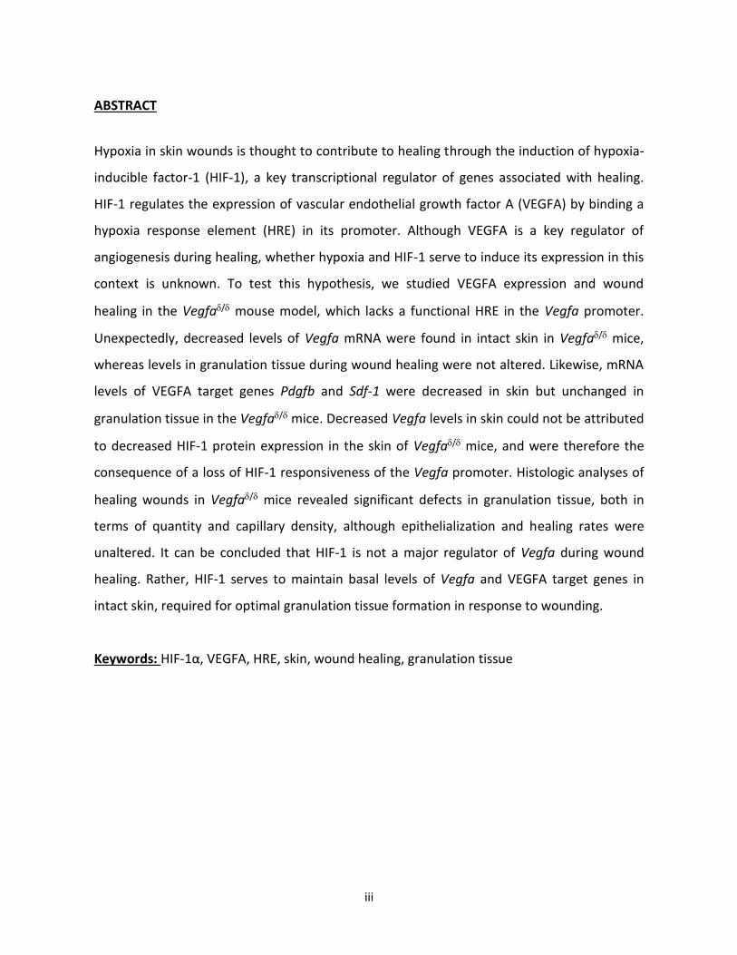
ABSTRACT
Hypoxia in skin wounds is thought to contribute to healing through the induction of hypoxia-
inducible factor-1 (HIF-1), a key transcriptional regulator of genes associated with healing.
HIF-1 regulates the expression of vascular endothelial growth factor A (VEGFA) by binding a
hypoxia response element (HRE) in its promoter. Although VEGFA is a key regulator of
angiogenesis during healing, whether hypoxia and HIF-1 serve to induce its expression in this
context is unknown. To test this hypothesis, we studied VEGFA expression and wound
healing in the Vegfa/ mouse model, which lacks a functional HRE in the Vegfa promoter.
Unexpectedly, decreased levels of Vegfa mRNA were found in intact skin in Vegfa/ mice,
whereas levels in granulation tissue during wound healing were not altered. Likewise, mRNA
levels of VEGFA target genes Pdgfb and Sdf-1 were decreased in skin but unchanged in
granulation tissue in the Vegfa/ mice. Decreased Vegfa levels in skin could not be attributed
to decreased HIF-1 protein expression in the skin of Vegfa/ mice, and were therefore the
consequence of a loss of HIF-1 responsiveness of the Vegfa promoter. Histologic analyses of
healing wounds in Vegfa/ mice revealed significant defects in granulation tissue, both in
terms of quantity and capillary density, although epithelialization and healing rates were
unaltered. It can be concluded that HIF-1 is not a major regulator of Vegfa during wound
healing. Rather, HIF-1 serves to maintain basal levels of Vegfa and VEGFA target genes in
intact skin, required for optimal granulation tissue formation in response to wounding.
Keywords: HIF-1α, VEGFA, HRE, skin, wound healing, granulation tissue
iv
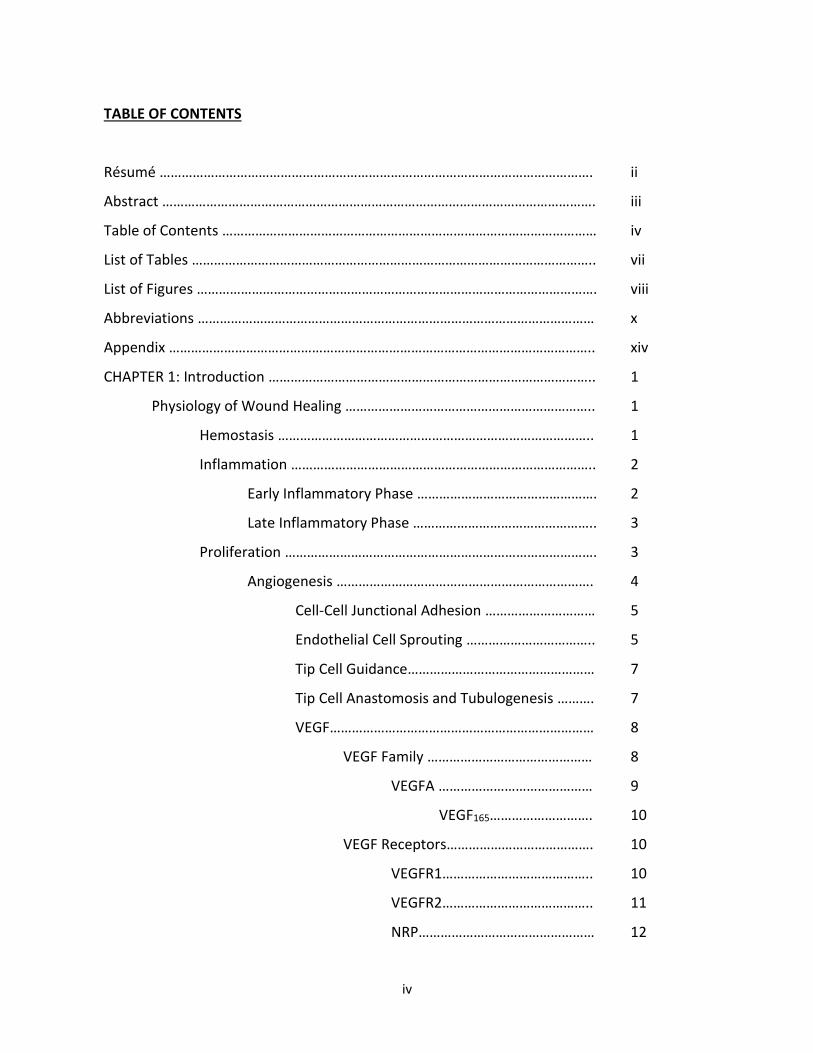
TABLE OF CONTENTS
Résumé ………………………………………………………………………………………………………. ii
Abstract ………………………………………………………………………………………………………. iii
Table of Contents ………………………………………………………………………………………… iv
List of Tables ……………………………………………………………………………………………….. vii
List of Figures ………………………………………………………………………………………………. viii
Abbreviations ……………………………………………………………………………………………… x
Appendix …………………………………………………………………………………………………….. xiv
CHAPTER 1: Introduction …………………………………………………………………………….. 1
Physiology of Wound Healing ………………………………………………………….. 1
Hemostasis ………………………………………………………………………….. 1
Inflammation ……………………………………………………………………….. 2
Early Inflammatory Phase …………………………………………. 2
Late Inflammatory Phase ………………………………………….. 3
Proliferation …………………………………………………………………………. 3
Angiogenesis ……………………………………………………………. 4
Cell-Cell Junctional Adhesion ………………………… 5
Endothelial Cell Sprouting …………………………….. 5
Tip Cell Guidance…………………………………………… 7
Tip Cell Anastomosis and Tubulogenesis ………. 7
VEGF……………………………………………………………… 8
VEGF Family ……………………………………… 8
VEGFA …………………………………… 9
VEGF165………………………. 10
VEGF Receptors…………………………………. 10
VEGFR1………………………………….. 10
VEGFR2………………………………….. 11
NRP………………………………………… 12

v
VEGFA Function…………………………………. 12
VEGFA Expression………………………………. 14
Remodelling and Scar Formation………………………………………….. 15
Oxygen and Wound Healing……………………………………………………………… 16
Oxygen and Cell Function……………………………………………………… 16
Energy Production…………………………………………………….. 16
Defence against Pathogens and ROS Production…….... 16
Oxygen Levels in Healing Wounds………………………………………... 17
Hypoxia-Inducible Factor-1……………………………………….. 18
Structure……………………………………………………….. 18
HIF-1α………………………………………………… 19
HIF-1β………………………………………………… 21
HIF-1 Complex Stability…………………………………………….. 21
HIF-α Residues………………………………………………. 24
Prolyl Hydroxylase Domain………………… 24
VCB Cul2 E3 ligase complex…………….…. 24
Arrest-Defective-1……………………………… 25
Factor inhibiting HIF-1……………………….. 25
HIF-1 Complex Activity………………………………………………. 26
MAP Kinase Pathway…………………………………….. 26
ERK1/ERK2 Pathway ………………………….. 26
Wound Healing Models and Measurements…………………………………….. 26
Research Hypotheses…………………………………………………………………………………… 28
Research Objectives…………………………………………………………………………………….. 29
CHAPTER 2: Article………………………………………………………………………………………. 30
Abstract……………………………………………………………………………………………. 31
Introduction…………………………………………………………………………………….. 32
Materials and Methods………………………………………………………………….... 34
Mice……………………………………………………………………………………… 34

vi
Part I: Molecular Profile………………………………………………………… 34
Real-time RT-PCR………………………………………………………. 35
Western Blot…………………………………………………………….. 36
Part II: Wound Healing Assay………………………………………………… 37
Histological Analyses…………………………………………………. 38
Statistical Analyses……………………………………………………………..... 38
Results ……………………………………………………………………………………………… 39
Part I: Molecular Profile………………………………………………………… 39
Part II: Wound Healing Assay………………………………………………… 42
Discussion………………………………………………………………………………………... 45
Acknowledgements………………………………………………………………………….. 46
References……………………………………………………………………………………….. 47
CHAPTER 3: Discussion and Future Experiments ……………………………………….... 49
References………..…………………………………………………………………………………………. 58

vii
LIST OF TABLES
Chapter 1: Introduction
Table I Factors capable of influencing Vegf gene expression………………………. 8
Table II Target genes of the HIF-1 complex…………………………………………………… 19
Chapter 2: Article – Materials and Methods
Table III Primers used for real-time PCR………………………………………………………… 36

viii
LIST OF FIGURES
Appendix
Figure S1…………………………………………………………………………………………………………………. xiv
MAP kinase cascades in mammalian cells
Figure S2………………………………………………………………………………………………………………….. xv
Hif-1α mRNA levels in intact skin and in granulation tissue in wild type and mutant (Vegfaδ/δ)
mice
Chapter 1: Introduction
Figure 1 Phases of Wound Healing………………………………………………………............ 1
Figure 2 Endothelial Cell Sprouting………………………………………………………………… 6
Figure 3 VEGFA Isoform Structures………………………………………………………………… 9
Figure 4 HIF-α Isoforms and Protein Domains………………………………………………… 20
Figure 5 HIF-1α Stability under Normoxic Conditions…………………………………….. 22
Figure 6 HIF-1α Stability under Hypoxic Conditions……………………………………….. 23
Chapter 2: Article – Results
Figure 7…………………………………………………………………………………………………………………… 39
ix
Vegfa mRNA levels in intact skin and in granulation tissue in wild type and mutant (Vegfaδ/δ)
mice
Figure 8…………………………………………………………………………………………………………………… 40
Pdgfb and Sdf-1 mRNA levels in intact skin and in granulation tissue in wild type and Vegfaδ/δ
mice
Figure 9…………………………………………………………………………………………………………………… 41
HIF-1α and VEGFA expression in intact skin of wild type and Vegfaδ/δ mice
Figure 10…………………………………………………………………………………………………………………. 42
Healing kinetics and macroscopic appearance of wounds in wild type and Vegfaδ/δ mice
Figure 11…………………………………………………………………………………………………………………. 43
Epithelialization of healing wounds in wild type and Vegfaδ/δ mice
Figure 12…………………………………………………………………………………………………………………. 44
Content of granulation tissue and capillaries in wild type and Vegfaδ/δ mice

x
ABBREVIATIONS
2-Oxoglutarate 2-OG
Activator protein AP
Adenosine triphosphate ATP
Angiopoietin ANGPT
Arrest-defective-1 ARD1
Aryl hydrocarbon nuclear translocator ARNT
Asparagine 803 N803
Basic fibroblast growth factor bFGF
Basix-helix-loop-helix-Per-ARNT-Sim bHLH-PAS
Beta-actin ACTB
c-Jun NH2-terminal kinase JNK
C-terminal transactivation domain C-TAD
Carbon dioxide CO2
Cluster of differentiation 31 CD31
Cobalt chloride CoCl2
Delta-like 4 Dll4
Early inflammatory phase EIP
Egg-laying nine EGLN
Epidermal growth factor EGF
Epidermal growth factor receptor EGFR
Erythropoietin EPO
Extracellular matrix ECM
Extracellular signal-regulated kinase ERK
Factor inhibiting HIF-1 FIH-1
Fetal liver kinase-1 Flk-1

xi
Fibroblast growth factor FGF
fms-like tyrosine kinase-1 Flt-1
Glucose transporter-1 GLUT-1
Hematopoietic stem cell HSC
Hematoxylin-eosin-phloxine-saffron HEPS
Hepatocyte growth factor HGF
HIF-prolyl hydroxylase HPH
Hydrogen peroxide H2O2
Hydroxyl ion OH-
Hypoxia-inducible factor HIF
Hypoxia response element HRE
Insulin-like growth factor IGF
Interferon-γ INF-γ
Interleukin IL
Internal ribosome entry site IRES
Jagged-1 Jag1
Keratinocyte growth factor KGF
Kinase domain region KDR
Late inflammatory phase LIP
Lysine 532 K532
Macrophage colony stimulating factor M-CSF
Matrix metalloproteinase MMP
Membrane type-1 matrix metalloproteinase MT1-MMP
Mitogen-activated protein MAP
N-terminal transactivation domain N-TAD
Neuropilin NRP
Nitric oxide NO

xii
Nitric oxide synthase NOS
Nuclear factor-1 NF-1
Nuclear factor kappa B NF-κB
Optimal cutting temperature OCT
Oxygen-dependent degradation domain ODDD
Peroxide anion HO2-
Phospholipase C PLC
Placental growth factor PlGF
Platelet derived growth factor PDGF
Platelet factor-IV PF4
Proline 402 P402
Proline 564 P564
Prolyl hydroxylase domain PHD
Quantitative polymerase chain reaction qPCR
Quantitative reverse transcription polymerase chain reaction RT-qPCR
Reactive oxygen species ROS
Receptor tyrosine kinase RTK
Ribosomal protein L19 RPL19
Specificity protein 1 Sp1
Stress-activated protein kinase SAPK
Stromal cell-derived factor-1 SDF-1
Superoxide anion O2-
Tissue inhibitors of metalloproteinases TIMPs
Tissue-type plasminogen activator tPA
Transforming growth factor TGF
Tumor necrosis factor TNF
Urokinase-type plasminogen activator uPA
xiii
Vascular endothelial cadherin VE-cadherin
Vascular endothelial growth factor VEGF
Vascular endothelial growth factor receptor VEGFR
Vesiculo-vacuolar organelles VVO
von Hippel-Lindau protein pVHL

xiv
APPENDIX
FIGURE S1| MAP kinase cascades in mammalian cells. Overview of the various branches involved in MAP kinase cell signalling. Image source Zhang and Liu, 2002 figure 1.

xv
A B
FIGURE S2|Hif-1α mRNA levels in intact skin and in granulation tissue in wild type and mutant (Vegfaδ/δ) mice: Quantitative PCR analysis was used to measure mRNA levels of (A) Hif-1α in intact skin ***P = 0.0010, WT: 1.3 ± 0.2, Vegfaδ/δ: 0.49 ± 0.06 and (B) Hif-1α in granulation tissue. No significant difference was observed when comparing granulation tissue Hif-1α mRNA levels between both populations of mice. Messenger RNA levels are relative to that of Rpl19. Values represent mean ± SEM (n = 6). A two-tailed unpaired t-test was utilized for the statistical analysis of the intact skin data set. A Sidak-corrected repeated measures 2-way ANOVA was utilized for the statistical analysis of the granulation tissue data set.

CHAPTER 1
INTRODUCTION
Physiology of Wound Healing
The complex series of events involved in the healing of a wound can be divided into 4
phases: hemostasis, inflammation, proliferation and remodelling/scar formation (Figure 1).
An alteration of either phase may lead to the development of a chronic, non-healing wound
[1-5].
Hemostasis
Tissue injury immediately triggers a transient vasoconstriction (lasting roughly 5-10
minutes) and initiates the activation of the extrinsic coagulation cascade [2, 6-8]. Through
2
platelet degranulation various growth factors and cell signalling molecules, platelet derived
growth factor (PDGF), fibroblast growth factor (FGF), β-thromboglobulin, serotonin,
bradykinin, prostaglandins, prostacyclins, thromboxane, histamine, insulin-like growth factor
(IGF)-1, epidermal growth factor (EGF), transforming growth factor (TGF)-β and platelet
factor-IV (PF4), among others, are released into the wound environment [2, 6, 8].
Approximately 20 minutes post-wounding, through the release of vasoactive substances such
as histamine, vasodilation and an increased vascular permeability are favored [8]. The
response initiated by these factors favors a healthy inflammatory response by attracting and
activating certain cells such as polymorphonuclear leukocytes, macrophages, fibroblasts and
endothelial cells, and contributes to hemostasis via the activation of the complement and
kinin cascades [2, 6, 8]. Ultimately, platelet aggregation within the fibrin blood clot at the site
of injury serves the purpose of diminishing blood loss [2, 6, 7].
Inflammation
The inflammatory phase quickly follows hemostasis and is characterized by the 5
cardinal signs of inflammation; redness, warmth, swelling, pain and loss of function [7]. The
inflammatory phase can be further divided into an early inflammatory phase (EIP) (days 1-2)
and a late inflammatory phase (LIP) (days 2-3) [2].
Early Inflammatory Phase
The classical and alternative pathways of the complement cascade are activated upon
initiation of the EIP. The activation of these pathways ultimately results in the attraction of
neutrophils to the wound site via chemoattractants such as TGF-β and/or complement
fragments C3a, C5a [2, 9]. Therefore, the first inflammatory cells to reach the wound are
neutrophils, which do so within 24-36h after wounding [2, 9]. Neutrophils then begin to
eliminate pathogens and foreign matter either by phagocytosis, enzymatic destruction (via
degranulation products such as collagenases and elastases) or through the production of free

3
radicals [2, 6, 8, 9]. Neutrophil activity usually ceases roughly 72 hours post-wounding, once
the affected region has been cleared of bacteria and debris [2, 10].
Late Inflammatory Phase
The cell type that predominates this portion of the inflammatory phase and that is
essential for normal wound progression is the monocyte/macrophage [2, 8]. Similarly to
neutrophils, blood monocytes are attracted to the wound by several chemoattractants (e.g.
PDGF, TGF-β) but make a tardier appearance, arriving at the wound site 48-72 hours after
injury [2, 9]. Once they have arrived at the wound site, blood monocytes become tissue
macrophages. The primary role of macrophages is that of repair. Although they possess
phagocytic and enzyme degrading capabilities to control and neutralize potential pathogens,
they are the main producers of various growth factors (PDGF, TGF-β, and FGF) that will
promote the proliferation of smooth muscle cells, the proliferation of endothelial cells, the
initial migration of fibroblasts, keratinocytes and endothelial cells within the wound, as well
as the production of the extracellular matrix (ECM) [2, 6-9].
Proliferation
Approximately 3 days after wounding and lasting until 2-4 weeks post-wounding, the
proliferative phase follows the inflammatory phase [2, 7, 9]. Fibroblast migration, deposition
of ECM and the formation of granulation tissue are all associated with the proliferative phase
[2, 7, 9, 11]. Similarly to neutrophils and monocytes, fibroblasts are attracted to the wound
by various factors [e.g. PDGF, TGF-β and basic fibroblast growth factor (bFGF)] [2, 7]. The
proliferation of fibroblasts results in the production of components (fibronectin, hyaluronan,
collagen and proteoglycans) essential for the formation of the new ECM [2].
The newly produced ECM allows for the regulation of differentiation, growth,
expansion, and migration of cells [2, 3]. The ECM is able to modify cytokine activity, which is
necessary for the regulation of its own components [12]. Overall, the ECM behaves as a

4
cellular scaffold, permitting the regeneration of wounded skin [3]. The main components of
the ECM are collagen, adhesive glycoproteins (fibronectin, laminin, thrombospondin and
integrins), and proteoglycans (glycosaminoglycans such as dermatan sulphate, heparan
sulphate and hyaluronic acid) [2].
Re-epithelialization is another essential process stimulated during the proliferative
phase [9, 11]. Two molecules are primarily responsible for the restoration of an epidermal
lining: EGF, whose production is attributed to keratinocytes and platelets, and TGF-α, which
is produced mainly by activated macrophages [9].
The proliferative phase also encompasses the production of another key component
in the healing of the wound: granulation tissue. Granulation tissue is mainly composed of a
network of capillaries and collagen and it serves as a transient dermal substitute until the
dermis can be fully repaired [6]. The quality of the granulation tissue within a wound may
serve as an indication of the wound health [2]. By 3-5 days after wounding, a healthy
granulation tissue bed should appear hyperaemic, moist, shiny and granular [2]. Conversely,
the granulation tissue of unhealthy wounds can be described as friable, soft, and of a beefy-
red color [2, 13]. Microscopically, granulation tissue is primarily characterized by
macrophages, proliferating fibroblasts and capillaries [2, 14].
As the proliferative phase ends, the ratio of hyaluronic acid to chondroitin sulfate is
altered (the amount of chondroitin sulfate increases), consequently reducing the
migratory/proliferative capabilities of fibroblasts. This favors fibroblast differentiation,
thereby denoting the initiation of the remodelling phase [7].
Angiogenesis
Angiogenesis is an essential component of the wound healing process occurring
within the proliferative phase and ensures the development of a network of capillaries [2, 6,
11]. Specific growth factors responsible for the creation of these blood vessels are vascular
endothelial growth factor (VEGF), bFGF and TGF-β, which can originate from epidermal cells,
fibroblasts, macrophages and endothelial cells [1, 9]. Angiogenesis is a process whose
5
function is directly proportional to oxygen tension levels and that consists of the formation of
new blood vessels within an organism [15-18]. Angiogenesis can occur due to endothelial cell
splitting, known as intussusception, or by endothelial cell sprouting [19-21]. Angiogenesis
differs from vasculogenesis, the latter being responsible for the formation of the very first
vascular networks within the viable embryo where mesodermal cells differentiate into
angioblasts, the precursors to endothelial cells [22, 23]. Angiogenesis mediated by VEGF is an
essential component of many physiological processes such as embryonic development, the
ovarian cycle and bone remodelling and it also contributes to pathological conditions such as
wound healing, retinopathy and tumor growth and development [17, 21, 24-28]. Many
factors are involved in the process of angiogenesis; these include but are not limited to PDGF,
TGF-β, tumor necrosis factor (TNF)-α, TNF-β, bFGF, FGF, TGF-α, hepatocyte growth factor
(HGF), interleukin (IL)-8, and angiopoietins, as well as one of the most important factors,
VEGF [15, 27, 29].
Cell-Cell Junctional Adhesion
Protein complexes present within the endothelium are responsible for stabilizing the
vascular endothelial cell lining and ensuring that a proper endothelial cell-cell adhesion
exists. These protein complexes are structurally altered throughout angiogenesis in order for
this process to successfully reach completion [22]. The major player regulating endothelial
cell-cell adhesion is a member of the cadherin family, vascular endothelial cadherin (VE-
cadherin) [19, 22].
Endothelial Cell Sprouting
Before endothelial tip cell sprouting can occur, specific steps are required for vascular
endothelial cells to become permissive to phenotypic changes. Matrix metalloproteinases
(MMP) such as membrane type-1 matrix metalloproteinase (MT1-MMP) and disintegrin as
well as VEGF-activated endothelial cell protease secretions are required for the

6
FIGURE 2| Endothelial cell sprouting: Figure demonstrating the influence of VEGF in the specialization of either a tip or stalk endothelial cell phenotype. This decision is mediated through the regulation of Notch signalling between neighbouring endothelial cells. Image source Eilken and Adams, 2010, figure 2
remodelling/degradation of the sub-endothelial basement membrane surrounding blood
vessels [19, 22]. Endothelial cell motility also must be enhanced through the modulation of
VE-cadherin. It has been postulated that exposure to VEGF promotes VE-cadherin
endocytosis, thereby leading to an increase in endothelial cell motility. Finally, endothelial
cell phenotypic specialization, tip cell or stalk cell, is required for angiogenesis to proceed
(Figure 2) [22]. This phenotypic specialization is regulated via the Notch pathway [20, 22]. In
the absence of VEGF, a balance exists regarding Notch cell signalling between adjacent
endothelial cells. A factor named Delta-like 4 (Dll4) is produced by adjacent endothelial cells
and activates the neighbouring cell’s Notch signalling pathway. In the presence of a VEGF
gradient, VEGF will stimulate one of the neighbouring endothelial cells to a greater extent
resulting in a higher degree of Dll4 production and therefore will induce a more robust Notch
pathway activation. Increased Notch signalling results in decreased expression of vascular

7
endothelial factor receptor (VEGFR)-2 and VEGFR3, increased expression of VEGFR1 and will
result in the undertaking of a stalk cell phenotype, whereas the adjacent Dll4-expressing cell
will undertake a tip cell phenotype [20, 22]. Stalk cells also express a factor named Jagged-1
(Jag1), which inhibits the characteristics of a motile, sprouting tip cell phenotype [22].
Tip Cell Guidance
Once endothelial cells have undertaken a tip cell phenotype, the sprouting cells’
motile filopodia respond to environmental cues which guide the path of new vessel growth
[22]. An essential component required for vessel support during its growth is the sheathing of
blood vessels by ECM components, primarily mature collagen [18]. The presence of an ECM
lacking mature collagen results in newly branched blood vessels that are delicate and poorly
assembled [18]. Tip cell guidance is another process in which the importance of heparin-
binding domains present in select VEGF isoforms can be observed. VEGF binds to heparin
sulfate proteoglycans present in the ECM and through this binding establishes a gradient that
aids in attracting tip cell direction [22].
Tip Cell Anastomosis and Tubulogenesis
VE-cadherin is also present on the filopodial tips of sprouting endothelial cells. In this
scenario, this cell-cell junctional adhesion complex may aid in the anastomosis of two
sprouting endothelial cells. The interaction of both VE-cadherin complexes on their
respective tip cells may initiate junction assembly, which will reduce tip cell motility. A
phenotypic cell conversion occurs, tip cell to stalk cell, as the process of tubulogenesis
commences. The tip cells are introduced into the endothelial cell lining where they become
stalk cells and the lumen of the future complete blood vessel is formed [22]. Finally a new
round of cell sprouting occurs as other endothelial cells are exposed to and modulated by
pro-angiogenic factors [22].
8
VEGF
Among the growth factors required for the development of a blood vessel network,
VEGF was the first to be identified, due to its ability to increase vascular permeability [15, 25,
30]. VEGF is essential for the viability of the developing embryo as well as being an important
factor for the proper maintenance and health of the adult vasculature [31, 32]. Embryonic
mice genetically engineered to lack one functional Vegf allele, Vegf+/-, die early in embryonic
life, at E9.5-E10.5, mainly due to severe vascular abnormalities that are predominantly
cardiovascular related [19, 21, 23, 25, 29, 31, 33-35]. On the other hand, overexpression of
Vegf (as little as twice the normal amount) leads to death of the organism [25, 31]. Many cell
types such as macrophages, glial cells, keratinocytes, endothelial cells, dermal fibroblasts and
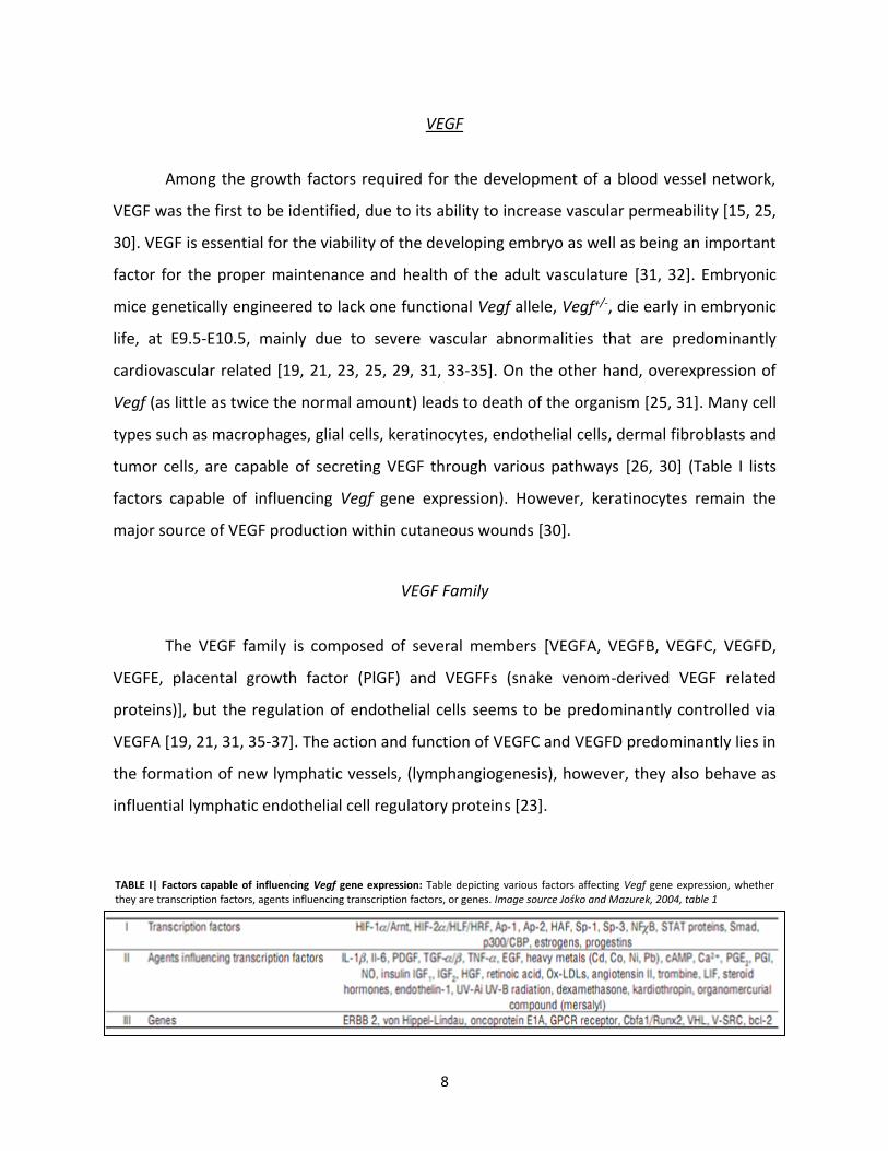
tumor cells, are capable of secreting VEGF through various pathways [26, 30] (Table I lists
factors capable of influencing Vegf gene expression). However, keratinocytes remain the
major source of VEGF production within cutaneous wounds [30].
VEGF Family
The VEGF family is composed of several members [VEGFA, VEGFB, VEGFC, VEGFD,
VEGFE, placental growth factor (PlGF) and VEGFFs (snake venom-derived VEGF related
proteins)], but the regulation of endothelial cells seems to be predominantly controlled via
VEGFA [19, 21, 31, 35-37]. The action and function of VEGFC and VEGFD predominantly lies in
the formation of new lymphatic vessels, (lymphangiogenesis), however, they also behave as
influential lymphatic endothelial cell regulatory proteins [23].
TABLE I| Factors capable of influencing Vegf gene expression: Table depicting various factors affecting Vegf gene expression, whether they are transcription factors, agents influencing transcription factors, or genes. Image source Jośko and Mazurek, 2004, table 1

9
FIGURE 3| VEGFA isoform structures: Major VEGFA isoforms found within humans (mice isoforms possess one less amino acid). Exon 1 encodes for the signal sequence [15]. Exon 3 confers the ability to dimerize as well as interact with VEGFR1 [15, 38]. VEGFR2-binding is enabled by the presence of exon 4 within the fully formed protein structure [15, 38]. Exon 5 ensures that VEGFA isoforms are vulnerable to plasmin cleavage [15, 38]. Exon 6 and 7 are heparin-binding domains. The presence/absence of these two domains explains why some VEGFA isoforms, such as VEGF189,
are mainly sequestered in
the ECM via binding to various heparan-containing ECM components, as opposed to VEGF121 which is a fully diffusible VEGFA isoform [38]. Furthermore, a portion of exon 7 and all of exon 8 are responsible for the ability to bind to coreceptors NRP1/NRP2. This interaction is required for enhanced VEGFR2 cell signalling via the binding of VEGFA to its receptor [38]. Image source Raimondi and Ruhrberg, 2013, figure 1.
VEGFA
The gene for VEGFA consists of 8 exons and 7 introns which, when subjected to RNA
splicing, gives rise to various isoforms [15, 38] (Figure 3). The currently known VEGFA
isoforms include VEGF206, VEGF189, VEGF183, VEGF165, VEGF148, VEGF145, VEGF121, VEGF189b,
and VEGF165b, where VEGF165 was found to be the predominantly active isoform in terms of
VEGF activity [15, 31, 36]. Consequently, in the following text describing functional activities
related to this growth factor, the use of the term VEGFA will refer to the most biologically
active isoform, VEGF165. The majority of VEGFA isoforms can exist in either a soluble or ECM
bound form, where each form plays a role in dictating the final functional and structural
outcome of the vasculature. Soluble VEGFA forms promote vascular hyperplasia, decreased
vascular density and poor vascular branching, whereas ECM bound forms favor highly
branched vessels and a vasculature characterized by thin walls [31].
10
VEGF165
VEGF165 possesses intermediary, exon inclusions allows this isoform to be bound to
the ECM as well as being secreted, and therefore optimal structural characteristics compared
to other VEGFA isoforms, thereby making this isoform the predominant mediator in carrying
out the various functions of VEGFA [27]. To further enforce that VEGF165 is the predominant
functionally active VEGFA isoform, mice solely expressing VEGF164 (mouse VEGFA isoforms
possess one less amino acid) do not possess any morphological vascular differences when
compared to wild type mice [21, 27, 31]. VEGF165 possesses a heparin-binding domain and
therefore is mitogenically active. A portion of VEGF165 is secreted, however a large portion of
VEGF165 remains bound to the cell surface and ECM [27, 39]. Similarly to VEGF189, VEGF165
bound to the ECM can be released as a secreted form via the action of plasmin [27].
VEGF Receptors
There exists 4 different VEGF receptors; VEGFR1, VEGFR2, VEGFR3 and Neuropilin-
1/Neuropilin-2 (NRP1/NRP2) [27]. VEGFR3 is not a suitable receptor for VEGFA, but rather
behaves as a receptor for VEGFC and VEGFD and therefore is the only receptor that will not
be outlined [21, 27, 33].
VEGFR1
VEGFR1, also known as fms-like tyrosine kinase-1 (Flt-1), is a receptor tyrosine kinase
(RTK) [27, 33, 40]. Ligands capable of interacting with this receptor include VEGFA, PlGF and
VEGFB [27]. Flt-1 expression is influenced by oxygen tension levels because it is a hypoxia-
inducible gene [33, 34, 41, 42]. Alternative splicing of Flt-1 can result in the expression of a
soluble form of the receptor that acts as a negative regulator of VEGFA activity through its
ability to bind and sequester the ligand VEGFA [19-22, 27, 31]. Therefore VEGFR1 has been
deemed a “decoy receptor” [19, 27, 31, 43]. Although this receptor primarily functions as a

11
“decoy receptor” it still plays an important role in monocyte chemotaxis, inflammation,
increasing vascular permeability, cell migration, hematopoietic stem cell (HSC) repopulation
and the induction of matrix metalloproteinase-9 (MMP-9), urokinase-type plasminogen
activator (uPA) and tissue-type plasminogen activator (tPA) [21, 23, 25, 27, 31, 33, 37].
Plasminogen activators (uPA and tPA) are responsible for the transformation of plasminogen
to plasmin, a predominant fibrinolytic agent, necessary for fibrin dissolution and
consequently permitting wound re-epithelialization [44]. MMP-9 on the other hand results in
the excision of collagenous anchors tethering keratinocytes to the basal lamina [44].
VEGFR2
VEGFR2, also known as kinase domain region (KDR) or fetal liver kinase-1 (Flk-1), is
structurally identical to VEGFR1, however the affinity of VEGFR2 for its ligand is 10 times
lower than is that of VEGFR1 [21, 27]. Known ligands for this receptor include VEGFA, VEGFC
and VEGFD. VEGR2 signalling is the main route through which VEGFA can carry out its
principal functions [22, 25, 27, 31, 36, 37, 43]. Flk-1-/- embryonic mice suffer the same fate as
Flt-1-/- mutants, which is embryonic death at E8.5-9.5 however vascularization does not occur
in the former [21, 25-27, 33, 34]. Oxygen tension levels alter the expression of Flk-1, which is
down-regulated under hypoxic conditions [23]. However, receptor phosphorylation is
increased under low oxygen tension levels [31]. Upon ligand binding, six tyrosine residues,
951, 996, 1054, 1059, 1175 (of particular importance), and 1214, located within VEGFR2, are
phosphorylated thereby permitting the subsequent activation of various signalling pathways,
such as extracellular-regulated kinase (ERK) 1/2 [19, 31]. Activation of this receptor has been
associated with many functions such as endothelial cell migration, maturation, proliferation,
survival, differentiation and vasculogenesis [19, 33].

12
NRP
NRP1 is a non-tyrosine kinase receptor that influences neuronal activity and whose
normal ligand belongs to the collapsin-semaphorin family [21, 27, 31, 35, 36]. NRP1
influences the effects of angiogenesis, explaining the specific interactions between NRP1 and
select VEGF family members (VEGFA, VEGFB, VEGFE and PLGF), whereas NRP2 regulates
lymphangiogenesis [21, 22, 36, 37]. Neuropilin receptors can act as powerful VEGFA signalling
enhancers when co-expressed on cells with VEGFR2. All VEGFA isoforms are capable of
interacting with NRP1 via the inclusion of exon 8 in their protein structure [21, 31]. Exon 7, a
heparin-binding domain within the VEGFA protein structure, along with the heparin-binding
domain found within the NRPs, leads to the formation of a surface whereby VEGFA/NRP can
interact with VEGFRs via the mediation of heparan [21]. This interaction is capable of
promoting an altered presentation of the VEGFA ligand to VEGFR2 in a manner that enhances
the signal pathway induced by receptor stimulation. Like Flt-1 and Flk-1, Nrp-1-/- embryonic
mice are not viable and are characterized by vascular abnormalities, demonstrating the
necessity of the enhanced VEGFA signal transduction induced by this coreceptor [27, 33, 36].
VEGFA Function
VEGFA possesses two main functions. The first is to increase the nutrient supply to
tissues and the second is playing a role in waste management by increasing the removal rate
of waste products [15, 45]. VEGFA accomplishes these fundamental functions via three
processes: vasodilation, angiogenesis and increased vascular permeability [15, 21, 45].
Vasodilation directly supports both of VEGFA’s main functions. VEGFA is a potent
vasodilator capable of dilating blood vessels very rapidly and to a large degree [15, 27]. The
increase in blood vessel diameter will result in increased blood flow, thereby allowing more
oxygen and glucose to reach the target tissue as well as increasing the removal rate of waste
products such as carbon dioxide (CO2), H+ and lactate [15]. The mechanism underlying the
VEGFA-induced vasodilation is based upon the action of nitric oxide (NO) [15, 27, 33]. VEGFA

13
binds to its receptor, VEGFR2, which will then activate the phospholipase C (PLC) pathway.
The end result of the activation of the PLC pathway is an increase in intracellular Ca2+ that
interacts with calmodulin to stimulate nitric oxide synthase (NOS) and therefore NO
production. NO is then capable of freely diffusing through the endothelial cells and acting on
the smooth muscle cells surrounding the blood vessels and relaxing them, leading to
vasodilation [15]. NO is also part of a positive feedback loop because NO is capable of
stimulating VEGFA expression [33].
VEGFA is one of the most potent angiogenic factors since it is capable of driving all the
important processes required for angiogenesis, including endothelial cell proliferation and
migration, basement membrane degradation, tube and lumen formation, increased vascular
permeability, and new vessel formation [15, 22, 26, 46-48]. This growth factor is able to
stimulate the growth of new vessels, whether they are arteries, veins or lymphatics [27]. In
the context of wound healing, angiogenesis allows essential nutrients and oxygen to be
delivered to mitotically active cells, thereby increasing healing rates [30]. VEGFA is also
capable of upregulating other factors, such as PDGFB and stromal cell-derived factor-1 (SDF-
1), which play an important role in angiogenesis [49-51]
VEGFA is capable of increasing the hydraulic conductivity of fluid as well as increasing
the diffusive permeability of certain macromolecules by altering the number or length of
pores present in the vasculature [15, 27]. The increase in vascular permeability allows for the
extravasation of certain proteins, fibrinogen and plasminogen, within the area surrounding
the wound. The presence and deposition of these factors within the ECM directly aids
subsequent tissue healing by creating a suitable ECM substrate for tissue growth as well as by
creating an environment that is pro-angiogenic [15]. The mechanism of VEGFA-induced
increased vascular permeability is similar to that of vasodilation. VEGFA will bind to its
receptor, VEGFR2, thereby leading to the activation of PLC and, ultimately, to an increase in
intracellular Ca2+. However, the increase in Ca2+ does not stimulate downstream factors but,
rather, it directly affects endothelial cells [15, 27]. Endothelial cell exposure to increased Ca2+
levels will result in contraction of the cells, clustering of vesicles inside the cells, and the

14
formation of vesiculo-vacuolar organelles (VVO) leading to an increased permeability to fluid
and macromolecules [15, 25].
VEGFA Expression
Oxygen tension levels play a very important role in the ability of the HIF-1 complex to
mediate VEGFA gene expression [27]. However, hypoxia-regulated VEGFA expression is not
the only pathway capable of stimulating this gene, which is expressed within nearly all tissues
in the adult [25, 52] (Table I). Hypoglycemia has been found to stimulate VEGFA expression
[33, 53, 54]. Furthermore, acidosis is also documented to lead to increased VEGFA levels [55].
Similarly to HIF-1α, many growth factors are able to stimulate VEGFA gene expression; these
include EGF, TGF-α, TGF-β, TNF-α, keratinocyte growth factor (KGF), IGF-1, FGF, bFGF and
PDGF [18, 25, 27, 33, 34, 56, 57]. Cytokines, IL-1α, IL-6 and IL-8, also play a role in the up-
regulation of this gene [25, 27, 33, 56]. Chemical induction of VEGFA expression is not the
only manner in which VEGFA expression is stimulated. Physical forces such as a simple stretch
can increase VEGFA gene expression [25].
Reactive oxygen species (ROS) are also mediators able to increase VEGFA expression
[15, 57, 58]; fibroblasts, endothelial cells and macrophages are all stimulated to express
VEGFA due to ROS exposure [58]. Furthermore, hydrogen peroxide (H2O2) is capable of
directly stimulating the VEGFA promoter resulting in increased expression and secretion
levels of this growth factor by keratinocytes and macrophages [18, 59]. This HIF-1
independent H202-induced VEGFA expression is mediated via the interaction of Sp1 with the
VEGFA promoter [57, 59].
Located within the Vegfa promoter are multiple binding sites for different
transcription factors such as HIF-1, activator protein (AP)-1, AP-2, Sp1, nuclear factor (NF)-1,
nuclear factor kappa B (NF-Κb) and Sp1-related factors, signifying a control of VEGFA
expression other than hypoxia via the activation of different signalling cascades [16, 34, 46,
60]. The MAP kinase signalling pathway documented to induce Vegfa expression is that of
ERK1/ERK2 pathway [34, 61]. The ERK1/ERK2 pathway has been shown to increase Vegfa
15
gene expression via the binding of transcription factors AP-2 and Sp1 to their appropriate
recognition DNA sequences within the Vegfa promoter [29, 56]. Moreover, the active
combinatory effect of Sp1 alongside AP-2 has been documented to increase Vegfa expression
[46, 52]. Macrophage colony stimulating factor (M-CSF) secreted by various cell types
including endothelial cells, fibroblasts, tumor cells, and monocytes, has been found to
upregulate Vegfa expression via the activation of the ERK pathways [46, 62].
Remodelling and Scar Formation
The process of remodelling has already begun as the first signs of granulation tissue
formation appear within the wound bed. Therefore the fourth and final wound healing phase
begins around 1 week post-wounding and can last ≥1-2 years [2, 9]. The ECM is continuously
being remodelled, due to constant remodelling of collagen. This dynamic state is present
until an equilibrium is reached at roughly 21 days post-wounding [2, 7]. MMPs are
responsible for proteolysis and for controlling cell migration through the ECM [2, 63]. Initially
MMPs are beneficial to the wound environment as they promote angiogenesis, cleanse the
wound via debridement, and aid in epithelialization [5]. MMPs vary considerably as several
different types exist, each with their own target. Interstitial collagenases alter collagen I, II,
and III, gelatinases degrade amorphous collagen and fibronectin and stromelysins alter
components of the ECM such as laminin [2]. Uncontrolled MMP activity impairs wound
healing since MMPs degrade collagen, essential for proper healing [2, 63]. Therefore MMPs
are usually found in the form of zymogens requiring an activating catalyst (proteases such as
plasmin) found only in regions of injured tissue [2].
As the wound progresses in its remodelling, certain changes can be observed. There is
an increased activity of MMP inhibitors [tissue inhibitors of metalloproteinases (TIMPs)] and
reduced MMP activity, mainly due to the activity of TGF-β [2, 9]. A reduction in the number
of macrophages and fibroblasts can be observed as can a reduction in cellular metabolic
activity [2, 9]. Blood perfusion to the wound site is reduced as well [2, 9]. Therefore, the
granulation tissue is slowly replaced by avascular scar tissue and, as the scar matures,
16
collagen fiber diameter increases and type III collagen is replaced by type I until a ratio
(collagen I : collagen III) of 4 : 1 is achieved [2, 6, 7, 9]. In addition to the superior
organizational state of the collagen fibers, cross-linking of these fibers also occurs, thereby
increasing the tensile strength of the scar tissue as the wound heals [6, 9]. The wound tensile
strength within the first week of healing is minimal. In humans, within 4-6 weeks post-
wounding the wound tensile strength increases to 30-50% of the strength of intact skin and
60% wound tensile strength is achieved by 6 months [4]. The maximum tensile strength
reached in a healed wound approximately 1 year after wounding is 80% of the strength of
healthy intact skin [2, 4, 7].
Oxygen and Wound Healing
Oxygen and Cell Function
Energy Production
Essential intracellular processes such as biosynthesis, movement, transport and
proliferation depend on an energy source in the form of adenosine triphosphate (ATP) [18,
59, 64]. ATP is synthesized via oxidative phosphorylation, requiring oxygen for its completion,
in amounts that are necessary to satisfy each cell’s purpose [18, 41, 58, 59].
Defence against Pathogens and ROS Production
Respiratory burst refers to the production of ROS such as peroxide anion (H02-),
hydroxyl ion (OH-) and superoxide anion (O2-), which are produced by various leukocytes and
serve to destroy invading pathogen and control against infection [18, 41, 58, 59]. This process
is highly dependent on the presence of oxygen in the environment [41, 57-59]. The active
enzyme responsible for this process, NADPH-linked oxygenase, requires a high concentration
of oxygen in order to be fully active since the production rate of the toxic radicals is directly
17
proportional to the degree of enzyme activity (50% activity at 45-80 mmHg and maximal
activity may require oxygen levels >300 mmHg) [18, 41, 59].
PDGF, EGF, TNF-α, and IL-1β are capable of increasing the synthesis of ROS through
fibroblast and leukocyte stimulation via Rac1 [57, 59]. ROS production alters the behaviour
and function of cells/processes via its influence on increasing cytokine release, stimulating
angiogenesis and increasing cell motility [18, 41, 58].
Oxygen Levels in Healing Wounds
Within the first few days after wounding, oxygen levels are drastically reduced giving
rise to a hypoxic environment [59]. A significant decrease in oxygen levels is present
approximately 48 hours following injury and maximal hypoxia (≤ 10 mmHg) is attained close
to 4 days post-wounding [65]. Oxygen tension levels vary within the wound itself with levels
as low as 10 mmHg recorded at the center of the wound and levels of 60 mmHg at the
wound periphery [18, 41]. Hypoxia is primarily the consequence of the traumatic destruction
of the vasculature as well as the augmentation of oxygen consumption rates due to increased
cellular density and metabolic activity [18, 41, 53, 58, 59, 66]. Acute hypoxia favors wound
healing because the low oxygen tension environment promotes fibroblast proliferation,
keratinocyte motility, procollagen synthesis, angiogenesis, ROS production and an increase in
the expression of various growth factors such as TGF-β1, PDGF and VEGFA [41, 58, 59, 64, 67,
68]. Furthermore, low oxygen tension levels have been documented as the predominant
inducer of VEGFA expression [23]. However, hypoxic conditions that persist (chronic hypoxia)
negatively impact wound healing since minimum oxygen tension levels of 30-40 mmHg are
required for efficient wound healing mechanisms [41, 58, 66]. Therefore angiogenesis, re-
epithelialization, fibroblast collagen deposition and the ability of leukocytes to control the
presence of pathogens, are all impaired under hypoxic conditions [18, 41, 58, 66]. Moreover,
fibroblast proliferation rates decrease, procollagen synthesis decreases, and TGF-β1
expression is downregulated [41, 58, 59]. Hence, the continued presence of hypoxia has been
linked to delayed wound healing [68]. In a normally healing wound, oxygen tension levels

18
return to normal and stabilize over time as a result of angiogenesis due to the influence of
VEGFA [41, 59]. Hypoxic conditions have been shown to favor the stability of Vegfa mRNA
due to the prolongation of the gene transcript’s half-life, normally approximately 30 minutes,
which is extended by 3-8-fold [32, 53, 69, 70]. Similarly to HIF-1α, VEGFA possesses an
internal ribosome entry site (IRES) that enables this growth factor to be adequately
expressed at the protein level in spite of hypoxic/stressful conditions [26, 33, 42, 53, 60].
Therefore under low oxygen tension levels, VEGFA levels ultimately increase due to increased
mRNA stability, increased transcription rates, and increased translation rates [16, 32, 33, 42,
56].
Hypoxia-Inducible Factor-1
Structure
Hypoxia-inducible factor-1 (HIF-1) is a heterodimeric transcription factor that induces
the expression of a plethora of genes important for cell survival, erythropoiesis, metabolic
regulation, cell proliferation and vascular biology [41, 59, 71, 72] (Table II). HIF-1 was first
discovered in the nuclear extracts of liver cells, Hep3B cells, exposed to hypoxic conditions. It
was discovered that the HIF-1 complex interacted with a specific DNA sequence, 5’-
TACGTGCT-3’, located within the enhancer region of the erythropoietin (EPO) gene [73].
Furthermore, the consensus sequence, 5’-RCGTG-3’, was not only found to be present at the
level of the EPO enhancer region but was also located within the DNA of various genes such
as VEGFA, angiopoietin (ANGPT)-1, ANGPT2, PDGFB, glucose transporter-1 (GLUT-1) and SDF-
1 [18, 71, 74, 75]. The protein subunits (α and β) constituting the HIF-1 complex are members
of the basic-helix-loop-helix-Per-ARNT-Sim (bHLH-PAS) protein family that utilize the bHLH
and PAS motifs to dimerize and consequently become functionally active [17, 18, 72, 73, 76].
The bHLH is not only necessary for proper heterodimerization between HIF-1 subunits but
also possesses a “basic” region responsible for HRE binding, subsequently resulting in the
promotion of target gene expression [71, 72, 76, 77]. HIF-α subunits contain three possible

19
TABLE II| Target genes of the HIF-1 complex: Genes are categorized according to their cell function contribution upon activation. The gene of particular importance for all the necessary steps of angiogenesis is VEGFA. Modified image source Ke and Costa, 2006, table 2.
isoforms, whereas only one possible configuration for the HIF-β subunit exists [72].
Furthermore, knockdown experiments have demonstrated that the HIF-1α isoform is of
predominant importance for target gene expression when exposed to hypoxic conditions
[77]. Figure 4 depicts the essential domains required for proper protein function as well as
the residues that are post-translationally modified in order to modulate the activity of this
transcription factor.
HIF-1α
The importance of HIF-1α is unequivocal since embryonic lethality arising at E10.5
accompanied by the development of a faulty vasculature characterizes Hif-1α-/- mice [17, 71,
77, 78]. HIF-1α possesses two transactivation domains located at each protein extremity,

20
FIGURE 4| HIF-α isoforms and protein domains: Graphical representation of the protein structures of the various components of the HIF-α subunits (HIF-1α/HIF-2α/HIF-3α) and HIF-1β subunit. The important functional domains are color coded between different subunits and the HIF-1α residues of importance are depicted in the image. These important proline residues that affect HIF-1α stability are conserved within the other HIF-α subunits as depicted in the image. The structure of the alternatively spliced HIF-3α subunit, IPAS, is also shown. Image source Ke and Costa, 2006, figure 1.
named N-terminal transactivation domain (N-TAD) and C-terminal transactivation domain (C-
TAD) [72, 77]. The transactivation domains permit the recruitment of essential transcription
enhancing co-activators such as p300/CBP and Ref-1 [72, 77]. Oxygen levels do not seem to
alter the transcription rate of this gene [72, 77]. HIF-1α expression levels are influenced by
the activity of various transcription factors such as AP-1, NF-1, NF-κB and constitutive
expression is predominantly maintained via Sp1 [77]. HIF-1α is ubiquitously expressed in all
murine tissues and its expression is upregulated in the presence of various growth factors
such as IGF-1, IGF-2, EGF and bFGF [71, 72, 77, 79]. Furthermore, due to the presence of an
IRES located within the 5’ untranslated region of Hif-1α, translation rates are not affected by
variable oxygen tension levels, as compared to a generalized reduction in protein synthesis
under hypoxic conditions [18, 32, 53, 72, 77]. Moreover, oxygen tension levels alter the
stability of HIF-1α/HIF-2α/HIF-3α, via their oxygen-dependent degradation domain (ODDD)
[18, 72, 77]. As a result, under normoxic conditions at the cellular level, minimal HIF-1α
protein levels are observed [34].

21
HIF-1β
HIF-1β, also known as the aryl hydrocarbon nuclear translocator (ARNT), is
constitutively expressed and is therefore present at stable levels under hypoxic/normoxic
conditions [18, 72, 76, 79]. HIF-1β also possesses a transactivation domain however this
domain has not been shown to have a significant effect on the positive modulation of HIF-1
gene target induction [77]. Similarly to null Hif-1α mutants, embryonic Hif-1β-/- mice are non-
viable and develop an abnormal vasculature [71].
HIF-1 Complex Stability
The stability of HIF-1α is strongly influenced by ambient oxygen tension levels. Under
normoxic conditions enzymes capable of hydroxylating specific HIF-1α amino acid residues
results in the ability of a polyubiquitinylating complex, VCB-Cul2-E3 ligase complex, to bind
and mark the protein for proteasomal degradation [72]. However, once oxygen is removed
from the environment (hypoxia), HIF-1α is no longer marked for proteasomal degradation
due to the absence of hydroxylated HIF-1α amino acid residues [72]. This therefore allows
HIF-1α to migrate to the nucleus where dimerization with HIF-1β occurs and target gene
expression is promoted via the recruitment of co-activators [72]. A detailed explanation of all
enzymes and factors influencing HIF-1α stability is found in the figure captions of figures 5
and 6.

22

23

24
HIF-α Residues
HIF-1α stability and activity is influenced by specific post-translational modifications
of certain residues encompassed within its ODDD: proline 402 (P402), proline 564 (P564) and
lysine 532 (K532) [72, 77]. Modifications of asparagine residue (N803), within its C-TAD, also
result in variable HIF-1α activity [72].
Prolyl Hydroxylase Domain
Prolyl Hydroxylase Domain (PHD), also known as HIF-prolyl hydroxylase (HPH) or Egg-
laying Nine (EGLN) is a member of the 2-oxoglutarate (2-OG) dependent dioxygenases,
enzymes capable of hydroxylating certain residues via molecular oxygen [18, 72]. Three
isoforms of this enzyme have been discovered: PHD1/HPH3/EGLN2, PHD2/HPH2/EGLN1 and
PHD3/HPH1/EGLN3 where PHD2 is the most potent in terms of its ability to hydroxylate HIF-
α residues [72]. The proper function of PHD requires two cofactors, ascorbate and iron (Fe2+).
Iron chelators and metal irons such as, Co2+, Ni2+ and Mn2+ are able to significantly reduce
PHD enzyme activity by altering the availability and stability of the ferrous ion [72, 76]. PHD
enzymatic function relies on splitting molecular oxygen and consequently hydroxylating HIF-
1α-containing P402 and P564 residues, thus promoting the interaction between HIF-1α and
the von Hippel-Lindau (pVHL) ubiquitin E3 ligase complex [18, 72, 80].
VCB-Cul2 E3 Ligase Complex
PHD-mediated hydroxylation of P402 and P564 found in HIF-1α allows for specific
binding of HIF-1α to pVHL. Once pVHL complexes with HIF-1α, other factors necessary for the
complete formation of the pVHL ubiquitin E3 ligase complex (VCB-Cul2 E3 ligase complex)
can be recruited. Elongin-C, elongin-B, cullin-2 and Rbx1 are recruited to form the VCB-Cul2
E3 ligase complex [72, 76]. HIF-1α is consequently polyubiquitinylated and therefore marked
for proteasomal degradation [18, 72, 76, 77, 80]. The phenotype for a null mutation of the
25
gene encoding for pVHL, a tumor suppressor gene, has been linked with contributing to the
progression of certain tumors [33, 71]. An inactive pVHL would increase HIF-1α stability and
therefore increase HIF-1 complex formation, thereby leading to the activation of genes
favoring tumor progression, such as the angiogenic factor VEGFA [71].
Arrest-Defective-1
Arrest-defective-1 (ARD1) is an acetyltransferase capable of post-translationally
modifying K532 found on HIF-1α [72, 76]. Acetylation of this residue favors the interaction of
HIF-1α with pVHL [72, 76]. Gene expression and translation of this protein are both
downregulated under hypoxic conditions, which may increase HIF-1α stability; however the
activity of this enzyme is not influenced by oxygen tension levels [72].
Factor inhibiting HIF-1
Factor inhibiting HIF-1 (FIH-1) is a 2-OG-dependent dioxygenase, like PHD [18, 72].
Asparagine residues found within the C-TAD (N803 in HIF-1α and N851 in HIF-2α) can be
hydroxylated via FIH-1, thereby preventing p300/CBP coactivator recruitment necessary for
successful gene target expression via the HIF-1 complex [18, 72, 76, 77, 80]. Oxygen tension
levels do not affect expression levels of FIH-1 and FIH-1’s function does not alter HIF-1α/HIF-
2α’s stability [72, 76]. However, the formation of a ternary complex between FIH-1, pVHL and
HIF-1α leading to the recruitment of a histone deacetylase via pVHL facilitates FIH-1’s
transcriptional repression of HIF-1 induced gene expression [72].

26
HIF-1 complex activity
MAP Kinase Pathway
Serine/threonine kinases are proteins that make up the mitogen-activated protein
(MAP) kinase signalling cascade family [34, 81]. There exists an abundance of factors capable
of triggering the various branches of the MAP kinase signalling cascade such as growth and
stress factors, hormones, and ECM components [34]. The p42/p44 (also known as
extracellular signal-regulated kinase: ERK2/ERK1) MAP kinase pathway as well as the stress-
activated protein kinase (SAPK) MAP kinase pathways, p38 and c-Jun NH2-terminal kinase
(JNK), are all components categorized under MAP kinase signalling (Figure S1) [34, 81, 82].
ERK1/ERK2 Pathway of the MAP Kinase Family
Various components are capable of activating the ERK pathway, such as ROS, Ca2+,
VEGFA, PDGF and TGF-β [59]. Activation of the ERK pathway leads to the phosphorylation of
HIF-1α and increases the transactivational capacity of HIF-1 [46, 59, 77]. The phosphorylation
of HIF-1α residues does not seem to affect the stability of this subunit but rather entices HIF-
1β to interact with its counterpart, thereby allowing dimerization to further carry out its
target gene expression activation [59, 76]. Furthermore, direct phosphorylation of the
coactivator p300/CBP via the ERK pathway has been shown to positively influence the
transcription-inducing activity of the HIF-1 complex [77].
Wound Healing Models and Measurements
Various animal wound models exist in the literature ranging from incisional,
excisional, burn and granulation tissue models [83]. For this study an excisional wound model
was selected for the same reasons outlined by Galiano et al [83]. The wounds are easily
27
harvested and specimens can be analyzed via immunohistochemistry or molecular profiling
[83]. Furthermore, an excisional mouse wound model allows for a simpler analysis of wound
healing processes such as epithelialization, granulation tissue formation, scar formation,
contraction and angiogenesis which was of particular importance for this study [83]. Wound
location selection, dorsum of mice, was based upon the area most unlikely to be affected by
post-operative auto-inflicted trauma. Auto-inflicted trauma to experimentally created
wounds would skew future data analyses regarding wound healing rates as well as gene
expression profiles.
A macroscopic as well as a histological approach was utilized in this study in order to
quantify and evaluate wound healing in the murine model. Macroscopically, surface area was
measured, using Image J software, from digital photographs taken from a standardized
distance from the wound, on pre-determined post-operative days, in order to determine the
rate of wound healing. With regards to the histological analysis of wound healing, specific
parameters (epidermal tongue length, epidermal gap, wound width) were used to evaluate
wound healing, reflecting measurements found within the literature [84]. As the wound heals
new epithelium originating from the wound margins will proliferate towards the center of
the wound until a complete new epithelial cover is formed. The new epidermis originating
from the wound margins is denoted as the epidermal tongue and therefore each wound
analyzed histologically will have a left and right epidermal tongue. The distance in between
both epidermal tongues, still uncovered by new epidermis, is denoted the epidermal gap.
Finally, the distance between wound margins (thereby including the left epidermal tongue,
epidermal gap and right epidermal tongue) represents the wound width. In this study these
histological parameters were used to calculate the percentage of neo-epidermal coverage.
This calculation resulted in the addition of the left and right epidermal tongues divided by the
wound width. It was decided to present the data in this manner as it depicted the
information in a more accurate and logical manner.
28
RESEARCH HYPOTHESES
It is well documented that a hypoxic environment allows for the formation of the active HIF-1
complex, which stimulates the induction of certain genes necessary for wound healing such
as Vegfa, a potent angiogenic agent. However, whether HIF-1 acts to induce Vegfa
expression in the context of cutaneous wound healing has not been established. This study
aimed to compare cutaneous wound healing between wild type and mutant mice incapable
of inducing Vegfa expression via HIF-1, in order to better understand the contribution of HIF-
1-induced Vegfa expression with regards to the healing process. The following hypotheses
were formulated:
1) A hypoxia response element (HRE) at the level of the Vegfa promoter is required for
normal Vegfa expression in cutaneous wounds.
2) HIF-1 induced Vegfa expression is required for optimal Vegfa downstream target
expression within a cutaneous wound healing setting.
3) HIF-1 induced Vegfa expression is required for optimal cutaneous wound healing
progression, including wound healing rates and granulation tissue formation.

29
RESEARCH OBJECTIVES
The objectives of this study were to determine the effect of a lack of HIF-1-induced Vegfa
expression on cutaneous wound healing within Vegfaδ/δ mice when compared to their wild
type counterparts.
Objective 1: To compare mRNA and protein expression levels of various pro-angiogenic
factors (Vegfa, Hif-1α, Sdf-1, Pdgfb) between wild type (WT) and mutant (Vegfaδ/δ) mice
throughout the course of cutaneous wound healing
• Real-time qPCR of Vegfa, Sdf-1, Pdgfb and Hif-1α throughout the course of wound
healing
• Western blot of HIF-1α and VEGFA within intact skin
Objective 2: To compare the rate of cutaneous wound healing between WT and mutant mice
• Measure the surface area of healing wounds on days 0, 3, 7, 10 and 14
Objective 3: To examine the differences in wound healing progression between WT and
mutant mice, at a histological level
• Measure various histological features (epidermal tongue length, epidermal gap,
wound width) to estimate progression of epidermal coverage;
• Measure the surface area of granulation tissue in cross sections of wound samples;
• Calculate the capillary density within the granulation tissue of healing wounds

CHAPTER 2 - Article in preparation for submission to PLoS One
A Hypoxia Response Element in the Vegfa Promoter is Required for Basal Vegfa Expression in
Skin and for Optimal Granulation Tissue Formation During Wound Healing in Mice
Domenic Ciarlillo1, Christophe Céleste2, Peter Carmeliet3, Derek Boerboom4, Christine
Theoret5
1,2,4,5Département de biomédecine vétérinaire, Faculté de médecine vétérinaire, Université
de Montréal, St-Hyacinthe, Canada.
3Laboratory of Angiogenesis and Vascular Metabolism, Vesalius Research Center, University
of Leuven, Leuven, Belgium.
Corresponding author: [email protected] (CT)
1 Conceptualization, formal analysis, investigation, methodology, validation, visualization,
writing – original draft preparation
2 Conceptualization, funding acquisition, investigation, methodology, supervision, writing -
review and editing
3 Resources, writing - review and editing
4 Conceptualization, formal analysis, supervision, resources, validation, writing - original
draft preparation
5 Conceptualization, funding acquisition, investigation, methodology, project administration,
resources, supervision, validation, writing – original draft preparation
Role of HIF-1 in Basal VEGFA Expression in Skin
Keywords: HIF-1α, VEGFA, HRE, skin, wound healing, granulation tissue

31
ABSTRACT
Hypoxia in skin wounds is thought to contribute to healing through the induction of hypoxia
inducible factor-1, a key transcriptional regulator of genes associated with healing. Hypoxia
inducible factor-1 regulates the expression of vascular endothelial growth factor A (VEGFA)
by binding a hypoxia response element in its promoter. Although VEGFA is a key regulator of
angiogenesis during healing, whether hypoxia and hypoxia inducible factor-1 serve to induce
its expression in this context is unknown. To test this hypothesis, we studied VEGFA
expression and wound healing in mutant mice that lack a functional hypoxia response
element in the VEGFA promoter. Decreased levels of VEGFA mRNA were found in intact skin
of mutant mice whereas levels in granulation tissue during wound healing were not altered.
Likewise, mRNA levels of VEGFA target genes, platelet-derived growth factor B and stromal
cell-derived factor-1, were decreased in skin but unchanged in granulation tissue of mutant
mice. Decreased VEGFA mRNA levels in skin of mutant mice could not be attributed to
decreased hypoxia inducible factor-1 protein expression, and were therefore a consequence
of the loss of hypoxia inducible factor-1 responsiveness of the VEGFA promoter. Histologic
analyses of healing wounds in mutant mice revealed significant defects in granulation tissue,
both in terms of quantity and capillary density, although epithelialization and healing rates
were unaltered. We conclude that hypoxia inducible factor-1 is not a major regulator of
VEGFA during wound healing; rather, it serves to maintain basal levels of VEGFA and its
target genes in intact skin, required for optimal granulation tissue formation in response to
wounding.
32
INTRODUCTION
Hypoxia within skin wounds arises primarily in response to traumatic destruction of
the cutaneous vasculature and augmentation of cellular oxygen consumption rates due to
temporary increases in cellular density and metabolic activity [1-4]. Acute hypoxia favors
wound healing because low oxygen tension levels promote angiogenesis, fibroplasia,
epithelialization, and extracellular matrix (ECM) synthesis [1, 2, 4-6]. These effects are
regulated, at least in part, by hypoxia-induced increases in the expression of various growth
factors including transforming growth factor (TGF)-β1, platelet-derived growth factor (PDGF)
and vascular endothelial growth factor (VEGFA) [1, 2, 4, 6].
Hypoxia triggers the accumulation of hypoxia-inducible factor (HIF)-1, the cellular
hypoxia sensor, which is the key element in the process of oxygen homeostasis and in the re-
establishment of blood vessels in hypoxic areas [1, 4]. HIF-1 is a heterodimeric transcription
factor that induces the expression of a plethora of genes that mediate adaptive responses to
hypoxia such as angiogenesis and cell proliferation/survival [1, 4, 7]. It exerts its
transcriptional regulatory effects by binding a site named the hypoxia-response element
(HRE) found within the promoters of various genes including VEGFA, angiopoietin (ANGPT)1,
ANGPT2, PDGFB, glucose transporter (GLUT)-1 and stromal cell-derived factor (SDF)-1 [3, 7-
9].
Angiogenesis is a physiological process whereby new blood vessels form from pre-
existing vessels [10, 11]. In the wound environment, these new vessels are required to
deliver essential nutrients and oxygen to mitotically active cells, thereby ensuring that
healing progresses normally [12]. VEGFA is a key regulator of angiogenesis in a variety of
developmental and physiological processes including wound healing [13, 14]. VEGFA is also
capable of upregulating other factors, such as PDGFB and SDF-1, which play an important
role in angiogenesis [15-17]. HIF-1 is thought to be a major regulator of Vegfa expression in
several cell types, and acts via a well-characterized HRE in the Vegfa promoter [7, 18].
However, whether HIF-1 acts to induce Vegfa expression in the context of cutaneous wound
healing has not been established. In the current study, we tested this idea in vivo using the

33
Vegfa/ mouse model, which lacks a functional HRE in the Vegfa promoter, and therefore
cannot respond to hypoxia/HIF-1 with an increase in Vegfa transcriptional activity [19]. We
predicted that Vegfa upregulation in response to wounding would fail to occur in mutant
mice, and that wound healing would be compromised due to poor angiogenesis and the
presence of granulation tissue of inferior quantity and/or quality. While our experimental
goal was achieved, the outcome of our study demonstrated that, contrary to prediction, HIF-
1 is required for basal expression of Vegfa in intact skin, rather than in granulation tissue
during wound healing.

34
MATERIALS AND METHODS
Mice
Vegfatm2Pec (hereafter Vegfa/) mice were as originally described [20]. Wild type and
mutant mice used for all experimentation were male and ranged from 6-8 weeks in age. All
mice were healthy upon commencement of experimentation. Animals were housed
individually under standardized conditions with controlled temperature, humidity, and a 12-
hour-day/12-hour-night light cycle. Animals had free access to water and standard mouse
chow. This study was conducted at the Faculté de médecine vétérinaire in strict accordance
with the guidelines for the care and use of laboratory animals, as sanctioned by the Canadian
Council on Animal Care. The protocol was approved by the Comité d’éthique de l’utilisation
des animaux de l’Université de Montréal (Permit Number: Rech-1635). All surgery was
performed under anesthesia, and all efforts were made to minimize suffering.
Part I: Molecular profile
For the first part of the study, 6 Vegfa/ and 6 wild type mice were used. On day 0,
mice were anesthetized by isoflurane inhalation 20 minutes after subcutaneous
administration of Metacam™ (meloxicam, 4mg/kg – Boehringer Ingelheim, St. Joseph,
Missouri, USA). The dorsum was clipped and prepared aseptically. Three full-thickness
excisional wounds were made to below the level of the panniculus carnosus (muscle layer
was removed), one on each side of midline and a third one centrally located just caudal to
the other two wound sites, using a sterile, disposable, 6 mm-diameter biopsy punch and
scissors; the excised tissue (intact skin) was kept as a day 0 sample. Wounds were left
uncovered to heal by secondary intention.
Post-operatively, mice were injected subcutaneously with 1 ml of warm, 0.9% sterile
saline solution for fluid replacement in accordance with CEAU mouse surgical protocol. All
mice were placed on a warming pad until mobile and were then returned to their cage

35
(cardboard paper bases were used as substrate) where they were further warmed until fully
awake. Mice received a subcutaneous dose of Metacam™ (4mg/kg, SID) on postoperative
days 1 and 2.
Animals were anesthetized as per wound creation surgery on days 5, 7 and 9; at each
of these times, a single wound was randomly selected for harvest. On day 9 the animals were
euthanized by carbon dioxide (CO2) inhalation after wound harvest. Wound sampling
consisted of creating a new 8 mm-diameter wound encompassing the initial wound to ensure
that the entire region of interest was collected. Tissue samples were embedded in Optimal
Cutting Temperature compound (OCT - Tissue-Tek, Torrance, CA, USA), snap-frozen in liquid
nitrogen and then stored at -80°C.
Real-time RT-PCR
Wound tissue samples were thawed and the peripheral and deeper wound sections
were discarded, keeping only granulation tissue. Conversely, the totality of day 0 samples
(i.e., intact skin) was kept. Total RNA was isolated and purified using the RNeasy Fibrous
Tissue Mini kit (Qiagen Sciences, Maryland, USA), following the manufacturer’s instructions.
Synthesis of cDNA was done using a SuperScript VILO cDNA synthesis kit (Thermo Fisher
Scientific, Life Technologies, Carlsbad, CA, USA) following the manufacturer’s instructions.
Quantitative polymerase chain reaction (qPCR) was carried out in a Bio-Rad CFX96 Touch
Real Time PCR Detection System, using the SSoAdvanced Universal SYBR Green Supermix
(Bio-Rad Laboratories, Hercules, CA, USA), following the manufacturer’s instructions. Custom
oligonucleotide primers were designed via Life Technologies, Inc (Table 3) and standard
curves were generated by serial dilution of a preparation of total cDNA. Expression levels of
genes were calculated relative to the housekeeping gene Ribosomal Protein-L19 (Rpl19) [21].
36
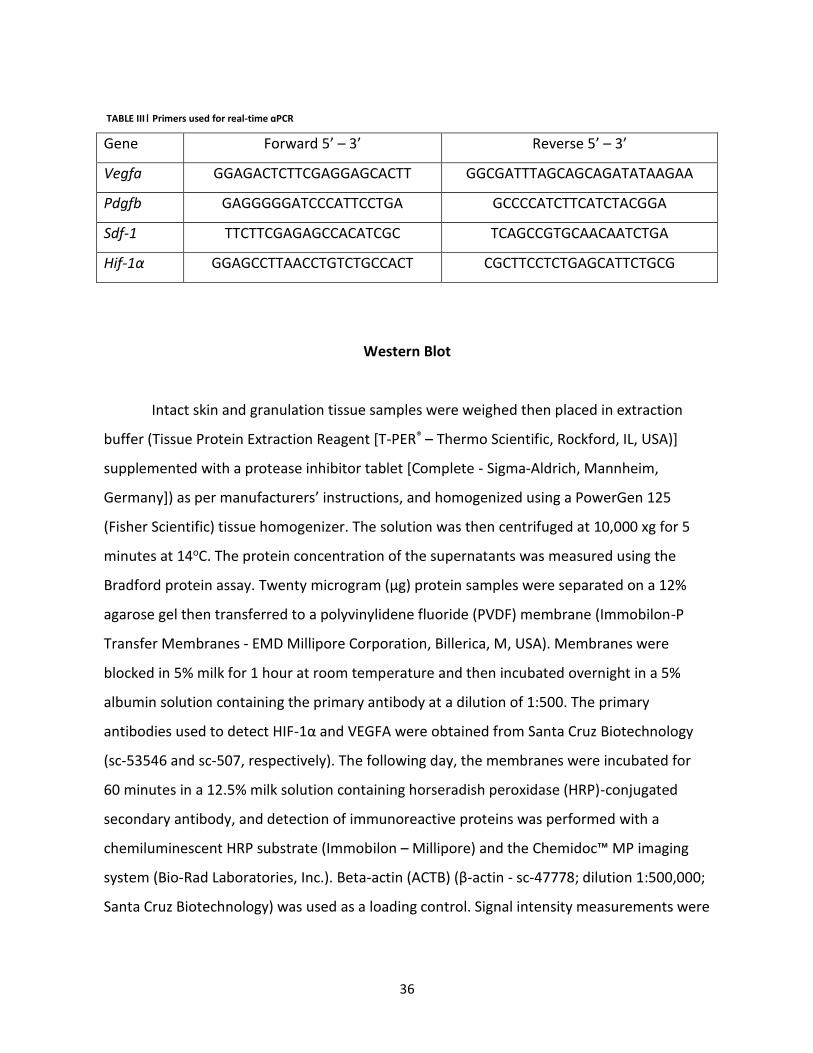
Gene Forward 5’ – 3’ Reverse 5’ – 3’
Vegfa GGAGACTCTTCGAGGAGCACTT GGCGATTTAGCAGCAGATATAAGAA
Pdgfb GAGGGGGATCCCATTCCTGA GCCCCATCTTCATCTACGGA
Sdf-1 TTCTTCGAGAGCCACATCGC TCAGCCGTGCAACAATCTGA
Hif-1α GGAGCCTTAACCTGTCTGCCACT CGCTTCCTCTGAGCATTCTGCG
Western Blot
Intact skin and granulation tissue samples were weighed then placed in extraction
buffer (Tissue Protein Extraction Reagent [T-PER® – Thermo Scientific, Rockford, IL, USA)]
supplemented with a protease inhibitor tablet [Complete - Sigma-Aldrich, Mannheim,
Germany]) as per manufacturers’ instructions, and homogenized using a PowerGen 125
(Fisher Scientific) tissue homogenizer. The solution was then centrifuged at 10,000 xg for 5
minutes at 14oC. The protein concentration of the supernatants was measured using the
Bradford protein assay. Twenty microgram (μg) protein samples were separated on a 12%
agarose gel then transferred to a polyvinylidene fluoride (PVDF) membrane (Immobilon-P
Transfer Membranes - EMD Millipore Corporation, Billerica, M, USA). Membranes were
blocked in 5% milk for 1 hour at room temperature and then incubated overnight in a 5%
albumin solution containing the primary antibody at a dilution of 1:500. The primary
antibodies used to detect HIF-1α and VEGFA were obtained from Santa Cruz Biotechnology
(sc-53546 and sc-507, respectively). The following day, the membranes were incubated for
60 minutes in a 12.5% milk solution containing horseradish peroxidase (HRP)-conjugated
secondary antibody, and detection of immunoreactive proteins was performed with a
chemiluminescent HRP substrate (Immobilon – Millipore) and the Chemidoc™ MP imaging
system (Bio-Rad Laboratories, Inc.). Beta-actin (ACTB) (β-actin - sc-47778; dilution 1:500,000;
Santa Cruz Biotechnology) was used as a loading control. Signal intensity measurements were
TABLE III| Primers used for real-time qPCR
37
obtained using Image Lab™ software version 5.2.1 (Bio-Rad Laboratories, Inc. Hercules, CA,
USA).
Part II: Wound healing assay
Eight mutant (Vegfa/) and 8 wild type mice were used for the second part of the
study. The same anesthetic, surgical and analgesic protocols applied in part I of
experimentation were followed in part II, except two wounds were created on either side of
midline using a sterile, disposable 10 mm-diameter biopsy punch and a scalpel blade instead
of three. The excised tissue (intact skin) was kept as a day 0 sample. Wounds were
photographed (baseline measurement) and were left uncovered to heal by secondary
intention.
Mice were immobilized and each wound was photographed using a digital camera
alongside a scale bar, on postoperative days 3, 7, 10 and 14. The wound surface area was
calculated in pixels, by a blinded observer, using ImageJ software (http://imagej.nih.gov/ij/),
and expressed as a percentage of the original wound surface area to determine the rate of
healing. On day 7 the animals were anesthetized as per wound creation surgery, and the
right-sided wounds were collected. Newly created wounds through harvesting were left to
heal by secondary intention. On day 14, the animals were euthanized by CO2 inhalation then
the left-sided wounds were collected. All samples were harvested with a biopsy punch to
include the remaining wound and the surrounding skin, placed flat on a piece of filter paper,
subcutis side down, then divided in half. One half was fixed in 10% neutral-buffered formalin
solution for 24h then stored in 70% alcohol at 4oC until processing in paraffin wax for
histology. The second half was embedded in OCT then snap-frozen in liquid nitrogen and
stored at -80°C until real-time (RT)-qPCR and Western Blot assays.

38
Histological analyses
Serial sections taken at the greatest wound width were stained with hematoxylin,
eosin, phloxine and saffron (HEPS) stain. The neo-epidermal coverage, based on the
percentage of the total wound width covered by new epithelium (sum of the epidermal
tongues migrating from the right and left wound margins divided by the wound width), and
the remaining wound width (epidermal gap length) were calculated for each section.
Measurements were determined relative to a constant reference (thickness of the intact
epidermis adjacent to the wound). Each measurement for each criterion was taken twice and
averages were used for statistical analyses. Total wound width was measured on days 7 and
14, whereas neo-epidermal coverage and the remaining wound width were measured on day
7.
The area of granulation tissue was quantified in day 7 wound samples using Zen Pro
2012 software and an Axioimager M1 microscope (Zeiss, Toronto, Ontario, Canada).
Capillaries within the granulation tissue were counted; only blood vessels running
perpendicularly to the wound surface were included in the count. The average number of
capillaries was then divided by the granulation tissue area measurement obtained for that
section, to obtain the capillary density.
Statistical analyses
Statistical testing was done using Prism v6.0d software (GraphPad software, Inc. La
Jolla, CA, USA). The threshold of statistical significance for all analyses was defined as P ≤
0.05. The statistical analyses applied to specific data sets are detailed in the corresponding
figure legends.
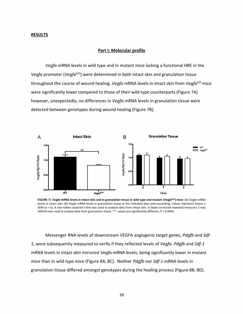
39
RESULTS
Part I: Molecular profile
Vegfa mRNA levels in wild type and in mutant mice lacking a functional HRE in the
Vegfa promoter (Vegfa/) were determined in both intact skin and granulation tissue
throughout the course of wound healing. Vegfa mRNA levels in intact skin from Vegfa/ mice
were significantly lower compared to those of their wild type counterparts (Figure 7A)
however, unexpectedly, no differences in Vegfa mRNA levels in granulation tissue were
detected between genotypes during wound healing (Figure 7B).
Messenger RNA levels of downstream VEGFA angiogenic target genes, Pdgfb and Sdf-
1, were subsequently measured to verify if they reflected levels of Vegfa. Pdgfb and Sdf-1
mRNA levels in intact skin mirrored Vegfa mRNA levels, being significantly lower in mutant
mice than in wild type mice (Figure 8A, 8C). Neither Pdgfb nor Sdf-1 mRNA levels in
granulation tissue differed amongst genotypes during the healing process (Figure 8B, 8D).
FIGURE 7| Vegfa mRNA levels in intact skin and in granulation tissue in wild type and mutant (Vegfaδ/δ) mice: (A) Vegfa mRNA levels in intact skin. (B) Vegfa mRNA levels in granulation tissue at the indicated days post-wounding. Values represent means ± SEM (n = 6). A two-tailed unpaired t-test was used to analyze data from intact skin. A Sidak-corrected repeated measures 2-way ANOVA was used to analyze data from granulation tissue. **: values are significantly different, P = 0.0044.
A B

40
HIF-1α and VEGFA expression in intact skin were analyzed by immunoblotting.
Whereas HIF-1α expression did not differ between genotypes, VEGFA expression in intact
skin was significantly lower in mutant mice than in wild type mice (Figure 9C).
A
C
B
D
FIGURE 8| Pdgfb and Sdf-1 mRNA levels in intact skin and in granulation tissue in wild type and Vegfaδ/δ mice: (A) Pdgfb mRNA levels in intact skin, *: values are significantly different, P = 0.0119. (B) Pdgfb mRNA levels in granulation tissue. (C) Sdf-1 mRNA levels in intact skin, *: values are significantly different, P = 0.0145. (D) Sdf-1 mRNA levels in granulation tissue. Messenger RNA levels are relative to that of Rpl19. Values represent means ± SEM (n = 6). A two-tailed unpaired t-test was used to analyze data from intact skin. A Sidak-corrected repeated measures 2-way ANOVA was used to analyze data from granulation tissue.

41
A
VEGFA
HIF-1α
ACTB
WT Vegfa/
B
C
FIGURE 9| HIF-1α and VEGFA expression in intact skin of wild type and Vegfaδ/δ mice: (A) Representative immunoblot analyses of HIF-1α and VEGFA expression in intact skin (n = 2/genotype). (B) Quantification of HIF-1α immunoblots (n = 6/genotype). Data were analyzed using a two-tailed unpaired t-test. (C) Quantification of VEGFA immunoblots (n = 6/genotype). Data were analyzed using a Mann-Whitney test. *: values are significantly different, P = 0.0152.
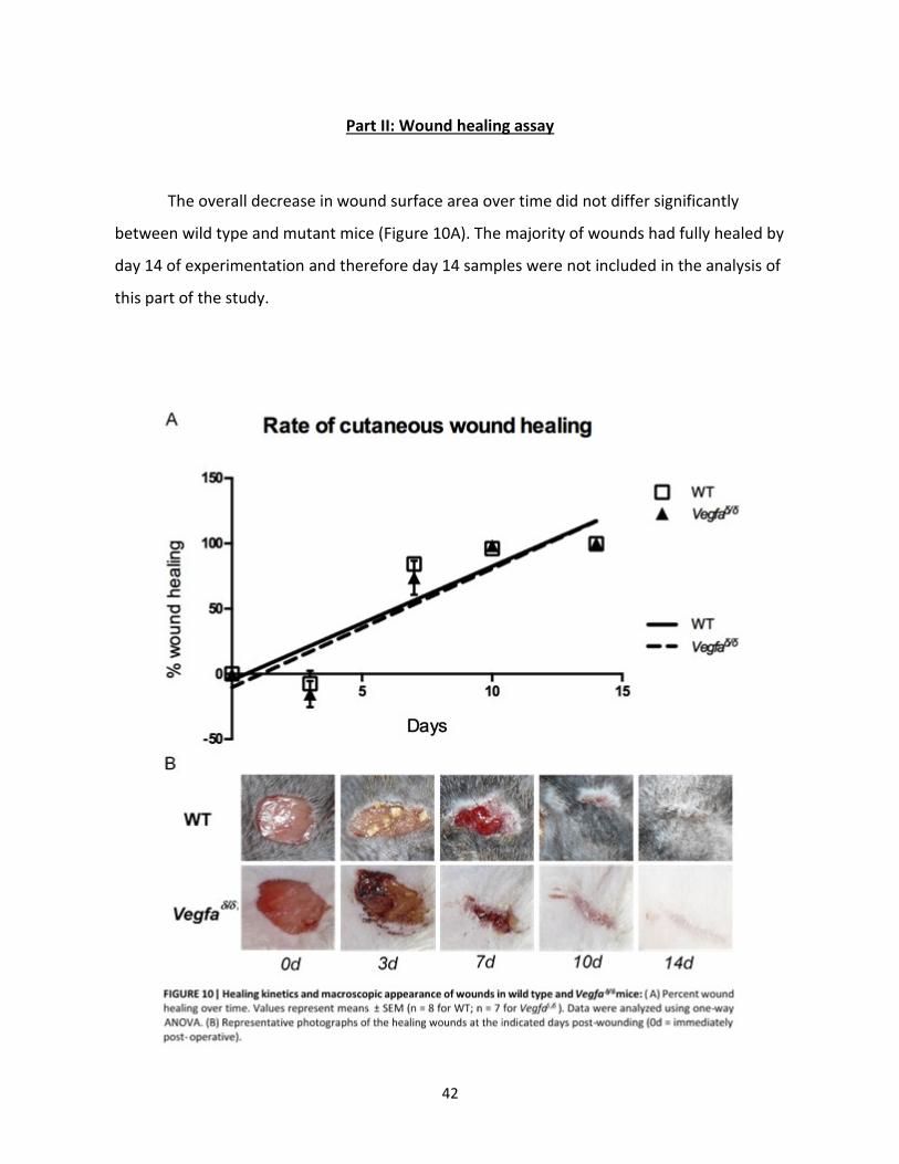
42
Part II: Wound healing assay
The overall decrease in wound surface area over time did not differ significantly
between wild type and mutant mice (Figure 10A). The majority of wounds had fully healed by
day 14 of experimentation and therefore day 14 samples were not included in the analysis of
this part of the study.

43
Since VEGF is known to contribute to keratinocyte migration [21], histometric
analyses were done to examine potential epidermal differences in the wounds of wild type
and mutant mice (Figure 11A). Quantification of neo-epidermal tongue lengths and epithelial
gaps showed no difference between the wounds of wild type and mutant mice at day 7 (not
shown). Consequently, no significant difference in neo-epidermal coverage was found
between the two populations of mice (Figure 11B).
Because VEGFA is a major stimulator of angiogenesis in response to the hypoxia
generated by wounding, the blood vessel-rich granulation tissue present within 7 day-old
wounds was examined. Granulation tissue surface area was measured in cross-sections as an
approximation of granulation tissue volume (Figure 12A). A significantly smaller surface area
of granulation tissue was present in the wounds of mutant mice (Figure 12B). Furthermore,
there was a trend towards reduced capillary density within the granulation tissue of 7 day-
old wounds in the mutant mice compared to the wild type mice (P = 0.0602) (Figure 12D).
B A
FIGURE 11| Epithelialization of healing wounds in wild type and Vegfaδ/δ mice: (A) Representative photomicrograph of a 7 day-
old wound in a wild type mouse. Measurements of the left (LET) and right epidermal tongues (RET), epidermal gap (EG) and wound width (WW) were used to evaluate the progress of wound healing. (B) Quantitative analysis of neo-epidermal coverage in the healing wounds of WT and Vegfaδ/δ
mice. Data were analyzed using a two-tailed unpaired t-test. Values represent means ± SEM (n = 8 for WT; n = 6 for Vegfaδ/δ).

44
B
D C
A
FIGURE 12| Content of granulation tissue and capillaries in wild type and Vegfaδ/δ mice: (A) Representative photomicrograph of a 7 day-old wound in a wild type mouse with the granulation tissue circumscribed. Adipose tissue or muscle found within the demarcated zone (grey hatched area) was excluded from the surface area measurement. (B) Surface area of the granulation tissue in 7 day-old wounds, *: values are statistically different, P = 0.0354. Values represent means ± SEM (n = 8 for WT; n = 6 for Vegfaδ/δ). A two-tailed unpaired t-test was used to analyze the data. (C) Representative photomicrograph showing examples of new capillaries (Cp) within the granulation tissue of 7 day-old wounds. (D) Capillary density (Cp / mm2 of granulation tissue). Values represent mean ± SEM (n = 8 for WT; n = 6 for Vegfaδ/δ). A two-tailed unpaired t-test was used to analyze the data.
45
DISCUSSION
Hypoxia associated with cutaneous wounding triggers the expression of a number of
genes required for the healing process, including the key angiogenic factor VEGFA [4, 7].
Although it is established that HIF-1 can regulate Vegfa expression in hypoxic cells via a HRE
[7], that this regulatory process drives Vegfa expression in the context of cutaneous wound
healing has long been presumed, but not actually demonstrated. In the present study, we
employed the Vegfa/ model, in which the lack of a functional HRE in the Vegfa promoter
provided the ideal system to determine the contribution of HIF-1 to Vegfa expression in
wounded skin.
Contrary to our hypothesis, Vegfa expression was not compromised in the
granulation tissue in the wounds of Vegfa/ mice. The simplest explanation for this finding is
that HIF-1 is simply not a regulator of Vegfa in the context of wound healing. Indeed, many
additional factors and signaling processes are known to regulate Vegfa expression and may
be relevant in granulation tissue. These include epidermal growth factor, platelet-derived
growth factor and interleukin-1α, which are released in response to wounding [14, 22].
Physicochemical stimuli such as acidosis, hypoglycemia, reactive oxygen species and
mechanical stretching all stimulate Vegfa expression [14, 23, 24, 25]. Likewise, the
extracellular signal-regulated kinase (ERK)1/ERK2 pathway induces Vegfa expression through
the binding of transcriptional factor complexes such as adaptor complex 2 and specificity
protein 1 to the Vegfa promoter [26, 27]. Whether these or other signaling processes drive
Vegfa expression in granulation tissue will be grounds for further study. Another explanation
for unaltered Vegfa expression in the granulation tissue of Vegfa/ mice is that
compensatory mechanisms (perhaps including the aforementioned Vegfa regulatory
processes) may have been induced to augment Vegfa expression, thereby masking any
deficit caused by deficient HIF-inducibility of the Vegfa promoter. Further experiments will
be required to test this possibility.
In this study, we readily detected HIF-1α protein in intact skin, confirming previous
reports of constitutive expression in several functionally diverse cell types including those
46
populating skin [28, 29, 30]. The decreased levels of Vegfa expression in the skin of Vegfa/
mice therefore indicate that one role of the constitutively expressed HIF-1α is to ensure
proper basal expression of Vegfa.
Despite normal expression of Vegfa (and Pdgfb and Sdf1) in their granulation tissue,
wounds in Vegfa/ mice featured a reduced abundance of granulation tissue, and capillary
density was decreased within the granulation tissue. Although we cannot provide a definitive
explanation for this, deficient Vegfa expression in the intact skin of Vegfa/ mice (i.e., prior
to wounding) provides a plausible explanation. Upon wounding, decreased VEGFA levels
present locally within the skin may have led to delayed and/or reduced capillary and
granulation tissue formation in the early phases of healing, leading to the changes observed
7 days post-wounding in the Vegfa/ model. This would suggest that a certain level of VEGFA
expression in intact skin is required for an optimal response to wounding and a rapid onset of
the healing process. Surprisingly, in spite of defects in the granulation tissue, the wounds of
Vegfa/ mice healed at the same rate as the wounds of wild type mice. This might be
explained by the highly contractile nature of wound healing in mice [31]. Indeed, several
studies of cutaneous wound healing in mice employ stents/splints to prevent wound
contraction [32, 33], thereby permitting a better analysis of the epithelialization process.
Future studies of the Vegfa/ mice could employ stenting in order to more conclusively
determine if the granulation tissue defects specifically affected epithelialization.
In summary, this study describes a novel role for HIF-1 in the expression of Vegfa in
skin. Constitutively expressed HIF-1α maintains a basal level of Vegfa expression, which is
required for optimal granulation tissue formation following wounding.
ACKNOWLEDGEMENTS
The authors thank Dr. Guy Beauchamp for help with statistical analyses, as well as Meggie
Girard, Dr. Mayra Tsoi and Dr. Gustavo Zamberlam for help with the mouse colony.

47
REFERENCES
1. Rodriguez, P.G., et al., The Role of Oxygen in Wound Healing: A Review of the Literature. Dermatologic Surgery, 2008. 34(9): p. 1159-1169.
2. Schreml, S., et al., Oxygen in acute and chronic wound healing. British Journal of Dermatology, 2010. 163(2): p. 257-268.
3. Sen, C.K., Wound healing essentials: Let there be oxygen. Wound Repair and Regeneration, 2009. 17(1): p. 1-18.
4. Tandara, A.A. and A.T. Mustoe, Oxygen in Wound Healing—More than a Nutrient. World Journal of Surgery, 2004. 28(3): p. 294-300.
5. Stadelmann, W.K., A.G. Digenis, and G.R. Tobin, Impediments to wound healing. The American Journal of Surgery, 1998. 176(2, Supplement 1): p. 39S-47S.
6. Davidson, J.D. and T.A. Mustoe, Oxygen in wound healing: More than a nutrient. Wound Repair and Regeneration, 2001. 9(3): p. 175-177.
7. Semenza, G.L., HIF-1: mediator of physiological and pathophysiological responses to hypoxia. Journal of Applied Physiology, 2000. 88(4): p. 1474-1480.
8. Manalo, D.J., et al., Transcriptional regulation of vascular endothelial cell responses to hypoxia by HIF-1. Blood, 2005. 105(2): p. 659-669.
9. Ceradini, D.J. and G.C. Gurtner, Homing to Hypoxia: HIF-1 as a Mediator of Progenitor Cell Recruitment to Injured Tissue. Trends in Cardiovascular Medicine, 2005. 15(2): p. 57-63.
10. Dimberg, A., et al., Chapter 236 - Vascular Endothelial Growth Factors and Receptors: Signaling in Vascular Development A2 - Bradshaw, Ralph A, in Handbook of Cell Signaling (Second Edition), E.A. Dennis, Editor. 2010, Academic Press: San Diego. p. 1927-1937.
11. Eichmann, A. and M. Simons, VEGF signaling inside vascular endothelial cells and beyond. Current opinion in cell biology, 2012. 24(2): p. 188-193.
12. Wilgus, T.A., et al., Regulation of scar formation by vascular endothelial growth factor. Lab Invest, 2008. 88(6): p. 579-590.
13. Rousseau, S., F. Houle, and J. Huot, Integrating the VEGF Signals Leading to Actin-Based Motility in Vascular Endothelial Cells. Trends in Cardiovascular Medicine, 2000. 10(8): p. 321-327.
14. Maharaj, A.S.R. and P.A. D'Amore, Roles for VEGF in the adult. Microvascular Research, 2007. 74(2–3): p. 100-113.
15. Romagnani, P., et al., CXC chemokines: the regulatory link between inflammation and angiogenesis. Trends in immunology, 2004. 25(4): p. 201-209.
16. Jain, R.K., Molecular regulation of vessel maturation. Nat Med, 2003. 9(6): p. 685-693. 17. Petit, I., D. Jin, and S. Rafii, The SDF-1–CXCR4 signaling pathway: a molecular hub
modulating neo-angiogenesis. Trends in Immunology, 2007. 28(7): p. 299-307. 18. Déry, M.-A.C., M.D. Michaud, and D.E. Richard, Hypoxia-inducible factor 1: regulation
by hypoxic and non-hypoxic activators. The International Journal of Biochemistry & Cell Biology, 2005. 37(3): p. 535-540.

48
19. Oosthuyse, B., et al., Deletion of the hypoxia-response element in the vascular endothelial growth factor promoter causes motor neuron degeneration. Nature genetics, 2001. 28(2): p. 131-138.
20. Carmeliet, P., et al., Abnormal blood vessel development and lethality in embryos lacking a single VEGF allele. Nature, 1996. 380: p. 4.
21. de Jonge, H.J.M., et al., Evidence Based Selection of Housekeeping Genes. PLoS ONE, 2007. 2(9): p. e898.
22. Ferrara, N., H.-P. Gerber, and J. LeCouter, The biology of VEGF and its receptors. Nat Med, 2003. 9(6): p. 669-676.
23. Neufeld, G., et al., Vascular endothelial growth factor (VEGF) and its receptors. The FASEB Journal, 1999. 13(1): p. 9-22.
24. Tsuzuki, Y., et al., Vascular Endothelial Growth Factor (VEGF) Modulation by Targeting Hypoxia-inducible Factor-1α → Hypoxia Response Element → VEGF Cascade Differentially Regulates Vascular Response and Growth Rate in Tumors. Cancer Research, 2000. 60(22): p. 6248-6252.
25. Sen, C.K., et al., Oxidant-induced Vascular Endothelial Growth Factor Expression in Human Keratinocytes and Cutaneous Wound Healing. Journal of Biological Chemistry, 2002. 277(36): p. 33284-33290.
26. Berra, E., G. Pagès, and J. Pouysségur, MAP Kinases and Hypoxia in the Control of VEGF Expression. Cancer and Metastasis Reviews, 2000. 19(1): p. 139-145.
27. Ripple, M.O., N. Kim, and R. Springett, Acute Mitochondrial Inhibition by Mitogen-activated Protein Kinase/Extracellular Signal-regulated Kinase Kinase (MEK) 1/2 Inhibitors Regulates Proliferation. Journal of Biological Chemistry, 2013. 288(5): p. 2933-2940.
28. Akakura N, Kobayashi M, Horiuchi I, Suzuki A, Wang J, Chen J, et al. Constitutive expression of hypoxia-inducible factor-1alpha renders pancreatic cancer cells resistant to apoptosis induced by hypoxia and nutrient deprivation. Cancer Res. 2001;61: 6548-6554.
29. Deschene K, Céleste C, Boerboom D, Theoret CL. Constitutive expression of hypoxia‐inducible factor‐1 α in keratinocytes during the repair of skin wounds in horses. Wound Repair Regen. 2011;19: 250-259.
30. Cho YS, Bae JM, Chun YS, Chung JH, Jeon YK, Kim IS, et al. HIF-1α controls keratinocyte proliferation by up-regulating p21 (WAF1/Cip1). Biochim Biophys Acta. 2008;1783: 323-333.
31. Galiano RD, Michaels J 5th, Dobryansky M, Levine JP, Gurtner GQ. Quantitative and reproducible murine model of excisional wound healing. Wound Repair Regen. 2004;12: 485-492.
32. Michaels J 5th, Churgin SS, Blechman KM, Greives MR, Aarabi S, Galiano RD, Gurtner GC. db/db mice exhibit severe wound-healing impairments compared with other murine diabetic strains in a silicone-splinted excisional wound model. Wound Repair Regen. 2007;15: 665-670.
33. Davidson JM, Yu F, Opalenik SR. Splinting strategies to overcome confounding wound contraction in experimental animal models. Adv Wound Care. 2013;2: 142-148.

CHAPTER 3
DISCUSSION AND FUTURE EXPERIMENTS
Since the mice used in this study were capable of producing and activating HIF-1, no
differences in HIF-1α upregulation in response to wounding were expected between wild
type and mutant mice. However, HIF-1-induced Vegfa expression in the mutant mice was
impossible due to genetic modifications. Considering that the only genetic difference
between the two populations of mice was the ability to express Vegfa via a HIF-1 pathway,
differences observed throughout the experiment were interpreted as being the result of an
absence of this HIF-1 dependent Vegfa induction.
Vegfa is a gene capable of influencing expression levels of various other factors, such
as Pdgfb and Sdf-1 [50, 51, 85, 86]. The results shown in figures 7 and 8 suggest that HIF-1 is
required for basal Vegfa expression in skin, itself influencing the cutaneous expression of
other transcriptional factors, such as Pdgfb and Sdf-1. Hif-1α mRNA levels were also
measured in intact skin and in granulation tissue over the course of wound healing and
displayed a similar expression profile when compared to Pdgfb and Sdf-1 (Figure S2). It has
been documented that the expression of various genes, such Pdgfb and IGF-1/2, are capable
of upregulating Hif-1α expression levels [79, 87]. Therefore, it is possible that the significant
difference in Hif-1α mRNA levels observed in figure S2 can be explained by downregulated
mRNA levels of Pdgfb and/or other Hif-1α expression influencing genes in Vegfaδ/δ mice that
were not included in the scope of this study. The results from the molecular profile assay
imply that absence of a HIF-1 induced Vegfa response alters Vegfa expression within the
context of skin biology. Given the importance of the HIF-1/VEGFA axis, it was expected that
Vegfa upregulation in response to the hypoxia caused by wounding would be blunted in mice
lacking the HRE within the Vegfa promoter however, this was not observed. This result may
be explained by two potential hypotheses. It is possible that a HIF-1 induced Vegfa response
within the context of cutaneous wound healing is not necessary for the production of Vegfa
in granulation tissue. Another possibility is that alternate Vegfa induction pathways,
50
activated by wounding, must have compensated for the effect of the mutation in the Vegfa/
mice.
Indeed, many additional factors and signaling processes are known to regulate Vegfa
expression and may be relevant in granulation tissue. These include but are not limited to
EGF, PDGF and IL-1α, which are released in response to wounding [25, 27]. Physicochemical
stimuli such as acidosis, hypoglycemia, ROS and mechanical stretching all stimulate Vegfa
expression [25, 33, 55, 57]. Likewise, the ERK 1/2 pathway induces Vegfa expression through
the binding of transcriptional factor complexes such as adaptor complex 2 and specificity
protein 1 to the Vegfa promoter [34, 61].
Wound healing environments are a complex mix of growth factors, cytokines and cell
populations [2]. During acute cutaneous wound healing a transient acidosis is present, due to
the accumulation of lactic acid, consumption of cellular oxygen and accumulation of local CO2
[88]. All 4 phases of the wound healing process (hemostasis, inflammation, proliferation,
remodelling/scar formation) are subjected to various influences of mechanical forces [89]. In
a cutaneous wound, fibroblasts, myofibroblasts, endothelial cells and epithelial cells are
affected by mechanical forces acting upon granulation tissue, whether it be intrinsic or
extrinsic in nature [89]. Platelet degranulation within the hemostasis phase immediately
results in the release of growth factors and cell signaling molecules (i.e. PDGF, EGF, FGF, TGF-
β) [2, 6, 8]. Neutrophils dominate the EIP and produce ROS in order to eliminate pathogens
and foreign matter [2, 6, 8, 9]. Macrophages dominate the LIP and through their role of
repair release various growth factors (PDGF, TGF-β, FGF) [2, 6, 8, 9]. PDGF, EGF, IL-1β and
TNF-α are capable of increasing the synthesis of ROS through fibroblast and leukocyte
stimulation via Rac1 [57, 59]. The aforementioned text not only demonstrates how these
processes/factors induce Vegfa expression, but also how they are present within a cutaneous
wound setting. In order to demonstrate the importance of the ERK1/2 pathways within the
world of cell signaling, the epidermal growth factor receptor (EGFR) signaling pathway within
keratinocytes will be studied. In an unstimulated environment, EGFR ligands (TGF-α,
amphiregulin, heparin binding-EGF, epiregulin) are continuously being produced and EGFR
activation is stabilized via an autocrine/paracrine feedback loop involving the activation of

51
ERK1/2 [90]. However, in a stimulated environment, an influx of pro-inflammatory cytokines,
TNF-α, interferon-γ (INF-γ) stimulate their respective receptors leading to the production of
the EGFR ligands previously mentioned, ultimately leading to the activation of ERK1/2.
Furthermore, cell-matrix and cell-cell interactions via fibronectin and e-cadherin leads to
stimulation of the EGFR and consequently ERK1/2. Finally, keratinocytes respond to the
presence of redox stress through the production of intracellular ROS, leading to the inhibition
of protein tyrosine phosphatases thereby maintaining an active EGFR resulting in ERK1/2
activation [90]. It can be seen that various circumstances can easily lead to the activation of
ERK1/2 within a single cell type, which may potentially result in the activation of Vegfa.
Given all the Vegfa signal transduction inducers; whether it be acidosis, mechanical
stimulation, or chemical stimulation (cytokines, growth factors, ROS) it is plausible to
hypothesize that the stimulation of alternate pathways, other than HIF-1, may have ensured
adequate Vegfa mutant mouse (Vegfaδ/δ) expression if such expression was sub-optimal.
Whether this is achieved through the stimulation of ERK1/2 remains to be examined.
The fact that normal Vegfa mRNA levels were attained in the granulation tissue of
Vegfa/ mice, attests to the importance of this growth factor in the wound healing setting.
Nevertheless, in spite of the normal levels of Vegfa expressed by mutant mice in response to
wounding, the temporal profile of other cytokines involved in wound healing may have been
influenced by the low baseline Vegfa levels in intact skin as well as the time required for
mutant mice to upregulate the aforementioned alternate Vegfa-inducing pathways. Indeed,
peak levels of Pdgfb, Sdf-1 (Figure 8) and Hif-1α (Figure S2) mRNA were delayed in mutant
mice relative to wild type mice in response to wounding.
Oxygen tension levels do not seem to alter HIF-1α transcriptional and translational
rates rather, they influence the stability of this complex subunit leading to its elimination via
proteasomal degradation under normoxic conditions [34, 72, 77]. Consequently,
measurement of HIF-1α at the protein level (Figure 9B) provides a more accurate portrait of
HIF-1’s biological activity when compared to the mRNA level (Figure S2). Although hypoxia
should, theoretically, not be present in intact skin, figure 9B shows a certain level of basal
HIF-1α protein expression in intact skin, as has been reported elsewhere [91, 92] . HIF-1α

52
protein levels in intact skin did not differ between wild type and Vegfa/ mice, which refutes
the possibility that the observed differences in Vegfa/VEGFA expression may be attributed to
altered HIF-1α levels, and further supports the conclusion that HIF-1 activity is required for
basal Vegfa expression in murine skin. Interestingly, the levels of VEGFA angiotropic protein
in the skin of Vegfa/ mice were significantly lower (Figure 9C), further supporting the
conclusion that HIF-1 is necessary for basal murine cutaneous Vegfa expression. Lower levels
of VEGFA protein may potentially lead to aberrant regulation of vessel function, possibly
contributing to the observed phenotype, as previously suggested by Oosthuyse et al [93] who
attributed the smaller birth size and lower weight gain of Vegfa/ mice to insufficient
vascular growth.
It was originally hypothesized that dampened upregulation of Vegfa in response to
the hypoxia generated by wounding would compromise healing in the Vegfa/ mice, partly as
a result of delays or decreases in angiogenesis and, consequently, deficient granulation
tissue. However, based on the molecular data obtained in the first part of this thesis, it then
seemed possible that the compensatory induction pathways ensuring sufficient Vegfa mRNA
levels throughout repair would, in fact, allow healing to progress normally. It was observed
with the wound progression assay that the mutant mice did not suffer delays in healing,
however the quantity and the quality of the granulation tissue within 7-day-old wounds were
deficient.
Observations suggest that decreased basal levels of Vegfa and its downstream targets
in the intact skin of Vegfa/ mice resulted in deficient granulation tissue formation and
angiogenesis during wound healing. Normal expression of Vegfa in the granulation tissue of
Vegfa/ mice likely mitigated this to some extent, resulting in a relatively mild phenotype
with no measurable decrease in the rate of wound healing. In summary, it appears that a
certain threshold of basal Vegfa is required for the formation of optimal granulation tissue in
response to cutaneous wounding.
Surprisingly, in spite of defects in the granulation tissue, the wounds of Vegfa/ mice
healed at the same rate as the wounds of wild type mice. This might be explained by the
highly contractile nature of wound healing in mice [83]. Wound contraction consists of the

53
process in which the wound area is decreased in size through the action of fibroblasts and
differentiated fibroblasts, myofibroblasts [94]. The granulation tissue within the wound bed
plays an important role in the process of wound contraction through the shortening of newly
deposited collagen fibers [95]. Wound contraction is an essential process in the healing of a
wound via secondary intention, influenced by the location of the wound, the wound size and
the mobility of the skin and deeper tissues [94, 95]. Indeed, many groups use stents to
prevent wound contraction in their studies of wound healing in mouse models so as to
enable the observation of epithelialization defects [83]. The degree of wound contraction
between both populations of mice was not directly measured, therefore it is not known
whether or not sub-optimal mutant mouse granulation tissue (quantity and capillary
abundance) reduced the efficacy of wound contraction. However, due to a lack of
insignificant macroscopic changes regarding murine wound healing, the amount of collagen
deposited within the granulation tissue of mutant mice was still sufficient to permit an
adequate level of wound contraction.
In conclusion, this study provides new evidence suggesting that hypoxic regulation of
Vegfa is not required for cutaneous wound healing to proceed in mice. However, HIF-1
induced gene expression is necessary for adequate Vegfa mRNA levels in intact skin.
Although a relative decrease in Vegfa mRNA levels in intact skin did not delay wound healing
as observed macroscopically, optimal granulation tissue formation and composition was
significantly altered. Given that Vegfa mRNA levels in granulation tissue were comparable
between the two populations of mice, it seems that both the mRNA levels of Vegfa as well as
its influence on target genes such as Pdgfb, Sdf-1 (decreased mRNA levels) in intact skin had
a significant impact on the initial formation and development of granulation tissue within a
cutaneous wound. Furthermore, this study demonstrates the importance of Vegfa within a
cutaneous wound setting. A certain level of Vegfa gene expression redundancy exists and,
therefore, it is possible that sub-optimal Vegfa mRNA levels in the intact skin of mutant mice
led to rescue by alternate non-hypoxic Vegfa-inducing pathways, thereby ensuring a normal
rate of cutaneous wound healing.
54
This study presents certain limitations. As previously stated, a portion of wound
healing within mice originates from contraction of the wound site [96]. The wounds created
experimentally in this study were not stented and therefore the portion of wound healing
arising from contraction was not eliminated. The effect of HIF-1 induced Vegfa expression on
re-epithelization of murine cutaneous wounds was therefore difficult to evaluate and
interpret. A HIF-1 induced Vegfa response may be necessary for adequate re-
epithelialization, however murine wound contraction may have possibly masked this result
leading to insignificant differences in the macroscopic rate of wound healing (Figure 10) and
histological neo-epidermal coverage (Figure 11). It would therefore be necessary to repeat
the experimental protocols with stented wounds in order to fully evaluate the effect of a lack
of a HIF-1 induced Vegfa response regarding re-epithelialization of murine cutaneous
wounds. Furthermore, the quantity of granulation tissue was calculated by considering the
amount of surface area measured using computer software. The positioning of the skin
samples directly impact the granulation tissue surface area measurements and therefore
poor specimen positioning leading to poor measurement recordings is another potential
experimental limitation. It is for this reason that the utmost care was taken to properly
position the specimens prior to embedding them in paraffin wax in order to reduce error due
to poor orientation. The results obtained demonstrate that significant changes in gene
expression were found in early stages of wound healing. The timeline chosen for this study
was not a limitation in itself as it permitted obtaining gene profile expressions of select genes
over a longer period. It is now known that future experiments should include more time
points within the early stages of wound healing.
While anti-CD31 antibody is often used to demonstrate the presence of endothelial
cells in tissue sections, new capillaries were easily identified on HEPS-stained sections of
wound samples in the current study. Furthermore, anti-CD31 staining of histological slides
led to inconsistencies in vessel identification (insufficiently or excessively stained) and
therefore was not an appropriate technique for slide analysis. In order to reduce the
occurrence of capillary count inconsistencies, counting was performed multiple times (5
times in total) and an average count was taken for statistical analyses. In addition, for all

55
processes of capillary counting, the same criterion was applied; the blood vessel diameter
needed to be of adequate size (diameter corresponding to one erythrocyte) and only blood
vessels perpendicular to the wound bed were considered new capillaries. Erroneous capillary
slide counts would be possible if these criteria had not been applied correctly or if unstained
capillaries falling within these criteria were not included. This would lead to a skewed
capillary density when considering the number of capillaries per mm2 of granulation tissue.
Phenotypic differences between wild type and mutant mice were observed.
Homozygous mutant mice obtained from Vegf+/δ breeding pairs were all growth retarded.
The weight of mutant mice at the start of experimentation was approximately 35% inferior to
that of their wild type counterparts. The skin of mutant mice seemed somewhat flaccid
compared to that of the wild type mice, as observed during surgery. Neurological deficits
(twitches, uncontrolled movements) were also observed in some mutant mice. In order to
improve the experimental design for future experiments, as previously mentioned,
cutaneous stents would be necessary. The use of stents would not only eliminate the
contribution of wound contraction to wound healing, but would also aid in controlling the
phenotypic skin inconsistencies found between both mice. It was observed that the
positioning of the skin post-operatively had affected the manner in which the wound healed.
As the skin of mutant mice lacked a certain level of rigidity, wounds tended to deform as the
wound healed. Deformed wounds would not only alter the evolution of the healing process
(altered epithelization and wound contraction) but may have also influenced the histological
analyses of this study (granulation tissue surface area and neo-epidermal coverage). Stenting
would therefore aid in holding the periphery of the wound margins in place, allowing for a
more accurate interpretation of the rate of wound healing and neo-epidermal coverage.
Another modification of the experimental design would be to not include any neurologically
affected mice, thereby eliminating any possible wound healing influences attributed to
neurological deficits.
This study has exposed interesting new advances in cutaneous wound healing
research, in particular that basal Vegfa expression in skin seems to depend on HIF-1α
expression levels. Furthermore, the formation of granulation tissue of optimal quality also

56
seems to depend on basal Vegfa expression, which in turn is dependent on HIF-1α expression
levels. It is also possible that pathways other than HIF-1α that directly/indirectly stimulate
Vegfa expression may be upregulated during the wound healing process in order to
compensate for reduced levels of basal Vegfa in mutant mice.
As presented in the introduction, the wound healing process is highly dynamic and
complex. Cell populations and cytokine/growth factor production vary with time and minor
environmental alterations may modify the manner in which a wound heals [2]. For this
reason, I believe the next experiment that should be carried out to further understand the
data obtained thus far is a DNA microarray. The data suggests that significant differences in
mRNA levels (Vegfa, Sdf-1, Pdgfb, Hif-1α) were observed in intact skin between wild type and
mutant mice, however no significant difference was observed when comparing granulation
tissue mRNA levels between both populations of mice. It therefore may be interesting to
perform a DNA microarray in intact skin and in granulation tissue at earlier time points,
perhaps day 0, 2 and 4. This would allow for the observation of which factors are
upregulated/downregulated, at what time point these changes occur and within which
population of cells. This data may shed light on the pathways and genes that are truly
important for the expression of Vegfa within a healing cutaneous wound. Chemically-induced
hypoxia using cobalt chloride (CoCl2), was attempted during this study, however Western
blot results showed that the CoCl2 trials were ineffective. Possible reasons for this failure
include overly thick tissue explants, insufficient concentrations of CoCl2 medium, or an
insufficient culture time. In order to determine which pathways may have potentially
upregulated Vegfa expression with regards to wound healing, the use of specific signal
transduction inhibitors in an in vitro model would be necessary. Altering the CoCl2
experimentation protocol with the use a ERK1/2 selective inhibitor, FR180204, may be a
viable option in researching how gene expression, in particular Vegfa, would be altered in the
absence of the downstream effects of ERK1/2 [97]. Therefore, tissue explants from both
populations of mice would be exposed to culture rich medium necessary for wound healing.
This medium would also contain CoCl2 in order to mimic a hypoxic environment as well as the
ERK1/2 inhibitor, FR180204. Tissues can then be cultivated at select times in order to

57
measure Vegfa mRNA level variations. It is to be expected that Vegfa mRNA levels within
mutant mice would remain at sub-optimal levels, comparable to those observed within intact
mutant mouse skin, due to the elimination of ERK1/2 Vegfa induction.
On a larger scale, due to an aging global population, elevated treatment costs and a
proportion of diabetic and obese individuals that continues to rise, the identification of
pathways essential for normal wound healing is of upmost importance [18]. The data
presented in this thesis provides new information on the manner in which a cutaneous
wound responds to a deficient hypoxia-driven Vegfa response.

REFERENCES
1. Borkow, G., J. Gabbay, and R.C. Zatcoff, Could chronic wounds not heal due to too low local copper levels? Medical Hypotheses, 2008. 70(3): p. 610-613.
2. Enoch, S. and D.J. Leaper, Basic science of wound healing. Surgery (Oxford), 2008. 26(2): p. 31-37.
3. Jayarama Reddy, V., et al., Nanofibrous structured biomimetic strategies for skin tissue regeneration. Wound Repair and Regeneration, 2013. 21(1): p. 1-16.
4. Rivera, A.E. and J.M. Spencer, Clinical aspects of full-thickness wound healing. Clinics in Dermatology, 2007. 25(1): p. 39-48.
5. Tsourdi, E., et al., Current Aspects in the Pathophysiology and Treatment of Chronic Wounds in Diabetes Mellitus. BioMed Research International, 2013. 2013: p. 6.
6. Hartoch, R.S., et al., Emergency Management of Chronic Wounds. Emergency Medicine Clinics of North America, 2007. 25(1): p. 203-221.
7. Wild, T., et al., Basics in nutrition and wound healing. Nutrition, 2010. 26(9): p. 862-866.
8. Stadelmann, W.K., A.G. Digenis, and G.R. Tobin, Physiology and healing dynamics of chronic cutaneous wounds. The American Journal of Surgery, 1998. 176(2, Supplement 1): p. 26S-38S.
9. Maxson, S., et al., Concise Review: Role of Mesenchymal Stem Cells in Wound Repair. Stem Cells Translational Medicine, 2012. 1(2): p. 142-149.
10. Menke, N.B., et al., Impaired wound healing. Clinics in Dermatology, 2007. 25(1): p. 19-25.
11. Weber, C.E., et al., Epithelial-Mesenchymal Transition, TGF-β, and Osteopontin in Wound Healing and Tissue Remodeling After Injury. Journal of Burn Care & Research, 2012. 33(3): p. 311-318.
12. Límová, M., Active Wound Coverings: Bioengineered Skin and Dermal Substitutes. Surgical Clinics of North America, 2010. 90(6): p. 1237-1255.
13. Siddiqui, A.R. and J.M. Bernstein, Chronic wound infection: Facts and controversies. Clinics in Dermatology, 2010. 28(5): p. 519-526.
14. Delavary, B.M., et al., Macrophages in skin injury and repair. Immunobiology, 2011. 216(7): p. 753-762.
15. Bates, D.O. and R.O.P. Jones, The Role of Vascular Endothelial Growth Factor in Wound Healing. The International Journal of Lower Extremity Wounds, 2003. 2(2): p. 107-120.
16. Milanini, J., et al., p42/p44 MAP Kinase Module Plays a Key Role in the Transcriptional Regulation of the Vascular Endothelial Growth Factor Gene in Fibroblasts. Journal of Biological Chemistry, 1998. 273(29): p. 18165-18172.
17. Richard, D.E., et al., p42/p44 Mitogen-activated Protein Kinases Phosphorylate Hypoxia-inducible Factor 1α (HIF-1α) and Enhance the Transcriptional Activity of HIF-1. Journal of Biological Chemistry, 1999. 274(46): p. 32631-32637.
59
18. Sen, C.K., Wound healing essentials: Let there be oxygen. Wound Repair and Regeneration, 2009. 17(1): p. 1-18.
19. Dimberg, A., et al., Chapter 236 - Vascular Endothelial Growth Factors and Receptors: Signaling in Vascular Development A2 - Bradshaw, Ralph A, in Handbook of Cell Signaling (Second Edition), E.A. Dennis, Editor. 2010, Academic Press: San Diego. p. 1927-1937.
20. Eichmann, A. and M. Simons, VEGF signaling inside vascular endothelial cells and beyond. Current opinion in cell biology, 2012. 24(2): p. 188-193.
21. Tugues, S., et al., Vascular endothelial growth factors and receptors: Anti-angiogenic therapy in the treatment of cancer. Molecular Aspects of Medicine, 2011. 32(2): p. 88-111.
22. Eilken, H.M. and R.H. Adams, Dynamics of endothelial cell behavior in sprouting angiogenesis. Current Opinion in Cell Biology, 2010. 22(5): p. 617-625.
23. NILSSON, I., et al., Vascular endothelial growth factor receptor-3 in hypoxia-induced vascular development. The FASEB Journal, 2004. 18(13): p. 1507-1515.
24. Gerber, H.-P., et al., Differential transcriptional regulation of the two vascular endothelial growth factor receptor genes Flt-1, but not Flk-1/KDR, is up-regulated by hypoxia. Journal of Biological Chemistry, 1997. 272(38): p. 23659-23667.
25. Maharaj, A.S.R. and P.A. D'Amore, Roles for VEGF in the adult. Microvascular Research, 2007. 74(2–3): p. 100-113.
26. Rousseau, S., F. Houle, and J. Huot, Integrating the VEGF Signals Leading to Actin-Based Motility in Vascular Endothelial Cells. Trends in Cardiovascular Medicine, 2000. 10(8): p. 321-327.
27. Ferrara, N., H.-P. Gerber, and J. LeCouter, The biology of VEGF and its receptors. Nat Med, 2003. 9(6): p. 669-676.
28. Klettner, A. and J. Roider, Constitutive and oxidative-stress-induced expression of VEGF in the RPE are differently regulated by different Mitogen-activated protein kinases. Graefe's Archive for Clinical and Experimental Ophthalmology, 2009. 247(11): p. 1487-1492.
29. Jośko, J. and M. Mazurek, Transcription factors having impact on vascular endothelial growth factor (VEGF) gene expression in angiogenesis. Medical Science Monitor, 2004. 10(4): p. RA89-RA98.
30. Wilgus, T.A., et al., Regulation of scar formation by vascular endothelial growth factor. Lab Invest, 2008. 88(6): p. 579-590.
31. Iruela-Arispe, M.L. and S. Lee, Chapter 235 - Mechanisms Underlying Context-Dependent VEGF Signaling For Distinct Biological Responses A2 - Bradshaw, Ralph A, in Handbook of Cell Signaling (Second Edition), E.A. Dennis, Editor. 2010, Academic Press: San Diego. p. 1919-1925.
32. Stein, I., et al., Translation of Vascular Endothelial Growth Factor mRNA by Internal Ribosome Entry: Implications for Translation under Hypoxia. Molecular and Cellular Biology, 1998. 18(6): p. 3112-3119.
33. Neufeld, G., et al., Vascular endothelial growth factor (VEGF) and its receptors. The FASEB Journal, 1999. 13(1): p. 9-22.

60
34. Berra, E., G. Pagès, and J. Pouysségur, MAP Kinases and Hypoxia in the Control of VEGF Expression. Cancer and Metastasis Reviews, 2000. 19(1): p. 139-145.
35. Zachary, I., Signaling mechanisms mediating vascular protective actions of vascular endothelial growth factor. American Journal of Physiology-Cell Physiology, 2001. 280(6): p. C1375-C1386.
36. Djordjevic, S. and P.C. Driscoll, Targeting VEGF signalling via the neuropilin co-receptor. Drug discovery today, 2013. 18(9): p. 447-455.
37. Wise, L.M., et al., The vascular endothelial growth factor (VEGF)‐E encoded by orf virus regulates keratinocyte proliferation and migration and promotes epidermal regeneration. Cellular microbiology, 2012. 14(9): p. 1376-1390.
38. Raimondi, C. and C. Ruhrberg. Neuropilin signalling in vessels, neurons and tumours. in Seminars in cell & developmental biology. 2013. Elsevier.
39. Ferrara, N., VEGF and the quest for tumour angiogenesis factors. Nature Reviews Cancer, 2002. 2(10): p. 795-803.
40. Meyer, M., et al., A novel vascular endothelial growth factor encoded by Orf virus, VEGF‐E, mediates angiogenesis via signalling through VEGFR‐2 (KDR) but not VEGFR‐1 (Flt‐1) receptor tyrosine kinases. The EMBO Journal, 1999. 18(2): p. 363-374.
41. Rodriguez, P.G., et al., The Role of Oxygen in Wound Healing: A Review of the Literature. Dermatologic Surgery, 2008. 34(9): p. 1159-1169.
42. Semenza, G.L., Regulation of hypoxia-induced angiogenesis: a chaperone escorts VEGF to the dance. The Journal of clinical investigation, 2001. 108(1): p. 39-40.
43. Tufro, A., et al., Crosstalk between VEGF-A/VEGFR2 and GDNF/RET signaling pathways. Biochemical and biophysical research communications, 2007. 358(2): p. 410-416.
44. Martin, P., Wound Healing--Aiming for Perfect Skin Regeneration. Science, 1997. 276(5309): p. 75-81.
45. Bates, D.O. and S.J. Harper, Regulation of vascular permeability by vascular endothelial growth factors. Vascular Pharmacology, 2002. 39(4–5): p. 225-237.
46. Curry, J.M., et al., M-CSF Signals through the MAPK/ERK Pathway via Sp1 to Induce VEGF Production and Induces Angiogenesis <italic>In Vivo</italic>. PLoS ONE, 2008. 3(10): p. e3405.
47. D'Amore, P.A., Vascular endothelial cell growth factor-a: not just for endothelial cells anymore. The American journal of pathology, 2007. 171(1): p. 14-18.
48. Yu, Y. and J.D. Sato, MAP kinases, phosphatidylinositol 3‐kinase, and p70 S6 kinase mediate the mitogenic response of human endothelial cells to vascular endothelial growth factor. Journal of cellular physiology, 1999. 178(2): p. 235-246.
49. Romagnani, P., et al., CXC chemokines: the regulatory link between inflammation and angiogenesis. Trends in immunology, 2004. 25(4): p. 201-209.
50. Jain, R.K., Molecular regulation of vessel maturation. Nat Med, 2003. 9(6): p. 685-693. 51. Petit, I., D. Jin, and S. Rafii, The SDF-1–CXCR4 signaling pathway: a molecular hub
modulating neo-angiogenesis. Trends in Immunology, 2007. 28(7): p. 299-307. 52. Berra, E., et al., Signaling angiogenesis via p42/p44 MAP kinase and hypoxia.
Biochemical pharmacology, 2000. 60(8): p. 1171-1178.
61
53. Dor, Y., R. Porat, and E. Keshet, Vascular endothelial growth factor and vascular adjustments to perturbations in oxygen homeostasis. American Journal of Physiology - Cell Physiology, 2001. 280(6): p. C1367-C1374.
54. Mazure, N.M., et al., Induction of vascular endothelial growth factor by hypoxia is modulated by a phosphatidylinositol 3-kinase/Akt signaling pathway in Ha-ras-transformed cells through a hypoxia inducible factor-1 transcriptional element. Blood, 1997. 90(9): p. 3322-31.
55. Tsuzuki, Y., et al., Vascular Endothelial Growth Factor (VEGF) Modulation by Targeting Hypoxia-inducible Factor-1α → Hypoxia Response Element → VEGF Cascade Differentially Regulates Vascular Response and Growth Rate in Tumors. Cancer Research, 2000. 60(22): p. 6248-6252.
56. Pagès, G., et al., Stress-activated Protein Kinases (JNK and p38/HOG) Are Essential for Vascular Endothelial Growth Factor mRNA Stability. Journal of Biological Chemistry, 2000. 275(34): p. 26484-26491.
57. Sen, C.K., et al., Oxidant-induced Vascular Endothelial Growth Factor Expression in Human Keratinocytes and Cutaneous Wound Healing. Journal of Biological Chemistry, 2002. 277(36): p. 33284-33290.
58. Schreml, S., et al., Oxygen in acute and chronic wound healing. British Journal of Dermatology, 2010. 163(2): p. 257-268.
59. Tandara, A.A. and A.T. Mustoe, Oxygen in Wound Healing—More than a Nutrient. World Journal of Surgery, 2004. 28(3): p. 294-300.
60. Shih, S.-C. and K.P. Claffey, Role of AP-1 and HIF-1 Transcription Factors in TGF-β Activation of VEGF Expression. Growth Factors, 2001. 19(1): p. 19-34.
61. Ripple, M.O., N. Kim, and R. Springett, Acute Mitochondrial Inhibition by Mitogen-activated Protein Kinase/Extracellular Signal-regulated Kinase Kinase (MEK) 1/2 Inhibitors Regulates Proliferation. Journal of Biological Chemistry, 2013. 288(5): p. 2933-2940.
62. Eubank, T.D., et al., M-CSF induces vascular endothelial growth factor production and angiogenic activity from human monocytes. The Journal of Immunology, 2003. 171(5): p. 2637-2643.
63. Ovington, L.G., Advances in wound dressings. Clinics in Dermatology, 2007. 25(1): p. 33-38.
64. Sano, H., S. Ichioka, and N. Sekiya, Influence of Oxygen on Wound Healing Dynamics: Assessment in a Novel Wound Mouse Model under a Variable Oxygen Environment. PLoS ONE, 2012. 7(11): p. e50212.
65. Albina, J.E., et al., HIF-1 expression in healing wounds: HIF-1α induction in primary inflammatory cells by TNF-α. American Journal of Physiology - Cell Physiology, 2001. 281(6): p. C1971-C1977.
66. Goldstein, L.J., Hyperbaric oxygen for chronic wounds. Dermatologic Therapy, 2013. 26(3): p. 207-214.
67. Davidson, J.D. and T.A. Mustoe, Oxygen in wound healing: More than a nutrient. Wound Repair and Regeneration, 2001. 9(3): p. 175-177.
62
68. Stadelmann, W.K., A.G. Digenis, and G.R. Tobin, Impediments to wound healing. The American Journal of Surgery, 1998. 176(2, Supplement 1): p. 39S-47S.
69. Levy, N.S., et al., Hypoxic stabilization of vascular endothelial growth factor mRNA by the RNA-binding protein HuR. Journal of Biological Chemistry, 1998. 273(11): p. 6417-6423.
70. Levy, A.P., N.S. Levy, and M.A. Goldberg, Post-transcriptional regulation of vascular endothelial growth factor by hypoxia. Journal of Biological Chemistry, 1996. 271(5): p. 2746-2753.
71. Semenza, G.L., HIF-1: mediator of physiological and pathophysiological responses to hypoxia. Journal of Applied Physiology, 2000. 88(4): p. 1474-1480.
72. Ke, Q. and M. Costa, Hypoxia-Inducible Factor-1 (HIF-1). Molecular Pharmacology, 2006. 70(5): p. 1469-1480.
73. Forsythe, J.A., et al., Activation of vascular endothelial growth factor gene transcription by hypoxia-inducible factor 1. Molecular and cellular biology, 1996. 16(9): p. 4604-4613.
74. Ceradini, D.J. and G.C. Gurtner, Homing to Hypoxia: HIF-1 as a Mediator of Progenitor Cell Recruitment to Injured Tissue. Trends in Cardiovascular Medicine, 2005. 15(2): p. 57-63.
75. Manalo, D.J., et al., Transcriptional regulation of vascular endothelial cell responses to hypoxia by HIF-1. Blood, 2005. 105(2): p. 659-669.
76. Brahimi-Horn, C., N. Mazure, and J. Pouysségur, Signalling via the hypoxia-inducible factor-1α requires multiple posttranslational modifications. Cellular Signalling, 2005. 17(1): p. 1-9.
77. Déry, M.-A.C., M.D. Michaud, and D.E. Richard, Hypoxia-inducible factor 1: regulation by hypoxic and non-hypoxic activators. The International Journal of Biochemistry & Cell Biology, 2005. 37(3): p. 535-540.
78. Yamakawa, M., et al., Hypoxia-inducible factor-1 mediates activation of cultured vascular endothelial cells by inducing multiple angiogenic factors. Circulation research, 2003. 93(7): p. 664-673.
79. Elson, D.A., et al., Coordinate up-regulation of hypoxia inducible factor (HIF)-1α and HIF-1 target genes during multi-stage epidermal carcinogenesis and wound healing. Cancer research, 2000. 60(21): p. 6189-6195.
80. Weir, L., et al., Hypoxia-mediated control of HIF/ARNT machinery in epidermal keratinocytes. Biochimica et Biophysica Acta (BBA) - Molecular Cell Research, 2011. 1813(1): p. 60-72.
81. Johnson, G.L. and R. Lapadat, Mitogen-activated protein kinase pathways mediated by ERK, JNK, and p38 protein kinases. Science, 2002. 298(5600): p. 1911-1912.
82. Zhang, W. and H.T. Liu, MAPK signal pathways in the regulation of cell proliferation in mammalian cells. Cell Res, 2002. 12(1): p. 9-18.
83. Galiano, R.D., et al., Quantitative and reproducible murine model of excisional wound healing. Wound Repair and Regeneration, 2004. 12(4): p. 485-492.

63
84. Mori, R., T.J. Shaw, and P. Martin, Molecular mechanisms linking wound inflammation and fibrosis: knockdown of osteopontin leads to rapid repair and reduced scarring. The Journal of Experimental Medicine, 2008. 205(1): p. 43-51.
85. Kano, M.R., et al., VEGF-A and FGF-2 synergistically promote neoangiogenesis through enhancement of endogenous PDGF-B–PDGFRβ signaling. Journal of Cell Science, 2005. 118(16): p. 3759-3768.
86. Grunewald, M., et al., VEGF-Induced Adult Neovascularization: Recruitment, Retention, and Role of Accessory Cells. Cell, 2006. 124(1): p. 175-189.
87. Liao, D. and R.S. Johnson, Hypoxia: A key regulator of angiogenesis in cancer. Cancer and Metastasis Reviews, 2007. 26(2): p. 281-290.
88. Schneider, L.A., et al., Influence of pH on wound-healing: a new perspective for wound-therapy? Archives of Dermatological Research, 2007. 298(9): p. 413-420.
89. Agha, R., et al., A Review of the Role of Mechanical Forces in Cutaneous Wound Healing. Journal of Surgical Research, 2011. 171(2): p. 700-708.
90. Pastore, S., et al., The Epidermal Growth Factor Receptor System in Skin Repair and Inflammation. Journal of Investigative Dermatology, 2008. 128(6): p. 1365-1374.
91. Deschene, K., et al., Constitutive expression of hypoxia‐inducible factor‐1 α in keratinocytes during the repair of skin wounds in horses. Wound Repair and Regeneration, 2011. 19(2): p. 250-259.
92. Cho, Y.-S., et al., HIF-1α controls keratinocyte proliferation by up-regulating p21 (WAF1/Cip1). Biochimica et Biophysica Acta (BBA)-Molecular Cell Research, 2008. 1783(2): p. 323-333.
93. Oosthuyse, B., et al., Deletion of the hypoxia-response element in the vascular endothelial growth factor promoter causes motor neuron degeneration. Nature genetics, 2001. 28(2): p. 131-138.
94. Horan, M.P., et al., Impaired wound contraction and delayed myofibroblast differentiation in restraint-stressed mice. Brain, Behavior, and Immunity, 2005. 19(3): p. 207-216.
95. Grillo, H.C., G.T. Watts, and J. Gross, Studies in Wound Healing: I. Contraction and the Wound Contents. Annals of Surgery, 1958. 148(2): p. 145-152.
96. Chen, L., et al., The murine excisional wound model: Contraction revisited. Wound repair and regeneration : official publication of the Wound Healing Society [and] the European Tissue Repair Society, 2015. 23(6): p. 874-877.
97. Ohori, M., et al., Identification of a selective ERK inhibitor and structural determination of the inhibitor–ERK2 complex. Biochemical and Biophysical Research Communications, 2005. 336(1): p. 357-363.