Reducing hypoxia and inflammation during invasive ...
12
1 SCIENTIFIC REPORTS | 6:26490 | DOI: 10.1038/srep26490 www.nature.com/scientificreports Reducing hypoxia and inflammation during invasive pulmonary aspergillosis by targeting the Interleukin-1 receptor Mark S. Gresnigt 1,2 , Abdessalem Rekiki 3 , Orhan Rasid 1 , Amélie Savers 4 , Grégory Jouvion 5 , Eric Dannaoui 6 , Marianna Parlato 7 , Catherine Fitting 1 , Matthias Brock 4 , Jean-Marc Cavaillon 1 , Frank L. van de Veerdonk 2 & Oumaïma Ibrahim-Granet 1 Hypoxia as a result of pulmonary tissue damage due to unresolved inflammation during invasive pulmonary aspergillosis (IPA) is associated with a poor outcome. Aspergillus fumigatus can exploit the hypoxic microenvironment in the lung, but the inflammatory response required for fungal clearance can become severely disregulated as a result of hypoxia. Since severe inflammation can be detrimental to the host, we investigated whether targeting the interleukin IL-1 pathway could reduce inflammation and tissue hypoxia, improving the outcome of IPA. The interplay between hypoxia and inflammation was investigated by in vivo imaging of hypoxia and measurement of cytokines in the lungs in a model of corticosteroid immunocompromised and in Cxcr2 deficient mice. Severe hypoxia was observed following Aspergillus infection in both models and correlated with development of pulmonary inflammation and expression of hypoxia specific transcripts. Treatment with IL-1 receptor antagonist reduced hypoxia and slightly, but significantly reduced mortality in immunosuppressed mice, but was unable to reduce hypoxia in Cxcr2 −/− mice. Our data provides evidence that the inflammatory response during invasive pulmonary aspergillosis, and in particular the IL-1 axis, drives the development of hypoxia. Targeting the inflammatory IL-1 response could be used as a potential immunomodulatory therapy to improve the outcome of aspergillosis. Humans continuously inhale spores of the fungus Aspergillus fumigatus, which is a ubiquitous mould in soil and decaying organic debris. Although rarely causing disease in immunocompetent individuals, A. fumigatus can cause lethal invasive pulmonary aspergillosis (IPA) in immunocompromised patients, with mortality varying between 30% and 90% 1 . erapies that result in neutropenia or neutrophil dysfunction are major predisposing factors 2–4 . In addition, patients that receive high doses of corticosteroids display an increased susceptibility to IPA 3–5 . Pulmonary hypoxia is commonly observed during experimental in vivo pulmonary aspergillosis models 6–8 and patients with IPA 9 , and is associated with poor outcome. It is believed that destruction of the pulmonary tissue by fungal invasion 6 and inhibition of angiogenesis 10 stands at the basis of pulmonary hypoxia in aspergillo- sis. In addition, collateral damage as a result of the inflammatory response contributes to hypoxia during asper- gillosis 6,7 . An important evolutionary adaptation of A. fumigatus has been to survive and proliferate in hypoxic environments, this is further illustrated by the fact that most Aspergillus mutants that are incapable of adapting to hypoxic conditions are avirulent or less virulent in mouse models 7,11–15 . It has even been suggested that the hypoxic microenvironment during aspergillosis contributes to fungal invasion 8 . 1 Unité de recherche Cytokines & Inflammation, Institut Pasteur, Paris. 2 Department of Medicine, Radboud University Medical Center, Nijmegen, The Netherlands. 3 PFID, Bioaster, Institut Pasteur, Paris France. 4 Fungal Genetics and Biology, School of Life Sciences, University of Nottingham, UK. 5 Unité Histopathologie Humaine et Modèles Animaux, Institut Pasteur, Paris France. 6 Paris-Descartes University, Faculty of Medicine, APHP, European Georges Pompidou Hospital, Parasitology-Mycology Unit, Microbiology department, Paris, France. 7 INSERM UMR S1163 Institut Imagine, Laboratoire d’Immunité Intestinale, Paris France. Correspondence and requests for materials should be addressed to O.I.-G. (email: [email protected]) Received: 10 February 2016 Accepted: 04 May 2016 Published: 24 May 2016 OPEN
Transcript of Reducing hypoxia and inflammation during invasive ...
1Scientific RepoRts | 6:26490 | DOI: 10.1038/srep26490
www.nature.com/scientificreports
Reducing hypoxia and inflammation during invasive pulmonary aspergillosis by targeting the Interleukin-1 receptorMark S. Gresnigt1,2, Abdessalem Rekiki3, Orhan Rasid1, Amélie Savers4, Grégory Jouvion5, Eric Dannaoui6, Marianna Parlato7, Catherine Fitting1, Matthias Brock4, Jean-Marc Cavaillon1, Frank L. van de Veerdonk2 & Oumaïma Ibrahim-Granet1
Hypoxia as a result of pulmonary tissue damage due to unresolved inflammation during invasive pulmonary aspergillosis (IPA) is associated with a poor outcome. Aspergillus fumigatus can exploit the hypoxic microenvironment in the lung, but the inflammatory response required for fungal clearance can become severely disregulated as a result of hypoxia. Since severe inflammation can be detrimental to the host, we investigated whether targeting the interleukin IL-1 pathway could reduce inflammation and tissue hypoxia, improving the outcome of IPA. The interplay between hypoxia and inflammation was investigated by in vivo imaging of hypoxia and measurement of cytokines in the lungs in a model of corticosteroid immunocompromised and in Cxcr2 deficient mice. Severe hypoxia was observed following Aspergillus infection in both models and correlated with development of pulmonary inflammation and expression of hypoxia specific transcripts. Treatment with IL-1 receptor antagonist reduced hypoxia and slightly, but significantly reduced mortality in immunosuppressed mice, but was unable to reduce hypoxia in Cxcr2−/− mice. Our data provides evidence that the inflammatory response during invasive pulmonary aspergillosis, and in particular the IL-1 axis, drives the development of hypoxia. Targeting the inflammatory IL-1 response could be used as a potential immunomodulatory therapy to improve the outcome of aspergillosis.
Humans continuously inhale spores of the fungus Aspergillus fumigatus, which is a ubiquitous mould in soil and decaying organic debris. Although rarely causing disease in immunocompetent individuals, A. fumigatus can cause lethal invasive pulmonary aspergillosis (IPA) in immunocompromised patients, with mortality varying between 30% and 90%1. Therapies that result in neutropenia or neutrophil dysfunction are major predisposing factors2–4. In addition, patients that receive high doses of corticosteroids display an increased susceptibility to IPA3–5.
Pulmonary hypoxia is commonly observed during experimental in vivo pulmonary aspergillosis models6–8 and patients with IPA9, and is associated with poor outcome. It is believed that destruction of the pulmonary tissue by fungal invasion6 and inhibition of angiogenesis10 stands at the basis of pulmonary hypoxia in aspergillo-sis. In addition, collateral damage as a result of the inflammatory response contributes to hypoxia during asper-gillosis6,7. An important evolutionary adaptation of A. fumigatus has been to survive and proliferate in hypoxic environments, this is further illustrated by the fact that most Aspergillus mutants that are incapable of adapting to hypoxic conditions are avirulent or less virulent in mouse models7,11–15. It has even been suggested that the hypoxic microenvironment during aspergillosis contributes to fungal invasion8.
1Unité de recherche Cytokines & Inflammation, Institut Pasteur, Paris. 2Department of Medicine, Radboud University Medical Center, Nijmegen, The Netherlands. 3PFID, Bioaster, Institut Pasteur, Paris France. 4Fungal Genetics and Biology, School of Life Sciences, University of Nottingham, UK. 5Unité Histopathologie Humaine et Modèles Animaux, Institut Pasteur, Paris France. 6Paris-Descartes University, Faculty of Medicine, APHP, European Georges Pompidou Hospital, Parasitology-Mycology Unit, Microbiology department, Paris, France. 7INSERM UMR S1163 Institut Imagine, Laboratoire d’Immunité Intestinale, Paris France. Correspondence and requests for materials should be addressed to O.I.-G. (email: [email protected])
Received: 10 February 2016
accepted: 04 May 2016
Published: 24 May 2016
OPEN
www.nature.com/scientificreports/
2Scientific RepoRts | 6:26490 | DOI: 10.1038/srep26490
Although the inflammatory response is required for tissue repair and clearance of A. fumigatus, unresolved chronic inflammation that occurs when Aspergillus spores are not cleared or restricted in germination can be detrimental to the host16,17. Hypoxia caused by tissue damage and ischemia will trigger necrotic cell death and the release of danger signals, such as interleukin (IL-1α )18, which drives the inflammatory response. The inflamma-tory response as a result of hypoxic cell death leads to the recruitment of immune cells, activation of downstream signalling pathways, and induction of proinflammatory cytokines and chemokines19,20. The ability of human mac-rophages to produce IL-1 and tumour necrosis factor (TNFα ) responses is enhanced in hypoxic environments21. Pulmonary hypoxia and inflammation intertwine at the cellular and molecular level22 with hypoxia being able to augment inflammation which in turn contributes to development of inflammation at the site of infection.
Here we studied hypoxia and its correlation with inflammation in two murine models of invasive pulmonary aspergillosis. First, Cxcr2−/− mice that are not able to efficiently recruit neutrophils to the site of infection were studied23,24. Second we investigated mice treated with cortisone-acetate that simulates corticosteroid immuno-suppression in patients and make them susceptible to aspergillosis. Our approach focused on in vivo real-time monitoring of hypoxia through the HS680 probe which was used previously to measure tumor hypoxia25. The goal of the current studies was to use HS680 for detecting up-regulation of carbonic anhydrase (CAIX) in vivo and to validate the specific accumulation of the agent in hypoxic regions of lungs tissues using in vivo imaging and quantification of hypoxia by fluorescence molecular tomography and by gene expression. After correlating hypoxia to cytokine mediated inflammation in the lung, we further investigated whether local tissue hypoxia could be reduced by targeting the IL-1 pathway that could be a driving force in the hypoxia- inflammatory loop during IPA.
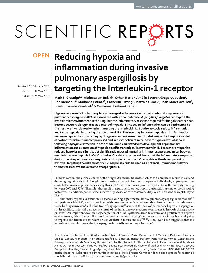
ResultsCxcr2−/− mice develop severe hypoxia and inflammation during invasive pulmonary aspergillosis. Neutrophils are, in addition to alveolar macrophages, essential for clearing Aspergillus spores from the lungs6,23,24,26. We investigated the role of CXCR2-mediated neutrophil recruitment in the development of hypoxia and inflammation during pulmonary aspergillosis by comparing Cxcr2−/− mice with wild-type mice.
Cxcr2−/− mice and WT controls were intranasally infected with a dose of 5 × 10⁷ live dormant conidia. At day 2 post infection (p.i) the number of alveolar macrophages (AM) and neutrophils (PMNs) was evaluated in the bronchoalveolar lavage (BAL). Compared to wild-type mice, Cxcr2−/− mice did not show a difference in abso-lute number of alveolar macrophages (Fig. 1A). In contrast and in line with previous studies23,24, Cxcr2−/− mice demonstrated an impaired recruitment of neutrophils to the lungs following Aspergillus infection, which was twofold lower than the amount of neutrophils recruited to the lungs of WT mice (Fig. 1A). During the course of
Figure 1. Hypoxia and inflammation during aspergillosis in Cxcr2−/− mice (A) Number of Alveolar macrophages and neutrophils (PMNs) in the BAL of control (naïve), PBS treated, wild-type (WT) and Cxcr2−/− mice 1 day p.i with live resting Aspergillus fumigatus spores (5 × 10⁷ in 25 μ l), shown as scatterplot and median and compared for significance using Mann Whitney U test. (B) Weight of WT (n = 13); and Cxcr2−/− mice (n = 13) during the course of infection and compared for significance by two way ANOVA. (C) Fungal burden measured by the luminescence signal by in vivo conversion of luciferin by the luciferase expressing A. fumigatus (strain 2/7/1) at day 1, 2 and 3 p.i. (D) Representative lung sections from WT and Cxcr2−/− mice at day 3 p.i. Methenamine silver staining shows, non-germinating conidia in WT and, foci of germinating conidia in Cxcr2−/− mice. (E) Proinflammatory mediators CXCL1, IL-1α , IL-1β , IL-6, and G-CSF measured in lung homogenates at day 3 p.i in WT and Cxcr2−/− mice, and are represented as mean ± SEM and were compared for significance using the Mann-Whitney U test. (F) Hypoxia measured by the fluorescent HypoxiSense probe 680 (Perkin Elmer) in the lungs of WT and Cxcr2−/− mice at day 3 pi. Data is represented as scatterplot and median and were compared for significance using the Mann-Whitney U test. *p < 0.05;**p < 0.005; ***p < 0.001.
www.nature.com/scientificreports/
3Scientific RepoRts | 6:26490 | DOI: 10.1038/srep26490
infection, both WT mice and Cxcr2−/− mice decrease in weight. While WT mice start to recover from day 3 p.i Cxcr2−/− continue to decrease in weight, and the average weight loss of the knock-out (KO) mice is significantly greater than that of WT mice (Fig. 1B) indicating their increased susceptibility to infection. The fungal burden was measured by in vivo bioluminescence imaging of the luciferase expressing A. fumigatus strain (Fig. 1C). The bioluminescence assay indicates that Cxcr2−/− mice do not have a different fungal burden than wild-type animals. It must be noted, however, that under extreme hypoxic conditions the luminescence might be biased towards indicating a lower fungal burden, since the firefly luciferase luminescence signal is oxygen dependent6,27. To control this possible limitation, lung sections were examined for fungal burden using Grocott methamine silver staining (Fig. 1D). Histological analysis revealed hyphae concentrated in inflammatory foci in the lungs of Cxcr2−/− mice, while in WT lungs no hyphal invasion was observed and only non germinating conidia were found.
Inflammation in the lungs was assessed by determining cytokine levels in lung homogenates at day 3 pi. Compared to wild-type mice, Cxcr2−/− mice had significantly higher inflammation in the lungs, as demonstrated by higher levels of all measured inflammatory mediators in the lung homogenates (Fig. 1E). Hypoxia in the lungs was assessed at day 3 p.i. by following the activity of carbonic anhydrase using the Perkin Elmer hypoxiSense 680 probe. The hyper-inflammatory state in the lungs of Cxcr2−/− mice correlated with significantly elevated hypoxia (Fig. 1F).
Cortisone-acetate immunocompromised mice demonstrate severe hypoxia and inflammation. Acquired immunodeficiency due to immunosuppressive therapy is one of the major predisposing factors for IPA2–5. Therefore, we investigated the development of hypoxia and inflammation in the setting of corticosteroid immunosuppression. In this setting immunocompetent as well as immunosuppressed mice received an inoculum of 5 × 10⁵ conidia. A higher concentration of 5 × 10⁷ results in the death of immunosuppressed mice within 24 to 48h (data not shown).
First, the effect of cortisone-acetate immunosuppression on the recruitment of alveolar macrophages and neutrophils to the lungs was investigated by measuring AMs and PMNs in the BAL at 2 days p.i. Following infection, no significant change in the recruitment of both cell subsets was observed between immunocompe-tent and cortisone acetate-treated mice (Fig. 2A). Yet it must be noted that corticosteroids affect effector cell function and lifespan, thereby compromising the mice rather than altering cell influx. During the course of infection, the average weight loss of immunosuppressed mice was significantly higher than that of immunocom-petent mice (Fig. 2B). Bioluminescence as well as lung sections revealed higher fungal burden with significantly higher luminescent signal and growing hyphae concentrated in inflammatory foci in the lungs of the immuno-suppressed mice. As expected, immunocompetent mice showed low luminescence signal and no germination of conidia (Fig. 2C,D). Three days after Aspergillus infection, cortisone-acetate immunosuppressed mice had severe pulmonary inflammation, reflected by elevated levels of CXCL1, IL-1α , IL-1β , IL-6, and G-CSF compared to immunocompetent mice (Fig. 2E). In correlation with severe pulmonary inflammation, cortisone-acetate immu-nosuppressed mice demonstrated severe pulmonary hypoxia (Fig. 2F) in comparison to immunocompetent and immunosuppressed PBS-infected mice. In addition to pulmonary hypoxia, hypoxia was also significantly higher in the sinus area of immunosuppressed infected mice (Fig. 2G).
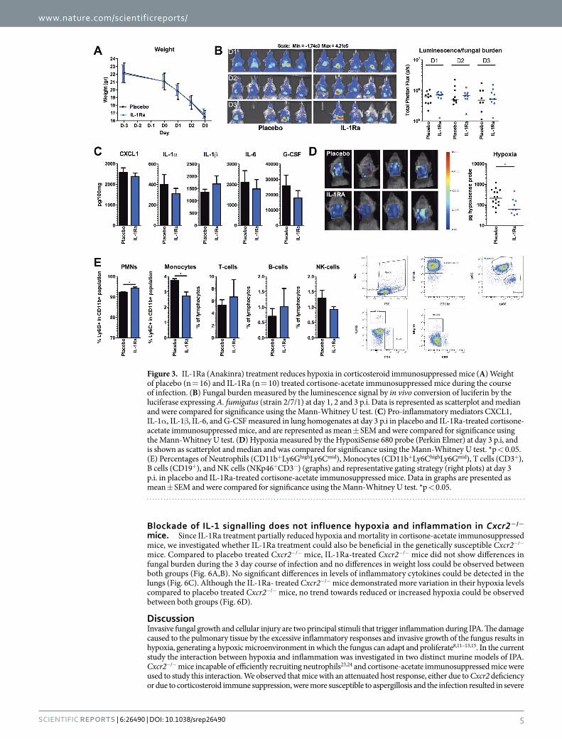
Blockade of IL-1 signalling reduces pulmonary hypoxia in cortisone-acetate immunosuppressed mice. Although crucial for initiating the primary host defence mechanisms against Aspergillus28,29, IL-1 can also play a key role in inducing detrimental hyper-inflammation17 and activates hypoxia-inducible factor 1 alpha (Hif1α )30,31. In turn Hif1α can activate transcription of IL-1α and IL-1β 32,33. We therefore hypothesized that inhi-bition of IL-1 signalling, by IL-1Receptor antagonist (Ra) (Anakinra), could break the loop between inflamma-tion and hypoxia. IL-1Ra reduces the biological activity of IL-1 by competitively binding to the Interleukin-1 type 1 receptor and can be therapeutically used to target IL-1 mediated inflammation34. Anakinra is the recombinant form of human IL-1Ra but has been shown to be active in murine models35,36.
Cortisone acetate immunosuppressed mice were infected with Aspergillus and IL-1Ra treatment was initiated one day p.i, to allow an initial immune response to clear fungal spores. The IL-1Ra-treated group and placebo group demonstrated similar weight loss during the course of infection (Fig. 3A). In addition, fungal burden shown by the luminescence in the lungs of IL-1Ra-treated mice was not significantly different from placebo treated ani-mals (Fig. 3B). Some mice (n = 6) clearly demonstrated the capacity of IL-1Ra to significantly reduce pulmonary inflammation compared to the control group. However, this was not consistently seen in all mice (total: n = 10) and no significant difference in pulmonary inflammation between IL-1Ra-treated, and placebo-treated mice at day 3 p.i could be demonstrated (Fig. 3C). However, IL-1Ra-treatment was effective since hypoxia measured by carbonic anhydrase activity was significantly decreased under this treatment (Fig. 3D).
To explore the effect of IL-1Ra treatment on the cellular composition of the leukocytes that are present in the lung, flow cytometry of lung homogenates was performed at day 3 p.i. IL-1Ra-treated mice show a minor increase in the population Ly6Ghigh neutrophils, whereas the population of Ly6Chigh monocytes among CD11b+ cells in the lungs was reduced (Fig. 3E).
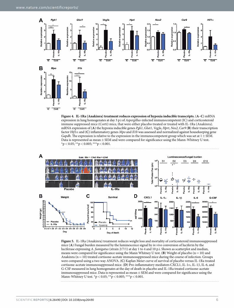
IL-1Ra treatment reduces expression of hypoxia inducible transcripts. To validate that IL-1Ra treatment of cortisone acetate immunosuppressed mice reduces pulmonary hypoxia during aspergillosis, the effect on expression of hypoxia inducible genes was assessed. Numerous genes are regulated by hypoxia respon-sive elements (HRE) that are targeted by the transcription factors Hif1α and Hif2α 37. The expression of several hypoxia inducible genes was assessed in lung homogenates of Aspergillus-infected immunocompetent mice and cortisone acetate immunosuppressed mice, with or without IL-1Ra treatment. Compared to immunocompetent mice immunosuppressed mice have significantly elevated expression of the hypoxia inducible genes Pgk1, Vegfa,

www.nature.com/scientificreports/
4Scientific RepoRts | 6:26490 | DOI: 10.1038/srep26490
Hprt and Nos2. In addition a trend towards increased expression of Glut1 and Hif1α itself could be observed. In contrast, immunocompromised mice that were treated with IL-1Ra did not as strongly induce the hypoxia induc-ible genes, and some genes Glut1 and Car9 (CAIX) were even significantly lower compared to placebo treated mice (Fig. 4A). In addition, expression of Il1b and Mpo were assessed as markers of inflammation. Both genes were significantly higher expressed in immunosuppressed mice, yet only a mild reduction could be observed following IL-1Ra treatment (Fig. 4B).
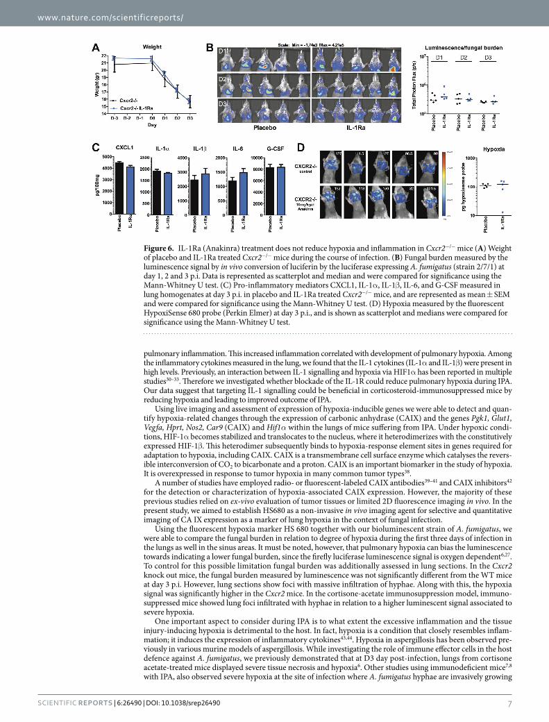
Blockade of IL-1 signalling decreases lethal outcome of aspergillosis in corticosteroid-treated mice. We further explored whether IL-1 receptor blockade, which results in reduction of inflammation and hypoxia, could increase survival in cortisone-acetate immunosuppressed mice. In mice that were infected with 5 × 10⁵ conidia no significant effect of IL-1Ra treatment on survival of the mice could be observed when com-pared to PBS treatment (data not shown). However, when the dose was reduced to 2 × 10⁵, treatment with IL-1Ra resulted in a significant reduction of fungal burden as shown by bioluminescence signal (Fig. 5A) associated with a significant difference in the body weights at day 3 p.i (Fig. 5B). Treatment with IL-1Ra for 10 days improved the survival of mice from 11 to 25% (Fig. 5C). Assessment of inflammation in the lungs at the day of death demon-strated that IL-1Ra treated mice had significantly lower levels of IL-6, CXCL1, and G-CSF in their lungs compared to placebo treated mice at the time of death (Fig. 5D).
In a real patient situation antifungal treatment is the standard regimen and immunomodulatory therapy could be added to the standard care. There is a possibility that immunomodulatory therapy could be more beneficial in combination with antifungal strategies. Therefore a pilot experiment comparing the survival of immunosuppressed mice challenged Aspergillus-conidia (2 × 10⁵) treated either with Caspofungin (10 mg/kg/day) or with a combi-nation of Caspofungin and IL-1Ra was performed. Interestingly, the combination was well tolerated by the mice and addition of IL-1Ra to the treatment regimen improved survival from 40% with caspofungin to 60% (Fig. S1).
Figure 2. Hypoxia and inflammation during aspergillosis in corticosteroid immunosuppressed mice (A) Number of Alveolar macrophages and neutrophils (PMNs) in the BAL of control, PBS treated and Aspergillus-infected (5 × 10⁵) immunocompetent (IC n = 5) and cortisone-acetate immunosuppressed mice (Corti n = 13) mice 1 day p.i, shown as scatterplot and median. (B) Weight of IC and cortisone-acetate immunosuppressed mice during the course of infection, and compared for significance using a two way ANOVA. (C) Fungal burden measured by the luminescence signal by in vivo conversion of luciferin by the luciferase expressing A. fumigatus (strain 2/7/1) at day 1, 2 and 3 p.i. Data is represented as scatterplot and median and were compared for significance using the Mann-Whitney U test. (D) Representative lung sections from (IC) and (Corti) mice at day 3 p.i. Methenamine silver staining showing, non-germinating conidia in (IC) and foci of germinating conidia in (Corti) mice. (E) Proinflammatory mediators CXCL1, IL-1α , IL-1β , IL-6, and G-CSF measured in lung homogenates at day 3 p.i in IC and cortisone-acetate immunosuppressed mice, and are represented as mean ± SEM and compared for significance using the Mann-Whitney U test. (F) Hypoxia measured by the fluorescent HypoxiSense 680 probe (Perkin Elmer) in the lungs of IC and Corti mice at day 3 p.i, and shown as scatterplot with median and were compared for significance using the Mann-Whitney U test. (G) Hypoxia measured in the sinus of IC and Corti mice at day 3 p.i, and shown as scatterplot with median and were compared for significance using the Mann-Whitney U test. *p < 0.05;**p < 0.005; ***p < 0.001.
www.nature.com/scientificreports/
5Scientific RepoRts | 6:26490 | DOI: 10.1038/srep26490
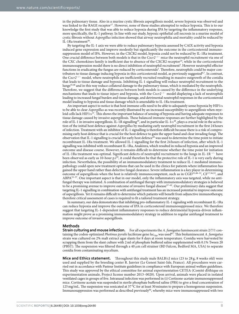
Blockade of IL-1 signalling does not influence hypoxia and inflammation in Cxcr2−/− mice. Since IL-1Ra treatment partially reduced hypoxia and mortality in cortisone-acetate immunosuppressed mice, we investigated whether IL-1Ra treatment could also be beneficial in the genetically susceptible Cxcr2−/− mice. Compared to placebo treated Cxcr2−/− mice, IL-1Ra-treated Cxcr2−/− mice did not show differences in fungal burden during the 3 day course of infection and no differences in weight loss could be observed between both groups (Fig. 6A,B). No significant differences in levels of inflammatory cytokines could be detected in the lungs (Fig. 6C). Although the IL-1Ra- treated Cxcr2−/− mice demonstrated more variation in their hypoxia levels compared to placebo treated Cxcr2−/− mice, no trend towards reduced or increased hypoxia could be observed between both groups (Fig. 6D).
DiscussionInvasive fungal growth and cellular injury are two principal stimuli that trigger inflammation during IPA. The damage caused to the pulmonary tissue by the excessive inflammatory responses and invasive growth of the fungus results in hypoxia, generating a hypoxic microenvironment in which the fungus can adapt and proliferate8,11–13,15. In the current study the interaction between hypoxia and inflammation was investigated in two distinct murine models of IPA. Cxcr2−/− mice incapable of efficiently recruiting neutrophils23,24 and cortisone-acetate immunosuppressed mice were used to study this interaction. We observed that mice with an attenuated host response, either due to Cxcr2 deficiency or due to corticosteroid immune suppression, were more susceptible to aspergillosis and the infection resulted in severe
Figure 3. IL-1Ra (Anakinra) treatment reduces hypoxia in corticosteroid immunosuppressed mice (A) Weight of placebo (n = 16) and IL-1Ra (n = 10) treated cortisone-acetate immunosuppressed mice during the course of infection. (B) Fungal burden measured by the luminescence signal by in vivo conversion of luciferin by the luciferase expressing A. fumigatus (strain 2/7/1) at day 1, 2 and 3 p.i. Data is represented as scatterplot and median and were compared for significance using the Mann-Whitney U test. (C) Pro-inflammatory mediators CXCL1, IL-1α , IL-1β , IL-6, and G-CSF measured in lung homogenates at day 3 p.i in placebo and IL-1Ra-treated cortisone-acetate immunosuppressed mice, and are represented as mean ± SEM and were compared for significance using the Mann-Whitney U test. (D) Hypoxia measured by the HypoxiSense 680 probe (Perkin Elmer) at day 3 p.i, and is shown as scatterplot and median and was compared for significance using the Mann-Whitney U test. *p < 0.05. (E) Percentages of Neutrophils (CD11b+Ly6GhighLy6Cmid), Monocytes (CD11b+Ly6ChighLy6Gmid), T cells (CD3+), B cells (CD19+), and NK cells (NKp46+CD3−) (graphs) and representative gating strategy (right plots) at day 3 p.i. in placebo and IL-1Ra-treated cortisone-acetate immunosuppressed mice. Data in graphs are presented as mean ± SEM and were compared for significance using the Mann-Whitney U test. *p < 0.05.
www.nature.com/scientificreports/
6Scientific RepoRts | 6:26490 | DOI: 10.1038/srep26490
Figure 4. IL-1Ra (Anakinra) treatment reduces expression of hypoxia inducible transcripts. (A–C) mRNA expression in lung homogenates at day 3 p.i of Aspergillus-infected immunocompetent (IC) and corticosteroid immune suppressed mice (Corti) mice, that were either placebo treated or treated with IL-1Ra (Anakinra). mRNA expression of (A) the hypoxia inducible genes Pgk1, Glut1, Vegfa, Hprt, Nos2, Car9 (B) their transcription factor Hif1α and (C) inflammatory genes Mpo and Il1b was assessed and normalized against housekeeping gene Gapdh. The expression is relative to the expression in the immunocompetent group which was set at 1 ± SEM. Data is represented as mean ± SEM and were compared for significance using the Mann-Whitney U test. *p < 0.05; **p < 0.005; ***p < 0.001.
Figure 5. IL-1Ra (Anakinra) treatment reduces weight loss and mortality of corticosteroid immunosuppressed mice (A) Fungal burden measured by the luminescence signal by in vivo conversion of luciferin by the luciferase expressing A. fumigatus (strain 2/7/1) at day 1 to 4 and 10 p.i. Shown as scatterplot and median, means were compared for significance using the Mann Whitney U test. (B) Weight of placebo (n = 10) and Anakinra (n = 10) treated cortisone-acetate immunosuppressed mice during the course of infection. Groups were compared using a two way ANOVA. (C) Kaplan Meier curve of survival of placebo versus IL-1Ra treated cortisone-acetate immunosuppressed mice. (D) Pro-inflammatory mediators CXCL1, IL-1α , IL-1β , IL-6, and G-CSF measured in lung homogenates at the day of death in placebo and IL-1Ra treated cortisone-acetate immunosuppressed mice. Data is represented as mean ± SEM and were compared for significance using the Mann-Whitney U test. *p < 0.05; **p < 0.005; ***p < 0.001.
www.nature.com/scientificreports/
7Scientific RepoRts | 6:26490 | DOI: 10.1038/srep26490
pulmonary inflammation. This increased inflammation correlated with development of pulmonary hypoxia. Among the inflammatory cytokines measured in the lung, we found that the IL-1 cytokines (IL-1α and IL-1β ) were present in high levels. Previously, an interaction between IL-1 signalling and hypoxia via HIF1α has been reported in multiple studies30–33. Therefore we investigated whether blockade of the IL-1R could reduce pulmonary hypoxia during IPA. Our data suggest that targeting IL-1 signalling could be beneficial in corticosteroid-immunosuppressed mice by reducing hypoxia and leading to improved outcome of IPA.
Using live imaging and assessment of expression of hypoxia-inducible genes we were able to detect and quan-tify hypoxia-related changes through the expression of carbonic anhydrase (CAIX) and the genes Pgk1, Glut1, Vegfa, Hprt, Nos2, Car9 (CAIX) and Hif1α within the lungs of mice suffering from IPA. Under hypoxic condi-tions, HIF-1α becomes stabilized and translocates to the nucleus, where it heterodimerizes with the constitutively expressed HIF-1β . This heterodimer subsequently binds to hypoxia-response element sites in genes required for adaptation to hypoxia, including CAIX. CAIX is a transmembrane cell surface enzyme which catalyses the revers-ible interconversion of CO2 to bicarbonate and a proton. CAIX is an important biomarker in the study of hypoxia. It is overexpressed in response to tumor hypoxia in many common tumor types38.
A number of studies have employed radio- or fluorescent-labeled CAIX antibodies39–41 and CAIX inhibitors42 for the detection or characterization of hypoxia-associated CAIX expression. However, the majority of these previous studies relied on ex-vivo evaluation of tumor tissues or limited 2D fluorescence imaging in vivo. In the present study, we aimed to establish HS680 as a non-invasive in vivo imaging agent for selective and quantitative imaging of CA IX expression as a marker of lung hypoxia in the context of fungal infection.
Using the fluorescent hypoxia marker HS 680 together with our bioluminescent strain of A. fumigatus, we were able to compare the fungal burden in relation to degree of hypoxia during the first three days of infection in the lungs as well in the sinus areas. It must be noted, however, that pulmonary hypoxia can bias the luminescence towards indicating a lower fungal burden, since the firefly luciferase luminescence signal is oxygen dependent6,27. To control for this possible limitation fungal burden was additionally assessed in lung sections. In the Cxcr2 knock out mice, the fungal burden measured by luminescence was not significantly different from the WT mice at day 3 p.i. However, lung sections show foci with massive infiltration of hyphae. Along with this, the hypoxia signal was significantly higher in the Cxcr2 mice. In the cortisone-acetate immunosuppression model, immuno-suppressed mice showed lung foci infiltrated with hyphae in relation to a higher luminescent signal associated to severe hypoxia.
One important aspect to consider during IPA is to what extent the excessive inflammation and the tissue injury-inducing hypoxia is detrimental to the host. In fact, hypoxia is a condition that closely resembles inflam-mation; it induces the expression of inflammatory cytokines43,44. Hypoxia in aspergillosis has been observed pre-viously in various murine models of aspergillosis. While investigating the role of immune effector cells in the host defence against A. fumigatus, we previously demonstrated that at D3 day post-infection, lungs from cortisone acetate-treated mice displayed severe tissue necrosis and hypoxia6. Other studies using immunodeficient mice7,8 with IPA, also observed severe hypoxia at the site of infection where A. fumigatus hyphae are invasively growing
Figure 6. IL-1Ra (Anakinra) treatment does not reduce hypoxia and inflammation in Cxcr2−/− mice (A) Weight of placebo and IL-1Ra treated Cxcr2−/− mice during the course of infection. (B) Fungal burden measured by the luminescence signal by in vivo conversion of luciferin by the luciferase expressing A. fumigatus (strain 2/7/1) at day 1, 2 and 3 p.i. Data is represented as scatterplot and median and were compared for significance using the Mann-Whitney U test. (C) Pro-inflammatory mediators CXCL1, IL-1α , IL-1β , IL-6, and G-CSF measured in lung homogenates at day 3 p.i. in placebo and IL-1Ra treated Cxcr2−/− mice, and are represented as mean ± SEM and were compared for significance using the Mann-Whitney U test. (D) Hypoxia measured by the fluorescent HypoxiSense 680 probe (Perkin Elmer) at day 3 p.i., and is shown as scatterplot and medians were compared for significance using the Mann-Whitney U test.
www.nature.com/scientificreports/
8Scientific RepoRts | 6:26490 | DOI: 10.1038/srep26490
in the pulmonary tissue. Also in a murine cystic fibrosis aspergillosis model, severe hypoxia was observed and was linked to the RAGE receptor17. However, none of these studies attempted to reduce hypoxia. This is to our knowledge the first study that was able to reduce hypoxia during IPA by targeting the inflammatory response, more specifically, the IL-1 pathway. In line with our study, hypoxic epithelial cell necrosis in a murine model of cystic fibrosis without Aspergillus infection showed that airway neutrophilia and mortality could be reduced by IL-1Ra treatment36.
By targeting the IL-1 axis we were able to reduce pulmonary hypoxia assessed by CAIX activity and hypoxia induced gene expression and improve modestly but significantly the outcome in the corticosteroid immuno-suppression model of IPA. However, in the CXCR2 model, hypoxia could not be reduced by IL-1Ra treatment. One crucial difference between both models is that in the Cxcr2−/− mice the neutrophil recruitment via one of the CXC chemokines family is inefficient due to absence of the CXCR2 receptor24, while in the corticosteroid immunosuppression model there is no direct inhibition of neutrophil recruitment5. However neutrophil effector functions in eradicating the fungus are reduced by corticosteroids5. Therefore, neutrophils could be major con-tributors to tissue damage inducing hypoxia in this corticosteroid model, as previously suggested6,7. In contrast, the Cxcr2−/− model, where neutrophils are inefficiently recruited resulting in massive outgrowth of the conidia that leads to tissue damage and hypoxia. Inhibiting IL-1 signalling will reduce neutrophil recruitment to the lungs45,46 and in this way reduce collateral damage to the pulmonary tissue, which is mediated by the neutrophils. Therefore, we suggest that the differences between both models is caused by the difference in the underlying mechanisms that leads to tissue injury and hypoxia, with the Cxcr2−/− model displaying a lack of neutrophils leading to increased fungal burden and tissue damage, and detrimental neutrophil responses in the corticosteroid model leading to hypoxia and tissue damage which is amendable to IL-1Ra treatment.
An important aspect to notice is that host immune cells need to be able to adequately sense hypoxia by HIF1α to be able to clear Aspergillus as was recently illustrated by an increased susceptibility to aspergillosis when mye-loid cells lack HIF1α 47. This shows the important balance of sensing of hypoxia and having adequate responses to tissue damage caused by invasive aspergillosis. These balanced immune responses are further highlighted by the role of IL-1 in invasive aspergillosis. IL-1R signalling29, and in particular IL-1α 28, plays a crucial role in the activa-tion of the initial host defence against Aspergillus by mediating early neutrophil recruitment in the first 24 hours of infection. Treatment with an inhibitor of IL-1 signalling is therefore difficult because there is a risk of compro-mising early host defence that is crucial for the host defence to gain the upper hand and clear invading fungi. The observation that IL-1 signalling is crucial for early host defence28 was used to determine the time point to initiate recombinant IL-1Ra treatment. We allowed IL-1 signalling for the first 24 hours of infection, after which IL-1 signalling was inhibited with recombinant IL-1Ra, Anakinra, which resulted in reduced hypoxia and an improved outcome and disease course. However, it remains difficult to determine whether the time point for initiation of IL-1Ra treatment was optimal. Significant defects of neutrophil recruitment to the lungs in IL-1R−/− have been observed as early as 10-hour p.i29, it could therefore be that the protective role of IL-1 is very early during infection. Nevertheless, the possibility of an immunomodulatory treatment to reduce IL-1 mediated immuno-pathology could open new treatment options that can be used in the clinics for patients where inflammation has gained the upper hand rather than defective fungal clearance. Severe inflammation is a key player in determining outcome of aspergillosis when the host is relatively immunocompetent, such as in CGD35,48–52, CF17,36,53, and ABPA53–57. One important aspect is that in our model, only the inflammatory axis was targeted, while no anti-fungal therapy was initiated. A combination of antifungal therapy with immunomodulatory strategies is believed to be a promising avenue to improve outcome of invasive fungal disease57–62. Our preliminary data suggest that targeting IL-1 signalling in combination with antifungal treatment has an increased potential to improve outcome of aspergillosis. Yet it remains difficult to determine which patients will benefit from adjunctive immunotherapy, therefore critical assessment of cases is required to fit a tailored treatment strategy.
In summary, our data demonstrates that inhibiting pro-inflammatory IL-1 signaling with recombinant IL-1Ra can reduce hypoxia and improve the outcome of IPA in corticosteroid immunosuppressed mice. We therefore suggest that targeting IL-1-dependent inflammatory responses to reduce detrimental hypoxia-driven inflam-mation might prove as a promising immunomodulatory strategy in addition to regular antifungal treatment to improve outcome of invasive aspergillosis.
MethodsStrain culturing and mouse infection. For all experiments the A. fumigatus luminescent strain 2/7/1 con-taining the codon-optimized Photinus pyralis luciferase gene lucOpt was used63. This bioluminescent A. fumigatus strain was cultured on 2% malt extract agar slants for 8 days at room temperature. Conidia were harvested by scrapping them from the slant culture with 2 ml of phosphate buffered saline supplemented with 0.1% Tween 20 (PBST). The suspension was filtered through a 40 μ m cell strainer (BD Falcon, Bedford MA, USA) to separate conidia from contaminating mycelium.
Mice and Ethics statement. Throughout this study male BALB/cJ mice (23 to 28 g, 8 weeks old) were used and supplied by the breeding center R. Janvier (Le Genest Saint-Isle, France). All procedures were car-ried out in accordance with Pasteur Institute guidelines in compliance with European animal welfare guidelines. This study was approved by the ethical committee for animal experimentation CETEA (Comité d’éthique en experimentation animale, Project license number 2013–0020). Upon arrival, animals were placed in isolated ventilated cages in groups of five. Intranasal infection was performed in (i) Cortisone-acetate immunosuppressed mice. Cortisone acetate was suspended in sterile phosphate buffered saline (PBS) to give a final concentration of 125 mg/mL. The suspension was sonicated at 37 °C for at least 30 minutes to prepare a homogenous suspension. Immunosuppression was performed as described previously64, whereby mice were immunosuppressed with two

www.nature.com/scientificreports/
9Scientific RepoRts | 6:26490 | DOI: 10.1038/srep26490
single doses of 25 mg cortisone acetate (Sigma Aldrich, St Louis, MO), which were injected intraperitoneally three days before and immediately prior to infection with conidia (day 0).
At the day of infection, mice were anesthetized by intramuscular injection of 150 μ L of a solution containing 10 μ g/mL of ketamine and 10 μ g/mL xylazine and inoculated intranasally with a dose of either 5 × 10⁵ or 2 × 10⁵ conidia in 25 μ L of PBS. (ii) Cxcr2 knock out mice. In comparison to the pharmacologic immunosuppressive treatment, we used mice deficient in the chemokine receptor CXCR2 (Cxcr2−/− mice). These mice display a defect in neutrophil recruitment and are susceptibile to A. fumigatus6,23,65. The susceptibility of these mice was compared to BALB/c wild type mice following inoculation with 5 × 10⁷ conidia in 25 μ L of PBS.
Measurement of hypoxia using HypoxiSense 680 probe. Several invasive and non-invasive approaches have been developed to measure tumor oxygenation including hypoxia bio-marker targeted agents with labels that can be detected by imaging techniques. There has been considerable interest in fluorescent optical reporters for CAIX expression. Among these agents, it has been reported that HS680 has high in vitro and in vivo specificity for CAIX. It accumulates preferentially in hypoxic regions of CAIX positive tumors and can be used to non-invasively detect and quantify CAIX25.
Pairing HypoxiSense with optical fluorescent imaging technology allows for the first time to image and quan-titate tumor sub-regions undergoing hypoxia-related changes, non-invasively and in vivo. In our settings, each mice received intravenously at day 2 p.i 100 μ L of the fluorescent imaging HypoxiSense 680 (2 nmol) from Perkin Elmer.
In vivo imaging(i) Bioluminescence. Images were acquired using an IVIS 100 and spectrum systems (Perkin Elmer) according to the manufacturer’s instructions and as previously described66. In brief, 100 μ L of PBS containing 3.33 mg D-luciferin was intraperitoneally injected in mice before each measurement. Mice were anesthetized using a con-stant flow of 2.5% isofluorane mixed with oxygen using an XGI-8 gas anesthesia system (Xenogen Corporation). Images from mice were acquired 10 min after luciferin injection. Acquisition and quantification were performed using Living Image software version 3.1 (Xenogen Corporation). Quantification of photons per second emitted by either the lungs or sinus areas was performed by defining regions of interest.
(ii) Fluorescent tomography (FMT 100 Perkin Elmer). Infected mice were anesthetized by isoflurane/oxygen mixture gas anesthesia system. Experimental and control mice were then imaged using fluorescence tomography (FMT 2500LX Pre-Clinical Imaging System, PerkinElmer, Boston, MA PerkinElmer, Boston, MA). Briefly, the anesthetized mice were positioned in the imaging cassette which was then placed into the FMT imag-ing chamber. A NIR laser diode trans illuminated (i.e. passed light through the body of the animal to be collected on the opposite side) the infected regions, with signal detection occurring via a thermoelectrically cooled CCD camera placed on the opposite side of the imaged animal. Appropriate optical filters allowed collection of both-fluorescence and excitation datasets, the entire image acquisition sequence taking approximately 5–6 min per mouse. The collected fluorescence data was reconstructed (TrueQuant™ software, PerkinElmer, Boston, MA) for the quantification of three-dimensional fluorescence signal within the tumors. Three-dimensional regions of interest (ROI) were drawn in the lung or sinus areas. For visualization and analysis purposes, TrueQuant software provided three dimensional (3D) images and quantification in pmoles of fluorescence within ROIs. Additional technical information on FMT imaging, image re-contruction, image analysis and quantification can be found in the following review articles67.
These protocols enabled us to monitor and quantify for each mouse (i) fungal growth by bioluminescence and (ii) hypoxia by the cell surface expression of carbonic anhydrase (CAIX).
Bronchoalveolar lavages. Bronchoalveolar lavage (BAL) fluid was harvested as previously described68. Mice were euthanized by injection of Pentobarbital (Sanofi Santé Animale, Libourne, France) and the respiratory tract was exposed by dissection. A small incision was made near the top of the trachea, and a blunt-end 20-gauge needle was inserted and tied in place with surgical thread around the trachea. BAL fluid was obtained by 10 rounds of filling the lungs with 0.7 mL PBS and withdrawing as much of the liquid as possible. The samples were centrifuged to collect BAL fluid cells. BAL fluid cells were washed and resuspended in 1 mL PBS.
Histopathology. Mice were euthanized at day 3 p.i. Lungs were immediately fixed in 4% neutral-buffered formalin and embedded in paraffin. Five μ m sections were cut and stained with Grocott’s methenamine silver staining (GMS) for detection of fungi69.
Gene expression. Mouse lungs were homogenized in 500 μ L TRIzol reagent (Invitrogen) and subsequently RNA was isolated according to the protocol supplied by the manufacturer. RNA was reverse transcribed into cDNA using iScript cDNA synthesis kit (Biorad, Hercules Ca). Quantitative real-time PCR (qPCR) was per-formed using power SYBR Green PCR master mix (Applied Biosystems, Carlsbad, CA) and following primers: Gapdh Fwd 5′ -AGGTCGGTGTGAACGGATTTG-3′ and Rev 5′ -TGTAGACCATGTAGTTGAGGTCA-3′ , Pgk1 Fwd 5′ -CAAATTTGATGAGAATGCCAAGACT-3′ and Rev 5′ -TTCTTGCTGCTCTCAGTACCACA-3′ 70, Glut1 Fwd 5′ -GGGCATGTGCTTCCAGTATGT-3′ and Rev 5′ -ACGAGGAGCACCGTGAAGAT-3′ 70, Vegfa Fwd 5′ -AGTCCCATGAAGTGATCAAGTTCA-3′ and Rev 5′ -ATC CGCATGATCTGCATGG-3′ 70, Hprt Fwd 5′ -TTATCAGACTGAAGAGCTACT-3′ and Rev 5′ -TTACCAGTGTCAATTATATCTTCAACAATC-3′ 70, Hif1α Fwd 5′ -ACCTTCATCGGAAACTCCAAAG-3′ and Rev 5′ -ACTGTTAGGCTCAGGTGAACT-3′ ,, Car9 (CAIX) Fwd 5′ - CCGGAACTGAGCCTATCCAAC-3′ and Rev 5′ - GCAAGGCCCGGTATTCCTG-3′, Il1b Fwd 5′ -GCAACTGTTCCTGAACTCAACT-3′ and Rev 5′ -ATCTTTTGGGGTCCGTCAACT-3′, Nos2 Fwd 5′ -CGTTTCGGGATCTGAATGTGA-3′ and Rev 5′ -GGGCAGCCTGTGAGACCTT-3′, and Mpo Fwd
www.nature.com/scientificreports/
1 0Scientific RepoRts | 6:26490 | DOI: 10.1038/srep26490
5′ -GTCAGCTGTAACACCCTTCCTAAAC-3′ and Rev 5′-CCGGCAGACTCCCAACCT-3′. PCR was performed using an Applied Biosystem StepOne PCR system using PCR conditions 2 min 50 °C, 10 min 95 °C followed by 40 cycles at 95 °C for 15 sec and 60 °C for 1 min. The RNA expression of genes of interest were corrected for differ-ences in loading concentration using the signal of housekeeping protein GAPDH. Subsequently, the expression of immunocompetent mice was set as 1+ /− SEM to measure changes relative expression in the cortisone acetate immunosuppressed mice and cortisone acetate immunosuppressed mice treated with IL-1Ra.
Flow Cytometry. Lung samples were processed to single cell suspension by use of commercial digestion kit (Milteny Biotec) according to manufacturer’s instructions. For surface staining cells from BAL and lungs were incubated for 30 minutes with appropriate amounts of antibodies in the presence of Fc block (BD Bioscience), after which cells were washed and stained with viability dye (eBioscience). Antibodies used were directed against mouse CD3 (145-2CII) and CD19 (ID3), NKp46 (29A1.4), F4/80 (BM8), Gr-1 (RB6-8C5), CD11c (HL3), CD11b (M1/70), Ly6C (HK1.4), Ly6G (1A8-Ly6g) were purchased from Biolegend, BD Biosciences and eBioscience. Flow cytometric data was acquired on a MACSQuant device (Miltenyi Biotec) and analysed using FlowJo soft-ware (TreeStar).
Enzyme-Linked Immunosorbent assay (ELISA). Lungs homogenates were obtained following disrup-tion in saline using the Retsch Mixer Mill 301 homogenizer. CXCL1, IL-1α , IL-1β , IL-6 and G-CSF, concentra-tions were determined in lungs supernatants by ELISA as specified by the manufacturer (DuoSet; R&D Systems).
Statistical analysis. Comparison of luminescence, hypoxia and inflammation within the different groups of mice was performed using two way ANOVA followed by Bonferoni post test. Differences in cytokine levels between experimental groups were assessed using the Mann-Whitney U test.
All tests were performed using Graph pad Prism software. *p < 0.05; **p < 0.005; ***p < 0.001; ****p < 0.0001.
References1. Brown, G. D. et al. Hidden killers: human fungal infections. Science translational medicine 4, 165rv113, doi: 10.1126/
scitranslmed.3004404 (2012).2. Kontoyiannis, D. P. et al. Prospective surveillance for invasive fungal infections in hematopoietic stem cell transplant recipients,
2001–2006: overview of the Transplant-Associated Infection Surveillance Network (TRANSNET) Database. Clin Infect Dis 50, 1091–1100, doi: 10.1086/651263 (2010).
3. Garcia-Vidal, C., Upton, A., Kirby, K. A. & Marr, K. A. Epidemiology of invasive mold infections in allogeneic stem cell transplant recipients: biological risk factors for infection according to time after transplantation. Clin Infect Dis 47, 1041–1050 (2008).
4. Marr, K. A., Carter, R. A., Boeckh, M., Martin, P. & Corey, L. Invasive aspergillosis in allogeneic stem cell transplant recipients: changes in epidemiology and risk factors. Blood 100, 4358–4366 (2002).
5. Lewis, R. E. & Kontoyiannis, D. P. Invasive aspergillosis in glucocorticoid-treated patients. Medical mycology: official publication of the International Society for Human and Animal Mycology 47 Suppl 1, S271–281, doi: 10.1080/13693780802227159 (2009).
6. Ibrahim-Granet, O. et al. In vivo bioluminescence imaging and histopathopathologic analysis reveal distinct roles for resident and recruited immune effector cells in defense against invasive aspergillosis. BMC Microbiol 10, 105 (2010).
7. Grahl, N. et al. In vivo hypoxia and a fungal alcohol dehydrogenase influence the pathogenesis of invasive pulmonary aspergillosis. PLoS pathogens 7, e1002145, doi: 10.1371/journal.ppat.1002145 (2011).
8. Hsu, J. L. et al. Aspergillus fumigatus invasion increases with progressive airway ischemia. PloS one 8, e77136, doi: 10.1371/journal.pone.0077136 (2013).
9. Takuma, T., Okada, K., Uchida, Y., Yamagata, A. & Sawae, Y. Invasive pulmonary aspergillosis resulting in respiratory failure during neutrophil recovery from postchemotherapy neutropenia in three patients with acute leukaemia. Journal of internal medicine 252, 173–177 (2002).
10. Ben-Ami, R., Lewis, R. E., Leventakos, K. & Kontoyiannis, D. P. Aspergillus fumigatus inhibits angiogenesis through the production of gliotoxin and other secondary metabolites. Blood 114, 5393–5399, doi: 10.1182/blood-2009-07-231209 (2009).
11. Blosser, S. J., Merriman, B., Grahl, N., Chung, D. & Cramer, R. A. Two C4-sterol methyl oxidases (Erg25) catalyse ergosterol intermediate demethylation and impact environmental stress adaptation in Aspergillus fumigatus. Microbiology (Reading, England) 160, 2492–2506, doi: 10.1099/mic.0.080440-0 (2014).
12. Chung, D. et al. ChIP-seq and in vivo transcriptome analyses of the Aspergillus fumigatus SREBP SrbA reveals a new regulator of the fungal hypoxia response and virulence. PLoS pathogens 10, e1004487, doi: 10.1371/journal.ppat.1004487 (2014).
13. Grahl, N., Dinamarco, T. M., Willger, S. D., Goldman, G. H. & Cramer, R. A. Aspergillus fumigatus mitochondrial electron transport chain mediates oxidative stress homeostasis, hypoxia responses and fungal pathogenesis. Molecular microbiology 84, 383–399, doi: 10.1111/j.1365-2958.2012.08034.x (2012).
14. Willger, S. D. et al. Dsc orthologs are required for hypoxia adaptation, triazole drug responses, and fungal virulence in Aspergillus fumigatus. Eukaryot Cell 11, 1557–1567, doi: 10.1128/EC.00252-12 (2012).
15. Willger, S. D. et al. A sterol-regulatory element binding protein is required for cell polarity, hypoxia adaptation, azole drug resistance, and virulence in Aspergillus fumigatus. PLoS pathogens 4, e1000200, doi: 10.1371/journal.ppat.1000200 (2008).
16. Carvalho, A. et al. Inflammation in aspergillosis: the good, the bad, and the therapeutic. Annals of the New York Academy of Sciences 1273, 52–59, doi: 10.1111/j.1749-6632.2012.06754.x (2012).
17. Iannitti, R. G. et al. Th17/Treg imbalance in murine cystic fibrosis is linked to indoleamine 2,3-dioxygenase deficiency but corrected by kynurenines. American journal of respiratory and critical care medicine 187, 609–620, doi: 10.1164/rccm.201207-1346OC (2013).
18. Kim, B. et al. The Interleukin-1alpha Precursor is Biologically Active and is Likely a Key Alarmin in the IL-1 Family of Cytokines. Frontiers in immunology 4, 391, doi: 10.3389/fimmu.2013.00391 (2013).
19. Eltzschig, H. K. & Carmeliet, P. Hypoxia and inflammation. The New England journal of medicine 364, 656–665, doi: 10.1056/NEJMra0910283 (2011).
20. Chen, G. Y. & Nunez, G. Sterile inflammation: sensing and reacting to damage. Nature reviews. Immunology 10, 826–837, doi: 10.1038/nri2873 (2010).
21. Ghezzi, P. et al. Hypoxia increases production of interleukin-1 and tumor necrosis factor by human mononuclear cells. Cytokine 3, 189–194 (1991).
22. Frohlich, S., Boylan, J. & McLoughlin, P. Hypoxia-induced inflammation in the lung: a potential therapeutic target in acute lung injury? American journal of respiratory cell and molecular biology 48, 271–279, doi: 10.1165/rcmb.2012-0137TR (2013).
23. Bonnett, C. R., Cornish, E. J., Harmsen, A. G. & Burritt, J. B. Early neutrophil recruitment and aggregation in the murine lung inhibit germination of Aspergillus fumigatus Conidia. Infect Immun 74, 6528–6539 (2006).

www.nature.com/scientificreports/
1 1Scientific RepoRts | 6:26490 | DOI: 10.1038/srep26490
24. Mehrad, B. et al. CXC chemokine receptor-2 ligands are necessary components of neutrophil-mediated host defense in invasive pulmonary aspergillosis. J Immunol 163, 6086–6094 (1999).
25. Bao, B. et al. In vivo imaging and quantification of carbonic anhydrase IX expression as an endogenous biomarker of tumor hypoxia. PloS one 7, e50860, doi: 10.1371/journal.pone.0050860 (2012).
26. Mircescu, M. M., Lipuma, L., van Rooijen, N., Pamer, E. G. & Hohl, T. M. Essential role for neutrophils but not alveolar macrophages at early time points following Aspergillus fumigatus infection. J Infect Dis 200, 647–656 (2009).
27. Robertson, J. B., Stowers, C. C., Boczko, E. & Johnson, C. H. Real-time luminescence monitoring of cell-cycle and respiratory oscillations in yeast. Proc Natl Acad Sci USA 105, 17988–17993 (2008).
28. Caffrey, A. K. et al. IL-1alpha Signaling Is Critical for Leukocyte Recruitment after Pulmonary Aspergillus fumigatus Challenge. PLoS pathogens 11, e1004625, doi: 10.1371/journal.ppat.1004625 (2015).
29. Jhingran, A. et al. Compartment-Specific and Sequential Role of MyD88 and CARD9 in Chemokine Induction and Innate Defense during Respiratory Fungal Infection. PLoS pathogens 11, e1004589, doi: 10.1371/journal.ppat.1004589 (2015).
30. Haddad, J. J. Recombinant human interleukin (IL)-1 beta-mediated regulation of hypoxia-inducible factor-1 alpha (HIF-1 alpha) stabilization, nuclear translocation and activation requires an antioxidant/reactive oxygen species (ROS)-sensitive mechanism. European cytokine network 13, 250–260 (2002).
31. Hellwig-Burgel, T., Rutkowski, K., Metzen, E., Fandrey, J. & Jelkmann, W. Interleukin-1beta and tumor necrosis factor-alpha stimulate DNA binding of hypoxia-inducible factor-1. Blood 94, 1561–1567 (1999).
32. Rider, P. et al. The transcription of the alarmin cytokine interleukin-1 alpha is controlled by hypoxia inducible factors 1 and 2 alpha in hypoxic cells. Frontiers in immunology 3, 290, doi: 10.3389/fimmu.2012.00290 (2012).
33. Zhang, W. et al. Evidence that hypoxia-inducible factor-1 (HIF-1) mediates transcriptional activation of interleukin-1beta (IL-1beta) in astrocyte cultures. Journal of neuroimmunology 174, 63–73, doi: 10.1016/j.jneuroim.2006.01.014 (2006).
34. Dinarello, C. A., Simon, A. & van der Meer, J. W. Treating inflammation by blocking interleukin-1 in a broad spectrum of diseases. Nat Rev Drug Discov 11, 633–652 (2012).
35. de Luca, A. et al. IL-1 receptor blockade restores autophagy and reduces inflammation in chronic granulomatous disease in mice and in humans. Proc Natl Acad Sci USA 111, 3526–3531 (2014).
36. Fritzsching, B. et al. Hypoxic Epithelial Necrosis Triggers Neutrophilic Inflammation via IL-1 Receptor Signaling in Cystic Fibrosis Lung Disease. American journal of respiratory and critical care medicine, doi: 10.1164/rccm.201409-1610OC (2015).
37. Lee, K. E. & Simon, M. C. SnapShot: Hypoxia-Inducible Factors. Cell 163, 1288–1288 e1281 (2015).38. Chen, C. L., Chu, J. S., Su, W. C., Huang, S. C. & Lee, W. Y. Hypoxia and metabolic phenotypes during breast carcinogenesis:
expression of HIF-1alpha, GLUT1, and CAIX. Virchows Arch 457, 53–61, doi: 10.1007/s00428-010-0938-0 (2010).39. Frangioni, J. V. In vivo near-infrared fluorescence imaging. Curr Opin Chem Biol 7, 626–634 (2003).40. Zatovicova, M. et al. Carbonic anhydrase IX as an anticancer therapy target: preclinical evaluation of internalizing monoclonal
antibody directed to catalytic domain. Curr Pharm Des 16, 3255–3263 (2010).41. Lou, Y. et al. Targeting tumor hypoxia: suppression of breast tumor growth and metastasis by novel carbonic anhydrase IX inhibitors.
Cancer Res 71, 3364–3376, doi: 10.1158/0008-5472.CAN-10-4261 (2011).42. Cecchi, A. et al. Carbonic anhydrase inhibitors. Design of fluorescent sulfonamides as probes of tumor-associated carbonic
anhydrase IX that inhibit isozyme IX-mediated acidification of hypoxic tumors. J Med Chem 48, 4834–4841 (2005).43. Desbaillets, I., Diserens, A. C., Tribolet, N., Hamou, M. F. & Van Meir, E. G. Upregulation of interleukin 8 by oxygen-deprived cells
in glioblastoma suggests a role in leukocyte activation, chemotaxis, and angiogenesis. The Journal of experimental medicine 186, 1201–1212 (1997).
44. Naldini, A., Pucci, A. & Carraro, F. Hypoxia induces the expression and release of interleukin 1 receptor antagonist in mitogen-activated mononuclear cells. Cytokine 13, 334–341, doi: 10.1006/cyto.2001.0842 (2001).
45. Hernandez, M. L. et al. IL-1 receptor antagonist reduces endotoxin-induced airway inflammation in healthy volunteers. The Journal of allergy and clinical immunology 135, 379–385, doi: 10.1016/j.jaci.2014.07.039 (2015).
46. Frank, J. A., Pittet, J. F., Wray, C. & Matthay, M. A. Protection from experimental ventilator-induced acute lung injury by IL-1 receptor blockade. Thorax 63, 147–153, doi: 10.1136/thx.2007.079608 (2008).
47. Shepardson, K. M. et al. Myeloid derived hypoxia inducible factor 1-alpha is required for protection against pulmonary Aspergillus fumigatus infection. PLoS pathogens 10, e1004378, doi: 10.1371/journal.ppat.1004378 (2014).
48. Meissner, F. et al. Inflammasome activation in NADPH oxidase defective mononuclear phagocytes from patients with chronic granulomatous disease. Blood 116, 1570–1573, doi: 10.1182/blood-2010-01-264218 (2010).
49. Morgenstern, D. E., Gifford, M. A., Li, L. L., Doerschuk, C. M. & Dinauer, M. C. Absence of respiratory burst in X-linked chronic granulomatous disease mice leads to abnormalities in both host defense and inflammatory response to Aspergillus fumigatus. The Journal of experimental medicine 185, 207–218 (1997).
50. Schappi, M. G., Jaquet, V., Belli, D. C. & Krause, K. H. Hyperinflammation in chronic granulomatous disease and anti-inflammatory role of the phagocyte NADPH oxidase. Semin Immunopathol 30, 255–271, doi: 10.1007/s00281-008-0119-2 (2008).
51. van Bruggen, R. et al. Human NLRP3 inflammasome activation is Nox1-4 independent. Blood 115, 5398–5400, doi: 10.1182/blood-2009-10-250803 (2010).
52. van de Veerdonk, F. L. et al. Reactive oxygen species-independent activation of the IL-1beta inflammasome in cells from patients with chronic granulomatous disease. Proc Natl Acad Sci USA 107, 3030–3033, doi: 10.1073/pnas.0914795107 (2010).
53. Knutsen, A. P. & Slavin, R. G. Allergic bronchopulmonary aspergillosis in asthma and cystic fibrosis. Clinical & developmental immunology 2011, 843763, doi: 10.1155/2011/843763 (2011).
54. Greenberger, P. A. Allergic bronchopulmonary aspergillosis. The Journal of allergy and clinical immunology 110, 685–692 (2002).55. Knutsen, A. P. et al. Fungi and allergic lower respiratory tract diseases. The Journal of allergy and clinical immunology 129, 280–291;
quiz 292–283, doi: 10.1016/j.jaci.2011.12.970 (2012).56. Kreindler, J. L. et al. Vitamin D3 attenuates Th2 responses to Aspergillus fumigatus mounted by CD4+ T cells from cystic fibrosis
patients with allergic bronchopulmonary aspergillosis. The Journal of clinical investigation 120, 3242–3254, doi: 10.1172/JCI42388 (2010).
57. Svirshchevskaya, E. V. & Kurup, V. P. Immunotherapy of allergic bronchopulmonary aspergillosis: a clinical and experimental approach. Frontiers in bioscience: a journal and virtual library 8, s92–101 (2003).
58. Bellocchio, S. et al. Immunity to Aspergillus fumigatus: the basis for immunotherapy and vaccination. Medical mycology: official publication of the International Society for Human and Animal Mycology 43 Suppl 1, S181–188 (2005).
59. Buddingh, E. P. et al. Interferon-gamma immunotherapy in a patient with refractory disseminated candidiasis. Pediatr Infect Dis J, doi: 10.1097/INF.0000000000000909 (2015).
60. Delsing, C. E. et al. Interferon-gamma as adjunctive immunotherapy for invasive fungal infections: a case series. BMC infectious diseases 14, 166, doi: 10.1186/1471-2334-14-166 (2014).
61. Lehrnbecher, T. et al. Immunotherapy in invasive fungal infection–focus on invasive aspergillosis. Curr Pharm Des 19, 3689–3712 (2013).
62. Kullberg, B. J., van de Veerdonk, F. & Netea, M. G. Immunotherapy: a potential adjunctive treatment for fungal infection. Curr Opin Infect Dis 27, 511–516, doi: 10.1097/QCO.0000000000000105 (2014).
63. Galiger, C. et al. Assessment of efficacy of antifungals against Aspergillus fumigatus: value of real-time bioluminescence imaging. Antimicrob Agents Chemother 57, 3046–3059 (2013).
www.nature.com/scientificreports/
1 2Scientific RepoRts | 6:26490 | DOI: 10.1038/srep26490
64. Schobel, F. et al. Aspergillus fumigatus does not require fatty acid metabolism via isocitrate lyase for development of invasive aspergillosis. Infect Immun 75, 1237–1244 (2007).
65. Mehrad, B., Moore, T. A. & Standiford, T. J. Macrophage inflammatory protein-1 alpha is a critical mediator of host defense against invasive pulmonary aspergillosis in neutropenic hosts. J Immunol 165, 962–968 (2000).
66. Brock, M. et al. Bioluminescent Aspergillus fumigatus, a new tool for drug efficiency testing and in vivo monitoring of invasive aspergillosis. Appl Environ Microbiol 74, 7023–7035 (2008).
67. Ntziachristos, V. & Weissleder, R. Experimental three-dimensional fluorescence reconstruction of diffuse media by use of a normalized Born approximation. Opt Lett 26, 893–895 (2001).
68. Ibrahim-Granet, O. et al. Phagocytosis and intracellular fate of Aspergillus fumigatus conidia in alveolar macrophages. Infect Immun 71, 891–903 (2003).
69. Sinha, B. K., Monga, D. P. & Prasad, S. A combination of Gomori-Grocott methenamine silver nitrate and hematoxylene and eosin staining technique for the demonstration of Candida albicans in tissue. Quad Sclavo Diagn 24, 129–132 (1988).
70. Park, S. K. et al. Hypoxia-induced gene expression occurs solely through the action of hypoxia-inducible factor 1alpha (HIF-1alpha): role of cytoplasmic trapping of HIF-2alpha. Mol Cell Biol 23, 4959–4971 (2003).
AcknowledgementsMG was supported by the Erasmus lifelong learning program. FvdV was supported by a Veni grant from ZonMw. OIG was supported by Institut Pasteur PTR 468 funding. OR was supported by a stipend from the Pasteur-Paris University (PPU) International PhD program. The authors are thankful to Marie-Anne Nicolas from Plateforme d’Imagerie Dynamique for her assistance in live imaging.
Author ContributionsConceived and designed the experiments: M.S.G., O.I.-G. and F.l.v.d.V. Performed the experiments: A.R., A.S., M.S.G., O.I.-G., O.R., M.P., C.F., G.J. and E.D. Analyzed the data: O.I.-G., M.S.G., O.R. and F.l.v.d.V. Wrote the manuscript: M.S.G., O.I.-G., O.R. and F.l.v.d.V. Amended the manuscript: O.I.-G., M.B., F.l.v.d.V., O.R. and J.M.C.
Additional InformationSupplementary information accompanies this paper at http://www.nature.com/srepCompeting financial interests: The authors declare no competing financial interests.How to cite this article: Gresnigt, M. S. et al. Reducing hypoxia and inflammation during invasive pulmonary aspergillosis by targeting the Interleukin-1 receptor. Sci. Rep. 6, 26490; doi: 10.1038/srep26490 (2016).
This work is licensed under a Creative Commons Attribution 4.0 International License. The images or other third party material in this article are included in the article’s Creative Commons license,
unless indicated otherwise in the credit line; if the material is not included under the Creative Commons license, users will need to obtain permission from the license holder to reproduce the material. To view a copy of this license, visit http://creativecommons.org/licenses/by/4.0/