Tram ligne C, arrêt Observatoire - Sciencesconf.org...Dr. Renaud CHOLLET Merck, Mosheim Dr....
45
jeudi 28 mars 2019 Collège Doctoral Européen 46 boulevard de la Victoire 67000 Strasbourg Tram ligne "C", arrêt "Observatoire" Organisé par le comité scientifique : Dr. GEOFFROY Valérie, Biotechnologie et signalisation cellulaire, UMR 7242, ESBS, Illkirch Dr. BRINGEL Françoise, Laboratoire de Génétique Moléculaire, Génomique, Microbiologie GMGM, UMR 7156, Strasbourg Pr. CANDOLFI Ermanno, Institut de Parasitologie et de Pathologie Tropicale de Strasbourg, Strasbourg Pr. ANDRE Philippe, Laboratoire de Biophotonique et Pharmacologie, UMR CNRS 7213, Faculté de Pharmacie, Illkirch Pr. LETT Marie-Claire, Laboratoire de Génétique Moléculaire, Génomique, Microbiologie GMGM, UMR 7156, Strasbourg
Transcript of Tram ligne C, arrêt Observatoire - Sciencesconf.org...Dr. Renaud CHOLLET Merck, Mosheim Dr....
jeudi 28 mars 2019
Collège Doctoral Européen 46 boulevard de la Victoire
67000 Strasbourg
Tram ligne "C", arrêt "Observatoire"
Organisé par le comité scientifique :
Dr. GEOFFROY Valérie, Biotechnologie et signalisation cellulaire, UMR 7242, ESBS, Illkirch
Dr. BRINGEL Françoise, Laboratoire de Génétique Moléculaire, Génomique, Microbiologie GMGM, UMR 7156, Strasbourg
Pr. CANDOLFI Ermanno, Institut de Parasitologie et de Pathologie Tropicale de Strasbourg, Strasbourg
Pr. ANDRE Philippe, Laboratoire de Biophotonique et Pharmacologie, UMR CNRS 7213, Faculté de Pharmacie, Illkirch
Pr. LETT Marie-Claire, Laboratoire de Génétique Moléculaire, Génomique, Microbiologie GMGM, UMR 7156, Strasbourg


Mutation dynamics and fitness effects at the single cell level
Dr Lydia ROBERT- Sorbonne Université, Laboratoire Jean Perrin, Paris
13h45
The last universal common ancestor: in search of microbe n°1
Dr William MARTIN - Institute of Molecular Evolution, Heinrich-Heine-Universität Dusseldorf, Germany
9h
sous la cotutelle de avec le soutien deco-organisé par
Laboratoire de bioimagerie et pathologies
> Programme de la journée : sms2019.sciencesconf.org/
Inscription gratuite mais obligatoireavant le 15 mars 2019
Séminaire de microbiologie de StrasbourgJeudi 28 mars 2019
Journée de rencontres entre chercheurs séniors, juniors, doctorants, masters travaillant dans le domaine de la bactériologie, la mycologie, la parasitologie et la virologie.
Deux conférences plénières :
Collège doctoral européen de Strasbourg
46 boulevard de la VictoireStrasbourg
C, E : arrêt Observatoire
Date limite de soumission d’un résumé (oral/poster) : 1er mars 2019
Nombre de places limité à 120 personnes
@ Dr Lydia Robert @ Dr William Martin


Séminaire de Microbiologie de Strasbourg
jeudi 28 mars 2019
Collège Doctoral Européen
Programme
8h30 Accueil
8h55 Introduction
Dr. Françoise BRINGEL Laboratoire de Génétique Moléculaire,
Génomique, Microbiologie, Strasbourg
9h00 Conférence plénière
The last universal common ancestor: in search of microbe n°1
Dr. William MARTIN - Institute of Molecular Evolution, Heinrich-Heine-Universität
Dusseldorf, Germany
Présentation du conférencier: Prof. Joseph MORAN, ISIS UMR7006
Modérateurs : Dr. Renaud CHOLLET, Merck
Dr. Anne FRIEDRICH, Laboratoire de Génétique Moléculaire,
Génomique, Microbiologie, Strasbourg
10h00 Communication orale
Survey of phenotypic expressivity at a species-wide scale using CRISPR-Cas9
Sabrina BIBI-TRIKI CNRS, Génétique Moléculaire, Génomique, Microbiologie Strasbourg
10h20 Présentation des sponsors
10H35 Pause-café
11h05 Communications orales
RsaC sRNA modulates the oxidative stress response during manganese starvation in
Staphylococcus aureus
David LALAOUNA, Jessica Baude, ZongFu Wu, Arnaud Tomasini, Stefano Marzi,
François Vandenesch, Pascale Romby, Isabelle Caldelari and Karen Moreau
UNISTRA CNRS, Architecture et Réactivité de l’ARN, UPR 9002, Strasbourg
La lugdulysine : un facteur de virulence potentiel et découverte d’un nouveau
mécanisme de maturation enzymatique
K. PROLA, Levy N., Argémi X., Strub J.M., Keller D., Cianferani S., Ruff M., Prévost G.
UNISTRA, CHRU de Strasbourg, FMTS, EA 7290 Virulence Bactérienne Précoce.
11h45 Présentation “flash” des posters en 3 minutes
12H30 Cocktail déjeunatoire offert par nos sponsors
13h00 Session posters

13h45 Conférence plénière
Mutation dynamics and fitness effects at the single cell level
Dr. Lydia ROBERT Sorbonne Université, Laboratoire Jean Perrin, Paris
Présentation de la conférencière : Dr. Françoise BRINGEL GMGM, Strasbourg
Modérateurs : Dr. Antoine GRILLON, IPPTS
Dr. Lysiane FOUGY, Aérial
14h45 Communications orales
Towards searching for novel dehalogenases in environmental samples by ultrahigh-
throughput screening
Emilie GEERSENS, Vuilleumier Stéphane and Ryckelynck Michaël. Institut de Biologie
Moléculaire et Cellulaire (IBMC) Strasbourg
Phosphatidic acid as a limiting host metabolite for the proliferation of the microsporidium
Tubulinosema ratisbonensis in Drosophila flies
Adrien Franchet, Gaëtan CARAVELLO, Sebastian Niehus, Gaëtan Caravello, and
Dominique Ferrandon Université de Strasbourg, CNRS, M3I UPR 9022, Strasbourg
15h45 Pause-café et session poster
16h15 Communications orales
Réseau d’interactions des protéines impliquées dans l’acquisition du fer par le
sidérophore pyoverdine chez Pseudomonas aeruginosa.
Anne Bonneau, Béatrice Roche and Isabelle Schalk
UMR7242, ESBS, Université de Strasbourg, CNRS, Bld Sébastien Brant, F-67412 Illkirch
Étude des bases moléculaires pour la vectorisation d’antibiotiques permettant de
contourner la faible perméabilité des membranes de Pseudomonas aeruginosa.
Q. PERRAUD, V. Gasser, P. Cantero, N. Zill, E. Baco, F. Hoegy, G. L. A. Mislin, L. Ehret-Sabatier,
I. J. Schalk, UNISTRA CNRS UMR 7242, Illkirch
Fine-tuning of predictive microbiology models through microlocal characterization of
foods by Nuclear Magnetic Resonance (NMR).
R. Recht, L. Fougy, V. Stahl, E. HAMON Aerial, Illkirch
17H15 Vote de la meilleure communication orale- Prix MERCK
Vote du meilleur poster- Prix CASDEN
Remise des prix
Dr. Renaud CHOLLET Merck, Mosheim
Dr. Valérie GEOFFROY Biotechnologie et Signalisation Cellulaire, UMR 7242, Illkirch
17h30 CONCLUSIONS
Prof. Ermano CANDOLFI Institut de Parasitologie et de Pathologie Tropicale de
Strasbourg, Strasbourg
Prof. Ph. ANDRE Laboratoire de Biophotonique et Pharmacologie, Illkirch
LISTE DES POSTERS
1. Regulation of IL-17A expression during Toxoplasma gondii infection?
Faïza FAHMI-BITTICH, Julie Brunet, Alexander W. Pfaff and Ermanno Candolfi
Institut de Parasitologie et de Pathologie Tropicale – EA 7292, Strasbourg
2. Spontaneous fermentation of sauerkraut: characterization of bacterial ecosystem
linked to physico-chemical changes
Lysiane FOUGY, Hezard B., Desmonts M-H. Aerial, Illkirch
3. Importance des voies d’import du fer dans la résistance aux antibiotiques
Gwenaelle GRAULIER, I. Schalk, P. Fechter
CNRS UMR7242 Biotechnologie et signalisation cellulaire, ESBS, ILLKIRCH
4. Intracellular localizations of PvdS ECF sigma factor in Pseudomonas aeruginosa
Léa HAAS, Isabelle Schalk, Julien Godet
Laboratoire de Biophotonique et Pharmacologie, UNISTRA CNRS 7021, Illkirch
5. Membrane aminoacyl-tRNA synthetases and signaling pathways
Marine HEMMERLE, H. Becker UNISTRA CNRS, Génétique Moléculaire, Génomique,
Microbiologie Strasbourg
6. Bacterial alteration of asbestos cement wastes
Agathe JAOUEN, S. David, V. A. Geoffroy CNRS UMR7242 Biotechnologie et
signalisation cellulaire, ESBS, ILLKIRCH
7. Triple element isotope fractionation of chloromethane from bacterial degradation by
two different pathways
Jing LUO, Jaime D Barnes, S Christoph Hartmann, Thierry Nadalig, Frank Keppler,
Stéphane Vuilleumier UNISTRA CNRS, Génétique Moléculaire, Génomique,
Microbiologie Strasbourg
8. Staphylococcus aureus Panton-Valentine Leukocidin causes an alternative NETosis in
human neutrophils presenting histone 3 citrullination on NETs
Viola MAZZOLENI, Gaëlle Zimmermann-Meisse, Daniel Keller, Gilles Prévost UNISTRA VBP
EA7290, Fédération de Medecine Translationnelle de Strasbourg, Institut de
Bactériologie, Strasbourg
9. Characterization of the interactions between icaR mRNA and regulatory RNAs in
Staphylococcus aureus
Noémie MERCIER, Eva Renard, Stefano Marzi, Pascale Romby, Alejandro Toledo-
Arana, Isabelle Caldelari UNISTRA CNRS, Architecture et Réactivité de l’ARN, UPR9002,
Strasbourg
10. Modes of action of antibiotics against multi-drug resistant pathogens
Eva RENARD, Emma Schenckbecher, Eric Ennifar, Gilles Prevost, Daniel Keller
UNISTRA Structure et dynamique des machines biomoléculaires, laboratoire
“Architecture et Réactivité de l’ARN”, IBMC, Strasbourg

11. Effet des bactéries associées à l’orge dans le métabolisme des terpénoïdes
Loïc WAECKERLE, Chloé Groh, Florence Arsène-Ploetze, Hubert Schaller CNRS IBMC,
Strasbourg
12. Bacterial adaptation to the utilization of chloromethane
Louis-François MEY, Yousra Louhichi, Stéphane Vuilleumier, Françoise BRINGEL
CNRS Laboratoire de Génétique Moléculaire, Génomique et Microbiologie
13. Ingénierie de la voie de biosynthèse du siderophore chez Pseudomonas pour
la production de pyoverdine fonctionnalisée
Sébastien MATHIEU, Coraline RIGOUIN
CNRS UMR7242 Biotechnologie et signalisation cellulaire, ESBS, ILLKIRCH
14. Antibacterial Potential of the Arab Mineral Pharmacopeia
Joshua MALSA, Anne FOSTER, Catherine VONTHRON, Elora AUBERT,
Véronique PITCHON, Isabelle SCHALK, and Pierre FECHTER
CNRS UMR7242 Biotechnologie et signalisation cellulaire, ESBS, ILLKIRCH.
15. Impact of the zinc finger motifs in HIV-1 Gag on the specific selection of
genomic RNA and its trafficking to the plasma membrane.
Emmanuel Boutant, Jeremy Bonzi, Halina Anton, Maaz Bin Nasim, Raphael
Cathagne, Eléonore Réal, Philippe Carl, Pascal Didier, Jean-Christophe Paillart,
Roland Marquet, Yves Mély, Hugues de Rocquigny, Serena Bernacchi.

RESUMES
DES
COMMUNICATIONS ORALES

The last universal common ancestor: In search of Microbe No. 1.
Prof. Dr. William Martin
Institute of Molecular Evolution
Heinrich-Heine-Universität Dusseldorf, Germany
The concept of a last universal common ancestor of all cells (LUCA) is central to the study of
early evolution and life's origin, yet information about how and where LUCA lived is lacking.
We investigated all clusters and phylogenetic trees for 6.1 million protein coding genes from
sequenced prokaryotic genomes in order to reconstruct the microbial ecology of LUCA.
Among 286,514 protein clusters, we identified 355 protein families (~0.1%) that trace to
LUCA by phylogenetic criteria. Because these proteins are not universally distributed, they
can shed light on LUCA's physiology. Their functions, properties, and prosthetic groups
depict LUCA as anaerobic, CO2-fixing, H2-dependent with a Wood-Ljungdahl (WL) pathway,
N2-fixing, and thermophilic. LUCA's biochemistry was replete with FeS clusters and radical
reaction mechanisms. Its cofactors reveal dependence upon transition metals, flavins, S-
adenosyl methionine (SAM), coenzyme A, ferredoxin, molybdopterin, corrins, and selenium.
Its genetic code required nucleoside modifications and SAM-dependent methylations. The
355 phylogenies identify clostridia and methanogens, whose modern lifestyles resemble
LUCA's, as basal among their respective domains. LUCA inhabited a geochemically active
environment rich in H2, CO2, and iron. The data support the view that the first organic
moleculaes — and life — arose from CO2 in a hydrothermal setting. As reconstructed from
genes, LUCA was half-alive.

Survey of phenotypic expressivity at a species-wide scale using CRISPR-Cas9
Sabrina Bibi-Triki, Téo Fournier and Joseph Schacherer
Université de Strasbourg, CNRS, GMGM UMR 7156, F-67000 Strasbourg, France
Elucidating the underlying rules that govern the phenotypic diversity observed in natural populations is an old but still unaccomplished goal in biology. Many genetic variants are known to have an impact on the phenotypic variation. However, a mutation can exhibit variable expressivity meaning that individuals who have this same mutation may express the phenotype to different degrees. The key to a better understanding of the genotype-phenotype relationship could benefit from a comprehensive view of expressivity cases within large populations. In this context, yeast and more precisely Saccharomyces cerevisiae is a powerful model system. Indeed, more than 50 rare genetic variants have been identified as involved in a given phenotypic variation. Taking advantage of these variants, we assessed the level of phenotypic expressivity of some of these genetic variants across a large population of S. cerevisiae isolates coming from a vast array of ecological and geographical origins. More specifically, we used the CRISPR-based editing methodology to introduce the selected genetic variants in a hundred S. cerevisiae genetic backgrounds. Using these CRISPR-engineered isolates, we determined the level of phenotypic expressivity of the different genetic variants tested. This allowed us to determine the complexity and degree to which phenotypic variation depends on genetic background. It also lays the foundation to dissect the genetic basis of the phenotypic expression variation.

RsaC sRNA modulates the oxidative stress response during manganese
starvation in Staphylococcus aureus
David Lalaouna, Jessica Baude, ZongFu Wu, Arnaud Tomasini, Stefano Marzi,
François Vandenesch, Pascale Romby, Isabelle Caldelari and Karen Moreau
Université de Strasbourg, CNRS, Architecture et Réactivité de l’ARN, UPR9002,
Strasbourg, France
The human pathogen Staphylococcus aureus produces numerous small regulatory RNAs
(sRNAs) for which functions are still poorly understood. Here, we focused on an atypical
sRNA called RsaC. Its length varies between different isolates due to the presence of
repeated sequences at the 5’ end, ranging up to 1,116 nt in HG001 strain. The 3’ part of
RsaC is highly conserved and contains C-rich sequences, which are characterized as
regulatory motifs in other staphylococcal sRNAs. Using MS2-affinity purification
coupled with RNA sequencing (MAPS) and quantitative differential proteomics, we
identified sodA mRNA as a main target of RsaC sRNA. SodA is a Mn-dependent
superoxide dismutase involved in oxidative stress response. We demonstrated that in
presence of RsaC, S. aureus cells were less resistant to oxidative stress, in relation with
lower activity of SodA enzyme. Remarkably, rsaC gene is co-transcribed with the major
manganese ABC transporter MntABC and, consequently, RsaC is mainly produced in
response to Mn starvation. This 3’UTR-derived sRNA is released from mntABC-RsaC
precursor after cleavage by RNase III which presumably recognizes a duplex formed by
RsaC and its antisens RNA. By negatively regulating non-essential Mn-containing
enzymes such as SodA, RsaC reduces the needs for Mn. SodM, an alternative Sod
enzyme using either Mn or Fe as co-factor, replaces SodA to response to oxidative stress.
Thus, RsaC may counteract the sequestration of Mn by the host organism, strategy used
to limit the virulence of S. aureus.
La lugdulysine : un facteur de virulence potentiel et
découverte d’un nouveau mécanisme de maturation
enzymatique
Prola K.1, Levy N.2, Argémi X.1, Strub J.M.3, Keller D.1, Cianferani S.3, Ruff M.2, Prévost G.1,
1. Université de Strasbourg, CHRU de Strasbourg, FMTS, EA 7290 : Virulence Bactérienne Précoce
2. Institut de Génétique, Biologie Moléculaire et Cellulaire
3. Laboratoire de Spectrométrie de Masse Bio-Organique, IPHC, UMR 7178
L’amélioration de l’identification bactérienne des staphylocoques à coagulase négative (CoNS)
(1) a permis de mettre en évidence la pathogénicité de Staphylococcus lugdunensis. Malgré sa
sensibilité aux antibiotiques, ce CoNS peut causer des infections chroniques variées telles que des
endocardites ou des infections ostéo-articulaires avec une virulence comparable à son homologue,
Staphylococcus aureus. L’étude clinique prospective VISLISI (2) collectant 81 souches de
Staphylococcus lugdunensis a permis de révéler et d’identifier la sécrétion d’un potentiel facteur de
virulence. En effet, les études statistiques ont démontré la corrélation entre la survenue d’infections
ostéo-articulaires et la détection d’une activité protéolytique due à cette enzyme, la lugdulysine.
Cette zinc métallopeptidase est synthétisée, puis sécrétée sous la forme d’un précurseur
inactif contenant un propeptide de 64 aa, localisé dans le domaine N terminal de la lugdulysine. Sous
sa forme active, la lugdulysine possède une activité optimale en présence de Ca2+ à 55°C et à pH 6.0.
Nous avons montré que la lugdulysine est principalement active sur les peptides et les protéines peu
structurées. La structure tri-dimensionnelle de la lugdulysine a été déterminée à l’IGBMC à une
résolution de 1.8 Å par cristallographie aux rayons X. L’analyse de cette structure a révélé la présence
de trois sites de fixation du calcium et a permis de confirmer la présence dans le site actif d’un atome
de zinc coordonné par le motif (241HEXX245H + 268E). De plus, 22 acides aminés du propeptide,
normalement éliminés lors de la maturation de l’enzyme, interagissent étroitement avec le domaine
mature. Ces interactions sont probablement importantes dans la maturation et l’activité enzymatique
puisque deux mutations (G299S et S412F), localisées au niveau du site d’interactions avec le
propeptide résiduel, et les mutations E242Q et Y315F dans le site actif enzymatique, empêchent toutes
la maturation de la pro-lugdulysine en enzyme mature.

1) Argemi, X., Riegel, P., Lavigne, T., Lefebvre, N., Grandpré, N., Hansmann, Y., Jaulhac, B., Prévost, G., and Schramm, F. (2015). Implementation of MALDI-TOF MS in routine clinical laboratories improves identification of coagulase negative staphylococci and reveals the pathogenic role of Staphylococcus lugdunensis. J. Clin. Microbiol. JCM.00177-15.
2) Argemi, X., Prévost, G., Riegel, P., Keller, D., Meyer, N., Baldeyrou, M., Douiri, N., Lefebvre, N., Meghit, K., Ronde Oustau, C., et al. (2017). VISLISI trial, a prospective clinical study allowing identification of a new metalloprotease and putative virulence factor from Staphylococcus lugdunensis. Clin. Microbiol. Infect. 23, 334.e1-334.e8.

Mutation dynamics and fitness effects at the single cell level
Lydia ROBERT
Sorbonne Université, Laboratoire Jean Perrin, Paris
Mutations are the source of genetic variation upon which natural selection acts and
therefore the driving force of evolution. In order to understand the generation of diversity
among life forms, from the variety of Galapagos finches to the spread of antibiotic resistant
bacterial strains, as well as the diversity between cells in an organism, such as in cancer
evolution, we need a quantitative characterization of the dynamics of mutation
accumulation as well as their effects on fitness.
Although commonly divided, according to their fitness effects, into three categories, good,
bad and neutral, in reality mutations show a distribution of fitness effects (DFE), from
strongly deleterious to highly beneficial. This distribution is an important quantity in
evolutionary biology but is difficult to measure experimentally. In previous studies on
microorganisms, the quality of DFE estimation was often limited either by a small sample of
mutations and/or by a sampling bias due to the effect of natural selection, which purges
strongly deleterious mutations. In studies involving higher organisms, the low number of
individuals that can usually be monitored limits the precision of DFE estimation. In addition,
the dynamics of the mutation accumulation process has never been experimentally
revealed, due to the lack of appropriate tools.
Using a microfluidic setup we followed the growth of thousands of individual Escherichia coli
cells for hundreds of generation as they accumulate mutations. Individual cells grow in
separate microchannels, thus avoiding any selection bias and therefore producing an
unbiased sample of tens of thousands of mutations. Lethal and strongly deleterious
mutations can also be detected as they appear, in contrast to previous studies. This high-
throughput data allowed a quantitative characterization of the DFE. Using a fluorescent
reporter of nascent mutations based on the expression of fluorescent Mismatch Repair
protein MutL, allowing detecting nascent mutations as fluorescent foci in the cells, we also
follow directly the dynamics of the mutation accumulation process in single cells.
Towards searching for novel dehalogenases in environmental samples by ultrahigh-throughput screening
Geersens Emilie, Vuilleumier Stéphane and Ryckelynck Michaël
Institut de Biologie Moléculaire et Cellulaire (IBMC), 5 rue René Descartes 67000
Strasbourg
Key words: Dehalogenation, enzymes, droplet-based microfluidics, single cell
The microbial biosphere represents a huge reservoir of enzymes with a wide palette of activities.
Dehalogenase biocatalysts involved in cleavage of carbon-halogen bonds, in particular, are of
great interest for many biotechnological and bioremediation applications. However, the
diversity of enzymes in the biosphere remains largely unexplored due to several limitations
such as the difficulty to cultivate the large majority of microorganisms in the laboratory. On the
other hand, metagenomic sequence analyses performed directly on environmental samples only
allow to identify enzyme-coding-genes that are already known.
Indeed, despite the large diversity of halogenated compounds found in nature, only a small
number of dehalogenase families, and furthermore most often of limited substrate range, have
been discovered and characterized to date. The goal of my PhD project is to develop and apply
a new single-cell resolution ultrahigh-throughput functional screening pipeline using droplet-
based microfluidics. This pipeline will serve to directly screen environmental samples and
metagenomic gene libraries using fluorescence-based detection of dehalogenation reactions.
As a first proof of concept, the pipeline was set up using a purified enzyme (DhlA) active with
the well-known halogenated contaminant 1,2-dichloroethane, and with the same dehalogenase
expressed in a model bacterium (E. coli).
Phosphatidic acid as a limiting host metabolite for the proliferation of the
microsporidium Tubulinosema ratisbonensis in Drosophila flies
Adrien Franchet*, Sebastian Niehus, Gaëtan Caravello, and Dominique Ferrandon@
Université de Strasbourg, CNRS, M3I UPR 9022, F-67000 Strasbourg, France
* Present address: The Francis Crick Institute, London, UK
Microsporidia are located at the base of the fungal evolutionary tree. They are obligate
intracellular parasites and harness host metabolism to fuel their growth and proliferation.
However, how the infestation of cells impacts the whole organism and how the organism
contributes to parasite proliferation remain poorly understood. Here, we have developed a
Tubulinosema ratisbonensis systemic infection model in the genetically-amenable Drosophila
melanogaster host in which parasite spores obtained in a mammalian cell culture infection
system are injected into adult flies. The parasites proliferate within flies and ultimately kill
their hosts. As commonly observed for microsporidia infecting insects, T. ratisbonensis
preferentially grows in the fat body and ultimately depletes the host metabolic stores. We find
that supplementing the fly diet with yeast does not benefit the host but the parasite, which
increases its proliferation. Unexpectedly, fatty acids and not carbohydrates nor amino-acids
are the critical components responsible for this phenomenon. Our genetic dissection of host
lipid metabolism identifies a crucial compound hijacked by T. ratisbonensis: phosphatidic
acid. We propose that phosphatidic acid is a limiting precursor for the synthesis of the parasite
membranes and hence of its proliferation.

Réseau d’interactions des protéines impliquées dans l’acquisition du fer par le
sidérophore pyoverdine chez P. aeruginosa.
Anne Bonneau, Béatrice Roche
and Isabelle Schalk
UMR7242, ESBS, Université de Strasbourg, CNRS, Bld Sébastien Brant, F-67412 Illkirch,
Strasbourg, France.
Pseudomonas aeruginosa est un pathogène opportuniste responsable d’infections chez les malades
immunodéprimés. Lors d’infections, P. aeruginosa sécrète de nombreux facteurs de virulence dont la
pyoverdine (PVD), un sidérophore produit par la bactérie pour acquérir le fer (un nutriment essentiel à
la croissance bactérienne). La fonction de la PVD est de chélater le fer ferrique dans l’environnement
de la bactérie et de le ramener dans la cellule bactérienne via un mécanisme impliquant diverses
protéines. Dans ce processus, le complexe PVD-Fe est reconnu dans un premier temps à la surface
bactérienne par un transporteur TonB-dépendant, FpvA, permettant l’import au travers de la
membrane externe (1). Dans le périplasme, le complexe PVD-Fe interagit avec deux protéines
périplasmiques, FpvC et FpvF qui vont participer au mécanisme de dissociation du fer de la PVD (2).
Cette réaction de dissociation implique d’une part une réduction du fer par la réductase de membrane
interne FpvG et d’autre part un transfert du fer du sidérophore vers la protéine périplasmique FpvC (3-
4). Le fer est ensuite transloqué vers le cytoplasme bactérien par le transporteur ABC FpvDE et l’apo-
PVD est recyclée vers le milieu extracellulaire par la pompe à efflux PvdRT-OpmQ (5). Des études de
dissociation du complexe PVD-Fe ont montré que l’activité optimale de la réductase FpvG nécessite
trois autres protéines, FpvH, FpvJ et FpvK, dont les rôles biologiques restent indéterminés (3).
L’objet de ce travail a été de caractériser par double hybride bactérien et des approches d’immuno-
précipitations, le réseau d’interaction protéique impliqué dans le mécanisme de dissociation du fer de
la PVD. Les différentes interactions mises en évidence nous permettent de proposer l’existence d’un
complexe multi-protéique faisant intervenir les deux protéines membranaires FpvG et FpvH ainsi que
trois protéines périplasmiques FpvJ, FpvF et FpvC.
1. Poole, K., Neshat, S. & Heinrichs, D. (1991) Pyoverdine-mediated iron transport in Pseudomonas aeruginosa:
involvement of a high-molecular mass outer membrane protein. FEMS Microbiol Lett 1:1-5
2. Brillet, K., Ruffenach, F., Adams, H., Journet, L., Gasser, V., Hoegy, F., Guillon, L., Hannauer, M., Page, A.,
& Schalk, I. (2012) An ABC transporter with two periplasmic binding proteins involved in iron acquisition in
Pseudomonas aeruginosa. ACS Chem Biol 7: 2036–45
3. Ganne, G., Brillet, K., Basta, B., Roche, B., Hoegy, F., Gasser, V. & Schalk, I.J. (2017) Iron release from
siderophore pyoverdine in Pseudomonas aeruginosa involves three new actors: FpvC, FpvG, and FpvH. ACS Chem
Biol 12:1056-1065
4. Greenwald, J., Hoegy, F., Nader, M., Journet, L., Mislin, G.L.A., Graumann, P.L., and Schalk, I.J. (2007)
Real-time FRET visualization of ferric-pyoverdine uptake in Pseudomonas aeruginosa: a role for ferrous iron. J
Biol Chem 282: 2987–2995
5. Yeterian, E., Martin, L.W., Guillon, L., Journet, L., Lamont, I.L., and Schalk, I.J. (2010a) Synthesis of the
siderophore pyoverdine in Pseudomonas aeruginosa involves a periplasmic maturation. Amino Acids 38: 1447–59

Étude des bases moléculaires pour la vectorisation d’antibiotiques permettant de
contourner la faible perméabilité des membranes de Pseudomonas aeruginosa Q. Perraud12, V. Gasser12, P. Cantero23, N. Zill12, E. Baco12, F. Hoegy12, G. L. A. Mislin12,
L. Ehret-Sabatier23, I. J. Schalk12 1UMR 7242 CNRS, Illkirch, 2Université de Strasbourg, Strasbourg, 3UMR 7178 CNRS,
Strasbourg
Les souches multirésistantes de Pseudomonas aeruginosa représentent une menace pour la
santé des patients atteints de mucoviscidose. Comme de nombreuses bactéries, P. aeruginosa
utilise des sidérophores, petites molécules organiques facilitant l’acquisition du fer, un
nutriment essentiel à la croissance bactérienne et limitant au cours d’une infection. Une
approche innovante pour surmonter la faible perméabilité des membranes de P. aeruginosa
aux antibiotiques est de détourner les voies d’acquisition du fer sidérophore-dépendantes pour
promouvoir l’import d’antibiotiques par une approche de Cheval de Troie. La stratégie consiste
à attacher des antibiotiques incapables de traverser la paroi bactérienne de P. aeruginosa de manière
covalente aux sidérophores. Ainsi, le pathogène en acquérant le fer via le ou les sidérophore(s),
incorporera également l’antibiotique lié. Au laboratoire, nous nous intéressons plus
particulièrement à des chélateurs du fer de type catéchol pour transporter des antibiotiques
dans P. aeruginosa. Nous avons cherché à identifier les différentes protéines et mécanismes
moléculaires impliqués dans l’import du fer par des vecteurs catéchols et leur conjugués
sidérophore-antibiotiques correspondant chez P. aeruginosa.
Nous avons employé une approche de protéomique label-free développée dans le but de
pouvoir analyser des échantillons complexes contenant à la fois des bactéries et des cellules
humaines. Les protéines différentielles impliquées dans l’acquisition du fer ont ensuite été
étudiées par qRT-PCR.
Cette approche nous a permis de déterminer que des composés de type catéchol liés au
linézolide sont capable de traverser la membrane externe de P. aeruginosa sur un modèle
d’infection de cellules épithéliales, d’induire l’expression des gènes de la voie du sidérophore
entérobactine et réprimer des gènes impliqués dans la biosynthèse et le transport des deux
sidérophores naturellement produit par P. aeruginosa.
1Augustin et al. Food Microbiology, 2015, 45, 205-215.
2Ferrier et al. Appl. Env. Micro, 2013, 79, 5870-5881
Fine-tuning of predictive microbiology models through microlocal
characterization of foods by Nuclear Magnetic Resonance (NMR)
R. Recht, L. Fougy, V. Stahl, E. Hamon
Aerial, Parc d'Innovation, 250 rue Laurent Fries, 67400 Illkirch.
Numerous mathematical models have been proposed to describe bacterial population
behaviors in foods. These models generally depict the growth kinetics of particular bacterial
strains based on their cardinal values. Recently, these traditional/macroenvironmental
approaches have been complemented for a food-borne pathogen Listeria monocytogenes by
an individual-based modelling (IBM) focusing only on a few cells and their surrounding
microenvironment. It takes into account the single-cell growth probability according to key
physicochemical parameters of the food matrix and its storage temperature1.
In this context, there is an increasingly prominent issue to accurately characterize the
physicochemical properties of foodstuffs. While robust methods are usually used to
individually investigate pH, water activity (aw), as well as NaCl, organic acid and total phenol
concentrations, one sample per analysis and per parameter is required. Besides for pH and aw,
there are few devices for microlocal scale analysis2.
This work describes an NMR-based multiparametric approach to characterize the
microenvironment of foods. It shows how NMR can simultaneously measure the
physicochemical parameters of interest for predictive microbiology purposes using a single
10-mg sample. The approach was designed and validated on four food matrices: a smear soft
cheese, cooked peeled shrimps, smoked salmon and smoked ham. This proof of concept and
application opens new doors for the improvement and fine-tuning of predictive microbiology
models through systematic spatial characterization of foodstuffs at microlocal scale. It also
paves the way to new practices in food safety management.

RESUMES
DES
COMMUNICATIONS PAR AFFICHE
Regulation of IL-17A expression during Toxoplasma gondii infection
Faïza Fahmi-Bittich, Julie Brunet, Alexander W. Pfaff and Ermanno Candolfi
Institut de Parasitologie et de Pathologie Tropicale (IPPTS) – EA 7292 - 1 rue Koeberlé
67000 Strasbourg
Toxoplasmosis is caused by the obligate intracellular protozoan; T. gondii wich is
capable of modulating different signaling pathways of the host cell in order to multiply and to
escape the immune response. However, severe clinical forms like ocular toxoplasmosis are
explained by an excessive inflammatory response. The inflammatory cytokine IL-17A has
been identified as a marker of disease severity. South American patients have more severe
forms of ocular toxoplasmosis than European patients, due to the existence of more virulent T.
gondii strains in South America. This difference in the virulence of the strains leads to
markedly different cytokine profiles. However, the mechanisms of IL-17A regulation in vitro
are still unknown.
During this work, we have shown that the regulation of the IL-17A promoter in
infected cells varies according to cell type and parasite strain. This strain-dependent
regulation is ruled by the polymorphic protein ROP16. Furthermore, the UHRF1 transcription
factor is overexpressed in cells infected with T. gondii via the action of parasite proteins as
ROP16. It has been shown in vitro that UHRF1 binds to the IL-17A gene promoter and
modulates its activity during Toxoplasma infection, but the mechanisms of action are still
unknown. In addition, we have shown that the activation of the IL-17A promoter can be
controlled by epigenetic modulation that may involves histone modifications, among other, by
acetylation or methylation. These phenomena could be due to the recruitment of epigenetic
enzymes such as HDACs, DNMTs and G9a via UHRF1.
Spontaneous fermentation of sauerkraut: characterization of bacterial
ecosystem linked to physico-chemical changes
Fougy L., Hezard B., Desmonts M-H.
Aerial, 250 rue Laurent Fries, C.S. 40443, 67412 Illkirch Cedex, France.
To produce sauerkraut, cabbage leaves, previously cut into strips, are salted and placed under
anaerobic conditions in containers, so that the natural lactic fermentation can start. Described
in literature, the spontaneous fermentation occurs in three different phases of pH lowering and
a succession of species of Leuconostoc, Lactobacillus and Pediococcus manages these phases.
The quality of sauerkraut obtained in this way depends on the substrate, the production
environment, but above all on the natural bacterial population. Currently, there is little
information on the bacterial ecosystems of cabbage and sauerkraut. To achieve a better control
of product quality, it is necessary to determine the diversity of the bacterial population in
cabbage and its dynamics during the fermentation in relation to physico-chemical changes.
Throughout the fermentation, 54 samples were taken (6 fermentation stages*9 replicates). The
physico-chemical monitoring of fermentation was carried out by measuring pH, titratable
acidity, lactic acid and acetic acid levels, glucose and fructose concentrations. To characterize
the bacterial diversity, the 16S rDNA of 29 samples (4 fermentation stages) was amplified for
Illumina Hiseq sequencing. Reads were then clustered into Operational Taxonomic Units
(OTU). Abundance of bacterial species was quantified by qPCR.
The 29 samples provided 1,221,090 bacterial 16S rRNA sequences, which were analyzed and
clustered into 111 OTUs. The bacterial diversity of salted cabbage was mainly composed of
Proteobacteria species such as Pectobacterium carotovorum, Pantoea agglomerans or even
Acinetobacter rhizosphaerae. At the beginning of fermentation, Leuconostoc mesenteroides
was dominant and remain during the two first stages of fermentation. Combined to this
Leuconostoc species, other bacteria, such as Lactobacillus curvatus or Leuconostoc fallax, were
predominant and certainly contributed to the fermentation process. At the end of the
fermentation, changes in bacterial diversity were observed. Pediococcus parvulus and
Lactobacillus plantarum were predominant whereas they were subdominant at the beginning
of fermentation.
The development of lactic acid bacteria, leading to the acidification of products, makes
sauerkraut unfavourable to the development of Proteobacteria and more specifically
Enterobacteria.
Importance des voies d’import du fer dans la résistance aux antibiotiques
G. Graulier1,2, I. Schalk1, P. Fechter1, 1UMR7242 CNRS, Illkirch ; 2Université de Strasbourg
Pseudomonas aeruginosa est une bactérie pathogène opportuniste causant notamment
de graves infections pulmonaires chez les personnes atteintes de mucoviscidose. Malgré les
traitements antibiotiques préventifs ou curatifs, une grande proportion de malades développe
des infections chroniques. Au cours de ces infections chroniques, P. aeruginosa évolue pour
s’adapter à son environnement, échapper plus facilement au système immunitaire et mieux
résister aux antibiotiques. Parmi les différents mécanismes d’adaptations, apparaissent souvent
des mutations dans les gènes impliquées dans l’import du fer.
Pour se développer, P. aeruginosa, comme la plupart des organismes, a absolument
besoin de fer. Cependant, il s’agit un élément très peu biodisponible, car il précipite à pH
physiologique, et il l’est encore moins en contexte infectieux dû notamment à la présence de
chélateurs de fer produits par l’hôte. Pour obtenir le fer dont elles ont besoin, les bactéries
synthétisent des sidérophores, petites molécules sécrétées dans le milieu extérieur où elles vont
lier le fer avant d’être re-importées dans les bactéries. P. aeruginosa produit deux sidérophores :
la pyoverdine (PVD) et la pyochéline. La synthèse de PVD fait intervenir une machinerie
complexe, qui est sous le contrôle d’un facteur de transcription, pvdS. Ce régulateur contrôle
non seulement l’expression des gènes permettant la synthèse de PVD, mais également
l’expression de nombreux autres gènes impliqués dans la virulence de la bactérie. Nous
étudions également l’implication de ce facteur de transcription dans la résistance aux
antibiotiques.
La délétion de ce gène, ou d’autres gènes de ces voies d’import du fer, ne modifie que
peu les concentrations minimales inhibitrices (MIC) vis-à-vis de plusieurs familles
d’antibiotiques. Par contre, des expériences de co-culture sur plusieurs jours entre souche
sauvage et mutantes des voies d’import du fer ont montré qu’une souche mutante pour pvdS
semble prendre le dessus en présence d’antibiotique. L’objectif sera à présent de comprendre
quels avantages apporte la délétion de ce gène, favorisant une meilleure résistance aux
antibiotiques.

Intracellular localizations of PvdS ECF sigma factor in Pseudomonas aeruginosa
Léa Haas1, Isabelle Schalk2, Julien Godet1
1. Laboratoire de Biophotonique et Pharmacologie, UMR CNRS 7021, Université de Strasbourg, Illkirch, France
2. Université de Strasbourg, UMR7242, ESBS, Bld Sébastien Brant, F-67413 Illkirch, Strasbourg, France
In Pseudomonas aeruginosa, as in most bacterial species, the expression of genes is tightly
controlled by a repertoire of transcriptional regulators, particularly the so-called sigma factors.
Sigma factors are an essential component of RNA polymerase and determine promoter selectivity.
The extra cytoplasmic function (ECF) sigma factors are small regulatory proteins that are quite
divergent in sequence relative to most other sigma factors. ECF sigma factors provide a means of
regulating gene expression in response to various extracellular changes.
PvdS is an ECF sigma factor implicated in iron regulation and is responsible for the biosynthesis of
pyoverdine. PvdS is also involved in the control of the expression of at least two other virulence
factors: exotoxin A, an endoprotease.
In order to better understand how the expression PvdS is regulated, its localizations have been
studied both in iron-rich and iron-deprived conditions by 3D-single-molecule localization
microscopy. Our preliminary data provide a cellular mapping of PvdS with an accuracy that has
never been achieved before.

Membrane aminoacyl-tRNA synthetases and signaling pathways
Marine Hemmerle, Hubert D. Becker
UMR7156 Génétique Moléculaire Génomique Microbiologie (GMGM) – Institut de
Physiologie et de Chimie Biologique, Université de Strasbourg 21 Rue René Descartes 67000
Strasbourg
Aminoacyl-tRNA synthetases are the family of ubiquitous enzymes essentially known for
their primary role which is to form aminoacyl-tRNA for protein synthesis. However, recent
studies have demonstrated their involvement in many other cellular processes including
apoptosis and autophagy 1,2
.
In the yeast Saccharomyces cerevesiae the methionyl-tRNA synthetase (MRS) and the
glutamyl-tRNA synthetase (ERS) are found in a complex together with the Arc1 protein.
Inside this complex the Arc1 protein acts as a cytosolic anchor and an aminoacylation
cofactor for the two aaRS. The formation of this complex is regulated by the metabolic status
of the cell. Indeed, the diauxic shift that occurs when the cell switches from fermentation to
respiration, leads to the transcriptional repression of the ARC1 gene and the release of the two
aaRS, which then relocate into the nucleus (for MRS) and the mitochondria (for ERS) 3.
Even if Arc1 was described as exclusively cytosolic, studies have shown that it interacts with
lipids such as phosphoinositides 4. Arc1 could thus bind to membranes and may trigger the
relocalization of the entire complex at the surface of membranes.
The interaction of Arc1 with phosphoinositids and membrane fractions was confirmed by our
lab using subcellular fractionation, vacuole purification experiments and a new microscopy
tool developed by our team. My work now focuses on the lipid-binding strategy of the AME
components and the vacuolar localization of the entire pool of cytosolic aminoacyl-tRNA
synthetases in the yeast Saccharomyces cerevisiae. In the future, we will study how, when
and why cytosolic aminoacyl-tRNA synthetases relocate at the vacuole surface.
1. Bonfils, G. et al. Leucyl-tRNA Synthetase Controls TORC1 via the EGO Complex. Mol. Cell 46, 105–110
(2012).
2. He, X. et al. Sensing and Transmitting Intracellular Amino Acid Signals through Reversible Lysine
Aminoacylations. Cell Metab. 27 1–16 (2018).
3. Frechin, M. et al. Expression of nuclear and mitochondrial genes encoding ATP synthase is synchronized by
disassembly of a multisynthetase complex. Mol. Cell 56, 763–776 (2014).
4. Fernández-Murray, J.P., McMaster C.R. Identification of novel phospholipid binding proteins in
Saccharomyces cerevisiae. FEBS Lett. 580, 82–86 (2006).

Bacterial alteration of asbestos cement waste
Agathe Jaouen, Sébastien David, Valérie Geoffroy UMR 7242, Biotechnologie et signalisation cellulaire, 300 Bd Sebastien Brant, Strasbourg
Asbestos are naturally occuring minerals composed of fibrous silicates. Their ability to resist
heat, fire, tension, electric and chemical agressions and their low cost made them good
candidates for the building industry. As a result, intensive utilisation of asbestos started in 1930
for the fabrication of rope packings, heatinsulating boards and fiber cement.
Fiber cement is a building and construction material, used mainly in roofing and facade products
because of its strength and durability. However, due to envionmental and health risks of asbestos,
many countries decided to ban these products during the 80s.
Asbestos containing waste is usually disposed of in controlled landfills, but this practice does not
definitively eliminate the problem related with asbestos fiber release and conflicts with
movements in support of sustainable land use and recycling. Another way to take charge of the
waste consist of extreme heating able to convert it into non toxic-glass, this process is called
vitrification. However, it requires huge amounts of energy and is very expensive.
Recent studies have shown that biological degradation of asbestos by micro-organisms could be
a possible alternative to treating the waste. Previous experiments in the laboratory highlighted a
bacterial degradation of asbestos waste by a Pseudomonas strain. These bacteria are able to
sequester iron with siderophores called pyoverdine, which makes absestos coumpouns less toxic
to humans. However, these studies were focused on flocking waste, and it remains undetermined
if a bacterial degradation could be possible on fiber cement. If this technic is successful,
biological degradation would be a cheaper way than vitrification and a more sustainable way
than storage.

Triple element isotope fractionation of chloromethane from bacterial degradation by two different pathways
Jing Luo1, Jaime D Barnes2, S Christoph Hartmann3, Thierry Nadalig1, Frank Keppler3,
Stéphane Vuilleumier1
1:Université de Strasbourg, Génétique Moléculaire Génomique Microbiologie, UMR 7156 CNRS,
Strasbourg, France
2: University of Texas, Department of Geological Sciences, Austin, TX USA
3: Heidelberg University, Institute of Earth Sciences, Heidelberg, Germany
Chloromethane (CH3Cl) is responsible for a significant part of stratospheric ozone destruction
by halogenated compounds, and has detrimental impacts on human health1. Microorganisms may
play important roles in both production and degradation of CH3Cl2. Previous research showed that
Leisingera methylohalidivorans MB2 grows with CH3Cl using a different enzyme than CmuAB from
the only pathway for CH3Cl degradation pathway characterized so far3. Stable isotope fractionation
is a valuable tool for characterization of modes and extent of contaminant degradation4. Here, we
investigated triple element (chlorine, carbon and hydrogen) isotopic fractionation patterns of CH3Cl
during growth of the chloromethane-degrading strain MB2 lacking cmu genes, as compared to that
observed for cmu pathway strain CM4. Isotope fractionation of chlorine was substantial, and very
similar for the two strains, unlike previously reported hydrogen fractionation3. Our analysis
provides new and useful data to help constrain the global budget of CH3Cl in the biosphere, and
will contribute to help define the still elusive chloromethane utilisation pathway of Leisingera
methylohalidivorans MB2, currently under investigation in our laboratory.
References
1. Tsai W-T (2017) "Fate of chloromethanes in the atmospheric environment: implications for
human health,ozone formation and depletion,and global warming impacts". Toxics 5, art.23.
2. Farhan M, Haque U, Besaury L, et al. (2017) Correlated production and consumption of
chloromethane in the Arabidopsis thaliana phyllosphere. Sci Rep 7, art.17589.
3. Nadalig T, Greule M, Bringel F, Keppler F, Vuilleumier S (2014) Probing the diversity of
chloromethane-degrading bacteria by comparative genomics and isotopic fractionation. Front
Microbiol 5, art.523.
4. Meckenstock RU, Morasch B, Griebler C, and Richnow HH (2004) Stable isotope fractionation
analysis as a tool to monitor biodegradation in contaminated acquifers. J Contam Hydrol 75,
215-255.

Staphylococcus aureus Panton-Valentine Leukocidin
Causes an alternative NETosis in human neutrophils
presenting histone 3 citrullination on NETs
Viola Mazzoleni *1, Gaëlle Zimmermann-Meisse 1, Daniel Keller 1, Gilles Prévost1
Université de Strasbourg, VBP EA7290, Fédération de Médécine Translationnelle de
Strasbourg, Institut de Bactériologie, 3 rue Koeberlé, 67000 Strasbourg, France.
*Corresponding author: [email protected]
Staphylococcus aureus is a major cause of nosocomial and community infections being able
to produce various virulence factors such as Panton-Valentine Leucocidin (PVL), one of
several bi-component toxins (BCTs). In particular, PVL targets C5aR-expressing cells, thus
activating mainly human leukocytes such as neutrophils, amongst other targets.
Here, we demonstrate that PVL, in the presence of 1mM of [Ca2+
]ext. , causes human
neutrophil extracellular trap (NETosis) process in vitro, presenting a parallel increased level
of autophagy and ending up with the ejection of chromatin fibers, decorated with neutrophil
elastase (NE). Nevertheless, unlike phorbol 12-myristate 13-acetate (PMA), a NETosis
inducer used as positive control, PVL NETosis is independent of NADPH oxidase for the
production of reactive oxygen species (ROS), rather involving mitochondria, xanthine oxidase
(XO) and myeloperoxidase (MPO). Surprisingly, the nuclear factor kappa (NF-kB) does not
seem to be involved in this process, while a small conductance channel (SK) is necessary for
the PVL NETosis.
Moreover, kinases are differently activated compared to PMA and only PVL presents
citrullinated histone 3 (citH3) on NETs.
These results suggest that PVL provokes a non-classical form of NETosis, an ad hoc modified
type of this immune response, rather than rapid membrane pore formation.

Characterization of The Interactions Between icaR mRNA and
Regulatory RNAs in Staphylococcus aureus
Noémie Mercier1, Eva Renard1, Stefano Marzi1, Pascale Romby1, Alejandro Toledo-
Arana2, Isabelle Caldelari1
1Université de Strasbourg, CNRS, Architecture et Réactivité de l’ARN, UPR9002, F-67000 Strasbourg,
France 2Instituto de Agrobiotecnología (IdAB). CSIC-UPNA-GN, 31192-Mutilva, Navarra, Spain.
Staphylococcus aureus is an opportunist pathogen, which produces surface
proteins involved in host adhesion or biofilm, and toxins to invade its host. Their
expression varies upon stress and environmental conditions and is tightly regulated
by transcription factors, two components systems and regulatory sRNAs. In S. aureus,
biofilms are mainly composed of the exopolysaccharides β-1,6 N-acetylglucosamine
(PNAG). The icaADBC operon encoding enzymes needed in the synthesis of PNAG is
transcriptionally inhibited by the regulatory protein IcarR. As published, the 3’ UTR of
the mRNA icaR repressed its own translation by an anti-sens mechanism. Recently,
the host laboratory identified a sRNA named RsaI binding to icaR mRNA and indirectly
inducing biofilm formation. The aim of the project is then to identify the factors
allowing icaR translation to counteract RsaI and to study their effect on biofilm
synthesis. To this end, RNAs interacting with icaR mRNA are identified in vivo by MS2
affinity purification coupled with RNA-Sequencing (MAPS). Electrophoretic Mobility
Shift Assay (EMSA) will validate the RNA-RNA hybrids and their functional effect on
IcaR will be established by western blot or gene fusions.
Previous MAPS experiences showed that two sRNAs, RsaA and RsaE, bind
icaR mRNA in accordance with in silico predictions. My task will be to confirm the role
of RsaA and RsaE in biofilm regulation. In summary, I will decipher the role of icaR
integrating both transcriptional regulatory networks and sRNA mediated post-
transcriptional regulatory signals to control biofilm formation in S. aureus. Moreover
preliminary results demonstrate that RsaA binds the 3’ end of icaA, which adds
another layer of potential regulation.

Modes of action of antibiotics against multi-drug resistant pathogens
Eva Renard1, Emma Schenckbecher1, Eric Ennifar1, Gilles Prevost2, Daniel Keller2
1. Structure et dynamique des machines biomoléculaires, laboratoire “Architecture et
Réactivité de l’ARN”, IBMC, CNRS/Université de Strasbourg, 15, rue René Descartes, 67084
Strasbourg, France
2. Université de Strasbourg, CHRU Strasbourg, Fédération de Médecine Translationnelle de
Strasbourg, EA 7290, Virulence Bactérienne Précoce, 67000 Strasbourg, France
The study of drug-target interactions always been a key step to design and produce
more effective antibiotics in order to tackle the emergence of resistance in bacteria
responsible for nosocomial infections. Almost half of all antibiotics target the bacterial
ribosome.
Structural aspects of ribosome/antibiotics interactions have been investigated(1), but
kinetic and thermodynamic data, yet essential for a complete understanding of mechanism of
action, are still sparse. Isothermal Titration Calorimetry (ITC) is a true in-solution and label-
free approach that directly provides all thermodynamic parameters of an interaction(2).
Here we use ITC to gain insights into the interaction between macrolides,
aminoglycosides and oxazolidinones or new compounds and the ribosome from diverse
pathogens (Pseudomonas aeruginosa, Klebsiella spp., Enterococcus spp., Acinetobacter
spp.). Our data will be completed with structural studies by Cryo-EM of theses complexes, in
order to obtain an integrative view of modes of action of antibiotics against bacterial
ribosomes.
(1) Wilson, D.N. (2014). Ribosome-targeting antibiotics and mechanisms of bacterial resistance.
Nature Reviews Microbiology 12, 35–48.
(2) Velazquez-Campoy, A., Luque, I., and Freire, E. (2001). The application of thermodynamic
methods in drug design. Thermochimica Acta 380, 217–227.

Effet des bactéries associées à l’orge dans le métabolisme des
terpénoïdes
Loïc Waeckerle, Chloé Groh, Florence Arsène-Ploetze, Hubert Schaller
IBMP-CNRS, 12 rue du général Zimmer 67084 Strasbourg, France
Les communautés de micro-organismes constituant des microbiotes peuvent avoir des effets
bénéfiques pour leurs hôtes. Si les microbiotes sont relativement bien étudiés chez les animaux,
les aspects fonctionnels des microbiotes végétaux sont encore peu connus. Parmi ces aspects,
les interactions métaboliques, impliquant l’échange de métabolites, peuvent jouer divers rôles
notamment dans la croissance et la résistance aux stress biotiques ou abiotiques. En particulier,
l’équipe étudie le rôle d’hormones d’origine terpénique, les brassinostéroides, dans les
interactions orge-bactéries. Pour cela, nous avons considéré trois génotypes d’orge brassicole :
le sauvage, un variant bri1 porteur d’un allèle faible du gène codant le récepteur des
brassinostéroïdes1, et un mutant suppresseur de bri1. L’ADN présent dans la phyllosphère, les
racines et la rhizosphère des plantules de ces trois génotypes a été extrait et purifié. Après
amplification du gène codant l’ARN 16S bactérien, un inventaire du microbiote a pu être
réalisé. En comparant les microbiotes de chaque génotype, nous avons identifié les genres
bactériens dont la présence semble être affectée par une modification de la réponse aux
brassinostéroïdes. Pour étudier l’effet de ces bactéries sur la croissance de la plante, nous avons
isolé certaines souches du microbiote d’orge et développé un système de culture axénique de
ces orges en hydroponie afin d’y inoculer une souche choisie. Cette étude nous permettra
d’identifier les bactéries à effet positif sur la germination ou la croissance de l’orge, ou
contribuant à une meilleure résistance aux stress abiotique ou biotique.
1. Villette, C., Zumsteg, J., Schaller, H. & Heintz, D. Non-targeted metabolic profiling of BW312 Hordeum
vulgare semi dwarf mutant using UHPLC coupled to QTOF high resolution mass spectrometry. Sci. Rep. 8,
13178 (2018).

Bacterial adaptation to the utilization of chloromethane
Louis-François Mey, Yousra Louhichi, Stéphane Vuilleumier, Françoise Bringel
Laboratoire de Génétique Moléculaire, Génomique et Microbiologie (GMGM)
UMR 7156 UNISTRA CNRS, bâtiment de l’IPCB, 21 rue René Descartes, 67000 Strasbourg
Chloromethane (CH3Cl, CM) is the most abundant volatile halocarbon in the atmosphere. Its
photolysis by UV radiation contributes up to 16% of the ozone layer destruction by halogenated
compounds. This toxic gas mainly produced naturally, can be degraded biotically by methylotrophic
bacteria. Methylotrophs are microorganisms that can use one-carbon compounds as carbon and
energy source. Among them, Methylorubrum extorquens strain CM4 is a well-studied facultative
methylotroph that can use CM, methanol and other multi-carbon compounds (such as succinate) to
grow. Most CM-degrading strains harbour a set of genes (cmuACB fmdB paaE hutI metF2 purU
folD), conserved in clusters, and including all known essential genes for CM growth of the cmu
pathway, the only pathway for CM degradation characterized so far1. The cmu genes are found
associated with mobile elements in a multitude of bacterial taxa suggesting that their acquisition via
Horizontal Gene Transfer (HGT)2. Transfer of this gene set cloned on a plasmid to Methylorubrum
strains of known genome sequence, lacking cmu genes and unable to grow on CM, resulted in only
poor growth with CM3. This suggests that acquisition of cmu pathway genes by HGT was not
sufficient for efficient CM utilisation, and required additional post-transfer adaptations that have not
been yet characterized.
In order to identify the key functions and determinants allowing growth on CM, an experimental
evolution approach was initiated. Transconjugants of Methylorubrum strains AM1, DM4 and PA1
harbouring cloned plasmid-borne cmu genes are currently cultivated for many generations on CM in
the presence of succinate provided in growth-limiting amounts. Variants capable of efficiently
utilizing CM for growth will be selected and isolated. Genome sequencing of evolved variants will
be used to identify beneficial mutations. This in labo evolution strategy was already used
successfully to identify genes required in adaptive growth with dichloromethane (CH2Cl2)4.
1 Nadalig, T., Greule, M., Bringel, F., Keppler, F., Vuilleumier, S. (2014) Front. Microbiol. 5, art. 523. 2 F. Bringel et al. (2019) Curr. Issues Mol. Biol. in press 3 Michener, J.K., Vuilleumier, S., Bringel, F., Marx, C.J. (2016) Front. Microbiol. 7, art. 1116. 4 Michener, J.K., Camargo Neves, A.A., Vuilleumier, S., Bringel, F., Marx, C.J. (2014) Elife. 3, art. 04279.
Ingénierie de la voie de biosynthèse du siderophore chez Pseudomonas pour
la production de pyoverdine fonctionnalisée
MATHIEU Sébastien
Encadré par : RIGOUIN Coraline
CNRS UMR7242 BSC, ESBS, 300 Bd Sébastien Brant CS 10413
67412 ILLKIRCH cedex
Les sidérophores sont des métabolites secondaires qui permettent aux bactéries de
subvenir à leur besoin en fer en cas de carence. En biotechnologie, ils sont utilisés (i)
comme biocapteurs puisqu’ils ont la capacité de chélater les métaux ; (ii) pour promouvoir
la croissance de plantes agricoles, (iii) dans de nouvelles stratégies thérapeutiques contre
les infections bactériennes. Toutes ces applications sont facilitées par une production
importante de sidérophore, et certaines nécessitent de conjuguer le sidérophore à une autre
molécule. Dans ce contexte, mon stage comporte deux objectifs : (i) concevoir une souche
châssis pour une production facilitée d’un sidérophore, la Pyoverdine, à partir de deux
souches de Pseudomonas (P. aeruginosa PAO1 et P. putida KT2440) ; (ii) fonctionnaliser
in vivo la Pyoverdine, par ingénierie enzymatique, pour faciliter la conjugaison du
sidérophore à d’autres molécules.
(i) Pour la souche châssis, l’objectif est de lever la régulation par le Fer (qui inhibe
l’expression de la voie de biosynthèse en milieu riche). Pour cela, nous
cherchons à substituer le promoteur natif du gène de l’activateur
transcriptionnel PvdS par un promoteur inductible (araCPBAD) ou constitutif
(Pc) chez les deux souches.
(ii) Concernant le second objectif, il s’agit de modifier par ingénierie enzymatique
la voie de biosynthèse de la Pyoverdine chez P. putida KT2440, pour pouvoir
incorporer un acide aminé non naturel très réactif dans la Pyoverdine, la
Propargylglycine.
La souche de P. aeruginosa PAO1 exprimant pvdS sous contrôle du promoteur PBAD est
capable de produire la Pyoverdine dans un milieu de culture riche, de façon indépendante
de la concentration en Fer et dépendante de la concentration en Arabinose.

Antibacterial Potential of the Arab Mineral Pharmacopeia
Joshua MALSA1, Anne FOSTER1, Catherine VONTHRON2, Elora AUBERT2,
Véronique PITCHON3, Isabelle SCHALK1, and Pierre FECHTER1
Metals have been used for centuries to fight bacteria, but their use fall down due to
the discovery of antibiotics, and to their toxicity. Nevertheless, the development of
multi-drug resistant bacteria leads to reconsider the antibacterial properties of metals.
A collaboration between historians, chemists and biologists allowed to investigate the
Middle Ages Arab pharmacopeia, which was the golden age of the Arabic medicine. More
than 100 remedies have been retrieved, revealing that most of those against cutaneous
infections contained metals. Some of these remedies will be reproduced and separated
in different fractions containing herbal extracts, metals or a combination of both.
The purposes of this studies will be to investigate the potential antibacterial activity of
these different fractions, their effect on the bacterial phenotypes and to start studies on
their mechanisms of action.
The minimum inhibitory concentration (MIC) of these fractions will be evaluated. For
that, pathogenic microorganisms will be grown in 96 well plates containing decreasing
concentration of herbal extract, metals, fractions or a combination between metals and
fractions. The results will allow to identify the fractions with antibacterial activities. The
effect of these active fractions on the phenotype of pathogenic bacteria (Pseudomonas
aeruginosa) will be studied through a proteomic approach. The results will be confirmed
at the mRNA level by qRT-PCR. Then, some of the genes whose expression varied in the
proteomic analysis will be deleted. Their function, their involvement in the effect of the
remedies will be tested by comparing the effect of the active fractions on the mutants
versus the wild-type strain. With this approach we hope to find new combinations of
molecules and metals that could help to fight the multi-drug resistance of bacteria.
1UMR7242, Biotechnologie et Signalisation Cellulaire, CNRS, Université de Strasbourg, 67413 Illkirch 2UMR 7200 Laboratoire d'Innovation Thérapeutique, CNRS, Université de Strasbourg, Faculté de Pharmacie, 67401 Illkirch, 3UMR 7044, Archimède, CNRS, Université de Strasbourg, 67081 Strasbourg

Impact of the zinc finger motifs in HIV-1 Gag on the specific selection of
genomic RNA and its trafficking to the plasma membrane.
Emmanuel Boutant 1*
, Jeremy Bonzi 2, Halina Anton
1, Maaz Bin Nasim
1, Raphael
Cathagne1, Eléonore Réal
1, Philippe Carl
1, Pascal Didier
1, Jean-Christophe Paillart
2, Roland
Marquet 2, Yves Mély
1, Hugues de Rocquigny
3*, Serena Bernacchi
2*
1 Laboratoire de Biophotonique et Pharmacologie, UMR 7213 CNRS, Faculté de Pharmacie, Université de
Strasbourg, 74, Route du Rhin, 67401 ILLKIRCH Cedex, France.
2 Architecture et Réactivité de l'ARN, Université de Strasbourg, CNRS, IBMC, 15 Rue R. Descartes, 67084
Strasbourg Cedex, France.
3 Morphogenèse et Antigénicité du VIH et des Virus des Hépatites, Inserm - U966 MAVIVH, 10 boulevard
Tonnellé - BP 3223 37032 Tours Cedex 1 – France.
Assembly of human immune deficiency virus-1 (HIV-1) is a highly regulated process which
includes the selection and packaging of host cellular and viral components to produce
infectious virion. The viral protein Gag orchestrates the assembly process1. Indeed, Gag
selects the gRNA from the bulk of cellular and spliced/unspliced HIV-1 RNAs by
specifically interacting with the packaging signal (Psi) which consists of four stem loops
(SL1-4) located in the 5’ end of genomic RNA. As the cellular trafficking of the viral
RiboNucleoProtein remains yet to be precisely defined, we investigated the role of the zinc
fingers in NC domain in cells to better understand the Gag mediated-specific selection of
gRNA, and its trafficking to the PM. More in detail, we compared the interactions of the
wild-type Gag precursor, several NC mutants and a non-myristoyled version of Gag, that
cannot interact with the PM (GagG2A) with gRNA by combining bio-imaging approaches
based on fluorescence such as confocal microscopy, PALM-STORM (Photo-Activated
Localization Microscopy and Stochastic Optical Reconstruction Microscopy), microinjection
assays, FRET-FLIM (Fluorescence Resonance Energy Transfer- Fluorescence Lifetime
Imaging Microscopy), and RISC (Raster Image Correlation Spectroscopy). Our data showed
that Gag interacts with viral RNAs in the cytoplasm, and at the PM as well, where they co-
localize. Importantly we found that this interaction is driven by ZF motifs in the NC domain,
and one ZF is sufficient to ensure the gRNA recruitment in the cytosol and its trafficking to
the assembly sites at PM. Besides, the two ZF motifs displayed a similar role. Finally, the
substitution of the Glycine at the N-terminus prevented as expected the co-localization with
the gRNA at the PM, but did not impair the specific recruitment of gRNA. Taken together,
our data clearly show that the discriminants for the specific Gag-gRNA selection exclusively
reside in the ZF motifs in the NC domain.
1. Wesley I. Sundquist and Hans-Georg Kräusslich. HIV-1 Assembly, Budding, and Maturation. Cold Spring
Harb Perspect Med. 2(8): a015420 (2012).
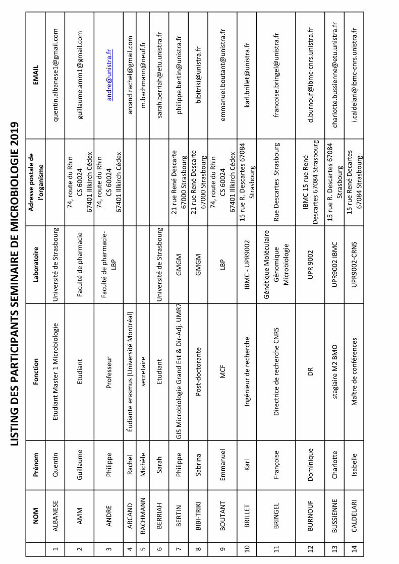
NO
MP
rén
om
Fon
ctio
nLa
bo
rato
ire
Ad
ress
e p
ost
ale
de
l'org
anis
me
EMA
IL
1A
LBA
NES
EQ
uen
tin
Etu
dia
nt
Mas
ter
1 M
icro
bio
logi
eU
niv
ers
ité
de
Stra
sbo
urg
qu
enti
n.a
lban
ese
1@
gmai
l.co
m
2A
MM
Gu
illau
me
Etu
dia
nt
Facu
lté
de
ph
arm
acie
74
, ro
ute
du
Rh
in
CS
60
02
4
67
40
1 Il
lkir
ch C
éd
ex
guill
aum
e.am
m1
@gm
ail.c
om
3A
ND
RE
Ph
ilip
pe
Pro
fess
eu
r Fa
cult
é d
e p
har
mac
ie-
LBP
74
, ro
ute
du
Rh
in
CS
60
02
4
67
40
1 Il
lkir
ch C
éd
ex
and
re@
un
istr
a.fr
4A
RC
AN
DR
ach
elÉu
dia
nte
era
smu
s (U
niv
ers
ité
Mo
ntr
éal)
arca
nd
.rac
hel
@gm
ail.c
om
5B
AC
HM
AN
NM
ich
èle
secr
etai
rem
.bac
hm
ann
@n
euf.
fr
6B
ERR
IAH
Sara
hEt
ud
ian
tU
niv
ers
ité
de
Stra
sbo
urg
sara
h.b
erri
ah@
etu
.un
istr
a.fr
7B
ERTI
NP
hili
pp
eD
ir. G
IS M
icro
bio
logi
e G
ran
d E
st &
Dir
-Ad
j. U
MR
71
56
GM
GM
21
ru
e R
ené
Des
cart
e
67
00
0 S
tras
bo
urg
ph
ilip
pe.
ber
tin
@u
nis
tra.
fr
8B
IBI-
TRIK
ISa
bri
na
Po
st-d
oct
ora
nte
GM
GM
21
ru
e R
ené
Des
cart
e
67
00
0 S
tras
bo
urg
bib
itri
ki@
un
istr
a.fr
9B
OU
TAN
TEm
man
uel
MC
FLB
P
74
, ro
ute
du
Rh
in
CS
60
02
4
67
40
1 Il
lkir
ch C
éd
ex
emm
anu
el.b
ou
tan
t@u
nis
tra.
fr
10
BR
ILLE
TK
arl
Ingé
nie
ur
de
rech
erch
eIB
MC
- U
PR
90
02
15
ru
e R
. Des
cart
es 6
70
84
Stra
sbo
urg
karl
.bri
llet@
un
istr
a.fr
11
BR
ING
ELFr
anço
ise
Dir
ectr
ice
de
rech
erch
e C
NR
S
Gén
étiq
ue
Mo
lécu
lair
e
Gén
om
iqu
e
Mic
rob
iolo
gie
Ru
e D
esca
rtes
Str
asb
ou
rgfr
anco
ise.
bri
nge
l@u
nis
tra.
fr
12
BU
RN
OU
FD
om
iniq
ue
DR
UP
R 9
00
2IB
MC
15
ru
e R
en
é
Des
cart
es 6
70
84
Str
asb
ou
rgd
.bu
rno
uf@
ibm
c-cn
rs.u
nis
tra.
fr
13
BU
SSIE
NN
EC
har
lott
est
agia
ire
M2
BM
OU
PR
90
02
IBM
C1
5 r
ue
R. D
esca
rtes
67
08
4
Stra
sbo
urg
char
lott
e.b
uss
ien
ne@
etu
.un
istr
a.fr
14
CA
LDEL
AR
IIs
abel
leM
aîtr
e d
e co
nfé
ren
ces
UP
R9
00
2-C
RN
S1
5 r
ue
Ren
é D
ecar
tes
67
08
4 S
tras
bo
urg
i.cal
del
ari@
ibm
c-cn
rs.u
nis
tra.
fr
LIST
ING
DES
PA
RTI
CIP
AN
TS S
EMIN
AIR
E D
E M
ICR
OB
IOLO
GIE
20
19

15
CA
ND
OLF
IEr
man
no
Pro
fess
eu
r -
Dir
ecte
ur
IPP
TS
Inst
itu
t d
e p
aras
ito
logi
e
et d
e p
ath
olo
gie
tro
pic
ale
3 r
ue
Ko
eber
lé 6
70
00
stra
sbo
urg
can
do
lfi@
un
istr
a.fr
16
CA
RA
DEC
Cla
ud
iaIE
GM
GM
21
ru
e R
ené
Des
cart
e
67
00
0 S
tras
bo
urg
cara
dec
@ig
bm
c.u
-str
asb
g.fr
17
CH
AM
AR
DJo
eyÉt
ud
ian
tjo
eych
amar
d@
gmai
l.co
m
18
CH
OLL
ETre
nau
dre
spo
nsa
ble
Mic
rob
iolo
gie
et
Mili
eu d
e C
ult
ure
MER
CK
39
Ro
ute
Ind
ust
riel
le d
e la
Har
dt,
67
12
0 M
ols
hei
m
ren
aud
.ch
olle
t@m
erc
kgro
up
.co
m
19
DEB
AN
DE
Lori
ne
Etu
dia
nte
Mas
ter
1 M
icro
bio
logi
eU
niv
ers
ité
de
Stra
sbo
urg
deb
and
e.lo
rin
e@gm
ail.c
om
20
DEM
UTH
Gu
illau
me
Etu
dia
nt
Un
ive
rsit
é d
e St
rasb
ou
rggu
illau
me.
dem
uth
68
@o
ran
ge.f
r
21
DU
PO
ND
Eugé
nie
Etu
dia
nte
Un
ive
rsit
é d
e St
rasb
ou
rgd
up
on
d.e
uge
nie
97
@gm
ail.c
om
22
FAR
JALL
AH
mai
ssen
étu
dia
nte
Un
ive
rsit
é d
e St
rasb
ou
rgm
aiss
en.f
arja
llah
@et
u.u
nis
tra.
fr
23
FOU
GY
Lysi
ane
Ingé
nie
ur
de
rech
erch
eA
eria
l2
50
ru
e La
ure
nt
Frie
s
67
40
0 Il
lkir
chl.f
ou
gy-h
ou
el@
aeri
al-c
rt.c
om
24
FRIA
NT
Sylv
ieD
irec
tric
e d
e R
ech
erch
e
GM
GM
Gé
nét
iqu
e
Mo
lécu
lair
e,
Gén
om
iqu
e,
Mic
rob
iolo
gie
21
ru
e D
esca
rtes
67
00
0 S
tras
bo
urg
s.fr
ian
t@u
nis
tra.
fr
25
FRIE
DR
ICH
An
ne
Ense
ign
ant
cher
cheu
r
Gén
étiq
ue
Mo
lécu
lair
e,
Gén
om
iqu
e,
Mic
rob
iolo
gie
21
ru
e D
esca
rtes
67
00
0 S
tras
bo
urg
ann
e.fr
ied
rich
@u
nis
tra.
fr
26
FRIT
SCH
Sara
hIn
gén
ieu
rU
MR
72
42
Pô
le A
PI,
30
0 B
d S
ébas
tien
Bra
nt,
67
40
0 Il
lkir
ch-
Gra
ffen
stad
en
sara
h.f
rits
ch2
@et
u.u
nis
tra.
fr
27
GA
SSER
Vér
on
iqu
eIn
gén
ieu
re d
'étu
des
UM
R7
24
2
Bio
tech
no
logi
e e
t
sign
alis
atio
n c
ellu
lair
e
ESB
S
30
0 B
ou
leva
rd S
ébas
tien
Bra
nt
CS
10
41
3
FR-6
74
12
ILLK
IRC
H
vero
niq
ue.
gass
er@
un
istr
a.fr

28
GEO
FFR
OY
Val
érie
MC
F
Bio
tech
no
logi
e e
t
sign
alis
atio
n c
ellu
lair
e,
Mét
aux
et m
icro
-
org
anis
mes
: b
iolo
gie,
chim
ie e
t ap
plic
atio
ns
ESB
S 3
00
Bd
Bra
nt
67
41
2
Illki
rch
Ced
exva
leri
e.ge
off
roy@
un
istr
a.fr
29G
ISM
ERO
RO
DR
ÍGU
EZLa
ura
stag
iair
e
Bio
tech
no
logi
e e
t
sign
alis
atio
n c
ellu
lair
e,
Mét
aux
et m
icro
-
org
anis
mes
: b
iolo
gie,
chim
ie e
t ap
plic
atio
ns
ESB
S 3
00
Bd
Bra
nt
67
41
2
Illki
rch
Ced
exla
ura
gis@
ucm
.es
30
GLO
DA
SC
lém
ent
Etu
dia
nt
Mas
ter
2 M
icro
bio
logi
eU
niv
ers
ité
de
Stra
sbo
urg
clem
ent.
glo
das
@gm
ail.c
om
31
GO
DET
Julie
nM
CU
-PH
UM
R 7
02
1jg
od
et@
un
istr
a.fr
32
GR
ENEY
Thie
rry
Étu
dia
nt
à la
fac
ult
é d
e p
har
mac
ie e
n M
1
assu
ran
ce q
ual
ité
Un
ive
rsit
é d
e St
rasb
ou
rgth
ierr
y.gr
eney
@o
ran
ge.f
r
33
GR
ILLO
NA
nto
ine
Pra
tici
en-h
osp
ital
o-U
niv
ers
itai
reLa
bo
rato
ire
de
Bac
téri
olo
gie
1, p
lace
de
l'hô
pit
al, 6
70
00
Stra
sbo
urg
a.gr
illo
n@
un
istr
a.fr
34
GR
UFF
AZ
Ch
rist
elle
Ass
ista
nt-
Ingé
nie
ur
UM
R 7
15
6c.
gru
ffaz
@u
nis
tra.
fr
35
GU
CC
IAR
DI
Flo
rian
eSt
agia
ire
M2
Inst
itu
t d
e V
iro
logi
e
INSE
RM
11
09
3 r
ue
Ko
eber
lé
67
00
0 S
TRA
SBO
UR
Gfl
ori
ane.
gucc
iard
i@et
u.u
nis
tra.
fr
36
HA
AS
Léa
Stag
iair
e M
aste
r
Lab
ora
toir
e d
e
Bio
imag
erie
et
Pat
ho
logi
es U
MR
70
21
CN
RS/
Un
istr
a
Facu
lté
de
Ph
arm
acie
74
ro
ute
du
Rh
in
CS
60
02
4
F-6
74
01
ILLK
IRC
H C
EDEX
lea.
haa
s@et
u.u
nis
tra.
fr
37
HA
NSM
AN
NTr
ista
nEt
ud
ian
t M
aste
r M
icro
bio
logi
eU
niv
ers
ité
de
Stra
sbo
urg
han
sman
ntr
ista
n@
gmai
l.co
m
38
HA
TTEM
ERH
elo
ïse
Etu
dia
nt
M1
AQ
ph
arm
acie
Un
ive
rsit
é d
e St
rasb
ou
rgh
elo
ise.
hat
tem
er@
ou
tlo
ok.
fr
39
HEL
FER
-LE
FOLL
An
ne-
Cat
her
ine
Ingé
nie
ure
d'é
tud
es C
NR
S
CN
RS
UP
R 9
00
2
Arc
hit
ect
ure
et
Réa
ctiv
ité
de
l'AR
N,
IBM
C
15
ru
e R
en
é D
esca
rtes
67
08
4 S
tras
bo
urg
ac.h
elfe
r@ib
mc-
cnrs
.un
istr
a.fr
40
HEN
RY
alex
isEt
ud
ian
t M
aste
r 2
UM
RS
11
09
Dr.
MO
OG
3 r
ue
Ko
eber
le S
tras
bo
urg
67
00
0al
exd
u6
_7@
ho
tmai
l.fr
41
HER
RG
OTT
Luca
sIn
gén
ieu
r d
'étu
de
l.her
rgo
tt@
ibm
c-cn
rs.u
nis
tra.
fr
42
HEZ
AR
Db
ern
ard
resp
on
sab
le la
bo
rato
ire
Aer
ial
25
0 r
ue
lau
ren
t Fr
ies
67
41
2 Il
lkir
ch c
edex
b.h
ezar
d@
aeri
al-c
rt.c
om
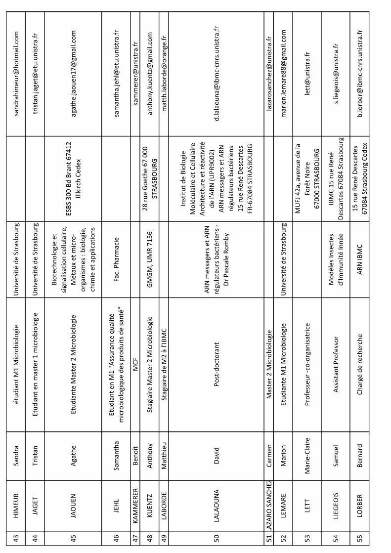
43
HIM
EUR
San
dra
étu
dia
nt
M1
Mic
rob
iolo
gie
Un
ive
rsit
é d
e St
rasb
ou
rgsa
nd
rah
imeu
r@h
otm
ail.c
om
44
JAG
ETTr
ista
nEt
ud
ian
t en
mas
ter
1 m
icro
bio
logi
eU
niv
ers
ité
de
Stra
sbo
urg
tris
tan
.jage
t@et
u.u
nis
tra.
fr
45
JAO
UEN
Aga
the
Etu
dia
nte
Mas
ter
2 M
icro
bio
logi
e
Bio
tech
no
logi
e e
t
sign
alis
atio
n c
ellu
lair
e,
Mét
aux
et m
icro
-
org
anis
mes
: b
iolo
gie,
chim
ie e
t ap
plic
atio
ns
ESB
S 3
00
Bd
Bra
nt
67
41
2
Illki
rch
Ced
exag
ath
e.ja
ou
en1
7@
gmai
l.co
m
46
JEH
LSa
man
tha
Etu
dia
nt
en M
1 "
Ass
ura
nce
qu
alit
é
mic
rob
iolo
giq
ue
des
pro
du
its
de
san
té"
Fac.
Ph
arm
acie
sam
anth
a.je
hl@
etu
.un
istr
a.fr
47
KA
MM
ERER
Ben
oît
MC
Fka
mm
erer
@u
nis
tra.
fr
48
KU
ENTZ
An
tho
ny
Stag
iair
e M
aste
r 2
Mic
rob
iolo
gie
GM
GM
, UM
R 7
15
62
8 r
ue
Go
eth
e 6
7 0
00
STR
ASB
OU
RG
anth
on
y.ku
entz
@gm
ail.c
om
49
LAB
OR
DE
Mat
thie
uSt
agia
ire
de
M2
à l'
IBM
Cm
atth
.lab
ord
e@o
ran
ge.f
r
50
LALA
OU
NA
Dav
idP
ost
-do
cto
ran
t
AR
N m
essa
gers
et
AR
N
régu
late
urs
bac
téri
ens
-
Dr
Pas
cale
Ro
mb
y
Inst
itu
t d
e B
iolo
gie
Mo
lécu
lair
e et
Ce
llula
ire
Arc
hit
ect
ure
et
réac
tivi
té
de
l'AR
N (
UP
R9
00
2)
AR
N m
essa
gers
et
AR
N
régu
late
urs
bac
téri
ens
15
ru
e R
ené
Des
cart
es
FR-6
70
84
STR
ASB
OU
RG
d.la
lao
un
a@ib
mc-
cnrs
.un
istr
a.fr
51
LAZA
RO
SA
NC
HEZ
Car
men
Mas
ter
2 M
icro
bio
logi
ela
zaro
san
chez
@u
nis
tra.
fr
52
LEM
AR
EM
ario
nEt
ud
ian
te M
1 M
icro
bio
logi
eU
niv
ers
ité
de
Stra
sbo
urg
mar
ion
.lem
are8
8@
gmai
l.co
m
53
LETT
Mar
ie-C
lair
eP
rofe
sse
ur
-co
-org
anis
atri
ce
MU
FJ 4
2a,
ave
nu
e d
e la
Forê
t N
oir
e
67
00
0 S
TRA
SBO
UR
G
lett
@u
nis
tra.
fr
54
LIEG
EOIS
Sam
uel
Ass
ista
nt
Pro
fess
or
Mo
dèl
es In
sect
es
d'Im
mu
nit
é In
née
IBM
C 1
5 r
ue
Re
né
Des
cart
es 6
70
84
Str
asb
ou
rgs.
liege
ois
@u
nis
tra.
fr
55
LOR
BER
Ber
nar
dC
har
gé d
e re
cher
che
AR
N IB
MC
15
ru
e R
ené
Des
cart
es
67
08
4 S
tras
bo
urg
Ce
dex
b.lo
rber
@ib
mc-
cnrs
.un
istr
a.fr

56
LOU
VEA
UA
naï
sEt
ud
ian
t m
aste
r 1
"as
sura
nce
qu
alit
é
mic
rob
iolo
giq
ue
des
pro
du
its
de
san
té"
Fac.
Ph
arm
acie
anai
s.lo
uve
au@
ora
nge
.fr
57
MA
AZ
BIN
Nas
imEt
ud
ian
t LB
PFa
cult
é d
e P
har
mac
ied
r.m
aaz_
nas
im@
ho
tmai
l.co
m
58
MA
HM
OU
DI-
KA
IDI
Nas
sira
Ch
erch
eur
Po
st-d
oct
ora
nt
UM
R5
67
1 G
MG
M
UM
R7
15
6 G
MG
M IP
CB
-
Pla
tefo
rme
de
bio
logi
e
67
00
0 S
tras
bo
urg
nm
ahm
ou
dik
aid
i@u
nis
tra.
fr
59
MA
LHER
BE
Lud
ivin
ete
chn
icie
nn
eG
MG
M U
MR
71
56
4 a
llée
Ko
nra
d R
oen
tgen
lmal
her
be@
un
istr
a.fr
60
MA
LSA
Josh
ua
Stag
iair
e M
aste
r 2
Mic
rob
iolo
gie
CN
RS
UM
R 7
24
2
ESB
S
30
0 B
d S
ébas
tien
Bra
nt
CS
10
41
3
67
41
2 IL
LKIR
CH
ced
ex
josm
al@
ho
tmai
l.fr
61
MA
NC
INEL
LIA
del
ine
Étu
dia
nte
M1
Man
cin
elli.
adel
ine@
gmai
l.co
m
62
MA
RTI
NIs
abel
leC
her
che
un
co
ntr
at p
ost
-do
cto
ral
m.iz
a@liv
e.fr
63
MA
RTI
N
Will
iam
Dr.
Inst
itu
te o
f m
ole
cula
r
evo
luti
on
, Hei
nri
ch-
Hei
ne-
Un
ive
rsit
ät
Du
ssel
do
rf, G
erm
any
bill
@h
hu
.de
64
MA
SQU
IDA
Ben
oît
Ch
erch
eur
CN
RS
GM
GM
UM
R 7
15
62
1 r
ue
Ren
é D
esca
rtes
67
08
4 S
tras
bo
urg
b.m
asq
uid
a
65
MA
THIE
USé
bas
tien
Mas
ter
2 e
n In
gén
ieri
e d
e l'U
niv
ers
ité
de
Lorr
ain
e C
NR
S U
MR
72
42
ESB
S
30
0 B
d S
ébas
tien
Bra
nt
CS
10
41
3
67
41
2 IL
LKIR
CH
ced
ex
seb
asti
en.m
ath
ieu
1@
etu
.un
iv-l
orr
ain
e.fr
66
MER
CIE
RN
oém
ieSt
agia
ire
M2
Mic
rob
iolo
gie
IBM
C5
Ru
e R
ené
Des
cart
es,
67
00
0 S
tras
bo
urg
mer
cier
.no
emie
@o
ran
ge.f
r
67
MEY
Lou
is-F
ran
çois
Stag
iair
e M
2 M
icro
bio
logi
eU
niv
ers
ité
de
Stra
sbo
urg
mey
lou
isf@
ho
tmai
l.fr
68
MEY
ERG
aëlle
Etu
dia
nt
M1
Ass
ura
nce
qu
alit
é
mic
rob
iolo
giq
ue
des
pro
du
its
de
san
téFa
c. P
har
mac
iem
.gae
lle2
@gm
ail.c
om
69
MO
OG
Ch
rist
ian
eD
Rc.
mo
og@
un
istr
a.fr
70
MO
RA
NJo
sep
hP
RIS
ISm
ora
n@
un
istr
a.fr
71
NA
DA
LIG
Thie
rry
Maî
tre
de
Co
nfé
ren
ces
GM
GM
UM
R 7
15
62
1 r
ue
Ren
é D
esca
rtes
67
08
4 S
tras
bo
urg
nad
alig
@u
nis
tra.
fr
72
NO
RM
AN
TV
ince
nt
po
st-d
oct
ora
nt
Mét
aux
et
mic
roo
rgan
ism
es :
bio
logi
e, c
him
ie e
t
app
licat
ion
s
ESB
S
30
0 B
ou
leva
rd S
ébas
tien
Bra
nt
CS
10
41
3
67
41
2 IL
LKIR
CH
no
rman
t@u
nis
tra.
fr
73
NO
VIK
OV
AO
xan
aEt
ud
ian
teo
xan
a.n
ovi
kova
@et
u.u
nis
tra.
fr
74
OLI
VA
RES
élo
die
Ch
ef d
e p
roje
t R
&D
EA7
29
0 (
Bio
film
Ph
arm
a)o
livar
es.e
lod
ie@
gmai
l.co
m
75
PA
HO
Nvi
cto
rét
ud
ian
t m
aste
r 1
Mic
rob
iolo
gie
Un
ive
rsit
é d
e St
rasb
ou
rgvi
cto
r.p
aho
n@
gmai
l.co
m
76
PIE
RR
AT
No
émie
Etu
dia
nt
Mic
rob
iolo
gie
Un
ive
rsit
é d
e St
rasb
ou
rgn
oem
ie.p
ierr
at@
etu
.un
istr
a.fr
77
PLO
ETZE
Flo
ren
ceM
CF
IBM
P-U
PR
23
57
12
ave
nu
e G
ener
al Z
imm
erp
loet
ze@
un
istr
a.fr
78
RA
EHM
Am
élie
Etu
dia
nt
Facu
lté
de
Ph
arm
acie
de
Stra
sbo
urg
74
Ro
ute
du
Rh
in, 6
74
00
Illki
rch
-Gra
ffen
stad
enam
elie
.rae
hm
@gm
ail.c
om
79
RIG
OU
INco
ralin
eM
aîtr
e d
e co
nfé
ren
ces
Bio
tech
no
logi
e e
t
sign
alis
atio
n c
ellu
lair
e
30
0 B
vd S
ébas
tien
Bra
nt,
CS1
04
13
.
67
41
2, I
llkir
ch
FRA
NC
E
rigo
uin
@u
nis
tra.
fr
80
RIN
ALD
IB
run
oIn
gén
ieu
rC
NR
S -
UM
R7
15
64
allé
e K
on
rad
Ro
etge
n
67
00
0 S
tras
bo
urg
b.r
inal
di@
un
istr
a.fr
81
RO
BER
TLy
dia
Dr
Sorb
on
ne
Un
ive
rsit
éLa
bo
rato
ire
Jean
Per
rin
lyd
ia.r
ob
ert@
up
mc.
fr
82
RO
MB
YP
asca
leD
U U
PR
90
02
- R
esp
on
sab
le d
e l'é
qu
ipe
AR
Nm
et
AR
N r
égu
late
urs
bac
téri
ens
Arc
hit
ect
ure
et
Réa
ctiv
ité
de
l'AR
N-
UP
R
90
02
IBM
C 1
5 R
ue
Ren
é
Des
cart
es 6
70
84
Str
asb
ou
rgp
.ro
mb
y@ib
mc-
cnrs
.un
istr
a.fr
83
SCH
ALK
Isab
elle
Dir
ectr
ice
de
Rec
her
che
CN
RS
UM
R7
24
2
UM
R 7
24
2, B
iote
chn
olo
gie
et S
ign
alis
atio
n C
ellu
lair
e
ESB
S
30
0, B
d S
ebas
tien
Bra
nt
CS
10
41
3
F67
41
2 Il
lkir
ch C
edex
isab
elle
.sch
alk@
un
istr
a.fr
84
TOU
BO
NG
aël
Etu
dia
nt
stag
iair
eIP
PTS
tou
bo
nga
el@
ho
tmai
l.co
m
85
VER
RIE
ZC
édri
cEt
ud
ian
t M
2IB
MC
- U
PR
90
02
15
Ru
e R
ené
Des
cart
es,
67
00
0 S
tras
bo
urg
ced
ric.
verr
iez@
wan
ado
o.f
r
86
VU
ILLE
UM
IER
Stép
han
eP
RG
MG
M U
MR
71
56
vuill
eum
ier@
un
istr
a.fr
87
WA
ECK
ERLE
Loïc
Mas
ter
2 M
icro
bio
logi
eP
lan
t is
op
ren
oid
Bio
logy
,
IBM
P-C
NR
S
12
ru
e d
u G
én
éral
Zim
mer
,
67
08
4 S
tras
bo
urg
loic
.wae
cker
le@
etu
.un
istr
a.fr
88
WA
GN
ERJe
rom
eC
RU
MR
72
42
ESB
S -
Pô
le A
PI
30
0, B
ou
leva
rd S
ébas
tien
Bra
nt
BP
10
41
3
67
41
2 IL
LKIR
CH
CED
EX
j.wag
ner
@u
nis
tra.
fr

89
WA
GN
ERR
enau
dIn
gén
ieu
r d
e re
cher
che,
res
po
nsa
ble
de
la p
late
form
e IM
PR
eSsU
MR
72
42
,
Bio
tech
no
logi
e e
t
Sign
alis
atio
n C
ellu
lair
e
ESB
S -
Pô
le A
PI
30
0, B
ou
leva
rd S
ébas
tien
Bra
nt
BP
10
41
3
67
41
2 IL
LKIR
CH
CED
EX
ren
aud
.wag
ner
@u
nis
tra.
fr
90
WIS
SLu
cille
Etu
dia
nt
Mas
ter
2U
MR
_S1
10
9 D
r
Ch
rist
ian
e M
OO
G
Inst
itu
t d
e V
iro
logi
e 3
ru
e
Ko
eber
lé 6
70
00
Str
asb
ou
rg,
Fran
ce
luci
lle.w
iss@
etu
.un
istr
a.fr
91
ZIN
E FI
LALI
No
uh
aM
aste
r 2
Mic
rob
iolo
gie
no
uh
afila
li21
@h
otm
ail.f
r
92
AN
TOIN
ELa
ura
do
cto
ran
tl.a
nto
ine@
ibm
c-cn
rs.u
nis
tra.
fr
93
AR
NO
UX
Lyd
iaTe
chn
icie
nn
eIn
stit
ut
de
Par
asit
olo
gie
3, r
ue
Ko
eber
lé 6
70
00
Stra
sbo
urg
lyd
ia.a
rno
ux@
un
istr
a.fr
94
BEN
OTM
AN
EIli
esD
oct
ora
nt
UM
R 1
10
9 /
Vir
olo
gie
3 r
ue
Ko
eber
le
67
00
0 S
tras
bo
urg
i.ben
otm
ane@
etu
.un
istr
a.fr
95
BIT
TIC
HFA
IZA
do
cto
ran
teIP
PTS
3 r
ue
Ko
eber
lé 6
70
00
Stra
sbo
urg
fa.f
aiza
60
@gm
ail.c
om
96
BO
NN
EAU
An
ne
Ph
DU
MR
72
42
ESB
S -
Pô
le A
PI
30
0, B
ou
leva
rd S
ébas
tien
Bra
nt
BP
10
41
3
67
41
2 IL
LKIR
CH
CED
EX
ann
e.b
on
nea
u@
etu
.un
istr
a.fr
97
BU
GU
ENO
Isaa
c
Max
imili
ano
DO
CTO
RA
NT
INSE
RM
UM
R 1
26
06
70
00
max
imili
ano
_1_1
6@
ho
tmai
l.co
m
98
CA
RA
VEL
LOG
aëta
nD
oct
ora
nt
UP
R 9
02
2 IB
MC
15
Ru
e R
ené
Des
cart
esca
rave
llo.g
aeta
n@
ho
tmai
l.co
m
99
CO
UR
TÈS
Mar
jori
eP
hD
Stu
den
tLN
CA
23
ru
e d
u L
oes
s
Stra
sbo
urg
mco
urt
es@
un
istr
a.fr
10
0D
AV
IDSé
bas
tien
Do
cto
ran
t
BSC
éq
uip
e m
étau
x et
mic
roo
rgan
ism
es:
bio
logi
e, c
him
ie e
t
app
licat
ion
s
ESB
S
30
0 B
d S
ébas
tien
Bra
nt
CS
10
41
3
67
41
2 IL
LKIR
CH
ced
ex
seb
asti
en.d
avid
@et
u.u
nis
tra.
fr
10
1D
ESG
RA
NG
ESEm
ma
Do
cto
ran
tIB
MC
15
Ru
e R
ené
Des
cart
esd
esgr
ange
sem
ma@
gmai
l.co
m
10
2FE
RH
ATI
Ferr
ou
dja
Etu
dia
nte
ferh
ati.f
erro
ud
ja@
gmai
l.co
m
10
3G
ALL
AIS
Flo
rian
eD
oct
ora
nte
1e
ann
ée /
Pra
tici
en H
osp
ital
ier
Co
ntr
actu
el e
n B
iolo
gie
Méd
ical
e
Inst
itu
t d
e V
iro
logi
e,
INSE
RM
11
09
, Un
iver
sité
de
Stra
sbo
urg
Inst
itu
t d
e V
iro
logi
e
3 R
ue
Koeb
erlé
67
00
0 S
tras
bo
urg
galla
is.f
lori
ane@
gmai
l.co
m
10
4G
EER
SEN
SEm
ilie
do
cto
ran
tIB
MC
15
Ru
e R
ené
Des
cart
esem
ilie.
gee
rsen
s@et
u.u
nis
tra.
fr
10
5G
RA
ULI
ERG
we
naë
lleD
oct
ora
nte
CN
RS-
UM
R7
24
2 B
SC
ESB
S
30
0 B
d S
ébas
tien
Bra
nt
CS
10
41
3
67
41
2 IL
LKIR
CH
ced
ex
g.gr
aulie
r@u
nis
tra.
fr
10
6G
RO
HC
hlo
éP
hD
stu
den
tIn
stit
ut
de
Bio
logi
e
Mo
lécu
lair
e d
es P
lan
tes
12
ru
e d
u G
én
éral
Zim
mer
67
00
0 S
TRA
SBO
UR
Gch
loe.
gro
h@
etu
.un
istr
a.fr
10
7H
EMM
ERLE
Mar
ine
Do
cto
ran
teU
MR
71
56
21
Ru
e R
ené
Des
cart
es,
67
00
0 S
tras
bo
urg
mar
ine.
hem
mer
le@
etu
.un
istr
a.fr
10
8H
EYD
MA
NN
Lau
raA
ssis
tan
te in
gén
ieu
r, P
HD
stu
den
tU
MR
_11
10
3 r
ue
Ko
eber
lé
67
00
0 S
tras
bo
urg
hey
dm
ann
@u
nis
tra.
fr
10
9K
LIN
GLE
RJé
rom
ine
Etu
dia
nte
en
th
èse
INSE
RM
UM
R_S
11
09
3 r
ue
Ko
eber
lé 6
70
00
Stra
sbo
urg
jero
min
e.kl
ingl
er@
gmai
l.co
m
11
0K
RU
PA
Eva
Do
cto
ran
te
Lab
ora
toir
e
d'e
nto
mo
logi
e m
édic
ale
-
IPP
TS
Lab
ora
toir
e d
'en
tom
olo
gie
méd
ical
e
Inst
itu
t d
e P
aras
ito
logi
e e
t
de
Pat
ho
logi
e Tr
op
ical
e d
e
Stra
sbo
urg
3 r
ue
Ko
eber
lé
67
00
0 S
TRA
SBO
UR
G
e.kr
up
a@u
nis
tra.
fr
11
1LA
NG
Céc
ileIn
gén
ieu
r d
'étu
de
IPP
TS3
ru
e K
oeb
erlé
ceci
le.la
ng@
un
istr
a.fr
11
2LE
FEU
VR
EB
asti
enD
oct
ora
nt
bas
tien
.lefe
uvr
e@
etu
.un
istr
a.fr
11
3LO
BST
EIN
An
ne-
Mar
ieD
oct
ora
nte
IBM
C1
5, r
ue
Re
né
Des
cart
esan
ne-
mar
ie.lo
bst
ein
@et
u.u
nis
tra.
fr
11
4LU
OJi
ng
Co
mm
un
icat
ion
jing.
luo
@et
u.u
nis
tra.
fr
11
5M
AZZ
OLE
NI
Vio
laD
oct
ora
nte
EA7
29
0-V
iru
len
ce
bac
téri
enn
e p
réco
ce :
fon
ctio
ns
cellu
lair
es e
t
con
trô
le d
e l'i
nfe
ctio
n
aigu
ë et
su
bai
guë
Inst
itu
t d
e b
acté
rio
logi
e
3 r
ue
Ko
eber
lé 6
70
00
Stra
sbo
urg
vio
la.m
azzo
len
i@et
u.u
nis
tra.
fr
11
6P
ERR
AU
DQ
uen
tin
Do
cto
ran
t
UM
R 7
24
2 -
Eq
uip
e
mét
aux
et
mic
roo
rgan
ism
es :
bio
logi
e, c
him
ie e
t
app
licat
ion
s
ESB
S
30
0 B
d S
ébas
tien
Bra
nt
CS
10
41
3
67
41
2 IL
LKIR
CH
ced
ex
q.p
erra
ud
@u
nis
tra.
fr
11
7P
RO
LAke
vin
Do
cto
ran
t (2
ème
ann
ée)
EA 7
29
0 :
Vir
ule
nce
Bac
téri
enn
e P
réco
ce
Inst
itu
t d
e B
acté
rio
logi
e
3 R
ue
Koeb
erlé
67
00
0 S
tras
bo
urg
kpro
la@
un
istr
a.fr
11
8R
AB
EHR
ou
kia
étu
dia
nt
rou
kia.
rab
eh@
etu
.un
istr
a.fr
11
9R
ENA
RD
Eva
Do
cto
ran
te
IBM
C -
Arc
hit
ect
ure
et
Réa
ctiv
ité
de
l'arN
(A
RN
UP
R9
00
2, d
irec
tric
e :
Pas
cale
Ro
mb
y) -
Eq
uip
e
"Str
uct
ure
et
dyn
amiq
ue
des
mac
hin
es
bio
mo
lécu
lair
es"
(dir
ecte
ur
: Eri
c En
nif
ar)
15
, ru
e R
en
é D
esca
rtes
e.re
nar
d@
ibm
c-cn
rs.u
nis
tra.
fr
12
0W
END
ENB
AU
MM
ario
nP
hD
Stu
den
tU
MR
71
56
IPC
B D
YPS
mar
ion
_wen
d@
ho
tmai
l.fr
CO
NT
AC
T
Mar
ie-L
uce
PA
LEN
NE
Lau
ren
t B
OTT
NER
Ren
aud
CH
OLL
ET
An
ne-
sop
hie
TO
RTR
OTA
U
Cat
her
ine
SCH
UST
ER
Jack
y D
e M
ON
TIG
NY
Jean
-Pie
rre
GIE
SS
Siam
ak B
AH
RA
M
EMA
IL
CA
SDEN
BA
NQ
UE
PO
PU
LAIR
Em
arie
-Lu
ce.P
ALE
NN
E@ca
sden
.ban
qu
epo
pu
lair
e.fr
FAC
ULT
E D
E M
EDEC
INE
siam
ak@
un
istr
a.fr
ECO
LE D
OC
TOR
ALE
VIE
ET
SAN
TEca
ther
ine.
sch
ust
er@
un
istr
a.fr
FAC
ULT
E D
ES S
CIE
NC
ES D
E LA
VIE
mo
nti
gny@
un
istr
a.fr
FAC
ULT
E D
E P
HA
RM
AC
IEje
an-p
ierr
e.gi
ess@
un
istr
a.fr
CR
YOTE
Cco
nta
ct@
cryo
tec.
fr
MER
CK
ren
aud
.ch
ole
t@m
erck
gro
up
.co
m
MP
BIO
MED
ICA
LSan
ne-
sop
hie
.to
rtro
tau
@m
pb
io.c
om
PA
RT
ENA
IRES