Simultaneous 13N-Ammonia and gadolinium first-pass ...
14
ORIGINAL ARTICLE Open Access Simultaneous 13 N-Ammonia and gadolinium first-pass myocardial perfusion with quantitative hybrid PET-MR imaging: a phantom and clinical feasibility study Muhummad Sohaib Nazir 1 , Sarah-May Gould 1 , Xenios Milidonis 1 , Eliana Reyes 1 , Tevfik F. Ismail 1 , Radhouene Neji 1,2 , Sébastien Roujol 1 , Jim O’Doherty 1 , Hui Xue 3 , Sally F. Barrington 1 , Tobias Schaeffter 1,4 , Reza Razavi 1 , Paul Marsden 1 , Peter Kellman 3 , Sven Plein 1,5 and Amedeo Chiribiri 1* * Correspondence: amedeo. [email protected] 1 Department of Cardiovascular Imaging, School of Biomedical Engineering and Imaging Sciences, King’s College London, St Thomas’ Hospital, Westminster Bridge, 4th Floor Lambeth Wing, London SE1 7EH, UK Full list of author information is available at the end of the article Abstract Background: Positron emission tomography (PET) is the non-invasive reference standard for myocardial blood flow (MBF) quantification. Hybrid PET-MR allows simultaneous PET and cardiac magnetic resonance (CMR) acquisition under identical experimental and physiological conditions. This study aimed to determine feasibility of simultaneous 13 N-Ammonia PET and dynamic contrast-enhanced CMR MBF quantification in phantoms and healthy volunteers. Methods: Images were acquired using a 3T hybrid PET-MR scanner. Phantom study: MBF was simulated at different physiological perfusion rates and a protocol for simultaneous PET-MR perfusion imaging was developed. Volunteer study: five healthy volunteers underwent adenosine stress. 13 N-Ammonia and gadolinium were administered simultaneously. PET list mode data was reconstructed using ordered subset expectation maximisation. CMR MBF was quantified using Fermi function-constrained deconvolution of arterial input function and myocardial signal. PET MBF was obtained using a one-tissue compartment model and image-derived input function. Results: Phantom study: PET and CMR MBF measurements demonstrated high repeatability with intraclass coefficients 0.98 and 0.99, respectively. There was high correlation between PET and CMR MBF (r = 0.98, p < 0.001) and good agreement (bias - 0.85 mL/g/min; 95% limits of agreement 0.29 to - 1.98). Volunteer study: Mean global stress MBF for CMR and PET were 2.58 ± 0.11 and 2.60 ± 0.47 mL/g/min respectively. On a per territory basis, there was moderate correlation (r = 0.63, p = 0.03) and agreement (bias - 0.34 mL/g/min; 95% limits of agreement 0.49 to - 1.18). Conclusion: Simultaneous MBF quantification using hybrid PET-MR imaging is feasible with high test repeatability and good to moderate agreement between PET and CMR. Future studies in coronary artery disease patients may allow cross-validation of techniques. Keywords: Hybrid imaging, PET-MR, Myocardial perfusion, Myocardial blood flow © The Author(s). 2019 Open Access This article is distributed under the terms of the Creative Commons Attribution 4.0 International License (http://creativecommons.org/licenses/by/4.0/), which permits unrestricted use, distribution, and reproduction in any medium, provided you give appropriate credit to the original author(s) and the source, provide a link to the Creative Commons license, and indicate if changes were made. European Journal of Hybrid Imaging Nazir et al. European Journal of Hybrid Imaging (2019) 3:15 https://doi.org/10.1186/s41824-019-0062-6
Transcript of Simultaneous 13N-Ammonia and gadolinium first-pass ...

ORIGINAL ARTICLE Open Access
Simultaneous 13N-Ammonia andgadolinium first-pass myocardial perfusionwith quantitative hybrid PET-MR imaging: aphantom and clinical feasibility studyMuhummad Sohaib Nazir1 , Sarah-May Gould1, Xenios Milidonis1, Eliana Reyes1, Tevfik F. Ismail1,Radhouene Neji1,2, Sébastien Roujol1, Jim O’Doherty1, Hui Xue3, Sally F. Barrington1, Tobias Schaeffter1,4,Reza Razavi1, Paul Marsden1, Peter Kellman3, Sven Plein1,5 and Amedeo Chiribiri1*
* Correspondence: [email protected] of CardiovascularImaging, School of BiomedicalEngineering and Imaging Sciences,King’s College London, St Thomas’Hospital, Westminster Bridge, 4thFloor Lambeth Wing, London SE17EH, UKFull list of author information isavailable at the end of the article
Abstract
Background: Positron emission tomography (PET) is the non-invasive referencestandard for myocardial blood flow (MBF) quantification. Hybrid PET-MR allowssimultaneous PET and cardiac magnetic resonance (CMR) acquisition under identicalexperimental and physiological conditions. This study aimed to determine feasibilityof simultaneous 13N-Ammonia PET and dynamic contrast-enhanced CMR MBFquantification in phantoms and healthy volunteers.
Methods: Images were acquired using a 3T hybrid PET-MR scanner. Phantom study:MBF was simulated at different physiological perfusion rates and a protocol forsimultaneous PET-MR perfusion imaging was developed. Volunteer study: fivehealthy volunteers underwent adenosine stress. 13N-Ammonia and gadoliniumwere administered simultaneously. PET list mode data was reconstructed usingordered subset expectation maximisation. CMR MBF was quantified using Fermifunction-constrained deconvolution of arterial input function and myocardialsignal. PET MBF was obtained using a one-tissue compartment model andimage-derived input function.
Results: Phantom study: PET and CMR MBF measurements demonstrated highrepeatability with intraclass coefficients 0.98 and 0.99, respectively. There was highcorrelation between PET and CMR MBF (r = 0.98, p < 0.001) and good agreement (bias− 0.85 mL/g/min; 95% limits of agreement 0.29 to − 1.98). Volunteer study: Mean globalstress MBF for CMR and PET were 2.58 ± 0.11 and 2.60 ± 0.47 mL/g/min respectively. Ona per territory basis, there was moderate correlation (r = 0.63, p = 0.03) and agreement(bias − 0.34 mL/g/min; 95% limits of agreement 0.49 to − 1.18).
Conclusion: Simultaneous MBF quantification using hybrid PET-MR imaging is feasiblewith high test repeatability and good to moderate agreement between PET and CMR.Future studies in coronary artery disease patients may allow cross-validation oftechniques.
Keywords: Hybrid imaging, PET-MR, Myocardial perfusion, Myocardial blood flow
© The Author(s). 2019 Open Access This article is distributed under the terms of the Creative Commons Attribution 4.0 InternationalLicense (http://creativecommons.org/licenses/by/4.0/), which permits unrestricted use, distribution, and reproduction in any medium,provided you give appropriate credit to the original author(s) and the source, provide a link to the Creative Commons license, andindicate if changes were made.
European Journal ofHybrid Imaging
Nazir et al. European Journal of Hybrid Imaging (2019) 3:15 https://doi.org/10.1186/s41824-019-0062-6

BackgroundPositron emission tomography (PET) is considered the non-invasive reference standard
for the quantification of absolute myocardial blood flow (MBF) (Bratis et al. 2013).
Quantification with PET has proven diagnostic (Farhad et al. 2013) and prognostic
value in patients with coronary artery disease (CAD) (Herzog et al. 2009). Quantifica-
tion of MBF is also feasible with CMR (Jerosch-Herold 2010), although it often involves
time-consuming and expert post-processing analysis that has hampered widespread up-
take in clinical routine. Fully automated methods for CMR-derived MBF quantification
with free breathing, motion corrected methods are now available and have been tested
in healthy volunteers with good reproducibility (Brown et al. 2018) and agreement with13N-Ammonia PET (Engblom et al. 2017). In recent observational studies, fully auto-
mated CMR pixel-wise MBF measurements showed good diagnostic accuracy in pa-
tients with suspected CAD (Hsu et al. 2018; Knott et al. 2019; Kotecha et al. 2019).
Previous studies that have compared MBF quantification between CMR and PET
have focussed on separate assessments of PET and CMR performed on common groups
of subjects but separated in time and thus being subject to intrasubject variation (Mor-
ton et al. 2012a; Miller et al. 2014; Fritz-Hansen et al. 2008; Parkka et al. 2006). The lar-
gest comparative study showed good agreement for myocardial perfusion reserve (ratio
of stress MBF:rest MBF) between PET and CMR, but poor agreement for absolute
MBF values (Morton et al. 2012b). Prior to adoption of quantitative CMR MBF in clin-
ical practice, it is important to understand the causes for discrepant findings. The inter-
study variation for stress MBF has been reported to be 19% with PET (Kitkungvan et
al. 2017) and 12–40% with CMR (Brown et al. 2018; Larghat et al. 2013). Hence, it is
unknown how much variation relates to intrinsic differences in modelling, imaging and
tracer kinetics or due to biological variation. Intrasubject biological variation between
PET and MR measurements can be avoided by performing simultaneous PET and CMR
imaging that provides an opportunity to ascertain the intrinsic magnitude of differences
in MBF measurements without physiological variation (Nazir et al. 2018).
The purpose of this study was to demonstrate the feasibility of simultaneous CMR
and PET perfusion quantification with hybrid PET-MR imaging under identical experi-
mental and physiological conditions ex vivo and in vivo.
MethodsAll data were acquired on a 3T hybrid PET-MR scanner (Biograph mMR, Siemens
Healthcare, Erlangen, Germany) with integrated PET and MRI systems housed in the
same gantry. CMR data were acquired with a flexible anterior 6-channel body transmis-
sion coil arrayed receiver, and posterior 6-channel spine arrayed receiver mounted in
the scanner table.
Phantom study
We have previously described a physical perfusion phantom that mimics the cardiac
chambers, great thoracic vessels and myocardium, making it possible to simulate first-
pass myocardial perfusion with high reproducibility (Chiribiri et al. 2013). In
this current study, a modified, 3D-printed version of the phantom has been used, which
has the potential to allow widespread validation and calibration across sites and
Nazir et al. European Journal of Hybrid Imaging (2019) 3:15 Page 2 of 14

different imaging modalities. Furthermore, preclinical evaluation of methodology and
quantification algorithms is possible in a controlled environment devoid of respiratory
or motion artefact. Details of the perfusion phantom are provided in Additional file 1.
Using a fixed cardiac output and heart rate of 60 beats per minute, a range of
myocardial perfusion rates were generated from 1 to 5 mL/g/min in 1 mL/g/min
increments. Radiotracer and gadolinium contrast were administered directly into
the inferior vena cava of the perfusion phantom via a power injector (Spectris
Solaris, Medrad®, Bayer AG) at a rate of 4 mL/s.
After completion of each experimental run, flow rates of the phantom were in-
creased to the maximum level for a minimum of 5 min in order to allow
complete washout of residual 13N-Ammonia and gadolinium contrast from the
entire phantom circuitry. As such, steady and repeatable experimental conditions
were created. Knowledge of the dispersion volume at site of sampling of the tis-
sue compartment allowed MBF values to be derived in units of mL/g/min for
measurements made with both modalities.
Gadobutrol (Gadovist, Bayer, Germany) contrast was administered as a dual
bolus with dose 0.0075 mmol/kg (dilute) + 0.075 mmol/kg (neat) as previously de-
scribed (Ishida et al. 2011; Hussain et al. 2012). The predilute bolus allows the
determination of the arterial input function (AIF), avoiding signal saturation ef-
fects (Jerosch-Herold 2010). A 120-s delay between the dilute and neat bolus of
contrast allowed for complete washout of the predilute gadolinium contrast from
the phantom circuitry before the arrival of the neat bolus of contrast agent.
Then, 200 MBq 13N-Ammonia was administered at the same time as the neat
bolus gadolinium contrast through the same intravenous line after preloading into
a long line to accommodate both PET tracer and gadolinium contrast. The whole
PET-MR perfusion measurement procedure was repeated once in order to deter-
mine its repeatability. The protocol for the phantom study is illustrated in Fig. 1.
CMR perfusion data were acquired using a saturation recovery sequence Fast
Low Angle Shot (FLASH) imaging readout over 240 dynamics. CMR parameters
were FOV 360 × 270 mm, pixel size 2.3 × 1.9 mm, slice thickness 8 mm, TI
100 ms, TR 1.9 ms, TE 0.99 ms, flip angle 10°, GRAPPA acceleration factor 2,
bandwidth 1002 Hz/px. PET data were acquired in 3D list mode for 7 min follow-
ing radiotracer administration. A 3D T1-weighted Dixon sequence was acquired
prior to each PET acquisition and used to generate an attenuation correction (AC)
map required to generate attenuation corrected PET data. PET list mode data were
binned into 60 × 3 s followed by 8 × 15 s consecutive time frames for subsequent
kinetic analysis. All PET frames were reconstructed using the ordered subsets ex-
pectation maximization (OSEM) algorithm with two iterations and 21 subsets using
a 4-mm smoothing filter and matrix size of 344 × 344.
Image analysis and perfusion quantification
CMR dynamic perfusion data were analysed in Osirix (OsiriX 64-bit, version 8.0.2, Pix-
meo SARL, Geneva, Switzerland). A circular region of interest (ROI) with diameter
16 mm was placed around the aortic vessel in order to derive the AIF, and another ROI
was placed around the myocardium compartment of the phantom (diameter 40 mm) in
Nazir et al. European Journal of Hybrid Imaging (2019) 3:15 Page 3 of 14

the same plane at a specified slice position of the myocardium with known volume of
dispersion.
CMR-derived quantitative MBF values were quantified using Fermi-constrained de-
convolution of the AIF and the myocardial signal intensity as previously described (Jer-
osch-Herold 2010; Jerosch-Herold et al. 1998), using in house software on Matlab
(MathWorks®, Natick, Massachusetts, USA).
Precise co-registration of PET and MR images was achieved by dedicated fusion soft-
ware—PMOD (v3.7, PMOD Technologies, Zurich, Switzerland). ROIs were placed over
the aorta for determination of the arterial input function and the myocardium.
PET-derived quantitative MBF values were calculated using a well-known simple
one-tissue compartment model described by De Grado et al. (1996) that was applied to
both phantom and human studies using PMOD software. In the perfusion phantom,
unlike tissue, there is no trapping of 13N-Ammonia and also no requirement for metab-
olite correction.
Volunteer study
The practical setup, administration of radiotracer and image acquisition utilised in the
phantom study was used to guide the clinical protocol for the volunteer study. In par-
ticular, the phantom study was used to determine the feasibility of administration of
gadolinium contrast using a dual bolus technique with simultaneous administration of
PET tracer.
Study population
Healthy volunteers (n = 5) were prospectively recruited to undergo a single cardiac
PET-MR scan. Exclusion criteria were age of less than 50 years, pregnancy and contra-
indication to gadolinium contrast or MRI. In addition, volunteers were carefully
screened prior to enrolment into the study to ensure absence of symptoms suggestive
of cardiac disease (such as chest pain, breathlessness, syncope) and were excluded if
there was a history of medical conditions, prior myocardial infarction or ischaemic
heart disease, presence of any cardiovascular risk factors (hypertension, hypercholester-
olaemia, diabetes mellitus, smoking history) or family history of premature myocardial
Fig. 1 Protocol for simultaneous PET-MR perfusion imaging. 13N-Ammonia was administered simultaneouslywith the neat bolus of gadolinium contrast. Cine imaging or adenosine was not acquired/administered forthe phantom study. AC attenuation correction, CH chamber, SR-FLASH saturation recovery prepared fast lowangle shot
Nazir et al. European Journal of Hybrid Imaging (2019) 3:15 Page 4 of 14

infarction. All volunteers underwent a rest 12-lead ECG and were specifically asked to
avoid caffeinated beverages for 24 h prior to the scan. The study was approved by the
National Research Ethics Service (16/WA/0271) and the United Kingdom Administra-
tion of Radioactive Substances Advisory Committee (ARSAC) with written informed
consent obtained from all patients for inclusion in the study.
Study protocol
The cardiac PET-MR protocol is shown in Fig. 1. Following acquisition of localiser im-
ages, cine images in two chamber, four chamber and short axis (basal mid, apical) orienta-
tion are acquired using the ‘3 of 5’ rule (Plein et al. 2015). A free breathing, motion
corrected saturation recovery FLASH pulse sequence was used (Kellman et al. 2017). Typ-
ical sequence parameters included FOV 360 × 270 mm, TR 1.5 ms, TE 1.0 ms, TI 115 ms,
flip angle 14°, slice thickness 8 mm, pixel size 2.4 × 1.9 mm, bandwidth 1085 hz/px over
120 dynamics. Gadolinium was administered intravenously using a dual bolus approach,
as in the phantom experiment, with eight baseline dynamics prior to contrast administra-
tion and 25-s interval between predilute and neat gadolinium contrast agent and was
injected at 4 mL/s with a power injector following a 25 mL saline flush, also injected at
4 mL/s. 550 MBq of 13N-Ammonia was injected at precisely the same time as the admin-
istration of the neat gadolinium contrast agent.
For the stress section of the protocol, adenosine was administered at 140 μg/kg/min
for 4 min prior to acquisition of PET and CMR data and continued for a further 3 min
following radiotracer and contrast administration. Blood pressure, heart rate and symp-
toms were evaluated during administration of adenosine. Following acquisition of stress
perfusion data, short axis cine imaging and late gadolinium enhancement (LGE) images
were acquired.
PET data were acquired in 3D list mode. AC maps were acquired using a Dixon MRI
sequence (Martinez-Moller et al. 2009), and following manual alignment of the AC
map and PET data to ensure no respiratory misalignment of data (Lassen et al. 2017),
AC-corrected PET data was reconstructed using OSEM with two iterations and 21 sub-
sets, with a 4-mm smoothing Gaussian filter and a matrix size of 344 × 344.
Image analysis and perfusion quantification
All PET data were quantified by an experienced PET operator with more than 10 years’
experience with cardiac PET imaging using PMOD software and blinded to the CMR
quantification results. CMR data was quantified and analysed by an operator with more
than 4 years’ experience in CMR and blinded to the PET quantification results. PET
and CMR MBF values were obtained on a global basis, and on a territory basis of the
main epicardial arteries based on 16 standard American Heart Association segmenta-
tion (Cerqueira et al. 2002).
Statistical analysis
All statistical analyses were performed using SPSS Statistics 23 (IBM, Armonk, NY,
USA). Results are expressed as mean ± standard deviation unless otherwise specified.
Correlation between PET and CMR MBF were determined by Pearson’s correlation and
Nazir et al. European Journal of Hybrid Imaging (2019) 3:15 Page 5 of 14
intraclass coefficients. Agreement was determined using Bland and Altman analysis. All
statistical tests were two-tailed and p values < 0.05 were considered significant.
ResultsPhantom study
All phantom experiments were completed successfully and simultaneous administration
of radiotracer and gadolinium contrast and image acquisition was achieved as demon-
strated in Fig. 2.
PET MBF measurements were highly repeatable with intraclass coefficients of 0.98.
CMR MBF measurements were highly repeatable with intraclass coefficients of 0.99.
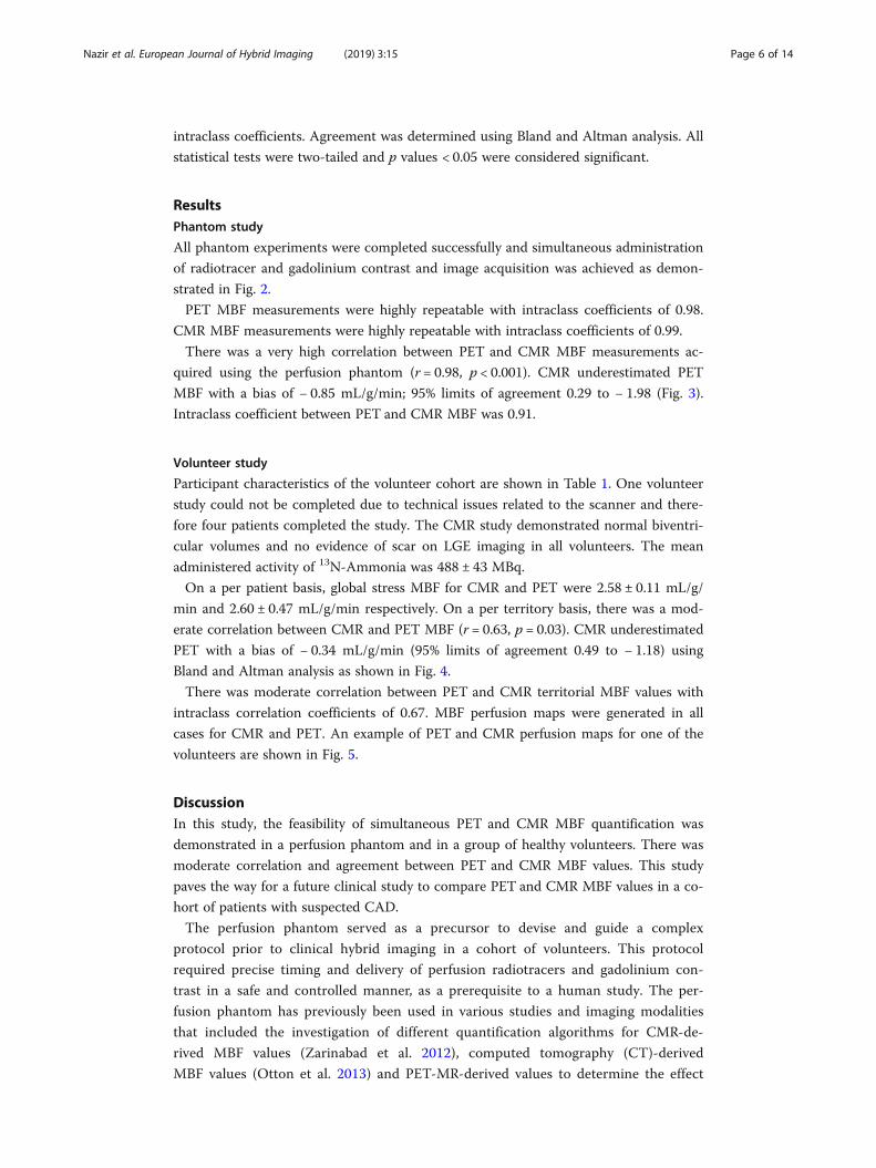
There was a very high correlation between PET and CMR MBF measurements ac-
quired using the perfusion phantom (r = 0.98, p < 0.001). CMR underestimated PET
MBF with a bias of − 0.85 mL/g/min; 95% limits of agreement 0.29 to − 1.98 (Fig. 3).
Intraclass coefficient between PET and CMR MBF was 0.91.
Volunteer study
Participant characteristics of the volunteer cohort are shown in Table 1. One volunteer
study could not be completed due to technical issues related to the scanner and there-
fore four patients completed the study. The CMR study demonstrated normal biventri-
cular volumes and no evidence of scar on LGE imaging in all volunteers. The mean
administered activity of 13N-Ammonia was 488 ± 43 MBq.
On a per patient basis, global stress MBF for CMR and PET were 2.58 ± 0.11 mL/g/
min and 2.60 ± 0.47 mL/g/min respectively. On a per territory basis, there was a mod-
erate correlation between CMR and PET MBF (r = 0.63, p = 0.03). CMR underestimated
PET with a bias of − 0.34 mL/g/min (95% limits of agreement 0.49 to − 1.18) using
Bland and Altman analysis as shown in Fig. 4.
There was moderate correlation between PET and CMR territorial MBF values with
intraclass correlation coefficients of 0.67. MBF perfusion maps were generated in all
cases for CMR and PET. An example of PET and CMR perfusion maps for one of the
volunteers are shown in Fig. 5.
DiscussionIn this study, the feasibility of simultaneous PET and CMR MBF quantification was
demonstrated in a perfusion phantom and in a group of healthy volunteers. There was
moderate correlation and agreement between PET and CMR MBF values. This study
paves the way for a future clinical study to compare PET and CMR MBF values in a co-
hort of patients with suspected CAD.
The perfusion phantom served as a precursor to devise and guide a complex
protocol prior to clinical hybrid imaging in a cohort of volunteers. This protocol
required precise timing and delivery of perfusion radiotracers and gadolinium con-
trast in a safe and controlled manner, as a prerequisite to a human study. The per-
fusion phantom has previously been used in various studies and imaging modalities
that included the investigation of different quantification algorithms for CMR-de-
rived MBF values (Zarinabad et al. 2012), computed tomography (CT)-derived
MBF values (Otton et al. 2013) and PET-MR-derived values to determine the effect
Nazir et al. European Journal of Hybrid Imaging (2019) 3:15 Page 6 of 14

of high counts rates of radiotracer (O’Doherty et al. 2017). This current study ex-
tends the utility of the perfusion phantom to demonstrate the feasibility and proto-
col development of fully quantitative PET and CMR-derived MBF simultaneously.
This is the first reported study in which simultaneous stress PET and CMR perfusion
acquisition was undertaken in a group of healthy volunteers using hybrid PET-MR im-
aging. MBF values were generated within the normal ranges reported in the literature for
healthy subjects, both for CMR (Brown et al. 2018) and for PET (Kitkungvan et al. 2017;
Chang et al. 2018). A previous study utilized PET-MR perfusion in patients with coronary
Fig. 2 Axial images of a PET only, b CMR only and c hybrid PET-MR images during passage of 13N-Ammonia and gadolinium contrast in the perfusion phantom. R right myocardial compartment, L leftmyocardial compartment
Nazir et al. European Journal of Hybrid Imaging (2019) 3:15 Page 7 of 14
artery disease, although used a dual sequence rather than a dual bolus approach and did
not have any preceding phantom studies (Kunze et al. 2018).
In this current study, CMR-derived MBF values underestimated PET values in both the
phantom and volunteer studies, and these findings are in keeping with previous clinical
studies that compared PET and CMR-derived MBF (Fritz-Hansen et al. 2008; Parkka et al.
2006). There are several plausible reasons to explain the underestimation of CMR-derived
MBF values observed in the phantom studies and the volunteer study.
A non-linear relationship exists between MRI signal intensity and gadolinium concen-
tration (Jerosch-Herold 2010). Mathematical derivation of MBF for CMR using Fermi de-
convolution requires the arterial input and myocardial output signals to behave in the
same linear relationship to that of the gadolinium concentration (Lee and Johnson 2009).
In the phantom study, signal saturation effects were alleviated by indirectly normalizing
the estimated impulse response functions, as native T1 values were not precisely known
(Hsu et al. 2018; Zierler 1962). Specifically, the AIF was scaled so that its time-inte-
gral matched that of the mean myocardial curve. In the volunteer study, the signal
intensity was converted to gadolinium concentration using a look-up table calcu-
lated by a Bloch simulation of the specific acquisition protocol used, as previously
described (Kellman et al. 2017). The quantification of MBF with PET does not suf-
fer from this non-linear relationship as the activity measured directly correlates
with tracer concentration.
CMR-derived MBF can underestimate MBF without a proper correction for the
non-linearity of the AIF from the blood pool (Jerosch-Herold 2010). This non-
linear effect is minimized by the use of a dual bolus (Christian et al. 2004) or
dual sequence (Gatehouse et al. 2004) method. In this study, we used dual bolus
to correct for the non-linear AIF relationship, although undertaking such a proto-
col is technically challenging. A dual sequence approach (Kellman et al. 2017;
Fig. 3 Bland and Altman plot of phantom MBF. CMR MBF underestimated PET MBF with a bias of −0.85 mL/g/min (95% limits of agreement + 0.29 to − 1.98). Dashed lines indicate 95% limits of agreementand bias
Nazir et al. European Journal of Hybrid Imaging (2019) 3:15 Page 8 of 14

Gatehouse et al. 2004), where the AIF is extracted from a low resolution FLASH
readout, can be used to correct for non-linearity and may be an alternative and
more practical approach that may facilitate clinical implementation.
An additional explanation for the underestimation observed with MRI is that differ-
ent analysis models are used in PET and MR. Quantification of absolute MBF with
CMR was achieved using a model independent approach, using a Fermi deconvolution
of the AIF and myocardial signal intensity. This is based on the central volume
principle (Zierler 1962), which allows calculation of flow based on the concentration of
a tracer that enters and exits a particular system. However, this model does not con-
sider the behaviour of gadolinium contrast in vivo. As such, tracer kinetic modelling
methods for quantification have been described (Bassingthwaighte et al. 1989) and may
provide a more accurate estimation of absolute MBF. Such an approach could not be
applied to the single compartmental model phantom and thus was not used for calcula-
tion of MBF in this study, but will be the focus of future work with multicompartmen-
tal perfusion models.
Fig. 4 Bland and Altman plot of territorial MBF. CMR MBF underestimated PET MBF with a bias of −0.34 mL/g/min (95% limits of agreement 0.49 to − 1.18). Dashed lines indicate 95% limits of agreementand bias
Table 1 Volunteer characteristics
Age 54 ± 3.5
Gender M (100%)
BMI (kg/m2) 25.5 ± 3.3
Stress HR (bpm) 93 ± 15
Rest HR (bpm) 66 ± 11
Stress systolic BP (mmHg) 140 ± 2.5
Rest systolic BP (mmHg) 132 ± 12
LVEDVi (mL/m2) 82.5 ± 27.6
LVEF (%) 61.8 ± 4.1
LGE scar (n) 0
BMI body mass index, HR heart rate, BP blood pressure, BPM beats per minute, LVEDVi Indexed left ventricular enddiastolic volume, LVEF left ventricular ejection fraction, LGE late gadolinium enhancement
Nazir et al. European Journal of Hybrid Imaging (2019) 3:15 Page 9 of 14

PET is considered the non-invasive reference standard for MBF quantification
(Bratis et al. 2013), and there are various perfusion models and radiotracers avail-
able for research and clinical application (Maddahi and Packard 2014). 13N-Ammo-
nia was employed as the radiotracer of choice, as it has an intermediate half-life
(9.96 min) which facilitates its practical use and has very good image quality
given its short positron range and retention in myocardial tissue (Maddahi and
Packard 2014). However, 13N-Ammonia does not have linear tracer uptake-myo-
cardial blood flow kinetics, particularly at high flow rates, and exhibits 85% myo-
cardial extraction fraction in vivo (Maddahi and Packard 2014). The ideal PET
perfusion radiotracer is 15O-Water as it is metabolically inert, freely diffusible,
with 100% extraction fraction and has linear tracer kinetics with near perfect cor-
relation between tracer uptake and MBF (Maddahi and Packard 2014). Neverthe-
less, 13N-Ammonia MBF quantification has been validated against the gold
standard PET perfusion tracer 15O-Water (Bol et al. 1993; Nitzsche et al. 1996).
In addition, whilst the quantification of absolute MBF with 13N-Ammonia has
good diagnostic accuracy (Gould et al. 1986), different published models and soft-
ware packages for the quantification of absolute MBF with 13N-Ammonia exist
(DeGrado et al. 1996; Hutchins et al. 1990; Nesterov et al. 2009; Adachi et al.
2007; PMOD 2018). De Grado et al. propose a one-tissue compartment model
with the use of the first 4 min after injection of tracer to avoid the effects of me-
tabolite build up and washout (DeGrado et al. 1996). Hutchins et al. propose a
two-tissue compartment model in which a second compartment accounts for the
build-up of metabolically trapped 13N-glutamine (metabolite of 13N-Ammonia)
with rate constants between each compartment (Hutchins et al. 1990). This
current study utilized a one tissue compartment model for the perfusion phantom
which does not have a second compartment or production of secondary metabo-
lites. As such, we also investigated absolute MBF values in the volunteer study
using the one-compartment De Grado model. Using this approach simplified the
quantification process and allowed a direct comparison of a one-tissue compart-
ment model comparable between CMR and PET MBF in phantoms and
volunteers.
Fig. 5 Perfusion PET-MR data from volunteer one. Side by side polar maps of MBF values with the samelook up table on a 16 American Heart Association (AHA) segments
Nazir et al. European Journal of Hybrid Imaging (2019) 3:15 Page 10 of 14

Another important factor that may explain the differences observed of MBF on a per
territory basis relates to quantification of MBF of a full 3D volumetric dataset with
PET, whereas in CMR, three distinct anatomical 8 mm slices of the heart at the base,
mid and apex are acquired.
During routine CMR image acquisition, patients are instructed to breath hold for
intermittent periods during the course of the scan. However, during routine PET im-
aging, patients are instructed to breathe normally. Thus, instructing patients to breath
hold during simultaneous PET-MR acquisition may interrupt the rhythmic respiratory
pattern, and it is unknown what the effect of this is on the PET data. In order to
minimize this as a possible confounder on the PET data, we used a free breathing
CMR perfusion sequence in this study, as opposed to isolated breath holds during PET
acquisition.
Limitations
The human circulatory system is a highly complex system composed of the large ves-
sels, pulsatile blood flow, the microcirculation, cardiac myocytes, plasma, and different
compartments such as the intra and extracellular space, which are all tightly regulated
by autoregulatory systems to maintain homeostatic conditions. In its current form, the
perfusion phantom cannot replicate the intricacies of the physiological processes of the
human body.
In addition, the exponential decrease of radiotracer in the perfusion phantom may
have a different profile for washout phase for the tracer kinetic system when tracer is
continuously washed out of the system in the perfusion phantom compared to the hu-
man circulatory system. However, a fixed cardiac output and heart rate similar to nor-
mal human physiology was employed for the phantom.
Furthermore, the fluid used in the phantom was normal saline, which has different
T1 values than blood in the human circulatory system. Nevertheless, the perfusion
phantom is free from respiratory artefact, ECG mis-triggering and allows preclinical de-
velopment in a highly controlled environment that mimics the uptake of tracer into a
compartment akin to that formed between the blood in the left ventricular cavity and
the myocardial tissue. Future developments of the perfusion phantom into a multicom-
partmental model may provide incremental steps to simulation of physiological condi-
tions that more closely align with the complexities of the human circulatory system,
that may also allow the modelling of multi-compartment tracer kinetics.
Finally, the number of repetitions of the study was limited, and this is in part due to
the build-up of radioactivity in the waste water tank, with a finite volume. The sample
size is relatively small, although this study serves as proof of principle of simultaneous
hybrid PET-MR perfusion prior to further clinical evaluation.
Future work
Having demonstrated the feasibility of simultaneous hybrid PET-MR perfusion imaging
in phantom and volunteers, the next step is to undertake a clinical study in a cohort of
patients with suspected CAD.
Several studies have compared the diagnostic accuracy of quantitative CMR for the
detection of significant CAD defined by invasive coronary angiography. A clinical
Nazir et al. European Journal of Hybrid Imaging (2019) 3:15 Page 11 of 14

validation study of CMR perfusion against 15O-Water PET that determines absolute
MBF, rather than fractional flow reserve measurements (which determine the haemo-
dynamic significance of epicardial coronary stenosis) is a more appropriate comparator.15O-Water is the gold standard perfusion tracer, and despite its practical challenges
due to short half-life, is the ideal perfusion tracer to compare against novel methods
for CMR perfusion which allow real-time, at point of care, fully automated in line
quantification and shows promise towards implementation into clinical routine (Kell-
man et al. 2017). Such a study may facilitate clinical and scientific acceptance for CMR
perfusion as a feasible alternative for MBF measurements which may allow widespread
use without the need for ionizing radiation and expensive radiochemistry facilities.
ConclusionHybrid PET-MR perfusion imaging with 13N-Ammonia and gadolinium contrast is
feasible. The correlation and agreement of PET and CMR-derived MBF values was high
in the perfusion phantom and moderate in healthy volunteers. Undertaking hybrid
PET-MR perfusion imaging with a dual bolus set up is complex and challenging, and a
single bolus dual sequence approach may be more practical and preferable. Future
evaluation in a cohort of patients with CAD is warranted.
Additional file
Additional file 1: Details of the perfusion phantom. (DOCX 513 kb)
AbbreviationsAC: Attenuation Correction; AIF: Arterial input function; CAD: Coronary artery disease; CMR: Cardiac magneticresonance; CT: Computed tomography; MBF: Myocardial blood flow; MR-IMPACT: Magnetic Resonance Imaging forMyocardial Perfusion Assessment in Coronary Artery Disease Trial; OSEM: Ordered subset expectation maximization;PET: Positron emission tomography; ROI: Region of interest
AcknowledgementsThe authors would like to thank the radiographers and administration team at King’s College London and the Guy’sand St Thomas’ NHS Hospital Cardiovascular MRI Service for their cooperation and assistance during the imaging andadministration processes.
FundingThe authors acknowledge financial support from the Department of Health through the National Institute for Health Research(NIHR) comprehensive Biomedical Research Centre award to Guy’s & St Thomas’ NHS Foundation Trust in partnership withKing’s College London and King’s College NHS Foundation Trust and by the NIHR Healthcare Technology Co-operative forCardiovascular Disease at Guy’s and St Thomas’ NHS Foundation Trust. This work was supported by the Wellcome/EPSRCCentre for Medical Engineering [WT 203148/Z/16/Z], the Rosetree’s Trust (under grant number A1380) and the EMPIR project15HLT05 PerfusImaging. The EMPIR initiative is co-funded by the European Union’s Horizon 2020 research and innovationprogramme and the EMPIR Participating States. MSN was funded by the UK Medical Research Council under grant numberMR/P01979X/1. SP was funded by a British Heart Foundation Chair under grant number CH/16/2/32089. SFB acknowledgessupport from the National Institute of Health Research (NIHR) [RP-2-16-07-001]. The views expressed are those of the authorsand not necessarily those of the NHS, the NIHR, the DoH, EPSRC, MRC or the Wellcome Trust.
Availability of data and materialsThe datasets used and analysed during the current study are available from the corresponding author on reasonablerequest.
Authors’ contributionsSN, AC and SP conceived the idea of the study. SN, JO, SMG, XM and PM devised the methodology for the study. SN,XM, SMG and ER recruited patients, undertook the research scans and analysed the data. RN and SR aided with dataanalysis. SB, TS, RR, PK, HX all provided critical review of the manuscript. All authors read and approved the finalmanuscript.
Ethics approval and consent to participateThe study was approved by the National Research Ethics Service (16/WA/0271) and the United KingdomAdministration of Radioactive Substances Advisory Committee (ARSAC) with written informed consent obtained fromall patients for inclusion in the study.
Nazir et al. European Journal of Hybrid Imaging (2019) 3:15 Page 12 of 14

Consent for publicationInformed consent was obtained from all participants to allow their data to be used for research purposes andpublication.
Competing interestsThe authors declare that they have no competing interests.
Publisher’s NoteSpringer Nature remains neutral with regard to jurisdictional claims in published maps and institutional affiliations.
Author details1Department of Cardiovascular Imaging, School of Biomedical Engineering and Imaging Sciences, King’s CollegeLondon, St Thomas’ Hospital, Westminster Bridge, 4th Floor Lambeth Wing, London SE1 7EH, UK. 2Siemens HealthcareLimited, Sir William Siemens Square, Frimley, Camberley GU16 8QD, UK. 3National Heart, Lung, and Blood Institute,National Institutes of Health, DHHS, Bethesda, MD, USA. 4Physikalisch-Technische Bundesanstalt, Berlin, Germany.5Leeds Institute of Cardiovascular and Metabolic Medicine, LIGHT Laboratories, Clarendon Way, University of Leeds,Leeds LS2 9JT, UK.
Received: 9 April 2019 Accepted: 15 July 2019
ReferencesAdachi I, Gaemperli O, Valenta I, Schepis T, Siegrist PT, Treyer V et al (2007) Assessment of myocardial perfusion by dynamic
O-15-labeled water PET imaging: validation of a new fast factor analysis. J Nucl Cardiol 14(5):698–705Bassingthwaighte JB, Wang CY, Chan IS (1989) Blood-tissue exchange via transport and transformation by capillary
endothelial cells. Circ Res 65(4):997–1020Bol A, Melin JA, Vanoverschelde JL, Baudhuin T, Vogelaers D, De Pauw M et al (1993) Direct comparison of [13N]ammonia
and [15O]water estimates of perfusion with quantification of regional myocardial blood flow by microspheres.Circulation. 87(2):512–525
Bratis K, Mahmoud I, Chiribiri A, Nagel E (2013) Quantitative myocardial perfusion imaging by cardiovascular magneticresonance and positron emission tomography. J Nucl Cardiol 20(5):860–870
Brown LAE, Onciul SC, Broadbent DA, Johnson K, Fent GJ, Foley JRJ et al (2018) Fully automated, inline quantification ofmyocardial blood flow with cardiovascular magnetic resonance: repeatability of measurements in healthy subjects. JCardiovasc Magn Reson 20(1):48
Cerqueira MD, Weissman NJ, Dilsizian V, Jacobs AK, Kaul S, Laskey WK et al (2002) Standardized myocardial segmentation andnomenclature for tomographic imaging of the heart. Circulation. 105(4):539–542
Chang C-Y, Hung G-U, Hsu B, Yang B-H, Chang C-W, Hu L-H et al (2018) Simplified quantification of 13N-ammonia PETmyocardial blood flow: a comparative study with the standard compartment model to facilitate clinical use. J NuclCardiol. https://doi.org/10.1007/s12350-018-1450-1
Chiribiri A, Schuster A, Ishida M, Hautvast G, Zarinabad N, Morton G et al (2013) Perfusion phantom: an efficient andreproducible method to simulate myocardial first-pass perfusion measurements with cardiovascular magnetic resonance.Magn Reson Med 69(3):698–707
Christian TF, Rettmann DW, Aletras AH, Liao SL, Taylor JL, Balaban RS et al (2004) Absolute myocardial perfusion in caninesmeasured by using dual-bolus first-pass MR imaging. Radiology 232:677–684
DeGrado TR, Hanson MW, Turkington TG, Delong DM, Brezinski DA, Vallee JP et al (1996) Estimation of myocardial blood flowfor longitudinal studies with 13N-labeled ammonia and positron emission tomography. J Nucl Cardiol 3(6 Pt 1):494–507
Engblom H, Xue H, Akil S, Carlsson M, Hindorf C, Oddstig J et al (2017) Fully quantitative cardiovascular magnetic resonancemyocardial perfusion ready for clinical use: a comparison between cardiovascular magnetic resonance imaging andpositron emission tomography. J Cardiovasc Magn Reson 19(1):78
Farhad H, Dunet V, Bachelard K, Allenbach G, Kaufmann PA, Prior JO (2013) Added prognostic value of myocardial blood flowquantitation in rubidium-82 positron emission tomography imaging. Eur Heart J Cardiovasc Imaging 14(12):1203–1210
Fritz-Hansen T, Hove JD, Kofoed KF, Kelbaek H, Larsson HB (2008) Quantification of MRI measured myocardial perfusionreserve in healthy humans: a comparison with positron emission tomography. J Magn Reson Imaging 27(4):818–824
Gatehouse PD, Elkington AG, Ablitt NA, Yang GZ, Pennell DJ, Firmin DN (2004) Accurate assessment of the arterial inputfunction during high-dose myocardial perfusion cardiovascular magnetic resonance. J Magn Reson Imaging 20(1):39–45
Gould KL, Goldstein RA, Mullani NA, Kirkeeide RL, Wong WH, Tewson TJ et al (1986) Noninvasive assessment of coronarystenoses by myocardial perfusion imaging during pharmacologic coronary vasodilation. VIII. Clinical feasibility of positroncardiac imaging without a cyclotron using generator-produced rubidium-82. J Am Coll Cardiol 7(4):775–789
Herzog BA, Husmann L, Valenta I, Gaemperli O, Siegrist PT, Tay FM et al (2009) Long-term prognostic value of 13N-ammonia myocardial perfusion positron emission tomography added value of coronary flow reserve. J Am CollCardiol 54(2):150–156
Hsu LY, Jacobs M, Benovoy M, Ta AD, Conn HM, Winkler S et al (2018) Diagnostic performance of fully automatedpixel-wise quantitative myocardial perfusion imaging by cardiovascular magnetic resonance. JACC CardiovascImaging 11(5):697–707
Hussain ST, Paul M, Plein S, McCann GP, Shah AM, Marber MS et al (2012) Design and rationale of the MR-INFORM study:stress perfusion cardiovascular magnetic resonance imaging to guide the management of patients with stable coronaryartery disease. J Cardiovasc Magn Reson 14:65
Hutchins GD, Schwaiger M, Rosenspire KC, Krivokapich J, Schelbert H, Kuhl DE (1990) Noninvasive quantification of regionalblood flow in the human heart using N-13 ammonia and dynamic positron emission tomographic imaging. J Am CollCardiol 15(5):1032–1042
Nazir et al. European Journal of Hybrid Imaging (2019) 3:15 Page 13 of 14

Ishida M, Schuster A, Morton G, Chiribiri A, Hussain S, Paul M et al (2011) Development of a universal dual-bolus injectionscheme for the quantitative assessment of myocardial perfusion cardiovascular magnetic resonance. J Cardiovasc MagnReson 13:28
Jerosch-Herold M (2010) Quantification of myocardial perfusion by cardiovascular magnetic resonance. J Cardiovasc MagnReson 12:57
Jerosch-Herold M, Wilke N, Stillman AE (1998) Magnetic resonance quantification of the myocardial perfusion reserve with aFermi function model for constrained deconvolution. Med Phys 25(1):73–84
Kellman P, Hansen MS, Nielles-Vallespin S, Nickander J, Themudo R, Ugander M et al (2017) Myocardial perfusioncardiovascular magnetic resonance: optimized dual sequence and reconstruction for quantification. J Cardiovasc MagnReson 19(1):43
Kitkungvan D, Johnson NP, Roby AE, Patel MB, Kirkeeide R, Gould KL (2017) Routine clinical quantitative rest stressmyocardial perfusion for managing coronary artery disease: clinical relevance of test-retest variability. JACCCardiovasc Imaging 10(5):565–577
Knott KD, Camaioni C, Ramasamy A, Augusto JA, Bhuva AN, Xue H et al (2019) Quantitative myocardial perfusion in coronaryartery disease: a perfusion mapping study. J Magn Reson Imaging. https://doi.org/10.1002/jmri.26668. Epub ahead ofprint
Kotecha T, Martinez-Naharro A, Boldrini M, Knight D, Hawkins P, Kalra S et al (2019) Automated pixel-wise quantitativemyocardial perfusion mapping by CMR to detect obstructive coronary artery disease and coronary microvasculardysfunction: validation against invasive coronary physiology. JACC Cardiovasc Imaging. Epub ahead of print
Kunze KP, Nekolla SG, Rischpler C, Zhang SH, Hayes C, Langwieser N et al (2018) Myocardial perfusion quantification usingsimultaneously acquired 13NH3-ammonia PET and dynamic contrast-enhanced MRI in patients at rest and stress. MagnReson Med 80:2641–2654
Larghat AM, Maredia N, Biglands J, Greenwood JP, Ball SG, Jerosch-Herold M et al (2013) Reproducibility of first-passcardiovascular magnetic resonance myocardial perfusion. J Magn Reson Imaging 37(4):865–874
Lassen ML, Rasul S, Beitzke D, Stelzmuller ME, Cal-Gonzalez J, Hacker M et al (2017) Assessment of attenuation correction formyocardial PET imaging using combined PET/MRI. J Nucl Cardiol. Epub ahead of print
Lee DC, Johnson NP (2009) Quantification of absolute myocardial blood flow by magnetic resonance perfusion imaging.JACC Cardiovasc Imaging 2(6):761–770
Maddahi J, Packard RR (2014) Cardiac PET perfusion tracers: current status and future directions. Semin Nucl Med44(5):333–343
Martinez-Moller A, Souvatzoglou M, Delso G, Bundschuh RA, Chefd'hotel C, Ziegler SI et al (2009) Tissue classification as apotential approach for attenuation correction in whole-body PET/MRI: evaluation with PET/CT data. J Nucl Med 50(4):520–526
Miller CA, Naish JH, Ainslie MP, Tonge C, Tout D, Arumugam P et al (2014) Voxel-wise quantification of myocardial blood flowwith cardiovascular magnetic resonance: effect of variations in methodology and validation with positron emissiontomography. J Cardiovasc Magn Reson 16:11
Morton G, Chiribiri A, Ishida M, Hussain ST, Schuster A, Indermuehle A et al (2012a) Quantification of absolute myocardialperfusion in patients with coronary artery disease: comparison between cardiovascular magnetic resonance and positronemission tomography. J Am Coll Cardiol 60(16):1546–1555
Morton G, Ishida M, Schuster A, Hussain S, Schaeffter T, Chiribiri A et al (2012b) Perfusion cardiovascular magnetic resonance:comparison of an advanced, high-resolution and a standard sequence. J Cardiovasc Magn Reson 14(1):34
Nazir MS, Ismail TF, Reyes E, Chiribiri A, Kaufmann PA, Plein S (2018) Hybrid positron emission tomography-magneticresonance of the heart: current state of the art and future applications. Eur Heart J Cardiovasc Imaging 19(9):962–974
Nesterov SV, Han C, Maki M, Kajander S, Naum AG, Helenius H et al (2009) Myocardial perfusion quantitation with 15O-labelled water PET: high reproducibility of the new cardiac analysis software (Carimas). Eur J Nucl Med Mol Imaging36(10):1594–1602
Nitzsche EU, Choi Y, Czernin J, Hoh CK, Huang SC, Schelbert HR (1996) Noninvasive quantification of myocardial blood flowin humans. A direct comparison of the [13N]ammonia and the [15O]water techniques. Circulation. 93(11):2000–2006
O’Doherty J, Chalampalakis Z, Schleyer P, Nazir MS, Chiribiri A, Marsden PK (2017) The effect of high count rates on cardiacperfusion quantification in a simultaneous PET-MR system using a cardiac perfusion phantom. EJNMMI Phys 4(1):31
Otton J, Morton G, Schuster A, Bigalke B, Marano R, Olivotti L et al (2013) A direct comparison of the sensitivity of CT and MRcardiac perfusion using a myocardial perfusion phantom. J Cardiovasc Comput Tomogr 7(2):117–124
Parkka JP, Niemi P, Saraste A, Koskenvuo JW, Komu M, Oikonen V et al (2006) Comparison of MRI and positron emissiontomography for measuring myocardial perfusion reserve in healthy humans. Magn Reson Med 55(4):772–779
Plein S, Greenwood J, Ridgway JP (2015) Cardiovascular MR ManualPMOD. http://www.pmod.com/files/download/v31/doc/pkin/2283.htm. Accessed 29th September 2018 [Available from:
http://www.pmod.com/files/download/v31/doc/pkin/2283.htm]Zarinabad N, Chiribiri A, Hautvast GL, Ishida M, Schuster A, Cvetkovic Z et al (2012) Voxel-wise quantification of myocardial
perfusion by cardiac magnetic resonance. Feasibility and methods comparison. Magn Reson Med 68(6):1994–2004Zierler KL (1962) Theoretical basis of indicator-dilution methods for measuring flow and volume. Circ Res 10(3):393–407
Nazir et al. European Journal of Hybrid Imaging (2019) 3:15 Page 14 of 14