Radioimmunotherapy with radioactive nanoparticles: First results of
10
Radioimmunotherapy with radioactive nanoparticles: First results of dosimetry for vascularized and necrosed solid tumors V. Bouchat, a V. E. Nuttens, and S. Lucas Laboratoire d’Analyses par Réactions Nucléaires (LARN), University of Namur (FUNDP), Rue de Bruxelles 61, B-5000 Namur, Belgium C. Michiels Unité de Recherche en Biologie Cellulaire (URBC), University of Namur (FUNDP), Rue de Bruxelles 61, B-5000 Namur, Belgium B. Masereel Department of Pharmacy (DP), University of Namur (FUNDP), Rue de Bruxelles 61, B-5000 Namur, Belgium O. Féron Unité de Pharmacothérapie (FATH), Université Catholique de Louvain (UCL), Avenue Mounier, 53, B-1200 Brussels, Belgium B. Gallez Laboratoire de resonance magnétique biomédicale (CMFA), Université Catholique de Louvain (UCL), Avenue Mounier, 73, B-1200 Brussels, Belgium T. Vander Borght Center for Molecular Imaging and Experimental Radiotherapy (IRME), Université Catholique de Louvain (UCL), Dr. G. Therasse, 1, B-5530 Yvoir, Belgium Received 29 June 2007; revised 7 September 2007; accepted for publication 7 September 2007; published 29 October 2007 Radioimmunotherapy uses monoclonal antibodies that are still labeled with only one radioactive atom. The aim of this paper is to assess, by means of MCNPX simulations, the doses delivered around and throughout a solid tumor when the radioactive atom linked to each antibody is replaced by a 5 nm diameter nanoparticle composed of numerous radionuclides. A new model for a spherical vascularized tumor has been developed in which the antibody distributions inside the tumor can be uniform or heterogeneous. It is also possible to simulate a central necrotic core inside the tumor where the concentration of radiolabeled antibodies is assumed to be zero. Dosimetry calculations have been performed for the beta-emitting radionuclide 90 Y 2 O 3 . Preliminary results show that the irregularity of vasculature and the presence of a necrotic core have a noticeable influence on the deposited dose profiles. Moreover, with a total activity of 5 and 34 MBq for tumor radii of 0.5 and 1.0 cm, respectively, viable tumor cells can receive doses of up to 50 Gy, even if high nonunifor- mity of the total activity is observed in the tumor. These simulations still require accurate informa- tion about antibody characteristics and necrosis sizes but clearly confirm that the use of monoclonal antibodies conjugated to nanoparticles could lead to a considerable enhancement of treatment efficacy against cancer. © 2007 American Association of Physicists in Medicine. DOI: 10.1118/1.2791038 Key words: radioimmunotherapy, dosimetry, Monte Carlo, tumor model, nanomedicine I. INTRODUCTION In radioimmunotherapy RIT, radionuclides coupled to monoclonal antibodies mAb’s are increasingly used to de- liver ionizing radiation to kill tumor cells while sparing nor- mal tissue. 1–3 The radiolabeled antibodies are directed against various antigens overexpressed on tumor cells or blood vessels formed during angiogenesis. 4–6 Each time, only one radioactive atom is linked to the antibody and the delivered tumor doses in RIT are influenced mainly by the antibody properties, the choice of radionuclide, and the bio- logical halflife of the complex. 1,7 This approach for treating cancer shows good results for hematopoietic malignancies such as non-Hodgkin’s lymphoma, which require only low doses of radiation 15 Gy. 8–12 Unfortunately, clinical studies have shown that solid tumors are less sensitive to ionizing radiation and doses higher than 60 Gy are necessary to observe therapeutic effects. 7 New strategies to increase antibody accumulation and penetration into the tumor are currently being investigated but the delivered doses remain insufficient to observe an important treatment response. 13–16 Today, it is possible to assemble several nonradioactive metal atoms to form clusters of nanometer dimensions. 17–20 Hence, we have conceptually designed a nanoparticle com- posed only of radioactive elements to enhance diagnostic sensitivity in medical imaging or to improve therapeutic ef- fectiveness in RIT. 4504 4504 Med. Phys. 34 „11…, November 2007 0094-2405/2007/34„11…/4504/10/$23.00 © 2007 Am. Assoc. Phys. Med.
Transcript of Radioimmunotherapy with radioactive nanoparticles: First results of

Radioimmunotherapy with radioactive nanoparticles: First resultsof dosimetry for vascularized and necrosed solid tumors
V. Bouchat,a� V. E. Nuttens, and S. LucasLaboratoire d’Analyses par Réactions Nucléaires (LARN), University of Namur (FUNDP),Rue de Bruxelles 61, B-5000 Namur, Belgium
C. MichielsUnité de Recherche en Biologie Cellulaire (URBC), University of Namur (FUNDP), Rue de Bruxelles 61,B-5000 Namur, Belgium
B. MasereelDepartment of Pharmacy (DP), University of Namur (FUNDP), Rue de Bruxelles 61, B-5000 Namur,Belgium
O. FéronUnité de Pharmacothérapie (FATH), Université Catholique de Louvain (UCL), Avenue Mounier, 53,B-1200 Brussels, Belgium
B. GallezLaboratoire de resonance magnétique biomédicale (CMFA), Université Catholique de Louvain (UCL),Avenue Mounier, 73, B-1200 Brussels, Belgium
T. Vander BorghtCenter for Molecular Imaging and Experimental Radiotherapy (IRME), Université Catholique de Louvain(UCL), Dr. G. Therasse, 1, B-5530 Yvoir, Belgium
�Received 29 June 2007; revised 7 September 2007; accepted for publication 7 September 2007;published 29 October 2007�
Radioimmunotherapy uses monoclonal antibodies that are still labeled with only one radioactiveatom. The aim of this paper is to assess, by means of MCNPX simulations, the doses deliveredaround and throughout a solid tumor when the radioactive atom linked to each antibody is replacedby a 5 nm diameter nanoparticle composed of numerous radionuclides. A new model for a sphericalvascularized tumor has been developed in which the antibody distributions inside the tumor can beuniform or heterogeneous. It is also possible to simulate a central necrotic core inside the tumorwhere the concentration of radiolabeled antibodies is assumed to be zero. Dosimetry calculationshave been performed for the beta-emitting radionuclide 90Y2O3. Preliminary results show that theirregularity of vasculature and the presence of a necrotic core have a noticeable influence on thedeposited dose profiles. Moreover, with a total activity of 5 and 34 MBq for tumor radii of 0.5 and1.0 cm, respectively, viable tumor cells can receive doses of up to 50 Gy, even if high nonunifor-mity of the total activity is observed in the tumor. These simulations still require accurate informa-tion about antibody characteristics and necrosis sizes but clearly confirm that the use of monoclonalantibodies conjugated to nanoparticles could lead to a considerable enhancement of treatmentefficacy against cancer. © 2007 American Association of Physicists in Medicine.�DOI: 10.1118/1.2791038�
Key words: radioimmunotherapy, dosimetry, Monte Carlo, tumor model, nanomedicine
I. INTRODUCTION
In radioimmunotherapy �RIT�, radionuclides coupled tomonoclonal antibodies �mAb’s� are increasingly used to de-liver ionizing radiation to kill tumor cells while sparing nor-mal tissue.1–3 The radiolabeled antibodies are directedagainst various antigens overexpressed on tumor cells orblood vessels formed during angiogenesis.4–6 Each time,only one radioactive atom is linked to the antibody and thedelivered tumor doses in RIT are influenced mainly by theantibody properties, the choice of radionuclide, and the bio-logical halflife of the complex.1,7 This approach for treatingcancer shows good results for hematopoietic malignancies
such as non-Hodgkin’s lymphoma, which require only low4504 Med. Phys. 34 „11…, November 2007 0094-2405/2007/34„
doses of radiation ��15 Gy�.8–12 Unfortunately, clinicalstudies have shown that solid tumors are less sensitive toionizing radiation and doses higher than 60 Gy are necessaryto observe therapeutic effects.7 New strategies to increaseantibody accumulation and penetration into the tumor arecurrently being investigated but the delivered doses remaininsufficient to observe an important treatment response.13–16
Today, it is possible to assemble several nonradioactivemetal atoms to form clusters of nanometer dimensions.17–20
Hence, we have conceptually designed a nanoparticle com-posed only of radioactive elements to enhance diagnosticsensitivity in medical imaging or to improve therapeutic ef-
fectiveness in RIT.450411…/4504/10/$23.00 © 2007 Am. Assoc. Phys. Med.
4505 Bouchat et al.: Monte Carlo dosimetry for radioactive nanoparticles 4505
A major obstacle in the use of nanoparticulate systems fordrug delivery is the process of opsonization responsible for arapid clearance of these clusters from circulation by thereticuloendothelial system �RES�, comprising mainly theKupffer Cells in the liver as well as spleen and bone marrowmacrophages.21–24 Nevertheless, progress in nanomedicinehas shown that the behavior and stability of a nanoparticlewithin the biological microenvironment can be controlled byits size and surface characteristics. For example, large mac-romolecules ��100 nm� move slowly in tissue, and this hasthe effect of enhancing their clearance by the host defenseRES.25 By creating spherical radioactive nanoparticleswhose diameter does not exceed 5 nm, the radiolabeled com-plex is small enough to reduce its uptake by the RES hencepromoting its diffusion through the target tissue. The surfaceof these nanoparticles can also be covered by nonionic sur-factants or polymeric macromolecules to form a protectivelayer against the absorption of opsonin proteins required formacrophage recognition. For example, coating nanoparticleswith polyethylene glycol �PEG� or PEG-containing copoly-mers increases their blood halflife after intravenousadministration.22,24–27 These long hydrophilic polymer chainslocated on the surface of the particles have the advantage ofreducing interparticulate attractive van der Waals forces andpreventing the aggregation of several nanoclusters. Finally,the polymeric coating of the nanoparticle can be functional-ized with targeting molecules such as the antibodies, thera-peutic drugs, or image contrast agents to create a multifunc-tional nanoparticle.21,28,29
Nanoparticles have already been investigated in medicineand have demonstrated promising application for tumortargeting.29,30 For example, quantum dots �QDs�, nanosizedfluorescent semiconductor particles, are rapidly becoming aninteresting tool for detecting tumors31 and therapeutic treat-ments could be considered by simply adding drugs aroundthese QDs.32–34 Magnetic28,35,36 or metal37–39 nanoparticlescoupled to antibodies are also in development for qualityimprovement of magnetic and optical imaging. Functional-ized with additive ligands, these nanoparticles could offernew opportunities for treating cancer. The advantages of us-ing radioactive nanoparticles in RIT are threefold. First, evenif half of the atoms encapsulated in the nanoparticle are nolonger radioactive when the antibodies reach the tumor, theactivity delivered by a nanoparticle is higher than the activitygiven by a single radioactive atom. Hence, antibodies conju-gated to nanoparticles should deliver higher radiation dose tothe tumors. Potential cluster radiolysis should however, bestudied. Second, different types of radionuclides can be com-bined ��, �, �, or x-ray emitters� or mixed with nonradioac-tive atoms used in medicine such as magnetic elements.Third, nanoparticles have more surface area to accommodatea large number of different types of functional groups so thatmore than one antibody can be conjugated.
As the energy deposition of such radioactive nanoparticlesis unknown, dosimetry simulations are required to calculatethe absorbed doses inside and around the tumor and to es-
tablish the best therapeutic treatment for the patient. An ap-Medical Physics, Vol. 34, No. 11, November 2007
proach using Monte Carlo N-Particles eXtended Software�MCNPX� to determine the radiation dose absorbed by tu-mor cells and healthy tissue has already been proposed byNuttens and co-workers.40 These simulations showed that itwas possible to significantly increase the radiation doseswhen the single radioactive atom coupled to each antibody inclassical radioimmunotherapy is replaced by a beta emittingnanoparticle. But the tumor model used for those simulationsis valid only for avascular cell clusters, which rarely exceed0.2 mm diameter.41–43
In this paper, we study the doses deposited inside andaround a spherical solid tumor when the single radionuclidelabeled to antibodies is replaced by a radioactive nanopar-ticle of 5 nm diam. For that purpose, new three-dimensionalvascular models representing the tumor have been devel-oped. For each model, shapes of deposited dose distributionswere calculated for the stable oxide molecules 90Y2O3,which are already experimentally produced and well adaptedto cure large tumors with nonhomogeneous vasculature. In-deed, the radionuclide yttrium-90 has the advantage of beinga pure � emitter with a high energy radiation �Emax
�2.3 MeV� responsible for a long penetration range in tis-sue �Rmax�11 mm�.44–46
II. TUMOR MODELS
The MCNPX software is capable of simulating the trans-port of photons and electrons in matter over a broad energyrange. The input file of this code is composed of three typesof data, those corresponding to the geometry of the problem�composition, form, and density of the tumor and surround-ing healthy tissues�, the radioactive sources �nature, emissionspectra, and positions inside and near the tumor�, and thenature of the desired result �i.e., the deposited energy�. Betaspectral data for yttrium-90 were taken from tables on theRADAR site �www.doseinfo-radar.com/RADARDecay.html�. The composition and density of malig-nant and healthy tissues were taken from ICRU Report 44,47
and three tumor models were considered.
II.A. Avascular model
In our previous work,40 the solid tumor was representedby a simple sphere of breast tissue surrounded by biologicalvectors �bVs�, and each of them was labeled with one radio-active nanoparticle. We have assumed that the maximum dis-tance between the tumor binding site of the biological vectorand the radioactive nanoparticle binding site was around15 nm.48–50 Radiolabeled bVs surrounding the tumor werethen modeled by a spherical surface that emits radiations.The radius of this surface was chosen 15 nm larger than thetumor radius to comply with the size of antibodies. The samematerial density was used for diseased and healthy tissues.The dose delivered to the tumor and normal tissues has beenevaluated using the pulse-height tally *F8 �MeV/g/nps� of-fered by MCNPX and converted into Gy.
This model is well adapted as long as the tumor measureshundreds of micrometers in diameter and is not vascularized.
However, most larger solid tumors are described by a set of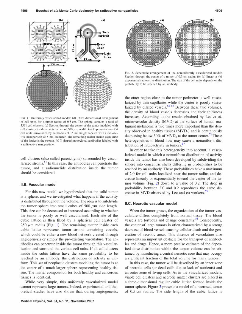
4506 Bouchat et al.: Monte Carlo dosimetry for radioactive nanoparticles 4506
cell clusters �also called parenchyma� surrounded by vascu-larized stroma.51 In this case, the antibodies can penetrate thetumor, and a radionuclide distribution inside the tumorshould be considered.
II.B. Vascular model
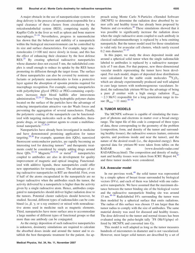
For this new model, we hypothesized that the solid tumoris a sphere, and we investigated what happens if the activityis distributed throughout the volume. The idea is to subdividethe tumor sphere into small cubes of 500 �m side length.This size can be decreased or increased according to whetherthe tumor is poorly or well vascularized. Each site of thecubic lattice is then filled by a spherical cell cluster of250 �m radius �Fig. 1�. The remaining matter inside eachcubic lattice represents tumor stroma containing vessels,which could be either a new blood network created throughangiogenesis or simply the pre-existing vasculature. The an-tibodies can penetrate inside the tumor through this vascular-ization and surround the various cell units. If all cell clustersinside the cubic lattice have the same probability to bereached by an antibody, the distribution of activity is uni-form. This set of neoplastic clusters modeling the tumor is atthe center of a much larger sphere representing healthy tis-sue. The matter composition for both healthy and canceroustissues is identical.
While very simple, this uniformly vascularized modelcannot represent large tumors. Indeed, experimental and the-
FIG. 1. Uniformly vascularized model: �d� Three-dimensional arrangementof cell units for a tumor radius of 0.5 cm. The sphere contains a total of3591 cell clusters. �c� Section through the center of the tumor modeled withcell clusters inside a cubic lattice of 500 �m width. �a� Representation of 4cell units surrounded by antibodies of 15 nm height labeled with a radioac-tive nanoparticle of 5 nm diameter. The remaining matter inside each cubeof the lattice is the stroma. �b� Y-shaped monoclonal antibodies labeled witha radioactive nanoparticle.
oretical studies have also shown that, during angiogenesis,
Medical Physics, Vol. 34, No. 11, November 2007
the outer region close to the tumor perimeter is well vascu-larized by thin capillaries while the center is poorly vascu-larized by dilated vessels.52–56 Between these two volumes,the density of blood vessels decreases and their thicknessincreases. According to the results obtained by Lee et al.microvascular density �MVD� at the surface of human ma-lignant melanoma is two times more important than the den-sity observed in healthy tissues �MVD0� and is continuouslydecreasing below 50% of MVD0 at the tumor center.55 Theseheterogeneities in blood flow may cause a nonuniform dis-tribution of radioactivity in tumors.57
In order to take this heterogeneity into account, a vascu-larized model in which a nonuniform distribution of activityinside the tumor has also been developed by subdividing thesphere into concentric shells differing in probabilities to bereached by an antibody. These probabilities have a maximumof 2.0 for cell units localized near the tumor radius and de-crease linearly or exponentially toward the center of the tu-mor volume �Fig. 2� down to a value of 0.2. The drop inprobability between 2.0 and 0.2 reproduces the same de-crease in MVD observed by Lee and co-workers.55
II.C. Necrotic vascular model
When the tumor grows, the organization of the tumor vas-culature differs completely from normal tissue. The bloodvessels are tortuous and change constantly.51 Consequently,the center of large tumors is often characterized by a strongdecrease of blood vessels causing cellular death and the gen-eration of necrotic areas. This absence of vasculature alsorepresents an important obstacle for the transport of antibod-ies and drugs. Hence, a more precise estimate of the depos-ited dose distribution within the tumor volume can be ob-tained by introducing a central necrotic core that may occupya significant fraction of the total volume for many tumors.
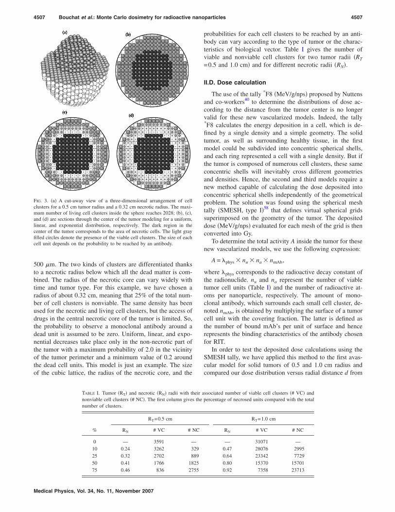
In this case, the tumor will be described by an inner zoneof necrotic cells �or dead cells due to lack of nutrients� andan outer zone of living cells. As in the vascularized models,viable cell clusters and necrotic matter clusters are placed ina three-dimensional regular cubic lattice formed inside thetumor sphere. Figure 3 presents a model of a necrosed tumor
FIG. 2. Schematic arrangement of the nonuniformly vascularized model:Section through the center of a tumor of 0.5 cm radius for �a� linear or �b�exponential radioactive distribution. The size of the cell units depends on theprobability to be reached by an antibody.
of 0.5 cm radius. The side length of the cubic lattice is
4507 Bouchat et al.: Monte Carlo dosimetry for radioactive nanoparticles 4507
500 �m. The two kinds of clusters are differentiated thanksto a necrotic radius below which all the dead matter is com-bined. The radius of the necrotic core can vary widely withtime and tumor type. For this example, we have chosen aradius of about 0.32 cm, meaning that 25% of the total num-ber of cell clusters is nonviable. The same density has beenused for the necrotic and living cell clusters, but the access ofdrugs in the central necrotic core of the tumor is limited. So,the probability to observe a monoclonal antibody around adead unit is assumed to be zero. Uniform, linear, and expo-nential decreases take place only in the non-necrotic part ofthe tumor with a maximum probability of 2.0 in the vicinityof the tumor perimeter and a minimum value of 0.2 aroundthe dead cell units. This model is just an example. The sizeof the cubic lattice, the radius of the necrotic core, and the
FIG. 3. �a� A cut-away view of a three-dimensional arrangement of cellclusters for a 0.5 cm tumor radius and a 0.32 cm necrotic radius. The maxi-mum number of living cell clusters inside the sphere reaches 2028; �b�, �c�,and �d� are sections through the center of the tumor modeling for a uniform,linear, and exponential distribution, respectively. The dark region in thecenter of the tumor corresponds to the area of necrotic cells. The light grayfilled circles denote the presence of the viable cell clusters. The size of eachcell unit depends on the probability to be reached by an antibody.
TABLE I. Tumor �RT� and necrotic �RN� radii with thnonviable cell clusters �# NC�. The first column givesnumber of clusters.
%
RT=0.5 cm
RN # VC # N
0 — 3591 —10 0.24 3262 325 0.32 2702 850 0.41 1766 1875 0.46 836 27
Medical Physics, Vol. 34, No. 11, November 2007
probabilities for each cell clusters to be reached by an anti-body can vary according to the type of tumor or the charac-teristics of biological vector. Table I gives the number ofviable and nonviable cell clusters for two tumor radii �RT
=0.5 and 1.0 cm� and for different necrotic radii �RN�.
II.D. Dose calculation
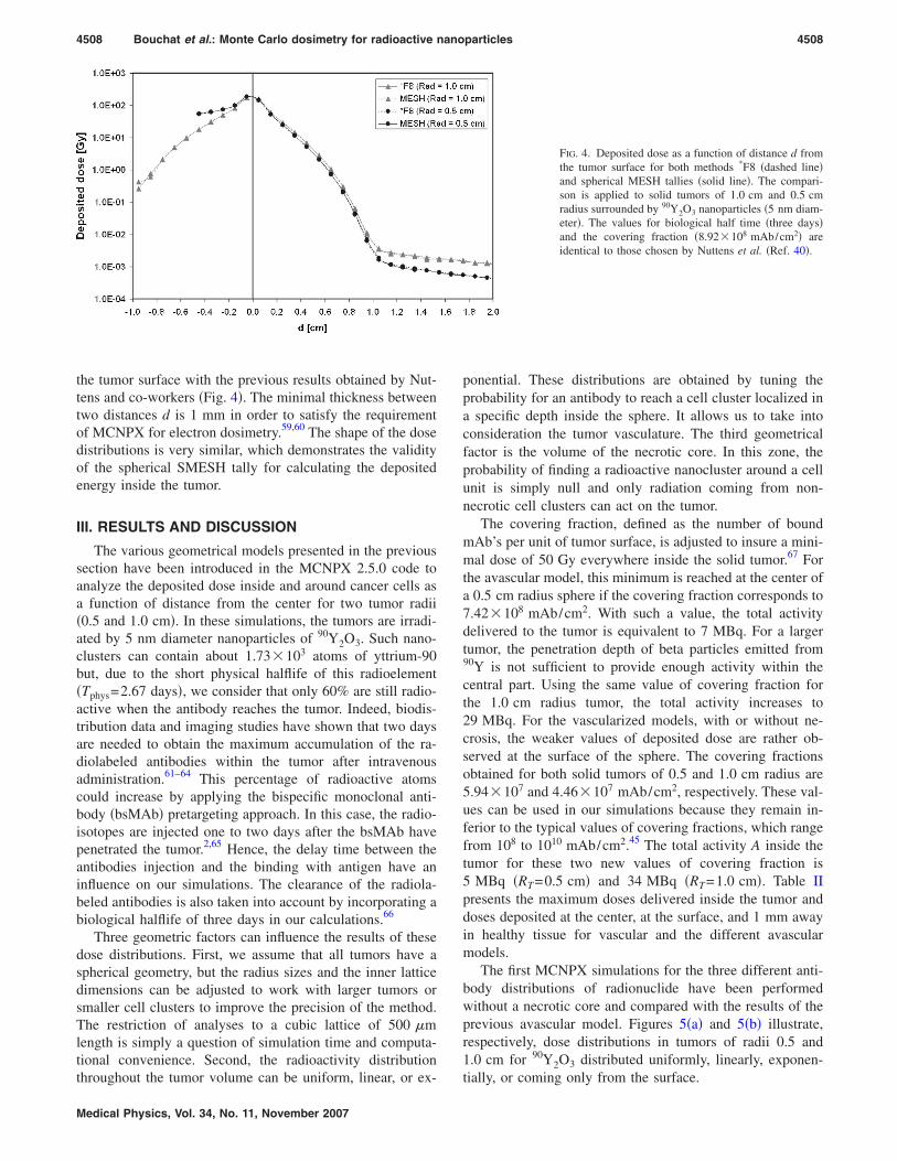
The use of the tally *F8 �MeV/g/nps� proposed by Nuttensand co-workers40 to determine the distributions of dose ac-cording to the distance from the tumor center is no longervalid for these new vascularized models. Indeed, the tally*F8 calculates the energy deposition in a cell, which is de-fined by a single density and a simple geometry. The solidtumor, as well as surrounding healthy tissue, in the firstmodel could be subdivided into concentric spherical shells,and each ring represented a cell with a single density. But ifthe tumor is composed of numerous cell clusters, these sameconcentric shells will inevitably cross different geometriesand densities. Hence, the second and third models require anew method capable of calculating the dose deposited intoconcentric spherical shells independently of the geometricalproblem. The solution was found using the spherical meshtally �SMESH, type I�58 that defines virtual spherical gridssuperimposed on the geometry of the tumor. The depositeddose �MeV/g/nps� evaluated for each mesh of the grid is thenconverted into Gy.
To determine the total activity A inside the tumor for thesenew vascularized models, we use the following expression:
A = �phys � nu � na � nmAb,
where �phys corresponds to the radioactive decay constant ofthe radionuclide. nu and na represent the number of viabletumor cell units �Table I� and the number of radioactive at-oms per nanoparticle, respectively. The amount of mono-clonal antibody, which surrounds each small cell cluster, de-noted nmAb, is obtained by multiplying the surface of a tumorcell unit with the covering fraction. The latter is defined asthe number of bound mAb’s per unit of surface and hencerepresents the binding characteristics of the antibody chosenfor RIT.
In order to test the deposited dose calculations using theSMESH tally, we have applied this method to the first avas-cular model for solid tumors of 0.5 and 1.0 cm radius andcompared our dose distribution versus radial distance d from
ssociated number of viable cell clusters �# VC� andpercentage of necrosed units compared with the total
RT=1.0 cm
RN # VC # NC
— 31071 —0.47 28076 29950.64 23342 77290.80 15370 157010.92 7358 23713
eir athe
C
29892555
4508 Bouchat et al.: Monte Carlo dosimetry for radioactive nanoparticles 4508
the tumor surface with the previous results obtained by Nut-tens and co-workers �Fig. 4�. The minimal thickness betweentwo distances d is 1 mm in order to satisfy the requirementof MCNPX for electron dosimetry.59,60 The shape of the dosedistributions is very similar, which demonstrates the validityof the spherical SMESH tally for calculating the depositedenergy inside the tumor.
III. RESULTS AND DISCUSSION
The various geometrical models presented in the previoussection have been introduced in the MCNPX 2.5.0 code toanalyze the deposited dose inside and around cancer cells asa function of distance from the center for two tumor radii�0.5 and 1.0 cm�. In these simulations, the tumors are irradi-ated by 5 nm diameter nanoparticles of 90Y2O3. Such nano-clusters can contain about 1.73�103 atoms of yttrium-90but, due to the short physical halflife of this radioelement�Tphys=2.67 days�, we consider that only 60% are still radio-active when the antibody reaches the tumor. Indeed, biodis-tribution data and imaging studies have shown that two daysare needed to obtain the maximum accumulation of the ra-diolabeled antibodies within the tumor after intravenousadministration.61–64 This percentage of radioactive atomscould increase by applying the bispecific monoclonal anti-body �bsMAb� pretargeting approach. In this case, the radio-isotopes are injected one to two days after the bsMAb havepenetrated the tumor.2,65 Hence, the delay time between theantibodies injection and the binding with antigen have aninfluence on our simulations. The clearance of the radiola-beled antibodies is also taken into account by incorporating abiological halflife of three days in our calculations.66
Three geometric factors can influence the results of thesedose distributions. First, we assume that all tumors have aspherical geometry, but the radius sizes and the inner latticedimensions can be adjusted to work with larger tumors orsmaller cell clusters to improve the precision of the method.The restriction of analyses to a cubic lattice of 500 �mlength is simply a question of simulation time and computa-tional convenience. Second, the radioactivity distribution
throughout the tumor volume can be uniform, linear, or ex-Medical Physics, Vol. 34, No. 11, November 2007
ponential. These distributions are obtained by tuning theprobability for an antibody to reach a cell cluster localized ina specific depth inside the sphere. It allows us to take intoconsideration the tumor vasculature. The third geometricalfactor is the volume of the necrotic core. In this zone, theprobability of finding a radioactive nanocluster around a cellunit is simply null and only radiation coming from non-necrotic cell clusters can act on the tumor.
The covering fraction, defined as the number of boundmAb’s per unit of tumor surface, is adjusted to insure a mini-mal dose of 50 Gy everywhere inside the solid tumor.67 Forthe avascular model, this minimum is reached at the center ofa 0.5 cm radius sphere if the covering fraction corresponds to7.42�108 mAb/cm2. With such a value, the total activitydelivered to the tumor is equivalent to 7 MBq. For a largertumor, the penetration depth of beta particles emitted from90Y is not sufficient to provide enough activity within thecentral part. Using the same value of covering fraction forthe 1.0 cm radius tumor, the total activity increases to29 MBq. For the vascularized models, with or without ne-crosis, the weaker values of deposited dose are rather ob-served at the surface of the sphere. The covering fractionsobtained for both solid tumors of 0.5 and 1.0 cm radius are5.94�107 and 4.46�107 mAb/cm2, respectively. These val-ues can be used in our simulations because they remain in-ferior to the typical values of covering fractions, which rangefrom 108 to 1010 mAb/cm2.45 The total activity A inside thetumor for these two new values of covering fraction is5 MBq �RT=0.5 cm� and 34 MBq �RT=1.0 cm�. Table IIpresents the maximum doses delivered inside the tumor anddoses deposited at the center, at the surface, and 1 mm awayin healthy tissue for vascular and the different avascularmodels.
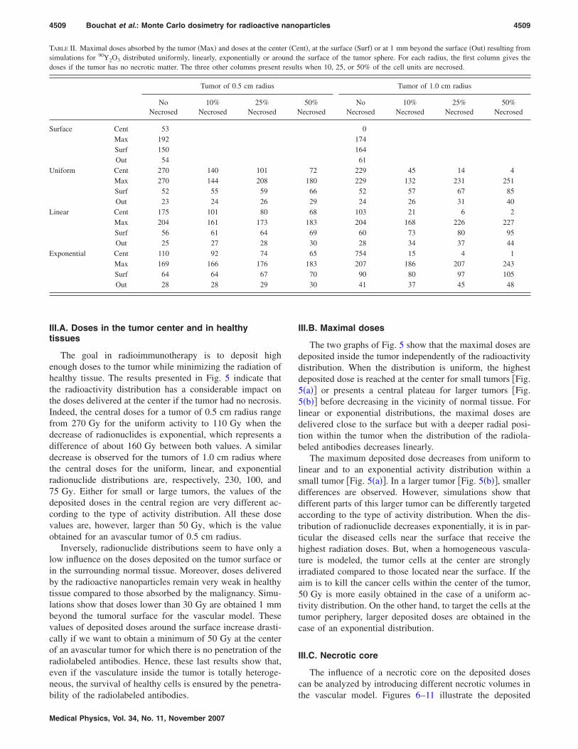
The first MCNPX simulations for the three different anti-body distributions of radionuclide have been performedwithout a necrotic core and compared with the results of theprevious avascular model. Figures 5�a� and 5�b� illustrate,respectively, dose distributions in tumors of radii 0.5 and1.0 cm for 90Y2O3 distributed uniformly, linearly, exponen-
FIG. 4. Deposited dose as a function of distance d fromthe tumor surface for both methods *F8 �dashed line�and spherical MESH tallies �solid line�. The compari-son is applied to solid tumors of 1.0 cm and 0.5 cmradius surrounded by 90Y2O3 nanoparticles �5 nm diam-eter�. The values for biological half time �three days�and the covering fraction �8.92�108 mAb/cm2� areidentical to those chosen by Nuttens et al. �Ref. 40�.
tially, or coming only from the surface.
4509 Bouchat et al.: Monte Carlo dosimetry for radioactive nanoparticles 4509
III.A. Doses in the tumor center and in healthytissues
The goal in radioimmunotherapy is to deposit highenough doses to the tumor while minimizing the radiation ofhealthy tissue. The results presented in Fig. 5 indicate thatthe radioactivity distribution has a considerable impact onthe doses delivered at the center if the tumor had no necrosis.Indeed, the central doses for a tumor of 0.5 cm radius rangefrom 270 Gy for the uniform activity to 110 Gy when thedecrease of radionuclides is exponential, which represents adifference of about 160 Gy between both values. A similardecrease is observed for the tumors of 1.0 cm radius wherethe central doses for the uniform, linear, and exponentialradionuclide distributions are, respectively, 230, 100, and75 Gy. Either for small or large tumors, the values of thedeposited doses in the central region are very different ac-cording to the type of activity distribution. All these dosevalues are, however, larger than 50 Gy, which is the valueobtained for an avascular tumor of 0.5 cm radius.
Inversely, radionuclide distributions seem to have only alow influence on the doses deposited on the tumor surface orin the surrounding normal tissue. Moreover, doses deliveredby the radioactive nanoparticles remain very weak in healthytissue compared to those absorbed by the malignancy. Simu-lations show that doses lower than 30 Gy are obtained 1 mmbeyond the tumoral surface for the vascular model. Thesevalues of deposited doses around the surface increase drasti-cally if we want to obtain a minimum of 50 Gy at the centerof an avascular tumor for which there is no penetration of theradiolabeled antibodies. Hence, these last results show that,even if the vasculature inside the tumor is totally heteroge-neous, the survival of healthy cells is ensured by the penetra-
TABLE II. Maximal doses absorbed by the tumor �Max� and doses at the centesimulations for 90Y2O3 distributed uniformly, linearly, exponentially or arodoses if the tumor has no necrotic matter. The three other columns present
Tumor of 0.5 cm radius
NoNecrosed
10%Necrosed
25%Necrosed
Surface Cent 53Max 192Surf 150Out 54
Uniform Cent 270 140 101Max 270 144 208Surf 52 55 59Out 23 24 26
Linear Cent 175 101 80Max 204 161 173Surf 56 61 64Out 25 27 28
Exponential Cent 110 92 74Max 169 166 176Surf 64 64 67Out 28 28 29
bility of the radiolabeled antibodies.
Medical Physics, Vol. 34, No. 11, November 2007
III.B. Maximal doses
The two graphs of Fig. 5 show that the maximal doses aredeposited inside the tumor independently of the radioactivitydistribution. When the distribution is uniform, the highestdeposited dose is reached at the center for small tumors �Fig.5�a�� or presents a central plateau for larger tumors �Fig.5�b�� before decreasing in the vicinity of normal tissue. Forlinear or exponential distributions, the maximal doses aredelivered close to the surface but with a deeper radial posi-tion within the tumor when the distribution of the radiola-beled antibodies decreases linearly.
The maximum deposited dose decreases from uniform tolinear and to an exponential activity distribution within asmall tumor �Fig. 5�a��. In a larger tumor �Fig. 5�b��, smallerdifferences are observed. However, simulations show thatdifferent parts of this larger tumor can be differently targetedaccording to the type of activity distribution. When the dis-tribution of radionuclide decreases exponentially, it is in par-ticular the diseased cells near the surface that receive thehighest radiation doses. But, when a homogeneous vascula-ture is modeled, the tumor cells at the center are stronglyirradiated compared to those located near the surface. If theaim is to kill the cancer cells within the center of the tumor,50 Gy is more easily obtained in the case of a uniform ac-tivity distribution. On the other hand, to target the cells at thetumor periphery, larger deposited doses are obtained in thecase of an exponential distribution.
III.C. Necrotic core
The influence of a necrotic core on the deposited dosescan be analyzed by introducing different necrotic volumes in
nt�, at the surface �Surf� or at 1 mm beyond the surface �Out� resulting fromhe surface of the tumor sphere. For each radius, the first column gives thes when 10, 25, or 50% of the cell units are necrosed.
Tumor of 1.0 cm radius
50%ecrosed
NoNecrosed
10%Necrosed
25%Necrosed
50%Necrosed
017416461
72 229 45 14 4180 229 132 231 25166 52 57 67 8529 24 26 31 4068 103 21 6 2
183 204 168 226 22769 60 73 80 9530 28 34 37 4465 754 15 4 1
183 207 186 207 24370 90 80 97 10530 41 37 45 48
r �Ceund tresult
N
the vascular model. Figures 6–11 illustrate the deposited
4510 Bouchat et al.: Monte Carlo dosimetry for radioactive nanoparticles 4510
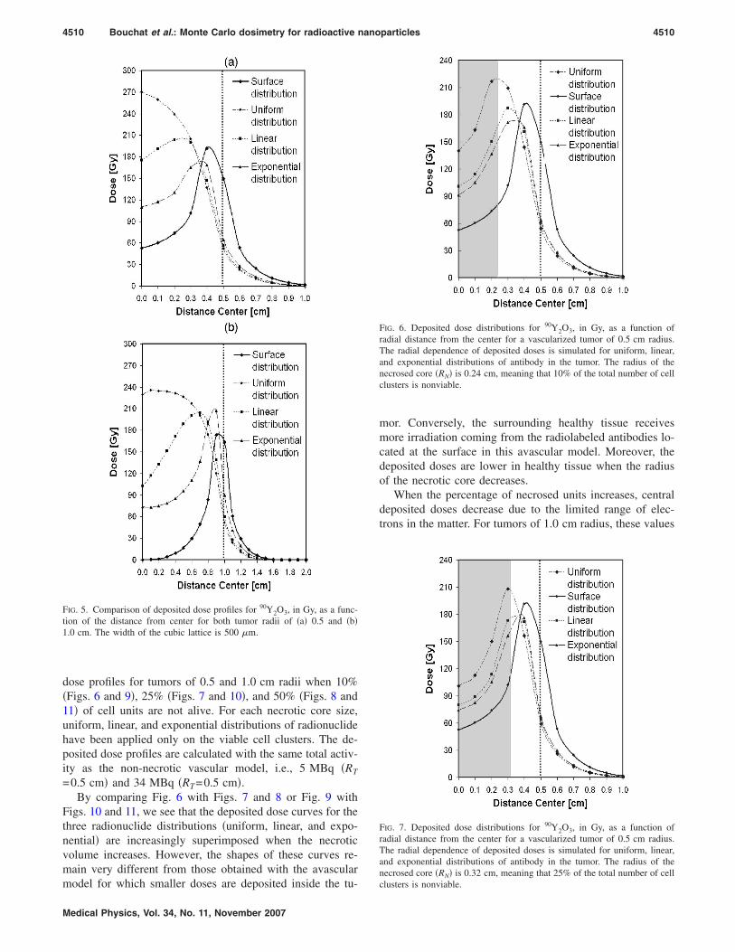
dose profiles for tumors of 0.5 and 1.0 cm radii when 10%�Figs. 6 and 9�, 25% �Figs. 7 and 10�, and 50% �Figs. 8 and11� of cell units are not alive. For each necrotic core size,uniform, linear, and exponential distributions of radionuclidehave been applied only on the viable cell clusters. The de-posited dose profiles are calculated with the same total activ-ity as the non-necrotic vascular model, i.e., 5 MBq �RT
=0.5 cm� and 34 MBq �RT=0.5 cm�.By comparing Fig. 6 with Figs. 7 and 8 or Fig. 9 with
Figs. 10 and 11, we see that the deposited dose curves for thethree radionuclide distributions �uniform, linear, and expo-nential� are increasingly superimposed when the necroticvolume increases. However, the shapes of these curves re-main very different from those obtained with the avascular
FIG. 5. Comparison of deposited dose profiles for 90Y2O3, in Gy, as a func-tion of the distance from center for both tumor radii of �a� 0.5 and �b�1.0 cm. The width of the cubic lattice is 500 �m.
model for which smaller doses are deposited inside the tu-
Medical Physics, Vol. 34, No. 11, November 2007
mor. Conversely, the surrounding healthy tissue receivesmore irradiation coming from the radiolabeled antibodies lo-cated at the surface in this avascular model. Moreover, thedeposited doses are lower in healthy tissue when the radiusof the necrotic core decreases.
When the percentage of necrosed units increases, centraldeposited doses decrease due to the limited range of elec-trons in the matter. For tumors of 1.0 cm radius, these values
FIG. 6. Deposited dose distributions for 90Y2O3, in Gy, as a function ofradial distance from the center for a vascularized tumor of 0.5 cm radius.The radial dependence of deposited doses is simulated for uniform, linear,and exponential distributions of antibody in the tumor. The radius of thenecrosed core �RN� is 0.24 cm, meaning that 10% of the total number of cellclusters is nonviable.
FIG. 7. Deposited dose distributions for 90Y2O3, in Gy, as a function ofradial distance from the center for a vascularized tumor of 0.5 cm radius.The radial dependence of deposited doses is simulated for uniform, linear,and exponential distributions of antibody in the tumor. The radius of thenecrosed core �RN� is 0.32 cm, meaning that 25% of the total number of cell
clusters is nonviable.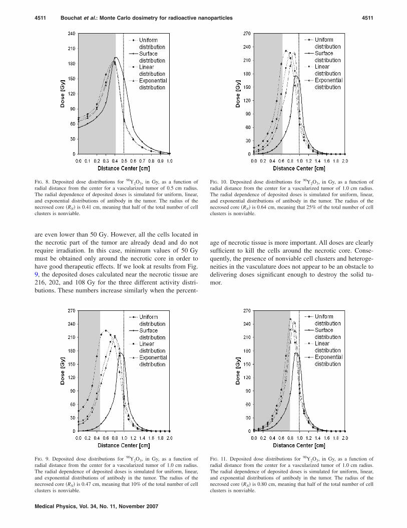
4511 Bouchat et al.: Monte Carlo dosimetry for radioactive nanoparticles 4511
are even lower than 50 Gy. However, all the cells located inthe necrotic part of the tumor are already dead and do notrequire irradiation. In this case, minimum values of 50 Gymust be obtained only around the necrotic core in order tohave good therapeutic effects. If we look at results from Fig.9, the deposited doses calculated near the necrotic tissue are216, 202, and 108 Gy for the three different activity distri-butions. These numbers increase similarly when the percent-
FIG. 8. Deposited dose distributions for 90Y2O3, in Gy, as a function ofradial distance from the center for a vascularized tumor of 0.5 cm radius.The radial dependence of deposited doses is simulated for uniform, linear,and exponential distributions of antibody in the tumor. The radius of thenecrosed core �RN� is 0.41 cm, meaning that half of the total number of cellclusters is nonviable.
FIG. 9. Deposited dose distributions for 90Y2O3, in Gy, as a function ofradial distance from the center for a vascularized tumor of 1.0 cm radius.The radial dependence of deposited doses is simulated for uniform, linear,and exponential distributions of antibody in the tumor. The radius of thenecrosed core �RN� is 0.47 cm, meaning that 10% of the total number of cell
clusters is nonviable.Medical Physics, Vol. 34, No. 11, November 2007
age of necrotic tissue is more important. All doses are clearlysufficient to kill the cells around the necrotic core. Conse-quently, the presence of nonviable cell clusters and heteroge-neities in the vasculature does not appear to be an obstacle todelivering doses significant enough to destroy the solid tu-mor.
FIG. 10. Deposited dose distributions for 90Y2O3, in Gy, as a function ofradial distance from the center for a vascularized tumor of 1.0 cm radius.The radial dependence of deposited doses is simulated for uniform, linear,and exponential distributions of antibody in the tumor. The radius of thenecrosed core �RN� is 0.64 cm, meaning that 25% of the total number of cellclusters is nonviable.
FIG. 11. Deposited dose distributions for 90Y2O3, in Gy, as a function ofradial distance from the center for a vascularized tumor of 1.0 cm radius.The radial dependence of deposited doses is simulated for uniform, linear,and exponential distributions of antibody in the tumor. The radius of thenecrosed core �RN� is 0.80 cm, meaning that half of the total number of cell
clusters is nonviable.
4512 Bouchat et al.: Monte Carlo dosimetry for radioactive nanoparticles 4512
IV. CONCLUSION
MCNPX can be used in radioimmunotherapy to evaluatethe energy deposition in tumor nodules and determine themost appropriate therapeutic treatment for a patient. How-ever, this software requires accurate information on the struc-ture and the microscopic distribution of radioactivity comingfrom the radiolabeled antibodies. In this work, the vascular-ized tumor was modeled by a set of spherical cell clusters of500 �m diameter arranged in a simple cubic lattice structure.Three different antibody distributions have been tested in thesimulations: Uniform, linear, and exponential. We have alsointroduced the possibility to have a central necrotic core in-side the tumor where the probability to observe an antibodyis supposed to be null. The shapes of spatial dose distribu-tions are strongly influenced by the morphology and the sizeof the solid tumors, indicating the importance of choosingthe optimal geometries to represent the tumor.
Spatial deposited dose distributions confirm the benefit ofusing radiolabeled nanoparticles containing hundreds of ra-dioactive atoms rather than antibodies coupled to a singleradionuclide to treat solid and poorly vascularized tumors inRIT. Indeed, even if the antibodies are distributed exponen-tially, doses higher than 50 Gy are delivered in the entiretumor. The use of radioactive nanoparticles thus limits theproblems of antibody penetrability often observed in classi-cal RIT when a single radioactive atom is labeled to eachspecific antibody.
ACKNOWLEDGMENTS
This research �Targan Project� is supported by the Wal-loon Region �Belgium�. C.M. and O.F. are senior researchassociates of FNRS �Fonds National de la Recherche Scien-tifique, Belgium�.
a�Author to whom correspondence should be addressed. Telephone: 0032-81-725479; Fax: 0032-81-725474. Electronic mail:[email protected]
1D. M. Goldenberg, “Targeted therapy of cancer with radiolabeled anti-bodies,” J. Nucl. Med. 43, 693–713 �2002�.
2D. M. Goldenberg and R. M. Sharkey, “Advances in cancer therapy withradiolabeled monoclonal antibodies,” Q. J. Nucl. Med. Mol. Imaging 50,248–264 �2006�.
3T. A. Waldmann, “Immunotherapy: Past, present and future,” Nat. Med.9, 269–277 �2003�.
4P. Carmeliet, “Angiogenesis in health and disease,” Nat. Med. 9, 653–660�2003�.
5J. Folkman, “Angiogenesis in cancer, vascular, rheumatoid and other dis-ease,” Nat. Med. 1, 27–31 �1995�.
6J. Ahlskog, G. Paganelli, and D. Neri, “Vascular tumor targeting,” Q. J.Nucl. Med. Mol. Imaging 50, 296–309 �2006�.
7A. Gruaz-Guyon, O. Raguin, and J. Barbet, “Recent advances in pretar-geted radio-immunotherapy,” Curr. Med. Chem. 12, 319–338 �2005�.
8E. J. Postema, O. C. Boerman, W. J. Oyen, J. M. Raemaekers, and F. H.Corstens, “Radioimmunotherapy of B-cell non-Hodgkin’s lymphoma,”Eur. J. Nucl. Med. 28, 1725–1735 �2001�.
9T. E. Witzig, “Radioimmunotherapy for patients with relapsed B-cell non-Hodgkin lymphoma,” Cancer Chemother. Pharmacol. 48, 91S-95S�2001�.
10C. A. White, J. R. Berlfein, and A. J. Grillo-Lopez, “Antibody-targetedimmunotherapy for treatment of non-Hodgkin’s lymphoma,” Curr. Pharm.Biotechnol. 1, 303–312 �2000�.
11D. M. Goldenberg, “The role of radiolabeled antibodies in the treatment
of non-Hodgkin’s lymphoma: the coming of age of radioimmunotherapy,”Medical Physics, Vol. 34, No. 11, November 2007
Crit. Rev. Oncol. Hematol. 39, 195–201 �2001�.12G. A. Wiseman, C. A. White, R. B. Sparks, W. D. Erwin, D. A. Podoloff,
D. Lamonica, N. L. Bartlett, J. Anthony Parker, W. L. Dunn, S. M. Spies,R. Belanger, T. E. Witzig, and B. R. Leigh, “Biodistribution and dosim-etry results from a phase III prospectively randomized controlled trial ofZevalin radioimmunotherapy for low-grade, follicular, or transformedB-cell non-Hodgkin’s lymphoma,” Crit. Rev. Oncol. Hematol. 39, 181–194 �2001�.
13W. A. Bethge and B. M. Sandmaier, “Targeted cancer therapy using ra-diolabeled monoclonal antibodies,” Technol. Cancer Res. Treat. 4, 393–405 �2005�.
14S. V. Govindan, G. L. Griffiths, H. J. Hansen, I. D. Horak, and D. M.Goldenberg, “Cancer therapy with radiolabeled and drug/toxin-conjugated antibodies,” Technol. Cancer Res. Treat. 4, 375–391 �2005�.
15R. M. Sharkey and D. M. Goldenberg, “Perspectives on cancer therapywith radiolabeled monoclonal antibodies,” J. Nuc. Med. Treat. 46, 115S-127S �2005�.
16D. M. Goldenberg, “Advancing role of radiolabeled antibodies in thetherapy of cancer,” Cancer Immunol. Immunother 52, 281–296 �2003�.
17D. L. Peng, T. Hihara, and K. Sumiyama, “Structure and magnetic prop-erties of FePt alloy cluster-assembled film,” J. Magn. Magn. Mater. 277,201–208 �2004�.
18A. L. Thomann, J. P. Salvetat, Y. Breton, C. Andreazza-Vignolle, and P.Brault, “Thermal stability of metal nanoclusters formed by low-pressureplasma sputtering,” Thin Solid Films 428, 242–247 �2003�.
19A. Naudon, D. Babonneau, D. Thiaudière, and S. Lequien, “Grazing-incidence small-angle x-ray scattering applied to the characterization ofaggregates in surface regions,” Physica B 283, 69–74 �2000�.
20Y. Saito, “Nanoparticles and filled nanocapsules,” Carbon 33, 979–988�1995�.
21S. M. Moghimi, A. C. Hunter, and J. C. Murray, “Long-circulating andtarget-specific nanoparticles: Theory to practice,” Pharmacol. Rev. 53,283–318 �2001�.
22S. M. Moghimi and J. Szebeni, “Stealth liposomes and long circulatingnanoparticles: Critical issues in pharmacokinetics, opsonization, andprotein-binding properties,” Prog. Lipid Res. 42, 463–478 �2003�.
23S. M. Moghimi and A. C. Hunter, “Recognition by macrophages and livercells of opsonized phospholipid vesicles and phospholipid headgroups,”Pharm. Res. 18, 1–8 �2001�.
24E. D. Owens, III and N. A. Peppas, “Opsonization, biodistribution, andpharmacokinetics of polymeric nanoparticles,” Int. J. Pharm. 307, 93–102�2006�.
25S. M. Moghimi, A. C. Hunter, and J. C. Murray, “Nanomedicine: Currentstatus and future prospect,” FASEB J. 19, 311–330 �2005�.
26R. Gref, M. Luck, P. Quellec, M. Marchand, E. Dellacherie, S. Harnish, T.Blunk, and R. H. Muller, “Stealth corona-core nanoparticles surfacemodified by polyethylene glycol �PEG�: influences of the corona �PEGchain length and surface density� and of the core composition on phago-cytic uptake and plasma protein adsorption,” Colloids Surf., B 18, 301–313 �2000�.
27Y. Yi, J. H. Kim, H.-W. Kang, H. S. Oh, S. W. Kim, and M. H. Seo, “Apolymeric nanoparticle consisting of mPEG-PLA-Toco and PLMA-COONa as a drug carrier: improvements in cellular uptake and biodistri-bution,” Pharm. Res. 22, 200–208 �2005�.
28C. C. Berry and A. S. G. Curtis, “Functionalisation of magnetic nanopar-ticles for applications in biomedicine,” J. Leukoc Biol. 78, 585–594�2005�.
29S. E. McNeil, “Nanotechnology for the biologist,” J. Leukoc Biol. 78,585–594 �2005�.
30M. Yokoyama, “Drug targeting with nano-sized carrier systems,” J. Artif.Organs 8, 77–84 �2005�.
31W. J. Parak, T. Pellegrino, and C. Plank, “Labelling of cells with quantumdots,” Nanotechnology 16, R9–R25 �2005�.
32X. Wu, H. Liu, K. N. Haley, J. A. Treadway, J. P. Larson, N. Ge, F. Peale,and M. P. Bruchez, “Immunofluorescent labeling of cancer marker Her2and other cellular targets with semiconductor quantum dots,” Nat. Bio-technol. 21, 41–46 �2003�.
33X. Gao, Y. Cui, R. M. Levenson, L. W. K. Chung, and S. Nie, “In vivocancer targeting and imaging with semiconductor quantum dots,” Nat.Biotechnol. 22, 969–976 �2004�.
34M. N. Rhyner, A. M. Smith, X. Gao, H. Mao, L. Yang, and S. Nie,“Quantum dots and multifunctional nanoparticles: new contrast agents for
tumor imaging,” Nanomedicine 1, 209–217 �2006�.
4513 Bouchat et al.: Monte Carlo dosimetry for radioactive nanoparticles 4513
35J. T. Kemshead and J. Ugelstad, “Magnetic separation techniques: theirapplication to medicine,” Mol. Cell. Biochem. 67, 11–18 �1985�.
36V. Choesmel, P. Anract, H. Hoifodt, J. P. Thiery, and N. Blin, “A relevantimmunomagnetic assay to detect and characterize epithelial cell adhesionmolecule-positive cells in bone marrow from patients with breast carci-noma: immunomagnetic purification of micrometastases,” Cancer 101,693–703 �2004�.
37C. Loo, A. Lowery, N. Halas, J. West, and R. Drezek, “Immunotargetednanoshells for integreted cancer imaging and therapy,” Nano Lett. 5, 709–711 �2005�.
38L. R. Hirsch, R. J. Stafford, J. A. Bankson, S. R. Sershen, B. Rivera, R. E.Price, J. D. Hazle, N. J. Halas, and J. L. West, “Nanoshell-mediatednear-infrared thermal therapy of tumors under magnetic resonance guid-ance,” Proc. Natl. Acad. Sci. U.S.A. 100, 13549–13554 �2003�.
39G. Wang, T. Huang, R. W. Murray, L. Menard, and R. G. Nuzzo,“Near-IR luminescence of monolayer-protected metal clusters,” J. Am.Chem. Soc. 127, 812–813 �2005�.
40V. E. Nuttens, A.-C. Wera, V. Bouchat, and S. Lucas, “Determination ofbiological vector characteristics and nanoparticle dimensions for radioim-munotherapy with radioactive nanoparticles,” Appl. Radiat. Isot. �ac-cepted�.
41D. Ribatti, A. Vacca, and F. Dammacco, “The role of vascular phase insolid tumor growth: A historical review,” Neoplasia 1, 293–302 �1999�.
42J. Folkman, E. Merler, C. Abernathy, and G. Williams, “Isolation of atumor factor responsible or angiogenesis,” J. Exp. Med. 133, 275–288�1971�.
43R. M. Sutherland, “Cell and environment interactions in tumor microre-gions: The multicell spheroid model,” Science 240, 177–184 �1988�.
44M. Cremonesi, M. Ferrari, L. Bodei, G. Tosi, and G. Paganelli, “Systemicand locoregional dosimetry in receptor radionuclide therapy with pep-tides,” Q. J. Nucl. Med. Mol. Imaging 50, 288–295 �2006�.
45R. W. Howell, D. V. Rao, and K. S. R. Sastry, “Macroscopic dosimetryfor radioimmuno-therapy: Nonuniform activity distributions in solid tu-mors,” Med. Phys. 16, 66–74 �1989�.
46E. B. Van Dieren, M. A. B. D. Plaizier, A. Van Lingen, J. C. Roos, G. W.Barendsen, and G. J. J. Teule, “Absorbed dose distribution of the augeremitters 67Ga and 125I and the �-emitters 67Cu, 90Y, 131I, and 186Reas a function of tumor size, uptake, and intracellular distributin,” Int. J.Radiat. Oncol., Biol., Phys. 36, 197–204 �1996�.
47ICRU Report 44, “Tissue substitutes in radiation dosimetry and measure-ment,” �International Commission on Radiation Units and Measurements,Bethesda, MD, 1989�, p 22.
48S. Lin, C.-K. Lee, Y.-H. Lin, S.-Y. Lee, B.-C. Sheu, J.-C. Tsai, and S.-M.Hsu, “Homopolyvalent antibody-antigen interaction kinetic studies withuse of a dual-polarization interferometric biosensor,” Biosens. Bioelec-tron. 22, 715–721 �2006�.
49A. S. Paulo and R. Garcia, “High-resolution imaging of antibodies bytapping-mode atomic force microscopy: attractive and repulsive tip-sample interaction regimes,” Biophys. J. 78, 1599–1605 �2006�.
50L. Cser, I. A. Gladkib, F. Franek, and Yu. M. Ostanevich, “Investigationof antibody structures by scattering techniques,” Colloid Polym. Sci. 259,625–640 �1981�.
51H. F. Dvorak, J. A. Nagy, and A. M. Dvorak, “Structure of solid tumorsand their vasculature: Implications for therapy with monoclonal antibod-ies,” Cancer Cells 3, 77–85 �1991�.
Medical Physics, Vol. 34, No. 11, November 2007
52B. Döme, S. Paku, B. Somlai, and J. Timar, “Vascularization of cutaneousmelanoma involves vessel co-option and has clinical significance,” J.Pathol. 197, 355–362 �2002�.
53J. A. M. Beliën, P. J. van Diest, and J. P. A. Baak, “Relation betweenvascularization and proliferation in invasive breast cancer,” J. Pathol.189, 309–318 �1999�.
54C. Schlueter, H. Weber, B. Meyer, P. Rogalla, K. Röser, S. Hauke, and J.Bullerdiek, “Angiogenic signaling through hypoxia,” Am. J. Pathol. 166,1259–1263 �2005�.
55D.-S. Lee, H. Rieger, and K. Bartha, “Flow correlated percolation duringvascular remodeling in growing tumors,” Phys. Rev. Lett. 96, 058104�2006�.
56K. Bartha and H. Rieger, “Vascular network remodeling via vessel coop-tion, regression and growth in tumors,” J. Theor. Biol. 241, 903–918�2006�.
57J. L. Humm and L. M. Cobb, “Nonuniformity of tumor dose in radioim-munotherapy,” J. Nucl. Med. 31, 75–83 �1990�.
58D. B. Pelowitz, ed., “MCNPX User’s Manual, Version 2.5.0,” LA-CP-05-0369 �2005�.
59N. Reynaert, H. Palmans, H. Thierens, and R. Jeraj, “Parameter depen-dence of the MCNP electron transport in determining dose distributions,”Med. Phys. 29, 2446–2454 �2002�.
60D. R. Schaart, J. Th.M. Jansen, J. Zoetelief, and P. F. A. de Leege, “Acomparison of MCNP4C electron transport with ITS 3.0 and experimentat incident energies between 100 keV and 20 MeV: influence of voxelsize, substeps and energy indexing algorithm,” Phys. Med. Biol. 47,1459–1484 �2002�.
61P. Karnami and K. Kairemo, “Targeting endothelial growth with mono-clonal antibodies against Tie-1 kinase in mouse models,” Clin. CancerRes. 9, 3821s–3826s �2003�.
62A. Jekunen, K. Kairemo, and P. Karnami, “In vivo modulators of antibodykinetics,” Acta Oncol. 35, 267–271 �1996�.
63S. Palm, R. M. Enmon, Jr., C. Matei, K. S. Kolbert, S. Xu, P. B. Zan-zonico, R. L. Finn, J. A. Koutcher, S. M. Larson, and G. Sgouros, “Phar-macokinetics and biodistribution of 86Y-Trastuzumab for 90Y dosimetryin an ovarian carcinoma model: correlative MicroPET and MRI,” J. Nucl.Med. 44, 1148–1155 �2003�.
64F. T. Lee, A. Rigopoulos, C. Hall, K. Clarke, S. H. Cody, F. E. Smyth, Z.Liu, M. W. Brechbiel, N. Hanai, E. C. Nice, B. Catimel, A. W. Burgess,S. Welt, G. Ritter, L. J. Old, and A. M. Scott, “Specific localization,gamma camera imaging, and intracellular trafficking of radiolabeled chi-meric anti-GD3 ganglioside monoclonal antibody KM871 in SK-MEL-28melanoma xenografts,” Cancer Res. 61, 4474–4482 �2001�.
65C.-H. Chang, R. M. Sharkey, E. A. Rossi, H. Karacay, W. McBride, H. J.Hansen, J.-F. Chatal, J. Barbet, and D. M. Goldenberg, “Molecular ad-vances in pretargeting radioimunotherapy with bispecific antibodies,”Mol. Cancer Ther. 1, 553–563 �2002�.
66K. Tobinai, Y. Kobayashi, and M. Narabayashi, “Feasibility and pharma-cokinetic study of a chimeric anti-CD20 monoclonal antibody �IDEC-C2B8, rituximab� in relapsed B-cell lymphoma,” Ann. Oncol. 9, 527–534�1998�.
67D. M. Goldenberg, David M. Goldenberg, R. M. Sharkey, G. Paganelli, J.Barbet, and J.-F. Chatal, “Antibody pretargeting advances cancer radio-immunodetection and radioimmunotherapy,” J. Clin. Oncol. 24, 823–834�2006�.