Partie 1 : Optimisation du matériel végétal et de...
123
Résultats et Conclusion Partie 1: Optimisation du matériel végétal et de l’extraction 65 Partie 1 : Optimisation du matériel végétal et de l’extraction 1. Introduction De nombreuses études effectuées partout dans le monde ont révélé que l'alimentation était la cause de nombreux problèmes de santé des populations occidentales. Il faut relever ce défi du "manger autrement", de l'alimentation santé, de la "functional food" pour mieux vivre, plus en forme et plus longtemps, ceci sans bouleverser des habitudes le plus souvent solidement ancrées, ni renoncer aux plaisirs de la table. Depuis la nuit des temps, les hommes apprécient les vertus des plantes pour vaincre la souffrance et améliorer leur santé. Certaines plantes ou organes comme les baies sont riches en composés phénoliques (Shahidi and Naczk, 2004 ; Zadernowski et al., 2005) tels que des flavonoïdes (Häkkinen et al., 1999). Ces composés peuvent avoir des effets sur la prévention de certaines pathologies (Cao et al., 1998 ; Landbo et al., 2001 ; Mazza et al., 2002), effets dus notamment à leurs propriétés antioxydantes (Wang et al., 1996 ; Skrede and Wrolstad, 2002). Les myrtilles (Prior et al., 1998) et le cassis (Moyer et al., 2002 ; Ehala et al., 2005) sont les baies qui possèdent les plus hauts taux en anthocyanes, ainsi qu’une capacité antioxydante très élevée. La myricétine, la quercétine, et le kaempférol sont les principaux flavonols, un autre groupe de flavonoïdes très important. Les teneurs en ces composés dans les baies sont fonction de différents facteurs comme le degré de maturation (Mikkonen et al., 2001 ; Raffo et al., 2004), les différences génétiques (cultivar), les conditions environnementales avant la récolte, le stockage après la récolte, les traitements pour la conservation (Shahidi and Naczk, 2004 ; Heiberg et al., 1992 ; Wang and Lin, 2000) ainsi que la saison (Howard et al., 2003).
Transcript of Partie 1 : Optimisation du matériel végétal et de...
Résultats et Conclusion Partie 1: Optimisation du matériel végétal et de l’extraction
65
Partie 1 : Optimisation du matériel végétal et de l’extraction
1. Introduction
De nombreuses études effectuées partout dans le monde ont révélé que l'alimentation était la
cause de nombreux problèmes de santé des populations occidentales. Il faut relever ce défi du
"manger autrement", de l'alimentation santé, de la "functional food" pour mieux vivre, plus en
forme et plus longtemps, ceci sans bouleverser des habitudes le plus souvent solidement
ancrées, ni renoncer aux plaisirs de la table.
Depuis la nuit des temps, les hommes apprécient les vertus des plantes pour vaincre la
souffrance et améliorer leur santé. Certaines plantes ou organes comme les baies sont riches
en composés phénoliques (Shahidi and Naczk, 2004 ; Zadernowski et al., 2005) tels que des
flavonoïdes (Häkkinen et al., 1999). Ces composés peuvent avoir des effets sur la prévention
de certaines pathologies (Cao et al., 1998 ; Landbo et al., 2001 ; Mazza et al., 2002), effets
dus notamment à leurs propriétés antioxydantes (Wang et al., 1996 ; Skrede and Wrolstad,
2002). Les myrtilles (Prior et al., 1998) et le cassis (Moyer et al., 2002 ; Ehala et al., 2005)
sont les baies qui possèdent les plus hauts taux en anthocyanes, ainsi qu’une capacité
antioxydante très élevée. La myricétine, la quercétine, et le kaempférol sont les principaux
flavonols, un autre groupe de flavonoïdes très important. Les teneurs en ces composés dans
les baies sont fonction de différents facteurs comme le degré de maturation (Mikkonen et al.,
2001 ; Raffo et al., 2004), les différences génétiques (cultivar), les conditions
environnementales avant la récolte, le stockage après la récolte, les traitements pour la
conservation (Shahidi and Naczk, 2004 ; Heiberg et al., 1992 ; Wang and Lin, 2000) ainsi que
la saison (Howard et al., 2003).
Résultats et Conclusion Partie 1: Optimisation du matériel végétal et de l’extraction
66
L'industrie agro-alimentaire doit actuellement faire face à une réduction des marges
bénéficiaires sur les produits traditionnels. Elle cherche donc à ajouter de la plus-value à
certains de ses produits et le développement des produits "santé" est une alternative très
intéressante, à une époque où la relation alimentation-santé est reconnue et où nous souffrons
de plus en plus de pathologies liées à l'alimentation. Un des principaux défis de l’industrie
alimentaire (fruits et légumes) est de valoriser les propriétés de ses produits. Grâce à leurs
propriétés de prévention de certaines pathologies, la richesse en composés phénoliques des
matières premières ou des produits transformés pourrait constituer un bon argument de vente.
Les baies domestiques et sauvages sont consommées en abondance et de nombreuses études
ont été réalisées pour évaluer leur contenu en composés phénoliques et leur pouvoir
antioxydant. Actuellement, peu d’études ont été faites sur les propriétés antioxydantes
d’autres explants tels que les bourgeons et les feuilles. Declume (1989) et Chrubasik (2000),
de leur coté, ont montré que les feuilles de cassis possédaient une activité anti-inflammatoire.
Cependant, aucune étude n'a comparé la capacité antioxydante des divers organes du cassis.
Dans cette partie de notre travail, nous avons voulu comparer les capacités antioxydantes de
différentes parties de divers cultivars de cassis dans le but de préparer un extrait présentant
une haute capacité antioxydante. Pour ce faire, nous avons testé différents paramètres comme
le cultivar, le type d’explant prélevé et son stade de développement: « Antioxidant capacity
of black currant varies with organ, season and cultivar. » (Journal of Agricultural and
Food Chemistry 54, 6271-6276, 2006).
Il est clair que, excepté les baies, d'autres explants ne peuvent être consommés tels quels mais
doivent être conditionnés comme complément alimentaire ou être ajoutés à des préparations.
Il faut noter que les feuilles et les bourgeons du cassis sont déjà utilisés dans la préparation de
compléments alimentaires.
La méthode d'extraction doit permettre l'extraction la plus complète possible des composés
d'intérêt et doit éviter leur modification chimique (Zuo et al., 2002). De nombreuses solutions
d’extraction sont proposées dans la littérature pour extraire les polyphénols de la matrice
végétale (Chavan et al., 2001). Le rendement dépend grandement de la solution extractive
mais aussi de la méthode utilisée (Goli et al., 2005) et de la matrice végétale. L'eau, des
Résultats et Conclusion Partie 1: Optimisation du matériel végétal et de l’extraction
67
mélanges aqueux d’éthanol, de méthanol et d’acétone sont généralement utilisés (Sun and Ho,
2005).
Dans cette partie du travail, nous avons tenté d'optimiser l'extraction des composés
phénoliques totaux afin d’obtenir un extrait dont la capacité antioxydante est la plus élevée
possible et cela dans des conditions compatibles avec l’alimentation.
Divers explants de cassis ont été testés ainsi que divers facteurs contribuant à l’efficacité de
l’extraction : le mélange d’extraction, le pH et l’état du matériel (frais, congelé ou lyophilisé).
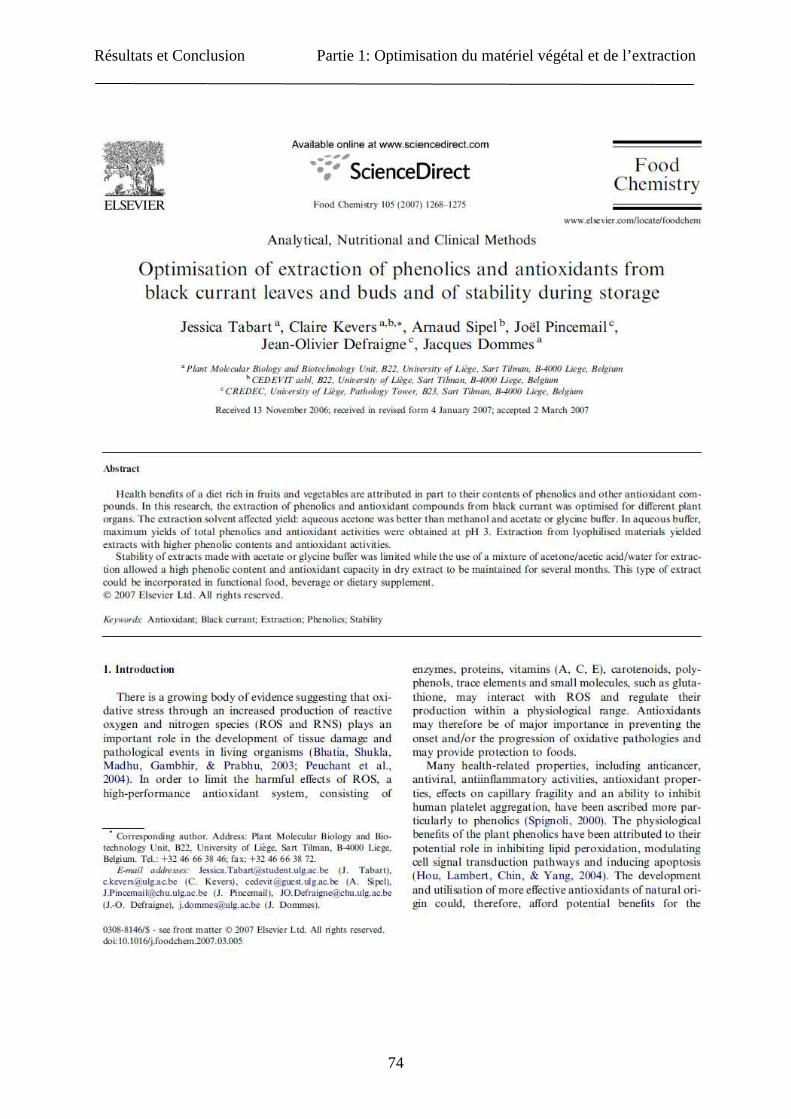
La stabilité des propriétés antioxydantes de l’extrait a également été étudiée : « Optimisation
of extraction of phenolics and antioxidants from black currant leaves and buds and of
stability during storage. » (Food Chemistry 105, 1268-1275, 2007).

Résultats et Conclusion Partie 1: Optimisation du matériel végétal et de l’extraction
68
2. Articles

Résultats et Conclusion Partie 1: Optimisation du matériel végétal et de l’extraction
69
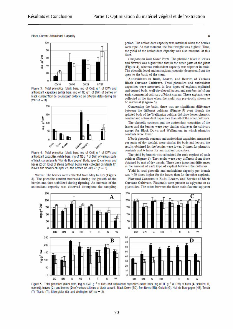
Résultats et Conclusion Partie 1: Optimisation du matériel végétal et de l’extraction
70

Résultats et Conclusion Partie 1: Optimisation du matériel végétal et de l’extraction
71
Résultats et Conclusion Partie 1: Optimisation du matériel végétal et de l’extraction
72
Résultats et Conclusion Partie 1: Optimisation du matériel végétal et de l’extraction
73
Résultats et Conclusion Partie 1: Optimisation du matériel végétal et de l’extraction
74
Résultats et Conclusion Partie 1: Optimisation du matériel végétal et de l’extraction
75
Résultats et Conclusion Partie 1: Optimisation du matériel végétal et de l’extraction
76
Résultats et Conclusion Partie 1: Optimisation du matériel végétal et de l’extraction
77

Résultats et Conclusion Partie 1: Optimisation du matériel végétal et de l’extraction
78
Résultats et Conclusion Partie 1: Optimisation du matériel végétal et de l’extraction
79

Résultats et Conclusion Partie 1: Optimisation du matériel végétal et de l’extraction
80

Résultats et Conclusion Partie 1: Optimisation du matériel végétal et de l’extraction
81
Résultats et Conclusion Partie 1: Optimisation du matériel végétal et de l’extraction
82
3. Conclusion
A l’heure actuelle, un vrai engouement pour les produits enrichis en phytonutriments secoue
la planète. Un vrai challenge s’opère au niveau industriel pour produire cette nouvelle
génération de produits, enrichis avec ces composés antioxydants ou autres, de base naturelle.
Au niveau des antioxydants, un des premiers points est de trouver un produit naturel riche en
composés polyphénoliques et de haute activité antioxydante. En prenant comme modèle le
cassis, nous avons pu remarquer que le taux en polyphénols ainsi que la capacité antioxydante
variaient en fonction de la saison de prélèvement et ce, quelque soit l’explant analysé
(feuilles, fruits, bourgeons ouverts ou fermés). En prenant aussi en compte la biomasse, il
s’avère que les feuilles prélevées en juin sont les plus intéressantes. Divers cultivars de cassis
ont aussi été analysés, mais aucune différence significative au niveau du contenu en composés
phénoliques et de la capacité antioxydante n’a été observée.
Pour tenter d’augmenter le rendement d’extraction des composés phénoliques du cassis,
l’étape d’extraction a été optimisée en jouant sur divers paramètres. Les résultats ont été
comparés au tampon glycine, tampon aqueux pouvant sans risque être utilisé dans la
production de compléments alimentaires. Pour l’extraction des composés phénoliques, les
solvants utilisés couramment sont le méthanol, l’acétone ainsi que leurs mélanges aqueux,
acidifiés ou non. Pour les feuilles de cassis, le mélange aqueux contenant 70% d’acétone
donne le meilleur rendement d’extraction. L’ajout de 2% d’acide acétique permet d’améliorer
ce rendement. Nous avons aussi analysé la stabilité de ces extraits au cours du temps et nous
avons pu constater que le mélange acétone/eau/acide acétique (70 :28 :2) permettait la
conservation des propriétés de nos extraits à plus long terme que le tampon glycine.
Pour analyser ces divers paramètres, nous avons utilisé deux techniques d’analyses courantes :
la méthode de décoloration du radical DPPH ainsi que le dosage des phénols totaux. Ces deux
techniques spectrophotométriques sont des techniques in vitro basées sur un principe
chimique propre ne reflétant qu’une infime partie des réactions potentielles des différents
composants de l’extrait. Dans la suite de ce travail, la caractérisation de l’activité
antioxydante ainsi que le contenu en composés phénoliques sera abordée en utilisant un
maximum de techniques tant in vitro que sur modèles cellulaires.
Résultats et Conclusion Partie 2: Caractérisation des extraits de cassis
83
Partie 2: Caractérisation des extraits de cassis
1. Introduction
Les molécules antioxydantes des matrices végétales peuvent être caractérisées par une
séparation chromatographique (par exemple HPLC, GC,…) suivie éventuellement d’une
identification par spectrométrie de masse ou plus globalement par des méthodes
spectrophotométriques détectant des groupes spécifiques chimiquement semblables de
composés réactifs. Les techniques chromatographiques sont très précises et permettent
l’identification d’un grand nombre de composés mais ces techniques sont très longues et
coûteuses (Stratil et al., 2006). Il est donc plus simple d’utiliser d’abord des méthodes
spectrophotométriques pour quantifier des familles de composés.
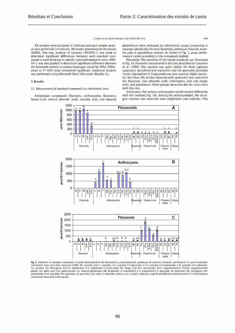
Dans un premier temps, nous avons utilisé des standards et tenté au moyen de ceux-ci de
valider les méthodes colorimétriques pour différents groupes de composés antioxydants
(flavonoïdes, anthocyanines, catéchines, acide ascorbique, et glutathion). Ces méthodes
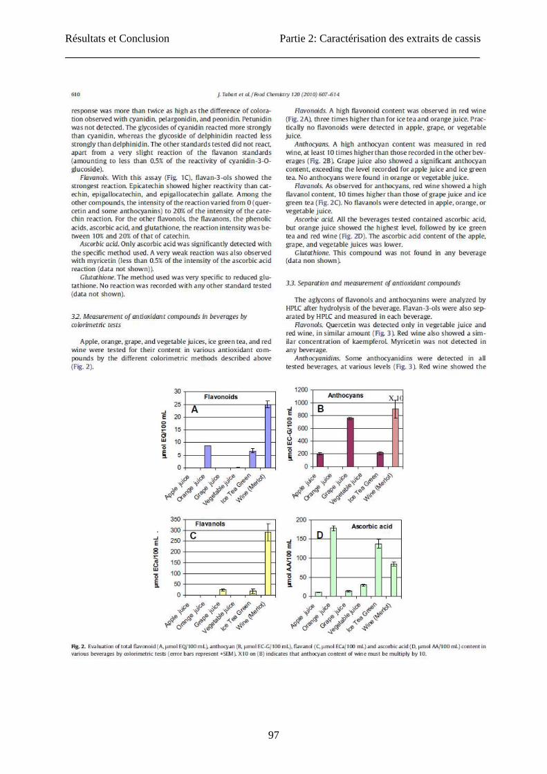
colorimétriques seront aussi appliquées à diverses matrices alimentaires (jus de pomme, jus
d’orange, jus de légumes, thé glacé et vin rouge) et les résultats mis en relation avec leur
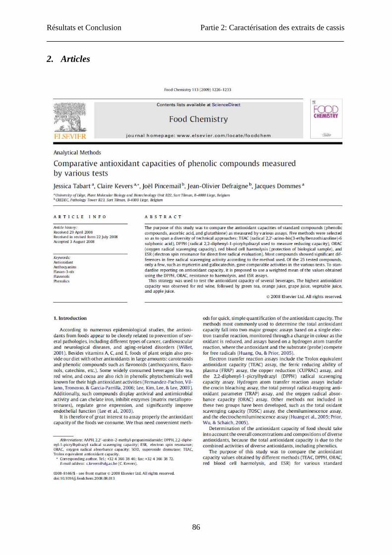
capacité antioxydante totale. Les résultats sont présentés dans l’article: « Evaluation of
spectrophotometric methods for antioxidant compound measurement in relation to total
antioxidant capacity in beverages. » (Food Chemistry 120, 607-614, 2010). Ensuite, ces
méthodes colorimétriques ont été appliquées à nos extraits de cassis, parallèlement à des
dosages de composés identifiés par chromatographie liquide: « Ascorbic acid, Phenolic acid,
Flavonoid and Carotenoid profiles of selected extracts from Ribes nigrum. » (Journal of
Agricultural and Food Chemistry, soumis en novembre 2010).
De nombreuses méthodes rapides et simples ont été développées pour l’évaluation de la
capacité antioxydante de matrices végétales. Les méthodes les plus utilisées sont classées en
deux groupes: les analyses basées sur un transfert d'électron, visualisées par un changement
de couleur lors de la réduction de l’oxydant (TEAC, FRAP, CUPRAC, DPPH), et les analyses
basées sur un transfert d'atome d'hydrogène, où l'antioxydant et le substrat (sonde) sont en
concurrence face aux radicaux libres (TRAP, ORAC, crossing blancking assay) (Huang et al.,
2005). D'autres méthodes non incluses dans ces deux groupes ont aussi été développées :
Résultats et Conclusion Partie 2: Caractérisation des extraits de cassis
84
TOSC, la chemiluminescence, l'électrochemiluminescence (Huang et al., 2005 ; Prior et al.,
2005), la RPE (piégeage de l’anion superoxyde par les molécules antioxydantes). Dans un
premier temps, nous avons comparé les valeurs de capacité antioxydante obtenues par les
différentes méthodes (TEAC, DPPH, ORAC et RPE) pour différents composés antioxydants
purs (principalement des composés phénoliques). Ensuite, nous avons proposé une méthode
permettant de normaliser la valeur de la capacité antioxydante. L'approche proposée a été
testée sur plusieurs boissons (jus de pomme, jus d’orange, jus de légumes, thé glacé et vin
rouge): « Comparative antioxidant capacities of phenolic compounds measured by
various tests. » (Food Chemistry 113, 1226-1233, 2009).
Toutes les méthodes décrites ci-dessus sont des techniques basées sur l’interaction in vitro
entre un système de production de radicaux libres, des antioxydants et une sonde. Elles ne
reflètent en aucun cas les aspects biologiques. D’autres techniques utilisant une approche plus
biologique ont aussi été développées. Nous en avons testé trois sur notre matériel. La
première analyse utilisée est le test de résistance des globules rouges soumis à un stress
oxydant en présence de composés antioxydants (Girodon et al., 1997). Le deuxième modèle
est l'analyse de l’activité antioxydante cellulaire (Wolfe and Liu, 2007) réalisée sur culture
cellulaire. Cette analyse permet de mesurer l'inhibition de l'oxydation de colorant par des
antioxydants dans des cellules en culture. Cette méthode donne une capacité antioxydante en
rapport avec la prise des composés antioxydants par des cellules. Un troisième modèle permet
de mettre en relation la production de radicaux libres et les situations inflammatoires.
L'activation des neutrophiles avec libération de myéloperoxydase (MPO) est impliquée dans
diverses pathologies chroniques inflammatoires chez l'homme et les chevaux. Une stimulation
excessive des neutrophiles avec production d’espèces réactives de l’oxygène (ROS) et
dégagement de la MPO mène à une activité préjudiciable d'oxydant sur les cellules, les tissus
et les molécules voisines. Quelques drogues et composés phénoliques ont été testés pour
limiter les effets délétères de l'activité excessive de MPO comme le resvératrol (Kohnen et al.,
2007). Ces effets ont été évalués par différentes analyses spécifiques à la MPO.

Résultats et Conclusion Partie 2: Caractérisation des extraits de cassis
85
Les extraits de cassis (feuilles, baies et bourgeons) ont été examinés pour leur potentiel
antioxydant par diverses analyses in vitro (DPPH, TEAC, ORAC, RPE et phénols totaux)
décrites ci-dessus. Nous avons complété notre recherche en étudiant l'effet de ces extraits sur
les modèles cellulaires en conditions oxydantes et, sur l'activité spécifique de la MPO équine
purifiée, sur la production de ROS et sur la dégranulation des neutrophiles équins :
« Antioxidant and anti-inflammatory activities of Ribes nigrum extracts. » (Journal of
Nutritional Biochemistry, soumis en janvier 2011).
Résultats et Conclusion Partie 2: Caractérisation des extraits de cassis
86
2. Articles
Résultats et Conclusion Partie 2: Caractérisation des extraits de cassis
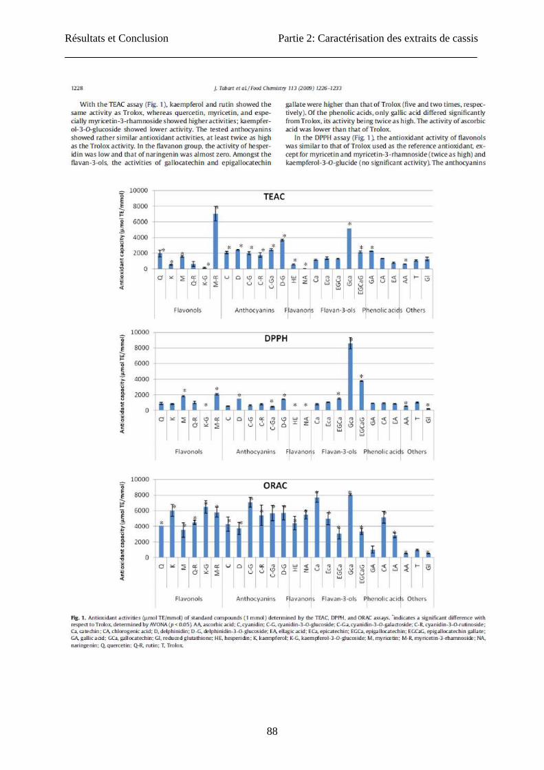
87
Résultats et Conclusion Partie 2: Caractérisation des extraits de cassis
88

Résultats et Conclusion Partie 2: Caractérisation des extraits de cassis
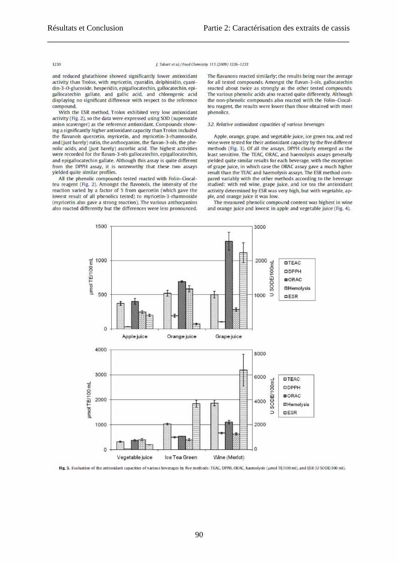
89
Résultats et Conclusion Partie 2: Caractérisation des extraits de cassis
90
Résultats et Conclusion Partie 2: Caractérisation des extraits de cassis
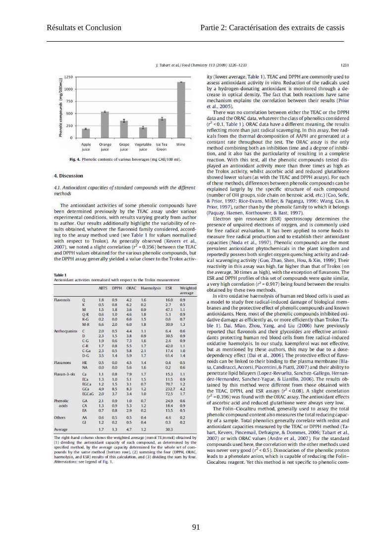
91
Résultats et Conclusion Partie 2: Caractérisation des extraits de cassis
92

Résultats et Conclusion Partie 2: Caractérisation des extraits de cassis
93
Résultats et Conclusion Partie 2: Caractérisation des extraits de cassis
94

Résultats et Conclusion Partie 2: Caractérisation des extraits de cassis
95
Résultats et Conclusion Partie 2: Caractérisation des extraits de cassis
96
Résultats et Conclusion Partie 2: Caractérisation des extraits de cassis
97
Résultats et Conclusion Partie 2: Caractérisation des extraits de cassis
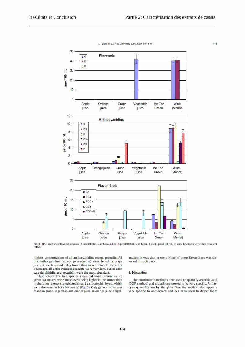
98
Résultats et Conclusion Partie 2: Caractérisation des extraits de cassis
99
Résultats et Conclusion Partie 2: Caractérisation des extraits de cassis
100
Résultats et Conclusion Partie 2: Caractérisation des extraits de cassis
101
Résultats et Conclusion Partie 2: Caractérisation des extraits de cassis
102
Ascorbic acid, Phenolic acid, Flavonoid and Carotenoid profiles of selected
extracts from Ribes nigrum.
Jessica TABARTa, Claire KEVERSa, Danièle EVERSb, Jacques DOMMESa
aPlant Molecular Biology and Biotechnology Unit, University of Liège, B22, Sart –Tilman, B-
4000 Liège (Belgium)
bPublic Research Center - Gabriel Lippmann, Rue du Brill 41, L-4422 Belvaux (Luxembourg)
Corresponding author: Jessica TABART: [email protected]
Other authors: [email protected], [email protected], [email protected];
TITLE RUNNING HEAD: Phenolics from Ribes nigrum
ABSTRACT
Small fruits such as berries have low energy contents, but high content of vitamins,
micronutrients, and dietary fibres and constitute a good source of natural antioxidant
compounds that are important constituents of the human diet. This study allowed to identify a
large number of compounds in an extract of blackcurrant showing high antioxidant activity
and to compare their profile in various parts of the plants (leaves, buds and berries). If it was
known that berries contained very high levels of natural phenolic compounds, here we
showed that leaves and buds could also be considered as a good sources of natural
antioxidants. Indeed, they contained high amount of ascorbic acid, phenolic acids, flavonoids
and carotenoids. An acetone mixture can extract several classes of phenolic compounds with a
good yield of ascorbic acid, flavonols, flavan-3-ols and anthocyanins. For phenolic acids, it
extracted fewer compounds than a specific extraction solution.
KEYWORDS: Antioxidant, flavonoids, flavonols, anthocyanins, flavanols, ascorbic acid,
phenolic acids, carotenoids.
Résultats et Conclusion Partie 2: Caractérisation des extraits de cassis
103
INTRODUCTION
Phenolic compounds are one of the most widely occurring groups of phytochemicals. In the
plant kingdom, these compounds can range from simple molecules such as phenolic acids, to
highly polymerized compounds such as tannins (1). Different classes of phenolic compounds
can be distinguished. Flavonoids and phenolic acids are the most abundant. Phenolic acids are
synthesised from hydroxybenzoic acid and hydroxycinnamic acid. The flavonoids are
subdivided in different classes: flavonols, anthocyanins, flavones, flavan-3-ols, flavanones,
isoflavones (2). In the plant, they play an important role in growth and reproduction,
providing protection against pathogens and predators as well as against abiotic stresses (1, 3).
In the human diet, fruits and vegetables have low energy content, but high content of
vitamins, essential micronutrients, and dietary fibres. They are also the predominant source of
flavonoids and phenolic acids. Many health-related properties, including anti-viral, anti-
inflammatory activities, antioxidant properties and ability to inhibit human platelet
aggregation have been described (4-6).
Carotenoids belong to another important group of natural pigments because of their wide
distribution, structural diversity and numerous functions for photosynthesis and for life in an
oxygen-containing atmosphere. The carotenoids are subdivided in two groups: the carotenes
and the xanthophylls (or oxycarotenoids) (7). Recently, these pigments have been described to
be implicated in the prevention of human health disorders such as heart disease and
photosensitivity disease but also of certain forms of cancer (8, 9).
Small fruits constitute a good source of natural antioxidant substances. Extracts of fruits from
various blackberry, raspberry and gooseberry cultivars act effectively as free radical inhibitors
(10, 11). In addition, the flavonoid content of small fruits has been investigated (12).
Blackcurrant berries contain very high amounts of phenolic compounds. Fresh blackcurrants
are particularly rich in anthocyanins. But other phenolic compounds like flavonols are also
present (13). Only a very small proportion of these berries are consumed fresh, most is
processed for juice concentrates. Leaves and buds can also be used (14); leaf micronisates and
glycerinate extracts of buds are especially commercialized as food supplements. Declume
(15) and Chrubasik (16) also demonstrate that leaf extracts of blackcurrant show significant
anti-inflammatory activity.
Résultats et Conclusion Partie 2: Caractérisation des extraits de cassis
104
The first objective of this study was to identify most of the compounds present in an extract of
blackcurrant with high antioxidant activity. We also compared the profile in ascorbic acid,
phenolic acids, flavonoïds and carotenoids of extracts from various parts of the plants (leaves,
buds and berries). The second objective was to check whether an acetone extraction method
optimised for high antioxidant capacity (17) did provide good yields of various classes of
antioxidant compounds.
MATERIAL AND METHODS
Materials
The buds, berries and leaves of two years old blackcurrant plants (Noir de Bourgogne) were
harvested respectively in March, July and August in the Belgian Ardennes (Bihain). The
various explants were directly cut out in pieces, frozen in liquid nitrogen, lyophilised and
stored at -20°C.
Sample preparation
Acetone Extraction:
One gram of fresh sample (berries, buds or leaves) was ground with 1 gram of quartz and 10
mL of extraction solution: acetone/water/acetic acid (70:28:2) (14). The mixture was shaken
during 1 h at 4°C and centrifuged at 17 000g for 15 min. The supernatant was removed, and
the sample was extracted again with the same procedure but incubated only 15 min. The
supernatants were pooled and then diluted as appropriate for the analyses.
Specific Extractions:
For ascorbic acid: 4 gram of fresh material (berries, buds, leaves) were ground with 1
gram of quartz and 80 mL of extraction solution (20 g/L metaphosphoric acid). The
mixture was shaken during 1 h at 4°C and centrifuged at 15 000 g for 15 min. For HPLC
analysis, 10 mL of a L-cystein solution (40g/L) were added to 20 mL of the supernatant
and the pH was adjusted between 7.0 and 7.2 with a solution of trisodium phosphate (200
g/L). After 5 minutes, the pH was adjusted between 2.5 and 2.8 with a solution of
metaphosphoric acid (20 g/L) (18).
Résultats et Conclusion Partie 2: Caractérisation des extraits de cassis
105
For phenolic acids (19): 3.4 gram of fresh material (berries, buds, leaves) were ground
with 1 gram of quartz and 49 mL of extraction solution (methanol/water/acetic acid;
90:1.5:8.5; v/v/v) containing 2 g/L of BHA (butylated hydroxyanisol). The mixture was
sonicated during 1 h at room temperature. 50 mL of 20 g/L ascorbic acid and 25 mL of 10
M NaOH were added for the hydrolysis of esterified phenolic acids. Then 12.5 mL of 12
N HCl were added for the extraction of glycoside forms of phenolic acids and the mixture
was incubated for 3 h at 85°C before cooling on ice. The mixture was extracted 5 times
with a solution of diethyl ether/ethyl acetate (50:50; v/v) and centrifuged at 1 200 g for 2
min. The organic phases were pooled and evaporated. All the residues were dissolved in 2
mL of 2% acetic acid and filtered for HPLC analysis.
For anthocyanins: One gram of fresh sample (berries, buds or leaves) was ground with 1
gram of quartz and 15 mL of 1% HCl in methanol (20). The mixture was shaken during 2
h at room temperature and incubated one night in the dark at -20°C before centrifugation
at 4 000g for 15 min. The supernatant was collected and the sample was extracted again
with 15 mL of 1% HCl in methanol. The supernatants were pooled and then diluted as
appropriate for the analyses.
For flavanols (based on Parva-Uzunalic et al. (21)): One gram of fresh sample (berries,
buds or leaves) was ground with 1 gram of quartz and 10 mL of acetone 100%. The
mixture was shaken during 1 h at 70°C and centrifuged at 17 000 g for 15 min. The
supernatant was removed and the sample was extracted again with the same procedure but
incubated only 15 min. The supernatants were pooled and then diluted as appropriate for
the analyses.
For carotenoids: One gram of fresh sample (berries, buds or leaves) was ground with 1
gram of quartz and 15 mL of 1% BHT (2,6-O-tert-butyl-4-methylphenol) in acetone (22).
The mixture was shaken during 30 min at 4°C in the dark and centrifuged at 17 000 g for
10 min. The supernatant was removed, and the sample was re-extracted until the sample
was colorless. The supernatants were pooled.
Colorimetric assays
Reduced ascorbic acid was measured with the 2,6-dichloroindophenol (DCIP) method of
the Association of Vitamin Chemists (23). Briefly, each molecule of vitamin C converted
a molecule of DCIP into a molecule of DCIPH2, and that conversion was monitored as a
decrease in the absorbance at 520 nm. A standard curve was prepared using a series of
Résultats et Conclusion Partie 2: Caractérisation des extraits de cassis
106
known ascorbic acid concentrations. 1 mL of diluted samples (in 5% metaphosphoric
acid) or ascorbic acid calibration solutions was mixed with 500 µL 10% metaphosphoric
acid. 300 µL citrate buffer (pH 4.15) and 300 µL DCIP (0.1 mg/mL) were added to 600
µL of this mixture. The optical density blanching was used; for each sample, the blank
value was determined after addition of 60 µL ascorbic acid (1 mg/mL) to take into
account interference due to the sample colour. The results were expressed as mg of
ascorbic acid (AA) per gram of fresh weight.
Total phenolic content was determined according to the Folin-Ciocalteu method (24). 3.6
mL of appropriate dilution of extracts were mixed with 0.2 mL of Folin-Ciocalteu reagent
and after 3 minutes of incubation, 0.8 mL of sodium carbonate solution (20% w/v) was
added. The mixture was heated at 100°C during one minute. The absorbance at 750 nm
was measured after cooling. A standard curve was done with chlorogenic acid. The results
were expressed in mg equivalent chlorogenic acid (CAE) per gram of fresh weight.
Total flavonol content was measured following the method of total flavonoids described
by Lamaison and Carmat (25). In a previous paper, we had demonstrated that this
technique appears adequate only for flavonols (26). Appropriately diluted extracts (1 mL)
were mixed with 1 mL of 2% AlCl3.6H2O in methanol. The absorbance at 430 nm was
measured 10 minutes later. Quercetin was used as standard, and results were expressed as
mg of quercetin equivalents (QE) per gram of fresh weight.
Total anthocyanin quantification was performed by the pH-differential method (27). The
extract was diluted in a pH 1.0 solution (0.1 M HCl, 25 mM KCl) and in a pH 4.5 solution
(0.4 M CH3COONa). The absorbance of the mixtures was then measured at 535 and 700
nm against distilled water. The value (Abs535-Abs700)pH1.0 - (Abs535-Abs700)pH4.5
corresponds to the absorbance due to the anthocyanins. Calculation of the anthocyanin
concentrations was based on a cyanidin 3-glucoside molar extinction coefficient of 25.965
cm-1. Results were expressed as µg of Kuromanin equivalents (KuE) per gram of fresh
weight.
Total flavanol content was evaluated by the vanillin assay (28). Each molecule of vanillin
reacted with a molecule of flavanol to produce a red chromophore. The conversion was
monitored as an increase in the absorbance at 500 nm. One volume of sample diluted in
methanol was mixed with 2.5 volumes of 1% vanillin in methanol and 2.5 volumes of 9 M
HCl in methanol. The mixture was incubated 20 minutes at 35° C before analysis. For
each sample, a blank value was measured where vanillin solution was replaced by

Résultats et Conclusion Partie 2: Caractérisation des extraits de cassis
107
methanol alone. Catechin (0 to 1 mg/mL) was used as standard and results were expressed
as mg of catechin equivalents (CaE) per gram of fresh weight.
Total carotenoid content was evaluated by a spectrophotometric method described by
Rodriguez-Amaya (29) using the measurement of the absorbance at 450 nm. From this
value we subtracted the turbidity of the sample (assayed through absorbance at 700 nm).
We used an extinction coefficient recommended for the mixture of carotenoids (ε = 2500).
The results were expressed as µg of carotenoids per gram of fresh weight.
Separation and measurement of compounds by HPLC
Analyses were performed in a liquid Elite Lachrom Merck Hitachi chromatograph equipped
with a L2450 photodiode array detector (sampling period: 400 ms, spectral bandwidth: 4 nm).
Separation was carried out using a LiChroCART steel cartridge (Merck), 250 mm x 4.6 mm,
filled with 5 µm particles RP 18 at 30°C for flavonols and 40°C for anthocyanidins and
flavan-3-ols. Other separations were carried out using a Grace Smart RP 18 (Grace Davison
Discovery Sciences), 250 mm x 4.6 mm filled with 5 µm particles RP 18 at 30°C for ascorbic
acid and phenolic acids.
For ascorbic acid analysis, the mobile phase was a gradient of water- metaphosphoric
acid (199:1; v/v) and 100% acetonitrile, at a flow rate of 1 mL/min. Optical density
was recorded at 254 nm.
For phenolic acid analysis, the mobile phase was a gradient of 2% ascorbic acid and
100% acetonitrile, at a flow rate of 0.5 mL/min. Optical densities were recorded at
260, 275 and 325 nm.
For flavonol aglycone analysis of hydrolyzed extracts, the mobile phase (14) was a
linear gradient of water-acetonitrile (50:50) adjusted to pH 1.8 with perchloric acid
and water-acetonitrile (95:5) adjusted to pH 1.8 with perchloric acid, at a flow rate of
1.2 mL/min. Optical density was recorded at 365 nm
For anthocyanidin analysis of hydrolyzed extracts, the mobile phase (30) was a
gradient of water-acetonitrile-formic acid (87:3:10) and (40:50:10), at a flow-rate of
0.8 mL/min. Optical density was recorded at 518 nm.
For flavan-3-ol analysis the mobile phase (31) was composed of 90% acetonitrile,
0.1% orthophosphoric acid and 9.9% water. A gradient of flow was used: 0.4 mL/min
to 3 min, a linear decrease to 0.3 mL/min at 10 min and to 0.2 mL/min at 13 min, a
Résultats et Conclusion Partie 2: Caractérisation des extraits de cassis
108
steady state to 25 min followed by a linear increase to 0.4 mL/min at 35 min. Optical
density was recorded at 230 nm.
All the samples were prepared in triplicates. Each sample analysis was performed in duplicate
or triplicate. All the results presented were the mean (± SE) of at least three independent
experiments.
Statistical analysis
The data were subjected to the statistical analysis of the variance (ANOVA-1) to evaluate
significant differences between various explants of blackcurrant. The difference was regarded
as significant when p < 0.05.
RESULTS AND DISCUSSION
The first aim of this study was to identify antioxidant compounds in the blackcurrant acetone
extracts (acetone/water/acetic acid, 70/28/2) from leaves, buds and berries. This method of
extraction of the antioxidant compounds was previously optimised (14) for the high
antioxidant capacity of the extracts. We first determined the contents of total phenolics,
phenolic acids, ascorbic acid, flavonols, anthocyanins, flavan-3-ols and carotenoids. The
second objective of this study was to check whether the acetone mixture was an adequate
solvent for high yield extraction of different antioxidant compounds (by comparing the
content of various antioxidant compounds in acetone extract and extracts obtained through
compound-specific methods).
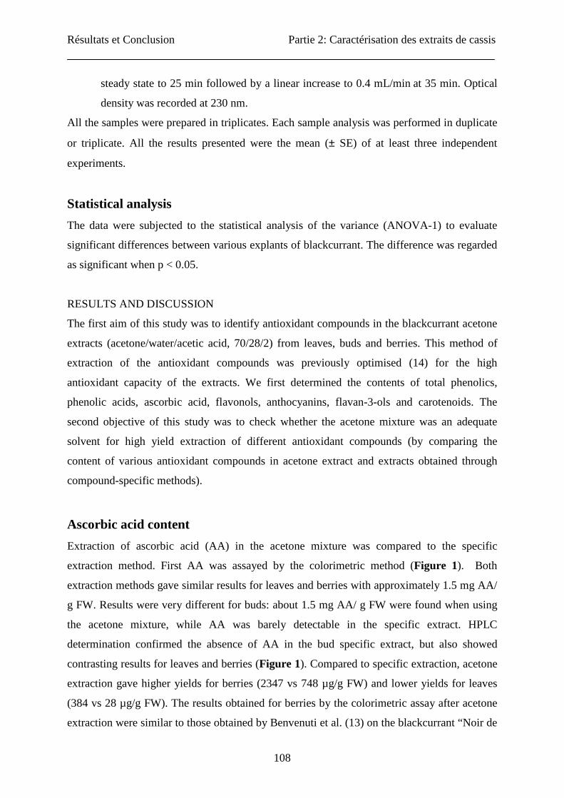
Ascorbic acid content
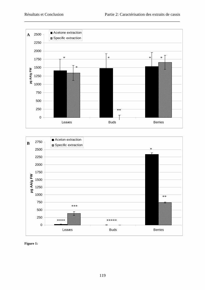
Extraction of ascorbic acid (AA) in the acetone mixture was compared to the specific
extraction method. First AA was assayed by the colorimetric method (Figure 1). Both
extraction methods gave similar results for leaves and berries with approximately 1.5 mg AA/
g FW. Results were very different for buds: about 1.5 mg AA/ g FW were found when using
the acetone mixture, while AA was barely detectable in the specific extract. HPLC
determination confirmed the absence of AA in the bud specific extract, but also showed
contrasting results for leaves and berries (Figure 1). Compared to specific extraction, acetone
extraction gave higher yields for berries (2347 vs 748 µg/g FW) and lower yields for leaves
(384 vs 28 µg/g FW). The results obtained for berries by the colorimetric assay after acetone
extraction were similar to those obtained by Benvenuti et al. (13) on the blackcurrant “Noir de
Résultats et Conclusion Partie 2: Caractérisation des extraits de cassis
109
Bourgogne” by HPLC. For the determination of the ascorbic acid, we could show great
differences between the two assays used. These observations were due to the characteristics
of the quantification methods: In the DCIP assay, only reduced L(+)-ascorbic acid was
measured but DCIP could also react with other reducing substances contained in the extracts
like myricetin (26). In HPLC assay, vitamin C was quantified in its two forms: reduced L(+)-
ascorbic acid and D(-)-deshydroascorbic acid. HPLC method had greater sensibility and
specificity than colorimetric assays. We could show also that bud extracts (by the two
extracting methods) did not contain ascorbic acid. Information about AA content in buds is
scarce. In soybean, the buds contain ascorbic acid and its concentration increases at flower
induction (32).
Total phenolic compounds’ content
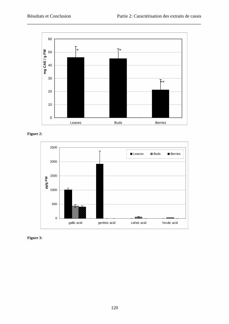
Total phenolic content was assayed in acetone extracts of leaves, buds and berries (Figure 2).
Leaves and buds showed significantly higher content (46 ± 8.4 and 45 ± 7.5 mg CAE/ g FW
respectively) than berries (21 ± 8.0 mg CAE/g FW). Benvenuti et al. (13) obtained contents in
berries of 5.5 ± 0.1 mg gallic acid equivalent / g of FW (extracted in methanol/HCl 2% (95:5,
v/v), while Cacace and Mazza (33) obtained 88.9 ± 2.4 mg CAE/g FW in an aqueous
sulphured dioxide extraction solution at pH 3.8 on milled berries.
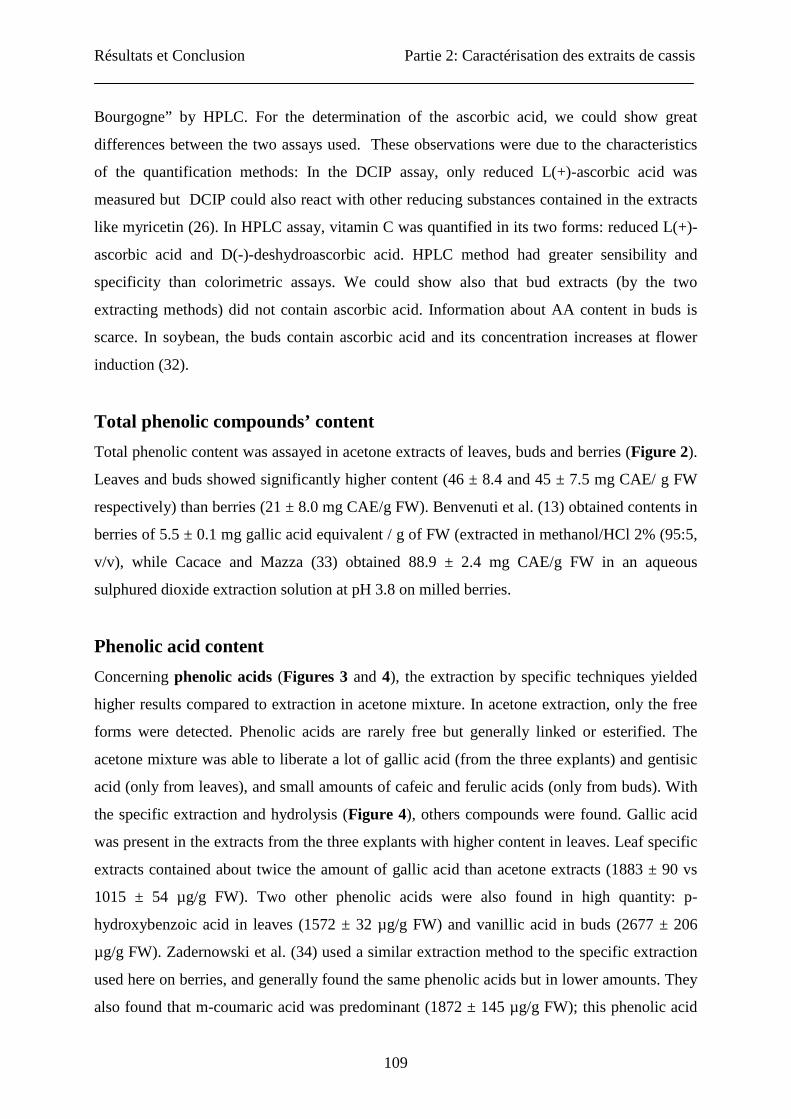
Phenolic acid content
Concerning phenolic acids (Figures 3 and 4), the extraction by specific techniques yielded
higher results compared to extraction in acetone mixture. In acetone extraction, only the free
forms were detected. Phenolic acids are rarely free but generally linked or esterified. The
acetone mixture was able to liberate a lot of gallic acid (from the three explants) and gentisic
acid (only from leaves), and small amounts of cafeic and ferulic acids (only from buds). With
the specific extraction and hydrolysis (Figure 4), others compounds were found. Gallic acid
was present in the extracts from the three explants with higher content in leaves. Leaf specific
extracts contained about twice the amount of gallic acid than acetone extracts (1883 ± 90 vs
1015 ± 54 µg/g FW). Two other phenolic acids were also found in high quantity: p-
hydroxybenzoic acid in leaves (1572 ± 32 µg/g FW) and vanillic acid in buds (2677 ± 206
µg/g FW). Zadernowski et al. (34) used a similar extraction method to the specific extraction
used here on berries, and generally found the same phenolic acids but in lower amounts. They
also found that m-coumaric acid was predominant (1872 ± 145 µg/g FW); this phenolic acid
Résultats et Conclusion Partie 2: Caractérisation des extraits de cassis
110
was not found in our specific berry extract. These contrasting results could be due to varietal
differences and/or to difference in ripening stage of the berries.
Flavonoid content
Flavonoids are important plant secondary metabolites accumulating in stressing conditions
(35). They are largely studied for their benefit on human health.
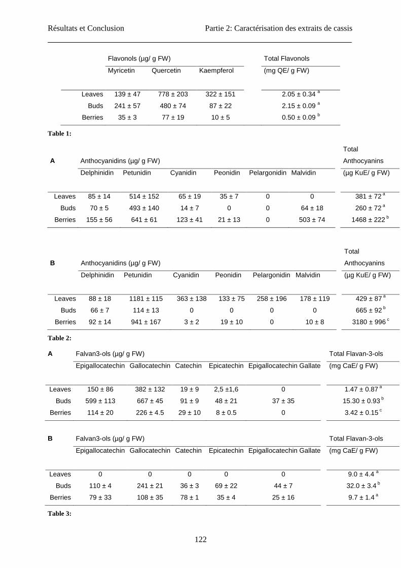
First, we evaluated the content in total flavonols in the three extracts (leaves, buds and
berries) (Table 1). The colorimetric technique used was described in the literature as able to
quantify the content in total flavonoids. But it is based on the formation of a complex between
the flavonoids and AlCl3. We showed previously that only the flavonols were measured (26)
by this technique. The contents in total flavonols of the berries (0.5 ± 0.1 mg QE/ g FW) were
significantly lower than in leaves and buds. The leaves and the buds had similar contents
(2.05 ± 0.34 and 2.15 ± 0.09 QE/ g FW respectively). Cacace and Mazza (33) assayed
flavonols in extracts from frozen berries by HPLC-MS analysis and reported slightly higher
contents (1.9 ± 0.14 mg QE / g of FW). After separation and HPLC analysis, we showed that
quercetin was more abundant than the two other aglycons (myricetin and kampferol) in the
three explants with a higher amount in leaf extracts (1.6 and 10 times more than in buds and
berries, respectively). Similar results were obtained by Borges et al. (36). This group
determined eight flavonols by HPLC-MS analysis with a better yield than in the present study.
Häkkinen et al. (37) also reported similar contents in berries for the three aglycons (quercetin:
3.3 to 6.8; K: <0.01 to 1, myricetin: 55 µg/g FW). The same conclusions were obtained by the
group of Jakobek (38): 44 µg myricetin / g FW, 21 µg quercetin / g FW and 8 µg kampferol /
g FW. For the majority of cultivars, quercetin is prevalent, followed by myricetin and finally,
kaempferol. But, the content in quercetin and myricetin considerably varied with the cultivars
(37). For the variety “Silvergieter”, no myricetin was present while for the “Rosenthals
langtraubige Schwarze” variety, myricetin was prevalent, followed by quercetin.
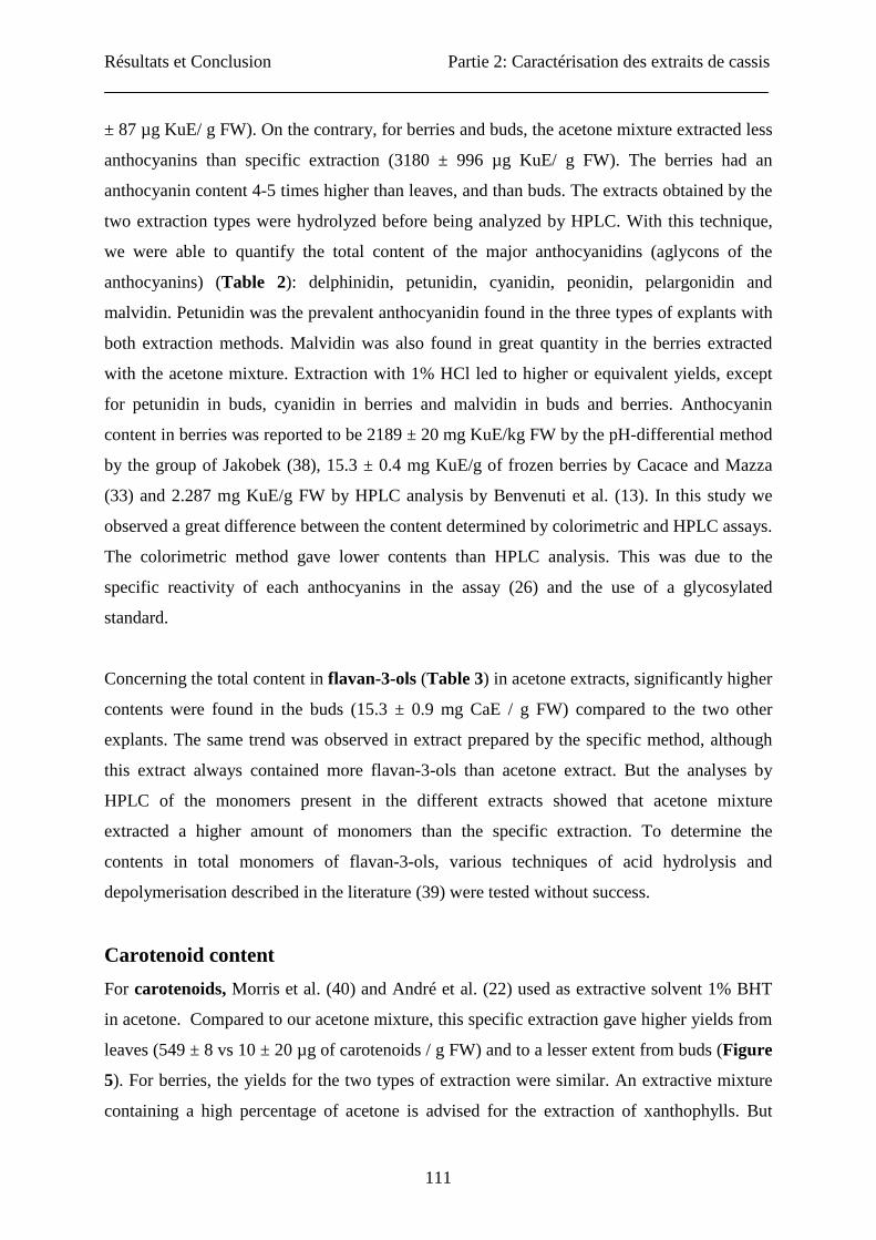
Anthocyanins are another group of pigments in plants showing health benefits. As shown in
table 3, total anthocyanins were significantly different among the explants studied and the
extraction methods used. For acetone extraction, the berry extract had a very high content
(1468 ± 222 µg KuE/ g FW) compared with the two other explants (381 ± 72 and 260 ± 72 µg
KuE/ g FW, for leaf and bud extracts respectively). The specific extraction for anthocyanins
described by Awika et al. (20) using 1% HCl in methanol gave similar results for leaves (429
Résultats et Conclusion Partie 2: Caractérisation des extraits de cassis
111
± 87 µg KuE/ g FW). On the contrary, for berries and buds, the acetone mixture extracted less
anthocyanins than specific extraction (3180 ± 996 µg KuE/ g FW). The berries had an
anthocyanin content 4-5 times higher than leaves, and than buds. The extracts obtained by the
two extraction types were hydrolyzed before being analyzed by HPLC. With this technique,
we were able to quantify the total content of the major anthocyanidins (aglycons of the
anthocyanins) (Table 2): delphinidin, petunidin, cyanidin, peonidin, pelargonidin and
malvidin. Petunidin was the prevalent anthocyanidin found in the three types of explants with
both extraction methods. Malvidin was also found in great quantity in the berries extracted
with the acetone mixture. Extraction with 1% HCl led to higher or equivalent yields, except
for petunidin in buds, cyanidin in berries and malvidin in buds and berries. Anthocyanin
content in berries was reported to be 2189 ± 20 mg KuE/kg FW by the pH-differential method
by the group of Jakobek (38), 15.3 ± 0.4 mg KuE/g of frozen berries by Cacace and Mazza
(33) and 2.287 mg KuE/g FW by HPLC analysis by Benvenuti et al. (13). In this study we
observed a great difference between the content determined by colorimetric and HPLC assays.
The colorimetric method gave lower contents than HPLC analysis. This was due to the
specific reactivity of each anthocyanins in the assay (26) and the use of a glycosylated
standard.
Concerning the total content in flavan-3-ols (Table 3) in acetone extracts, significantly higher
contents were found in the buds (15.3 ± 0.9 mg CaE / g FW) compared to the two other
explants. The same trend was observed in extract prepared by the specific method, although
this extract always contained more flavan-3-ols than acetone extract. But the analyses by
HPLC of the monomers present in the different extracts showed that acetone mixture
extracted a higher amount of monomers than the specific extraction. To determine the
contents in total monomers of flavan-3-ols, various techniques of acid hydrolysis and
depolymerisation described in the literature (39) were tested without success.
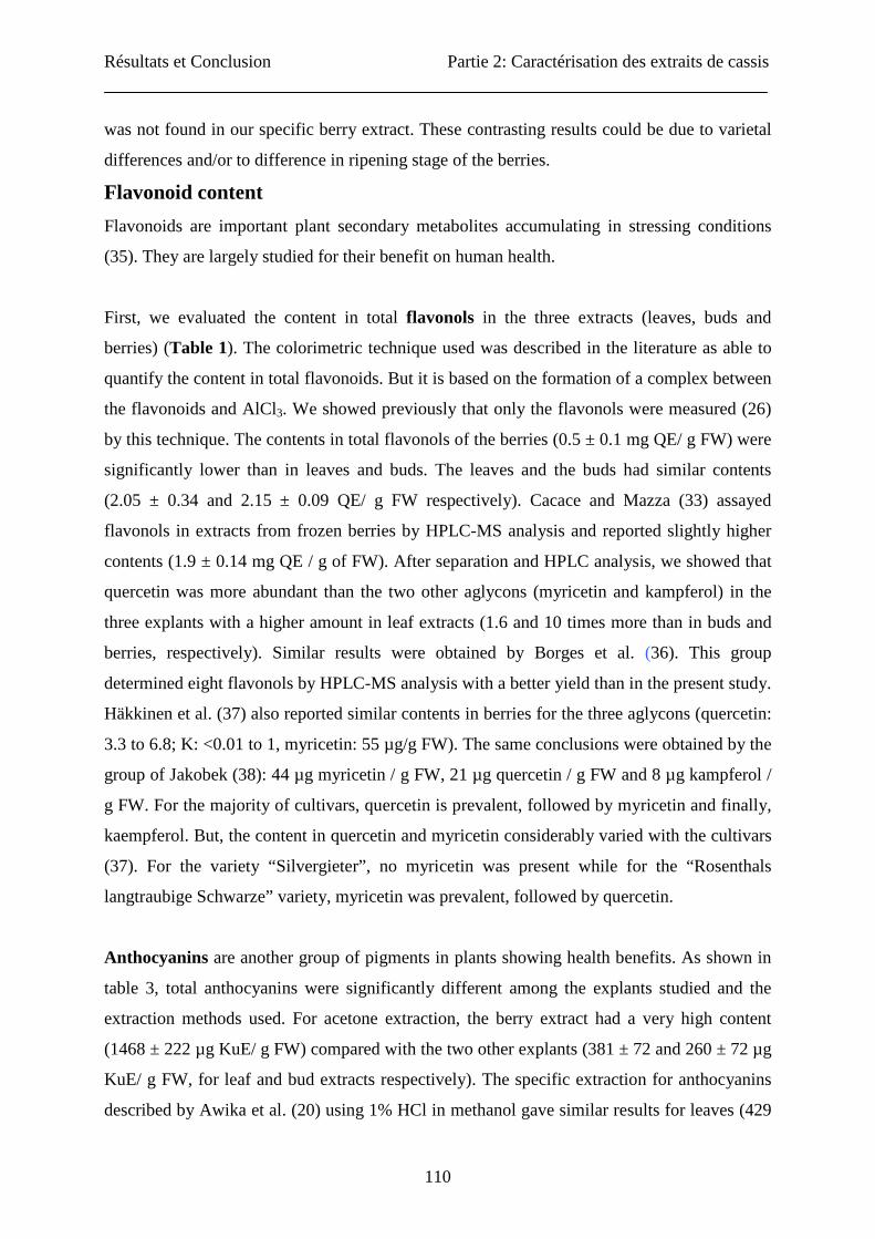
Carotenoid content
For carotenoids, Morris et al. (40) and André et al. (22) used as extractive solvent 1% BHT
in acetone. Compared to our acetone mixture, this specific extraction gave higher yields from
leaves (549 ± 8 vs 10 ± 20 µg of carotenoids / g FW) and to a lesser extent from buds (Figure
5). For berries, the yields for the two types of extraction were similar. An extractive mixture
containing a high percentage of acetone is advised for the extraction of xanthophylls. But
Résultats et Conclusion Partie 2: Caractérisation des extraits de cassis
112
those were probably degraded in the acetone method because no precaution was taken here to
allow their correct extraction and their conservation.
In general, the black currant berries contained higher levels of natural phenolic compounds
than other fruits like Rubus fruticosus, Rubus Idaeus L., Ribes Rubum and Sambucus nigra
(13, 37-38). The blackcurrant variety “Noir de Bourgogne” has a lower level in phenolic
compounds and ascorbic acid than other varieties of blackcurrant but a higher level in
anthocyanins (13-14). In this work, we have shown that leaves and buds could be considered
as a good source of natural antioxidant compounds.
CONCLUSION
To summarize, the acetone extracts of leaves and buds had total phenolic contents largely
higher than the berry extracts, which was also confirmed after analyzing the content in total
flavonoids. The most abundant flavonol aglycon in these extracts was quercetin. The content
in ascorbic acid was similar in the three types of extracts if measured by spectrophotometric
method while by HPLC analysis, berries showed a higher content in ascorbic acid than the
two other plant organs. The extracts also contained phenolic acids: gallic acid in extracts from
the three explants, gentisic acid in leaves and ferulic acid in buds and berries. The most
abundant anthocyanidin, whatever the explants, was petunidin. Concerning the flavan-3-ols,
the bud extracts presented a higher content compared to other explants. The predominant
monomers of flavan-3-ols were gallocatechin followed by epigallocatechin.
In conclusion, the acetone mixture can extract several classes of phenolic compounds with a
good yield for ascorbic acid, flavonols, flavan-3-ols and anthocyanins. This method is not
adequate for carotenoids. For phenolic acids, it extracts fewer compounds than the specific
method.
ACKNOWLEDGMENT:
J.T. gratefully acknowledges the AFR Luxembourg and Mr Andrianne from HerbalGem for
providing the material. The skilful assistance of the APE staff (provided to CEDEVIT by the
regional government of Wallonia) was greatly appreciated.

Résultats et Conclusion Partie 2: Caractérisation des extraits de cassis
113
REFERENCES:
(1) Bravo, L. Polyphenols: Chemistry, dietary sources, metabolism, and nutritional
significance. Nutr. Rev. 1998, 56, 317-333.
(2) Williamson, G.; Santos-Buelga, C. In Methods in polyphenol analysis; The Royal
Society of Chemistry: United Kingdom, 2003.
(3) Teutter, D. Significance of flavonoïds in plant resistance: a review. Environ. Chem. Lett.
2006, 4, 147-157.
(4) Guardia, T.; Rotelli, A. E.; Juarez, A. O.; Pelzer, L. E. Anti-inflammatory properties of
plant flavonoids; Effects of rutin, quercetin and hesperetin on adjuvant arthiris in rat. Il
Farm. 2001, 56, 683-687.
(5) Ghedria, K. Les Flavonoïdes: structure, propriétés biologiques, rôle prophylactique et
emplois en thérapeutique. Phytothér. 2005, 4, 162-169.
(6) Scalbert, A.; Claudine, C.; Morand, C.; Rémésy, C. Dietary polyphenols and the
prevention of diseases. Crit. Rev. Food Sci. Nutr. 2005, 45, 287-306.
(7) Olivier, J.; Palou, A. Chromatographic determination of carotenoids in foods. J.
Chromatogr. A 2000, 881, 543-555.
(8) Mayne, S. T. Beta-carotene, carotenoids, and disease prevention in humans. FASEB J
1996, 10, 691-701.
(9) Kanofsky, J. R.; Sima, P. D. Activity of a cationic carotenoid derivative in a mouse
model of protoporphyria. J. Photochem. Photobiol. B Biol. 2007, 87, 124-129.
(10) Heinonen, I. M.; Meyer, A. S.; Frankel, E. N. Antioxidant activity of berry phenolics
on human low density lipoprotein and liposome oxidation. J. Agric. Food Chem. 1998,
46, 4107–4112.
(11) Su, M. S.; Silva, J. L. Antioxidant activity, anthocyanins, and phenolics of rabbiteye
blueberry (Vaccinim ashei) by-products as affected by fermentation. Food Chem.
2006, 97, 447–451.
(12) Pantelidis, G. E; Vasilakakis, M.; Manganaris, G. A.; Diamantidis, G. Antioxidant
capacity, phenol, anthocyanin and ascorbic acid contents in raspberries, blackberries,
red currants, gooseberries and Cornelian cherries. Food Chem. 2007, 102, 777–783.
Résultats et Conclusion Partie 2: Caractérisation des extraits de cassis
114
(13) Benvenuti, S.; Pellati, F.;Melegari, M.; Bertelli, D. Polyphenols, anthocyanins, ascorbic
acid, and radical scavenging activity of Rubus, Ribes, and Aronia. Food Chem.
Toxicol. 2004, 69, 164-169.
(14) Tabart, J.; Kevers, C.; Pincemail, J.; Defraigne, J. O.; Dommes, J. Antioxidant capacity
of black currant varies with organ, season, and cultivar. J. Agric. Food Chem. 2006,
54, 6271-6276.
(15) Declume, C. Anti-inflammatory evaluation of hydroalcoholic extract of black currant
leaves (Ribes nigrum). J. Ethnopharmacol. 1989, 27, 91-98.
(16) Chrubasik, S. Pain therapy using herbal medicines. Gynakol. 2000, 33, 59-64.
(17) Tabart, J.; Kevers, C.; Sipel, A.; Pincemail, J.; Defraigne, J. O.; Dommes, J.
Optimisation of extraction of phenolics and antioxidants from black currant leaves and
buds and stability during storage. Food Chem. 2007, 105, 1268-1275.
(18) AFNOR. Foodstuffs – Determination of vitamin C by high performance liquid
chromatography. NF EN 1430 2003.
(19) Barberousse, H.; Roiseux, O.; Robert, C.; Paquot, M.; Deroanne, C.; Blecker, C.
Analytical methodologies for quantification of ferulic acid and its oligomers. J. Sci.
Food Agric. 2008, 88, 1494–1511.
(20) Awika, J. M.; Rooney, L. W.; Waniska, R. D. Anthocyanins from black sorghum and
their antioxidant properties. Food Chem. 2004, 90, 293-301.
(21) Parva-Uzunalic, A.; Skerget, M.; Knez, Z.; Einreich, B.; Otto, F.; Grüner, S. Extraction
of active ingredients from green tea (Camellia sinensis): Extraction efficiency of
major catechins and caffeine. Food Chem. 2006, 96, 597-605.
(22) Andre, C. M.; Ghislain, M.; Bertin, P.; Oufir, M.; Del Rosario Herrera, M.; Hoffman,
L.; Hausman, J. F.; Larondelle, Y.; Evers, D. Andean potato cultivars (Solanum
tuberosum L.) as a source of antioxidant and mineral micronutrients. J. Agric. Food
Chem. 2006, 55, 366-378.
(23) Association of vitamin chemists. Methods of vitamin assay, 3rd ed.; Interscience
Publishers: New York, 1961.
(24) Caboni, E.; Tonelli, M. G.; Lauri, P.; Iacovacci, P.; Kevers, C.; Damiano, C.; Gaspar,
T. Biochemical aspects of almond microcuttings related to in vitro rooting ability.
Biol. Plantar. 1997, 39, 91-97.
Résultats et Conclusion Partie 2: Caractérisation des extraits de cassis
115
(25) Lamaison, J. L.; Carmat, A. Teneur en principaux flavonoïdes des fleurs et des feuilles
de Crataegus monogyna Jacq. et de Crataegus laevigata (Poiret) DC en fonction de la
végétation. Plant. Méd. Phytothér. 19990, XXV, 12-16.
(26) Tabart, J.; Kevers, C.; Pincemail, J.; Defraigne, J. O.; Dommes, J. Evaluation of
spectrophotometric methods for antioxidant compound measurement in relation to
total antioxidant capacity in beverages. Food Chem. 2010, 120, 607-614.
(27) Nielsen, I. L.; Haren, G. R.; Magnussen, E. L.; Dragsted, L. O.; Rasmussen, S. E.
Quantification of anthocyanins in commercial black currant juices by simple high-
performance liquid chromatography, Investigation of their pH stability and
antioxidative potency. J. Agric. Food Chem. 2003, 51, 5861-5866.
(28) Nakamura, Y.; Tsuji, S.; Tonogai, Y. Analysis of proanthocyanidins in grape seed
extracts, health foods and grape seed oils. J. Health Sci. 2003, 49, 45-54.
(29) Rodriguez-Amaya, D. B. A guide to carotenoid analysis in foods, Omni Research:
ILSI Press, 2001.
(30) Dutruc-Rosset, G. Détermination par CLHP de neuf anthocyanes principales dans le
vin rouge et rosé. Résolution OENO 22/2003.
(31) Sharma, V.; Gulati, A.; Ravindranath, S. D.; Kumar, V. A simple and convenient
method for analysis of tea biochemicals by reverse phase HPLC. J. Food Comp. Anal.
2005, 18, 583-594.
(32) Bharti, S.; Garg, P. P. Changes in the ascorbic acid content of the lateral buds of
soybean in relation to flower induction. Plant Cell Physiol. 1970, 11, 723-727.
(33) Cacace, J. E.; Mazza, G. Extraction of anthocyanins and other phenolics from black
currants with sulfured water. J. Agric. Food Chem. 2002, 50, 5939-5946.
(34) Zadernowski, R.; Naczk, M.; Nesterowicz, J. Phenolic acid profiles in some small
berries. J. Agric. Food Chem. 2005, 53, 2118-2124.
(35) Hardorne, J. B.; Williams, C. A. Advances in flavonoid research since 1992.
Phytochem. 2000, 55, 481-504.
(36) Borges, G.; Degeneve, A.; Mullen, W.; Crozier, A. Identification of flavonoid and
phenolic antioxidants in black Currants, blueberries, red currants, and cranberries. J.
Agric. Food Chem. 2010, 58, 3901-3909.
Résultats et Conclusion Partie 2: Caractérisation des extraits de cassis
116
(37) Häkkinen, S.; Heinonen, M.; Kärenlampi, S.; Mykkänen, H.; Ruuskanen, J.; Törrönen,
R. Screening of selected flavonoids and phenolic acids in 19 berries. Food Res.
Internat. 1999, 32, 345-353.
(38) Jakobek, L.; Seruga, M.; Novak, I.; Medvidovic-Kosanovic, M. Flavonols, phenolic
acids and antioxidant activity of some red fruits. Deutsche Lebensmit.Rund. 2007, 103,
369-378.
(39) Kelm, M. A., Hammerstone, J. F.; Schmitz, H. H. Identification and quantitation of
flavanols and proanthocyanidins in foods: How good are the datas? Clin. Developm.
Immunol. 2005, 12, 35-41.
(40) Morris, W. L; Ducreux, L.; Griffiths, D.; Steward, D.; Davies, H. V.; Taylor, M.
Carotenogenesis during tuber development and storage in potatoes. J. Exp.l Bot. 2004,
399, 975-982.
Résultats et Conclusion Partie 2: Caractérisation des extraits de cassis
117
FIG. AND TABLE CAPTIONS:
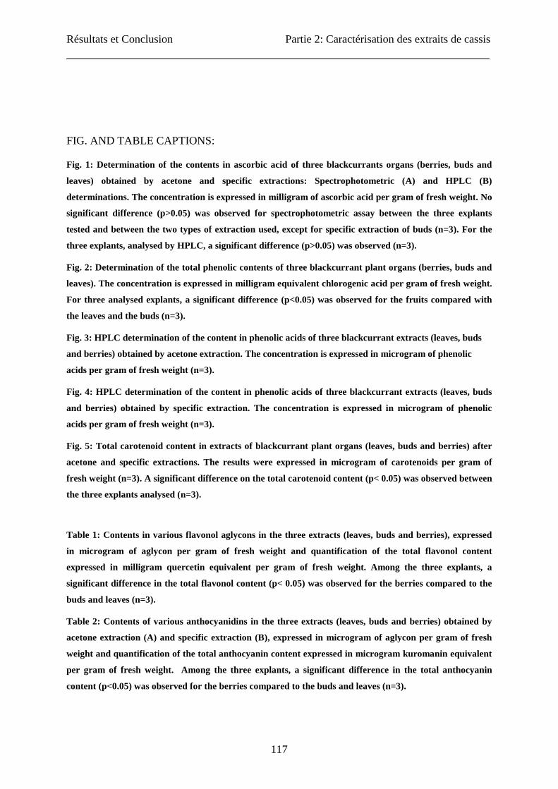
Fig. 1: Determination of the contents in ascorbic acid of three blackcurrants organs (berries, buds and
leaves) obtained by acetone and specific extractions: Spectrophotometric (A) and HPLC (B)
determinations. The concentration is expressed in milligram of ascorbic acid per gram of fresh weight. No
significant difference (p>0.05) was observed for spectrophotometric assay between the three explants
tested and between the two types of extraction used, except for specific extraction of buds (n=3). For the
three explants, analysed by HPLC, a significant difference (p>0.05) was observed (n=3).
Fig. 2: Determination of the total phenolic contents of three blackcurrant plant organs (berries, buds and
leaves). The concentration is expressed in milligram equivalent chlorogenic acid per gram of fresh weight.
For three analysed explants, a significant difference (p<0.05) was observed for the fruits compared with
the leaves and the buds (n=3).
Fig. 3: HPLC determination of the content in phenolic acids of three blackcurrant extracts (leaves, buds
and berries) obtained by acetone extraction. The concentration is expressed in microgram of phenolic
acids per gram of fresh weight (n=3).
Fig. 4: HPLC determination of the content in phenolic acids of three blackcurrant extracts (leaves, buds
and berries) obtained by specific extraction. The concentration is expressed in microgram of phenolic
acids per gram of fresh weight (n=3).
Fig. 5: Total carotenoid content in extracts of blackcurrant plant organs (leaves, buds and berries) after
acetone and specific extractions. The results were expressed in microgram of carotenoids per gram of
fresh weight (n=3). A significant difference on the total carotenoid content (p< 0.05) was observed between
the three explants analysed (n=3).
Table 1: Contents in various flavonol aglycons in the three extracts (leaves, buds and berries), expressed
in microgram of aglycon per gram of fresh weight and quantification of the total flavonol content
expressed in milligram quercetin equivalent per gram of fresh weight. Among the three explants, a
significant difference in the total flavonol content (p< 0.05) was observed for the berries compared to the
buds and leaves (n=3).
Table 2: Contents of various anthocyanidins in the three extracts (leaves, buds and berries) obtained by
acetone extraction (A) and specific extraction (B), expressed in microgram of aglycon per gram of fresh
weight and quantification of the total anthocyanin content expressed in microgram kuromanin equivalent
per gram of fresh weight. Among the three explants, a significant difference in the total anthocyanin
content (p<0.05) was observed for the berries compared to the buds and leaves (n=3).
Résultats et Conclusion Partie 2: Caractérisation des extraits de cassis
118
Table 3 : Identification and quantification of monomers of flavan-3-ols in blackcurrant extracts (leaves,
buds and berries) obtained by acetone (A) and specific (B ) extraction in microgram of monomer per gram
of fresh weight and quantification of the total content in flavanols expressed in milligram catechin
equivalent per gram of fresh weight. Among the three explants, a significant difference on the total
flavanol content (p<0.05) was observed for buds compared to leaves and berries (n=3).
Résultats et Conclusion Partie 2: Caractérisation des extraits de cassis
119
Figure 1:
0
250
500
750
1000
1250
1500
1750
2000
2250
2500
Leaves Buds Berries
µg A
A/g
FW
Acetone extraction
Specific extraction
0
250
500
750
1000
1250
1500
1750
2000
2250
2500
2750
Leaves Buds Berries
µg A
A/g
FW
Aceton extraction
Specific extraction
A
*
** *** **** *****
B
* * * * * **
Résultats et Conclusion Partie 2: Caractérisation des extraits de cassis
120
0
10
20
30
40
50
60
Leaves Buds Berries
mg
CA
E /
g F
W
Figure 2:
0
500
1000
1500
2000
2500
gallic acid gentisic acid cafeic acid ferulic acid
µg/g
FW
Leaves Buds Berries
Figure 3:
* *
**

Résultats et Conclusion Partie 2: Caractérisation des extraits de cassis
121
0
500
1000
1500
2000
2500
3000
gallic acid gentisic acid p-hydroxybenzoic acid ferulic acid p-couramic acid vanillic acid
µg/g
FW
Leaves Buds Berries
Figure 4:
0
100
200
300
400
500
600
Leaves Buds Berries
µg o
f car
oten
oids
/ g
FW
Acetone extraction
Specific extraction
Figure 5:
*
** *** *** **** ****
Résultats et Conclusion Partie 2: Caractérisation des extraits de cassis
122
Flavonols (µg/ g FW) Total Flavonols
Myricetin Quercetin Kaempferol (mg QE/ g FW)
Leaves 139 ± 47 778 ± 203 322 ± 151 2.05 ± 0.34 a
Buds 241 ± 57 480 ± 74 87 ± 22 2.15 ± 0.09 a
Berries 35 ± 3 77 ± 19 10 ± 5 0.50 ± 0.09 b
Table 1:
A Anthocyanidins (µg/ g FW)
Total
Anthocyanins
Delphinidin Petunidin Cyanidin Peonidin Pelargonidin Malvidin (µg KuE/ g FW)
Leaves 85 ± 14 514 ± 152 65 ± 19 35 ± 7 0 0 381 ± 72 a
Buds 70 ± 5 493 ± 140 14 ± 7 0 0 64 ± 18 260 ± 72 a
Berries 155 ± 56 641 ± 61 123 ± 41 21 ± 13 0 503 ± 74 1468 ± 222 b
B Anthocyanidins (µg/ g FW)
Total
Anthocyanins
Delphinidin Petunidin Cyanidin Peonidin Pelargonidin Malvidin (µg KuE/ g FW)
Leaves 88 ± 18 1181 ± 115 363 ± 138 133 ± 75 258 ± 196 178 ± 119 429 ± 87 a
Buds 66 ± 7 114 ± 13 0 0 0 0 665 ± 92 b
Berries 92 ± 14 941 ± 167 3 ± 2 19 ± 10 0 10 ± 8 3180 ± 996 c
Table 2:
A Falvan3-ols (µg/ g FW) Total Flavan-3-ols
Epigallocatechin Gallocatechin Catechin Epicatechin Epigallocatechin Gallate (mg CaE/ g FW)
Leaves 150 ± 86 382 ± 132 19 ± 9 2,5 ±1,6 0 1.47 ± 0.87 a
Buds 599 ± 113 667 ± 45 91 ± 9 48 ± 21 37 ± 35 15.30 ± 0.93 b
Berries 114 ± 20 226 ± 4.5 29 ± 10 8 ± 0.5 0 3.42 ± 0.15 c
B Falvan3-ols (µg/ g FW) Total Flavan-3-ols
Epigallocatechin Gallocatechin Catechin Epicatechin Epigallocatechin Gallate (mg CaE/ g FW)
Leaves 0 0 0 0 0 9.0 ± 4.4 a
Buds 110 ± 4 241 ± 21 36 ± 3 69 ± 22 44 ± 7 32.0 ± 3.4 b
Berries 79 ± 33 108 ± 35 78 ± 1 35 ± 4 25 ± 16 9.7 ± 1.4 a
Table 3:
Résultats et Conclusion Partie 2: Caractérisation des extraits de cassis
123
Antioxidant and anti-inflammatory activities of
Ribes nigrum extracts.
Jessica TABART 1, Thierry FRANCK 2, Claire KEVERS 1, Joël PINCEMAIL 3, Didier
SERTEYN 2, Jean-Olivier DEFRAIGNE 3, Jacques DOMMES 1
University of Liège,
1Plant Molecular Biology and Biotechnology Unit, B22
2CORD, B6
3CREDEC, B35
Sart Tilman,
B-4000 LIEGE
Belgium
Corresponding author: Jessica TABART: [email protected]
Other authors: [email protected], [email protected], [email protected],
[email protected], JO. [email protected], [email protected].
TITLE RUNNING HEAD: Antioxidant activity of blackcurrant extracts
ABSTRACT
Blackcurrant berries contain very high amounts of flavonoids with a number of health benefit
attributed to the antioxidant potential. Recent studies indicate that blackcurrant flavonoids
also exhibit anti-inflammatory properties. Leaves and buds actually used to produce food
complement could also exhibit such interesting properties.

Résultats et Conclusion Partie 2: Caractérisation des extraits de cassis
124
In the literature, many methods are used to evaluate the antioxidant capacity of food
compounds or biological systems, the main ones being the DPPH, TEAC, ORAC and ESR
assays. These methods are valid indicators of the antioxidant potential of dietary substances.
However this type of assay does not provide evidence that a substance or a mixture of
antioxidants has an in vivo antioxidant activity when consumed. To obtain more biologically
relevant information, the antioxidant activities of the extracts were evaluated on cellular
models implicating the measurement of blood haemolysis, of the Cellular Antioxidant
Activity on endothelial cells and of the anti-inflammatory activities on isolated equine
stimulated neutrophils and purified myeloperoxidase.
The classical in vitro tests, as well the cellular models generally showed that the blackcurrant
leaf extract have the highest antioxidant capacity followed by buds and berries. These
antioxidant activities are correlated to the total phenolics content of the extracts.
KEYWORDS: Antioxidant, Cellular Antioxidant Activity, Myeloperoxidase, EAHy926,
Neutrophils, SIEFED
INTRODUCTION
Oxidative stress results from a decrease of endogenous antioxidant capacities or an increase of
reactive oxygen species (ROS) concentration in organisms. It can cause deleterious damages
on cell constituents like DNA, proteins, … and finally, could induce several pathologies [1,
2]. Phytochemicals of fruits and vegetables such as polyphenols have been considered of
crucial nutritional importance in the prevention of chronic diseases such as cancer,
cardiovascular and neurodegenerative diseases. This may be related to their antioxidant
activity as well their ability to regulate cellular activities of inflammation-related cells (mast
cells, macrophages, lymphocytes and neutrophils).
Blackcurrant berries contain very high amounts of phenolic compounds [3, 4]. They are
sometime called “super fruit” because they show a number of health benefits [5]. These
benefits are attributed to the antioxidant potential of berries but recent studies indicate that
blackcurrant flavonoids exhibit also anti-inflammatory properties. A supplementation with
blackcurrant fruit concentrate in monocytic THP-1 cell culture modulates inflammatory
response with a decrease of NFκB, TNFα and IL-6 production by an unknown mechanism [5].
Résultats et Conclusion Partie 2: Caractérisation des extraits de cassis
125
Several studies demonstrated that berries have an effect on the prevention of the eye fatigue
[6]. There are only few studies on involved mechanisms and the majority of these studies
focused on berries [6, 7, 8, 9]. Leaves and buds can also used to produce food supplement.
Leaves are used in European traditional medicine for treatment of inflammatory disorders
such as rheumatic disease [10]. The proanthocyanidins contained in blackcurrant leaves
interfere with the accumulation of circulatory leukocytes, associated with a decrease of pro-
inflammatory factors such as TNFα, IL-1β, NO and CINC-1 [11]. To our knowledge, bud
health properties were never studied but they are used for the production of food
complements.
The objective of this work was to evaluate the antioxidant capacities of the blackcurrant
extracts from leave, buds and berries. To perform this study, we used several in vitro classical
techniques applied for the measurement of antioxidant capacity and efficacy of food
antioxidants. The most popular assay is the ORAC assay (Oxygen Radical Antioxidant
Capacity) initially developed by Cao and Prior [12] and using 2,2’-azobis(2-aminopropane)
dihydrochloride (AAPH) as free radical generator. Other tests include the reaction of
antioxidant with stable free radicals (2,2-diphenyl-1-picrylhydrazyl (DPPH) and 2.2’-
azinobis(3-ethylbenzthiazoline-6-sulfonic acid cation radical (ABTS.+)) or the ability of
antioxidants to reduce ferric (FRAP assay) and cupric ions (CUPRAC assay). Less unknown
is the use of the Electron Spin Resonance (ESR) spectroscopy, the only one method allowing
to directly evidence the free radical formation in a biological sample. In presence of 5,5-
dimethyl-1-pyrroline-N-oxide (DMPO) as spin trapping agent, the system xanthine/xanthine
oxidase is used to generate and detect superoxide anion radical (O2.-) which has the great
advantage to be a physiological species.
In order to get more biologically relevant information, in vitro oxidative stress models on cells
have also been developed. The first assay used is the haemolysis assay where blood cells were
placed in oxidative conditions with or without antioxidant compounds [13]. The second model
is the Cellular Antioxidant Activity (CAA) assay on hepatic cells (HepG2). This assay can
measure the inhibition of dye oxidation by antioxidants in cell cultures. This method is more
biologically relevant because it gives an antioxidant capacity taking into account the uptake of
compounds by cells [14]. A third model is linked to ROS production in connection to
inflammatory situations. The activation of neutrophils leads to the formation of superoxide
Résultats et Conclusion Partie 2: Caractérisation des extraits de cassis
126
anion through NADPH oxidase cascade. At the same time, activated neutrophils also release
myeloperoxidase (MPO) [15] which catalyses the conversion of hydrogen peroxide (H202) to
the more potent oxidant hypochlorous acid (HOCl).
For different reasons linked to instruments limitations, mechanisms, endpoint, quantification
method and biological relevance, there is often a lack of correlation between results obtained
through these differentassays. Actually, it is well admitted that there is no simple universal
assay by which antioxidant capacity of food can be assessed accurately and quantitatively. It
is the reason why we used in this study several in vitro test including cellular models to better
evidence and understand the antioxidant and anti-inflammatory potency of blackcurrant
extracts (leaves, berries and buds).
MATERIALS AND METHODS
Sample preparation
The buds, berries and leaves of blackcurrant (noir de Bourgogne), two years old, were
harvested respectively in March, July and August in the Belgian Ardennes (Bihain). The
various explants were directly cut out into pieces, frozen in liquid nitrogen, lyophilized and
stored at -20°C for some months.
For extraction, one gram of fresh sample (berries, buds or leaves) was ground with 1 gram of
quartz and 10 ml of extraction solution: acetone/water/acetic acid (70:28:2) [16]. The mixture
was shaken during 1 h at 4°C and centrifuged at 17 000 g for 15 min. The supernatant was
removed, and the sample was extracted again with the same procedure but incubated only 15
min. The supernatants were pooled and then diluted as appropriate for the immediate analysis.
For experiments on EAHy926 cells and neutrophils, the acetone contained in the supernatant
was evaporated; the remainder of supernatant was freeze-dried and stored at – 20°C. For
different assays, the extracts were suspended in the adequate medium and filtered.
Determination of total phenolic contents
Total phenolics were determined according to the Folin-Ciocalteu method described in a
previous study [17] The results were expressed in mg equivalent chlorogenic acid (CAE) per
gram of fresh weight.
Résultats et Conclusion Partie 2: Caractérisation des extraits de cassis
127
In vitro evaluation of antioxidant capacity
To determine the antioxidant capacity, the first method used was the TEAC assay (scavenging
of the radical 2,2-azino-bis(3-ethylbenzothiazoline)-6 sulphonic acid, ABTS). Antioxidant
capacity was also determined by scavenging of the radical 2,2-diphenyl-1-picryhydrazyl
(DPPH). ORAC assays were carried out on a Fluoroskan Ascent FL Plate Reader
(ThermoLabsystems, Finland) at 37°C. For these three methods described in a previous study
[17], Trolox was used as standard and the antioxidant capacity was expressed in mg Trolox
equivalent (TE) per gram of fresh weight. All samples were analysed in duplicate.
Superoxide anion (O2-)-scavenging capacities were measured by electron spin resonance
(ESR) spectroscopy described in a previous study [17]. Measurements were performed at
room temperature on a JEOL–Jes-FR30 spectrophotometer. The results were expressed in
units of SOD equivalent (U SODE) per gram of fresh weight. The running conditions were as
follows: 9.5 GHz frequency, 100 kHz modulation frequency, 4mW microwave power, 335.6
mT centre field, 2500G Gauss modulation amplitude, 1 s time constant, and 4 min sweep
time.
Evaluation of antioxidant capacity on cells
a) Red blood cell resistance to oxidative stress (haemolysis)
The resistance of red blood cell was measured on a microliter plate reader (Labsystems IEMS
reader MF) as described by Tabart et al. [17]. The results were expressed in mmole Ascorbic
Acid (AA) equivalent per gram of fresh weight.
b) Cellular Antioxidant Activity (CAA) on EAHy926 cell s
Cell Culture: The human endothelial-like EAhy926 cell line [18] derived from the fusion of
human umbilical vein endothelial cells with the A549 carcinoma cell line. EAHy926 were
grown in DMEM (Biowithaker, Lonza) supplemented with 10% of heat-inactived FBS
(Fetal serum bovine), 10 µM HEPES, 5000 unit of penicillin and streptomycin, 1 mM
sodium pyruvate, 2 mM glutamine and 2% HAT (hypoxanthin, aminoprotein and thymidin)
and were maintained at 37°C and 5% CO2.
Résultats et Conclusion Partie 2: Caractérisation des extraits de cassis
128
Cellular Antioxidant Activity (CAA) was measured using the method of Wolfe and Liu
[14] with various modifications. EAHy926 cells were seeded at a density of 5000 cells/ well
on a 96 well microplate in 100 µl of supplemented DMEM. 100 µL of blackcurrant extract
were added in each well, together with 100 µl of DCFH-DA (dichlorofluorescein diacetate,
25 µM, Sigma) dissolved in PBS at 37°C. After 1 h at 37°C, wells were washed with 100 µl
PBS (3 times). Then, 100 µl of APPH (600 µM) were added in each well. The plate was
placed into a Fluoroskan Ascent FL Plate Reader at 37°C. Emission of fluorescence was
recorded every five minutes during 1h (emission at 538 nm and excitation at 485 nm). We
used the method of quantification described by Wolfe and Liu [14]. Results were expressed
in microgram quercetin equivalent (QE) per gram of freeze-dried extract. Each experiment
was repeated with different cell batches.
Evaluation of anti-inflammatory activity of ribes extracts on the oxidant
response of neutrophils and on MPO activity
Isolation of equine neutrophils: They were isolated from EDTA (1.6 mg/ml) treated blood
drawn from jugular vein of horses not under medical treatment (Faculty of veterinary
Medicine, University of Liège, Belgium). They were isolated at room temperature by
centrifugation on a percoll density gradient according to the method of Pycock et al. [19].
After washing in physiological saline solution, the cell pellets were suspended in 20 mM
phosphate buffer saline (PBS) at pH 7.4. The cells were used within four hours after isolation,
and each assay was performed in triplicate. Each experiment was repeated at least twice with
different cell batches.
Effect of the extract on the ROS production by stimulated neutrophils
Neutrophil suspensions (106 neutrophils/200 µL PBS) were distributed in the wells of a 96-
well microtiter plate (White Combiplate 8, Fischer Scientific, Tournai, Belgium) and
incubated for 10 min at 37°C with the extracts at final concentration of 50, 25, 10, 7.5, 5, 2.5,
1, 0.5 µg mL-1 After incubation, neutrophils were stimulated with phorbol myristate acetate
(PMA) according to the method of Kohnen et al. (2007). Immediately after the addition of
PMA, the CL response of the neutrophils was monitored for 30 min (Multiscan Ascent,
Fischer Scientific, Tournai, Belgium) and expressed as the integral value of the total CL
emission. Control assays made with the vehicle solution of extracts (PBS) were taken as
Résultats et Conclusion Partie 2: Caractérisation des extraits de cassis
129
100% of chemiluminescence response. Each experiment was repeated with different cell
batches.
Effect of the extracts on the release of MPO by stimulated neutrophils:
The neutrophil suspensions (106 cells/ml) were incubated for 10 min at 37 °C with the
extracts at final concentration of 50, 25, 10, 7.5, 5, 2.5, 1, 0.5 µg mL-1 and then activated for
30 min at 37°C with PMA as described in Kohnen et al. (2007). After activation, the
neutrophil suspensions were centrifuged (450 x g, 10 min), and the supernatants were
collected. MPO released by the neutrophils was measured in the supernatants by an ELISA
assay raised against equine MPO (Franck et al., 2005) and distributed by BIOPTIS (Liege,
Belgium). The control assay taken as 100% of MPO release was made with the supernatant
obtained from PMA stimulated neutrophils where the extracts were replaced by PBS. Each
experiment was repeated with different cell batches.
Effect of the extracts on the specific activity of MPO measured by SIEFED
The Specific Immunological Extraction Followed by Enzymatic Detection (SIEFED) is an
original method developed for the specific detection of active equine neutrophil MPO (Franck
et al., 2006) The first step is an immunoextraction of MPO from a solution or a biological
sample by specific immobilized antibodies; the next step consist of washings to eliminate
unspecifically bound compounds or interfering substances and the third step is the detection
of MPO enzymatic activity. The extracts at final concentrations of 50, 25, 10, 7.5, 5, 2.5, 1,
0.5 µg mL-1 were incubated for 10 min with pure equine MPO (50 ng/ml) in the dilution
buffer (PBS 20 mM with 5 g/L BSA and 0.1 % Tween 20) before the immunoextraction step.
The control taken as 100 % MPO activity was performed with purified MPO in PBS.
Statistical analysis
For antioxidant capacity assays, the data were subjected to the statistical analysis of the
variance (ANOVA-1) to evaluate the significant differences between various explants of
blackcurrant. The difference was regarded as significant when p < 0.05 or 0.01.
For anti-inflammatory assays, a one-way ANOVA followed by Dunnett’s post-test was
performed to compare the inhibitory percentage of the extract with the corresponding 100 %
controls. A p value < 0.05 was considered as significant.

Résultats et Conclusion Partie 2: Caractérisation des extraits de cassis
130
RESULTS
Evaluation of antioxidant capacity of blackcurrant extracts by in vitro assays
Blackcurrant extracts were prepared from leaves, berries and buds. Their antioxidant capacity
was evaluated by several commonly used assays (Table 1). With all these methods, we can
show a similar profile for the three explants analyzed. Leaf and bud extracts showed a
significant (p<0.01) higher antioxidant capacity than berry extracts. In the ESR assay, leaves
and buds also showed a higher activity than berries.
For all assays, the observations were correlated with the total phenol content: leaves and buds
contained two times more phenolic compounds (46.1 ± 8.4 and 45.1 ± 7.5 mg ACE/ g of FW,
respectively) than berries (21.25 ± 8.0 mg ACE/ g of FW).
Evaluation of antioxidant capacity of blackcurrant extracts on cellular models
The cellular antioxidant activity of blackcurrant extracts (leaves, berries and buds) was
measured using the CAA assay on EAHy926 cells (Table 2). Leaf extracts had the highest
CAA value (12.89 ± 0.77 µmole QE/ g of LE) followed by buds (8.8 ± 1.92 µmole QE/ g of
LE) and finally, berries (0.40 ± 0.10 µmole QE/ g of LE). For each extract, we also
determined the concentration to obtain 50% of fluorescence inhibition (EC50): 113 ± 7 µg /ml
for leaf extract, 166 ± 36 µg /ml for bud extract and 3688 ± 910 µg /ml for berry extract. For
the haemolysis assay, also called “Resistance Test of blood cells in oxidative stress
conditions”, the antioxidant capacities were the same for leaves and buds (around 1.2 mmole
AA/ g of FW) and lower for berries (0.394 ± 0.053 mmole AA/ g of FW).
To determine if this inhibition was really due to a decreased of ROS production in the
presence of the extracts and not to a cytotoxic effect on endothelial cells, a viability assay
using MTT (3-[4,5-dimethylthiazol-2-yl]2,5-diphenyltatrazolium bromide; Cell Growth
Determination Kit MTT, Sigma-Aldrich) was made in the range of concentrations from 0 to 1
mg extract/ml. The extracts (data not shown) were not cytotoxic.
Effects of blackcurrant extracts on total ROS production by neutrophils
Résultats et Conclusion Partie 2: Caractérisation des extraits de cassis
131
The influence of blackcurrant extracts (leaves, berries and buds) on the ROS production of
PMA-activated neutrophils was determined through a lucigenin dependent
chemiluminescence assay (Figure 1). A significant dose-dependent inhibition was observed
for all extracts. We reached 50% of inhibition with a concentration of 5 ± 0.5 µg of leaf
extract/ml, 13 ± 4.3 µg of bud extract/ml and 44 ± 20 µg of berry extract/ml. To determine if
this inhibition was really due to a decrease of ROS production in the presence of the extracts
and not by a toxic effect on equine neutrophils, a tryptan blue exclusion test was made in the
range of concentrations used and above. The extracts (data not shown) were not cytotoxic.
Only some cell aggregates were formed with high extract concentrations (1 mg/ml) versus
PBS controls.
Effects of blackcurrant extracts on the total MPO released by stimulated neutrophils
The effects of blackcurrant extracts (leaves, berries and buds) on the total MPO released by
activated neutrophils were measured by an ELISA assay. Extract addition did not induce
significant decrease of MPO release versus control (data not presented). The higher
concentration of blackcurrant extracts (1 mg extract/ ml) gave only 15 to 20 % of inhibition.
Thus, only slight effects of blackcurrant extracts were observed on MPO release by
neutrophils.
Effects of blackcurrant extracts on MPO activity (in vitro)
To determine the effects of blackcurrant extracts (leaves, berries and buds) on MPO activity,
we used the SIEFFED method developed by Franck et al. [21]. We observed a dose-
dependent inhibition of the MPO activity with all the explants tested (Figure 2). For leaves,
the percentage of inhibition was superior to 50% with the smallest concentration tested
(around 60 % inhibition at 0.5 µg/ml for leaf extract). For buds, we reached 50% inhibition at
0.9 ± 0.2 µg/ml; and for berries, 14 ± 4 µg/ml. At the smallest concentrations of berry extract
(1 µg/ml), a significantly increased MPO activity was observed.
DISCUSSION
Many methods are used in different studies to evaluate the antioxidant capacity of food
compounds or biological systems. The two oldest and most usually used in vitro methods are
the assays based on DPPH and ABTS radicals, two methods based on the transfer of hydrogen
between the free radicals and the antioxidants. These two methods give similar results for the

Résultats et Conclusion Partie 2: Caractérisation des extraits de cassis
132
blackcurrant extracts as well as for the majority of plant extracts [22, 23]. But, the technique
of discolouration of ABTS.+ radical presents nevertheless a disadvantage compared to the
DPPH method. Indeed, radical ABTS.+ is very unstable, a loss of absorbance of 30% is
already observed over a 1 hour incubation while radical DPPH is very stable [23]. The ORAC
method is more widely used in the agro-food sector. At the present time, it is considered as
the method of reference to evaluate the total antioxidant capacity of a food sample. This assay
largely differs from the two other methods: a fluorescent dye is destroyed by AAPH radical
and the antioxidants present in the sample will protect this molecule against oxidation. This
technique gives higher values of antioxidant capacity than the two first methods. With regard
to another technique, the ESR assay, the results are not easily comparable with the methods
described above owing to the fact that the results are not expressed in the same units. ESR
gives us information as for the action of antioxidants on a free radical, the anion superoxide,
found abundantly in the human body. O2.- is mainly produced in cellular respiratory processes
in mitochondria and by the NADPH oxidase on endothelial cells and phagocytes.
In this first part of the study, we can observe that the choice of the method has a notable
influence on the determination of the antioxidant potential of a sample. This is due to the
various antioxidant compounds found in the food matrices and the sources of free radicals
used in the assays. But we observed a similar profile on the antioxidant capacity: leaves ≥
buds > berries, even if the intensity of the answer is variable from one technique to another
due to the type of reaction implemented but also to the choice of standard (Trolox,
chlorogenic acid or SOD). These methods are valid indicators of the antioxidant potential of
dietary substance but this type of assays did not provide evidence that a substance or a
mixture of antioxidant compounds has an in vivo antioxidant activity when consumed.
Complementary to the classical in vitro tests to measure the antioxidant potential, we also
studied the effect of blackcurrant extracts on the CAA and anti-inflammatory activities of
cellular models. The CAA assay, developed by Wolfe and Liu [14], and the haemolysis assays
are complementary but are more biologically relevant than in vitro assays since they used
cellular models to evaluate the antioxidant capacity of fruits, vegetables, and foods. The
haemolysis assay is on in vivo situation since this method proposes the influence of an
antioxidant contribution on red blood cells near to an oxidizing stress state. The rate of cell
lysis can be regarded as an in vitro marker of the oxidative damage. The susceptibility of the

Résultats et Conclusion Partie 2: Caractérisation des extraits de cassis
133
red blood cells could be determined by their submission to an oxidative system that produces
ROS and by measuring at regular laps time the rate of haemolysis. The latency time before the
beginning of haemolysis is proportional to the quantity of antioxidant content present in the
reaction mixture [24]. For the CAA, the dye is taken up by cells and it is deacetylated by
intracellular enzymes (making it unable to exit the cell). The CAA assay is based on the
ability of hydroxyl radicals to react with a fluorescent probe in the cell and on the prevention
of this reaction in the presence of antioxidant compounds inside the cell. The DCFH-DA is a
commonly used dye in biological systems [25, 26]. DCFH-DA is apolar and it diffuses
through the biological membranes. In the cell, the dye is deacetylated by cellular esterases to
form DCFH that is more polar and is trapped in the cell. In presence of ROS, DCFH is
oxidized into DCF that is fluorescent. We have worked in the conditions described by Wolfe
and Liu [14] but we have chosen endothelial cells for our investigations. Endothelial cells are
involved in the development of several vascular pathologies through endothelial dysfunction
[27] by various mechanisms like increasing endothelial oxidase activity like NADPH oxidase
enzyme [28], decreased the endothelial nitric oxide (vasodilator agent) bioavailability by
using it as a substrate [28, 29].
DCFH-DA was already used on EAHy926 cells [26] to measure the protection afforded by
honey on oxidative stress. The optimization of the CAA assay on EAHy926 cells did need
some modifications: cell density was 5000 cells/ well and PBS was used for dye solubilisation
instead of William’s E medium. This assay was used to determine the antioxidant potential of
some berries by Wolfe and Liu [30]: pomegranate, blackberry, blueberry, strawberry,
raspberry and cranberry. The extracts had an antioxidant action in the CAA model at two
levels. First, they can act at the cell membrane, breaking peroxyl radical chain reactions at the
cell surface. Second, they can be taken up by the cells and then react with ROS intracellulary
[14]. In our study, we found with these two tests the same profile than in vitro assays.
A second cellular model is used and consists of polymorphonuclear neutrophils which are the
primary effector cells in host responses to injury and infection [15]. The antimicrobial
function of stimulated PMNs is based on their phagocytic capacity and ability to release
oxidant and proteolytic enzymes and ROS involved in the destruction of microorganisms.
ROS play important roles in the pathogenesis of various diseases, such as neurodegenerative
disorders, cancer, cardiovascular diseases, atherosclerosis, cataracts, and inflammation [31,
Résultats et Conclusion Partie 2: Caractérisation des extraits de cassis
134
32]. Myeloperoxidase (MPO) is an haemic enzyme stored in the azurophilic granules of
neutrophil and has an essential role in the destruction of bacteria inside PMNs. By using
hydrogen peroxide (H2O2) and chloride anion (Cl-), it produces hypochlorous acid (HOCl), an
important microbial killer [15]. MPO also catalyzes tyrosine nitration in proteins from nitrite
and H2O2 [20]. In some conditions of acute and chronic inflammations, MPO is released from
cells and can cause severe damage to macromolecules in cell membranes and in extracellular
space [33]. Thus, the decreasing oxidant activity of neutrophils and MPO may prove
beneficial for the treatment of inflammation-related diseases.
When neutrophils were stimulated in the presence of blackcurrant extracts, a dose dependent
inhibitory effect on ROS production was observed for each extract. These results showed that
some molecules contained into the extract have the abilities to scavenge ROS produced by
neutrophils or to inhibit the activation of NADPH oxidase by interfering with the intracellular
pathway or membrane elements involved in the activation of the enzymatic complex. We
suspected that the effect of ribes extract on the oxidative burst of PMNs is mainly
extracellular. Indeed, ribes extracts had poor effect on the degranulation pathway of MPO
probably because the molecules which contain do not interfere with membrane receptors,
cytoskeleton, intracellular Ca2+ and PKC activation pathway [34, 35]. Such hypothesis should
be confirmed by testing longer incubation time between the cell and the extract or by adapting
on PMNs, the DCFH technique used with endothelial cells. Nevertheless, our results showed
that blackcurrant extract had important extracellular scavenging activity on ROS produced by
stimulated neutrophils.
Although blackcurrant extract had a poor effect on the release of total MPO, we were
interested about their effect on the activity of this oxidant enzyme. Evidence has emerged that
MPO-derived oxidants such HOCl and nitrogen dioxide (●NO2) contribute to tissue damage
and the initiation and propagation of acute and chronic vascular inflammatory disease that
attracts considerable interest in the development of therapeutically useful MPO inhibitors
including natural compounds [36]. All the tested extracts, mainly leaves and buds, exerted a
strong dose-dependent inhibitory effect on the specific activity of equine MPO as measured
by the SIEFED assay. In SIEFED assay, the interferences of the extract with the revelation
system were not possible since the tested compound or the extract was washed before the
measurement of the activity. Therefore, this technique constitute a useful pharmacological
Résultats et Conclusion Partie 2: Caractérisation des extraits de cassis
135
tool to study specific inhibitors of MPO because if an inhibition of the enzyme is measured
after the washing of the tested compound, it means that this compound can interact directly
with the enzyme by inducing conformational changes in the protein moiety or by blocking or
hindering the active site of the enzyme.
Interestingly, all the antioxidant activities of the blackcurrant extracts are correlated with their
content in total polyphenolic compounds. In a previous study [37], we showed that leaf and
bud extracts had a higher flavonol content (2.05 ± 0.34 and 2.15 ± 0.09 mg quercetin
equivalent per gram of fresh weight, respectively) than berry extract (0.5 ± 0.1 mg quercetin
equivalent per gram of fresh weight), with a prevalence of quercetin. Some flavonoids, like
quercetin, rutin, resveratrol, are especially known for their scavenging properties [38]. In
addition, several studies showed that some polyphenolic compounds could have a potential
inhibitory effect on inflammation by decreasing the ROS production and degranulation [39]
of stimulated neutrophils, but also by decreasing the activities of NADPH oxidase [40, 41]
and MPO [42]. Other studies showed that flavonoids could prevent cardiovascular diseases
[43] by inhibiting the activity of MPO, which could be responsible of the oxidation of low-
density lipoproteins in arteries [43].
In conclusion, blackcurrant extracts possess strong antioxidant activities as demonstrated by
in vitro classical assays, but also by cellular models such as endothelial cells and
polymorphonuclear neutrophils. Our study emphasized the intracellular and extracellular
scavenging properties of blackcurrant extracts showing not any cell toxicity. Although
blackcurrant extracts do not interfere with the degranulation of MPO by stimulated
neutrophils, they strongly inhibited the activity of MPO. By inhibiting the ROS production by
endothelial cells and polymorphonuclear neutrophils, blackcurrant extract present an
interesting potential against inflammatory and cardiovascular diseases. For all assays, a better
effect was observed for leaves and buds in comparison to berries, and these effects seemed to
be correlated to the total polyphenolic contents of the extracts. Very few studies have
characterized other explants than berries. The fruits are a good source of anthocyanins and
flavonols [44, 45]. Leaves contain small quantity of essential oil but also numerous flavonoid
glycosides, especially derivates of quercetin and kampferol, and prodelphinidins [10]. Further
investigations will focus on the characterization of the compounds present in the extracts to
identify the active compounds or to show possible synergic interactions between several
Résultats et Conclusion Partie 2: Caractérisation des extraits de cassis
136
compounds which could have benefit effect against inflammatory diseases in animals and
inhuman.
ACKNOWLEDGMENT:
J.T. gratefully acknowledges the AFR Luxembourg and the memberships of CEDEVIT
A.S.B.L (Liège).
REFERENCES
[1] Halliwell B, Aruoma OI. DNA and free radicals. FEBS Lett. 1993; 335: 140-1.
[2] Halliwell B. Lipid peroxidation, antioxidants and cardiovascular disease: How should we move forward?
Cardiovasc Res. 2000; 47: 410-8.
[3] Häkkinen S, Heinonen M, Kärenlampi S, Mykkänen H, Ruuskanen J, Törrönen R. Screening of selected
flavonoids and phenolics acids in 19 berries. Food Res Intern. 1999; 32: 345-53.
[4] Benvenuti S, Pellati F, Melegari M, Bertelli D. Polyphenols, anthocyanins, ascorbic acid, and radical
scavenging activity of Rubus, Ribes, and Aronia. Food Chem Toxicol. 2004; 69: 164-9.
[5] Lyall KA, Hurst SM, Cooney J, Jensen D, Lo K, Hurst RD, Stevenson LM. Short-term blackcurrant extract
consumption modulates exercise-induce oxidative stress and lipopolysaccharide-stimulated inflammatory
responses. Am J Physiol Regul Integr Comp Physiol. 2009; 297: R70-R81.
[6] Nakamura Y, Matsumoto H, Todoki K. Endothelium-dependent vasorelaxation induced by black currant
concentrate in rat thoracic aorta. Jpn J Pharmacol. 2002; 89: 29-35.
[7] Moller P, Loft S, Alfthan G, Freese R. Oxidative DNA damage in circulatoring mononuclear blood cells
after ingestion of blackcurrant juice or anthocyanin-rich drink. Mut Res. 2004; 551:119-26.
[8] McGhie TK, Walton MC, Barnett LE, Vather R, Martin H, Au J, Alspach PA, Booth CL, Kruger MC.
Boysenberry and blackcurrant drinks increased the plasma antioxidant capacity in an elderly population bud
had little effect on other markers of oxidative stress. J Sci Food Agric. 2007; 87: 2519-27.
[9] Dalgard C, Nielsen F, Morrow JD, Enghusen-Poulsen H, Jonung T, Horder M, and de Maat MPM.
Supplementation with orange and blackcurrant juice, but not vitamin E, improves inflammatory markers in
patients with peripheral arterial disease. Br J Nutr. 2009; 101: 263-9.
[10] Wichtl M, Anton R. Plantes thérapeutiques : Tradition, pratique officinale, science et thérapeutique. Ed.
Tec. & Doc. Paris; 1999.
[11] Garbacki N, Kinet M, Nusgens B, Desmecht D, Damas J. Proanthocyanidins, from Ribes nigrum leaves,
reduce endothelial adhesion molecules ICAM-1 and VCAM-1. J Inflamm. 2005; 2: 9-21.
[12] Cao G, Prior RL. Measurment of oxygen radical absorbance capacity in biological samples. Methods
Enzymol. 1999; 299: 50-62.
[13] Girodon F, Blache D, Monget AL, Lombart M, Brunet-Lecompte P, Arnaud J, Richard MJ, Galan P.
Effect of two-year supplementation with low doses of antioxidant vitamins and/ or minerals in elderly subjects
on levels of nutrients and antioxidant defense parameters. J Am Col. Nutr. 1997; 4: 357-65.
[14] Wolfe KL, Liu RH. Cellular antioxidant activity (CAA) assay for assessing antioxidants, foods, and
dietary supplements. J Agric Food Chem. 2007; 55: 8896-07.
Résultats et Conclusion Partie 2: Caractérisation des extraits de cassis
137
[15] Klebanoff SJ. Myeloperoxidase: friend and foe. J Leukoc Biol. 2005; 77: 598-625.
[16] Tabart J, Kevers C, Sipel A, Pincemail J, Defraigne JO, Dommes J. Antioxidant capacity of black currant
varies with organ, season, and cultivar. J Agric Food Chem. 2006; 54: 6271-6.
[17] Tabart J, Kevers C, Pincemail J, Defraigne JO, Dommes J. Comparative antioxidant capacities of
phenolic compounds measured by various tests. Food Chem. 2009; 113: 1226-33.
[18] Edgell CIS, McDonald CC, Graham JB. Permanent cell line expressing human factor VIII-related antigen
established by hybridization. Proc Natl Acad Sci USA. 1983; 80: 3734-7.
[19] Pycock JF, Allen WE, Morris TH. Rapid, single-step isolation of aquine neutrophils on a discontinuous
Percoll density gradient. Res Vet Sci. 1987; 42: 411-2.
[20] Kohnen S, Franck T, Van Antwerpen P, Zouaoui Boudjeltia K, Mouithys-Mickalad A, Deby C,
Moguilevsky N, Deby-Dupont G, Lamy M, Serteyn D. Resveratrol inhibts the activity of equine neutrophil
myeloperoxidase by a direct interaction with the enzyme. J Agric Food Chem. 2007; 55: 8080-7.
[21] Franck T, Kohnen S, Deby-Dupont G, Grulke S, Deby C., Serteyn D. A specific method for measurement
of equine active myeloperoxidase in biological samples and in in vitro tests. J Vet Diagn Invest. 2006; 18:
326-34.
[22] Awika JM, Rooney LW, Waniska RD. Anthocyanins from black sorghum and their antioxidant
properties. Food Chem. 2004; 90: 293-301.
[23] Stratil P, Klejdus B, Kuban V. Determination of total Content of Phenolic Compounds and Their
Antioxidant Activity in Vegetables – Evaluation of Spectrophotometric Methods. J Agric Food Chem. 2006;
54: 607-16.
[24] Pincemail J, Meurisse M, Limet R, Defraigne JO. L’évaluation du stress oxydatif d’un individu : une
réalité pour le médecin. Vaisseaux, Cœur, Poumons. 1999 ; 4 (5).
[25] Wang H, Joseph JA. Structure-activity relationships of quercetin in antagonizing hydrogen peroxide-
induced calcium dysregulation in PC12 cells. Free Radical Biol Med. 1999; 27: 683-94.
[26] Beretta G, Orioli M, Facino RM. Antioxidant and radical scavenging activity of honey in endothelial cell
cultures (EAHy926). Planta Med. 2007; 73: 1182-9.
[27]Cines DB, Pollak ES, Buck CA, Loscalzo J, Zimmerman GA, McEver RP, Pober JS, Wick TM, Konkle
BA, Schwartz BS, Barnathan ES, McCrae K, Hug BA, Schmidt AM, Stern DM. Endothelial cells in
physiology and in pathophisiology of vascular disorders. Blood. 1998; 91: 3527-61.
[28] Ray R, Shah AM. NADPH oxidase and endothelial cell function. Clin Sci.2005; 109: 217-26.
[29] Baldus S, Rudolph V, Roiss M, Ito WD, Rudolph TK, Eiserich JP, Sydow K, Lau D, Szöcs K, Linke A,
Kubala L, Berglund L, Schrepfer S, Deuse T, Haddad M, Risius T, Klemm H, Reichenspurner HC, Meinertz T
and Heitzer T. Heparins increase endothelial nitric oxide bioavailability by liberating vessel-immobilized
myeloperoxidase. Circ. 2006; 113: 1871-8.
[30] Wolfe KL, Liu RH. Cellular antioxidant activity of common fruits. J Agric Food Chem. 2008; 56: 8418-
26.
[31] Parke A, Parke DV. The pathogenesis of inflammatory disease: Surgical shock and multiple system organ
failure. Inflammopharmacol. 1995; 3: 168-78.
Résultats et Conclusion Partie 2: Caractérisation des extraits de cassis
138
[32] Conforti F, Sosa S, Marrelli M, Menichini F, Statti GA, Uzunov D, Tubaro A, Menichini F, Loggia RD.
In vivo anti-inflammatory and in vitro antioxidant activities of Mediterranean dietary plants. J
Ethnopharmacol. 2008; 116: 144-51.
[33] Deby-Dupont G, Deby C, Lamy M. Neutrophil myeloperoxidase revisited: its role in health and disease.
Intensivmed. 1999; 36: 500-13.
[34] Sheppard FR, Kelher MR, Moore EE, Mclaughlin NJD, Banerjee A., Silliman C.C. Structural
organization of the neutrophil NADPH oxidase: phosphorylation and translocation during priming and
activation. J Leukoc Biol. 2005; 78: 1025-42.
[35] Jog NR, Rane MJ, Lominadze G, Luerman GC, Ward RA, McLeish KR. The actin cytoskeleton regulates
exocytosis of all neutrophil granule subsets. Am J Physiol Cell Physiol. 2007; 292: 1690- 00.
[36] Malle E, Furtmuller PG, Sattler W, Obinger C. Myeloperoxidase: a target for new drug development? Br
J Pharmacol. 2007; 152: 838-54.
[37] Tabart J, Kevers C, Evers D, Dommes J. Ascorbic acid, Phenolic acid, Flavonoid and Carotenoid profiles
of selected extracts from Ribes nigrum. J Agric Food Chem.; article submit in November.
[38] Iacopini P, Baldi M, Storchi P, Sebastiani L. Catechin, epicatechin, quercetin, rutin and resveratrol in red
grape: Content, in vitro antioxidant activity and interactions. J Food Comp Anal. 2008; 21: 589-598.
[39] Blackburn WD, Heck LW, Wallance RW. The bioflavonid quercetin inhibits neutrophil degranulation,
superoxide production, and the phosphorylation of specific neutrophil proteins. Biochem Biophys Acta. 1987;
144: 1229-36.
[40] Schneider C, Berton G, Spisani S, Traniello S, Romeo D. Quercetin, a regulator of polymorphonuclear
leukocyte (PMNL) functions. Adv Exp Biol Med. 1978; 221: 371-9.
[41] Tauber AI. Fay JR, Marletta MM. Flavonoid inhibition of the human neutrophil NADPH-oxidase.
Biochem Pharmacol. 1984; 33: 1367-9.
[42] Pincemail J, Deby C, Thirion A, de Bruyn-Dister M, Goutier R. Human myeloperoxidase activity is
inhibited in vitro by quercetin, comparison with three related compounds. Experient. 1988 ; 44 : 450–3.
[43] Shiba Y, Kinoshita T, Chuman H, Taketani Y, Takeda E, Kato Y, Naito M, Kawabata K, Ishisaka A,
Terao J, Kawai Y. Flavonoids as sibstrates and inhibitors of myeloperoxidase : molecular actions of aglycone
and metabolites. Chem Res Toxicol. 2008; 21: 1600-9.
[44] Wu X, Gu L, Prior RL, McKay S. Characterization of anthocyanins and proanthocyanidins in some
cultivars of Ribes, Aronia, and Sambucus and their antioxidant capacity. J Agric Food Chem. 2004; 52: 7846-
56.
[45] Borges G, Degeneve A, Mullen W, Crozier A. Identification of flavonoid and phenolic antioxidants in
black currants, blueberries, raspberries, red currants, and cranberries. J Agric Food Chem. 2010; 58: 3901-9.
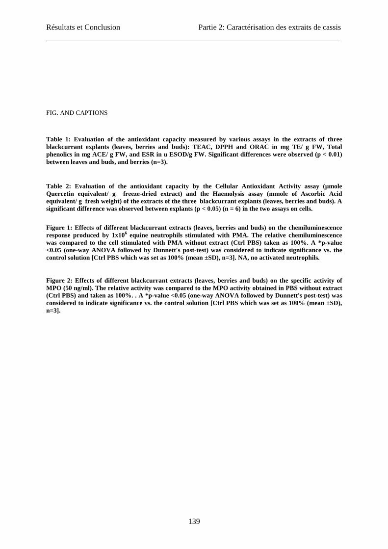
Résultats et Conclusion Partie 2: Caractérisation des extraits de cassis
139
FIG. AND CAPTIONS
Table 1: Evaluation of the antioxidant capacity measured by various assays in the extracts of three blackcurrant explants (leaves, berries and buds): TEAC, DPPH and ORAC in mg TE/ g FW, Total phenolics in mg ACE/ g FW, and ESR in u ESOD/g FW. Significant differences were observed (p < 0.01) between leaves and buds, and berries (n=3).
Table 2: Evaluation of the antioxidant capacity by the Cellular Antioxidant Activity assay (µmole Quercetin equivalent/ g freeze-dried extract) and the Haemolysis assay (mmole of Ascorbic Acid equivalent/ g fresh weight) of the extracts of the three blackcurrant explants (leaves, berries and buds). A significant difference was observed between explants (p < 0.05) (n = 6) in the two assays on cells.
Figure 1: Effects of different blackcurrant extracts (leaves, berries and buds) on the chemiluminescence response produced by 1x10⁶ equine neutrophils stimulated with PMA. The relative chemiluminescence was compared to the cell stimulated with PMA without extract (Ctrl PBS) taken as 100%. A *p-value <0.05 (one-way ANOVA followed by Dunnett's post-test) was considered to indicate significance vs. the control solution [Ctrl PBS which was set as 100% (mean ±SD), n=3]. NA, no activated neutrophils.
Figure 2: Effects of different blackcurrant extracts (leaves, berries and buds) on the specific activity of MPO (50 ng/ml). The relative activity was compared to the MPO activity obtained in PBS without extract (Ctrl PBS) and taken as 100%. . A *p-value <0.05 (one-way ANOVA followed by Dunnett's post-test) was considered to indicate significance vs. the control solution [Ctrl PBS which was set as 100% (mean ±SD), n=3].
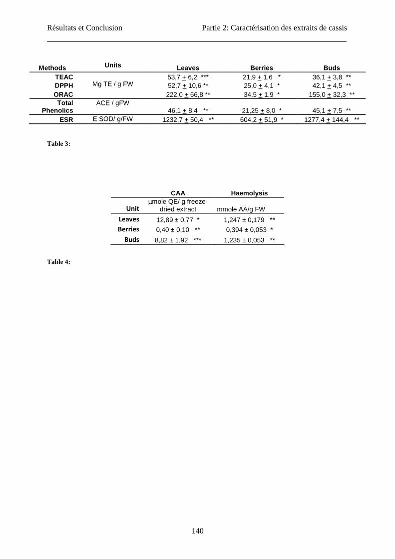
Résultats et Conclusion Partie 2: Caractérisation des extraits de cassis
140
Methods Units Leaves Berries Buds TEAC 53,7 + 6,2 *** 21,9 + 1,6 * 36,1 + 3,8 ** DPPH 52,7 + 10,6 ** 25,0 + 4,1 * 42,1 + 4,5 ** ORAC
Mg TE / g FW
222,0 + 66,8 ** 34,5 + 1,9 * 155,0 + 32,3 ** Total
Phenolics ACE / gFW
46,1 + 8,4 ** 21,25 + 8,0 * 45,1 + 7,5 ** ESR E SOD/ g/FW 1232,7 + 50,4 ** 604,2 + 51,9 * 1277,4 + 144,4 **
Table 3:
CAA Haemolysis
Unit
µmole QE/ g freeze-dried extract mmole AA/g FW
Leaves 12,89 ± 0,77 * 1,247 ± 0,179 ** Berries 0,40 ± 0,10 ** 0,394 ± 0,053 *
Buds 8,82 ± 1,92 *** 1,235 ± 0,053 **
Table 4:
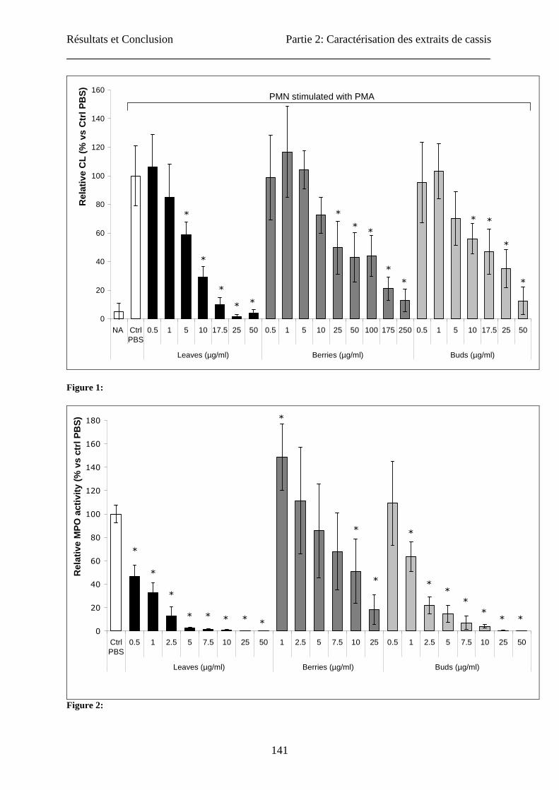
Résultats et Conclusion Partie 2: Caractérisation des extraits de cassis
141
0
20
40
60
80
100
120
140
160
NA CtrlPBS
0.5 1 5 10 17.5 25 50 0.5 1 5 10 25 50 100 175 250 0.5 1 5 10 17.5 25 50
Leaves (µg/ml) Berries (µg/ml) Buds (µg/ml)
Rel
ativ
e C
L (%
vs
Ctr
l PB
S) PMN stimulated with PMA
*
*
**
*
*
*
*
*
*
*
*
*
*
Figure 1:
0
20
40
60
80
100
120
140
160
180
CtrlPBS
0.5 1 2.5 5 7.5 10 25 50 1 2.5 5 7.5 10 25 0.5 1 2.5 5 7.5 10 25 50
Leaves (µg/ml) Berries (µg/ml) Buds (µg/ml)
Rel
ativ
e M
PO
act
ivity
(%
vs
ctrl
PB
S)
*
**
*
** *
*
*
*
* * * *
*
*
*
*
Figure 2:
Résultats et Conclusion Partie 2: Caractérisation des extraits de cassis
142
3. Conclusion
La caractérisation des molécules antioxydantes des matrices végétales peut être réalisée par de
nombreuses approches différentes. Avant d’utiliser cette large gamme de techniques en vue de
caractériser au mieux nos extraits de cassis, nous avons mené un certain nombre
d’investigations sur la validation de ces techniques au moyen d’une large gamme de standards
commerciaux ainsi que de divers produits alimentaires. Etant donné la diversité des résultats
obtenus pour le dosage de la capacité antioxydante, nous avons proposé une méthode
permettant de normaliser la valeur de la capacité antioxydante en employant une moyenne
pondérée basée sur les résultats de quatre méthodes aux principes différents : DPPH
(oxydoréduction), ORAC (cinétique de piégeage du radical peroxyle), Hémolyse (protection
d’un échantillon biologique) et l’EPR (évaluation d’un radical libre particulier).
Au niveau des extraits de cassis obtenus par le mélange acétone-eau- acide acétique (70 :28
:2), nous avons caractérisé le contenu en composés antioxydants par des analyses
chromatographiques et/ou spectrophotométriques. Les feuilles et les bourgeons présentent un
contenu plus élevé en composés phénoliques et en flavonols, avec une prédominance de la
quercétine. Par contre, les fruits possèdent un contenu en acide ascorbique très élevé. Au
niveau des flavan-3-ols, les bourgeons sont les plus riches, avec une prédominance pour la
gallocatéchine et l’épigallocatéchine. Au niveau de la caractérisation des propriétés
antioxydante et anti-inflammatoire de nos extraits, notre étude a mis en évidence des
propriétés intracellulaires et extracellulaires en tant que piégeurs de radicaux libres et une
activité inhibitrice de la myéloperoxydase chez les neutrophiles. Nous avons observé un
meilleur effet pour les feuilles, suivit par les bourgeons et finalement, loin derrière, les baies.
Cette différence semble être corrélée avec le taux en phénols totaux.
En empêchant la production de radicaux libres dans les cellules comme les neutrophiles ou les
cellules endothéliales, l’extrait de feuilles de cassis possèderait un potentiel élevé pour le
traitement de pathologies tant inflammatoires que cardio-vasculaires. Dans la suite de ce
travail, les effets de l’extrait de feuilles de cassis seront étudiés sur le processus de
vasorelaxation en utilisant des modèles cellulaires et animaux.
Résultats et Conclusion Partie 3: Biodisponibilité et Processus de vasorelaxation
143
Partie 3 : Biodisponibilité et Processus de vasorelaxation
1. Introduction
En plus des effets antioxydants et anti-inflammatoires, les polyphénols agissent aussi au
niveau de la vasomotricité. Ces polyphénols peuvent agir directement au niveau de
l’endothélium, en interagissant avec des facteurs endothéliaux (monoxyde d’azote et
endothéline-1). Ils peuvent également contribuer à préserver l’intégrité de l’endothélium par
divers mécanismes : la régulation de la prolifération, de la migration et de l’apoptose des
cellules endothéliales. Les polyphénols, de par leurs multiples effets vasculaires (anti-
inflammatoires, vasorelaxants, ...) présentent un intérêt certain pour la prévention de
nombreuses pathologies.
Des études utilisant un concentré de jus de cassis comme modèle ont déjà permis de démonter
les effets protecteurs du cassis sur le processus de relaxation vasculaire (Nakamura et al.,
2002). Comme vu dans les précédents résultats, les feuilles ont des propriétés beaucoup plus
importantes que les baies. Dans cette dernière partie des résultats, nous avons tenté de montrer
les effets de cet extrait de feuilles de cassis sur la relaxation vasculaire dépendante de
l’endothélium en utilisant des modèles cellulaires mais aussi un modèle d’organes isolés. Un
deuxième point abordé dans cette partie consiste à voir si notre extrait peut être consommé
sans danger et si oui, quelle est la partie ingérée potentiellement active au niveau de
l’organisme. Ce dernier aspect est abordé en utilisant un modèle animal.
Résultats et Conclusion Partie 3: Biodisponibilité et Processus de vasorelaxation
144
2. Article
Bioavailability and vasorelaxant effect of Ribes
nigrum leaf extract on human endothelial cells and
rat aorta. Jessica TABART 1, Claire KEVERS 1, Joël PINCEMAIL 2, Jean-Olivier DEFRAIGNE 2,
Sophie VINCENT 3, Arlette DELAMARCHE 3, Jacques DOMMES 1
University of Liège,
1Plant Molecular Biology and Biotechnology Unit, B22
2CREDEC, B35
Sart Tilman,
B-4000 LIEGE
Belgium
University of Rennes 3Laboratory « Mouvement Sport Santé » (EA1274), and ENS Cachan, UFR-APS,
F-Rennes Cedex
France
Corresponding author: Jessica TABART: [email protected]
Other authors: [email protected], [email protected], [email protected],
[email protected], [email protected], [email protected];
TITLE RUNNING HEAD: Vasorelaxant effect of blackcurrant leaf extract
Résultats et Conclusion Partie 3: Biodisponibilité et Processus de vasorelaxation
145
ABSTRACT
The dysfunction of endothelium is involved in an important cause of mortality in developed
countries, atherosclerosis. Many plants contain compounds that cause endothelium-dependent
vasorelaxation in vivo. In our study, we used a leaf extract of blackcurrant to investigate the
endothelium-dependent relaxing properties in cell model and on isolated organ model, and
complementary, the toxicity and the bioavailability in an animal model. Our results indicated
the regulation of eNOS mRNA with an increase of NO production. In organ bath chambers,
we could also show a relaxant effect of the extract on vessel. With the animal model, we did
not observe any toxic effect after 28-days of consumption. But we showed an increase of
some flavonoids in plasma and a regulating effect on body weight.
KEYWORDS: Endothelium-dependent relaxation, antioxidant, endothelium, NO synthase,
bioavailability, toxicity, blackcurrant.
INTRODUCTION
Atherosclerosis is the leading overall direct or indirect cause of mortality in developed
countries (Beaudeux et al., 2006). The physiopathological mechanisms for formation and
evolution of vascular atheromogenic damages are correlated with various risk factors like
oxidative stress. The abnormal increase of reactive oxygen species (ROS) and reactive
nitrogen species (RNS) in endothelial cells and vascular smooth muscle cells induces an
endothelial activation, an inflammatory response and the proliferation of vascular muscular
smooth cells which cause atherosclerotic damages (Beaudeux et al., 2006).
Endothelium has many functions like control of vascular permeability, inflammatory cell
adhesion, coagulation and platelet aggregation. Therefore, the functional integrity of the
endothelium is essential to prevent vascular leakage and formation of the atherosclerosis. The
vascular endothelium plays an important role in homeostasis by modulating vascular smooth
muscle tone and acts as a main target site in hypertension and atherosclerosis (Moncada et al.,
1991). Endothelial cells produce various vasodilating substances such as endothelium-derived
relaxing factors or nitric oxide (NO), endothelium-derived hyperpolarizing factors (EDHF)
(Fletou and Vanhoutte, 1988) and vasodilator prostaglandins.
Résultats et Conclusion Partie 3: Biodisponibilité et Processus de vasorelaxation
146
NO produced by endothelial-type nitric oxide synthase is a key factor in vascular protection
as vasodilator agent. NO can act also as antithrombic agent by inhibiting platelet aggregation
and adhesion. Besides these properties, NO possesses also many antiartherosclerotic
properties which include: the prevention of leukocyte adhesion to vascular endothelium and
their migration into the vascular wall, the decrease of endothelium permeability, the inhibition
of LDL oxidation, and the inhibition of proliferation of vascular smooth cells (Li and
Föstermann, 2000). One of the mechanisms involved in the endothelium dysfunction is a
reduced NO synthesis and/or an increase of vascular NO degradation (Barton et al., 1997; Li
and Föstermann, 2000).
Many plants including grapes contain extractable compounds that cause endothelium-
dependent vasorelaxation in vitro (Fitzpatrick et al., 1993). Plant flavonoids exhibit various
biological properties that have been demonstrated in a variety of experimental systems. The
benefit of flavonoid-rich food has been attributed to the antioxidant activity of these
compounds (Di Carlo et al., 1999) but also to the modulation of various factors involved in
the development of vascular diseases. In rats, polyphenols could decrease arterial pressure,
prevent low density lipoprotein (LDL) oxidation, inhibit vascular smooth muscle cells, and
prevent platelet aggregation (Martin and Andriantsihaina, 2002).
Blackcurrants are among the berries with the higher amount of phenolic compounds,
especially anthocyanins. Other major phenolics present in blackcurrants include flavonols,
procyanidins and various phenolic acids (Benvenuti et al., 2004). Only a very small portion of
these berries is consumed fresh; most is processed for industrial use in making juice and
liquors. Nakaishi et al. (2000) found that oral intake of blackcurrant concentrate prepared
from blackcurrant juice had significant and beneficial effects on visual functions. Nakamura
et al. (2002) suggested that the prevention of eye fatigue resulted probably from increased
blood supply to these areas caused by vasorelaxation induced by blackcurrant concentrate.
Their work demonstrated that blackcurrant concentrate causes endothelium-dependent
vasorelaxation in vitro in rat aorta rings by increasing NO production, via the histamine H1-
receptors on the endothelium. Leaves and buds can also be used (Tabart et al., 2006). In a
previous study (Tabart et al., 2011), we have demonstrated that leaf and bud extracts have
higher antioxidant and anti-inflammatory properties than berry extract.
Résultats et Conclusion Partie 3: Biodisponibilité et Processus de vasorelaxation
147
This study aimed at investigating the effect of blackcurrant leaf extract on the endothelium
relaxation by examining in vitro the expression modulation of eNOS and the NO production
by human endothelial cells. Its effect was also observed on the relaxation of isolated vessels.
The second aim of this study was to examine, in vivo, the toxicity and the bioavailability of
leaf extract in a chronic experiment on an animal model.
MATERIAL AND METHODS
Sample preparation
The leaves of blackcurrant (Noir de Bourgogne), two years old, were harvested in August in
the Belgian Ardennes (Bihain). The various explants were directly cut out into pieces and
were frozen in liquid nitrogen, lyophilized and preserved at -20°C for a few months.
For preparing extract, 50 grams of frozen sample (berries, buds or leaves) were ground with a
few grams of quartz and 500 ml of extraction solution: acetone/water/acetic acid (70:28:2)
(Tabart et al., 2006). The mixture was shaken during 1 h at 4°C and centrifuged at 17 000 g
for 15 min. The supernatant was removed, and the sample was extracted again with the same
procedure but incubated only 15 min. The acetone was evaporated and the residue was filtered
and lyophilised before storage at -20°C. The residue was resuspended in appropriate medium
for analysis (DMEM supplemented for cell analysis, Water for chronic administration of
extract in rats and DMSO for analysis on isolated vessels).
Cell culture
The human endothelial-like EAhy926 cell line (Edgell et al., 1983), is derived from the fusion
of human umbilical vein endothelial cells with the A549 carcinoma cell line. EAHy926 cells
were grown in DMEM (Biowithaker, Lonza) supplemented with 10% of heat-inactivated fetal
bovine serum, 10 µM 4-(2-hydroxyethyl)-1-piperazineethanesulfonic acid (HEPES), 5000
units/l of penicillin and streptomycin, 1 mM pyruvate sodium, 2 mM glutamin and 2% HAT
medium supplement (hypoxanthin, aminoprotein and thimidin), and were maintained at 37°C
and 5% CO2.
Résultats et Conclusion Partie 3: Biodisponibilité et Processus de vasorelaxation
148
3-[4,5-dimethylthiazol-2-yl]2,5-diphenyltetrazolium bromide viability assay
Potential toxic effects of blackcurrant leaf extracts were tested using the Cell Growth
Determination kit MTT (3-[4,5-dimethylthiazol-2-yl]2,5-diphenyltetrazolium bromide,
Sigma-Aldrich, St Louis, MO). The EAhy926 cell line was plated in a 96-well plate at 5000
cells/well and samples treated in sestuplicate with different concentrations of blackcurrant leaf
extract (0-1 mg/ml) or complete DMEM as a control. The cell density was assessed at 24 hour
time intervals over 5 consecutive days. Cell viability was evaluated as recommended by the
manufacturer. The absorbance was measured on a microliter plate reader (Labsystems IEMS
reader MF) at 570 nm. The background absorbance at 690 nm was subtracted.
Determination of nitrite content
Nitrite accumulation in EAhy926 cell culture supernatant was measured using the Griess
method for the determination of the total nitrite content (nitrite and nitrate), adapted for 96-
wells plates (Bani et al., 1998). 500 000 cells were incubated in 6-well plates with leaf extract
at different concentration (0 to 1 mg/ml) or quercetin solution (50 µM) during 5 min, 6 h, 12
h, 24 h and 48 h). At the end of incubation, 100 µl culture supernatant were mixed with 25 µl
of NADPH (93 µg/ml final concentration) and 10 µl of nitrate reductase (0.2 U) in a 96-well
plates. After incubation at 25°C for 1.5 h, 100 µl of Griess reagent (1 part 1% sulfanilamide
and 1 part 0.1% N-naphtylethyl-ethylenediamine (NED) in 5% ortho-phosphoric acid) were
added. The absorbance at 540 nm was measured after 10 minutes of incubation. Various
concentrations of sodium nitrate (0–500 µM) were used to obtain a calibration curve. The
results were expressed as µM of NaNO3.
Semi-quantitative RT-PCR for eNOS mRNA analyses
eNOS mRNA expression was analyzed with semi-quantitative RT-PCR. In the first part,
EAhy926 cell cultures were incubated with leaf extract at various concentrations (0 to 1 mg/
ml) during 24 h. In the second part, cell cultures were incubated with leaf extract at 250 µg/ml
for different times (5 min, 6 h, 12 h, 24 h and 48 h). After incubation, total RNA was isolated
with the RNeasy mini kit (Qiagen) as recommended by the supplier. RNA (1µg) was
transcribed into cDNA by reverse transcription by M-MLV RT RNase H(-) Point Mutant
enzyme (Promega). For the PCR, 2.5 µl of cDNA product of each sample was amplified on
Gene Amp® PCR System 9700 (PE Applied BioSystems) with Taq DNA polymerase using a
primer pair specific to eNOS (forward primer: 5’-GCT GCG CCA GGC TCT CAC CTT C-3’,
Résultats et Conclusion Partie 3: Biodisponibilité et Processus de vasorelaxation
149
reverse primer: 5’-GGC TGC AGC CCT TTG CTC TCA A-3’, 35 cycles, 554 bp, annealing
temperature: 55°C), a primer pair specific to iNOS (forward primer: 5’-TCT TGG TCA AAG
CTG TGC TC-3’, reverse primer: 5’-CAT TGC CAA ACG TAC TGG TC-3’, 35 cycles, 237
bp, annealing temperature: 60°C) and two primer pairs specific to β-actin ([forward primer:
5’-GGC ATC GTG ATG GAC TCC G-3’, reverse primer: 5’-GCT GGA AGG TGG ACA
GCG A-3’, 23 cycles, 603 bp, annealing temperature: 68 °C] [forward primer: 5’- ATG GAT
GAT GAT ATC GCC GCG -3’, reverse primer: 5’ - TCT CCA TGT CGT CCC AGT TG -
3’, 30 cycles, 248 bp, annealing temperature: 58°C]). At last, the PCR amplified samples were
visualized by electrophoresis through 1.5% agarose gel in the presence of ethidium bromide.
The quantification was carried out using the G:Box Imaging System (Syngene, Wesburg) and
the GeneSnap and GeneTools softwares.
Animals and ethical approval
Male Wistar rats purchased in the central animalery of University of Liege (Belgium) (12-14
weeks old) were housed in cages under standard experimental conditions (23 ± 1°C, 12 h
light/dark cycle). All experimental protocols were approved by the Animal Care and Use
Committee of University of Liège (Belgium), and conduced humanely.
Characterization of the relaxant effect of leaf extract on vascular reactivity
Male Wistar rats were killed by an injection of pentobarbital (80 mg/Kg bodyweight) and then
exsanguinated. The aorta was removed and carefully cleaned of adhering fat and connective
tissue, and cut into rings (3-4 mm). The rings were mounted in standard organ baths (IOX,
EMKA Technologies, France) filled with physiological salt solution (Krebs buffer: 118 mM
NaCl, 25 mM NaHCO3, 5 mM Glucose, 4.7 mM KCl, 1.2 mM KH2PO4, 1.2 mM MgSO4 and
2.5mM CaCl2), maintained at 37°C and continuously bubbled with 95% O2 - 5% CO2
mixture. Resting tension was adjusted to 3 g. Tension was measured with an isometric force
transducer.
After an equilibration period of 90 min, the vessels were contracted three times with KCl
(0.08 M) in order to test their contractile capacity. The presence of functional endothelium
was assessed in all preparations by determining the ability of acetylcholine (Ach, 10 µM) to
induce more than 50% relaxation of rings precontracted with noradrenalin (0.1 µM). Vessels

Résultats et Conclusion Partie 3: Biodisponibilité et Processus de vasorelaxation
150
were considered to be devoid of functional endothelium when there was no relaxation
response to Ach.
After washing and returning to baseline tension, aortic rings with functional endothelium were
pre-contracted to 80% of KCl maximal tension with phenylephrine (0.5 µM). When the
contraction reached a steady state, increasing concentrations of Ach, quercetin or leaf extract
were added cumulatively.
Oral toxicity and bioavailability of leaf extract f lavonoids
The rats were fed with a standard diet. The animals were divided into three groups of 5
animals (n=5). The diet of two groups was supplemented with blackcurrant leaf extract
(dissolved in water) amounting to 896 ± 30 and 1528 ± 137 mg/kg bodyweight every day
during 28 days. Weight of animals was monitored for 28 consecutive days. The general
behaviour of each animal was observed every day throughout the study period, such as food
consumption.
At the end of the treatment, animals were sacrificed by an injection of pentobarbital (80
mg/Kg of bodyweight) and then exsanguinated. The blood and the liver were collected for
analysis.
Blood was analysed for toxicity markers and bioavailability of phenolic compounds. For the
toxicity analysis, various markers were quantified versus control by the Department of
Clinical Biology of CHU-Liège (Belgium): oral orientation, hepatic and cardiac markers. The
plasma was used to analyse the content of total phenolics (by a spectrophotometric method),
flavonol aglycons and favan-3-ols monomers (by HPLC following methods described by
Tabart et al., 2009; 2010b).
Statistical analysis
The data were subjected to the statistical analysis of the variance (ANOVA-1) to evaluate the
significant differences between various explants of blackcurrant. The difference was regarded
as significant when p < 0.05.

Résultats et Conclusion Partie 3: Biodisponibilité et Processus de vasorelaxation
151
RESULTS
1. Toxicity of blackcurrant leaf extract
The toxicity of blackcurrant leaf extract was determined at two levels: on cell model and on
animal model.
a. Cytotoxicity on human endothelial cells
Before testing the cytotoxic effect of the extract on animal, a viability assay was used to
determine the effect of leaf extract on the viability of cells during 120 hours (Figure 1).
0,00
0,05
0,10
0,15
0,20
0,25
0,30
0 10 50 100 250 500 750 1000
µg of extract / ml of medium/20 000 cells
DO 24H
48H
72H
96 H
120 H
Figure 1: Evolution of cell viability during 120 h of incubation with leaf extract at different concentrations
(0 to 1 mg/ml). Data represented the means ± SD (n = 6).
In control conditions, the viability increased to 72h. The addition of leaf extract at a
concentration lower than 500 µg/ ml did not modify the cell viability during the first 72 hours.
With higher concentrations of leaf extract (1 mg/ ml), the viability decreased and a
cytotoxicity was observed.
b. Toxicity on rat models after 28-days chronic ingestion
In the 28-d chronic oral toxicological tests in rats, leaf extract was administrated orally to rats
at doses of 0 (control), 896 ± 30 and 1528 ± 137 mg/Kg of body weight per day (Figure 2).
All animals survived in all groups.

Résultats et Conclusion Partie 3: Biodisponibilité et Processus de vasorelaxation
152
0
50
100
150
200
250
300
350
400
0 7 11 14 21 25 28Days
Wei
ght o
f rat
(g) Control
1st amount
2 nd amount
Figure 2: Evolution of body weight during 28-d chronic toxicological test with leaf extract (0 (control), 896
± 30 (1st amount) and 1528 ± 137 (2nd amount) mg/Kg of body weight). Data represented the means ± SD.
A significantly difference was observed at p>0.05 between the three groups (a, b, c) (n = 5).
At the end of the period, the body weight of treated animals was significantly lower than that
of control rats, without any abnormal clinical-related parameter. Liver weight was lower in
animal fed with the highest amount leaf extract as compared to control (Figure 3).
02468
1012141618
Control 1st amount 2nd amount
Wei
ght o
f liv
er
Figure 3: Weight of liver after 28-d chronic toxicological test with leaf extract (0 (control), 896 ± 30 (1st
amount) and 1528 ± 137 (2nd amount) mg/Kg of body weight). Data represented the means ± SD. A
significantly difference was observed at p>0.05 between control and the 2nd group (a, b) (n = 5).
Concerning toxicological markers, no difference was found between the three groups (Table
1).
a b c
a a,b b

Résultats et Conclusion Partie 3: Biodisponibilité et Processus de vasorelaxation
153
Table 1: Analysis of different markers of toxicity after 28-days chronic toxicological ingestion of leaf
extract (0 (control), 896 ± 30 (1st amount) and 1528 ± 137 (2nd amount) mg/Kg of body weight) (n = 5).
(GGT: gamma glutamyl transpeptidase, GOT: glutamic oxalo-acetic transaminase, GPT: glutamic
pyruvic transaminase, LDL: low-density protein).
2. Bioavailability of blackcurrant leaf extract after 28-days chronic
ingestion
After the 28-days chronic oral test in rats, we determined the concentration of various
compounds of blackcurrant leaf extract in the plasma (Table 2).
Table 2: Analysis of the flavanols, flavonols and total phenol content of plasma after 28-d chronic
ingestion of two amount (896 ± 30 (1st amount) and 1528 ± 137 (2nd amount) mg/Kg of body weight) of leaf
extract versus control. Data represent the means ± SD. A significantly difference was observed between
the three ingested amounts at p<0.05 (n = 5 for total phenolics analysis and n = 3 for the others).
Total phenols content significantly increased in correlation with the administrated amount.
Some flavonoids were analysed by HPLC: flavonols, anthocyanins and flavan-3-ols. For
flavonols, the quantity present in the plasma of rats that have ingested the extract was
significantly higher versus control, without correlation with the amount administrated except
for myricetin. No anthocyanin could be detected. For flavan-3-ols, only epigallocatechin
(EGC) monomer was detected. The concentration of this compound was not correlated with
General parameters Hepatic exploration Cardiac Markers
Urea (g/L) Creatin (mg/L)
Alkalin
Phosphatase GGT GOT GPT LDH
Creatin kinase
(UI/L)
Control 0.662 ± 0.008 4.20 ± 0.15 136.4 ± 13.7 <5 174.2 ± 28.4 60 ± 8.1 661 ± 138 6551 ± 1981
1st amount 0.632 ± 0.020 4.52 ± 0.47 130.6 ± 9.8 <5 148.0 ± 47.2 53.7 ± 2.3 570 ± 217 6093 ± 1403
2nd amount 0,618 ± 0.127 5.50 ± 0.23 115.0 ± 37.8 <5 137.8 ± 23.4 49 ± 1.3 573 ± 252 4464 ± 951
Myricetin Quercetin Kampferol EGC Total Phenols
(µg/ml) (µg/ml) (µg/ml) (µg/ml) (mg/ml)
Control 0* 0.03 ± 0.02* 0* 23.16 ± 5.46* 4.65 ± 0.22*
1st amount 1.29 ± 1.15* 9.28 ± 2.75** 4.93 ± 2.01** 54.04 ± 5.22** 5.14 ± 0.15**
2nd amount 9.29 ± 3.01** 6.40 ± 3.18** 3.34 ± 2.05** 16.59 ± 9.14* 5.88 ± 0.42***
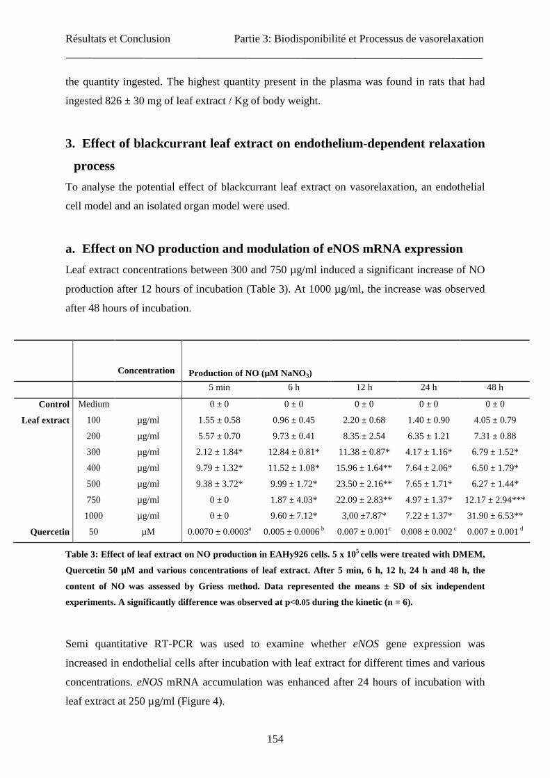
Résultats et Conclusion Partie 3: Biodisponibilité et Processus de vasorelaxation
154
the quantity ingested. The highest quantity present in the plasma was found in rats that had
ingested 826 ± 30 mg of leaf extract / Kg of body weight.
3. Effect of blackcurrant leaf extract on endothelium-dependent relaxation
process
To analyse the potential effect of blackcurrant leaf extract on vasorelaxation, an endothelial
cell model and an isolated organ model were used.
a. Effect on NO production and modulation of eNOS mRNA expression
Leaf extract concentrations between 300 and 750 µg/ml induced a significant increase of NO
production after 12 hours of incubation (Table 3). At 1000 µg/ml, the increase was observed
after 48 hours of incubation.
Concentration
5 min 6 h 12 h 24 h 48 h
Control Medium 0 ± 0 0 ± 0 0 ± 0 0 ± 0 0 ± 0
Leaf extract 100 µg/ml 1.55 ± 0.58 0.96 ± 0.45 2.20 ± 0.68 1.40 ± 0.90 4.05 ± 0.79
200 µg/ml 5.57 ± 0.70 9.73 ± 0.41 8.35 ± 2.54 6.35 ± 1.21 7.31 ± 0.88
300 µg/ml 2.12 ± 1.84* 12.84 ± 0.81* 11.38 ± 0.87* 4.17 ± 1.16* 6.79 ± 1.52*
400 µg/ml 9.79 ± 1.32* 11.52 ± 1.08* 15.96 ± 1.64** 7.64 ± 2.06* 6.50 ± 1.79*
500 µg/ml 9.38 ± 3.72* 9.99 ± 1.72* 23.50 ± 2.16** 7.65 ± 1.71* 6.27 ± 1.44*
750 µg/ml 0 ± 0 1.87 ± 4.03* 22.09 ± 2.83** 4.97 ± 1.37* 12.17 ± 2.94***
1000 µg/ml 0 ± 0 9.60 ± 7.12* 3,00 ±7.87* 7.22 ± 1.37* 31.90 ± 6.53**
Quercetin 50 µM 0.0070 ± 0.0003a 0.005 ± 0.0006 b 0.007 ± 0.001c 0.008 ± 0.002 c 0.007 ± 0.001 d
Table 3: Effect of leaf extract on NO production in EAHy926 cells. 5 x 10⁵ cells were treated with DMEM,
Quercetin 50 µM and various concentrations of leaf extract. After 5 min, 6 h, 12 h, 24 h and 48 h, the
content of NO was assessed by Griess method. Data represented the means ± SD of six independent
experiments. A significantly difference was observed at p<0.05 during the kinetic (n = 6).
Semi quantitative RT-PCR was used to examine whether eNOS gene expression was
increased in endothelial cells after incubation with leaf extract for different times and various
concentrations. eNOS mRNA accumulation was enhanced after 24 hours of incubation with
leaf extract at 250 µg/ml (Figure 4).
Production of NO (µM NaNO3)
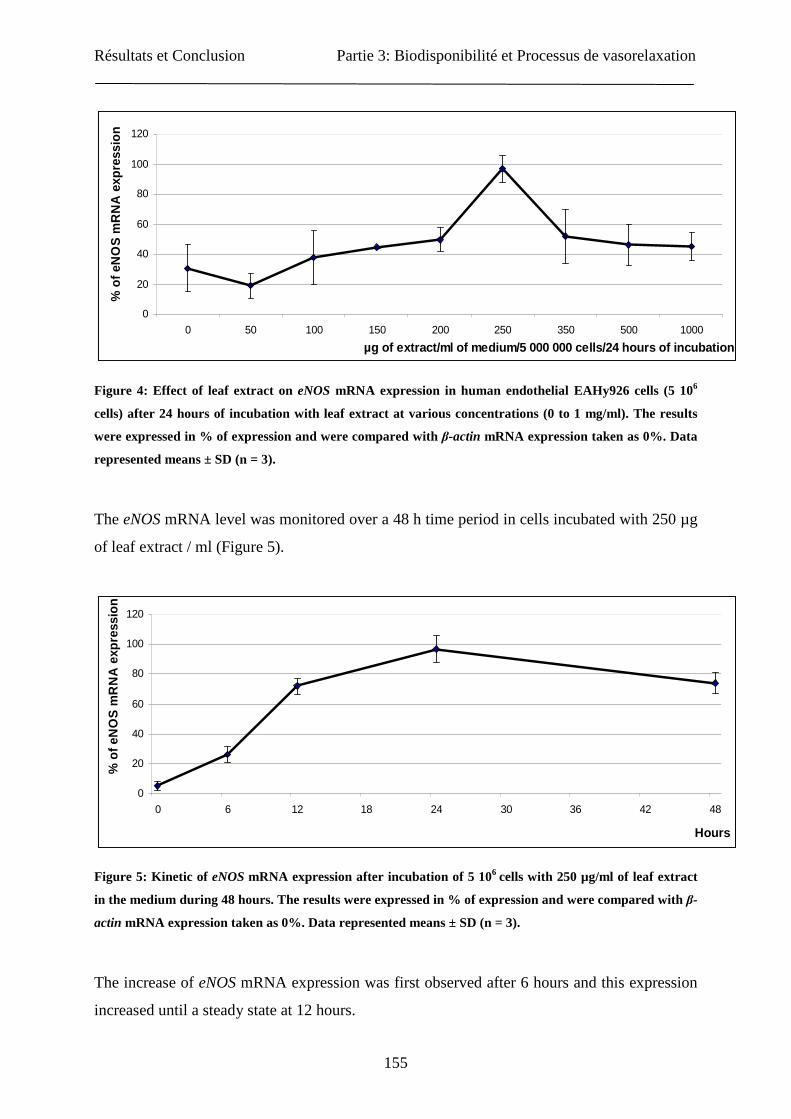
Résultats et Conclusion Partie 3: Biodisponibilité et Processus de vasorelaxation
155
0
20
40
60
80
100
120
0 50 100 150 200 250 350 500 1000
µg of extract/ml of medium/5 000 000 cells/24 hours of incubation
% o
f eN
OS
mR
NA
exp
ress
ion
Figure 4: Effect of leaf extract on eNOS mRNA expression in human endothelial EAHy926 cells (5 106
cells) after 24 hours of incubation with leaf extract at various concentrations (0 to 1 mg/ml). The results
were expressed in % of expression and were compared with β-actin mRNA expression taken as 0%. Data
represented means ± SD (n = 3).
The eNOS mRNA level was monitored over a 48 h time period in cells incubated with 250 µg
of leaf extract / ml (Figure 5).
0
20
40
60
80
100
120
0 6 12 18 24 30 36 42 48
Hours
% o
f eN
OS
mR
NA
exp
ress
ion
Figure 5: Kinetic of eNOS mRNA expression after incubation of 5 106 cells with 250 µg/ml of leaf extract
in the medium during 48 hours. The results were expressed in % of expression and were compared with β-
actin mRNA expression taken as 0%. Data represented means ± SD (n = 3).
The increase of eNOS mRNA expression was first observed after 6 hours and this expression
increased until a steady state at 12 hours.

Résultats et Conclusion Partie 3: Biodisponibilité et Processus de vasorelaxation
156
b. In vitro determination of vascular reactivity
To investigate the functional consequences of the enhanced eNOS mRNA expression, we
studied the effects of quercetin and blackcurrant leaf extract on vasorelaxation in comparison
to acetylcholine (Figure 6).
Figure 6: Cumulative concentration-relaxation curves for Acetylcholine (0 to 10-5 M, A), Quercetin (0 to 3
µM, B) and leaf extract of blackcurrant (0 to 1.60 mg/ ml, C) in endothelium-intact Rat aortic rings
precontracted with phenylephrine (µM). The results were the means ± SD (n = 6).
0
20
40
60
80
100
120
0,000000 0,000002 0,000004 0,000006 0,000008 0,000010
Acetylcholine (M)
% o
f rel
axat
ion
0
20
40
60
80
100
120
0,000 0,001 0,002 0,003
Quercetin (M)
% o
f rel
axat
ion
0
20
40
60
80
100
120
0 250 500 750 1000 1250 1500 1750
Leaf extract (µg/ml)
% o
f rel
axat
ion
A
B
C
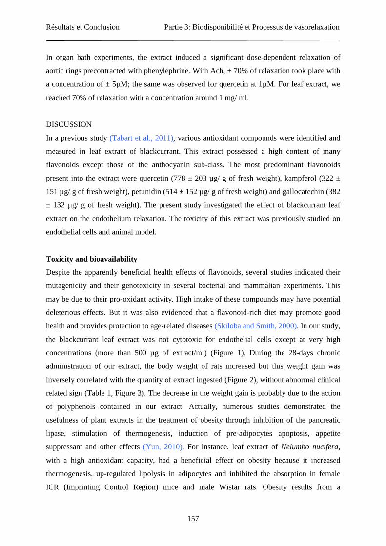
Résultats et Conclusion Partie 3: Biodisponibilité et Processus de vasorelaxation
157
In organ bath experiments, the extract induced a significant dose-dependent relaxation of
aortic rings precontracted with phenylephrine. With Ach, ± 70% of relaxation took place with
a concentration of ± 5µM; the same was observed for quercetin at 1µM. For leaf extract, we
reached 70% of relaxation with a concentration around 1 mg/ ml.
DISCUSSION
In a previous study (Tabart et al., 2011), various antioxidant compounds were identified and
measured in leaf extract of blackcurrant. This extract possessed a high content of many
flavonoids except those of the anthocyanin sub-class. The most predominant flavonoids
present into the extract were quercetin (778 ± 203 µg/ g of fresh weight), kampferol (322 ±
151 µg/ g of fresh weight), petunidin (514 ± 152 µg/ g of fresh weight) and gallocatechin (382
± 132 µg/ g of fresh weight). The present study investigated the effect of blackcurrant leaf
extract on the endothelium relaxation. The toxicity of this extract was previously studied on
endothelial cells and animal model.
Toxicity and bioavailability
Despite the apparently beneficial health effects of flavonoids, several studies indicated their
mutagenicity and their genotoxicity in several bacterial and mammalian experiments. This
may be due to their pro-oxidant activity. High intake of these compounds may have potential
deleterious effects. But it was also evidenced that a flavonoid-rich diet may promote good
health and provides protection to age-related diseases (Skiloba and Smith, 2000). In our study,
the blackcurrant leaf extract was not cytotoxic for endothelial cells except at very high
concentrations (more than 500 µg of extract/ml) (Figure 1). During the 28-days chronic
administration of our extract, the body weight of rats increased but this weight gain was
inversely correlated with the quantity of extract ingested (Figure 2), without abnormal clinical
related sign (Table 1, Figure 3). The decrease in the weight gain is probably due to the action
of polyphenols contained in our extract. Actually, numerous studies demonstrated the
usefulness of plant extracts in the treatment of obesity through inhibition of the pancreatic
lipase, stimulation of thermogenesis, induction of pre-adipocytes apoptosis, appetite
suppressant and other effects (Yun, 2010). For instance, leaf extract of Nelumbo nucifera,
with a high antioxidant capacity, had a beneficial effect on obesity because it increased
thermogenesis, up-regulated lipolysis in adipocytes and inhibited the absorption in female
ICR (Imprinting Control Region) mice and male Wistar rats. Obesity results from a
Résultats et Conclusion Partie 3: Biodisponibilité et Processus de vasorelaxation
158
disequilibrium between energy intake and expenditure and is known to be a strong risk factor
for type 2-diabeted associated with insulin resistance (Ono et al., 2006). Lipid disorders are
also induced in abnormal vasomotor process through the high concentration of oxidized LDL,
the release of endothelin by endothelial cells, the increase of vessel permeability and so on
(Lebranchu, 2000).
The metabolism of polyphenols is now well understood. Actually, numerous studies show that
the quantities of polyphenols found intact in urine vary from one phenolic compound to
another. Inter-individual variations have also been observed; probably due to the different
composition of the intestinal flora which can differently affect their metabolism (D’Archivio
et al., 2007). The concentration of polyphenols reached after their consumption varies highly
according to the nature of polyphenols and the food sources. The plasma concentrations of
intact flavonoids rarely exceeded 1 µM and the maintenance of high concentration in plasma
requests repeated ingestions over time; in fact the maximum concentrations are most often
reached 1-2 hours after ingestion, except for polyphenols which require to be modified prior
to absorption (D’Archivio et al., 2007). Quercetin is the most predominant flavonoid in the
human diet and is one of the most extensively studied polyphenols. It serves as a good
example because its metabolism in humans is well understood. Estimated human consumption
is in the range of 4 to 68 mg per day based on epidemiological studies (Knekt et al., 1997;
Hertog et al., 1993, 1995; Rimm et al., 1996). Due to absorption differences of different
conjugated forms of quercetin, its bioavailability varies greatly in different foods. Generally,
the absorption of glycosylated flavonol was far greater than the absorption of the aglycon
(Skiloba and Smith, 2000). In our study (Table 2), the total phenolics in plasma increased
with the quantity of extract ingested. However when we analyzed different sub-classes of
flavonoids, only myricetin increased in a dose-dependent manner. Quercetin and kampferol
also increased in plasma but with no significant difference between the two amounts
administrated. The healthy effects of polyphenols depend both on their intake and
bioavailability. The concept of bioavailability integrates several variables such as intestinal
absorption, metabolism by the microflora, binding to albumin, cellular uptake, accumulation
in tissues, and biliary and urinary excretion (D’Archivio et al., 2007).
Résultats et Conclusion Partie 3: Biodisponibilité et Processus de vasorelaxation
159
Nitric oxide and vasorelaxation process
Numerous in vitro studies showed that polyphenols induced at low concentration (<10 g/L) an
endothelium-dependent vasorelaxation and at high concentration (> 10 g/L) a vascular smooth
muscle-dependent vasorelaxation (Andriambeloson et al., 1997, 1998; Flesch et al., 1998).
The released nitric oxide contributes to the relaxation of vascular smooth muscle. On another
hand, many studies focused on the beneficial vascular effect of phenolic compounds in
humans. The current study provides evidence that blackcurrant leaf extract can up-regulate the
eNOS gene in human endothelial cells (Figure 5). The enhanced mRNA expression results in
an increased production of bioactive nitric oxide, as measured by the Griess reagent (Table 3).
Similar results were often found in studies on other food products such as wine (Wallerath et
al., 2003) or strawberry aqueous extract (Soares et al., 2006). The increase of NO production
was observed after 6 hours of incubation with a maximum at 12 hours. Such results were also
obtained by Xu et al. (2007) focusing on icariin effect. Increased production of eNOS mRNA
(Figure 4) most probably contributes to increased NO production. Further investigations must
be done to analyze the activity of eNOS protein and his expression.
An increased production of NO may contribute to the vascular protection and
antiatherosclerotic properties through a number of mechanisms. Therefore, we hypothesized
that the generation of NO induced by blackcurrant leaf extract may be associated with its
protective capacity on the vascular system. In this study, we have investigated the effect of
blackcurrant leaf extract on aortic rings of rats with intact endothelium. 70% of relaxation was
reached with leaf extract at ± 1 mg/ ml (Figure 6C). Nishida and Satoh (2004) showed that
Gingko biloba extract at 0.03 to 3 mg/ ml had a potent concentration-dependent relaxation: 70
± 4.5 % at 3 mg/ml. For strawberry extract (Edirisinghe et al., 2008), 70% of relaxation was
reached with a concentration of 1 mg/ml. In a recent study (Tabart et al., 2011), we have
shown that the predominant flavonoid constituent of our extract was quercetin. The potent
relaxation of this compound was assessed. Effectively, we quercetin showed an endothelium-
dependent vasorelaxation potential at concentrations between 1 µM and 1 mM. In 1 mg /ml
leaf extract, the concentration of quercetin was around 15 µM (Figure 6B). At 1 mM, we had
approximatively 60% of relaxation. Therefore the vasorelaxation effect of the blackcurrant
leaf extract should depend on other compounds in addition to quercetin. Various effects of
quercetin were reported in the literature. The group of Benito (2002) showed that quercetin
increased eNOS protein activity and induced endothelium-dependent vasorelaxation in
Résultats et Conclusion Partie 3: Biodisponibilité et Processus de vasorelaxation
160
hypersensitive rats. The same result on eNOS protein activity was obtained by Xu et al.
(2007) with quercetin at 50 µM. But Wallerath et al. (2005) found that quercetin (1 to 33 µM)
reduced eNOS mRNA expression in a concentration dependent manner in EAHy926 cells for
24 hours. To explain this surprising result, they suggested mRNA destabilization, because
eNOS promoter activity was not modified by quercetin.
The relaxation of blood vessels is subjected to a complex control mechanism. Increased
relaxation of vascular smooth muscle can result from increased eNOS expression and/or NOS
activity or from activation of guanylate cyclase, all leading to the accumulation of cyclic
GMP (Moncada et al., 1991; Sennequier and Vadon-Le Goff, 1998).
In conclusion, our results indicate that blackcurrant extract are not cytotoxic and that its oral
uptake leads to an increase of some flavonoids in plasma with, perhaps, a regulation of body
weight. We show also that blackcurrant leaf extract can moderately up-regulate the production
of NO and the expression of eNOS mRNA. These effects could contribute to the
cardiovascular protection from atherosclerosis. Further investigations must be done for a good
comprehension of the mechanism by which our extract acts on endothelium-dependent
vasorelaxation.
ACKNOWLEDGMENT:
J.T. gratefully acknowledges the AFR Luxembourg and the memberships of CEDEVIT and
A.S.B.L (Liège).

Résultats et Conclusion Partie 3: Biodisponibilité et Processus de vasorelaxation
161
REFERENCES
1. Andriambeloson E., Kleschoyv A., Muller N., Beretz A., Stoclet J.C., and Andrianrsitohaina R., 1997. Nitric
oxide production and endothelium-dependent vasorelaxation induced by wine polyphenols in rat aorta.
British Journal of Pharmacology, 120, 1053-1058.
2. Andriambeloson E., Magnier C., Haan-Archipoff G., Lobstein A., Anton R., Beretz A., Stoclet J.C., and
Andriantsitohaina R., 1998. Natural dieatry polyphenolic compounds cause endothelium-dependant
vasorelaxation in rat thoracic aorta. The Journal of Nutrition, 128, 2324-2333.
3. Bani D., Failli P., Bello M.G., Thiemermann C., Bani Sacchi T., bigazzi M., and Masini E., 1998. Relaxin
activates the L-Arginine-nitric oxide pathway vascular smooth muscle cells in culture. Hypertension, 31,
1240-1247.
4. Barton M., Cosentino F., Brandes R.P., Moreau P., Shaw S., and Lüsher T.F., 1997. Anatomic heterogeneity
of vascular aging: Role of nitric oxide and endothelin. Hypertension, 30, 817-824.
5. Beaudeux J.L., Delattre J., Therond P., Bonnefont-Rousselot D., Legrand A., and Peynet J., 2006. Oxidative
stress in the atherosclerotic process. Immuno-analyse & Biologie spécialisée, 21, 144-150.
6. Benito S., Lopez D., Saiz M.P., Buxaderas S., Sanchez J., Puig-Parellada P., and Mitjavila M.T., 2002. A
flavonoid-rich diet increases nitric oxide production in rat aorta. British Journal of Pharmacology, 135, 910-
916.
7. Benvenuti S., Pellati F., Melegari M., and Bertelli D., 2004. Polyphenols, anthocyanins, ascorbic acid, and
radical scavenging activity of Rubus, Ribes, and Aronia. Food and Chemical Toxicology., 69, 164-169.
8. D’Archivio M., Filesi C., Di Benetto R., Gargiulo R., Giovannini C., and Masella R., 2007. Polyphenols,
dietary sources and bioavailability. Annali Dell’Istituto Superiore di Sanità, 43, 348-361.
9. Di Carlo G., Mascolo N., and Izzo A.A., 1999. Flavonoids: old and new aspects of a class of natural
therapeutic drugs. Life Sciences, 65, 337-353.
10. Edgell C.-I. S., McDonald C.C., and Graham J.B., 1983. Permanent cell line expressing human factor VIII-
related antigen established by hybridization. Proceedings of the National Academy of Sciences USA, 80,
3734-3737.
11. Edirisinghe I., Burton-freeman B., Varelis P., and Kappagoda T., 2008. Strawberry extract caused
endothelium-dependent relaxation through the activation of PI3 kinase/Akt. Journal of Agricultural and
Food Chemistry, 56, 9383-9390.
12. Fitzpatrick D.F., Hirscjfield S.L., and Coffey R.G., 1993. Endothelium-dependent vasorelaxing activity of
wine and other grape products. American Journal of Physiology Heart and Circulatory Physiology, 265,
774-778.
13. Flesch M., Schwarz A., and Bohm M., 1998. Effects of red and white wine on endothelium-dependent
vasorelaxation of rat aorta and human coronary arteries. American Journal of Physiology Heart and
Circulatory Physiology, 275, 1183-1190.
14. Fletou M., and Vanhoutte P.M., 1988. Endothelium dependent hyperpolarization of canince coronary
smooth muscle. British Journal of Pharmacology, 93, 515-524.
15. Hertog M.G.L., Freskens E.J.M., Hollman P.C.H., Katan M.B., and Kromhout D., 1993. Dietary antioxidant
flavonoids and risk of coronary heart disease: the Zutphen Elderly Study. Lancet, 342, 1007-1011.
Résultats et Conclusion Partie 3: Biodisponibilité et Processus de vasorelaxation
162
16. Hertog M.G.L., Kromhout D., Aravanis C., Blackburn H., Buzina F., Fidanza R., Giampaoli S., Jansen A.,
Menotti A., Nedeljkovic S., Pekkarinen M., Simic B.S., Toshima H., Freskens J.M., Hollman P.S.H., and
Katan M.B., 1995. Flavonoid intake and long-term risk of coronary heart disease and cancer in the seven
countries study. Archives of Internal Medicine, 155, 381–386.
17. Knekt P., Järvinen R., Seppänen R., Heliövaara M., Teppo L., Pukkula E., and Aromaa A., 1997. Dietary
flavonoids and the risk of lung cancer and other malignant neoplasms. American Journal of Epidemiology,
146, 223-230.
18. Lebranchu Y., 2000. Lipides et dysfonctionnement de l’endothélium. Néphrologie, 21, 373-376.
19. Li H., and Föstermann U., 2000. Nitric oxide in the pathogenesis of vascular disease. Journal of Pathology,
190, 244-254.
20. Martin S., and Andriantsitohaina R., 2002. Cellular mechanism of vasculo-protection induced by
polyphenols on the endothelium. Annales de Cancérologie et d’Angéiologie, 51, 304-315.
21. Moncada M., Palmer R.M., and Higgs E.A., 1991. Nitric oxide: physiology, pathophysiology and
pharmacology. Pharmacological Reviews, 43, 109-141.
22. Nakaishi H., Matsumoto H., Tominage S., and Hirayama M., 2000. Effects of blackcurrant anthocyanoside
intake on dark adaptation and VDT work-induced transient refractive alternation in healthy humans.
Alternative Medicine Review, 5, 553-562.
23. Nakamura Y., Matsumoto H., and Todoki K., 2002. Endothelium-dependent vasorelaxation induced by
blackcurrant concentrate in rat thoracic aorta. Japanese Journal of Pharmacology, 89, 29-35.
24. Nishida S., and Satoh H., 2004. Comparative vasodilating actions among terpenoids and flavonoids
contained in Ginkgo biloba extract. Clinica Chimica Acta, 339, 129-133.
25. Ono Y., Hattori E., Fukaya Y., Imai S., and Ohizumi Y., 2006. Anti-obesity effect of Nelumbo nucifera
leaves extract in mica and rats. Journal of Ethnopharmacology, 106, 238-244.
26. Rimm E.B., Katan M.B., Ascherio A., Stampfer M.J., and Willett W.C., 1996. Relation between intake of
flavonoids and risk for coronary heart disease in male health professionals. Annals of Internal Medicine,
125, 384-389.
27. Sennequier N., and Vadon-Le Goff S., 1998. Biosynthèse du monoxyde d’azote (NO) : mécanisme,
régulation et contrôle. Médecine/Sciences, 14, 1185-1195.
28. Skiloba C.F., and Smith M.T., 2000. Potential health impact of excessive flavonoid intake. Free Radical
Biology and Medicine, 29, 375-383.
29. Soares V.C., Varanda E.A., and Raddi M.S., 2006. In vitro basal and metabolism-mediated cytotoxicity of
flavonoids. Food and Chemical Toxicology, 44, 835-838.
30. Tabart J., Kevers C., Sipel A., Pincemail J., Defraigne J.O., and Dommes J. 2006. Antioxidant capacity of
black currant varies with organ, season, and cultivar. Journal of Agricultural and Food Chemistry, 54, 6271-
6276.
31. Tabart J., Kevers C., Pincemail J., Defraigne J.-O., and Dommes J., 2009. Comparative antioxidant
capacities of phenolic compounds measured by various tests. Food Chemistry, 113, 1226-1233.
32. Tabart J., Franck T., Kevers C., Pincemail J., Serteyn D., Defraigne J.-O., and Dommes J., 2010a.
Antioxidant and anti-inflammatory activities of Ribes nigrum extracts. Journal of Nutritional Biochemistry,
submitted.

Résultats et Conclusion Partie 3: Biodisponibilité et Processus de vasorelaxation
163
33. Tabart J., Kevers C., Pincemail J., Defraigne J.-O., and Dommes J., 2010b. Evaluation of
spectrophotometric methods for antioxidant compound measurement in relation to total antioxidant capacity
in beverages. Food Chemistry 120, 607-614.
34. Tabart J., Kevers C., Evers D., and Dommes J., 2011. Ascorbic acid, phenolic acid, flavonoid and
carotenoid profiles of selected extracts from Ribes nigrum. Journal of Agricultural and Food Chemistry,
submitted.
35. Wallerath T., Poleo D., Li H., and Föstermann U., 2003. Red wine increases the expression of human
endothelial nitric oxide synthase. Journal of the American College of Cardiology, 41, 471-478.
36. Wallerath T., Li H., Gödtel-Ambrust U., Schwarz P.M., and Föstermann U., 2005. A blend of polyphenolic
compounds explains the stimulatory effect of red wine on human endothelial NO synthase. Nitric Oxide, 12,
97-104.
37. Xu H.B., and Huang Z.Q., 2007. Icariin enhances endothelial nitric-oxide synthase expression on human
endothelial cells in vitro. Vascular Pharmacology, 47, 18-24.
38. Yun J.W., 2010. Possible anti-obesity therapeutics from nature-A review. Phytochemistry, 71, 1625-1641.

Résultats et Conclusion Partie 3: Biodisponibilité et Processus de vasorelaxation
164
3. Conclusion
L’extrait de feuilles de cassis a une potentialité non négligeable au niveau du processus de
relaxation vasculaire dépendante de l’endothélium : augmentation de l’expression de l’eNOS,
ce qui se traduit par une augmentation du taux de NO. D’autres investigations devraient
également être réalisées sur le modèle des aortes isolées pour une meilleure compréhension du
mécanisme par lequel les composants de l’extrait agissent sur la motricité vasculaire
dépendante de l’endothélium, par exemple des inhibiteurs enzymatiques comme le L-NAME
(NG-nitro-L-arginine methyl ester, inhibiteur de l’eNOS) et l’ODQ (1H-[1,2,4] oxadiazolo-
[4,3-α] quinoxalin-1-one, inhibiteur de la Guanylate Cyclase).
La toxicité de l’extrait de feuilles de cassis a été analysée par une expérience d’administration
de l’extrait quotidiennement et ce, durant 28 jours. A la fin de cette expérience, aucun signe
clinique n’a laissé apparaître d’effet toxique de l’extrait sur les animaux. Il semblerait
seulement que l’administration de cet extrait régule la prise de poids puisque l’on peut voir
une corrélation inverse entre la dose de l’extrait administré et la prise de poids. Ce qui
pourrait être expliqué par le fait que de nombreuses études montrent un effet positif des
polyphénols dans le traitement de l’obésité (Ono et al., 2006 ; Yun, 2010). Au niveau de la
biodisponibilité, il semblerait que le taux plasmatique de certains composés ne soit pas
directement corrélé avec les quantités ingérées.
Une des revues pouvant convenir pour la soumission de l’article serait la revue Nitric Oxide,
avec un facteur d’impact de 2,506.

Discussion et Conclusions Générales
165
Discussion et Conclusions Générales
Les petits fruits (mûres, framboises et groseilles) constituent une bonne source de substances
antioxydantes, plus importante que les autres fruits, et cela principalement parce qu’ils ont un
contenu assez élevé en anthocyanes. Ces fruits agissent effectivement comme des inhibiteurs
de radicaux libres (Heinonen et al., 1998 ; Su and Silva, 2006) du fait notamment de leur
teneur élevée en flavonoïdes (Pantelidis et al., 2007). Dans le cas du cassis, les baies
largement utilisées dans la production industrielle de sirops et de concentrés, contiennent des
quantités très élevées de composés phénoliques, particulièrement des anthocyanes mais aussi
des flavonols (Benvenuti et al., 2004). Les bourgeons et les feuilles de cassis sont également
utilisés en tant que complément alimentaire conseillés comme adaptogène tonique, diurétique
ainsi que dans le traitement d’affections rhumatismales.
L’objectif de ce travail était de mettre au point, à l’échelle du laboratoire, un extrait de cassis
riche en flavonoïdes et stable dans le temps, de le caractériser au niveau de sa composition en
composés phénoliques et, enfin, de démontrer ses activités biologiques : capacité
antioxydante, capacité anti-inflammatoire et effets sur le processus de relaxation des
vaisseaux sanguins. Pour ce faire, différents points ont été traités :
1. L’optimisation du matériel végétal et l’optimisation de l’extraction des flavonoïdes
Il est communément admis que le contenu en composés phénoliques des baies est affecté
par divers facteurs environnementaux, le degré de maturité, les différences génétiques, la
saison (Heiberg et al., 1992 ; Wang and Lin, 2000 ; Howard et al., 2003). Les conditions
de stockage et les procédés de fabrication industriels influencent également ce contenu.
Au niveau des autres explants de ces plantes (feuilles et bourgeons), très peu de données
existent (Swiderski et al., 2004). Nous avons dans un premier temps examiné divers
explants de cassis (feuilles, fruits et bourgeons) en fonction du cultivar ainsi qu’à
différents stades du développement. Nous avons pu remarquer que le taux en polyphénols
ainsi que la capacité antioxydante (DPPH) variaient en fonction de la saison de
prélèvement et ce, quelque soit l’explant analysé. Par contre, aucune différence
significative n’a été observée entre ces explants prélevés sur divers cultivars. En prenant
en compte la biomasse, il s’avère que les feuilles prélevées en juin sont les plus
intéressantes.
Discussion et Conclusions Générales
166
Pour tenter d’augmenter le rendement d’extraction des composés phénoliques du cassis,
l’étape d’extraction a été optimisée en jouant sur divers paramètres comme le pH, la
température, le nombre d’étapes durant l’extraction, le solvant d’extraction, la
fragmentation du matériel végétal. Les résultats ont été comparés à ceux obtenus au
moyen de tampon glycine, tampon aqueux pouvant être utilisé dans la production de
compléments alimentaires. Dans notre étude, le mélange aqueux contenant 70% d’acétone
et 2% d’acide acétique donne le meilleur rendement d’extraction, en utilisant du matériel
lyophilisé. Des observations similaires ont été faites lors d’étude portant sur le chocolat
(Counet and Collin, 2002). Au niveau de la stabilité, nous avons pu constater que le
mélange acétone permettait l’obtention d’extraits plus stables que ceux obtenus avec le
tampon glycine, et stabilité qui se maintenait durant plusieurs mois.
2. La caractérisation des extraits et l’évaluation de propriétés-santé (capacités
antioxydante et anti-inflammatoire)
Avant d’utiliser la large gamme de techniques couramment utilisées en vue d’évaluer la
capacité antioxydante, nous avons mené un certain nombre d’investigations sur leur
validation. Le groupe de Prior (2005) a émis certaines recommandations pour standardiser
les méthodes d’évaluation de la capacité antioxydante. Le premier facteur à prendre en
considération est de mettre en relation le mécanisme de réaction avec l’application cible à
analyser. Exemple, pour étudier l’activité classique des antioxydants, il est préférable
d’utiliser les méthodes basées sur un transfert de proton et d’électron (HAT) plutôt que
des méthodes basées uniquement sur un transfert d’électron (SET) car le radical peroxyl
est le radical le plus abondamment trouvé dans les systèmes biologiques. Un autre facteur
à prendre en considération est la pertinence biologique. De nouvelles techniques utilisant
d’autres radicaux libres actifs au sein des systèmes biologiques (anion superoxyde,
peroxynitrite, radical hydroxyl, …) devraient être développées et utilisées afin d’avoir un
bon aperçu des potentiels antioxydants (Prior et al., 2005). Il est clair qu’il n’y a pas de
technique simple et unique permettant de mettre en évidence la capacité antioxydante
globale d’un échantillon, permettant ainsi des comparaisons entre extraits de différentes
origines et diverses matrices alimentaires. Dans le cadre d’une standardisation, nous avons
proposé une méthode permettant de normaliser la valeur de la capacité antioxydante en
employant une moyenne pondérée basée sur les résultats de quatre méthodes choisies pour
leurs principes différents : DPPH (oxydoréduction), ORAC (cinétique de piégeage du
radical peroxyl), résistance à l’hémolyse (protection d’un échantillon biologique) et l’EPR
Discussion et Conclusions Générales
167
(évaluation d’un radical libre particulier). Sur cette base, la gallocatéchine apparaît comme
le meilleur antioxydant et pourrait dès lors être utilisée comme standard de référence dans
les diverses analyses en remplacement du traditionnel trolox. Nous avons également
analysé les méthodes spectrophotométriques de quantification des groupes de flavonoïdes
où nous avons pu constater quelques anomalies. Par exemple, pour le dosage
spectrophotométrique des flavonoïdes totaux, seuls les flavonols réagissent très bien. Les
autres classes de flavonoïdes n’interagissent pas ou très peu. Au contraire, dans le test de
la vanilline pour le dosage des flavan-3-ols et le test des anthocyanes, seules quelques
interférences sont observées. Cependant pour évaluer correctement le contenu en
composés phénoliques, le recourt à l’utilisation des techniques de chromatographie est
indispensable mais ces méthodes sont assez coûteuses et lourdes à mettre en œuvre.
Après cette analyse critique des diverses méthodes utilisées dans la littérature, nous avons
dans un premier temps caractérisé le contenu des extraits de cassis obtenus par l’extraction
au mélange acétone-eau-acide acétique (70 /28/2) en utilisant des techniques
spectrophotométriques et chromatographiques (HPLC). Les feuilles et les bourgeons
présentent un contenu plus élevé, par rapport aux baies, en composés phénoliques et en
flavonols, avec une prédominance de la quercétine. Par contre, nous avons observé un
contenu en acide ascorbique plus élevé dans les fruits que dans les autres explants, avec
une concentration de 2,35 µg/ g de poids frais. Au niveau des flavan-3-ols, les bourgeons
sont les plus riches, avec une prédominance pour la gallocatéchine et l’épigallocatéchine.
Les trois extraits ont aussi un contenu non négligeable en acides phénoliques avec des
prédominances propre à chaque explant : acide gentisique pour les feuilles, l’acide
férulique dans les bourgeons et les baies. Au niveau des anthocyanines, on retrouve
abondamment la pétunidine, avec, comme attendu, un contenu plus élevé dans les baies.
Ces rendements d’extraction ont été comparés à des techniques d’extraction spécifiques à
chaque classe de composés phénoliques. Les résultats permettent de démontrer que le
mélange acétone-eau-acide acétique (70/28/2) permet d’extraire efficacement plusieurs
classes de flavonoïdes et d’autres composés phénoliques : acide ascorbique, flavonols,
flavan-3-ols et anthocyanes. Par contre, cette technique n’est pas adéquate pour
l’extraction des caroténoïdes, probablement suite au caractère plus hydrophobe de ceux-ci.
Discussion et Conclusions Générales
168
Nous avons ensuite évalué deux propriétés santé connues des mélanges de polyphénols :
propriétés antioxydante et anti-inflammatoire. Au niveau de la capacité antioxydante, nous
avons utilisé les méthodes traditionnelles in vitro (DPPH, TEAC, phénols totaux, RPE et
ORAC) ainsi que deux techniques sur modèles cellulaires. La première technique
cellulaire utilisée est le test de résistance des globules rouges face à un stress oxydant
(APPH). Par cette technique, l’effet protecteur des flavonoïdes peut être lié à leur
interaction avec la membrane plasmique des globules rouges (Blasa et al., 2007) et leur
capacité à pénétrer dans la couche lipidique (Lopez-Revuelta et al., 2006). Le deuxième
test cellulaire utilisé est l’évaluation de l’activité antioxydante cellulaire (AAC),
développée par Wolfe et Liu (2007). Cette technique est un dérivé de la technique ORAC
mais la protection de la sonde se fait à l’intérieur même de la cellule. Une corrélation
significative des résultats de ce test avec le contenu en phénols totaux (R2= 0.793) et les
résultats ORAC (R2= 0.522) est observée en analysant les résultats obtenus pour 25 fruits
(Wolfe and Liu, 2008a). Par contre, en utilisant des standards des composés antioxydants,
Wolfe et Liu (2008b) n’ont observé aucune corrélation entre les résultats AAC (Activité
antioxydante Cellulaire) et ORAC (R2= 0.080). La plupart des antioxydantes ayant une
haute activité dans le test ORAC n’ont pas (rutine, apigénine, naringénine, genistein) ou
ont une faible activité (taxifolin, catéchine, épicatéchine, kampférol) dans le test AAC
(Wolfe and Liu, 2008b). La lipophilicité des flavonoïdes joue également un rôle dans le
test AAC puisqu’il tient compte de leur accessibilité aux radicaux libres intracellulaires.
Les flavonoïdes ayant une forte lipophilicité ont, comme attendu, une faible activité
antioxydante cellulaire. Cependant, les flavonoïdes présentant une très faible lipophilicité
présentent aussi cette faible activité (Wolfe and Liu, 2008b).
D’une manière générale, nous avons pu observer que le choix de la méthode a une
influence non négligeable sur les capacités antioxydantes mesurées. Cela est dû entre autre
à la grande variabilité des composés présents dans les matrices végétales mais aussi aux
différentes sources de radicaux libres utilisés dans les méthodes d’analyses. De telles
différences ont déjà été observées, notamment par le groupe de Bors (Bors et al., 1990).
L’activité antioxydante ou la propriété de piégeur de radicaux libres des flavonoïdes est
influencée par trois de leurs propriétés structurales : (1) le groupement o-hydroxyl sur le
cycle B, (2) la liaison double entre les carbones en position 2 et 3 combinée à un groupe 4-
oxo sur le cycle C, et (3) les groupements hydroxyls en position C3 et C5 (Bors et al.,

Discussion et Conclusions Générales
169
1990 ; Wolfe and Liu, 2008b). Les flavonoïdes cumulant ces caractéristiques possèdent
généralement une haute capacité antioxydante, mais le choix de la technique d’évaluation
a aussi son importance (Bors et al., 1990). Pour les trois extraits de cassis et quelque soit
la technique utilisée, nous avons observé un profil semblable même si l’intensité de la
réponse est variable d’une technique à l’autre: feuilles > bourgeons > baies. Cette
corrélation presque parfaite entre les différentes méthodes n’est pas couramment obtenue
pour d’autres matrices végétales. Généralement, on trouve une assez bonne corrélation
uniquement entre la technique des phénols totaux et les techniques TEAC et DPPH
(Tabart et al., 2006, 2007) ou avec les valeurs ORAC (André et al., 2007).
Un autre modèle cellulaire utilisé met en relation la production de radicaux libres et les
processus inflammatoires. Ce modèle a été développé sur des neutrophiles équins activés
au Centre de l’Oxygène, Recherche et Développement (CORD) de l’Université de Liège.
Ces modèles expérimentaux ont déjà été utilisés pour mettre en avant les propriétés anti-
inflammatoires de composés antioxydants comme les curcuminoïdes, les
tétrahydrocurcuminoïdes (Franck et al., 2008), le resvératrol (Kohnen et al., 2007) ainsi
que sur certains flavonols (Pincemail et al., 1988). Pour les extraits de cassis, notre étude a
mis en évidence à la fois au niveau intracellulaire qu’extracellulaire des propriétés de
piégeurs de radicaux libres et une activité inhibitrice de la myéloperoxydase chez les
neutrophiles. Nous avons observé un meilleur effet des extraits de feuilles, suivis des
extraits de bourgeons et finalement et loin derrière ceux des baies. Cette différence est
corrélée avec la teneur en phénols totaux. Tout comme le resvératrol (Kohnen et al.,
2007), nos extraits ne montrent aucun effet au niveau du processus de dégranulation.
D’autres flavonoïdes ont également montré une activité anti-inflammatoire comme la
quercétine, ses analogues et ses métabolites (Shiba et al., 2008). Ces composés induisent
une inhibition de l’activité de la myéloperoxydase dans les modèles de lésions
athéroscléreuses chez l’homme mais aussi une activité de piégeurs de l’acide
hypochloreux (Shiba et al., 2008). Ce groupe met également en évidence le fait que
l’activité des flavonoïdes ne dépend pas uniquement de leur activité de piégeurs de
radicaux libres mais également de leur hydrophobicité.
Discussion et Conclusions Générales
170
En empêchant ainsi la production de radicaux libres dans les cellules comme les
neutrophiles ou les cellules endothéliales, l’extrait de feuilles de cassis possèderait un
potentiel élevé pour le traitement de pathologies tant inflammatoires que cardio-
vasculaires.
3. Effet des extraits de cassis sur la motricité vasculaire
De nombreuses études ont montré l’effet bénéfique des composés phénoliques au niveau
de la motricité vasculaire. Un intérêt croissant leurs est donc porté en vue de développer
des traitements alternatifs de pathologies vasculaires comme l’athérosclérose, les maladies
neurodégénératives ainsi que les maladies cardiovasculaires. Des travaux récents ont
montré que des extraits de plantes riches en polyphénols pouvaient induire une
vasorelaxation dépendante de l’endothélium, probablement via le relargage de monoxyde
d’azote (Nakamura et al., 2002). Nos investigations se sont portées sur l’effet vasorelaxant
dépendant de l’endothélium. Pour ce faire, nous avons utilisé dans un premier temps des
modèles cellulaires (production de NO et modulation de l’expression de l’ARNm eNOS
dans les cellules endothéliales) et finalement, un modèle d’organe isolé (aortes isolées
placées dans un bain à organes). Nos résultats, sur cellules, ont montré une augmentation
de la production de monoxyde d’azote simultanément avec une augmentation du taux
d’ARNm eNOS lorsque les cellules sont incubées en présence d’extrait de feuilles de
cassis. Des observations similaires ont été faites pour l’icariine, flavonoïde présent dans
Epimedii herba, plante médicinale chinoise (Xu et al., 2007). Ces observations ont été
renforcées en utilisant les aortes isolées pré-contractées, où l’extrait de cassis permet
d’obtenir 70% de relaxation avec une concentration aux alentours de 1 mg/ ml. Des
résultats similaires ont été obtenus avec un extrait de fraise (Edirisinghe et al., 2008). Une
autre plante médicinale, le Gingko biloba, utilisée dans le traitement des désordres
circulatoires, les insuffisances cérébrovasculaires et autres pathologies, présente une
activité de relaxation vasculaire dépendante de l’endothélium. Une concentration de 3
mg/ml sur aorte isolée donne une relaxation de 70 ± 0.45%. Cette propriété est
principalement due à deux fractions : la fraction des flavonoïdes (22 à 27%) et la fraction
des terpénoïdes (6%) (Nishida and Satoh, 2004). Nous avons également testé sur ce
modèle la quercétine, flavonol le plus abondamment trouvé dans notre alimentation et
aussi le plus étudié. Ce flavonol permet une augmentation de l’expression de l’ARNm
eNOS et a également un potentiel vasorelaxant dépendant de l’endothélium à une
Discussion et Conclusions Générales
171
concentration allant de 1 µM à 1 mM (± 60% de relaxation). Les résultats obtenus en
utilisant l’extrait de feuille de cassis ont donc mis en évidence ses potentialités
vasorelaxantes. Il pourrait donc contribuer à la protection des vaisseaux. Le mécanisme
par lequel les polyphénols induisent une relaxation vasculaire n’est pas encore connu.
Mendes et al. (2003) ont montré que la vasorelaxation induite par les polyphénols était
induite par un relarguage d’ATP et probablement d’autres nucléotides comme l’ADP et
l’UTP des cellules vasculaires via les récepteurs P2Y1 et/ou P2Y2. Nakamura et al. (2002)
ont montré que la relaxation des vaisseaux par un concentré de jus de cassis était causée
par une augmentation de la production de monoxyde d’azote, via les récepteurs histamines
H1 de l’endothélium. D’autres extraits de plantes n’agissent pas via la voie du NO. C’est
le cas d’extraits d’ail dont l’effet relaxant se fait par une voie dépendante de l’endothélium
(inhibition des cyclooxygénases et production de facteurs relaxants autres que le NO) et
par une voie indépendante de l’endothélium (hyperpolarisation des muscles lisses) (Ashraf
et al., 2004).
Des investigations plus poussées devraient être réalisées afin de mieux comprendre les
processus impliqués. Ainsi, au niveau cellulaire, il serait intéressant de compléter nos
travaux avec des études sur l’expression et l’activité de la protéine eNOS. Au niveau des
fragments d’aortes isolées, des investigations devraient être menées en vue d’une
meilleure compréhension des mécanismes par lesquels l’extrait de cassis agit sur la
motricité vasculaire. Diverses drogues pourraient être utilisées, dans un premier temps,
pour démontrer à quel niveau agissent les composants de l’extrait pour la production du
NO : le L-NAME (inhibiteur des NO synthases) et l’ODQ (inhibiteur de la guanylate
cyclase). Dans un deuxième temps, nous pourrions également chercher à comprendre
comment les composants phénoliques modulent l’activité de certaines enzymes
impliquées dans la motricité vasculaire. Après une meilleure compréhension de l’action de
cet extrait sur la motricité vasculaire, nous pourrions le tester sur des modèles
expérimentaux de développement de pathologies comme l’athérosclérose.
Discussion et Conclusions Générales
172
4. Toxicité et biodisponibilité des composés phénoliques de l’extrait de feuilles de cassis
Les polyphénols peuvent agir comme antioxydants mais aussi comme pro-oxydants, ce
qui suggère que ces composés peuvent être potentiellement dommageables comme
bénéfiques. En effet, les cycles phénols contenus dans les flavonoïdes, sous oxydation par
les peroxydases, peuvent conduire à la formation de radicaux phénoxyls, extrêmement
cytotoxiques. Ces radicaux peuvent oxyder les lipides insaturés, le GSH, le NADH et les
acides nucléiques et ainsi, conduire à la production d’autres radicaux (Galati et al., 1999,
2000 ; Chan et al., 1999 ; Galati and O’Brien, 2004). Dans notre étude, la toxicité des
extraits de feuilles de cassis a été analysée au niveau cellulaire (cellules endothéliales et
neutrophiles) et dans une étude sur modèle animal. Au niveau cellulaire, les extraits n’ont
pas montré d’effets cytotoxiques. Au niveau du modèle animal, une administration
quotidienne de l’extrait sur une période de 28 jours n’a montré aucun effet toxique. Mais
nous avons pu observer une réduction de la prise de poids par rapport aux témoins. De
nombreuses études démontrent l’effet des composés phénoliques dans le traitement de
l’obésité : inhibition de la lipase pancréatique, stimulation de la thermogenèse, diminution
de l’appétit, … (Ono et al., 2006 ; Yun, 2010).
Comme mentionné plus haut, les concentrations nécessaires à une cytotoxicité ne
devraient pas être atteintes dans le cadre d’une alimentation normale. Par contre,
l’utilisation de compléments alimentaires dits « antioxydants » pourrait conduire à une
exposition à des taux potentiellement toxiques. Certains producteurs de ce type de
complément recommandent des doses de 500 à 1000 mg de quercétine par jour, ce qui est
10 à 20 fois supérieur aux doses ingérées dans une alimentation typiquement végétarienne
(Skiloba and Smith, 2000). Donc, des investigations devront être menées en vue de
déterminer les potentiels toxiques associés aux flavonoïdes et autres composés
phénoliques avant l’utilisation de ces produits dérivés des plantes en tant que traitement
(Galati and O’Brien, 2004).
Les effets positifs ou négatifs pour la santé d’extraits végétaux dépendent non seulement
des quantités ingérées mais également de la biodisponibilité des différents composés
phénoliques. Cette biodisponibilité varie grandement en fonction de la source alimentaire
mais aussi au niveau même des molécules (classe, type de sucre, …). Ce sont les
flavanones ainsi que les isoflavones, retrouvées principalement dans le citron et le soja,
Discussion et Conclusions Générales
173
qui montrent les meilleurs profils de biodisponibilité avec des concentrations plasmatiques
pouvant atteindre 5 µM. Pour les flavonols, les flavanols et les flavones, la concentration
plasmatique atteint rarement 1 µM (Scalbert and Williamson, 2000 ; Manach et al., 2004).
Un des flavonoïdes les plus étudiés est la quercétine. L’aglycon seul a une biodisponibilité
de 24 ± 9% et les formes glycosylées ont une biodisponibilité de 52 ± 15 % (Hollman et
al., 1997). Dans notre étude, la biodisponibilité des composants de l’extrait de feuilles de
cassis a été testée après une consommation journalière constante et sur 28 jours. Nous
avons pu observer des changements au niveau plasmatique par rapport aux témoins, mais
ces changements ne sont pas dose-dépendants sauf pour la myricétine et les phénols
totaux. Vu la faible biodisponibilité des polyphénols, il serait intéressant de développer
des techniques en vue d’augmenter cette biodisponibilité, et ce, dans le but d’augmenter
l’action bénéfique de ces composés dans la prévention de pathologies.
Ce travail a aussi permis d’ouvrir d’autres voies éventuelles d’investigation comme
l’utilisation d’extraits de cassis dans le traitement de l’obésité et des pathologies dérivantes
comme le diabète de type II. Une autre approche, encore très peu étudiée, est l’étude des
effets synergiques ou antagonistes des mélanges de polyphénols. A l’heure actuelle, les
principales investigations concernant les effets santé des polyphénols sur modèle animal ou
cellulaire sont réalisées avec des composés standards comme la quercétine ou le resvératrol,
alors que l’on sait que les mélanges ne se comportent pas simplement comme une addition
d’effets. Ayant un extrait végétal très bien caractérisé au niveau des flavonoïdes, des études
sur les effets synergiques ou antagonistes pourraient être réalisées avec les principaux
composants de l’extrait.
En conclusion, l’extrait de feuilles de cassis obtenu par une extraction au mélange acétone-
eau-acide acétique possède de nombreuses qualités tant au niveau nutritionnel qu’au niveau
de la protection santé. Cet extrait, riche en composés phénoliques, possède une haute activité
antioxydante. Lors de cette étude, nous avons pu le tester sur divers modèles. Il peut agir tant
au niveau extracellulaire qu’au niveau intracellulaire comme piégeurs de radicaux libres. Il
peut également interagir sur l’activité de certaines enzymes dans divers processus comme
l’inflammation et la motricité vasculaire.

Références bibliographiques
175
Références bibliographiques
� Aldini G., Carini M., Piccoli A., Rossoni G., and Maffei Facino R., 2003. Procyanidins from grape
seeds protect endothelial cells from peroxinitrite damage and enhance endothelium-dependent
relaxation in human artery: new evidences for cardio-protection. Life Sciences, 73, 2883-2898.
� Anderson T.J., 2003. Nitric oxide, atherosclerosis and the clinical relevance of endothelial dysfunction.
Heart Failure Reviews, 8, 71-86.
� André C.M., Ghislain M., Bertin P., Oufir M., Del Rosario Herrera M., Hoffman L., Hausman J.F.,
Larondelle Y., and Evers D., 2006. Andean potato cultivars (Solanum tuberosum L.) as a source of
antioxidant and mineral micronutrients. Journal of Agricultural and Food Chemistry, 55, 366-378.
� Andriambeloson E., Kleschoyv A., Muller N., Beretz A., Stoclet J.C., and Andrianrsitohaina R., 1997.
Nitric oxide production and endothelium-dependent vasorelaxation induced by wine polyphenols in rat
aorta. British Journal of Pharmacology, 120, 1053-1058.
� Andriambeloson E., Magnier C., Haan-Archipoff G., Lobstein A., Anton R., Beretz A., Stoclet J.C., and
Andriantsitohaina R., 1998. Natural dieatry polyphenolic compounds cause endothelium-dependant
vasorelaxation in rat thoracic aorta. The Journal of Nutrition, 128, 2324-2333.
� Arts I.C.W., Hollman P.C.H., Bueno de Mesquita H.B., Feskens E.J.M., and Kromhout D., 2001a.
Dietary catechins and epithelial cancer incidence: The Zutphen Elderly Study. International Journal of
Cancer, 92, 298-302.
� Arts I.C.W., Hollman P.C.H., Feskens E.J.M, Bueno de Mesquita H.B., and Kromhout D., 2001b.
Catechin intake might explain the inverse relation between tea consumption and ischemic disease: The
Zutphen Elderly Study. American Journal of Clinical Nutrition, 74, 227-232.
� Arts I.C.W., Jacobs Jr D.R., Gross M., Harnack L.J., and Folsom A.R., 2002. Dietary catechins and
cancer incidence among postmenopausal women: the Iowa Women’s Health Study (United States).
Cancer Causes and Control, 13, 373-382.
� Ashraf M.Z., Hussain M.E., and Fahim M., 2004. Endothelium-mediated vasorelaxant response of
garlic in isolated rat aorta: role of nitric oxide. Journal of Ethnopharmacology, 90, 5-9.
� Barouki R., 2006. Stress oxydant et vieillissement. Médecine/Sciences, 22, 266-272.
� Beattie J., Crozier A., and Duthie G.G., 2005. Potential health benefits of berries. Current Nutrition and
Food Science, 1, 71-86.
� Benito S., Lopez D., Saiz M.P., Buxaderas S., Sanchez J., Puig-Parellada P., and Mitjavila M.T., 2007.
A flavonoid-rich diet increases nitric oxide production in rat aorta. British Journal of Pharmacology,
135, 910-916.
� Benkhalti F., Leggssyer A., Gomez P., Paz E., Lopez-Miranda J., Perez-Jimenez F., and Boustani E.,
2003. Effects of virgin olive oil phenolic compounds on LDL oxidation and vasorelaxation activity.
Therapie, 58, 133-137.
� Benvenuti S., Pellati F., Melegari M., and Bertelli D., 2004. Polyphenols, anthocyanins, ascorbic acid,
and radical scavenging activity of Rubus, Ribes, and Aronia. Food Chemistry and Toxicology, 69 (3),
164-169.
Références bibliographiques
176
� Blasa M., Candiracci M., Accorsi A., Piacentini M.P. and Piatti E., 2007. Honey flavonoids as
protection agent against oxidative damage to human red blood cells. Food Chemistry, 104, 1635-1640.
� Bors W., Heller W., Michel C. and Saran M., 1990. Flavonoids as antioxidants:determination of
radical-scavenging efficiencies. Methods of Enzymology, 186, 343-355.
� Bosetti C., Bravi F., Talamini R., Parpinel M., Gnagnarella P., Negri E., Montella M., Lagiou P.,
Franseschi S., and La Vecchia C., 2006. Flavonoids and prostate cancer risk : a study in Italy. Nutrition
and Cancer, 56, 123-127.
� Bosetti C., Rossi M., McLaughlin J.K., Negri E., Talamini R., Lagiou P., Montella M., Ramazzotti V.,
Franceschi S., and La Vecchia C., 2007. Flavonoids and the risk of renal cell carcinoma. Cancer
Epidemiology, Biomarkers and Prevention, 16, 98-101.
� Bravo L., 1998. Polyphenols: Chemistry, dietary sources, metabolism, and nutritional significance.
Nutrition Review, 56, 317-333.
� Bruneton, 1999. Plantes médicinales (3ème édition). Pharmacognosie, Phytochimie, Plantes médicinales.
Editions médicinales internationales, éditions Tec & Doc Lavoisier.
� Burns J., Gardner P.Y., O’Neil J., Crawford S., Morecroft I., McPhail D.B., Lister C., Matthews D.,
MacLean M.R., Lean M.E.J., Duthie G.G., and Crozier A., 2000. Relationship among antioxidant
activity, vasodilation capacity, and phenolic content of red wines. Journal of Agricultural and Food
Chemistry, 48, 220-230.
� Cacace J.E., and Mazza G., 2002. Extraction of anthocyanins and other phenolics from black currants
with sulfured water. Journal of Agricultural and Food Chemistry, 50, 5939-5946.
� Cai H., and Harrison D.G., 2000. Endothelial dysfunction in cardiovascular diseases: the role of oxidant
stress. Circulation Research, 87, 840-844.
� Cao G., Sofic E., and Prior R.L., 1997. Antioxidant and prooxidant behaviour of flavonoids: structure-
activity relationships. Free Radical Biology and Medicine, 22: 749-760.
� Cao G., Russell R.M., Lischner N., and Prior R.L., 1998. Serum antioxidant capacity is increased by
consumption of strawberries, spinach, red wine or vitamin C in elderly women. Journal of Nutrition,
128, 2383-2390.
� Chan T., Galati G., and O’Brien P.J., 1999. Oxygen activation during peroxidase catalysed metabolism
of flavones or flavanones. Chemico-Biological Interactions, 122, 15-25.
� Chavan U.D., Shahidi F., and Naczk M., 2001. Extraction of condensed tannins from beach pea
(Luthyrus maritimus L.) as affected by different solvents. Food Chemistry, 75, 509–512.
� Chrubasik S., 2000. Pain therapy using herbal medicines. Gynakologe 2000, 33, 59-64.
� Cillard J., Morel I., Cillard P., Lescoat G., and Gicquel M., 1990. Flavonoids as free radical scavengers.
Flavonoids in Biology and Medicine III: Current Issues in Flavonoid Research; N.P. Das; National
University of Singapore, 143-160.
� Cillard J., and Cillard P., 2006. Mécanismes de la peroxydation lipidique et des anti-oxydations.
Oléagineux, Corps Gras, Lipides ; 13, 24-29.
Références bibliographiques
177
� Commenges D., Scolet V., Renaud S., Jacqmin-Gadda H., Barberger-Gateau P., and Dartigues J.F.,
2000. Intake of flavonoids and risk of dementia. European Journal of Epidemiology, 16, 357-363.
� Cook N.C., and Samman S., 1996. Flavonoids-Chemistry, metabolism, cardioprotective effects, and
dietary source. Nutritional Biochemistry, 7, 66-76.
� Corder R., Douthwaite J.A., Lees D.M., Khan N.Q., Viseu dos Santos A.C., Wood E.G., and Carrier
M.J., 2001. Endothelin-1 synthesis reduced by red wine. Nature, 414, 863-864.
� Cosentino F., and Luscher T.F., 2002. Les mécanismes moléculaires de la dysfonction endothéliale dans
le diabète. Flammarion Médecine-Sciences, journées de diabétologie 2002.
� Counet C., and Collin S., 2003. Effect of the number of flavonols units on the antioxidant activity of
procyanidin fractions isolated from chocolate. Journal of Agricultural and Food Chemistry, 51, 6816-
6822.
� D’Archivio M., Filesi C., Di Benetto R., Gargiulo R., Giovannini C., and Masella R., 2007.
Polyphenols, dietary sources and bioavailability. Annali Dell’Istituto Superiore di Sanità, 43, 348-361.
� De Nigris F., Willimas-Ignarro S., Lerman L.O., Crimi E., Mansueto G., D'Armiento F.P., De Rosa G.,
Sica V., Ignarro L.J., and Napoli C., 2005. Beneficial effects of pomegranate juice on oxidation-
sensitive genes and endothelial nitric oxide synthase activity at sites of perturbes shear stress.
Proceedings of the National Academy of Sciences, 29, 4896-4901.
� Declume C., 1989. Anti-inflammatory evaluation of hydroalcoholic extract of black currant leaves
(Ribes nigrum). Journal of Ethnopharmacology, 27, 91-98.
� Deplanque D., 2003. Physiopathologie de l’ischémie cérébrale. Encyclopédie Médico-Chirurgicale,
Editions Scientifiques et Médicales Elsevier SAS, Paris, Neurologie, 17-045-A-80, 1-10.
� Di Carlo G., Mascolo N., and Izzo A.A., 1999. Flavonoids: old and new aspects of a class of natural
therapeutic drugs. Life Sciences, 65, 337-353.
� Diebolt M., Bucher B., and Andriantsitohaina R., 2001. Wine polyphenols decrease blood pressure,
improve NO vasodilatation, and induce gene expression. Hypertension, 38, 159-165.
� Duthie G., and Crozier A., 2000. Plant-derived phenolic antioxidants. Current Opinion in Lipidology,
11, 43-47.
� Edirisinghe I., Burton-freeman B., Varelis P., and Kappagoda T., 2008. Strawberry extract caused
endothelium-dependent relaxation through the activation of PI3 kinase/Akt. Journal of Agricultural and
Food Chemistry, 56, 9383-9390.
� Ehala S., Vaher M., and Kaljurand M., 2005. Characterization of phenolic profiles of northern European
berries by capillary electrophoresis and determination of their antioxidant activity. Journal of
Agricultural and Food Chemistry, 53, 6484-6490.
� Fernandez-Pachon M.S., Villano D. Troncoso A.M., and Garcia-Parrilla M.C., 2006. Determination of
the phenolic composition of sherry and table white wines by liquid chromatography and their relation
with anioxidant activity. Analytica Chimca Acta, 563, 101-108.
� Flesch M., Schwarz A., and Bohm M., 1998. Effects of red and white wine on endothelium-dependent
vasorelaxation of rat aorta and human coronary arteries. American Journal of Physiology: Heart and
Circulatory Physiology, 275, 1183-1190.
Références bibliographiques
178
� Fitzpatrick D.F., Fleming R.C., Bing B., Maggi D.A., and O'Malley R., 2000. Isolation and
characterization of endothelium-dependent vasorelaxing compounds from grape seeds. Journal of
Agricultural and Food Chemistry, 48, 6384-6390.
� Franck T., Kohnen S., Grulke S., Neven P., Goutman Y., Peters F., Pirotte B., Deby-Dupont G. and
Serteyn D., 2008. Inhibitory effect of curcuminoids and tetrahydrocurcuminoids on equine activated
neutrophils and myeloperoxidase activity. Physiological Research, 57, 577-587.
� Furchgott R.F., and Zawadski J.V., 1980. The obligatory role of endothelial cells in the relaxation of
arterial smooth muscle by acetylcholine. Nature, 288, 373-376.
� Galati G., Chan T.S., Wu B. and O’Brien P.J., 1999. Glutathione-dependent generation of reactive
oxygen species by the peroxidase-catalysed redox cycling of flavonoids. Chemical Research in
Toxicology, 12, 521-525.
� Galati G., Teng S., Moridani M.Y., Chan T.S. and O’Brien P.J., 2000. Cancer chemoprevention and
apoptosis mechanisms induced by dietary polyphenolics. Drug Metabolism and Drug Interactions, 17,
311-349.
� Galati G., and O’Brien P.J., 2004. Potential toxicology of flavonoids and other dietary phenolics:
significance for their chemopreventive and anticancer properties. Free Radical Biology and Medecine,
37, 287-303.
� Galley H.F., and Webster N.R., 2004. Physiology of the endothelium. British Journal of Anaesthesia,
93, 105-113.
� Garbacki N., Kinet M., Nusgens B., Desmecht D., and Damas J., 2005. Proanthocyanidins, from Ribes
nigrum leaves, reduce endothelial adhesion molecules ICAM-1 and VCAM-1. Journal of Inflammation,
2, 1-12.
� Geleijne J., Launer L.J., Van der Kuip D.A.M., Hofman A., and Witteman J.C.M., 2002. Inverse
association of tea and flavonoid intakes with incident myocardial infarction: the Rotterdam Study.
American Journal of Clinical Nutrition, 75, 880-886.
� Geleijne J., and Hollman P.C.H., 2008. Flavonoids and cardiovascular health: which compounds, what
mechanism? American Journal of Clinical Nutrition, 88, 12-13.
� Ghedria K., 2005. Les flavonoïdes: structure, propriétés biologiques, rôle prophylactique et emplois en
thérapeuthique. Phytothérapie, 4, 162-169.
� Gilgun-Sherki Y., Melamed E., and Offen D., 2001. Oxidative stress induced-neurodegenerative
diseases: the need for antioxidants that penetrate the blood brain barrier. Neuropharmacology 40, 959-
975.
� Girodon F., Blache D., Monget A.L., Lombart M., Brunet-Lecompte P., Arnaud J., Richard M.J., and
Galan P., 1997. Effect of two-year supplementation with low doses of antioxidant vitamins and/or
minerals in elderly subjects on levels of nutrients and antioxidant defence parameters. Journal of the
American College of Nutrition, 4, 357–365.
� Goli A.H., Barzegar M., and Sahari M.A., 2005. Antioxidant activity and total phenolic compounds of
pistachio (Pistachia vera) hull extracts. Food Chemistry, 92, 521–525.
Références bibliographiques
179
� Goodwin J.L., Uemura E., and Cunnick J.E., 1995. Microglial release of nitric oxide by the synergetic
action of β-amyloid and IFN-γ. Brain Research, 692, 207-214.
� Gorman A.M., McGowan A., O’Neill C., and Cotter T., 1996. Oxidative stress and apoptosis in
neurodegeneration. Journal of the Neurological Sciences 139, 45-52.
� Gosh D., and Scheepens A., 2009. Vascular action of polyphenols. Molecular Nutrition and Food
Research, 53, 322-331.
� Häkkinen S., Heinonen M., Kärenlampi S., Mykkänen H., Ruuskanen J., and Törrönen R., 1999.
Screening of selected flavonoids and phenolics acids in 19 berries. Food Research International, 32,
345-353.
� Halliwell B., and Aruoma O.I., 1993. DNA and free radicals. FEBS Letters 335-1, 140-141.
� Halliwell B., 1994. Free radicals and antioxidants: a personal view. Nutrition Reviews, 52: 253-265.
� Halliwell B., 2000. Lipid peroxidation, antioxidants and cardiovascular disease: How should we move
forward? Cardiovascular Research 47, 410-418.
� Harborne J.B., and Williams C.A., 2000. Advances in flavonoid research since 1992. Phytochemistry,
55, 481-504.
� Hayoz D., and Silacci P., 2006. Biologie de la cellule endothéliale, apport des modèles expérimentaux.
Sang Thrombose Vaisseaux, 18, 318-324.
� He F.J., Nowson C.A., and MacGregor G.A., 2006. Fruit and vegetable consumption and stroke: meta-
analysis of cohort studies. Lancet, 367, 320-326.
� Heiberg N., Mage F., and Haffner K., 1992. Chemical composition of 10 black currant (Ribes nigrum)
cultivars. Acta Agriculturae Scandinavica Section B, 42, 251-254.
� Heinonen I.M., Meyer A.S., and Frankel E.N., 1998. Antioxidant activity of berry phenolics on human
low density lipoprotein and liposome oxidation. Journal of Agricultural and Food Chemistry, 46, 4107–
4112.
� Hertog M.G.L., Freskens E.J.M., Hollman P.C.H., Katan M.B., and Kromhout D., 1993. Dietary
antioxidant flavonoids and risk of coronary heart disease: the Zutphen Elderly Study. Lancet, 342,
1007-1011.
� Hertog M.G.L., Kromhout D., Aravanis C., Blackburn H., Buzina F., Fidanza R., Giampaoli S., Jansen
A., Menotti A., Nedeljkovic S., Pekkarinen M., Simic B.S., Toshima H., Freskens J.M., Hollman
P.S.H., and Katan M.B., 1995. Flavonoid intake and long-term risk of coronary heart disease and
cancer in the Seven Countries Study. Archives of Internal Medicine, 155, 381–386.
� Hertog M.G.L., Sweetnam P.M., Fehily A.M., Elwood P.C., and Kromhout D., 1997. Antioxidant
flavonols and ischemic heart disease in a Welsh population of men: the Caerphilly Study. American
Journal of Clinical Nutrition, 65, 1489-1494.
� Hirvonen T., Virtamo J., Korhonen P., Albanes D., and Pietnen P., 2000. Intake of flavonoids,
carotenoids, vitamins C and E, and risk of stroke in male smokers. Stroke, 31, 2301-2306.
� Hirvonen T., Virtamo J., Korhonen P., Albanes D., and Pietinen P., 2001. Flavonol and flavone intake
and the risk of cancer in male smokers (Finland). Cancer Causes and Control, 12, 789-796.
Références bibliographiques
180
� Ho C.T., Osawa T., Huang M.T., and Rosen R.T., 1994. Food phytochemicals for cancer prevention. II.
Teas, spices, and herbs. Austio, TX: American Chemical Society.
� Hollman P.C.H., Hertog M.G.L., and Katan M.B., 1996. Analysis and health effects of flavonoids. Food
Chemistry, 57, 43-46.
� Hollman P.C.H., Van Trijp J.M.P., Mengelers M.J.B., De Vries J.H.M., and Katan M.B., 1997.
Bioavailability of the dietary antioxidant flavonol quercetin in man. Cancer Letters, 114, 139-140.
� Hollman P.C.H., and Katan M.B., 1999. Dietary flavonoids: Intake, health effects and bioavailability.
Food and Chemical Toxicology, 37, 937-942.
� Horie T., Mizuma T., Kasai S., and Azawu S., 1988. Conformational change in plasma albumin due to
interaction with isolated rat hepatocytes. American Journal of Physiology, 254, 465-470.
� Howard L.R., Clark J.R., and Brownmiller C., 2003. Antioxidant capacity and phenolic content in
blueberries as affected by genotype and growing season. Journal of the Science of Food and
Agriculture, 83, 1238- 1247.
� Hsieh T.C., Juan G., Darzynkiewicz Z., and Wu J.M., 1999. Resveratrol increases nitric oxide synthase,
induces accumulation of p53 and p21(WAF1/CIP1), and suppresses cultured bovine pulmonary artery
endothelial cell proliferation by perturbing progression through S and G2. Cancer Research, 59, 2596–
2601.
� Hu M., 2007. Commentary: Bioavailability of Flavonoids and Polyphenols: Call to Arms. Mol.
Pharmaceutics, 4, 803–806.
� Huang D., Ou B., and Prior R. L., 2005. The chemistry behind antioxidant capacity assays. Journal of
Agricultural and Food Chemistry, 53, 1841–1856.
� Ignarro L.J., Byrns R., Buga G.M., Wood R.S., and Chaudhuri G., 1987. Pharmacological evidence that
endothelium-derived relaxing factors is nitric oxide: use of pyrogallol and superoxide dismutase to
study the endothelium-dependent and nitric oxide elicited vascular smooth muscle relaxation. Journal of
Pharmacology and Experimental Therapeutics, 244, 1-9.
� Ignarro L.J., Byrns R.E, Sumi D., De Nigris F., and Napoli C., 2006. Pomegranate juice protects nitric
oxide against oxidative destruction and enhances the biological actions of nitric oxide. Nitric oxide, 15,
93-102.
� Jochmann N., Lorenz M., Krosigk A., Martus P., Böhm V., Baumann G., Stangl K., and Stangl V.,
2008. The efficacy of black tea in ameliorating endothelial function is equivalent to that of green tea.
British Journal of Nutrition, 99, 863-868.
� Karim M., McCormick K., and Kappagoda C.T., 2000. Effects of cocoa extracts on endothelium-
dependent relaxation. Journal of Nutrition, 130, 2105S-2108S.
� Kim H.P., Son K.H., Chang H.W., and Kang S.S., 2004. Anti-inflammatory plant flavonoids and
cellular action mechanisms. Journal of Pharmacological Sciences, 96, 229-245.
� Knasmüller S., Nersesyan A., Misik M., Gerner C., Mikulits W., Ehrlich V., Hoelzl C., Szakmary A.,
and Wagner K.H., 2008. Use of conventional and –omics based methods for health claims of dietary
antioxidants: a critical overview. British Journal of Nutrition, 99 (E-suppl. 1), ES3-ES52.
Références bibliographiques
181
� Knekt P., Järvinen R., Reunanen A., and Maatela J., 1996. Flavonoid intake and coronary mortality in
Finland: a cohort study. British Medical Journal, 312, 478-481.
� Knekt P., Järvinen R., Seppänen R., Heliövaara M., Teppo L., Pukkula E., and Aromaa A., 1997.
Dietary flavonoids and the risk of lung cancer and other malignant neoplasms. American Journal of
Epidemiology, 146, 223-230.
� Knekt P., Kumpulainen J., Järvinen R., Rissanen H., Heliövaara M., Reunanen A., Hakulinen T., and
Aromaa A., 2002. Flavonoid intake and risk of chronic diseases. American Journal of Clinical
Nutrition, 76, 560-568.
� Koechlin-Ramonatxo C., 2006. Oxygen, oxidative stress and anti oxidant supplementation, or an other
way for nutrition in respiratory diseases. Nutrition Clinique et métabolisme, 20, 165-177.
� Kohnen S., Franck T., Van Antwerpen P., Zouaoui Boudjeltia K., Mouithys-Mickalad A., Deby C.,
Moguilevsky N., Deby-Dupont G., Lamy M., and Serteyn D., 2007. Resveratrol inhibits the activity of
equine neutrophil myeloperoxidase by a direct interaction with the enzyme. Journal of Agricultural and
Food Chemistry, 55, 8080-8087.
� Kolluru G.K., Siamwala J.H., and Chatterjee S., 2010. eNOS phosphorylation in health and disease.
Biochimie, doi: 10.1016/j.biochi.2010.03.20.
� Kris-Etherton P.M., Lefevre M., Beecher G.R., Gross M.D., Keen C.L., and Etherton T.D., 2004.
Bioactive compounds in nutrition and health-research methodologies for establishing biological
function: The antioxidant and anti-inflammatory effects of flavonoids on artherosclerosis. Annual
Review of Nutrition, 24, 511-538.
� Landbo A.K., and Meyer A.S., 2001. Enzyme-assisted extraction of antioxidative phenols from black
currant juice residue (Ribes nigrum). Journal of Agricultural and Food Chemistry, 49, 3169-3177.
� Landolfi R., Mower R.L., and Steiner M., 1984. Modification of platelet function and arachidonic acid
metabolism by bioflavonoids. Structure-activity relations. Biochemical Pharmacology, 33:1525-1530.
� Laurin D., Masaki K.H., Foley D.J., White L.R., and Launer L.J., 2004. Midlife dietary intake of
antioxidants and risk of late-life incident dementia: The Honolulu-Asia aging study. American Journal
of Epidemiology, 159, 959-967.
� Lebranchu Y., 2000. Lipides et dysfonctionnement de l’endothélium. Néphrologie, 21, 373-376.
� Lee K.W., Kim Y.J., Lee H.J., and Lee C.Y., 2003. Cocoa has more phenolic phytochemicals and a
higher antioxidant capacity than teas and red wine. Journal of Agricultural and Food Chemistry, 51,
7292-7295.
� Leikert F.J., Räthel T.R., Wohlfart P., Cheynier V., Vollmar A.M., and Dirsch V.M., 2002. Red wine
polyphenols enhance endothelial nitric oxide synthase expression and subsequent nitric oxide release
from endothelial cells. Circulation, 106, 1614-1617.
� Le Marchand L., Murphy S.P., Hankin J.H., Wilkens L.R., and Kolonel L.N., 2000. Intake of
flavonoids and lung cancer. Journal of the National Cancer Institute, 92, 154-160.
� Li H., Xia N., Brausch I., Yao Y., and Förstermann U., 2004. Flavonoids from artichoke (Cynara
Scolymus L.) up-regulated endothelial-type nitric oxide synthase gene expression in human endothelial
cells. The Journal of Pharmacology and Experimental Therapeutics, 310, 926-932.
Références bibliographiques
182
� Lin J., Rexrode K.M., Hu F., Albert C.M., Chae C.U., Rimm E.B., Stampfer M.J., and Manson J.E.,
2007. Dietary intake of flavonols and flavones and coronary heart disease in US women. American
Journal of Epidemiology, 165 (11), 1305-1313.
� Lopez-Figueroza M.O., Caamano C., Marin R., Guerre B., Alonso R., Morano M.I., Akil H., and
Watson S.J., 2001. Characterization of basal nitric oxide production in livings cells. Biochemica et
Biophysica Acta, 1540, 253-264.
� Lopez-Revuelta A., Sanchez-Gallego J.I., Hernandez-Hernandez A., Sanchez-Yague J., and Llanillo
M., 2006. Membrane cholesterol contents influence the protective effects of quercetin and rutin in
erythrocytes damaged by oxidative stress. Chemico-Biological Interactions, 161, 79-91.
� Lorenz M., Wessler S., Follmann E., Michaelis W., Düsterhöft T., Baumann G., Stangl K., and Stangl
V., 2004. A constituent of green tea, epigallocatechin-3-gallate, activates endothelial nitric oxide
synthase by a phosphatidylinositol-3-OH-kinase-, cAMP-dependent protein kinase-, and Akt-dependent
pathway and leads to endothelial-dependent vasorelaxation. The Journal of Biological Chemistry, 276,
6190-6195.
� Manach C., Scalbert A., Morand C., Rémésy C., and Jiménez L., 2004. Polyphenols: food sources and
bioavailability. American Journal of Clinical Nutrition, 79, 727-747.
� Mariani E., Polidori M.C., Cherubini A., and Mecocci P., 2005. Oxidative stress in brain aging,
neurodegenerative and vascular diseases: An overview. Journal of Chromatography B, 827, 65-75.
� Marin J., and Rodriguez-Martinez M.A., 1997. Role of vascular nitric oxide in physiological and
pathological conditions. Pharmacology and Therapeutics, 75, 111-134.
� Marletta M.A., 1993. Nitric oxide synthase structure and mechanism. The Journal of Biological
Chemistry, 268, 12231-12234.
� Martin S., and Andriantsitohaina R., 2002. Cellular mechanism of vasculo-protection induced by
polyphenols on the endothelium. Annales de Cancérologie et d’Angéiologie, 51, 304-315.
� Mazza G., Kay C.D., Cottrell T., and Holub B.J., 2002. Absorption of anthocyanins from blueberries
and serum antioxidant status in human subjects. Journal of Agriculture and Food Chemistry, 50, 7731-
7737.
� McKenna E., Smith J.S., Coll K.E., Mazack E.K., Mayer E.J., Antanavage J., Wiedmann R.T., and
Johnson R.G., 1996. Dissociation of phospholamban regulation of cardiac sarcoplasmic reticulum Ca2+
ATPase by quercetin. The Journal of Biological Chemistry, 271, 24517-24525.
� Mendes A., Desgranges C., Cheze C., Vercauteren J., and Frelson J.L., 2003. Vasorelaxant effects of
grape polyphenols in rat isolated aorta. Possible involvement of a purinergic pathway. Fundamuntal and
Clinical Pharmacology, 17, 673-681.
� Mikkonen T.P., Määttä K., Hukkanen A.T., Kokko H.I., Törrönen A.R., Kärenlampi S.O., and
Karjalainen R.O., 2001. Flavonol content varies among black currant cultivars. Journal of Agricultural
and Food Chemistry, 49, 3274-3277.
� Mink P.J., Scrafford C.G., Barraj L., Harnack L., Hong C.P., Nettleton J.A., and Jacobs Jr. D.R., 2007.
Flavonoid intake and cardiovascular disease mortality: a prospective study in postmenopausal women.
American Journal of Clinical Nutrition, 85, 895-909.
Références bibliographiques
183
� Mladenka P., Zatloukalova L., Filipsky T., and Hrdina R., 2010. Cardiovascular effects of flavonoids
are not caused only by direct antioxidant activity. Free Radical Biology and Medicine, article in press,
doi:10.1016/j.freeradbiomed.2010.06.010.
� Moon Y.J., Wang X., and Morris M.E., 2006. Dietary flavonoids: Effects on xenobiotic and carcinogen
metabolism. Toxicology In Vitro, 20, 187-210.
� Moyer R.A., Hummer K.E., Finn C.E., Frei B., and Wroldtad R.E., 2002. Anthocyanins, phenolics, and
antioxidant capacity in diverse small fruits: Vaccinium, Rubus, and Ribes. Journal of Agricultural and
Food Chemistry, 50, 519-525.
� Murad F., Forstermann U., Nakane M., Schmidt H., Pollock J., Sheng H., Matsumoto T., Warner T.,
Mitchell J., Tracey R., and Buechler W., 1992. The nitricoxide-cyclic GMP signal transduction pathway
in vascular smooth muscle preparations and other tissues. The Japanese Journal of Pharmacology, 58,
150-157.
� Nakamura Y., Matsumoto H., and Todoki K., 2002. Endothelium-dependent vasorelaxation induced by
black currant concentrate in rat thoracic aorta. Japanese Journal of Pharmacology, 89, 29-35.
� Ness A.R., and Powles J.W., 1997. Fruit and vegetables, and cardiovascular disease: a review.
International Journal of Epidemiology, 26, 1-13.
� Ndiaye M., Chataigneau T., Andriantsitohaina R., Stoclet J.C., and Schini-Kerth V.B., 2003. Red wine
polyphenols cause endothelium-dependent EDHF-mediated relaxations in porcine coronary arteries via
a redox-sensitive mechanism. Biochemical and Biophysical Research Communications, 310, 371-377.
� Ndiaye M., Chataigneau T., Chataigneau M., and Schini-Kerth V.B., 2004. Red wine polyphenols
induced EDHF-mediated relaxations in porcine coronary arteries through the redox-sensitive activation
of the PI3-kinase/Akt pathway. British Journal of Pharmacology, 142, 1131-1136.
� Nishida S., and Satoh H., 2004. Comparative vasodilating actions among terpenoids and flavonoids
contained in Ginkgo biloba extract. Clinica Chimica Acta, 339, 129-133.
� O’Leary K.A., Day A.J., Needs P.W., Mellon F.A., O’Brien N.M., and Williamson G., 2003.
Metabolism of quercetin-7-and quercetin-3-glucuronides by an in vitro hepatic model: the role of
human β-glucuronidase, sulfotransferase, catechol-O-methyltransferase and multi-resistant protein 2
(MRP2) in flavonoid metabolism. Biochemical Pharmacology, 65, 479-481.
� Ono Y., Hattori E., Fukaya Y., Imai S., and Ohizumi Y., 2006. Anti-obesity effect of Nelumbo nucifera
leaves extract in mica and rats. Journal of Ethnopharmacology, 106, 238-244.
� Pantelidis G.E., Vasilakakis M., Manganaris G.A., and Diamantidis G., 2007. Antioxidant capacity,
phenol, anthocyanin and ascorbic acid contents in raspberries, blackberries, red currants, gooseberries
and Cornelian cherries. Food Chemistry, 102, 777–783.
� Pincemail J., Thirion A., Dupuis M., Braquet P., Drieu K., and Deby C., 1987. Ginkgo biloba extract
inhibits oxygen species production generated by phorol acetate stimulated human leukocytes.
Experientia, 43, 181-184.

Références bibliographiques
184
� Pincemail J., Deby C., Thirion A., De Bruyn-Dister M., and Goutier R., 1988. Human myeloperoxidase
activity is inhibited in vitro by quercetin. Comparaison with three radical compounds. Experientia, 44,
450-453.
� Pincemail J., Deby C., Drieu K., Anton R., and Goutier R., 1990. Flavonoids as “antioxidants”.
Flavonoids in Biology and Medicine III: Current Issues in Flavonoid Research; N.P. Das; National
University of Singapore, 161-179.
� Pincemail J., Meurisse M., Limet R., and Defraigne J.O., 1999. Espèces oxygénées activées,
antioxydants et cancers. Medi-Sphere, 97, 29-33.
� Prior R.L., Wu X., and Schaich K., 2005. Standardized methods for the determination of antioxidant
capacity and phenolics in foods and dietary supplements. Journal of Agricultural and Food Chemistry,
53, 4290- 4302.
� Raffo A., Paoletti F., and Antonelli M., 2004. Changes in sugar, organic acid, flavonol and carotenoid
composition during ripening of berries of three seabuckthorn (Hippophae rhamnoides L.) cultivars.
European Food Research and Technology, 219, 360-368.
� Rice-Evans C.A., Miller N.J., and Paganga G, 1996. Structure-antioxidant activity relationships of
flavonoids and phenolic acids. Free Radical Biology and Medicine, 20, 933-956.
� Rimm E.B., Katan M.B., Ascherio A., Stampfer M.J., and Willett W.C., 1996. Relation between intake
of flavonoids and risk for coronary heart disease in male health professionals. Annals of Internal
Medicine, 125, 384-389.
� Roberfroid M., 2002. Aliments Fonctionnels. Editions TEC & DOC, Collection Sciences & Techniques
Agroalimentaires.
� Ross J.A., and Kasum C.M., 2002. Dietary flavonoids: Bioavailability, metabolic effects and safety.
Annual Review of Nutrition, 22, 19-34.
� Rossi M., Garavello W., Talamini R., Negri E., Bosetti C., Dal Maso L., Lagiou P., Tavani A., Polesel
J., Barzan L., Ramazzotti V., Franceschi S., and La Vecchia C., 2007a. Flavonoids and the risk of oral
and pharyngeal cancer: a case-control study from Italy. Cancer Epidemiology, Biomarkers and
Prevention, 16, 1621-1625.
� Rossi M., Garavello W., Talamini R., La Vecchia C., Franceschi S., Lagiou P., Zambon P., Dal Maso
L., Bosetti C., and Negri E., 2007b. Flavonoids and risk of squamous cell esophageal cancer.
International Journal of Cancer, 120, 1560-1564.
� Rusznyak S., and Szent-Gyorgyi A., 1936. Vitamin P: Flavonols as vitamins. Nature, 138, 27.
� Scalbert A., and Williamson G., 2000. Dietary intake and bioavailability of polyphenols. The Journal of
Nutrition, 130, 2073S-2085S.
� Scalbert A., Johnson I.T., and Saltmarsh M., 2005a. Polyphenols: antioxidants and beyond. American
Journal of Clinical Nutrition, 81 (suppl), 215-217.
� Scalbert A., Manach C., Morand C., and Rémésy C., 2005b. Dietary Polyphenols and the prevention of
diseases. Critical Reviews in Food Science and Nutrition, 45, 287-306.
� Scarmeas N., Stern Y., Mayeux R., and Luchsinger J.A., 2006a. Mediterranean diet, Alzheiler disease,
and vascular mediation. Archives of Neurology, 63, 1709-1717.
Références bibliographiques
185
� Scarmeas N., Stern Y., Tang M.X., Mayeux R., and Luchsinger J.A., 2006b. Mediterranean diet and
risk for Alzheiler’s disease. Annals of Neurology, 59, 912-921.
� Schmitt C.A., and Dirsch V.M., 2009. Modulation of endothelial nitric oxide by plant-derived products.
Nitric oxide, 21, 77-91.
� Sennequier N., and Vadon-Le Goff S., 1998. Biosynthèse du monoxyde d’azote (NO) : mécanisme,
régulation et contrôle. Médecine/Sciences, 14, 1185-1195.
� Serteyn D., Grulke S., Franck T., Mouithys-Mickalad A., and Deby-Dupont G., 2003. La
myéloperoxydase des neutrophiles, une enzyme de défense aux capacités oxydantes. Annales de
médecine Vétérinaire, 147, 79-93.
� Sesso H.D., Gaziano J.M., Liu S., and Buring J., 2003. Flavonoid intake and the risk of cardiovascular
disease in women. American Journal of Clinical Nutrition, 77, 1400-1408.
� Shahidi F., and Naczk M., 2004. Phenolics in Food and Nutraceuticals. CRC Press: Boca Raton,
FL;131-155.
� Shiba Y., Kinoshita T., Chuman H., Taketani Y., Takeda E., Kato Y., Naito M., Kawabata K., Ishisaka
A., Terao J., and Kawai Y., 2008. Flavonoids as substrates and inhibitors of myeloperoxidase :
molecular actions of aglycone and metabolites. Chemical Research in Toxicology, 21, 1600-1609.
� Shin W.H., Park S.J., and Kim E.J., 2006. Protective effect of anthocyanins in middle cerebral artery
occlusion and reperfusion model of cerebral ischemia in rats. Life Science, 79, 130-137.
� Singh M., Arseneault M., Sanderson T., Murthy V., and Ramassamy C., 2008. Challenges for research
on polyphenols from foods in Alzheimer’s disease: bioavailability, metabolism, and cellular and
molecular mechanisms. Journal of Agricultural and Food Chemistry, 56, 4855-4873.
� Skiloba C.F., and Smith M.T., 2000. Potential health impact of excessive flavonoid intake. Free Radical
Biology and Medicine, 29, 375-383.
� Skrede G., and Wrolstad R.E., 2002. Flavonoids and other polyphenolics in grapes and other berry fruit.
In Functional Foods-Biochemical Processing Aspects; Shi J., Mazza G., Le Maguer M., Eds.; CRC
Press: Boca Raton FL; Vol II, 71-130.
� Stevens A., and Lowe J., 1997. Histologie Humaine. De Boeck Edition.
� Stoclet J.C., Chataigneau T., Ndiaye M., Oak M.H., El Bedoui J., Chataigneau M., and Schini-Kerth
V.B., 2004. Vacular protection by dietary polyphenols. European Journal of Pharmacology, 500, 299-
313.
� Stratil P., Klejdus B., Kuban V., 2006. Determination of total Content of Phenolic Compounds and
Their Antioxidant Activity in Vegetables – Evaluation of Spectrophotometric Methods. Journal of
Agricultural and Food Chemistry, 54, 607-16.
� Strube M., Dragsted L.O., and Larsen J.C., 1993. Naturally occurring antitumourigens. I. Plant Phenols.
Nordiske Seminar-og Arbejdsrapporter, 605. Copenhague, Denmark: Nordic Council of Ministers.
� Su M.S., and Silva J.L., 2006. Antioxidant activity, anthocyanins, and phenolics of rabbiteye blueberry
(Vaccinim ashei) by-products as affected by fermentation. Food Chemistry, 97,447–451.
� Sun T., and Ho C., 2005. Antioxidant activities of buckwheat extracts. Food Chemistry, 90, 743–749.
Références bibliographiques
186
� Swiderski A., Muras P., and Koloczeck H., 2004. Flavonoid composition in frost-resistant
Rhododendron cultivars grown in Poland. Scientica Hortica, 100, 139-151.
� Tabart J., Kevers C., Pincemail J., Defraigne J.O., and Dommes J., 2006. Antioxidant capacity of black
currant varies with organ, season, and cultivar. Journal of Agricultural and Food Chemistry, 54, 6271-
6276.
� Tabart J., Kevers C., Sipel A., Pincemail J., Defraigne J.O., and Dommes J., 2007. Optimisation of
extraction of phenolics and antioxidants from black currant leaves and buds and stability during storage.
Food Chemistry, 105, 1268-1275.
� Tabart J., Kevers C., Pincemail J., Defraigne J.O., and Dommes J., 2009. Comparative antioxidant
capacities of phenolic compounds measured by various tests. Food Chemistry, 113, 1226-1233.
� Tavani A., Spertini L., Bosetti C., Parpinel M., Gnagnarella P., Bravi F., Peterson J., Dwyer J., Lagiou
P., Negri E., and La Vecchia C., 2006. Intake of specific flavonoids and risk of acute myocardial
infarction in Italy. Public Health Nutrition, 9, 369-374.
� Teutter D., 2006. Significance of flavonoïds in plant resistance: a review. Environmental Chemistry
Letters, 4, 147-157.
� Wallerath T., Poleo D., Li H., and Föstermann U., 2003. Red wine increases the expression of human
endothelial nitric oxide synthase. Journal of the American College of Cardiology, 41, 471-478.
� Wallerath T., Li H., Gödtel-Ambrust U., Scharz P.M., and Föstermann U., 2005. A blend of
polyphenolic compounds explains the stimulatory effect of red wine on human endothelial NO
synthase. Nitric Oxide, 12, 97-104.
� Wang H., Cao G., and Prior R.L., 1996. Total antioxidant capacity of fruits. Journal of Agricultural and
Food Chemistry, 44, 701-705.
� Wang S.Y., and Lin H., 2000. Antioxidant activity in fruits and leaves of blackberry, raspberry and
strawberry varies with cultivar and developmental stage. Journal of Agricultural and Food Chemistry,
48, 140-146.
� Williamson G., Day A.J., Plumb G.W., and Couteau D., 2000. Human metabolic pathways of dietary
flavonoids and cinnamates. Biochemical Society Transactions, 28, 16-22.
� Williamson G., and Santos-Buelga C., 2003. Methods in polyphenols analysis.
� Wink D.A., and Mitchell J.B., 1998. Chemical biology of nitric oxide: insights into regulatory,
cytotoxic, and cytoprotective mechanisms of nitric oxide. Free Radical Biology and Medicine, 25, 434-
536.
� Wolfe K.L., and Liu R.H., 2007. Cellular antioxidant activity (CAA) assay for assessing antioxidants,
foods, and dietary supplements. Journal of Agricultural and Food Chemistry, 55, 8896-07.
� Wolfe K.L., and Liu R.H., 2008a. Cellular antioxidant activity of common fruits. Journal of
Agricultural and Food Chemistry, 56, 8418-26.
� Wolfe K.L., and Liu R.H., 2008b. Structure-activity relationships of flavonoids in the cellular
antioxidant activity assay. Journal of Agricultural and Food Chemistry, 56, 8404-8411.
� Xu H.B., and Huang Z.Q., 2007. Icariin enhances endothelial nitric-oxide synthase expression on
human endothelial cells in vitro. Vascular Pharmacology, 47, 18-24.
Références bibliographiques
187
� Yao L.H., Jiang Y.M., Shi J., Tomas-Barberan F.A., Datta N., Singanusong R., and Chen S.S., 2004.
Flavonoids in food and their health benefits. Plant Foods for Human Nutrition, 59, 113-122.
� Yochum L., Kushi L.H., Meyer K., and Folsom A.R., 1999. Dietary flavonoid intake and risk of
cardiovascular disease in postmenopausal women. American Journal of Epidemiology, 146, 943-949.
� Yun J.W., 2010. Possible anti-obesity therapeutics from nature - A review. Phytochemistry, 71, 1625-
1641.
� Zadernowski R., Naczk M., Nesterowicz J., 2005. Phenolic acid profiles in some small berries. Journal
of Agricultural and Food Chemistry, 53, 2118-2124.
� Zago A.S., and Zanesco A., 2006. Nitric oxide, cardiovascular disease and physical exercise. Arquivos
Brasileiros de Cardiologia, 87, e227-e233.
� Zenebe W., Pechanova O., and Andriantsitohaina R., 2003. Red wine polyphenols induce
vasorelaxation by increased nitric oxide bioactivity. Physiological Research, 52, 425-432.
� Zuo Y., Chen H., and Deng Y., 2002. Simultaneous determination of catechins, caffeine and gallic acids
in green, oolong, black and pu-erh teas using HPLC with a photodiode array detector. Talanta, 57, 307–
316.