Ontogeny of osmoregulatory functions and structures of - ePIC - AWI
138
Ontogeny of osmoregulatory functions and structures of three decapod crustaceans from the North Sea Die Ontogenie osmoregulatorischer Funktionen und Strukturen dreier Zehnfunkrebse der Nordsee Ude Cieluch Ber. Polarforsch. Meeresforsch. 493 (2004) ISSN 1618 - 3193
Transcript of Ontogeny of osmoregulatory functions and structures of - ePIC - AWI
Ontogeny of osmoregulatory functions and structures of three decapod crustaceans from the North Sea
Die Ontogenie osmoregulatorischer Funktionen und Strukturen dreier Zehnfunkrebse der Nordsee
Ude Cieluch
Ber. Polarforsch. Meeresforsch. 493 (2004) ISSN 1618 - 3193
PREFACE
The present thesis is based on a cooperation of the research groups of Dr.
Klaus Anger (Biologische Anstalt Helgoland, Germany) and Prof. Dr. Guy
Charmantier (Equipe AEO, Universite Montpell ier 11, France). For several
years, this cooperation has revealed excellent scientific results. Without the
boundless exchange of methodical skills and scientific knowledge, this thesis
wou ld have not been possible in its present form. The studies of t h i s work
are based On the knowledge of rearing crustacean larvae under control led
laboratory conditions, and among the magnif icent technique o f taking
hemolymph samples f rom an animal, which in some cases was no t much
bigger than the dot at the end o f this sentence. Rearing crustacean larvae
requires an unbelievable amount of time. Someone l would like to thank at
this point is Uwe Nettelmann. He spent a lot of t ime in the laboratory taking
care of countless larvae for the success of this, and many other works. l like
to thank Dr. Jil l Nicola Schwarz for being such a great "native speaker". l am
thankful t o Prof. Dr. Dietrich Siebers for accepting the Part as second
supervisor. l would also like to thank Prof. Dr. Friedrich Buchholz, w h o took
over the supervision of this thesis, but most of all, gave me the opportunity
to work at one of the most unique places for marine science in Germany, the
island of Helgoland. l really appreciate the help by Klaus Anger, Guy
Charmantier, and Mirei l le Charmantier-Daures. They always posi t ive ly
influenced m y scientific career, particularly by showing me that cooperation
is one of the most important things in science.
Ude Cieluch
Die vorliegende Arbeit ist die leicht verändert Fassung einer kumulativen Dissertation,
die in der Sektion ,,Schelfmeerökologie bei Prof. Dr. Friedrich Buchholz angefertigt und
dem Fachbereich Biologie der Universitä Hamburg im Juli 2004 vorgelegt wurde.
CONTENTS
CONTENTS
SUMMARY ......................................................................................................... V
ZUSAMMENFASSUNG .................................................................................... VII
1 INTRODUCTION .......................................................................................... I
1.1 ONTOGENY OF OSMOREGULATION ........................................................... 1
1.2 TRANSPORTING EPITHELIA ......................................................................... 3
1.3 THE POLE OF THE NA+/K+-ATPASE ................................................................ 5
1.4 SPECIES STUDIED ........................................................................................ 7
1.5 OUTLINE OF THE THESIS ............................................................................. 9
2 MATERIAL AND METHODS .................................................................... 12
2.1 ANIMAL SAMPLING AND LARVAL REARING ............................................. 12 2.1.1 Carcinus maenas ................................................................................................................ 12 2.1.2 Eriocheir sinensis ............................................................................................................... 13 2.1.3 Crangon crangon ............................................................................................................... 13
2.2 OSMOREGULATION AND SALINITY TOLERANCE ...................................... 14 2.2.1 Preparation of media .......................................................................................................... 14 2.2.2 Hemolymph sampling ........................................................................................................ 15 2.2.3 Osmoregulatory capacity and salinity tolerante ............................................................... 15 2.2.4 Statistics ............................................................................................................................. 15
2.3 MICROSCOPY ............................................................................................. 16 2.3.1 Immunofluorescence microscopy ...................................................................................... 16 2.3.2 Transmission electron microscopy .................................................................................... 17
3 RESULTS AND DISCUSSION ................................................................. 18
3.1 ONTOGENY OF OSMOREGULATION ......................................................... 18 3.1.1 Carcinus maenas ................................................................................................................ 18 3.1.2 Eriocheir sinensis ............................................................................................................... 19 3.1.3 Crangon crangon ............................................................................................................... 22
3.2 TRANSPORTING EPITHELIA AND N m - A T P A S E ....................................... 23 3.2.1 Carcinus maenas ................................................................................................................ 23 3.2.2 Eriocheir sinensis ............................................................................................................... 25 3.2.3 Crangon crangon ............................................................................................................... 26
3.3 ECOLOGICAL CONSIDERATIONS ............................................................... 27 3.3.1 Salinity adaptation in early zoeal Stages ............................................................................ 28 3.3.2 Ecophysiological changes after metarnorphosis ............................................................... 29
CONTENTS --
3.4 FUTURE PERSPECTIVES ............................................................................. 30
4 PUBLICATIONS ......................................................................................... 32
Publication I: Ontogeny of osmoregulatory structures and functions in the .................................. green crab Carcinus maenas (Crustacea, Decapoda) 35
Publication II: Salinity tolerance, osmoregulation, and immunolocalization of Na+/KATPase in larval and early juvenile Stages of the Chinese mitten crab, Eriocheir sinensis (Decapoda, Grapsoidea) ........................................ 49
Publication III: Osmoregulation, immunolocalization of Na+/K+-ATPase, and ultrastructure of branchial epithelia in the developing brown shrimp, Crangon crangon (Crustacea, Decapoda) ................................................... 87
5 REFERENCES ............................................................................................ 121
SUMMARY
SUMMARY
Aspects of osmoregulation such as salinity tolerance, osmoregulatory
capacity, the location of transporting epithelia, and the expression of the
enzyme NaVK+-ATPase were investigated during the ontogeny of three
euryhaline decapod crustacean species from the North Sea: in the green
crab, Carcinus maenas, in the Chinese mitten crab, Eriocheir sinensis, and in
the brown shrimp, Crangon crangon.
Hemolymph osmolal i ty was measured i n laboratory-reared
developmental stages that were exposed to a wide range of salinities, and
osmoregulatory capacity was calculated in relation to the osmolality o f the
external medium. Salinity tolerance was determined by survival rates. With
the exception of the slightly hyper-regulating zoea I, zoeal development in C.
maenas was stenohaline. The ability to hyper-regulate appeared after the
first metamorphosis, in the megalopa, and increased in subsequent juvenile
crab stages. In E. sinensis, hyper-regulation was strong at hatching,
decreased in later stages and reappeared in the megalopa. The strong hyper-
hypo-regulating capability of adult mitten crabs was established in the first
juvenile instar. In C. crangon, an ability to hyperliso-regulate was present at
hatching and remained in zoeal stages and decapodids. The hyper-hypo-
regulating ability of adults was established at the transition f rom the
decapodid to the first juvenile stage.
The expression of the Na+/K+-ATPase and ion-transporting cells were
located by means of immunofluorescence microscopy and transmission
electron microscopy, respectively. During the zoeal development of C.
maenas, Organs of the branchial chamber did not possess ionocytes or
positive immunoreactivity. In the megalopa, Na+lK+-ATPase was located in
ionocytes of the posterior gills, but was not detectable in the anterior gills.
This remained the case in subsequent crab l juveniles and adults. In E.
sinensis, positive immunolabelling of Na+/K+-ATPase was noted in the
branchiostegites of the zoeal stages l and 11, but not in the last zoeal stage V.
In the megalopa and the first juvenile crab, Na+/K+-ATPase was located in the
most posterior gills, whereas the anterior gills lacked immunolabelling of the
enzyme. In the zoeal stages l and VI of C. crangon, specific immunoreactivity
SUMMARY
of the Na+/K+-ATPase was observed in the epithelia l in ing the
branchiostegites and the pleura. In subsequent decapodids and juveniles,
immunolabeled Na+/K+-ATPase remained located in ionocytes i n the
branchiostegite epithelium, but i t disappeared from the pleurae and
appeared in the epipodites. In larger juveniles of C. crangon, the shaft of the
gills showed specific immunoreactivity.
Regardless of species, newly hatched zoeal Stages showed an adaptation
to low andlor varying salinities. The osmoregulatory capabilities were closely
related to the development of ion-transporting cells, and with the expression
of the NaVKATPase. In all three species, metamorphosis to the first juvenile
instar marked the appearance of the adult Pattern of osmoregulation.
ZUSAMMENFASSUNG
ZUSAMMENFASSUNG
Aspekte der Osmoregu la t ion w i e Salzgehal ts to leranz,
osmoregulatorische Kapazität die Ausbildung von Transportepithelien und
die Expression des Enzyms Nai/K+-ATPase wurden währen der Ontogenie
dreier euryhaliner dekapoder Crustaceen-Arten der Nordsee untersucht, bei
der Strandkrabbe, Carcinus maenas, der chinesischen Wollhandkrabbe,
Eriocheir sinensis, und der Nordseegarnele, Crangon crangon.
Im Labor aufgezogene Entwicklungsstadien wurden einem weiten
Spektrum unterschiedlicher Salinitäte ausgesetzt und anschließen die
Osmolaritä der Haemolymphe gemessen. Die osmoregulatorische Kapazitä
wurde im Verhältni zur Salinitä des umgebenden Mediums berechnet und
die Salzgehaltstoleranz anhand der Ãœberlebensrat bestimmt. Mit Ausnahme
einer leichten Hyperregulation im ersten Zoeastadium verl ief die
Larvalentwicklung von C. m a e n a s stenohalin. Nach der ersten
Metamorphose zeigte das Megalopastadium noch eine begrenzte Fähigkei
zur Hyperregulation, welche anschließend im ersten juvenilen Krebs
zunahm. Eine starke Fähigkei zur Hyperregulation war bei E. sinensis direkt
nach dem Schlüpfe im ersten Larvalstadium ausgepragt, nahm dann i n den
folgenden Zoeastadien ab und nach der Metamorphose zur Megalopa wieder
zu. Die hyper-hypo-regulatorische Fähigkei adulter Wollhandkrabben war im
ersten juvenilen Stadium ausgepragt. Bei C. crangon zeigte sich eine
Fähigkei zur Hyperregulation direkt nach dem Schlupf der Larven, welche
unveränder bis zum ersten Dekapodidstadium erhalten blieb. Die hyper-
hypo-regulatorische Fähigkei adulter Tiere erschien beim Übergan vom
letzten Dekapodidstadium zur juvenilen Garnele.
Die Expression des Enzyms Na+/K+-ATPase wurden mi t Hilfe von
Immunofluoreszenz-Mikroskopie ermittelt. Die Morphologie spezieller
lonentranspor tze l len ( lonozy ten) wurde i m Transmiss ions-
Elektronenmikroskop untersucht. In den Zoeastadien von C. maenas konnten
im Bereich der Kiemenhöhl keine lonozyten erkannt werden. In der
Megalopa, dem ersten juvenilen Krebs und bei adulten Tieren wurde Na+/K+-
ATPase in lonozyten der hinteren Kiemen lokalisiert, währen die vorderen
Kiemen keine solche Spezialisierung aufzeigten. Im ersten und zweiten
ZUSAMMENFASSUNG
Zoeastadium von E. sinensis konnte im inneren Epithel der Branchiostegiten
Na*/Ki-ATPase nachgewiesen werden, welche dort in der letzten Zoea
(Stadium V) nicht mehr erkennbar war. Im Megalopastadium und im ersten
juvenilen Krebs wurde Na+/K+-ATPase in den hinteren Kiemen lokalisiert. Im
ersten und letzten Zoeastadium (Stadien l und VI) von C. crangon konnte
Na+/K+-ATPase in den Epithelien des Branchiostegiten und der Pleura
nachgewiesen werden. In den folgenden Dekapodid- und Juvenilstadien war
markierte Na+/K+-ATPase weiterhin in den Branchiostegiten vorhanden,
verschwand aus dem Epithel der Pleura und war in Epithelien der Epipoditen
zu erkennen. In ältere Juvenilstadien erschienen die Kiemen als weiteres
osmoregulatorisches Organ.
Unabhängi von der Art zeigten die ersten Larvalstadien eine Anpassung
an niedrige undIoder variierende Salzgehalte. Die osmotischen Fähigkeite
standen dabei in einem engen Zusammenhang mit der Ausbildung von
Transportzellen und der Expression des Enzymes Na+/K+-ATPase. In allen drei
Arten markierte jeweils die Metamorphose zum ersten Juvenilstadium die
osmoregulatorischen Fähigkeite der adulten Tiere.
1 INTRODUCTION
1 INTRODUCTION
1.1 ONTOGENY OF OSMOREGULATION
Salinity and its potential variations are known as key factors influencing
reproduction, dispersal and recruitment of organisms in marine, coastal and
estuarine habitats (Anger, 2003). Decapod crustaceans have adapted to a
variety of different habitats, including areas of fluctuating and /or constantly
low osmotic conditions such as estuaries and shallow coastal regions. One of
the major traits of estuarine species is the potential for osmoregulation, i.e.
the regulation of the inner osmotic pressure independent to that o f the
surrounding medium. This adaptation is, at least in part, achieved by
ionocytes, cells that are specialized in ionic exchanges (see 1.21, and through
the enhanced activity of the Na+/Kt-ATPase, an enzyme that is abundantly
located in ion-transporting cells and tissues (see 1.3).
The ability to osmoregulate is thus a major adaptive trait of aquatic
species. A large amount of information on this topic is now available for a
great variety of decapod crustaceans, but i t is mostly restricted to adults
(reviews in Mantel and Farmer, 1983; Pequeux, 1995). Some species spend
their entire life cycle in the Same environment, while others display complex
migratory life-history Patterns, where successive developmental Stages are
exposed to different osmotic conditions. Investigations into the ontogeny of
the osmoregulatory capacity (defined as the difference between hemolymph
osmolality and the osmolality of the external medium), and of specialized
ion-transporting structures are thus of great demand in the study of
ecophysiological traits in species living under fluctuating salinity conditions.
1 INTRODUCTION
Based on available data, three alternative developmental patterns were
recognized in the ontogeny of osmoregulation in decapod crustaceans
(Charmantier, 1998): (a) osmoregulation is weak and varies only little during
the Course of development; (b) the first postembryonic stage possesses a
capability of osmoregulation similar to that in conspecific adults; ( C ) the
osmoregulatory pattern changes during development, usually at o r after
metamorphosis, from an osmoconforming or slightly regulating to an
osmoregulating response.
The first ontogentic category usually comprises true marine
osmoconformers such as the rock crab Cancer spp. (Charmantier and
Charmantier-Daures, 1991). A species of the second category, in which the
pattern of osmoregulation is established at hatching, is for example the
palaemonid shrimp Palaemonetes argentinus (Charmantier and Anger, 1999).
In this species, the adult pattern of osmoregulation (hyper-regulation at low
salinities < I 7 % o ) is present in the first zoeal stage and, only slightly
increasing, persists in subsequent Stages. Most of the osmoregulating
species investigated so far belong to the third category, in which the pattern
of osmoregulation changes during development (reviewed by Charmantier,
1998). This category also includes estuarine species such as the strongly
regulating grapsoids Sesarma reticulatum (Foskett, 19771, Armases miersii
(Charmantier et al., 1998), Sesarma curacaoense (Anger and Charmantier,
2000), and Chasmagnathus granulata (Charmantier et al., 20021, or the
ocypodid Uca subcylindrica (Rabalais and Cameron, 1985).
Since the ability to osmoregulate depends On specialized tissues and
Organs, the structural and functional ontogeny of transporting cells and
epithelia is of great importance while investigating the physiological abilities
I INTRODUCTION
of early life-history stages in species living under harsh environmental
conditions (Charmantier, 1998; Anger, 2001 ).
1.2 TRANSPORTING EPITHELIA
The ability to osmoregulate is based On the functionality of cells
spezialized in ion transport, the ionocytes. These cells show characteristic
features such as apical microvilli, basolateral infoldings of the cytoplasmic
membrane, and an increased number of mitochondria often found in close
association with the basolateral infoldings (reviewed by Mantel and Farmer,
1983; Pequeux, 1995). Osmoregulation and the location of ionocytes have
therefore been studied extensively in a great variety of decapod crustacean
species and other aquatic invertebrates, but predominantly in adults. In
brachyuran crabs, osmoregulatory structures are mainly located in the
posterior giils, whereas the anterior gills generally possess thin respiratory
epithelia, allowing diffusive gas exchange (reviewed by Mantel and Farmer,
1983; Gilles and Pequeux, 1985; Pequeux and Gilles, 1988; Lucu, 1990; Taylor
and Taylor, 1992; Pequeux, 1995). While the number of investigations into
the ontogeny of osmoregulation in decapod species has recently increased,
only a few studies have approached the development of transporting
epithelia.
Among the few species in which the ontogeny of osmoregulatory
structures has been investigated are Farfantepenaeus aztecus (Talbot et al.r
19721, Penaeus japonicus (Bouaricha et al., 19941, Callianassa jamaicense
(Felder et al., 19881, and Homarus gammarus (Lignot and Charmantier, 2001).
From these studies i t appears that appart from the gills, other Organs can

I INTRODUCTION
also play a major role in ion-transport, and that the location of transporting
epithelia can change during development (reviewed by Charmantier, 1998).
For instance, throughout the larval and post-larval development of the
shrimp F, aztecus, ionocytes were located in the branchiostegite and along
an area of the inner body wall (Talbot et al., 1972). In the shrimp P, japot~icus,
ion-transporting epithelia developed progressively in the protozoeal and
early mysis stages, located along the inner epithelia of the branchiostegite
and the pleura, then disappeared from these locations and appeared in the
gills of later mysis, juvenile and adult stages (Bouaricha et al., 1994).
Recently, immunolocalization using monoclonal antibodies has been
used as a tool to identify transporting epithelia in different decapod
crustaceans. Based On the technique provided by Ziegler (1997) who
localized Nai/K+-ATPase in the sternal epithelium of the terrestrial isopod
Porcellio scaber, the enzyme was located in transporting epithelia i n larvae
and juveniles of the lobster Homarus gammarus (Lignot et al., 1999; Lignot
and Charmantier, 2001), and in the crayfish Asfacus ieptodactyius (Barradas
et al., 1999). In juvenile H. gammarus, osmoregulatory structures were
located in the epipodites and in the branchiostegites (Lignot et al., 1999).
Na+/K+-ATPase was already present in the embryonic epipodites, whereas the
branchiostegite appeared as an additional osmoregulatory Organ only after
metamorphosis (Flik and Haond, 2000; Lignot and Charmantier, 2001).
In conclusion, the precise ontogenetic localization of transporting cells
and of the associated enzyme is essential in the study of osmo-physiological
capabilities of euryhaline decapod crustaceans (Flik et al., 1994; Haond et al.,
1998; Lignot et al., 1999; Lignot and Charmantier, 2001).
I INTRODUCTION
1.3 THE ROLE OF THE NA+/K+-ATPASE
Na+/K+-ATPase is one of the most important enzymes in the process of
ionic regulation (reviewed by Towle, 1981, 1984a,b; Pequeux, 1995;
Charmantier, 1998; Lucu and Towle, 2003). The protein is composed o f W o
subunits, an a-subunit of 95-101 kDa, and a smaller ß-subuni of 38-40 kDa,
which form a holo-enzyme of two a-subunits and two ß-subunit w i th a
molecular weight of 274-280 kDa (reviewed by Lucu and Towle, 2003). Using
ATP as a source of energy, this enzyme enables an active exchange of ions
with the external medium achieved by the uptake or excretion of mainly Na+
and Cl- across epithelial membranes (Neufeldt et al., 1980; de Renzis and
Bornancin, 1984). Osmotic regulation is achieved either by direct exchanges
of ions across epithelial membranes, or by indirect movements of ions
mediated by potential differences between the cytosol and the surrounding
medium (reviewed by Pequeux, 1995).
The gills of brachyuran crabs have been recognized as the main sites of
osmoregulation. In addition to the reported morphological differences
between anterior and posterior gills (see 1.21, the latter were also found to
have significantly higher specific Na+/K+-ATPase activity than anterior gills.
This underlined their presumed involvement in osmoregulation (reviewed by
Lucu and Towle, 2003). However, no differences in the levels of Na+/K+-
ATPase activity were observed, for instance in the gills of crayfish, Astacus
/eptodacty/us (Barradas et al., 1999), or in homogenates of the gills of the
lobster Homarus gammarus (Lucu and Devescovi, 1999; Flik and Haond,
2000).
I INTRODUCTION
Na+/K+-ATPase activity is closely related to variations in external salinity,
wi th significant increases under conditions of osmotic response. When
euryhaline crabs were transferred from seawater to more dilute salinities
(hypo-osmotic conditions), a significant increase in the Na+/K+-ATPase activity
in posterior gills was observed in several hyper-regulating species, including
Callinectes sapidus (Neufeld et al., 1980), Carcinus maenas (Siebers et al.,
1982, 1985; Henry et al., 2002) and Cbasmagnathus granulatus (Castilho et
al., 2001; Schleich et al., 2001). In the weakly regulating lobster H o m a r ~ ~ s
g a m m a r u s , an increase in the Na+/K+-ATPase activity was observed in
homogenates of the gills, epipodites and branchiostegites after transfer to
dilute seawater (Lucu and Devescovi, 1999; Flik and Haond, 2000). With most
of the activity found in epipodites and branchiostegites, these Organs were
considered as addit ional sites of Na+/K+-ATPase activation and
osmoregulatory ion exchange (Lignot and Charmantier, 2001).
The role of the Na+/K+-ATPase in the process of hypo-osmoregulation is
less weil understood in crustaceans. A few studies suggested a crucial role of
the enzyme in the process of ion excretion at higher salinities. For instance,
an increasing Na+/K+-ATPase activity was observed in the metepipodites of
the brine shrimp Artemia salina after transfer to 200-400 % seawater
(Holliday et al., 1990). A strongly regulating shore crab, Pachygrapsus
marmoratus, showed an increase of the a-subunit mRNA in several gills after
transfer to dilute media, but only in one posterior gill after transfer to
concentrated seawater (Spanings-Pierrot and Towle, 2003).
I INTRODUCTION
I .4 SPECIES STUDIED
Despite the great variability of environmental factors in estuaries, these
regions are usually very productive and characterized by a high abundance
of various aquatic vertebrate and invertebrate species. Among the decapod
crustacean species widely distributed in shallow coastal and estuarine
regions of the North Sea are (i) the green crab, Carcinus maenas Linnaeus
1758, (ii) the Chinese mitten crab, Eriocheir sinensis Milne-Edwards 1854, and
(iii) the common brown shrimp, Crangon crangon Linnaeus 1758. A common
trait among these three species is their pronounced euryhalinity, i.e. their
ability to cope with low andlor fluctuating salinities. Accordingly, i t was
expected that considerable ontogenetic changes occur and that these species
may serve as suitable models in crustacean osmoregulation.
(i) The green crab, C. maenasr is widely distributed in European waters
covering a geographical area from the Baltic Sea to the Azores, where
salinity ranges from 9 %O to 35 %o (Winkler et al., 1988). lts euryhalinity has
been a factor enabling i t to be an invasive species in coastal and estuarine
habitats of the east and West coasts of the USA and Canada, as well as in
West and South Africa and Australia (Cohen et al., 1995; Grosholz and Ruiz,
1995; Lafferty and Kuris, 1996). In contrast to the broad salinity tolerance of
adults, embryogenesis and larval development of this species requires much
higher salt concentrations (Green, 1968; Kinne, 1971; Nagaraj, 1993). A
laboratory study On the osmotic tolerance of C. maenas larvae from the
North Sea indicated that a salinity of at least 25 x0 is needed for successful
development (Anger et al., 1998).
1 INTRODUCTION
The developmental cycle of C. maenas comprises four zoeal stages and a
megalopa, which is followed by the first juvenile crab Stage (Crothers, 1967;
Anger et al., 1998).
(ii) The Chinese mitten crab, E. sinensis, is an invasive species originating
from South-east Asia. During the 20th century, i t was introduced to other
regions, and has spread over great Parts of Europe and North America
(Panning, 1938; for a recent review, See Herborg et al., 2003; Rudnick et al.;
2003). Soon after its introduction to German waters, this crab became so
abundant that i t was increasingly considered a harmful pest and a serious
fish predator (Peters, 1936; Thiel, 1936). Due to its ability to cope equally well
with seawater and freshwater (termed holo-euryhalinity; Kinne, 1971), it
spread rapidly in habitats with basically no interspecific competition from
other crabs.
The complex migratory life-cycle of E. sinensis includes a long period of
growth in freshwater and a shorter breeding period in near-shore brackish
waters. During growth to adult size, juvenile mitten crabs can migrate, e.g. in
the river Elbe, up to several hundred kilometers upstream. Mature adults
migrate back downstream to estuaries for reproduction (Schellenberg, 1928;
Panning, 1938). The life cycle of E. sinensis includes five (occasionally six)
zoeal stages and a megalopa, which is followed by the first juvenile crab
(Kim and Hwang, 1995; Montu et al., 1996). A detailed description of the
larval morphology of E. sinensis was provided by Montu et al. (1996),
including the extra larval stages occasionally occurring under unfavourable
environmental conditions, which were previously observed in the laboratory
by Anger (1991).
1 INTRODUCTION
(ii i) With landings exceeding 20.000 t/y (Temming and Damm, 2002), the
brown shrimp C. crangon is one of the most important commercially
exploited crustacean species in northern European waters. The shr imp is
widely distributed in estuarine and coastal areas where, particularly i n the
North Sea, salinity fluctuates with tides. Its euryhalinity even enables
populations to exist in the Baltic Sea, where the animals are rather exposed
to constantly low salinity compared to populations of the North Sea.
The developmental cycle of C. crangon contains at least six zoeal stages,
one or more decapodid stages, followed by the first juvenile shrimp. Larval
development in the laboratory was found to be highly variable in number
and morphology of stages under varying extrinsic factors such as light,
temperature, larval density or salinity (Criales, 1985; Criales and Anger,
1986).
1.5 OUTLINE OF THE THESIS
Several studies have been published On the ontogeny of osmoregulation
of various decapod crustacean species. Remarkably, only a few studies
investigate the ontogenetic development of Organs specialized in ionic
exchanges. Concluding from the Information available, it was assumed that
osmoregulating abilities might be directly related to the ontogenetic
development of structures specialized in ion-transport. While in the past the
results obtained On osnioregulatory functions and structures were mostly
presented separately, i t was the aim of this study to investigate physiological
and structural aspects concurrently, and to combine the results with regard
1 INTRODUCTION
to ecological and life-history traits of three decapod crustacean species from
the North Sea.
This was achieved by:
- extensive rearing of larvae under constant laboratory conditions.
- direct measurements of hemolymph osmolality in developmental
stages after exposure to a wide range of salinities.
- locating ion-transporting epithelia in selected stages of development
by means of light microscopy and transmission electron microscopy.
- immunolocalization of Na+/K+-ATPase in transporting epithelia using
monoclonal antibodies.
- discussing the implications of the findings in terms of interspecific
relevance and roles of the organisms in the ecosystem.
The adults of the species studied in this thesis live in diverse habitats with
stable, varying, andlor constantly low salinities, where successful larval
development depends on ontogenetic migration to areas of higher and more
constant osmotic conditions. This process, termed "export strategy", is
mainly based On tidal vertical migrations of larvae and tidal currents, which
provide early larval seaward transport and re-immigration of later
developmental stages (Anger, 2001).
HYPOTHESIS:
(i) With such complex life-history Patterns, osmoregulatory functions and
specialized transporting structures change in successive ontogenetic stages,
which have to cope with changing environmental conditions.
1 INTRODUCTION
( i i ) Aspects of osmoregulation such as salinity tolerance and
osmoregulatory capacity are directly related to the development of
transporting cells and epithelia, and to the enhanced expression of Na+/K+-
ATPase.
2 MATERIAL AND METHODS
2 MATERIAL AND METHODS
2.1 ANIMAL SAMPLING AND LARVAL REARING
2.1.1 Carcinus maenas
Ovigerous females and juveniles of C. maenas were collected from the
rocky intertidal Zone of the island of Helgoland, North Sea, Germany. After
transfer to the laboratory, females were kept individually in 5 l plastic aquaria
connected to an overflow System using running seawater (salinity 32 %J.
Aquaria were maintained in a constant-temperature room at 15 ' C and a
12h:12h 1ight:dark cycle.
Hatched larvae were collected with sieves (200 um mesh size) and
individually reared through metamorphosis using glass vials ( 50 ml ) at a
constant temperature of 18 'C and the Same lightldark regime. Water and
food (freshly hatched Ar temia spp. nauplii) were changed daily. The
developmental stages used in the osmoregulation experiment comprised all
zoeal stages (I-IV), the megalopa, the first and second crab instars and larger
juveniles collected in the field (carapace width 14-18 mm). Stages used for
immunohistochemistry and electron microscopy comprised the zoea IV, the
megalopa, the first juvenile crab and adults (carapace width 32 - 41 mm).
Adult crabs were acclimated to a salinity of 25 %o for at least 2 weeks prior to
use.
2 MATERIAL AND METHODS
2.1.2 Eriocheir sinensis
Ovigerous females of E. sinensis were dredged from the Elbe estuary
near the harbour of Cuxhaven, Germany. In the laboratory, females were
kept individually in 25 l plastic aquaria connected to a closed recirculating
system with water of salinity 20 %o, at a constant temperature of 9 O C and a
12h:lZh light:dark cycle. The animals were fed thawed mussels (Myt i lus
edulis) every second day. The water in the system was changed twice a
week.
Hatched larvae were collected with sieves (200 um mesh size) and
individually reared through metamorphosis at a salinity of 25 %O using glass
vials ( 50 ml), at a constant temperature of 18 'C and the Same lightldark
regime. In early larvae (zoea l and II), water and food (freshly hatched
Artemia sp. nauplii) were changed daily. In all following stages, water and
food was changed every other day. The developmental stages used i n the
osmoregulation experiment comprised the zoeal stages I, II, IV, and V, the
megalopa and the juvenile crab stages l and II. The zoea I , 11, V, megalopa
and first juvenile crab were used for immunohistochemistry and electron
microscopy.
2.1.3 Crangon crangon
Shrimps were dredged from sand flats north of the island of Helgoland,
Germany, and ovigerous females were selected aboard. After transfer to the
laboratory, females were kept individually in 10 l plastic aquaria connected to
an overflow system using running sea water (salinity 32 %o) at an ambient
temperature of 15 'C and a 12h:12 h 1ight:dark regime. Thawed mussels
(Mytilus edulis) were given as food every second day.
2 MATERIAL AND METHODS
Hatched larvae were collected with sieves (200 um mesh size) and reared
individually in 100 ml plastic beakers at a constant temperature of 18 'C and
a 12h:12h lightldark cycle. Water and food (freshly hatched Ar temia sp.
nauplii) were changed daily. The developmental stages used were: zoeae l to
VI, the first decapodid (postembryonic instar VII), the first juvenile (reached
after 1 or 2 decapodid stages) and larger juveniles from the field (0.8-1.1 cm
total carapace length).
2.2 OSMOREGULATION AND SALINITY TOLERANCE
2.2.1 Preparation of media
Experimental media were obtained by diluting 1 um-filtered sea water
(salinity 32 %o) with desalinated freshwater or by adding Tropic Marine salt
(Wartenberg, Germany). Salinity was expressed as osmotic pressure (in
mOsm kg") and as salt content of the medium (in %o); a value of 3.4 %o is
equivalent to 100 mOsm kg" (29.41 mOsm kg" = 1 %o). The osmotic pressure
of the media was measured with a micro-osmometer Model 3 MO plus
(Advanced Instruments, Needham Heights, MA, USA) requiring 20 pl per
sample. The following media were prepared, stored at 18 'C and used in the
osmoregulation experiments: 5 mOsm kg" (0.16 %o, referred to as
freshwater), 30 mOsm kg-I (1.0 %o), 155 mOsm kg-I (5.3 %o), 300 mOsm kg-'
(10.2 %o), 500 mOsm kg.' (17.0 %o), 749 mOsm kg-I (25.5 %o), 947 mOsm kg-'
(32.2 %o, seawater) and 1302 mOsm kgl (44.3 %o).

2 MATERIAL AND METHODS
2.2.2 Hemolymph sampling
The experiments were carried out at a constant temperature of 18 'C.
Larvae and juveniles were transferred directly to the experimental media and
exposed to it for 24 h in covered petri dishes. The exposure time of large
juvenile stages varied according to the species (see puplications in chapter 4
for details).
The specimens were superficially dried On filter Paper and quickly
immersed into mineral oil to prevent evaporation and desiccation. Remaining
adherent water was removed using a glass micropipette. A new micropipette
was then inserted into the heart for hemolymph sampling. For all
experimental stages, hemolymph osmolality was measured with reference to
the medium osmolality on a Kalber-Clifton nanoliter osmometer (Clifton
Technical Physics, Hartford, NY, USA) requiring about 30 nl.
2.2.3 Osmoregulatory capacity and salinity tolerance
Results were expressed either as hemolymph osmolality o r as
osmoregulatory capacity. The latter is defined as the difference between the
osmolality of the hemolymph and that of the medium. Dead animals were
counted at the end of the exposure time to obtain survival rates. Salinity
tolerance was measured as % survival at different salinities.
2.2.4 Statistics
Analysis of variance (ANOVA) and Student's t-test were used for multiple
and pair-wise comparisons of OC data after appropriate checks for normal
distribution and equality of variance (Sokal and Rohlf, 1995).
2 MATERIAL AND METHODS
2.3 MICROSCOPY
2.3.1 Immunofluorescence microscopy
Gills, epipodites and branchiostegites of adults and large juveniles were
dissected, cut into small pieces, and fixed for 24 h in Bouin's fixative. Zoea
larvae, megalopae and first juveniles were used as whole and fixed b y direct
immersion in the Same fixative. After rinsing in 70 % ethanol, samples were
fully dehydrated in graded ethanol and embedded in Paraplast-extra (Sigma).
Sections of 4 um were cut on a Leitz Wetzlar microtome, collected on poly-L-
lysine-coated slides, and stored overnight at 38 'C. Sections were pre-
incubated for 10 min in 0.01 m M Tween 20, 150 m M NaCI in 10 m M
phosphate buffer, pH 7.3. To remove free aldehyde groups of the fixative,
samples were treated for 5 min with 50 m M NH4CI in phosphate-buffered
saline (PBS), pH 7.3. The sections were then washed in PBS and incubated
for 10 min with a blocking solution (BS) containing 1 % bovine Serum
albumin (BSA) and 0.1 % gelatin in PBS. The primary antibody (purchased
from the DSHB, University of lowa, IA, USA) was diluted in PBS to 20 pg*ml-
I, placed in small droplets (10 PI) on the sections, and incubated for 2 h at
room temperature in a wet chamber. Control sections were incubated in BS
without primary antibody. To remove unbound antibodies, the sections were
then washed (6x5 min) in BS and incubated for 1 h with small droplets of the
secondary antibody, fluorescein isothiocyanate (FITCI-labelled goat anti-
rabbit IgG (HandL; Jackson Immunoresearch, West Baltimore, MD, USA).
After extensive washes in BS, sections were fixed with mounting medium
(Sigma) and examined with a fluorescent microscope (Leitz Diaplan coupled
2 MATERIAL AND METHODS
to a Ploemopak 1-Lambda lamp) with a filter Set (450 nm to 490 nm band-
pass excitation filter) and a phase-contrast device.
2.3.2 Transmission electron microscopy
Gills, epipodites and branchiostegites of adults and large juveniles were
dissected, cut into small pieces, and fixed for 1.5 h at 4 ' C i n 5 %
glutaraldehyde solution buffered at pH 7.4 with 0.1 M cacodylate buffer.
Zoeal larvae, megalopae and first juveniles were fixed as whole. For
adjustment to the osmotic pressure of the hemolymph, sodium chloride was
added to the fixative and buffer to give a final osmolality of 735 mOsm kg-I.
Samples were then rinsed in 0.1 M cacodylate buffer and postfixed for 1.5 h
at room temperature in buffered 1 % Os04. After extensive washes in buffer,
the samples were fully dehydrated in graded acetone and embedded in
Spurr's low viscosity medium. Semi-thin sections (1 um) were prepared
using glass knifes with an LKB microtome and stained with methylene blue
for observations under the light microscope. Ultra-thin sections were
obtained using a diamond knife, contrasted with uranyl acetate (Watson,
1958) and lead citrate (Reynolds, 1963) and examined with a transmission
electron microscope (EM 902, Zeiss, Germany) operated at 80 kV.
3 RESULTS and DISCUSSION
3 RESULTS AND DISCUSSION
3.1 ONTOGENY OF OSMOREGULATION
3.1.1 Carcinus maenas
Adult Carcinus maenas are euryhaline hyper-osmoregulators while
exposed to low and/or fluctuating salinities (Theede, 1969; Siebers et al.,
1982, 1985). In contrast to the euryhalinity of the adults, successful larval
development trough metamorphosis requires higher salinities (Nagaraj,
1993; Anger et al., 1998).
Except for a temporary hyper-osmoregulating ability of the stage-1 zoea
in dilute medium (17 %o), the zoeal stages are stenohaline osmoconformers.
Similar observations were reported in other estuarine species, such as the
strongly osmoregulating grapsoids Armases miersii (Charmantier et al.,
19981, Sesarma curacaoense (Anger and Charmantier, ZOOO), Chasmagnathus
granulata (Charmantier et al., 2002), and Eriocheir sinensis (Cieluch et al.,
submitted). The later zoeal stages (11-IV) of C. maenas were iso-osmotic with
maximum survival at 25 %o and increasing mortality at decreasing salinities.
These findings agree with observations by Anger et al. (1998), who found
decreasing rates of zoeal survival, development, growth, respiration and
assimilation in C, maenas at salinities below 20 %o.
A conspicuous shift in the osmoregulatory pattern of C. maenas occured
after metamorphosis, from the zoeal Stage IV to the megalopa. The megalopa
was still osmoconforming in salinities 232 %o, but showed an ability to hyper-
regulate in dilute media down to 10 %o. Compared to the following stages
3 RESULTS and DISCUSSION
(crabs l and 11, later juveniles), its capability to hyper-regulate was still
limited, but the osmoregulatory pattern of adult C. maenas was established
in the megalopa. After the second metamorphic moult, the first juvenile crab
Stages showed a substantially increased ability for hyper-regulation. Juvenile
crabs effectively hyper-regulated in media of salinity 25 %o and were able to
tolerate salinities as low as 5 %o. Except for hyper-regulation of larger
juveniles at the lowest salinity (5 %o), not much variation was found in the
osmoregulatory capacity and salinity tolerance of early and late juvenile
crabs. This suggests that the osmoregulatory capabilities of adult C. maenas
are in principle established at metamorphosis, shifting from a weakly
regulating megalopa to an effectively hyper-regulating first juvenile crab.
Hence, the crab clearly belongs to Charmantier's third pattern, in which the
pattern of osmoregulation changes during the postembryonic development
(Charmantier, 1998).
Conclusions - Carcinus maenas:
>- Zoeal development is mostly stenohaline.
>- Establishment of the adult pattern of osmoregulation after
metamorphosis from the zoea IV to the megalopa.
>- The second metamorphic moult, from the megalopa to the first juvenile
crab, marks the osmoregulating capabilities of larger juveniles and adult
crabs.
3.1.2 Eriocheir sinensis
Similar ontogenetic changes in the pattern of osmoregulation were found
in the Chinese mitten crab, Eriocheir sinensis (Cieluch et al., submitted). An
export strategy was assumed for this freshwater-invading species, where
3 RESULTS and DISCUSSION
larval development occurs in near-shore waters with higher salinity (Anger,
1991).
In contrast to the osmoconforming zoeae of C. maenas (Cieluch et al.,
2004), a hyper-regulating ability was present at hatching and persisted
throughout zoeal development. Similarly to C. maenas, and other estuarine
species, the highest osmoregulatory capacity and salinity tolerance was
observed in the zoea l (Charmantier et al., 1998; Anger and Charmantier,
2000; Charmantier et al., 2003). The ability to hyper-regulate varied only
slightly after metamorphosis to the megalopa, but it increased significantly
after metamorphosis to the subsequent juvenile crab. It was thus, similar to
C. maenas, the second metamorphic moult that marked the strong regulating
abilities of adult mitten crabs. This timing of ontogenetic changes usually
classifies E. sinensis into Charmantier's last ontogenetic category. However,
differing in the hyper-regulating abilities of their larval stages, the ontogeny
of osmoregulation might be considered a variation of this Pattern
(Charmantier and Carmantier-Daures, 2001; Anger, 2001).
- P-
Conclusions - Eriocheirsinensis:
>- The function of hyper-regulation is strong at hatching, is retained
(although at a lower level) in later zoeal stages, and reappears in the
megalopa.
The second metamorphosis, from the megalopa to the first juvenile crab,
marks the strong osmoregulating abilities of adult mitten crabs.
Although not as clearly developed, the ability of hypo-osmoregulation is
a feature that E. sinensis shares with other estuarine species, such as the
strongly regulating grapsoids Armases miersii (Charmantier et al., 1998) and
3 RESULTS and DISCUSSION
Sesarma curacaoense (Anger and Charmantier, 2000). During development,
this ability appears in the megalopa and persists in juvenile crab Stages when
exposed to seawater (salinity 32 %o) or to more concentrated media (salinity
44 %o). This ability is a trait common in terrestrial and semi-terrestrial
crustaceans, presumably compensating increased hemolymph osmolality
caused by desiccation during terrestrial activity (Anger, 2001). Able to survive
longer periods outside the water, for example during seasonal migrations
from land-locked freshwater habitats into rivers, but generally not considered
as semi-terrestrial, E. sinensis may be classified as transitional between
aquatic and semi-terrestrial crustaceans. This assumption is supported by a
reduced number of gills in E. sinensis (Barra et al., 1983), another feature
apparently typical of semi-terrestrial and terrestrial crabs (Bliss, 1968; Taylor
and Greenaway, 1979).
Conclusions - Eriocheir sinensis:
>- The ability to hypo-osmoregulate is established in the megalopa.
>- E. sinensis is probably a transitional species between aquatic and semi-
terrestrial crustaceans.
A similar timing of ontogenetic changes as in E. sinensis and C. maenas
was previously observed in Armases miersii (Charmantier et al, 1998),
Sesarma curacaoense (Anger and Charmantier, 2000), Chasmagnathus
granulata (Charmantier et al., 2002) and Uca subcylindrica (Rabalais and
Cameron, 1985). Concluding that these species belong to four different
families but have in common that they tolerate great salinity fluctuations, the
3 RESULTS and DISCUSSION
observed ontogenetic patterns of osmoregulation may be typical of
euryhaline decapod species (Cieluch et al., submitted).
3.1.3 Crangon crangon
The natural environment of Crangon crangon is characterized b y rapid
salinity changes, for which the shrimp compensates by effective hyper-lhypo-
regulation (Hagerman, 1971; McLusky et al., 1982). This is in contrast to the
relatively narrow salinity tolerance of their larvae (Criales and Anger, 1984).
An ability to hyperliso-regulate was established at hatching and persisted
throughout larval development. The larval stages showed a salinity tolerance
that ranged from 17 - 44 %o. This pattern remained unchanged in decapodids,
whereas the first juveniles displayed the adult pattern of osmoregulation, i.e.
hyperlhypo-osmoregulation in media ranging from 10 - 44 %o w i th an
isosmotic point at 25 %o. Even though the first juveniles were still limited in
their osmoregulatory capacity and salinity tolerance, similar regulating
capabilities were observed in later juveniles, as well as being previously
reported in adults (Hagerman, 1971; McLusky et al., 1981). Following
development, we found a close correlation between the salinity tolerance
and osmoregulatory capacity. The weak regulating abilities of the larvae
limited their survival to salinities 217 %o. With regulating abilities increasing
with further development, juvenile stages effectively hyper/hypo-regulated
and survived in media of salinity ranging from 10 - 44 %o. It is thus the
transition from the decapodids to the juvenile stages which marks the adult
pattern of osmoregulation in C. crangon, and which classifies this shrimp
into the third ontogenetic category (Charmantier, 1998).
3 RESULTS and DISCUSSION
Conclusions - Crangon crangon:
>- A weak hyperliso-regulation is present at hatching and persisted
throug hout larval development.
>- Larval survival is limited to salinities 217 %o.
>- The adult Pattern of osmoregulation is established at the transition from
the decapodid to the first juvenile.
>- Euryhalinity increases further with development.
3.2 TRANSPORT1 N G EPITHELIA AND NAVK+-ATPASE
The process of osmoregulation is based On transporting epithelia
specialized in ionic exchanges, mainly of Na+ and Cl-, where the enzyme
Na+lK+-ATPase is abundantly located (Thuet et al., 1988; Lignot et al., 1999;
Lignot and Charmantier, 2001; reviewed by Lucu and Towle, 2003). The
ontogeny of osmoregulation of the species in the present study correlated
with the development of transporting epithelia as well as with the enhanced
expression of the Na+/K+-ATPase, which were located in different regions of
the branchial chamber.
3.2.1 Carcinus maenas
In the osmoconforming zoea IV of C. maenas, undifferentiated gill buds
were present within the branchial chamber (for a detailed discription of gill
development in C. maenas, See Hong, 1988). Na+/K+-ATPase was found to be
almost absent in these Organs, and the epithelial cells lacked the typical
differentiation of ionocytes. Hence, the gill buds are probably not involved in
ionic regulation. The gills appeared to be morphologically differentiated after
metamorphosis to the megalopa Stage, which has a limited ability to hyper-
3 RESULTS and DISCUSSION
regulate. The gill filaments possessed epithelial cells with typical features of
ionocytes, suggesting their involvement in ionic regulation. This assumption
is also supported by the conspicuous presence of Na+/K+-ATPase within the
epithelial gill cells, which can be related to an involvement of the cells in
ionic exchange (Lignot et al., 1999; Lignot and Charmantier, 2001). Several
studies report a functional differentiation of the gills of adult C. maenas, with
the anterior gills mainly fulfilling a respiratory function, and the posterior
gills as the main site of osmoregulatory activity (Compere et al., 1989; Taylor
and Taylor, 1992; Lawson et al., 1994; Hebel et al., 1999). Following the
ontogeny of osmoregulatory structures in C, maenas, this differentiation is
already present in the megalopa, and is maintained in the subsequent
juvenile crab.
Our study also corresponds with the observation that, in the gills of adult
C. maenas, Na+/K+-ATPase is mainly restricted to the basolateral infoldings of
thick epithelial cells in posterior gills (Towle and Kays, 1986). In addition, we
found the Na+/K+-ATPase to be mainly restricted to the proximal Parts of the
gills, whereas the distal area appeared free of specific immunolabelling,
suggesting the absence of Na+lK+-ATPase.
Conclusions - Carcinus maenas:
>- Branchial transporting cells are lacking in the osmoconforming zoeal
Stages.
>- lonocytes with presence of Na+/K+-ATPase are located in posterior gill
filaments of the megalopa and the first juvenile crab Stage.
>- Na+/K-ATPase is restricted to basolateral infoldings of the cellsof the
proximal gill filaments.
3 RESULTS and DISCUSSION
3.2.2 Eriocheir sinensis
In mitten crabs, all zoeal stages are moderate hyper-isoregulators, with
the highest osmoregulatory capacity and salinity tolerance present in the
zoea I. At this stage, the cells of the inner epithelium of the branchiostegite
showed typical features of ionocytes. The presence of Na+/K+-ATPase at that
Same location suggests that the branchiostegites are indeed involved in ionic
regulation. Although this has previously been observed in other regulating
decapods (Bouaricha et al., 1994; Cieluch, 2000; Lignot and Charmantier,
2001; Cieluch et al., submitted), our findings represent the first observation of
the branchiostegite as an osmoregulatory Organ in a brachyuran crab. The
presence of Na+/K+-ATPase in the branchiostegite persisted in the zoea II. In
the last zoeal stage (zoea V), the branchiostegites and gill buds did not
possess ionocytes, nor did they show positive immunoreactivity. The origin
of the moderate hyper-regulating ability in the last zoeal stage of E. sinensis
thus remains unclear. Other Organs such as the digestive tract or the
excretory System may be involved, but this remains to be studied (see also
chapter 3.4).
lonocytes with a conspicious presence of Na+/K^ATPase reappeared in
the megalopa, but located in the three most posterior gills. This was also
noted in the subsequent juvenile crab stages. In conclusion, we observed a
change in the location of the ion-transporting epithelia in E. sinensis, from
the branchiostegites in early larval stages, to the posterior gills of the
megalopa and juvenile crabs. In contrast to proximally restricted Nai/K+-
ATPase in the gills of C. maenas, the expression of the Na+lK+-ATPase in E.
sinensis was abundantly located in the entire gill, inciuding the proximal and
distal areas of the gill as well as the gill shaft.
3 RESULTS and DISCUSSION
Conclusions - Eriocheir sinensis:
>- lonocytes with presence of immunolabeled Na+/K+-ATPase are observed
in zoeal stages l and 11, but are absent in zoea V.
Transporting epithelia re-appear after metamorphosis in posterior gills
of the megalopa and juvenile crabs.
3.2.3 Crangon crangon
A shift in the location of the transporting epithelia was also observed
during the post-embryonic development of C. crangon. In the zoea l and VI
stages, the epithelia lining the branchiostegite and the pleurae showed
typical features of ion-transporting cells. Na+/K+-ATPase was abundantly
located within these cells. In the subsequent decapodids and the first juvenile
stages, ionocytes were identified in the branchiostegites and epipodites, but
they disappeared from the pleurae. Thus, the osmoregulatory functions
appear to shift from the pleurae to the epipodites. The branchiostegites and
epipodites then appear as the main osmoregulatory Organs.
The gills of C. crangon may attain a regulatory function only in later
juvenile stages. Gill buds were present in the branchial chamber of the first
decapodid as simple evaginations, and became differentiated in the first
juvenile Stage. In these stages, the gi l ls showed no specific
immunoreactivity, suggesting the level of the Na+/K+-ATPase to low to be
detected. In larger juveniles from the field, ionocytes with positive
immunolabelling were noted along the gill shaft, which implies that this Part
of the gill may also be involved in the process of ionic regulation in later
developmental stages. In regulating decapod crustaceans, the gill filaments
3 RESULTS and DISCUSSION
are usually specialized in ionic exchanges. However, the epithelial cells o f the
gill filaments in C. crangon were rather undifferentiated without any
immunolabeled presence of the Na+/K+-ATPase. This observation suggested
that the gill filaments are most probably involved in respiration. Compared to
the differentiation of the mainly respiratory function in the anterior gills, and
ion-regulation in the posterior gills of brachyuran crabs, the gills o f C.
crangon showed a functional differentiation within a single gill.
Conclusions - Crangon crangon:
>- lonocytes with presence of NaVKATPase are found in the
branchiostegites and the pleurae of the zoea l and VI.
>- In decapodids and the first juveniles, ionocytes remain in the
branchiostegites, appear in epipodites, but disappear from the pleura.
>- Gill shafts of later juveniles possess ionocytes with presence of Na+/K+-
ATPase.
>- The gill filaments appear free of specific immunolabelling.
3.3 ECOLOGICAL CONSIDERATIONS
Crustaceans have adapted to a variety of environments, including
freshwater and terrestrial habitats. However, in aquatic Systems, salinity is
one of the main factors influencing the distribution and recruitment of
organisms. It was stated that the successful establishment of a species in a
particular habitat depends On the ability of each of its developmental stages
to adapt to this environment (Charmantier, 1998). The ontogeny of
osmoregulation and the development of osmoregulatory structures are thus
important ecological adaptations in estuarine species.
3 RESULTS and DISCUSSION
3.3.1 Salinity adaptation in early zoeal stages
Low andlor fluctuating salinities are common in the natural environments
of the shore crab C. maenas. Adult crabs are able to compensate such
variations by effective hyper-osmoregulation (Theede, 1969; Siebers et al.,
1982, 1985). Although only weakly developed, this capability was observed
as early as zoeal Stage I. Similarly but more distinct, this effect was
previously observed in the first zoeal stages of the estuarine species
Chasmagnathus granulata (Charmantier et al., 2002; Gimenez, 2003) and,
during this study, also in E. sinensis (Cieluch et al., submitted). The strong
regulating abilities of the zoea l stages of C. granulata were presumed to be
beneficial at hatching within the brackish parental habitat, and during the off-
shore transport towards marine waters (Charmantier et al., 2002).
Hence, our observations support the assumption that in species which
display a reproductive export-strategy, for instance C. maenas and E .
sinensis, the temporarily regulating abilities of the newly hatched larvae
account for an adaptation to varying andlor low salinities at hatching, and
during off-shore export in regions with higher salt concentrations. We also
found this Pattern, although to a lesser extent, in the first zoeal stages of C.
crangon (Cieluch et al., submitted). It might thus be regarded as a typical
feature of estuarine crustacean species.
Conclusions:
>- Temporary hyper-regulating ability of zoea l stages appears to be a
common trait in estuarine species.
>- Zoea l stages adapt to low andlor fluctuating salinity at hatching.
3 RESULTS and DISCUSSION
3.3.2 Ecophysiological changes after metamorphosis
Typical of brachyuran crabs, the metamorphosis of C. maenas and E.
sinensis is accomplished over two moults. After metamorphosis, the
megalopa resembles an intermediate stage between the planktonic zoeae
and the benthic crabs, and this study indicates that they are also intermediate
in terms of osmoregulation. Defined by their moderate hyper-regulating
abilities, the megalopa may initiate the re-invasion of estuaries w i th
fluctuating or low salinities. The second metamorphosis, with another
substantial increase in the hyper-regulating ability and, consequently, an
enhanced salinity tolerance, marks a crucial osmo-physiological shift that
allows young crabs to cope with fluctuating andlor low salinities, or as in the
case of E. sinensis, even with freshwater.
Conclusions:
>- Megalopa are intermediate between the osmoconforming1weak
regulating zoeal stages and the strongly regulating, euryhaline juvenile
cra bs.
* Megalopa are able to initiate re-immigration into estuaries for further
development.
The hyper-osmoregulating abilities of young developmental stages of the
brown shrimp, C. crangon, appear as a major trait allowing larval
development in estuarine or coastal regions with fluctuating andlor low
salinity. This ability was observed as early as in the first larval stage, which
hatches and, most probably, remains in the adult habitat with varying
environmental conditions. The ability to hyper-regulate at low salinity
persists throughout larval development. Although still limited compared to
3 RESULTS and DISCUSSION
the juvenile stages, larvae and decapodids are well adapted to an
environment where rapid changes in salinity occur with the tides, as in
estuaries, or due to desiccation or intense rainfalls, for example i n shallow
coastal regions of the Wadden Sea.
Conclusions:
>- Larvae of C. crangon adapt to low andlor fluctuating salinity at hatching.
>- C. crangon develops close to the parental habitat.
Similarly to the observed ontogentic changes in C. maenas and E .
sinensis, the shift in the osmoregulatory Pattern of C. crangon also correlated
with a morphological change. Regarded as transitional between the zoeal
larvae and the juvenile stages (Anger, 2001), decapodids still show a pelagic
life-style. The subsequent juvenile stages show an increasingly benthic
behaviour, and, concluding from this study, this transition correlates with an
increase in their salinity tolerance and osmoregulatory capacity. Hence, the
observed morphological and physiological changes suggest that
metamorphosis in the development of C. crangon is a gradual process rather
than a dramatic change, as observed e.g. in C. maenas (Cieluch et al., 2004)
and E. sinensis (Cieluch et al., submitted).
3.4 FUTURE PERSPECTIVES
While the number of studies of the ontogeny of osmoregulation
increased in recent years (for a review, See Charmantier, 19981, there is still a
substantial lack of knowledge about the development of transporting
3 RESULTS and DISCUSSION
epithelia and about the ontogenetic expression of Nai/K+-ATPase in larvae
and young post-larval stages.
Furthermore, the cellular basis of the (temporary) regulating capabilities
of young larval stages requires further investigation.
The euryhalinity of Carcinus maenas also sustains populations in the
Western part of the Baltic Sea where, in contrast to their Counterparts living
in the North Sea, the crabs are exposed to rather constant conditions o f low
osmotic pressure. It is still unclear whether the populations of the Baltic Sea
are capable of reproduction, or if they are dependent On larval advection
from the North Sea, with the megalopae probably the first Stage of
development able to invade such low salinities. It was reported that adult C.
maenas from the Baltic Sea have higher regulating capacities than crabs
from the North Sea (Theede, 19691, suggesting potential genetic differences
between these populations (Anger et al., 1998). A comparative study on the
ontogeny of osmoregulation of populations from the North Sea and the
Baltic Sea might thus provide valuable information On the processes of
functional diversity, such as adaptation of decapod crustacean species to
environments with constantly low salinity.
4 PUBLICATIONS
4 PUBLICATIONS
The present cumulative thesis is based On three publications as listed
below. My participation in each of the publications is explained:
PUBLICATION I
Cieluch, U., Anger, K., Aujoulat, F., Buchholz, F., Charmantier-Daures, M., and
Charmantier, G. (2004)
Ontogeny of osmoregulatory structures and functions in the green crab
Carcinus maenas (Crustacea, Decapoda)
The Journal of Experimental Biology 207 (2): 325-336
The general concept of the study was developed by the first, second, and the
last author. l performed the sampling of adult crabs and most of the larval
rearing. l did the histology, the electron microscopy, the final image
processing, and the data interpretation. l wrote the manuscript and the final
version of the article was discussed with all authors.
PUBLICATION I1
Cieluch, U., Anger, K., Charmantier-Daures, M., and Charmantier, G. (2004)
Salinity tolerance, osmoregulation, and immunolocalization of Na+/K+-ATPase
in larval and post-larval Stages of the Chinese mitten crab Eriocher sinensis
(Decapoda, Grapsoidea)

4 PUBLICATIONS
Marine Ecology Progress Series; 35p.
The idea of this study was developed in cooperation with all authors. I
supervised the sampling of female crabs, did most of the larval rearing, and
all the histological and electron microscope work. l wrote the manuscript and
the final version of the publication was discussed with all authors.
PUBLICATION 111
Cieluch, U., Charmantier, G., Grousset, E., Charmantier-Daures, M., and
Anger, K. (2004)
Osmoregulation, immunolocalization of Na+/K+-ATPase, and ultrastructure of
branchial epithelia in the developing brown shrimp, Crangon crangon
(Decapoda, Caridea)
Physiological and Biochemical Zoology (submitted); 30 p.
I did the sampling of shrimps as well as the rearing and adaptation of larvae.
I performed the histology work, the electron microscopy, the final image
processing, and the data interpretation. l wrote the manuscript and the final
version of the article was discussed with all authors.


PUBLICATION I
Publication I
Ontogeny of osmoregulatory structures and functions in the green
crab Carcinus maenas (Crustacea, Decapoda)
Ude Cieluchl, Klaus Anger1, Fabien Aujoulat2, Friedrich Buchholzl,
Mireille Charmantier-Daures2, Guy Charmantier2
Biologische Anstalt Helgoland / Stiftung Alfred-Wegener-Institut fü Polar-
und Meeresforschung, Bremerhaven, Germany
'Equipe Adaptation Ecophysiologique et Ontogenese, UMR 5000 GPIA,
Universite Montpellier 11, Montpellier, France
The Journal of Experimental Biolog y 207 (2): 325-336 (2004)

Ontogeny of osmoregulatory structures and functions in the green crab Carcinus maenas (Crustacea, Decapoda)
Ude Cieluchl-*, Klaus Anger1, Fabien Aujoulat2, Friedrich Buchholzl, Mireille Charmantier-~aures~ and Guy Charmantier2
Biologische Anstalt Helgoland/St(ftung Alfred-Wegener-Insfituf fŸ Polar- und h4eeresforschung, Meere.s',~/c~tion, D-27498 Helgoland, Germany and ^Equipe Adaptation Ecophysiologique e f Ontogen&e, UMR 5000 GI'IA,
Universite? Montpellier 11, F-34095 Monfpellier cedex 05, France 'Author for correspondence (e-mail: ucieluch@a\vi-bremerhaven.de)
Accepfed 16 October 2003
Sumniary
The ontogeny of osnioregulation, the development of branchial transporting epithelia and the expression of the enzyme Nat/Kt-ATPase were studied in Carcinus maenas (L.) obtained from the North Sea, Germany. Laboratory- reared zoea larvae, megalopae and young crabs were exposed to a wide range of salinities, and hemolymph osmolality was measured after 24 h exposure time (72 h in juveniles). Zoea I larvae slightly hyper-regulated in dilute media (10.2% and 17.0%) and osmoconformed at >17%. All later zoeal stages (11-IV) osmoconformed in salinities from 10.2% to 44.3%0. The megalopa hyper-regulated at salinities from 10.2 to 25.5%. Young crabs hyper- regulated at salinities from 5.3%0 to 25.5%0, showing an increase in their osmoregulatory capacity. The development of transporting epithelia and the expression of Na'^/K+-ATPase were investigated by means of transmission electron microscopy and immunofiuorescence microscopy. In the zoea IV, only a very light fluorescence staining was observed in gill buds. Epithelial cells were rather undifferentiated, without
showing any features of ionocytes. Gills were present in the megalopa, where Na+/K+-ATPase was located in basal filaments of the posterior gills. In crab I juveniles and adults, Nat/K+-ATPase was noted in the three most posterior pairs of gills, but lackimg in anterior gills. lonocytes could first be recognized in filaments of megalopal posterior gills, persisting through subsequent stages at the Same location. Thus, the development of the gills and the expression of Na+/K+-ATPase are closely correlated with the ontogeny of osmoregulatory abilities. The niorphological two-step metamorphosis of C. maenas can also be regarded as an osmo-physiological metamorphosis, (i) froni the osmoconforming zoeal stages to the weakly regulating megalopa, and (ii) to the effectively hyper-regulating juvenile and adult crabs.
Key words: osmoregulation, ontogeny, l~emolymph osmolality, immunolocalizatio~i, NaL/IC'-ATPase, gill, larva, ionocyte. Carcinus
maenns.
Introduction
Salinity and its potential variations are among the main factors influencing reproduction, dispersal and recruitment of organisms in marine, coastal and estuarine habitats (Anger, 2003). Adaptation to constantly low or fluctuating salinity is, at least in part, achieved by cells specialized in ionic exchanges, the ionocytes. At low salinity, the ionocytes compensate the ion loss caused by osmotic gradients between the hemolymph and the surrounding medium by active ion pumping (uptake of Na^ and C l ) . Along with apical microvilli and numerous mitochondria, basolateral infoldings of the cytoplasmic membrane are typical characteristics of ion- transporting cells (reviewed by Mantel and Farmer, 1983; Pequeux, 1995). Osmoregulation and the location of ion- transporting cells and tissues have beeil extensively studied, so that a considerable amount of information is now available on
tliis topic in a great variety of decapod crustacean species and other aquatic invertebrates. In osmoregulating brachyuran crabs, numerous studies have pointed out that osmoregulatory structures are inainly located in the posterior gills, whereas anterior gill lamellae generally possess tlun respiratory epithelia, which enable diffusive gas exchange (reviewed by Mantel and Farmer, 1983; Gilles and Pequeux, 1985; Pequeux and Gilles, 1988; Lucu, 1990; Taylor and Taylor, 1992; Pequeux, 1995).
In the process of ionic regulation, Na*/KA-ATPase is one of the most important enzyines (reviewed by Towle, 1981, 1984a,b; Pkqueux, 1995; Charmantier, 1998; Lucu and Towle, 2003). By using ATP as a source of energy, it enables an active ion-exchange across epithelial membranes (Neufeld et al., 1980; De Renzis and Bomancin, 1984). Immunolocalization of
326 U. Cieluch and others
Na/K'-ATPase usiiig nionoclonal antibodies has recently beenused as a tool to identify transporting epithelia. e.g. in the terrestrial isopod Porcellio scaber (Ziegler, 1997): lobster Homariis ganmanis (Lignot et al., 1999; Lignot aiid Charn~antier, 2001): aiid in crayfkh Astacus leptodactyliis (Barradas et al., 1999). By investigating the development, location and functionality of transporting epithelia, the precise cellular location of Na'/K'-ATPase is of special interest (Flik et al., 1994; Haond et al., 1998; Lignot et al., 1999; Lignot and Cliarmantier, 2001).
Several studies have been conducted On the ontogeny of osmoregulation in various species (reviewed by Cliarmantier, 1998). However, investigations On the ontogeny of osmoregulating tissues and its potential variations tliroughout development are still very limited (Hong, 1988; Bouaricha et al., 1994; Charmantier, 1998; Anger, 2001; Lignot and Charmantier, 2001). Ainong the few species in which the ontogeny of ion-transporting epithelia have been investigated by histological andlor electron microscopical studies are Farfuntepetmeus aztecus (Talbot et al., 19721, Callia~~assa janiaicense (Felder et al., 19861, Penaeiisjuponicus (Bouaricha et al., 1994) and Honiams gamiiiarus (Lignot and Charmantier, 200 1). From these studies it appears that Organs other than gills can also play a inajor role in ion-transport and that the location of epithelia iiivolved in ion-exchange can change during developinent (reviewed by Charmantier, 1998).
The adult green crab Carcimts maetias (L.) is a eurylialine species that exhibits the ability of effective hyperosmoregulatioii in habitats of low andlor fluctuating salinity (Theede, 1969; Siebers et al., 1982, 1985). In European waters, tliis ability has enabled the crab to Cover a wide geographical area from the Baltic Sea to the Azores, living in habitats where salinity ranges from 9x0 to 35%0 (Winkler et al., 1988). Its euryhalinity has also aided in it becon~ing an invasive species in estuarine habitats of the east and west coasts of tlie USA and Canada, as weil as in West and Soutli Africa aiid Australia (Cohen et al., 1995; Grosholz aiid Ruiz, 1995; Lafferty and Kuris, 1996).
The gills of adult C. maenas have received much attention as the potential site of ioiiic exchange and much information, including the location and fine structure of ionocytes, is known (e.g. Coiiipere et al., 1989; Taylor and Taylor, 1992; Lawson et al., 1994; Hebel et al., 1999). In addition, an ultracytochemical approach conducted in gills of C. maeitus showed that the presence of Na7/K^-ATPase is mainly restricted to basolateral infoldings of epithelial cells in posterior gill lamellae (Towle and Kays, 1986).
In contrast to the ability of adult C. tnuenas to live over extended periods in habitats with low salinity, the reproduction, embryogenesis and larval development of this species require liigher salt concentrations (Green, 1968; Kinne, 1971; Nagaraj, 1993). A laboratory study on the tolerante of C. maenas larvae from the North Sea facing hypo-osinotic Stress showed that a salinity of at least 25%0 is needed for successful development (Anger et al., 1998). At reduced salinities (<20%0), significaiit decreases were found in the rates
of early zoeal survival, development, growtli, respiratioi and assimilation (Anger et al., 1998). I t is thns likely that t1-u osmo-physiological Pattern changes during the Course o development.
The present investigation was condxictecl ( i ) to study thi ontogeny of osmoregulation by direct measurcments of thi hemolymph osniolality, (ii) to locate and follow thi development of osmoregulatory epithelia and the expression o Na/KL-ATPase using transmission electron microscop! (TEM) and iinmunofluorescence light niicroscopy (ILM), am (iii) to relate the ontogeny of osnioregi~lation to thi development of transporting epithelia and to ecological traits
Materials and methods
Ovigerous females and juveniles of Cai'riiiii,\ 111(1enas L were collected from the rocky intertidal Zone of the island o Helgoland, North Sea, Germany. After transfer to th' Helgoland Marine Station, females were kept individually ii 5 1 plastic aquaria connected to an oveiflow svstein usiii! running seawater (salinity =32%0). Aquaria were maintained ii a constant-ten~perature room at 15OC and subjected to 12 h:12 h 1ight:dark cycle. Hatched larvae were collected ii sieves (200 um mesh size) and individually reared throne! metamorphosis using glass vials (==SO ml) at a constar temperature of 18'C and under the Same lig1it:dark regime. Fo the osmoregulation experiment, larvae were reared at 32% (941 mOsm kg-I'); the larvae used for iniiii~~nofluoresceiic light microscopy (ILM) and transmission electron n~icroscop: (TEM) were reared at 25%" (735 mOsm kg-I). Water and foo, (freshly hatched Artemiu sp. nauplii) were changed daily. Th developmental stages tested in the osmoreg~lation experimer comprised all zoeal stages (I-IV), the megalopa, the first an. second crab instars (I and 11), and larger juveniles collected i the field (carapace width 14-18 mm). Tlie following stage were chosen for ILM and TEM: zoea IV, n~egalopa, first cra and adults (body size 3 2 4 1 mm, acclimated to 25%" salinit for at least 2 weeks). Adult C. maetias were fed tliawed mussel ( ~ ~ ~ ~ t i h i s ediilis) every second day. For all experinieiits. animal in the niiddle of an instar, i.e. in intermolt stage C (Dracf 1939) were exclusively used. Prior to tissue sampling fc histology, adult crabs were anesthetized by immersion i cooled water (=3OC).
Pxpamtion of media Experimental media were obtained by diluting 1 uni-filtere
sea water ( 3 2 % ~ ) with desalinated freshwater or by addin Tropic Marinm salt (Wartenberg, Germany). Saliiiity wa expressed as osmotic pressure (in inosni k g l ) and as sa content of the medium (in %o); a value of 3.4%" is equivaler to 100 mOsm kg-I (29.41 m0sm kg-l=1%0). The osmoti pressure of the media was ineasured witli a niicro-osmomete Model 3 M O plus (Advanced Instruments, Needham Height; MA, USA), requiring 20 pl per sample. The following medi were prepared, stored at 18OC and used in the osmoregulatio
experiment: 30 in0siii kg-I (1.0%0), 155 mOsm k g J (5.3%"). 300 m0sm kg- ' ( 10.2%0), 500 mOsm kg-I ( 17.0%0), 749 mOsm kg ' (25.5%0), 947 mOsm kg-J (32.2%0, referred to as seawater) and 1302 m0sm kg4 (,44.3%0).
The experiment was carried out at a constant temperature of 18OC, representative of typical summer conditions in tlie area of origin of our material, the North Sea, and known to be favourable for both larval and adult C. maenus, both in the laboratory (Dawirs, 1985; Anger et al., 1998) and in the field (Harms et al., 1994).
Larvae and juveniles were transferred directly to the experimental media and exposed for 24 h (72 h in large juveniles from the field) in covered Petri dishes. Following their capture, large juvenile crabs from the field were kept in seawater (=32%0) for 48 h at 18OC. The number of exposed aninials was kept to a minimuin level of 9-1 1 individuals per condition. Dead animals were counted at the end of tlie exposure time to obtain survival rates. The surviving specimens were superficially dried On filter paper and quickly immersed into mineral oil to prevent evaporation and dessication. Any ren~aining adherent water was removed usiiig a glass micropipette. A new micropipette was then inserted into the heart for hemolymph sampling. For all experimental stages, hemolymph osmolality was measured witli reference to the medium osmolality on a Kalber-Clifton nanoliter osinometer (Clifton Technical Physics, Hartford, CT, USA) requiring about 30 nl. Results were expressed either as hemolymph osmolality or as osmoregulatory capacity. The latter is defined as the difference between tlie osmolality of the hemolymph and that of the niedium. Analysis of variance (ANOVA) and Student's t-tests were used for multiple and pairwise statistical comparisons of mean values, respectively, after appropriate checks for normal distribution and equality of variance (Sokal and Rohlf, 1995).
Inimunofluoresce~ice light microscopv After removal of the carapace, anterior and posterior gills of
adult C. niaenas were dissected from the inner body wall and fixed for 24 h in Bouin's fixative. Zoeae, megalopae and crab I were fixed by direct irnmersion in the same fixative. After rinsing in 70% ethanol, samples were fully dehydrated in a graded ethanol series and enlbedded in Paraplast-extra (Sigma). Sections (4 pm) were cut on a Leitz Wetzlar microtome, collected On poly-L-lysine-coated slides and stored overnight at 38OC. Sections were then pre-incubated for 10 min in 0.01 mmol 1-I Tween 20, 150 mmol 1-J NaCl in 10 mmol I-I
phosphate buffer, pH 7.3, To reinove the free aldehyde groups of the fixative, samples were treated for 5 min with 50 mmol l--I N H K l in phosphate-buffered saline (PBS), pH 7.3. The sections were tlien washed in PBS and incubated for 10 min with a hlocking solution (BS) containing 1% bovine seruin albumin (BSA) and 0.1% gelatine in PBS. The primary antibody (monoclonal antibody IgGu5, raised against the avian U-subunit of the N a K - A T P a s e ) was diluted in PBS to
20 we; ml-I, placed in small droplets of 100 [il an the scctions aiid incubated for 2 h at rooni temperature in a wet chainber. Control sections were incubated in BS without primary antibody. To remove unbound antihodies, the sections were tlien washed ("3x 5 min) in BS and incubated for l h with small droplets (I00 pl) of secondary antibody, fborescein- isothiocyanate (F1TC)-labeled goat anti-niouse IgG (Jackson Iinniunoresearch, West Baltimore, USA). After extensive washes in BS (4x 5 min), the sections were covered with a inounting medium and examined using a fluorescence niicroscope (Leitz Diaplan coupled to a Ploemopak 1 -Lambda lamp) with an appropriate filter set (450-490 um band-pass excitation filter) and a phase-contrast device.
Transmission elecfron microscopy Anterior and posterior gills of adult crabs were cut into sinall
pieces and fixed for 1.5 h in 5% glutaraldeliyde solution buffered at pH 7.4 with 0.1 mol 1"' cacodylate buffer. Zoeae, megalopae and early crab stages were fixed for 1 h by direct ininiersion in the sanie fixative. For adjustment to the osmotic pressure of the hemolymph, NaCl was added to the fixative and buffer to give a final osmolality of 735 mOsm k g ' . Sainples were then rinsed in buffer and postfixed for 1.5 11 at room temperature in buffered 1 % 0 ~ 0 4 . After extensive waslies in buffer, the samples were fully dehydrated in graded acetone and embedded in Spurr low viscosity niedium. Seinitliin sections (1 pm) were prepared using glass knives with a LKB microtome and stained with Methylene Blue for light microscopic observatioiis. Ultrathin sections were obtained using a diamond knife, contrasted witli uranyl acetate (Watson, 1958) and lead citrate (Reynolds, 1963) and examined with a transmission electron microscope (EM 902, Zeiss, Gerinany) operated at 80 kV.
Results
Survival of the different stages during exposure for 24 h (72 11 in juveniles) to the experimental media is given in Table I. Survival was 89-100% for all developmental stages in media >749 mOsm kg-I (25.5%0). At 500 and 300 mOsm k g l (17.0 and 10.2%0, respectively), 80-100% of young and juvenile crabs survived, but survival rates were lower in zoeae and megalopae (except for zoeae I) at 500 mOsm kg-I (17.0%0). At 300 mOsni kg-' (10.2%0j, zoeal survival was low, with complete mortality in zoea IV. At a salinity of 155 m0sm kg-I (5.3%0), 40-100% of the juvenile and adult crabs survived, but all zoea larvae and megalopae died, so that no data of larval osmoregulation could be obtained from this treatment. Only juvenile crabs were exposed to the lowest salinity (30 mOsm kg-I, 1.0%0), in which the mortality rate reached 80-100%.
Tlie developmental stages were exposed to a wide range of salinities. The experimental results are given as variations in
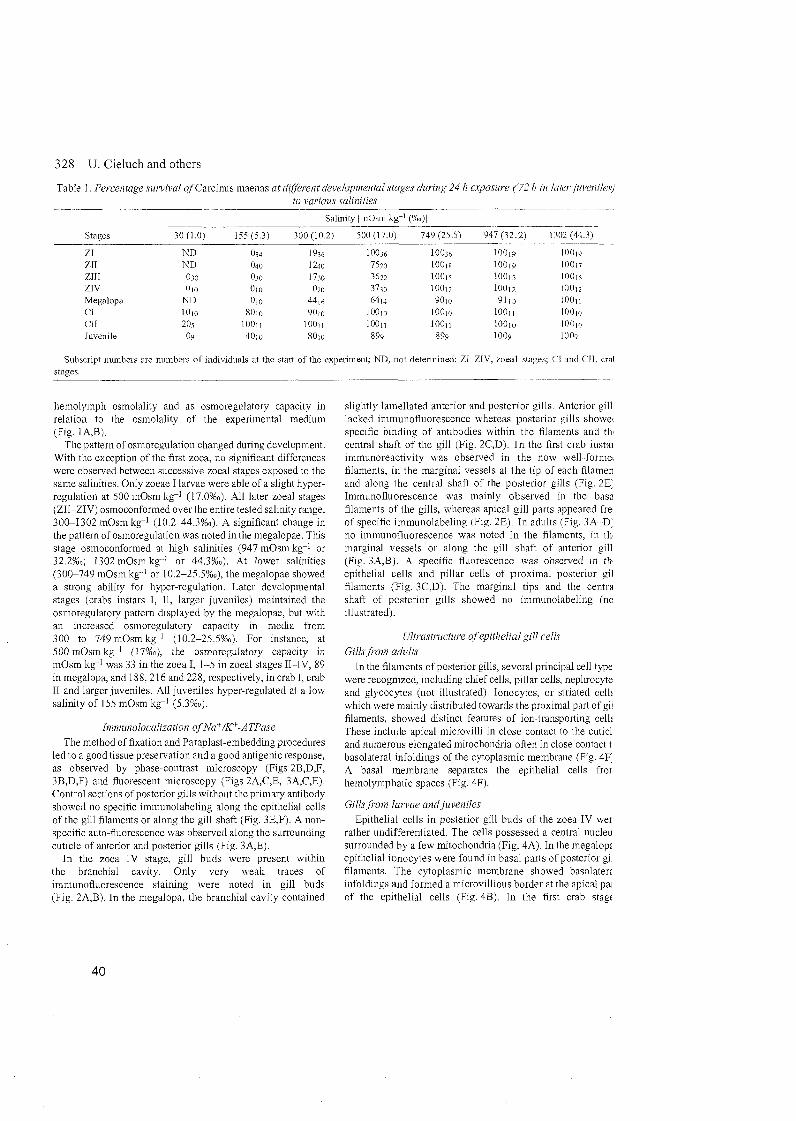
328 U. Cieluch and others
Table 1. Percentage sunival ofCarcinus maenas at dif'ferent develoj~nieiital stages during 24 h exposiire (72 h in /dH'r,jm'enik'.~j to various salinities
Stages
ZI zu ZIII 7,IV Megalopa CI CI1 Juvenile
Subscript munbers are munbers of individuals at the start of the experiment; ND, not determined; ZI-ZIV, zoeal stages: C'I and ('11, cral sfaoes.
I~eniolyiiiph osmolality and as osmoregulatory capacity in relation to the osmolality of the experimental medium (Fig. 1 A,B).
The pattern of osn~oregulation changed during development. With the exception of the first zoea, 110 significant differences were observed between successive zoeal stages exposed to the Same salinities. Only zoeae I larvae were able of a slight hyper- regulation at 500 in0sni k c l (17.0%~). All later zoeal stages (ZII-ZIVI osmoconformed over the entire tested salinity range, 300-1302 m0sm kg-I (10.2-44.3%0). A significant change in the pattern of osmoregulation was noted in tlie megalopae. This stage osmoconfornied at high salinities (947 mOsm kg4 or 32.2%~; 1302 m0sm kg-I or 44.3%0). At lower salinities (300-749 niOsm kg--' or 10.2-25.5%0), the megalopae showed a strong ability for hyper-regulation. Later developmental stages (crabs instars I, 11, larger juveniles) maintained the osmoregulatory pattern displayed by the megalopae, but with an increased osmoregulatory capacity in media from 300 to 749 m0sm kg-I (10.2-25.5%0). For instance, at 500 m0sm kg-l (17%0), the osmoregulatory capacity in mOsni kg-I was 33 in the zoea I, 1-5 in zoeal stages 11-IV, 89 in megalopa, and 188,216 and 228, respectively, in crab I, crab I1 and larger juveniles. All juveniles hyper-regulated at a low salinity of 155 mOsm kg-X (5.3%0).
The method of fixation and Paraplast-embedding procedures led to a good tissue preservation and a good antigenic response, as observed by phase-contrast microscopy (Figs 2B,D,F, 3B,D,F) and fluorescent microscopy (Figs 2A,C,E, 3A,C,E). Control sections of posterior gills without the primary antibody showed no specific immunolabeling along the epithelial cells of the gill filaments or along the gill shaft (Fig. 3E.F). A non- specific auto-fluorescence was observed along the surrounding cuticle of anterior and posterior gills (Fig. 3A,E).
I11 tlie zoea IV stage, gill buds were present within the branchial cavity. Only very weak traces of imniunofluorescence staining were noted in gill buds (Fig. 2A,B). In the megalopa, the branchial cavity contained
slightly lamellated anterior and posterior gills. Anterior gill lacked immunofluorescence whereas posterior gills showei specific binding of antibodies withm tlie filaments and th central shaft of the gill (Fig. 2C,D). In the first crab instai in~munoreactivity was obsen~ed in the now well-forniei filaments, in the marginal vessels at the tip of each filamen and along the central shaft of the posterior gills (Fig. 2E' Immunofluorescence was mainly observed in tlie basa filaments of the gills, whereas apical gill parts appeared fre. of specific immunolabeling (Fig. 2E). In adults (Fig. 3A-D' no immunofluorescence was noted in tlie filan~ents, in tli marginal vessels or along the gill shaft of anterior gill (Fig. 3A,B). A specific fluorescence was obseived in th epithelial cells and pillar cells of proximal posterior gil filaments (Fig. 3C,D). The marginal tips and tlie centra shaft of posterior gills showed no iinmunolabeling (nc illustrated).
I11 the filaments of posterior gills, several principal cell type were recognized, including chef cells, pillar cells, nephrocyte and glycocytes (not illustrated). Ionocytes, or striated celli whch were mainly distributed towards the proximal Part of gi'. filaments, showed distinct features of ion-transporting celli These include apical microvilli in close contact to the cuticl and numerous elongated mitochondria often in close contact t basolateral infoldings of the cytoplasmic membrane (Fig. 4F' A basal membrane separates the epithelial cells fror hemolymphatic spaces (Fig. 4Fi.
Epithelial cells in posterior gill buds of the zoea IV wer rather undifferentiated. The cells possessed a central nucleu surrounded by a few mitochondria (Fig. 4A). In the megalop; epithelial ionocytes were found in basal parts of posterior gi filaments. The cytoplasmic niembrane showed basolaterz infoldings and formed a microvillious border at the apical pa: of tlie epithelial cells (Fig. 4B). In the first crab stagc
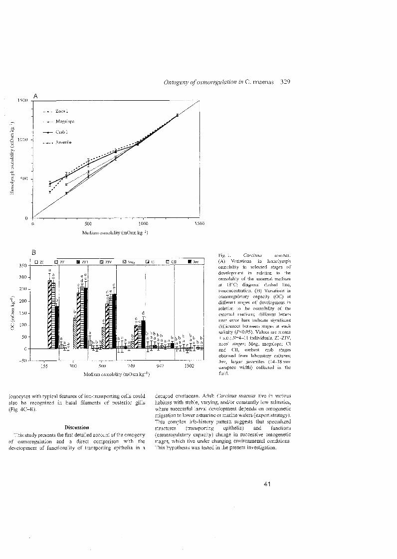
500 1000
Medium osmolality (mOsin k g ' )
Medium osmolality (mOsm kg')
Fig.1. Carciniis muct~as. (A) Variations in l~cmolyinph osmolality in selected stages of development in relation to t11e osmolality of the extcmal medi~im at 1S0C; diagonal dashed line, isoconcentration. (B) Variations m osmoregulatory capacity (OC) at different stages of development iu relation to the osmolality of the extemal medium; different letters near error bars indicate sigilificant differentes between stages at eacli salinity (P<0.05). Values are ineans  s.D.: N=4-11 individuals. ZI--ZIV. zoeal Stages; Meg. megalopa; CI and CII. earliest crab stages obtaiued from laboratory cultures; Juv. larger juveniles (14-1 8 nun carapace width) collectcd in the Seid.
ionocytes with typical features of ion-transporting cells could decapod crustacean. Adult Curcinus maenus live in various also be recognized in basal filaments of posterior gills habitats with stable, varying, andlor constantly low salinities, (Fig. 4C-E). where successful larval development depends On ontogenetic
migration to lower estuarine or marine waters (export strategy). This complex life-history Pattern suggests that specialized
Discussion structures (transporting epithelia) and functions This stzidy presents the first detailed account of the ontogeny (osmoregulatory capacity) change in successive ontogenetic
of osmoregulation and a direct comparison with the stages, which live under changing environrnental conditions. development of functionality of transporting epithelia in a This hypothesis was tested in the present investigation.
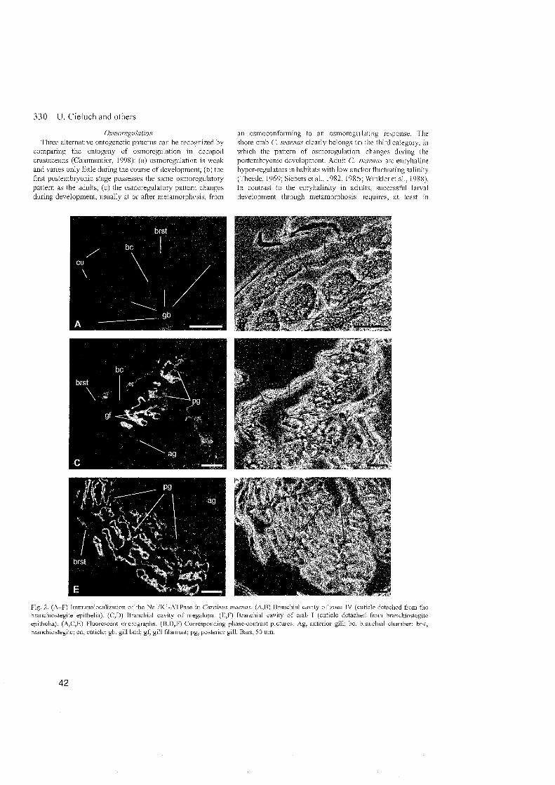
330 U. Cieluch and others
O'iiiioregula~ion Three alternative ontogenetic patterns can be recognized by
comparmg the ontogeny of osmoregulation in decapod crustaceans (Charmantier, 1998) (a) osinoreg~~latioii 1s weak and vanes only little during the course of development, (b) the first postembryonic Stage possesses the Same osinoregulatory pattern as the adults, (C) the osn~oregulatory pattern changes dunng development, usually at 01 after metamorphosis. from
Fig. 2. (A-F) Immunolocalizatioii of the NaLiKL-ATPase in Carcimis branchiostegite epithelia). (C,D) Branchial cavity of megalopa. (E,I epithelia). (A,C,E) Fluorescent micrographs. (B.D,F) Comesponding 1 branchiostegite: CU, cuticle; gb. gill bud; gf, gill filament; pg, posterior <-
an osmoconforming to an osinoregulatiiig rcsponse. The shore crab C. niuenu.~ clearly belongs t o tlic third category, in which the pattern of osmoregulation changes during tlie postembryonic development. Adult C. ~iuii'iia.~ are euryhaline hyper-regulators in habitats with low andlor fi~ictuating salinity (Theede, 1969; Siebers et aL. 1982, 1985; Winkleret al., 1988). In contrast to the euryhalinity in adults, s~iccessfui larval development through metamorphosis requires, at least in
maenas. @,B) Branchial cavity of zoea IV (culicle detached from the ) Braiichial cavity of crab I (cuticle detached from braiichiostegite pliase-contrast pictures. Ag, anterior gill: bc. braiichial chainber; brst. ;ill. Bars, 50 um.
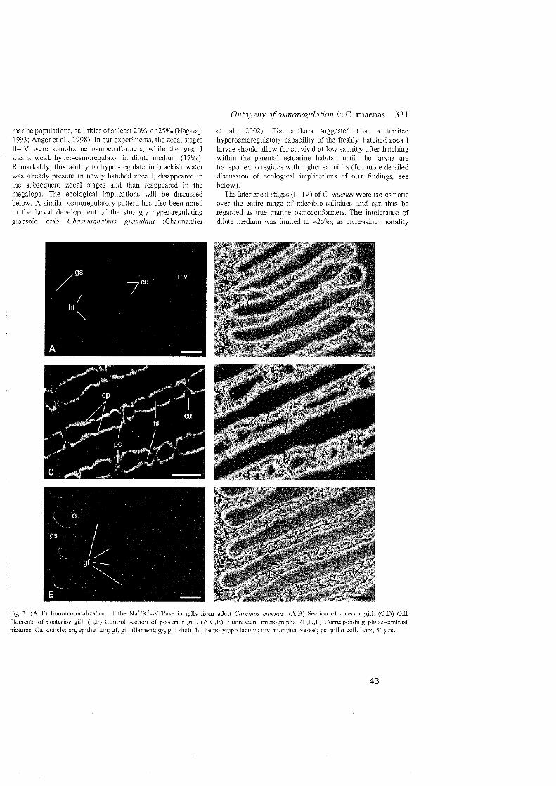
marine populations, salinities of at least 20%0 or 25%0 (Nagaraj, 1993; Anger et al., 1998). In our experiments, the zoeal stages 11-IV were stenohaline osmocoiiformers, wliile the zoea I was a weak hyper-osmoregulator in dilute medium (l7%0). Remarkably, this ability to hyper-regulate in brackisli water was already present in newly liatched zoea I, disappeared in the subsequent zoeal stages and than reappeared in the megalopa. The ecological implications will be discussed below. A similar osmoregulatory pattern has also been noted in the larval development of the strongly hyper-regulating grapsoid crab Cfismagiiufhzis gramilafa (Charmantier
Fig. 3. (A-F) Immunolocalization of the NaIK-ATPase in gills fro filaments of posterior gill. (E.F) Control section 01' posterior gill. (A pictures. Cu, cuticle; ep. epithelium: gf, gill filament: gs, gill shaft. hl, 11
et al., 2002). The authors suggested that a limited hyperosmoregulatory capability of the freshly hatclied zoea I larvae should allow for sui-vival at low saliiiity after liatchine; witliin the parental estuarine habitat, until the larvae are transported to regions with higher salinities (for more detailed discussion of ecological implications of o u r findings, See below).
The later zoeal stages 01-IV) of C. niaenas were iso-osinotic over the entire range of tolerable saliiiities and can tlius be regarded as tme marine osmoconformers. The intolerante of dilute medium was limited to =25%0, as increasing mortality
n~ adult Carcimis iiraenas. (&B) Section of anterior gill. (C,D) Gill ,C,E) Fluorescent microgi'aphs. (B.D,F) Corresponding phase-contrast emolymph lacuna; mv, marginal vessel; pc, pillar cell. Bars, 50 km.

332 U. Cieluch and others
Fig. 4. (-4-F) Transmission electron inicrographs of Carciiins inaenas epithelial gill cells in zoea IV(A). megalopa (B), juveiiile crab I (C- m d adult (F). (A) Two epithelial cells of gill buds with central nuclei sun'oimdcd by a few mitochondria. (B) Marginal tip of a posterior 2 filament in megalopal Stage. The development of basolateral infoldings is visible around the liemolymph lacmia. and numerous vcsicles t
located within the cyioplasm of the epithelial cells. (C) Apical cell part of an ionocyte with initochondria in dose contact to apical microvi note endocuticle detached from epidermal layer. (D) High inagilification of apical microvilli showing a band desmosome conuection (amo between two neigl~bouringcells. (E) Basal cell part of an ionocyte with deep basolateral iiifoldiii~s of the cytoplasmic membrane. Numerc elongated mitochondria are visible in close contact with basolateral infoldings. (F) Epithelial ionocyte of a postenor gill filaincnt with apic nucrovilli and numerous mitochondria in close contact with basolateral mfoldings of the cyloplasinic membranc. Vesicles are noticeable apical and basal cell poles. Bm, basal meinbrane; CU, cuticle; ld, hemolymph lacuna; mi. mitochondrium; mv. microvilli; im. nucleus: T
vesicle. Bars, 2 pm (AB): l uni (C-F); 0.2 um (D).
levels occurred at lower salinities. This limited osmotic tolerance of tlie zoeal stages Supports the findings by Anger et al. (19981, in which salinities below 25%0 led to decreasing early zoeal survival, development, growth, respiration and assimilation.
A considerable shift in tlie osinoregulatory patterii occured after the first n~etamorphic inolt, froin the last zoeal stage (IV) to the megalopa. The megalopa was still osmoconformiiig in salinities >32%0, but was able to hyper-regulate in dilute media down to =10%0. Although its capability to hyper-regulate was still limited coiiipared to the followiiig instars (crabs I and 11, later juveniles), the osn~oregulatory pattern (liyper- isoregulation) of adult C. maenas is in principle already established in the megalopa. Tlie iiext nietamorphic n~olt, from the megalopa to the first juvenile crab stage, showed considerably increased ability for hyper-regulation, allowing now for a tolerance of salinities down to as low as 4 % 0 . Tlie osmoregulatory capacity of the crab I did not differ greatly froin that in the following stage (crab 11), and it iiicreased only slightly in later juveniles. However, survival rates at salinities from 1.0%0 to 25.5%0 as well as hemolymph osmolality at 5.3%0 observed in larger juveniles from the field were below those of laboratory-reared crab I and 11. Other factors such as temperature, saliiiity, water and food quality, which can be controlled and kept constant in the laboratory, are known to influence larval development and overall fitness (reviewed by Anger, 2001). Unknown natural variabilities in those factors in the field might tlius account for the slight but significant reduction in salinity tolerance and osmoregulatory capability observed in later juvenile crabs.
As a preliminary conclusion, the establishment of the adult osmoregulatory pattern in C niaenas is accomplished through two metainorpluc steps (I) the zoea-megalopa transition with the appearance of limited hyper-iegulation. and (2) tlie megalopa-crab transition with a further substantial increase in the osmoregulatory capacity and, in consequence, a hgher tolerance of lower salinities A similar tiining of metaniorphosis-related clianges 111 osmoregulatory pattems has been reported for other bracliyuran crabs such as the strongly regulating grapsoids Aniiasev iiiiersn (Charmant~er et al . 1998), Sevariiia curucaoerise (Anger and Charmantier, 2000), and Chasii~ug~~afl~~is gramilata (Charmantier et al ,2002). or in the ocypodid Uca wbcyl~iic/ricu (Rabalais and Cameron. 1985) An exception was found in tlie grapsoid S e w m a reticiilufuni, where the megalopa maintained the initial zoeal osmoregulatory Pattern and the osmo-pliys~ological shift only appeared after the megalopa-crab traiisition (Foskett, 1977) Sharing a pattem of ontogenetic changes in osmoregulation sinular to those of strongly regulating grapsoids, but still limited by its osmotic tolerance, this study confirms that C, maeiius is a transitional species between true marine osmoconformers like Cancer spp. (Charmantier and Channantier-Daures, 199 1 ), and very strongly regulating, freshwater-invading species such as Eriocheir sinensis (G. Charmantier, unpublished data).
Imnwnoloculizafio~~ o f Na+/K+-ATPase und gill tiltrastructttre Osmoregulation is based On efficient ionic regulation
(inainly of Na1 and C I ) , accomplislied by spccialized transportiiig epithelia where tlie enzyme Nai-/K -ATPase is abundantly located (Thuet et al., 1988; Lignot ct al., 1999; Lignot and Charmantier, 2001; reviewed by Lucn and Towle, 2003). The ontogeny of osmoregulation of C'. ~ i n i m m is correlated with the expression of NaL/K-ATPase (preseiit study) and the developinent of gills (for detailed discription of gill development in C. inuerias, see Hong, 1988).
In the last zoeal stage (zoea IV), whicli is an osmoconfornier, undifferentiated gill buds are formed within the branchial chamber. NaL/K'-ATPase was alniost abseilt within these organs, suggesting that these simple branchial extensions are not yet involved in effective ionic exchange. This suggestion is supported by the ultrastmcture of epithelial cells found within these organs (see Fig. 4A). Tliey lack typical features of ionocytes such as apical microvilli and basolateral infoldings. Gill morphology begins to differentiate after metamoiphosis to the megalopa, which lias liniited ability to hyper-osmoregulate. Tlie arthobraiichs and pleurobranchs arising from the thoracal appendages then beconie differentiated into a central gill shaft and partially lainellated filaments. Epithelial cells witli typical ion-tansporting features can now be found within the gill filaments. This morphological and ultrastructural change coincides witli a possible involvement of g l l s in osinoregulation, also supported by the presence of Na/KL-ATPase in epithelia of the gill shaft and of the filaments of the posterior two pleurobranchs. The presence of Na7/K-ATPase can be related to an involvement of the epithelial cells in ionic exchange (Lignot et al., 1999; Lignot and Charmantier, 2001). Different iminunoreactivity between anterior and posterior gills was obsenled in the megalopa and in the following juvenile stages. In tlie crab I, a strong hyper-osmoregulator, tlie three posterior gills (1 arthobranch and 2 pleurobranchs) are well developed witliin the branchial chamber, and Na7/K-ATPase is distributed mainly in the basal filaments. Ionocytes found in the filaments are siinilar to those obsenred in adults, including typical features such as a microvillous border and mnnerous mitochondria in close contact to basolateral infoldings of the cytoplasn~ic membrane. Thus, most posterior gills are involved in osn~oregulatioii, whereas tlie anterior gills with thin epithelial cells and lack of Na/KL-ATPase seem to attain respiratory functions. These fiiidings agree with previous studies conducted On the crabs Callinectes sapidiis and Carcinus maeiius (Towle and Kays, 1986). Our study supports also the observation tliat, in adult C. maeiius, Nar/K-ATPase is mainly restricted to basolateral infoldings of thick epithelial gill cells in posterior gills (Towle and Kays, 1986).
In other decapod cnistaceans species, Organs like branchiostegites and epipodites play, at least at certain points during development, an iinportant role in ionic exchange. For instance, an inimunohstochemical approacli in juvenile Homuriis gannuariis showed that NaL/K-ATPase is mainly restricted to epithelia of tlie epipodite and the inner epithelium of the branchiostegite (Lignot et al., 1999). Following tlie oiitogeny of osmoregulatory functions in H. ga~iiii~ariis, the
334 U. Cieluch and others
presence of NaL/K'"-ATPase in epipodites has been already established in embryos, and the branchiostegite appears as an additional osmoregulatory Organ after metamorphosis (Haond et al., 1998; Flik and Haond, 2000; Lignot and Charmantier, 2001). In C. tiiaenas, no shift in location or function of ion- transporting epithelia was observed in this study. Tlie temporary and low hyper-osmoregulatory ability that we report in zoea I might originate from the temporary occurrence of ionocytes along the branchiostegites. but this remains to be studied. The posterior gills appear as the dominant Organs involved in the process of ionic regulation. An increase in Na'/KF-ATPase after abrupt transfer to low salinity has been observed in anterior gills of C inaenas (Lucu and Flik, 1999), but the present study confirms the major role of posterior gills of C. niaenas in the process of ionic exchange (Towle and Kays, 1986; Goodman and Cavey, 1990; Taylor and Taylor, 1992; Lawson et al., 1994; Hebel et al., 1999). The development of gills and the expression of NaiKL-ATPase can therefore be considered as olle of the main processes enabling the effective hyper-osmoregulatory abilities of C. maenas.
In tlie natural environments of the shore crab C. maenas. low and/or fluctuating salinities are common, but osmotic stress is initiated or compensated by effective hyper-regulation of internal ionic concentration (Theede, 1969; Siebers et al., 1982, 1985). This mechanisn~ was observed, although only weakly developed, as early as in the zoea I stage, whicli hatches in the same liabitat where adults live. In contrast, later zoeal Stages of C. maenas from the North Sea are true marine osinoconformers, which suffer osmotic stress when they are exposed to constantly low or varying salinities (Anger et al., 1998). Behavioural mechanisms such as tide-related release of larvae and endogenous vertical migration rhythms are known from estuarine crab populations living adjacent to the sea (Queiroga et al., 1994; Zeng and Naylor, 1996a,b,c). These mechanisms provide a rapid off-shore export of larvae to regions with higher salt concentrations (Anger, 2001). In the case of a retention in areas with lower salinity, zoea larvae must face hypo-osmotic stress. During a shor-term exposure to such conditions, mechanisms of isosmotic intracellular regulation may allow for survival, as observed in the lobster Homarus gumtnu~iis (Haond et al., 1999).
Typical of a brachyuran crab, the morphological metamorphosis of C. maenas is accomplished over two molts (Rice and Ingle, 1975). After the first metamorphic molt, the megalopa resembles an intermediate stage between the planktonic zoeae and the benthc crabs. Towards the end of this instar, the megalopa settles and molts to the first juvenile crab instar (Crothers, 1967,). The megalopa can be also regarded as an intermediate stage in terms of osmoregulation, differing from the zoeae by its hyper-osmoregulatory ability in salinities <25%0, yet limited in its osmotic tolerance and ion-regulating capacity compared to the subsequent juvenile crab instars. However, the osmoregulatory ability in the megalopa allows for a reinvasion of areas with low salt concentrations. The
second shift, with another substantial increase in tlie hyp< regulating ability and, consequently, a n enhanced tolerance low salinities, takes place at the transition from tlie mega10 to the first crab Stage. in whicli the ~norpliological, anatomi( and osmo-physiological metamorphosis of C miieni/,~ can considered as complete. The young crab is able to cope wi low andlor fluctuating saliiiities, whic11 extends its liabitat areas with brackish water conditions, e.g. estnaries (Siebers al., 1982, 1985).
Populations of C. niaenas that live in coastal areas a. estuaries of the North Sea are influenced by tidally fluctuati: salinities, while their counteiparts living in the Baltic Sea a exposed to rather constant conditions o f low osn~otic press111 It is still unknown whether the population of C. inaettas in t Western Baltic Sea is capable of reproduction. Although fresh hatched zoeae of C. maenas are seasonally abundant in t. surface plankton at average salinities of 15%" in tlie Kiel Fjoi Baltic Sea (Kandler, 1961 ). advanced developmental stag have not so far been observed. Con~parative studies hti shown that adult C. maenas f ron~ the Baltic Sea liave a high capacity of hyper-regulation than crabs from the North Se and a cross-wise adaptation to higher o r lower salinities w only partially reversible (Theede, 1969). Anger et al. (199 suggested that there might be genetic differences between the populations. Although physiological variations have bei studied in adult crabs from geographically separat1 populations (Theede, 1969) and in different colour morp (McGaw and Naylor, 1992a,b), no information is availab about the larval response of crabs from the Baltic Sea salinity variations. A comparative study 011 the ontogeny osmoregulation and of reproductive traits in C. titaeiias fro the North Sea and the Baltic Sea might thus provide valuab information on the processes required for a succesf establishinent of decapod cmstacean species in habitats wi constantly 10% salinity.
The authors would like to thank Jessica Wintz and Uv Nettelmann for their extensive help in rearing crabs ai larvae, and Kerstin Beyer for her technical assistance electron microscopy. U.C. was fmancially supported by tl University of Hamburg by a SokratesIErasnius-grant perform research in Montpellier. The monoclonal antibot was purchased from the DSHB, University of Iowa, USA.
References Anger, K . (2001). The biology of decapod cmstacean larvae. In Cmstace
Issiies, vol.14 (ed. R. Vonk). pp. 263-318. Abingdon, Exton (PA): Lis: Tokyo: A. A. Balkema.
Anger, K. (2003). Salinity as a key Parameter m the larval biology of decap cmstaceans. Im'. Repr. Da'. 43, 29-45.
Anger, K . and Charmantier, G . (2000). Ontogeny of osmoregulation a salin~ty tolerance in a mangrove crab. Sesarma curacaoense (Decapoc Grapsidae). J. fip. Mar. Biol. Ecol. 251. 265-274.
Anger, K. , Sph-ak, E. and Luppi, T . (1998). Effects of reduced salinities t
deveiopme~t and bioenergetics of early larval shore crab, Ccircinus nioeiu J. Exp. Mo?. Biol. Ecol. 220, 287-304.
Barradas. C.. Dunel-Erb. S.. Lienon. J . and Peaneiix. A. (199; . . A , ~
Supenmposed morphofimctional study of ion regulation and respiration
Ontogeny of osmoregulation in C. maenas 335
Single gill filanicnts of the crayfish Astacus Leptodacyliis. J. Crnst. Biol. 19. 14-25.
Bonaricha, N., Cliarniantier-Daures, M., Thuet, P , Trilles, J. P. and Charmantier, G. (1994). Ontogeny of osmoregulatoiy structures in the shnmp Penaeusjaponicus (Crustacea, Decapoda). Biol. Bull. 186. 29-40.
Charmantier, G. (1998). Ontogeny of osmoregulation in crustaceans: a review. Ins. Repr. Dev. 33. 177-190.
Charmantier, G. an0 Charmantier-Daures, M. (1991). Onfogeny of osmoregulation iind saliuity tolerante in Cancer in'oratus': elements of companson with C boreali.~ (Crustacea, Decapoda). Biol. Bull. 180, 125- 134.
Charmantier, G., Charmantier-Daiires, M. and Anger, K. (1998). Ontogeuy of osinoregulation in the grapsid crab Amarses miersii (Cmsfacea: Decapoda). Mur. Ecol. Prog. Sei: 164, 285-292.
Charmantier, G,, Gimknez, L., Charmantier-Danres, M. and Anger, K. (2002). Ontogeny of osmoregulation, physiological plasticity and larval export strategy in thc grapsid crab Chasmag~iafhus r m ~ u l a t a (Crustacea. Decapoda). Mm: Ecol. Prog. Ser. 229, 185-194.
Cohen, A. D., Carlton, J. T. and Fonntain, M. C. (1995). Introduction. dispersal and potential impacts of Ehe green crab Carcimis maeiias in San Francisco Bay. Califon~ic~ Mor. Biol. 122, 225-237.
Compere, P , Wanson, S., Pequeux, A., Gilles, R. and Goffiuet, G. (1989). Ultrastructural changcs in the gill epithehum of fhe green crab Carcinus mamas in relation to the external medium. Tissue Ce11 21, 299-318.
Crothers, J. H. (1967). 'Ihe biology of the shore crab Carcimis maenas (L.). I. The background-anatomy, growth and life history. Field Studies 2. 407- 434.
Dawirs, R. R. (1985). Temperature and larval deveiopinent of Carcinus maenas (Decapcia) in fhe laboratory; predictions of larval dynamics in the sea. Mnr. Ecol. Prog Ser. 24, 297-302.
De Renzis, G. and Bornancin, M. (1984). Ion Transport und Gd1 ATPase (ed. W. S. Hoar and D. J. Randall), pp. 65-104. Orlando: Academic Press.
Drach, P. (1939). Mue et cycle d'intermue chez les Cnistac6s Dkapodes. h à § Inst. O c e a n o ~ . Monaco 19, 103-391.
Felder, J., Felder, D. and Hand, S. (1986). Ontogeny of osmoregulation in the estuariue ghost shnmp Callianassa jamuicmse var. louisianensis Schmitt (Decapoda, Thalassinidae). .L Exp. Mar. Biol. Ecol. 99, 91-105.
Flik, G. and Haond, C. (2000). Na" and Ca2^ pumps in the gills, epipodites and branchiostegites of the European lobster Hornams guniniunis: effecfs of dilute sea water. .I Exp. Biol. 203, 213-220.
Flik, G.. Verbost, P. M., Atsma, W. and Lucu, C. (1994). Calcium transport m gill piasma membranes ofthe ciab Carcimis maenas: evidence for camers dnven by ATP and a Na" gradient. J. Exp. Biol. 195. 109-122.
Foskett, J. (1977). Osmoregulation in fhe larvae and adults of the grapsid crab Sesarma reticiilatum. Biol. Bull. 153. 505-562.
Giiles, R. and P6qneux, A. J. R. (1985). Ion transport in crustacean gills: physiological and ultrastnictural approaches. In Transporl Processes, Iono- atid Osinoregdation (ed. R. Gilles and M. Gilles-Baillieu), pp. 136-158. Berlin. Heidelberg, New York, Tokyo: Springer-Verlag.
Goodman, S. H. and Cavey, M. J. (1990). Organization of a phyllobranchiate gill from the green crab Carcinns maenus (Cnistacea, Decapoda). Ce11 Tissue Res. 260, 495-505.
Green, J. (1968). The Biology of Estuuril~e Ammcils. pp. 401 pp. London: Sigwick and Jackson.
Grosholz, E. D. and Ruiz, G. M. (1995). Spread and potential impact of the recently introduced European greeii crab, Carcinns niaenas. in central California. >Wal-. Biol. 122. 239-247.
Haond, C., Bonnal, L., Sandeaiix, R.. Charmantier, G. and Trilles, J.-P. (1999), Onlogeny of intracellular isosinotic regulation in the European lobster Homarus gammarns. Pliysiol. Biochem. Zoo?. 72, 534-544.
Haond, C., Flik, G. and Charmantier, G. (1998). Confocal laser scanning and elecfron microscop~cal studies 011 osmoregulatory epithelia in the branchial cavity of the lobster Ho~ncirlis gcini7nai¥;is J. Exp. Bol. 201, 1817- 1833.
Harms, J., Meyer-Harms, B., Dawirs, R. R. and Anger, K. (1994). Growth and physiology of Carcirnis maenas (Decapoda, Portunidae) lanrae in the ficld and in laboratory expernnents. Mrir. Ecol, Prog. Ser. 108, 107-118.
Hebel, D. K., Jones, M. B., Moate, R. M. and Depledge, M. H. (1999). Differmg sensitivities of respiratory and osmoregulatory gill tissue of Cacinus maenas (Crustacea: Decapoda) to watet-bome capper. Mur. Biol. 133, 675-681.
Hong, S. Y. (1988). Development of epipods and gilis in some pagunds and brachyurans. J. Na/. Hist. 22, 1005-1040.
Kändler R. (1961). Eber das Vorkommen von Fischbrut, Dccapodcniarven und Medusen i n der Kieler Förde Kieler Meeresforsch. 17, 48-64,
Kinne, 0. (1971). Salinily: 4.3. Aniinals: 4.31 Invcrtchrates, In 11 Coniprehensive. Integr~ted Treatise on Life in Oceans a n d ('oastul Wuters, vol. 1 (ed- 0. Kinne), pp. 683-1244. London: Wiley and Sons.
Laffe*, K. D. and Kuris, A. M. (1996). Biological control OS manne pests. Ecology 77. 1989-2000.
Lawson, S. L., Jones, M. B. and Moate, R. M. (1994). Structuriil vanability and distnbutioi~ of cells in a postenor gill of Carcinus nzwnas (Decapmia: Brachyura). .L Mar. Biol. Assn. UK 74. 771-785.
Lignot, J. H., Charmantier-Daures, M. and Cliarmanlicr, C. (1999). Im~nunolocalization of "Na/KL-ATPase m the Organs of the brancliial cavity of the european lobster Homams gammariis (Crustacea: Dccapocla). Ce// Tissue Res. 296, 417-426.
Lignot, J.-H. and Charmantier, G. (2001). Iininunolocali~~ition of Na ' ,KL- ATPase in the branchial cavity dunng the early developmcnt OS the European lobsterHomcmtsgammams (Crustacea. Decapch). .I, flistoc/i~~~~i. Qtocliem. 49. 1013-1023.
Lucu, C. (1990). Ionic regulalory mechanisms in crustacean gill epitheha. Conip. Bioct'lem. Pl t~~siol 97.4, 297-306.
Lucu, C. and Flik, G. (1999). Na--K^-ATPase and Na-/Ca2- exchange activities in gtlls of hyperregulating Carcimis maenas. Am. J. Pfiysiol. 276, R490-R499.
LUCH, C. and Towle, D. W. (2003). 'NaT+K'-ATPase i n gills of aquatic crustacea. Cor~ip. Biochem. Pli~~siol. 135A. 195-214,
Mantel. L. II. and Farmer. L. L. (1983). Osmotic and Ionic Regiilation, pp. 53-161. New York, London: Academic Press. Inc.
McGaw, I. J. and Naylor, E. (1992a). The effect of slielter on sahiiity preference behavior of the shore crab Carci~iiis ~nuenas. Mar. Je11av. P ~ s i o l . 11, 145-152.
McGaw, 1. J. and Naylor, E. (1992b). Salinity preferences of the sliorc crab Carciuns maeiias m relation 10 coloration dunng intermolt arid to prior acclimation. J. Exp. Mal.. Biol. Ecol. 155, 145-159.
Nagaraj, M. (1993). Combinedeffects oftemperature and sahnity on thezoeal development of the green crab. Carci~tus maenas (Lmnaeus, 1758) (Decapoda, Poriunidae). Sei. Mnr. 57, 1-8.
Neufeld, G. J., HoUiday, C. W. and Pritchard, J . B. (1980). Salinity adaptation of gill NaL/K-ATPase in Ehe blue crab, Callinectes supidiis. Exp. Zoo/. 211, 215-224.
Pequeux, A. (1995). Osmotic regulation in crustaceans. J. Cmst. Biol 15. 1- an
Pkqiieux, A. and Giiies, R. (1988). NaCl Transport in Gills <md Related Structfires, pai? I: Imjeriebrates (ed. R. Gregor), pp. 2-47. Berlin: Spritiger- Verlag.
Queiroga, H.. Costlow, J. D. and Moreira, M. H. (1994). Lama1 abundante Patterns of Cwcinus maenas (Decapoda. Brachyura) in Canal de Mira (Ria de Aveiro. Portugal). ,War. Ecol. Prog. Sei: 111, 63-72.
Rabalais, N. N. and Cameron, J . X. (1985). The effects of factors imprtant in seini-and environments on the early development of Ucu s~bc.yl i~~d~~ica. Biol. Bull. 168. 135-146.
Reynolds, E. S. (1963). The use of lead Citrate at high pH as an electron- opaque stain in electron microscopy. Ce11 Biol. 35. 313-323.
Rice, A. L. and Ingle. R. W. (1975). The larval developrnent of Carciriiis mcielias (L.) and C. medi~errmieus Czerniavsky (Crustacea, Brachyura, Portumdae) reared in the laboratory. Biill. Br. Mus. Nut. His!. /Zoo/./ 28. 103-119.
Siebers, D., Leweck, K., Markus, H. and Winkler, A. (1982). Sodnim regulation in the shore crab Carci~ius maenas as related to ambient salinity. Mur. Biol. 69, 37-43.
Siebers, D., Winider, A., Lucu, C., Thedens, G. and Weichart, D. (1985). Na-K-ATPase generates an active transpori potential in Ehe giils of the hyperi-egulating shore crab Carcimis maefias. Jfar. Biol. 87. 185.192.
Sokal, R. R. and RoMf, F. J. (1995). Biomelv: The Principles und Practice of Statislics m Biological Research. San Francisco: W. H. Freeman and Co.
Talbot, P., Clark, W. H. and Lawence, A. L. (1972). Ultrastnictural observations of muscle insertion and modified branchiostegite epidennis in larval brown shninp, Pwaeus azteciis. Tissiie Cell4. 613-628.
Taylor, H. H. and Taylor, E. W. (1992). Gills und L q s : The Exchange of Gases wid Ions (ed. F. W. Harrison and A. G. Humes), pp. 203-293. New York: Wiley-Liss, Inc.
Theede, H. (1969). Einige neue Aspekte bei der Osmo~egulation von Carcinus maeiias. Mar. Biol. 2, 114-120.
Thiiet. P., Charmantier-Daures, M. and Charmantier, G. (1988), Relation
336 U. Cieluch and o t h e r s
entre osmor.4giilat1on et activit&s d'ATPase N a - K et d'anhydrase chez lanes et postlanres de Homarns gammarits (L.) (Cmstacea: Decapoda). .1. E.IP. Mm-. Biol. Ecol. 115. 249-261
Towle. I). W. (1984a). Membrane-bound ATPase in arihropod Ion- traasportnig tissues. Am. Zool. 24. 177-185.
Towle, D. W. (1984b). Regiilatoiy Functions ofNa'- K'-ATPase in Marine m d Esmarine Ai7imah (ed. A. P&queux and L. ßolls) pp. 157-170. Berlin, IIeidelberg. New York, Tokyo: Springer Verlag.
Towle. D. W. (1981). Role of N a i - K ' - ATPase in ionic reguiation by manne and esiuanne animals. Mur. Biol. Leu. 2. 107-121.
Towle. D. W. and Kays, W. T. (1986). ßasolatera localization of Na^KL- ATPase 111 gill ep~thehum of two osmoregulating crabs. Calli~~ecte'i sc~pidiis and Ccn-cmiis maenas. J. Exp. Zoo! 239. 311-318.
Watson, M. L. (1958). Stammg of tissue sections with heavy metals. J. Bioplz~s. Wachem. Cytol. 4. 475-478.
Winkler, A.. Siebers. D. and Becker, W. (1988). Osmotic and ionic
regulation in shore crabs Carcmus mciai(i.7 in11:1biting ii liii;il csi~tiii: Helgola~~der Meeresunters 42. 99-1 1 1.
Zens. C. and Naylor, E. (1996a). Endogenous tiii;il ihyihiiis O S vertic. inigailon in field collectcd zoea-1 larvae O S tlie siiorc ciali Cardin mfiepws: nnplications for ebb fide offshore dispe1s:ii. Mur. Ecol. Pros. Se 132, 71-82,
Zeng, C. and Naylor, E. (1996b). Her~tabiiiiy oScirc:ii~~tal veilical ~ ~ ~ i g r a i i c rhyfhins in xoea lamae of ihe shore crab Carcifins mamm (I,.). J. Lxp. Ma Biol. Ecol. 202, 239-257.
Zeug, C. and Naylor, E. (1996~). Occurencc in coasial waten ar endogenous swimmmg rhythms ofiate inegalopa ol'thc sliore cnib Cracim mawas: imphcafions for onshore recruitment. Mur. EroL P r o g S m 13, 69-79.
Ziegler, A. (1997). Inununocytocliem~cal iocals/'-iidon of N:I' ,KL-ATPase : the calcm~n-transporting sternal epithelium of iiic lerrestnal isopc PorceUio scaber L. (Cnistacea). J. Histochew. C\'[ochtw 45, 437-446.
PUBLICATION II
Publication I1
Salinity tolerance, osmoregulation, and immunolocalization o f
Na+/K+-ATPase in larval and early juvenile stages of the Chinese
mit ten crab, Eriocheir sinensis (Decapoda, Grapsoidea)
Ude Cieluchl, Klaus Anger1, Mire i l le Charmantier-Daures2, G u y
Charmantier2
Biologische Anstalt Helgoland / Stiftung Alfred-Wegener-Institut fü Polar-
und Meeresforschung, Bremerhaven, Germany
Equ ipe Adaptation Ecophysiologique et Ontogenese, UMR 5000 GPIA,
Universite Montpellier 11, Montpellier, France
Marine Ecology Progress Series (submitted)

PUBLICATION I 1
Salinity tolerance, osmoregulation, and immunolocalization of N~+/K^-
ATPase in larval and early juvenile stages of the Chinese mitten crab,
Eriocheir sinensis (Decapoda, Grapsoidea)
Udc cieluchl*, Klaus ~ n g e r ' , Mireille ~hannantier-~aures ' , Guy chamantier2
1 . Biologische Anstalt Helgoland, Stiftung Alfred-Wegener-Institut fü Polar- und
Meeresforschung, Meeresstation, D-27498 Helgoland, Gennany
~ ~ u i ~ e Adaptation Ecophysiologique et Ontogenese, UMR 5171 GPIA, Universitk
Montpellier 11, F-34095 Montpellier cedex 05, France
Running head: "Ontogeny of osmoregulation in Eriocheir sinensis"
* Corsesponding author, phone: +49 (0) 4725 819-344, fax: +49 (0) 4725 819-369
e-niail: [email protected]
PUBLICATION II
ABSTRACT
The ontogeny of osinoregulation was studied in the Chinese mitten crab Eriocheir siiiensis
from the Elbe estuary, North Sea, Germany. Laboratoiy-reared developmental stagcs vvcre
exposcd to salinities ranging from 0.16-44.3 %o. Salinity tolerancc (ST) was detennincd by
survival rates. Hemolymph osmolality was measured and os~noregulatory capacity (OC)
calculated as the difference between the osmolality of the hemolymph in relation to the
external medium. Zoea I larvae hyper-regulated in dilute media from 5.3 - 25.5 %o, and
osmoconfonned in seawater (32.2 %o) and concentrated seawater (44.3 %o). All latcr zocal
stages slightly hypcr-regulated at salinities from 10.2-17.0 %o, and were osn~oconformcrs
at 25.5-44.3 %o. The megalopae hyper-regulated at low salinities from 5.3-17.0 %", and
hypo-regulated at 32.2 and 44.3 %o. Juvenile crabs I and I1 hyper-regulatcd a t salinitics
ranging froin 1 .O-25.5 %o, and hypo-regulated at 32.2 and 44.3 %o. ST was high with a
survival rate of 90-100 % in all developinental stages at salinities ranging from 10.2-32.2
%o. Except for zoea I (30 % survival), all zoeal stages died at 5.3 %O and below, At that
salinity, 46 % of megalopae survived and 100 % survival was notcd in juvenile crabs I and
11. At 44.3 %o, ST was high in zoeae I and I1 (100 % survival), decreased in the zoca IV (25
% survival), and increased during later development with survival rates of 33, 77, and 100
% in zoea V, megalopae, and juvenile crabs 1/11, respectively. The expression of Na/KA-
ATPase and the developinent of transporting epithelia were studied by means of
i i~~~~~unof luorescence light n~icroscopy (ILM) and transmission electron microscopy
(TEM). In the stage-I and stage-TI zoeae, fluorescence staining was observed along the
inner epithelium of the branchiostegite, and epithelial cells showed typical features of
ionocytes including apical microvilli and basolateral infoldings of the cytoplasinic
men~brane. In the zoea V, gill buds were present within the branchial chamber, but
PUBLICATION I 1
epithelial cells were rather undifferentiatcd without showing typical features of ionocytcs.
In the megalopa and the first juvenile crab, ionocytes and iniinunolabeled N ~ K - A T P ~ S C
were located in the filaments of the most posterior gills, while the epithelial cells of tl1c
antcrior gills appeared undifferentiated and showed no positive immunodetection of thc
enzyme. Comparison of histological results with physiological capabilities shows a closc
correlation between the ontogeny of osmoregulation and the expression of IVa'K-~Tpasc
within transporting epithelia located in the branchial chamber. The adult pattern of
osmoregulation in E. sinensis develops through two molts, (1) froin the moderate hypcr-
iso-regulating zoeal phase to the moderate hyper-/hypo-regulating megalopa, (2) from the
megalopa to the strongly euryhaline, hyper-/hypo-regulating juvenile crab Stages. Thc
results of this study support the hypothesis of an export strategy in this holo-euryhalinc
crab species.
Keywords: ontogeny; osn~oregulation; ionocyte; export strategy; cmstacea
PUBLICATION I1
INTRODUCTION
A major adaptive trait of crustacean species living under varying andlor low salinity
conditions is the ability to osmoregulate (for recent reviews see Chamantier 1998, Angcr
2003). At low salinity, the loss of ions through the body surface and urine is compc~~sated
by effective ion uptake, mainly o f N a and CF. This process is based upon the functionality
of cells specialized in ion-exchange, the ionocytes. Mainly located in the posterior gills of
brachyuran crustaceans, ionocytes show distinct morphological features such as apical
microvilli and basolateral infoldings of the cytoplasmic membrane, while the latter are
often in close contact with numerous mitochondria (reviewed by Mantel & Farmer 1983,
Gilles & Pequeux 1985, Pequeux & Gilles 1988, Lucu 1990, Taylor & Taylor 1992,
Pequeux, 1995).
NaL/K-ATPase is one of the most important enzymes involved in the process of
osmoregulation (reviewed by Towle 1981, 1984a,b, Pequeux 1995, Charmantier 1998,
Lucu & Towle 2003). This enzyme enables active ion-exchange across cpithclial
membranes (Neufeld et al. 1980, De Remis & Bornancin 1984), and its precise cellular
location is therefore of great importance in investigations of the ontogeny of
osmoregulation and the development of ion-transporting cells and epithelia (Flik et al.
1994. Haond et al. 1998, Lignot et al. 1999, Lignot & Charmantier 2001). Recently,
in~munolocalization of Nzi^/~^-AT~ase using monoclonal antibodies has been recognized
as a useful tool for locating ionocytes in tissues and Organs of decapod crustaceans, for
instance in the lobster Homarus gammurus (Lignot et al. 1999, Lignot & Charmantier
2001), the crayfish Asfucus lepfodactyltis (Barradas et al. 1999), or the green crab
Carcinns maenas (Cieluch et al. 2004).
PUBLICATION I1
As an extremely cusyhaline specics, the Chinese mitten crab, Eriocheir sinensis H.
Milnc-Edwards 1854 (possibly identical with the Japanese mitten crab, E. japonica dc
Haan, 1835; for recent discussion of generic taxonomy and phylogcnetic position OS
Eriocheir spp., see Tang et al. 2003), has become a model for studics of gill morphology
(Barra et al. 1983) and physiology (Riestenpatt et al. 1994), in particular in relation to ionic
regulation and excretion (Pkqueux & Gilles 1981, Rathmeyer & Siebers 2001),
electrophysiology (Onken & Graszynski 1989, Onken et al. 1991), and enzyme activitics in
gills (Olsowski et al. 1995, Mo et al. 1998). E. sinensis is an invasive spccies which
originales fsom south-east Asia and has spread, most probably via ballast water, to Europe
and North America (Panning 1938, for recent review, see Herborg et al. 2003, Rudnick et
al. 2003). Its successful dispersal in brackish coastal lagoons, rivers, and land-lockcd
inland waters is based on its capability to cope equally well with freshwater, brackish
watcr, or scawater (temed holo-euryhalinity; Kinne 1971).
In contrast to the tremendous amount of information available for adult mitten
crabs, very little is known about the development of pl~ysiological traits in the larval and
early juvenile stages. The life cycle of E. sinensis includes 5 (occasionally 6) zoeal stages,
and a megalopa, which is followed by the first juvenile crab (Kim & Hwang 1995, MontÅ
et al. 1996). Anger (1991) studied the effects of various combinations of salinity and
temperature on the larval development of this species in thc laboratory, observing highest
larval survival at salinities between 15 and 25 %o. These experimental results showed that
the larval stages, in contrast to the adults, do not tolerate freshwater conditions, which
suggests an export strategy with larval hatching within brackish estuaries, later zoeal
development in lower estuarine or coastal waters with higher salinities, and megalopal
andlor juvenile reiinmigration into the upper reaches of estuaries and rivers. Later
PUBLICATION II
development and growth to adulthood and sexual maturation occurs under frcshwater
conditions.
In a recent review, Chamiantier (1998) postulated that the successful establishmcnt of
a species in a particular habitat depends on the ability of each of its developmental stages
to adapt to this environment. This implies that a h l l understanding of the ecology and lifc
history of a species, including the spreading potential of invasive species, rcquircs a
detailed knowledge of the ontogeny of osmoregulation and other physiological traits.
Particularly in estuarine species, the ontogeny of osmoregulation is of special rclcvance,
since salinity may vary during development through a con~plex life cycle, from truly
marine to brackish or even freshwater conditions (Anger 2001,2003).
A fair amount of information on the ontogeny of regulatory capacity is now
available, while the number of investigations on developmental changes in the structures
and functions of osn~oregulating tissues and organs is still limited (Hong 1988, Bouaricha
et al. 1991, 1994, Charmantier 1998, Anger 2001, Lignot & Chasmantier 2001). Among
the few species in which the ontogeny of ion-transporting epithelia has been investigated
by means of histological andlor electron microscopical techniques are
Farfantepenaeus aztecns (Talbot et al. 1972), Cullianassa janzaicense (Felder et al. 1986),
Penaeus japonicus (Bouaricha et al. 1994) and Homarus gamrnarus (Lignot &
Channantier 2001). In a recent study on the green crab, Carcimis nzuenas, a close
correlation between the ontogeny of osmoregulation and the development of ion-regulating
tissues and organs has been shown (Cieluch et al. 2004).
In the present investigation, we studied the ontogeny of osmoregulation in
laboratory-reared early developmental stages of E. sinensis. The osmorcgulatory capacity
(OC) was defined in relation to extemal salt concentrations, and salinity tolerante (ST) was
PUBLICATION I 1
determined as survival rates. Furthermorc, we invcstigated the cxpression of N~^/K*-
ATPase and the location of ion-transporting cells by means of immunofluorescence and
electron microscopy. This study focused on the relationships between ontogenetic changcs
in physiological and morphological traits and presumable patterns of ontogenctic
migrations in this freshwater-invading species.
PUBLICATION I1
MATERIAL AND METHODS
Animals
Ovigerous females of Eriocheir sinensis were dredged in April from the Elbe
estuasy near the harbor of Cuxhaven (North Sea, Gemany), and transported in water from
the capture site to the Helgoland Marine Biological Station. In the laboratory, females werc
kept individually in 25 l plastic aquaria connected to a closed reeirculating System.
Constant conditions in the laboratoiy were 20 %O salinity, a temperaturc of 9 'C and a
12:12 h light/dark cycle. The water in the System was replaced twice a week and crabs
were fed thawed mussels (Mvtilus edulis) evesy second day. Newly hatched larvae wcre
collected in sieves (200 mmesh size) receiving water from an overflow and individually
reared through metamorphosis in glass vials ( 50x111) at 25 %o, a constant temperature of
18 'C, and a 12:12 h lightldark regime. In early larvae (zoea I and 11), water and food
(freshly hatched Artemia sp. nauplii) were changed daily. In all following stages, water and
food were changed every second day. The developmental stages tested in the
osmoregulation experin~ent comprised the zoeal stages I, 11, IV, and V, the megalopa, and
the juvenile crab stages I and 11. The following stages were chosen for ILM and TEM: zoea
I, zoea V, megalopa, juvenile crab I, and larger juveniles (carapace width 0.8-1.2 cm). For
all experiments, only animals in the middle of an instar, i.e. in intermolt stage C (Drach
1939) were used. Larger juvenile crabs were anesthetized in cooled water ( 3 'C) prior to
tissue sampling for ILM and TEM.
PUBLICATION I 1
Preparation of media
Experimental media were obtained by diluting 1 pm-filtered and UV-sterilizcd
seawater (32 %o) with desalinated freshwater or by adding Tropic ~ a r i n @ salt (Wartenbcrg,
Gerniany). Salinity was expressed as osinotic pressure (in mOsin kg") and as sah contcnt
of the medium (in %o); a value of 3.4 %O is equivalent to 100 mOsm kg"' (29.41 n1Osm kg"
= 1 %o). The osinotic pressure of the media was measured with a microosmon~eter Model 3
MO plus (Advanced Instruments, Needham Heights, MA, USA) requiring 20 1 pcr
sample. The following media were prepared, stored at 9 'C and used in the osinoregulation
experiment: 5 mOsm kg'l (0.16 %o, referred to as freshwater), 30 mOsin k g l (1.0 %o), 155
mOsm kg-l (5.3 %o), 300 mOsm kg-' (10.2 %o), 500 mOsm kg-' (17.0 %o), 749 mOsm kg"
(25.5 %o), 947 nlOsn1 kg"' (32.2 %o, seawater), and 1302 mOsm kg"' (44.3 %o).
Osmoregulafion and salinity tolerante
The experiment was carried out at a constant temperature of 18 ¡C According to
previous results on different species (Channantier 1998), the young developmental Stages
werc exposcd directly to the experimental media for 24 h in covcrcd Petri dishes; exposure
time in juvenile crabs was 72 h. Dead animals were counted at the end of the cxposure time
to obtain survival rates according to salinity and developmental stage.
The surviving specimens were superficially dried on filter Paper and quickly immersed
in mineral oil to prevent evaporation and desiccation. Remaining adherent water was
removed using a glass micropipette. A new micropipette was thcn inserted into the heart to
obtain hemoly~nph samples, which were then ineasured with reference to the medium
osmolality on a Kalber-Clifton nanolitre osmometer (Clifton Technical Physics, Hartford,
NY, USA) requiring about 30 nl. Results were expressed either as hemolymph osinolality
PUBLICATION II --
or as OC. The latter is defmcd as the difference between hemolymph osmolality and the
osmotic prcssure of the medium. Analysis of variance (ANOVA) and Student's t-tcsts
were used for multiple and pairwise statistical comparisons of mean values, respcctivcly,
after appropriate Checks for normal distribution and equality of variance (Sokal and Rolilf
1995).
In~munofliiorescence light microscopy
Samples were fixed for 24 h by direct immersion in Bouin's fixative. After rinsing
in 70 % ethanol, samples were fully dchydrated in a graded ethanol series and enibeddcd in
Paraplast X-tra (Sigma). Sections (4 m)were cut on a Leitz Wetzlar microtome, collectcd
on poly-L-lysine-coated slides and stored overnight at 38 'C. Sections were then pre-
incubated for 10 min in 0.01 111M Tween 20, 150 mM NaCl in 10 n1M phosphate buffer,
pH 7.3. To remove the free aldehyde groups of the fixative, sainples were treated for 5 min
with 50 inM m 4 C 1 in phosphatc-buffered saline (PBS), pH 7.3. The sections were thcn
washed in PBS and incubated for 10 min with a blocking solution (BS) containing 1 ?4
bovine serum albumin (BSA) and 0.1 % gelatin in PBS. The primary antibody
(monoclonal antibody 1gG 5 , raised against the avian -subunit of the N ~ I K - A T P ~ S ~ )
was diluted in PBS to 20 gml", placed in small droplets of 100 Ion thc sections and
incubated for 2 h at roon~ temperature in a wet chamber. Control sections were incubated
in BS without primary antibody. To remove unbound antibodies, the sections were then
washed (3x5 min) in BS and incubated for 1 h with sn~all droplets (100 I) of the
secondary antibody, fluoresceinisothiocyanate (F1TC)-labeled goat anti-inouse IgG
(Jackson Inm~unoresearch, West Baltimore, USA). After extensive washes in BS (4x5
n~in), the sections were covered with a rnounting medium and examined with a fluorescent
PUBLICATION I 1
inicroscope (Lcitz Diaplan coupled to a Ploemopak 1-Lambda lan~p) with an appropriatc
filter Set (450 nin to 490 n111 band-pass cxcitation filter).
Transmission electron inicroscopy
Sainples were fixed by direct immersion for 1.5 h in 5 % glutaraldehyde solution buffcrcd
at pH 7.4 with 0,l mol 1 cacodylate buffer. For adjustment to the osmotic pressure of the
hemolymph, NaCl was added to the fixative and buffer to get a final osn~olality o f 735
mOsm kg". Samples were then rinsed, stored ovemight at 4 'C in buffcr, and post-fixcd
for 1 h at room temperature in buffered 1 % 0 ~ 0 ~ . After extensive washes in buffer, thc
samples were fully dehydrated in graded acetone and embedded in Spurr low viscosity
medium. Semi-thin sections (1 m) were prepared using glass knives with a Leica
microtoine and stained with Methylene Blue for light n~icroscopic observations. Ultra-thin
sections were obtained using a diainond knife, contrasted with uranyl acetatc (Watson
1958) and lead citrate (Reynolds 1963), and examined with a transmission electron
inicroscope (EM 902, Zeiss, Gemiany) operated at 80 kV.

PUBLICATION I1
RESULTS
Salinip tolerante
Survival of the different stages during exposure for 24 h (72 h in juveniles) to thc
experimental media is given in Table 1. Survival rates of 92-100 % in all devclopmcntal
stages were observed in salinities from 300-947 mOsm kg"' (10.2 to 32.2 960). In
concentrated seawater (1302 n10sm kg", 44.3 %o), 100 % survival was noted only in zoeae
I and 11, and in juvenile crabs I and 11. At that salinity, survival was lowest in zoea IV (25
X), but increased in subsequent stages with survival of 33 % and 77 % in the zoea V and
the inegalopa, respectively. Except for the zoea I and the inegalopa at 155 mOsm kg" (5.3
%o), larval stages did not survive exposure to media 155 m0sm kg'l ( 5.3 %o). In
freshwater (5 mOsm kg l , 0.16 %o), all zoeae I died, whereas survival rates were 82 % and
100 % in juvenile crab stages I and 11, respectively.
Osmoregiilafion
Results are given as variations in hemolymph osmolality (Fig. lA) and as OC in
relation to the osmolality of the experimental medium (Fig. ]B).
The pattem of os~noregulation changed during devclopment. Zoea I larvae hypcr-
regulated in media from 155-749 mOsm kg'l (5.3-25.5 %o) and osmoconformed in media
>947 mOsm kg" (232.2 %o). All later zoeal stages (stages 11, IV, and V) slightly hyper- -
regulated at 300 and 500 mOsm kg ' (10.2 and 17.0 %o, respectively) and osmoconformed
in media 3749 mOsm kg-l (225.5 %o). A change in the Pattern of osmoregulation, from
hyper-isoregulation to hyper-hypo-regulation, was noted in the megalopa. This stage
hyper-regulated in media from 155-500 mOsm kg"' (5.3-17.0 %o), was isosmotic at 749
PUBLICATION I 1
1n0sn1 kg" (25.5 %o), and weakly hypo-regulated in salinities 2947 i110si11 kg" (232.2
Juvcnile crabs, which survived over the entire range of tested media (0.16-44.3 %U),
nlaintained the Pattern of osmoregulation (hyper-hypo-regulation), with an increased O C at
low salinities from 5-500 mOsin kg" (<0.2 to 17.0 %o) compared to the n~egalopa. No
significant difference in the OC was observed bctween juvenile crab Stages I and I1
exposed to 5 mOsm kg"' (0.16 %o) or 30 mOsin kg'* (1.0 %o).
Immunolocalization of N U / K - A T P ~ S ~
The nlethod of fixation and embedding proccdures yielded good tissuc preservation
and antigenic response as observed by fluorescent inicroscopy (Fig. 2A-F). Control
sections without the primary antigen showed no specific immunolabeling in posterior gills
of juvenile crabs I (Fig. 2G).
In the zoeae I and 11, iin~nunofluorescence staining was noted along the cpithelium
of the inner sidc of the branchiostegite (Fig. 2A.B). Gill buds of the zoea I1 appeared free
of inununostaining (Fig. 2B). In the zoea V, gill buds were present in the branchial
chainber, but no positive iinmunolabeling was noted in the gills or along the epitheliuin of
the branchiostegite (Fig. 2C). In the n~egalopa, the branchial cavity possessed 5 pairs of
differentiated gills. No fluorescence staining was noted in the anteiior gills (Fig. 2D). The
three most posterior gills showed specific immunoreactivity in the filaments and along thc
gill shaft (Fig. 2D,E). Posterior gills of the first juvenile crab showed positive
iinmunoreactivity along the gill filaments, at the marginal vessels located at the tip of each
filament, and along the gill shaft (Fig. 2F). As in the megalopa, the anterior gills of
juvenilc crabs appeared free of specific immunolabeling (not illustrated).
PUBLICATION I 1
Ultrastrucfure of epithelial cells
In the zoea I, a transporting epithelium was found along the inner sidc of the
branchiostegite facing the branchial chamber (Fig. 3A). The epithelial cells showcd a fcw
basolateral infoldings often in contact to the numerous mitochondria, and apical microvilli
in close contact to the cuticle (Fig. 3A,B). The early gills of the zoea V appearcd slighily
lamellated, but epithelial cells did not present a clear differentiation, with a central nuclcus
and only a few mitochondria (Fig. 3C). In the megalopa, anterior and posterior gills wcre
found differentiated with a central gill shaft and numerous parallel oriented filamcnts.
Ionocytes showing typical features such as apical microvilli and basolateral infoldings of
the cytoplasmic n~embrane associated with n~itochondria were found in the filaments of tllc
posterior gills (Fig. 3D,E). Epithelial cells of anterior gill filaments were thin and
undifferentiated (not illustrated). In the f i s t juvenile crab, ionocytes with typical features
of ion-transporting cells such as deep basolateral infoldings often in close association to
partly elongated mitochondria, as well as a distinct apical n~icrovillious border wcre
present in filaments of the posterior gills (Fig. 3F,G).
PUBLICATION I1
DISCUSSION
Onfogenv ofosmoregulation
Summarizing the available data on the ontogeny of osnioregulation in decapod
crustaceans, Charniantier (1998) proposed three alternative ontogenetic Patterns: (1)
osmoregulation varies little with developmental Stages, adults are normally wcak
regulators or osmoconformers; (2) the adult type of osmoregulation is established i n thc
first postembryonic stage; (3) n-ictaniorphosis marks the appearance of the adult type of
osmoregulation. The last pattem was reported, for instancc, for Homarus umericui~us
(Charmantier et al. 1984a, 1988), Uca subcylindrica (Rabalais & Cameron 1985),
Homarus gammarus (Thuet 1988), Cancer irroratus (Cham-iantier & Chamantier-Daures
1991), and Carcinus maenas (Cieluch et al. 2004).
The regulating abilities of Eriocheir sinensis may be conipared to those observed in
the grapsoid crabs Armases miersii (Charmantier et al, 1998), Sesarma curacaoense
(Anger & Charmantier 2000) and, in particular, Chasmagnathus granulata (Charmantier et
al. 2003). In contrast to other species of the third ontogenetic category, these semi-
terrestrial crabs differ ontogenetically by an ability to hyper-regulate in their first zocal
stage, which is already present at hatching. The adult capability of hypo-osmoregulation is
established at metamorphosis to the megalopa stage. Hence, the ontogeny of
osmoregulation in these species is regarded a variation of the third pattem (Charniantier &
Chasmantier-Daures 2001). A similar pattern is reported here in E. sinensis. In this species,
the ability of hyper-regulation is already present at hatching, and it persists throughout
zoeal developnient. It increases slightly in the n-iegalopa stage, and significantly after
metamosphosis to the subsequent juvenile crab. The second metan-iorphic molt thus n-iarks
the appearance of a strong hyper-regulating ability, which is typical of adult mitten crabs.
PUBLICATION II
However, the Pattern of hyper-hypo-regulation appears for the Erst time in thc mcgalopa
stage, which is similar to the ontogeny of osmoregulation in A. miersii, S. curacaopnse and
C. graniilata. Thc ontogeny of osnioregulation in these species as well as in E s i n e n ~ i s
may thus be considcrcd as a variation of Charmantier's third categoiy, which may bc a
physiological fcature of grapsoid crabs (Charmantier et al. 1998, 2002, Angcr &
Charmantier 2000, Charmantier & Chamiantier-Daures 200 1).
Although to a lesser extent, E. sinensis shares with A. miersii and S. curacaoense
the ability of hypo-osmoregulation. The mcgalopae and juvenile crab Stages I and I1 hypo-
rcgulated their internal osmotic concentration during exposure to seawater (32.2 %") and in
more concentrated media (44.3 %o). This regulating ability has been identified as a typical
fcature of terrestrial and scmi-terrestrial crustaceans, presutnably compensating higher
hemolymph osn~olality caused by desiccation during terrestrial activity (Angcr 2001).
Although not considered semi-terrestrial, mitten crabs survive some time outside thc watcr,
for instance during seasonal migrations from land-locked freshwater habitats into rivers
and estuaries (Pctcrs & Panning 1933, Peters 1936). As anothcr feature apparently typical
of terrestrial and semi-terrestrial crabs (Bliss 1968, Taylor & Greenaway 1979), E. sinensis
shows a reduced number of gills (Barra et al. 1983), and may thus be considered as
transitional between aquatic and se~ni-terrestrial crustaceans.
Although the megalopae presented the adult type of hyper-/hypo-regulation, their
OC and ST under both hypo- and hyperosmotic conditions was limited comparcd to the
following juvenile instar. For instance, in a medium of 5.3 %o, the OC of the megalopae
was 262 ±2 mOsm kg", with a survival rate of 46 %, while a value of 396 ± mOsm kg"'
and 100 % survival was observed in the juvenile crab stage I. Hence, the second
metamorphic inolt marks the crucial ontogenetic shift that enables young crabs to cope
PUBLICATION I1
with marine conditions equally well as with freshwater. However, during the coursc of
subsequent juvenile developmcnt from the crab I to crab 11, a slight but significant (p
<0.05) increase of the osinorcgulating ability was observed at media 5.3 %o. It is thus
likely that the regulating abilities still increase in later juvenile stages. In freshwater, for
example, the hemolyn~ph osinolality of the juvenile crab I1 was 473 s16 mOsm kg"
(present data), eon~pared to 615 inOsin kg"' in adults (De Leersnyder 1967).
A similar timing of ontogenetic changes was also found in Armases miersii
(Charmantier et al. 1998), Sesarma curacaoense (Anger & Charmantier 2000),
Chasmagnafhus granulafa (Charrnantier et al. 2002), Carcinzis maenas (Cieluch et al.
2004), and Uca subcylindrica (Rabalais & Cameron 1985b). Since these species belong to
four different families but have in common that they tolerate great salinity fluctuations,
those ontogenetic Patterns of osmorcgulation may be typical of euiyhaline decapod
species.
Immunolacalizafion of ~a'/&?-~TPase und epithelial ulfrastniefwe
Effective ionic regulation, mainly the exchange of N a and Cl-, is generally based
upon specialized ion-pun~ping cells called ionocytes, and upon the activity of the enzyme
N ~ " ' / K - A T P ~ s ~ (reviews in Mantel & Farnier 1983, Pkqueux 1995, Charmantier 1998,
Lucu & Towle 2003). Recent investigations on the euryhaline green crab Carcinus naenas
have shown that the ontogeny of OC and ST is closely correlated with the development of
ionocytes, as well as with the expression of the N a " ' i K ' - ~ ~ ~ a s e within Organs of the
branchial chamber (Cielueh et al. 2004). In contrast to the osmoconforming zoeae of
C. maenas, all larval stages of the mitten crabs are moderate hyper-regulators. During the
larval phase, we found the highest OC and ST temporary present in the zoea I. In this
PUBLICATION I1
stage, cells with typical features of ionocytes were located along thc inner epithclium of
the branchiostegite. The tenlporary presence of enlianced amounts of NaIK-ATPasc at
that same location within this Organ iinplics the involveinent of the branchiostcg~tcs in
ionic exchange. Although this has not been observed in brachyuran crabs so Sar, the
branchiostegites may act as osinoregulatory Organs, at least at a certain point of
dcvclopment, as previously obscrvcd in the shriinp Penaeus japonicus (Bouaricha ct al.
1994) and the lobstcr Homarus gammarus (Lignot & Charmantier 2001). The prcscncc of
NaIK-ATPase in the branchiostegite persisted through the zoea 11. In the zoca V, the
branchiostegites and gills did not possess ionocytes or a conspicuous presence of
detectable ainounts of N ~ / K ^ - A T P ~ s ~ . The origin of their moderate regulating abilities
remains unclear, Other sites including the digestive tract or the excretory systcni n~ight be
involved, but this requires furthcr invcstigation. In conclusion, during the larval phase the
function of hyper-osmoregulation is strong at hatching, significantly declines in later zoeal
stages (p <0.05), and reappears in the megalopa, This correlates with a conspicuous shift in
the location of the transporting epithelia. In the zoeae I and 11, ionocytes are present along
the inner cpithelia of the branchiostegites, absent there in the zoea V, reappear in the
megalopa stage, but now in the posterior gills, a situation retained in juvenile crabs.
In adult E. sinensis, ion loss occurring during exposure to dilute media is known to
be counterbalanced mainly by the activity of the thsee inost posterior gills (Pequeux &
Gilles 198 1). This observation was underlined by an ultrastructural study, which located
ionocytes inainly in posterior gill filaments, whereas the anterior gills, with thin and
undifferentiated epithelia, were believed to have mainly a respiratory function (Barra et al.
1983). Our study shows that this differentiation of anterior and posterior gills occurs in the
megalopa, and is kept in later juvenile stages.
PUBLICATION I1
Ecological relevante o f osmoregulationfor migrations of fhe mitten c ~ a b
The ontogeny of osmoregulatoiy functions of the gills and adjacent tissues has
important ecological implications in estuarine species. This applies also to the complex lifc
cycle of E. sinensis. The strong euryhalinity of juvenile and adult E. sinensis originales
from the developinent of differentiated gills and from high amounts of N ~ K - A T P ~ S C
within these Organs.
The adult type of osmoregulation in E. sinensis is established over two molts: (1)
from the hyper-isoregulating zoeae to the moderately hyper-/hypo-regulating n~egalopa,
and (2) from the n~egalopa to a strongly euryhaline hyper-/hypo-regulating first juvenilc
crab Stage. This shift in the Pattern of osmoregulation iinplies two important ecological
adaptations: (1) an enhanced OC and ST, and (2) a transition from the pelagic zoeal phase
to a (seini-)benthic n~egalopal phase,
The present study indicates that the ontogeny of osmoregulation in E. sinensis is
ciosely related to the ecology and reproductive migration behavior of this species. As in
other estuarine crustaceans, the complex migratory life cycle of E. sinensis includes a long
period of growth in freshwater and a shorter breeding period in brackish and near-shore
marine waters. Hence, Anger (1991) assumed an export strategy for E. sinensis, where the
zoeae hatch in lower estuaries. Young larvae are then transported out of the estuary, most
probably by outflowing surface currents, and later development would take place at higher
salinity in near-shore marine waters. After the first metamorphic inolt, the megalopa
progressively acquires a semi-benthic behavior and initiates the re-invasion of estuaries,
presumably supported by near-bottom Counter currents. Subsequent juvenile crabs may
stay in the estuaries or immediately begin upstream reiinmigration (Anger 199 1).

PUBLICATION I 1
Our fmdings are congment with this presun~ed ontogenetic migration pattcrn. Wc
found that all zoeal stages are moderately able to hyper-osmoregulate at low salinity, with
the highest OC and ST in the first larval stage (zoea I). Although to a limited dcgrcc, all
zoeal stages are thus able to compensate possible low andlor fluctuating salinitics, which
are typical conditions occurring at hatching in estuaries and during transport to ncar-sliorc
waters. The rest of the larval development may then be acco~nplished in near-shorc watcrs
with higher and more stable salinities (Anger 1991). Similar capabilities werc found in
newly hatched larvae of the estuarine grapsoid crab Chasmagnathus gran~i la fa
(Charmantier et al. 2002), Compared to later zoeal stages of this species which wcrc all
osn~oconfor~ners, a strong hyper-regulating ability was noted only in the zoea I stage,
which presumably is beneficial during the initial period at hatching within the brackish
parental habitat and during the off-shore transport towards marine waters (Charmantier et
al. 2002). On the other hand, all zoeal stages of E. sinensis are moderate hypcr-regulators.
While the larval development of C. granulata tlirough n~etamorphosis depends on a rapid
transport of the larvae towards coastal marine waters with more stable osmotic conditions
(Channantier et al. 2002, Gimknez 2003), a wider range of ST and moderate hypcr-
regulating abilities in the larval stages of E. sinensis would favor a successful development
closer to the parental habitat (Anger 1991).
The megalopae of E. sinensis showed a notable incsease in their regulating abilities at low
salinities, allowing for an on-shore migration and further development within the estuary,
where salinity fluctuates with the tides. Even though the first metarnorphic molt leads in
principle to the adult type of os~noregulation in E. sinensis, euryhalinity increases hrther
in subsequent juvenile crab stages, which allow the development of the remarkable ability
to cope equally well with freshwater and seawater, enabling the young crabs to develop in
PUBLICATION II
upper parts of thc cstuary, to migratc up-streain, and eventually, to initiate the re-invasion
of the parental habitats.
PUBLICATION II
Acknowledgements
The authors thank C. ßlasc and J.-P. Sclzner for technical assistance i n clccison
microscopy, and F. Aujoulat for his support in immunocytochemistry. We woulcl also likc
to thank U. Nettelmann and B. Preidl for helping in rearing the crabs and larvac, and thc
Crew of the RV Uthör for providing mitten crabs. The monoclonal antibody was
purchased from the DSHß University of Iowa, USA.
PUBLICATION I1
REFERENCES
Anger K (2001) The biology of decapod crustacean larvae. In: Csustacean issues, Vol. 14.
R. Vonk, Lisse, Abingdon, Exton (PA), Tokyo: A.A. Balkema
Anger K (1991) Effects of temperature and salinity on the larval development of thc
Chinese mitten crab Eriocheir sinensis (Decapoda: Grapsidae). Mar Ecol Prog Ses
72: 103-1 10
Anger K (2003) Salinity as a key Parameter in the larval biology of decapod crustaceans.
Inv Repr Dev 43:29-45
Anger K, Charmantier G (2000) Ontogeny of osmoregulation and salinity tolerante in a
mangrove crab, Sesarma curacaoense (Decapoda, Grapsidae). J Exp Mar Bio1 Ecol
25 1 :265-274
Barra J-A, Pequeux A, Humbert W (1983) A morphological study on gills of a crab
acclimated to fresh water. Tissue & Cell 15:583-596
Barradas C, Wilson JM, Dunel-Erb S (1999) Na^-ATPase activity and
immunocytochemical labeling in podobranchial filament and lamina of thc
freshwater crayfish, Astacus leptodactylus Eschscholtz: Evidence for the existence of
sodium transpost in the filaments. Tissue & Ce11 31523-528
Bliss DE (1968) Transition from water to land in decapod crustaceans. Am Zoo1 8:355-392
Bouaricha N, Charmantier-Daures M, Thuet P, Trilles JP, Charmantier G (1994)
Ontogeny of osmoregulatory stmctures in the shrimp Penaeus japonicus (Cmstacea,
PUBLICATION I I
Decapoda). Biol Bull 186:29-40
Bouaricha N, Charmantier G, Charmaiitier-Daures M, Thuet P, Trilles JP (1 991)
Ontog6nese de l'osmorigulation chez la Crevette Penaeus japonicus. Cah Biol Mar
32:149-158
Charmantier G (1998) Ontogeny of osmoregulation in crustaceans: a review. Inv Rcpr
Dev 33: 177-190
Charmantier G, Charmantier-Daures M (1991) Ontogeny of osmoregulation and
salinity tolerance in Cancer irroratus; clemcnts of comparison with C. borealis
(Crustacea, Decapoda). Biol Bull 180: 125-134
Charmantier G, Charmantier-Daures M (2001) Ontogeny of osmoregulation in
crustaceans: the embryonic phase. Am Zoo1 41:1078-1089
Charmantier G, Charmantier-Daures M, Anger K (1998) Ontogeny of osmoregulation
in the grapsid crab Amarses miersii (Crustacea: Decapoda). Mar Ecol Prog Ses
164:285-292
Charmantier G, Charmantier-Daures M, Bouaricha N, Thuet P, Aiken DE, Trilles J-
P (1988) Ontogeny of osmoregulation and salinity tolerance in two decapod
crustaceans: Homurus americanus and Penaeus japonicus. Biol Bull 175: 102-1 10
Charmantier G. Charmantier-Daures M, M. and Aiken DE (1984a) Variation des
capacitis osmorigulatrices des larves et post-larves de Homarus americunus H.
Milne-Edwards, 1837 (Crustacea, Decapoda). C R Acad Sci Paris 299:863-866
Charmantier G, Gimknez L, Charmantier-Daures M, Anger K (2002) Ontogeny of
PUBLICATION II
osmoregulation, physiological plasticity and larval export strategy in the grapsid crab
Chasmagnathus granulata (Csustacea, Decapoda). Mar Ecol Prog Ses 229: 185-194
Cieluch U, Anger K, Aujoulat F, Buchholz F, Charmantier-Daures M, Charmantier
G (2004) Ontogeny of osmoregulatory stsuctures and functions in the green crab,
Carcinus maenas (Cmstacea, Decapoda). J Exp Biol 207:325-336
De Leersnyder M (1967) Le milieu intkrieur d'Eriocheir sinensis Milne-Edwards e t ses
variations. I. -Etude dans le milieu nature1 . Cahiers de Biologie Marine 8:195-218
De Renzis G, Bornancin M (1984) Ion transport and gill ATPase. In: Hoar WS, Randall
DJ (eds) Academic Press, Orlando, p 65- 104
Drach P (1939) Mue et cycle d'intermue chez les Cmstacks Dkcapodes. Ann Inst
Oceanogr Monaco 19: 103-391
Felder J, Felder D, Hand S (1986) Ontogeny of osmoregulation in the estuarine ghost
shrimp Callianassa jamaicense var. louisianensis Schmitt (Decapoda, Thalassinidae).
J Exp Mar Biol Ecol99:91-105
Flik G, Verbost PM, Atsma W, Lucu C (1994) Calcium transport in gill plasma
membranes of the crab Carcinus maenas: evidence for carriers driven by ATP and a
Na+ gradient. J Exp Biol 195: 109- 122
Gilles R, Pkqueux A (1985) Ion transport in crustacean gills: Physiological and
ultrastructural approaches. In: Gilles, R., Gilles-Baillien, M. (eds), Transport
Processes: Iono- and Osmoregulation. Springer-Verlag Berlin, p 136-158
Gimbnez L (2003) Potential effects of physiological plastic responses to salinity on
PUBLICATION II
population networks of the estuarine crab Chasmagnathus granulata. Helgol Mar Rcs
56:265-273
Haond C, Flik G, Charmantier G (1998) Confocal laser scanning and clectron
microscopical studies on osmoregulatory epithelia in the branchial cavity of the
lobster Homarus gammarus. J Exp Biol. 201: 1817-1833
Herborg L-M, Rushton SP, Clare AS, Bentley MG (2003) Spread of the Chinese mitten
csab (Eriocheir sinensis H. Milne Edwards) in Continental Europc: analysis of a
historical data Set. Hydrobiologia 503:21-28
Hong SY (1988) Development of epipods and gills in some pagurids and brachyurans. J
Nat Hist 22:1005-1040
Kim CH. Hwang SG (1995) The complete larval development of the mitten crab
Eriocheir sinensis H. Milne Edwards. 1853 (Decapoda, Brachyura, Grapsidae) reared
in the laboratory and a key to the known zoeae of the Vasuninae. Crustaceana
68:793-812
Kinne 0 (1971) Salinity: 4.3. Animals; 4.31 Invertebrates. In: Kinne 0 (ed) A
Comprehensive, Integrated Treatise on Life in Oceans and Coastal Waters. Wiley and
Sons, London, p 683-1244
Lignot JH, Cliarmantier-Daures M, Charmantier G (1999) Immunolocalization of
Na^VK+-ATPase in the Organs of the branchial cavity of the european lobster
Homarus gammarus (Crustacea: Decapoda). Ce11 Tissue Res 296:417-426
Lignot JH, Charmantier G (2001) Immunolocalization of Na+,K+-ATPase in the
branchial cavity during the early development of the European lobster Homarus
ga;nnzarus (Csustacea, Decapoda). J Histochem Cytochem 49:1013-1023
Lucu C (1990) Ionic regulatory mechanisms in c~ustacean gill epithelia. Comp. Biochcm.
Physiol. A Physiol97:297-306
Lucu C, Towle DW (2003) Na+ + K+ -ATPase in gills of aquatic ci-ustacea. Comp.
Biochem. Physiol. A Physiol 135: 195-214
Mantel LH, Farmer LL (1983) Osmotic and Ionic Regulation. Academic Press, Inc. New
York, London, p 53-161
Mo JL, Devos P, Trausch G (1998) Dopamine as a modulator of ionic transport and
Nat/Kt-ATPase activity in the gills of the chinese crab Eriocheir sinensis. J Crust
Bio1 18:442-448
MontÅ M, Anger K, de Bakker C (1996) Larval development of the Chinese mitten crab
Eriocheir sinensis H. Milne-Edwards (Decapoda: Grapsidae) reared in the laboratory.
Helgol Meeresunters 50:223-252
Neufeld GJ, Holliday CW, Pritchard JB (1980) Salinity adaptation of gill Na-TKt-
ATPase in the blue crab, Callinectes sapidus. J Exp Zoo1 21 1:215-224
Olsowski A, Putzenlechner M, Böttche K, Graszynski K (1995) The carbonic
anhydrase of the Chinese crab Eriocheir sinensis: effects of adaptation from tap to
salt water. Helgol Meeresunters 49:727-735
Onken H, Graszynski K (1989) Active Cl- absorption by the Chinese crab (Eriocheir
sinensis) gill epithelium measured by transepithelial potential difference. J Comp
PUBLICATION I 1
Physiol B 159:21-28
Onken H, Graszynski K, Zeiske W (1991) Na+-indepcndent electrogenic Cl uptakc
across the posterior gills of the Chinese crab (Eriocheir sinensis): voltagc-clamp
microelcctrode studics. J Comp Physiol A 161:293-301
Panning A (1938) The Chinese mitten crab. An Rep Smithsonian Inst 361-375
Pkqueux A (1995) Osmotic regulation in cn~staceans. J Cnist Biol 15:l-60
Pkqueux A, Gilles R (1981) Na4' fluxcs across isolated perfused gills of the chinesc crab
Eriocheir sinensis. J Exp Biol 92: 173-186
Pkqueux A , Gilles R (1988) NaCl transport in gills and related structures. Part I:
Invertebrates. In: Gregor R (ed) Springer-Verlag, Berlin, p 2-47
Peters N (1936) Die Verbreitiing der chinesischen Wollhandkrabbe in Europa. In:
Zoologisches Staatsinstitut und Zoologisches Museum in Hamburg (ed) Die
chinesische Wollhandkrabbe in Europa. Der Fischmarkt 4/5:2-4
Peters N, Panning A (1933) Die Chinesische Wollhandkrabbe in Deutschland. Zoo1
Anzeiger 104:
Rabalais NN, Cameron JN (1985) The effects of factors important in semi-arid
environments on the early development of Uca subcvlindrica. Bio1 Bull 168:135-146
Rathmayer M, Siebers D (2001) Ionic balance in the freshwater-adapted Chinese crab,
Eriocheir sinensis. J Comp Physiol B 17 1 :27 1-28 1
Reynolds ES (1963) The use of lead citrat at high pH as an electron-opaque stain in
PUBLICATION I1
electron microscopy. J Ce11 Biol 35:313-323
Riestenpatt S, Zeiske W, Onken H (1994) Cyclic AMP stimulation of electrogcnic
uptake of Na+ and Cl across the gill epithelium of the chinese crab Eriocheit-
sinensis. J. exp. Biol. 188: 159-174
Rudnick DA, Hieb K, Grimmer KF, Resh VH (2003) Patterns and processes of
biological Invasion: the Chinese mitten crab in San Francisco Bay. Basic and Applicd
Ecology 4:249-262
Sokal RR, Rohlf FJ (1995) Biometry: The principles and practice of statistics in
biological research. WH Freeman and Co, San Francisco
Talbot P, Clark WH, Lawrence AL (1972) Light and electron microscopic studies on
osmoregulatory tissue in the developing brown shrimp, Paneus aziecus. Tissuc &
Ce11 4:271-286
Tang BP, Zhou KY, Song DX, Yang G, Dai AY (2003) Molecular systematics of the
Asian mitten crabs, genus Eriocheir (Crustacea: Brachyura). Molecular
Phylogenetics and Evolution 29:309-316
Taylor HH, Greenaway P (1979) The structure of the gills and lungs of the arid-zone
crab, Holthuisana (Ausiroihelphusa) transversa (Brachyura: Sundathelphasidae)
including obsevations On asterial vessels within the gills. J Zoo1 Lond 189:359-884
Taylor HH, Taylor EW (1992) Gills and lungs: The exchange of gases and ions. In:
Harrison FW, Humes AG (eds) In: Microscopic Anatomy of Invertebrates, Vol.10
Decapod Crustacea. Wiley-Liss, Inc. New York, p 203-293
PUBLICATION I1
Thuet P, Charmantier-Daures M, Charmantier G (1988) Relation entre osmor6gulation
et activitks d' ATPase Na+-Kt et d'anhydrase chez larves et postlarves d e f-lomarus
gammarus (L.) (Crustacea: Decapoda). J Exp Mar Biol Ecol 115:249-261
Towle DW (1984a) Membrane-bound ATPase in arthropod ion-transporting tissucs. Am
Zoo1 24: 177-185
Towle DW (1984b) Regulatory functions of Na%K+ - ATPase in marine and estuarine
animals. In: Pkqueux A, Bolis L (eds) Berlin. Heidelberg. New York. Tokyo:
Springer Verlag, p 157-170
Towle DW (1981) Role of Na++K+- ATPase in ionic regulation by marine and cstuarinc
animals. Mar Biol Lctt 2: 107-121
Watson ML (1958) Staining of tissue sections with hcavy metals. J Biophys Biochem
Cytol4:475-478
PUBLICATION I1
LEGENDS OF FIGURES
Fig. 1. Eriocheir sinensis. (A) Variations in hemolymph osmolality in selected stagcs of
development in rclation to thc osmolality of the external medium at 18 'C; diagonal dashcd
line, isoconcentration. (B) Variations in osmoregulatory capacity (OC) at different stagcs
of dcvelopnient in relation to tlie osmolality of the external medium; different letters ncar
error bars indicate significant differentes beiween stages at each salinity (P < 0.05). Values
are means  S.D. (n = see Tablc 1). ZI-ZV, zoeal stages; Meg: n~egalopa; CI and CII: fkst
and second crab stages.
Fig. 2. Eriocheir sinensis. (A-G) Imn~unolocalization of the Na+/K+-ATPase. (A)
Branchial cavity of the zoea I. Immunostaining is visible along the inner epithelium of thc
branchiostegite. (B) Branchial cavity of the zoea 11. Inm~unofluorescence is displayed in
the branchiostegite, but not in gill buds. (C) Gill buds of the zoea V showing absence of
immunostaining. (D) Horizontal section of the branchial chamber of the n~egalopa.
Positive immunostaining is noted in the three posterior gills. (E) Higher magnification of
megalopal gills comparing posterior (left) and anterior (right) gills. (F) Branchial cavity of
juvenile crab I showing positive immunoreactivity in the two inost posterior gills. (G)
Control section of a posterior gill of the first juvenile crab. Ag, anterior gill; bc, branchial
cavity; brst, branchiostegite; gb, gill bud; gf, gill filament; gs, gill shaft; pg, posterior gill;
pl, pleurae. Bars, 50 m.
Fig. 3. Eriocheir sinensis. (A-G) Transmission electron micrographs of branchial epithelial
cells of zoea I (A,B), zoea V (C), megalopa (D,E), and juvenile crab I (F,G). (A) Ionocyte
PUBLICATION II
OS the inner epithclium OS the branchiostegite showing numerous mitochondria and apical
microvilli in the zoea I. Artefacts caused by prcparation (asteriks). (B) High magnification
of apical microvilli showing a desmosome connection (an-ows) between two cpilhclial
cells. (C) Gill filament showing a central hen~olymph lacuna and undifferentiated cpithclial
cells with a central nucleus and a few mitochondria. (D) Central hemolyph lacuna in a
posterior gill filament of the megalopa and basal part of ionocytes displayitig dcep
basolateral infoldings of the cytoplasmic meinbrane in close contact to numcrous
mitochondria. A basal inembrane separates the epithelial cells fsom the heniolyn~phatic
space. (E) Apical cell part of an ionocyte of a posterior gill with numerous mitochondria
and microvilli, note endocuticle detached from epidemial layer. (F) Membrane infoldings
and numerous mitochondria foming a basolateral infolding system in a posterior gill ccll
of the first juvenile crab. (G) Apical regions of two epithelial gill cells showing a distinct
inicrovillious border in close contact to the cuticle. Bc, branchial cavity; bi, basolateral
infoldings; bin, basal inembrane; CU, cuticle; 111, hemolymph lacuna; mi, mitochondrium;
mv, inicrovilli; nu, nucleus. Bars, 2 m (A,C,E); 1 rn (D,F,G); 0.2 m (B).
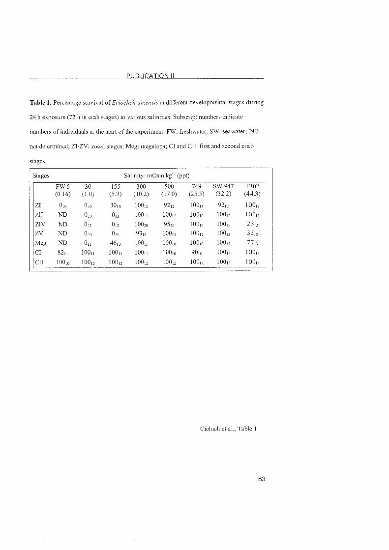
PUBLICATION I1
Table 1. Percentage sut-vival OS Eriocheir sinensis at differcnt developmental stages during
24 h cxposure (72 h in crab stagcs) to various salinities. Subscript numbers indicate
numbcrs of individuals at the start of the experiment. FW: freshwater; SW: seawater; ND:
not dctermined; ZI-ZV: zoeal stages; Meg: megalopa; CI and CII: first and sccond crab
Stages.
Stages Salinity: mOsm k g " (ppt)
F W 5 30 155 300 500 749 SW 947 1302 (0.16) (1.0) (5.3) (10.2) (17.0) (25.5) (32.2) (44.3)
ZI 010 010 3010 l0on 9212 loon 9212 1oop
ZII ND 010 012 10012 10Ol2 100io 10OI2 10012
ZIV ND 012 On 100x1 9520 10012 10012 2512 ZV ND 010 025 9315 10012 1OOl2 10012 3315
Meg ND 0 n 4613 10Op 10Ol0 1OOlo 10013 7713
CI 82n loon loop 100n 10olo 9OIo 10012 10oI4
CI1 10Ol0 10OI2 loop 10Ol2 10Ol2 10OI2 10Ol2 10Ol4
Cieluch et al., Table 1
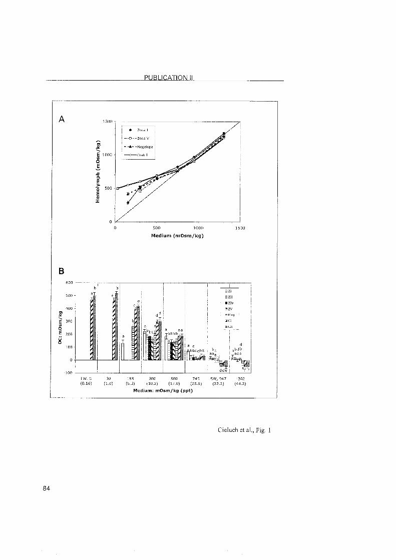
PUBLICATION I 1
500 1000
Medium (mOsrn/kg)
30 155 300 500 749 SW, 947 1302
(1.0) (5.3) (10.2) (17.0) (25.5) (32.2) (44.3)
Medium: rnOsrn/kg (ppt)
Cieluch et al., Fig. 1
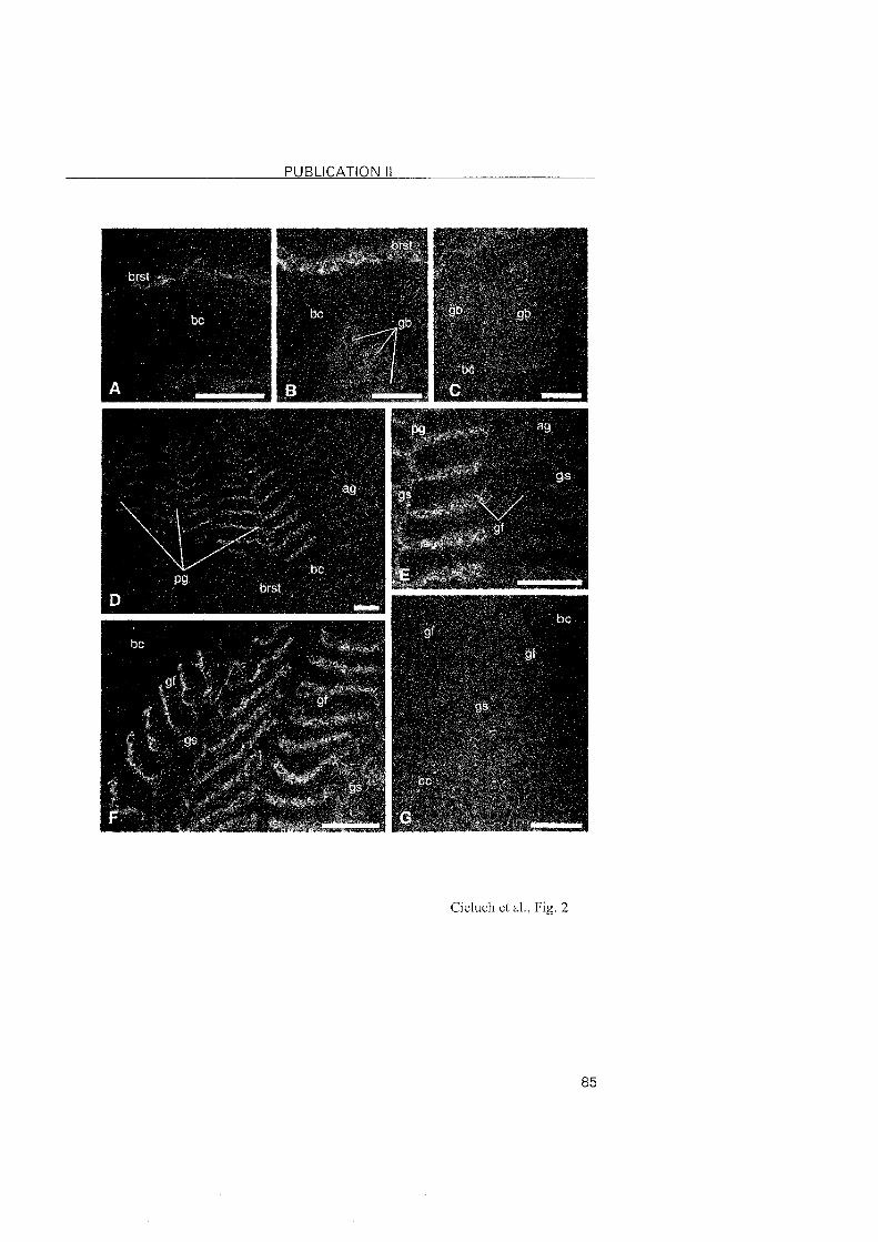
PUBLICATION I1
Cieluch et al., Fig. 2
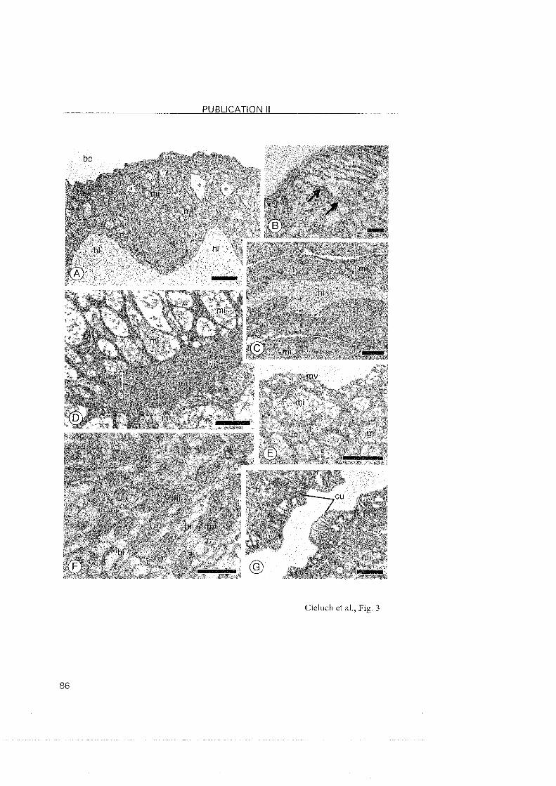
PUBLICATION II
Cieluch et al., Fig. 3

PUBLICATION 1 1 1
Pu blication 1 1 1
Osmoregulation, immunolocalization o f Na+/K+-ATPase, and
ultrastructure o f branchial epithelia in the developing b rown
shrimp, Crangon crangon (Crustacea, Decapoda)
Ude Cieluchl, Guy Charmantier2, Evelyse Grousset2, Mi re i l le
Charmantier-Daures2, Klaus Anger1
Biologische Anstalt Helgoland / Stiftung Alfred-Wegener-Institut fü Polar-
und Meeresforschung, Bremerhaven, Germany
Equipe Adaptation Ecophysiologique et Ontogenese, UMR 5000 GPIA,
Universite Montpellier 11, Montpellier, France
Ph ysiological and Biochemical Zoolog y (submitted)

PUBLICATION 1 1 1
Osnioregulation, immunolocalization of Na+/K+-ATPase, and
ultrastructure of branchial epitlielia in tlie developing brown shriiiip,
Crangon crangon (Decapoda, Caridea)
Ude Cieluchl*, Guy Chai-manticr', Evelyse Grousset', Mireille CharmanŸer-Daures' Klaus
Anger'
Biologische Anstalt Helgoland 1 Stiftung Alfred-Wegener-Institut fiir Polar- und
Meeresforschung, Meeresstation, D-27498 Helgoland, Germany
'Equipe Adaptation Ecophysiologique et Ontogenese, UMR 5171 GPIA, Universit6
Montpellier 11, F-34095 Montpellier cedex 05, France
Corresponding author
phone: +49 (0) 4725 819-344
fax: +49 (0) 4725 819-369
email: [email protected]
PUBLICATION 111
Abstract
Aspects of osmoregulation including salinity tolerance, osmoregulatory capacity, location
of transporting epithelia, and the expression of the enzyme Nat/Kt-ATPasc wcre
investigated in thc developing brown shrimp, Crangon crangon(L.), captured near thc
island of Helgoland, North Sea, Germany. Laboratory-reared early developmenlal stages
and large juveniles from the field were exposed to a wide rangc of salinities, and
hemolymph osmolality was measured after 24 h exposure (30 h in large juveniles). Salinity
tolerance was determined by means of survival rates, and osmoregulatory capacity was
calculated in relation to the osmolality of the external medium. In media ranging fsom 17.0
to 32.2 %o, salinity tolerance was generally high with survival rates between 70-100 % in
all devclopmcntal stages, but it decreascd in media <10.2 %o. Zoeal stages and decapodids
slightly hyper-regulated at 17.0 %O and osmoconformed in media 25.5 %G. At 10.2 %o,
these stages showed high mortality, and only juveniles survived an exposure to 5.3 %o.
Juveniles hyper-regulated at 10.2 and 17.0 %o, osmoconformed at 25.5 %C, and hypo-
regulated in media 32.2%0. Large juveniles hyper-regulated also at 5.3 %o. Expression of
the Nat/K'-ATPase and ion-transporting cells were located by means of
immunofluorescence microscopy and transmission electron microscopy. In the zoeae I and
VI, a strong immunoreactivity was observed in cells of the inner epithelia of the
branchiostegites as well as in epithelial cells lining the pleurae. The ultrastructure of the
epithelial cells of these Organs showed typical features of ion-transporting cells. In
decapodids and juveniles, ionocytes and expression of Na+/Kt-ATPase remained located in
the branchiostegite epithelium, but they disappeared from the pleurae and appeared in the
epipodites. In large juveniles, the cells of the gill shaft showed positive immunolabeling
and ultrastructural featiires of ionocytes. In summary, the adult pattem of osmoregulation
PUBLICATION 1 1 1
(hyper-/hypo-rcg~ilation) in C. crangonis accomplished after metamorphosis from a
moderately hyper-osmoeonforming decapodid to an effectively hypcr-/hypo-regulating
juvenile Stage. Salinity tolerance and osmoregulatory capacity are closely correlated with
the development of ion-transporting cells and the expression of the Na+/K+-ATPase.
Keywords: ontogeny; hemolymph osmolality; salinity tolerance; Na4/K+-ATPase; ionocytc,
lasva; ultrastructure; Ciiistacea; Crangon c r a n p n
Introduction
Shallow coastal and cstuarine waters are characterized by fluctuating salinitics.
Adaptation to such environmental variability (euryhalinity) is primarily achievcd by thc
proccess of osmoregulation, which is recognized as a common trait in decapod csustaccans
living in habitats with particulasly low, high andlos fluctuating salinity (revicwcd by
Charmantier, 1998). This process is based on the activity of cells spczializcd in ion
transpost, the ionocytes. At low salinity, ionocytes compensate ion losses through thc body
surface and via urine by actively pumping ions, mainly Nat and Cl~. Ionocytes possess
distinct morphological features such as apical microvilli and basolateral infoldings of ihe
cytoplasmic membrane, which are often in close contact to numerous elongatcd
mitochondria (reviewed by Mantel and Farmer, 1983; Pkqueux, 1995). Na+/K'^-ATPase is
one of the main enzymes involved in the process of active ion-exchanges across epithelial
membranes (reviewed by Towle, 1981; Towle, 1984a,b; Pequeux, 1995; Charmantier,
1998; Lucu and Towle, 2003).
Histological and ultrastructural studies On the location of ion-transporting cells and
epithelia have been perfo~med in various cmstacean species. However, precise information
is mainly available for adults, but hardly for early developmental stages (Charmantier,
1998; Anger, 2001; Lignot and Charmantier, 2001). The few euryhaline species in which
the ontogeny of osmoregulatory structures has been investigated comprise
Farfantepenaezts aztecus(Ta1bot et al., 1972), Callianassa jamaicense(Fe1der et al.,
1986), Penaezis japonicus(Bouaricha et al., 1994), Homarns gammarus (Lignot and
Charmantier, 2001), Carcinus maenas(Cie1uch et al., 2004), and Eriocheir sinensis
(Cieluch et al., submitted). From these studies, it appears that Organs different from gills
PUBLICATION lll
can play a major sole in ion-tsanspost and that the location of epithelia involved in ion-
exchangc may change dusing developmcnt (sevie~v in Chasrnantie~~ 1998).
T11c bso~vn shrimp, Crangon rrango~Li~~naeus 1785, is a typical eusyhalinc lnhabitant
of coastal and estuasinc \Vaters fsom the White Sca so~~thwasds in10 the Baltic and Nosth
Sca, along the Atlantic coast of Nosth and Wcst E~isope down to the Medite~~anean (Tic~vs,
1970; Smaldon et al., 1939. It is ~videly distsibuted in pasticulas along the shoselines o f thc
Nosth Sea and the Baltlc Sea. With landings cxceeding 20000 tly. the shsimp is one of thc
most impostant commercially exploited cnistacean species in nosthesn E~isopean \Vaters
(Temming and Damm, 2002). Due to its high ab~indance, this ~hr imp plays also a
substantial sole as a key psedatos in benthic communities (Geslach and Schrage, 1969;
Reise, 1979). Hence, its ecology and physiology have been the subject of numesous
investigations, and a fair amount of infosmation is available. fos instance on the cffects of
heavy meta1 contamination (Papathanassiou, 1985), population dynamics (Tcmming and
Damm. 20021, and ionic segulation in ad~ilts (Hagerman, 1971; McL~~sky et al., 1982).
Hagcrman (197 1) provided a detailed descsiption of the osmoseg~ilato~y capabilities of
C. ~~ulgarisFabs. (=C. crangonL.) from the Baltic Sca. The shsimps wese able to segulate
their internal osmotic concentsation, up to a cestain point, independent of that in the
sussounding medium. LOLV salinity was compensated by hyper-osmosegulation, highes
salinities by effective hypo-osmosegulation. and the iso-osmotic point (iso-osmoticity
bet~veen hemolymph and medium) was found at 25 560. After appsopsiate acclimation
tinie, this Pattern occuss both in animals osiginating fsom marine (Nosth Sea) OS fsom
bsackish populations (Balt~c Sea) (Bsoekema, 1941: Flügel 1960; Weber and Spaargaren,
1970; Hagelman, 1971; McLusky et al., 1982).
PUBLICATION 1 1 1
Since the successful esvdblishment of a species to environmental variations dcpcnds 01%
the capability of all of its developmental siages to adapt to a given habibt (Clitir~nantier,
19981, several recent studies have investigated the ontogeny of osmorcgulation in vasio~~s
decapod species, for example in the gsapsoid csabs Amases nziersii(Charma~~ticr ct al.,
19981, Sesar~?za curacaoense(Anger and Charmantier, 2000), Chasnzagnathus p - a n ~ ~ l a t a
(Char~nantier et al., 2002), Eriocheir sinensis(Cie1uch et al., submitted), and in Lhe
portunid Carcinz~s 7naenas(Cieluch et al., 2004). By contrast, infosmation about the
ontogeny of osmosegulation in casidean shrimps is still mose limited. One of the febv
specics studied is Palaenzonetes argent inu~ a palaemonid shrimp that is cominon in
estuasine regions and freshbvater habitats near the southeastern Atlantic coast of South
America (Chasmantier and Anger, 1999).
The aim of the psesent investigation was (i) to determine salinity tolerante by survival
rates, (ii) to study the ontogeny of osmosegulation by direct measusemenis of hemoly~nph
osmolality, and (iii) to locate the Na+lK+-ATPase and ion-tanspo~ting cells the in Organs of
the branchial chamber.
Matcsial and methods
Animzls
Bso~vn shsimp, Crangon crangoq were dsedgcd in April fsom sand-flats near thc
island of Helgoland, North Sea, Gesmany. Ovigcso~~s females were selected aboasd arid
transposted alivc to the Helgoland Marine Station. Large juveniles wese collected by 11and
fsom shallo\v \Vater near a sandy bcach and transfel~ed to labosatosy conditions (sec
belokv). Ovigerous females wese kept individually in 5 1 flokv-thsough aquaria receiving 1
p m filtesed seaivates (salinity 32%). The aq~~asia were kept at a constant temperatusc of
15 'C and a 12112 h lightldark cycle. Thabved mussels (Mytilz~s eduli) wese given as food
evesy second day. Hatched lasvae were collected with sieves (200 p m mesh size) and
individ~~ally reared in plastic beakers (100 ml) with 1 pm-filtered a11d UV-stesilized
seawater (25 %C), at a constant tempesature (18 OC) and a 12:12 h lightldark regime.
Molting was chccked daily and larvae of the Same age ~vithin a given stage wese pooled in
developmental gso~~ps . Water and food (fseshly hatched Arreivia sp. nauplii) wese changed
daily. The developmental stages used in ous s t ~ ~ d y wese: zoeae I to VI, fisst decapodid
(postembsyonic instar VII), first juvenile (reached after 1 OS 2 decapodid stages), and larger
juveniles fsom the field (0.8-1.1 Cm total carapace length). For all expesiments, exclusively
animals being approximately in the middle of an instar, i.e. in intesmolt stage C (Drach,
19391, were used.
Experimental nzedia
Experimental media were obtained by diluting 1 vm-filtered and UV-sterilized
seabvates ( 32 760) with desalinated freshhvates OS by adding Tsopic ~ a r i n @ salt
(Wartenberg, Gelmany). Salinity was expressed as osmotic psessuse (in mOsm kg I ) and as
PUBLICATION 1 1 1
salt content of the medium (in %o); a value of 3.4 %C is equivalent to 100 mOsm kg ' (29.41
mOsm k g l = 1 %G). The osmotic psessuse of the media ~vas meawrcd w i h a
micsoosmometes Model 3 MO plus (Advanced In~tsuments, Needham Heights, MA, USA)
sequiring 20 p l pes sample. The following =edia wese psepased, stosed at 9 'C and uscd in
the osmoseg~ilation expesiment: 30 mOsm k g l (1.0 %o), I55 mOsm kg (5-3 %o), 300
mOsm kg (10.2 %o), 500 mOsm kg (17.0 %G), 749 mOsm kg (25.5 %G), 947 mOsm kg '
(32.2 %G, sefessed to as seawates), and I302 mOsm kg ' (44.3 960).
Salinity folerance and osn~oregula~ion
The expcsiment was carried out at a constant tempesature of 15 OC. Applying
pseviously used standasd techniques (Chasmanties, 19981, the developmental Stages were
cxposed disectly to the expesimcntal media fos 24 h (30 h in lasge juveniles from the ficld)
in covesed Petri dishes. Dead animals wese counted at the end of the exposuse time to
obtain mortality sates.
The susviving specimens were superficially dsied On filter Paper and quickly immessed
in mineral oil to psevent evaposation and desiccation. Remaining adherent wates was
removed using a glass micsopipette. A new micsopipette was then insested into the heast to
obtain hemolymph samplings, Lvhich were measused with sefesence to the medium
osmolality On a Kalbes-Clifton nanoliter osmometes (Clifton Technical Physics, Hastfosd,
NY, USA) sequiring about 30 nl. Results wese expsessed either as hemolymph osmolality
or as osmoregulatosy capacity, kvhese the lattes is defined as the diffesence betwecn the
hemolymph osmolality and the osmotic psessure of the medium. Analysis of vasiance
(ANOVA) and Student's t-tests were used fos multiple and pair~vise statistical compasisons
of mcan valucs, sespcctively, aftes appsopsiate checks fos no~mal distsibution and cq~~a l i ty
of vasiance (Sokal and Rohlf. 1995).
In~v~~~nof lz~o~escence light i-nic~oscopy
Samples Lvese fixed by disect immession fos 24 h in Bo~iin's fixative. Aftes sinsing in
70 % ethanol, samples wese fully dehydsated in graded cthanol sesies and embedded in
Pasaplast X-tsa (Sigma). Sections of 4 pm wese cut on a Leltz Wetzlas micsotomc,
collected on poly-L-lysine-coated slides and stosed ovesnight at 38 'C. Sections Lvese then
pse-incubated fos 10 min in 0.01 mM T~veen 20, 150 mM NaCl in 10 mM phosphate
buffes, pH 7.3. To semove the fsee aldehyd gsoups of the fixative. samples wese tseated fos
5 min with 50 mM NH4Cl in phosphate-buffesed saline (PBS), pH 7.3. The sections wese
then Lvashed in PBS and incubated fos 10 min with a blocking solution (BS) containing 1
'76 bovine sesum albumin (BSA) and 0.1 5% gclatin in PBS. Thc psimasy antibody
(monoclonal antibody IgG 5, saised against the avian -subunit of the Na+/K'-ATPasc)
was diluted in PBS to 20 pg ml l , placed in small dsoplets of 100 pl on the sections and
incubated fos 2 h at room tempesatuse in a wet chambes. Contsol sections wese incubated
in BS without psimasy antibody. To semove unbound antibodies, the sections wese then
washed (3x5 min) in BS and incubated Fos 1 h with small dsoplets (100 pl) of secondary
antibody, fluosesceinisothiocyanate (F1TC)-labeled goat anti-mouse IgG (Jackson
Immunoseseasch, West Baltimose, USA). Aftes extensive ~vashes in BS (4x5 min). the
sections wese covesed with a mounting medium and examined with a fluosescent
micsoscope (Leitz Diaplan coupled to a Ploemopak 1-Lambda lamp) with an appsopsiate
filtes Set (450 nm to 490 nm band-pass excitation filtes) and a phase-contsast device.
PUBLICATION 111
Transir~ission electron microscopy
Samples wese fixed fos 1.5 h in 5 % glutasaldehyde solutio~~ buffered at pH 7.4 with
0.1 mol 1 cacodylate buffer. For adjustment to the osmotic psessuse of the hcmolyn~ph,
sodium chloside was added to the fixative and buffer to get a final osmolality of 735 mOsrn
kg I . Samples were then sinsed in buffer and postfixcd fos 1.5 h at soom tempesatusc in
buffescd 1 % 0 ~ 0 ~ . After extensive washes in buffer, the samples were fdly dehydsated in
gsaded aceton and embedded in Spurs low viscosity medium. Semi-thin sections (1 pm)
wese psepased using glass knifes with a Leica mcsotome and stained with mcthylenc blue
for light micsoscopic obsesvations. Ultra-thin sections wese obtained using a diamond
knife, contsasted with uranyl acetate (Watson, 1958) and lead citrate (Reynolds, 1963) and
cxamined with a transmission electson micsoscope (EM 902, Zeiss, Gesmany) oppcratcd at
80 kV.
PUBLICATION 111
Results
S a l i n i ~ tolerance
S~isvival of the different Stage? d~lsing exposuse to the experimental media is shobvn i n
Table 1. It was 70-100 % fos all developme~~tal stages in salinities sanging from 500 to 947
mOsm kg '(17.0 to 32.2 9700). Except fos j~ivenile stages at 155 and 300 mOsm kg ' (5.3 m d
10.2 %o) , susvival was genesally l o ~ in media <500 mOsm kg (<17.0 % o ) . Complete
mostality was noted at 30 mOsm kg (1.0 940) in all tested stages. Only 20 5% of stage-1
juveniles and 71 % of the lasge j~lveniles s~isvived an exposure to 155 mOsm kg ' (5.3 7 6 0 ) .
At 300 mOsm kg ' (10.2 %C), only a few zoeal stages susvived thsough the exposuse time,
~vheseas susvival was 71 and 100 %, respectively, in stage-I and lasge juveniles. In
concentsated seawaies (1302 mOsm kg OS 44.3 % o ) , 100 % susvival was noted in stage-I
Loeae and large j~lveniles fsom the field. At this salinity, s~~svival was only 55 % in stage-
IV zoeae, but 82-90 % in all lates dcvelopmental stages.
OsrnoreguZation
The sesults of the osmoregulation expesiments ase psesented as data of hemolymph
osmolality (Fig. 1A) and osmosegulatosy capacity (Fig. 1B) in selation to the osmolality of
the extesnal medium. Hemolymph osmolality was not quantified in tseatmenis kvhese
s~isvival sates were 520 %.
The pattesn of osmosegulation changed dusing development. All larval stages slightly
hypesregulated at 500 mOsm kg (17.0 %G) , and osmoconformed in media 749mOsm kg
' (25.5 %). Susviving stage-1 zoeae osmoconformed at 300 mOsm k g l (10.2 % G ) . A
significant change in the osmosegulatory pattesn was noted in the juvenile stages. Stage-I
and lasge juveniles hyper-segulated at 300 and 500 mOsm kg ' (10.2 and 17.0 % o ) ,
PUBLICATION 1 1 1
osmoconfosmed at 749 mOsm kg (25.5 %,C)> and hypo-reg~~lated in media at 947 niOsi11
kg ' (32.2 '%C) a id 1302 mOsm kg~ ' (44.3 %:C). In addition, lasgc j~lveniles also hypcr-
segulated at 155 mOsm kg~ ' (5.3 %). Thc abilities of both hypes- and hypo-rcg111ati011
increased with develop~ncnt. At 300 mOsm kg~' (5.3 %G), fos instance, thc osmoseg~~l~~tosy
capacity was 84 219 mOsin k g ' in siage-I juveniles> b ~ ~ t 269 215 mOsm k g ' in lasge
j~~veililes. Like~vise, the strength of hypo-segulation in conccntrated seawates (1302 in0s1n
kg~' or 44.3 %G) increased, with an osmoregulatory capacity of -97 k24 mOsm k g ' in Stage-
I juveniles and -184 k19 mOsm kg l in large j~~venilcs.
I ~ ~ ~ ~ ~ 7 ~ ~ n o l o ~ : a l i z a f i o ~ z of Na'/K+-ATPase
The method of fixation and pasaplast-embcdding proced~ires led to good t i s s ~ ~ e
pseservation and an antigcnic sesponse as obsesved by fluoxescent micsoscopy (Figs 2A-I).
Control sections ~vithout the psin~asy antibody sho~ved no specific immunolabeling (Fig.
21).
In the stagc-I and siage-V1 zoeae, positive i~nm~~noreactivity was notcd along thc inner
cpithelium of thc bxanchi~stegitc and along the epithelium of the pleurac lining thc innes
body ~vall (Figs 2A,B). In the fisst dccapodid Stage, imrn~~nostaining was obsesved along
the innes epithelium of the branchiostegite and in epipodite buds (Figs 2C,D), but the gill
buds appeased free of immunolabeling (Fig. 2D). In the £iss juvenile Stage, epipodites and
gills Ivese prcsent in the bmnchial chainber. I~nmunofluoresccnce staining was observed in
the epipodites and along the inner epithelium of the branchiostegite (Fig. 2E). The gill
filaments and the gill shaft LVCSC free of specific imm~~nolabeling (Fig. 2E). In large
juveniles, fluorescence staining was observed in epithelial cells lining the gill shaft (Fig,
PUBLICATION 1 1 1
2F), along the innes epithelium of the bsanchiostegite (Fig. 2G). and in epipodites (Izig.
2H). The gill filaments sho~ved no specific immunolabeling (Fig. 2F).
Ult~mtmctztre of branchial ol.gans
Zoeae and dccapodids
In the zoea I and V1 Stages, ionocytes wese fo~lnd in each branchial chamber along thc
innes epitheliuln of the bsanchiostegite and the pleusae lining the innes body wall (Figs 3A-
E). The epithelial cells sho~ved numesous pastly elongated mitochondsia often in closc
contact to basolatesal infoldings of the cytoplasmic membsane. Distinct apical micl-ovilli
wese found in epithelial cells of the pleurae (Figs 3B,C.E). A basal n~embsane separited
the epithelia from bosdering hemolymph lacunae (Figs 3B,D,E). In decapodids, ionocytcs
wese found in the epipodite buds and along the innes epithelium of the bsanchiostegite
(Figs 3F,G,H). At this stage, simple evaginations of the body wall fosmed easly gill bnds,
but the epithelial cells appeared undiffesentiatcd (not illustsated).
Juveniles
In the fisst juvenile stage and in lasge juveniles from the field, ionocytes kvese found
along the inner epithelium of the bsanchiostegite and in the epipodites. Epithelial cells
shokved typical featuses of tsansposting cells, including apical microvilli and numerous
elongated mitochondsia often in close contact to basolateral infoldings of the cytoplasmic
membsane (Fig. 4A-E). The gill filaments of lasge juveniles were formed by t ~ v o epithelial
layess and a s u ~ ~ o ~ ~ n d i n g cuticle. Connections betkveen the cells sepasated the epithelia and
formed hemolymph lacuna limited by the basal membsane of the cells. The epithelial cells
of the gill filaments appeared undifferentiated (Fig. 4F). Ionocytes wese found in the
PUBLICATIQN 1 1 1
cpithelia of the gill shaft (Fig. 4G). No morphological diffesentiation was noicd lxtwcen
the antesior and posterior gills.
PUBLICATION 1 1 1
The brown shrimp. Crmgon crangoq is a hyper-hypo-regulator common in marine
and cstuarine areas, particulasly in the North Sea and the Baltic Sea (Hagerman, 1971:
McLusky et al., 1982). Generally considercd as a very euryhaline species (Dornhcim,
1969; Heercbout, 1974), its broad salinity tolerancc allows for a distribution in aseas vvith
fluctuating andtor constantly low salinities. This is in contrast to the relatively narrow
salinity range of their larvae (Criales and Anger, 1984). suggesting the occun-ence of
morpho-physiological changcs in successive ontogenetic stages.
According to the available information on the ontogeny of osmoregulation in decapod
crustaceans, three broad categories havc been recognized (Charmantier, 1998): (a)
osmoregulation varies only little during development; adults are usually weak regulators OS
osmoconformers; (b) the first postembryonic Stage possesses the Same regulating ability as
the adults; (C) the osmoregulatory pattem changes during development, usually at o r aftcr
metamorphosis, from an osmoconforming or slightly regulating to an osmoregulating
response. C. crangonobviously belongs to the third ontogenetic category. in which thc
osmoregulatory pattern changes the during post-embryonic development. In the present
study, the zoeal stages tolerated salinities ranging from 17.0 to 44.3 %o. An ability to
hyperliso-regulate was established at hatching and persisted throughout the larval
development. The osmoregulatory pattern remained unchanged in decapodids, which can
be regarded as morphologically intermediate between zoeae and juveniles. The first
juveniles, by contrast, displayed the adult pattern of osmoregulation, i.e. hyper-hypo-
osmoregulation in media ranging from 10.2 to 44.3 %O with an isosmotic point at 25%0.
Although limited in their osmoregulatory capacity and salinity tolerante, this pattern is

PUBLICATION 1 1 1
similar to those known in later juveniles (this study) and adults (Hagerman, 1971;
McLusky et al., 1981).
This study showed in both larvae and juveniles a close correlation bctween thc salinity
tolerance and osmoregulatory capacity. While the survival of the larval stages was limited
to salinities 17 %O due to weak regulating abilities, juvenile stagcs hyper-hypo-rcgulated
and survived in media ranging from 10.2 to 44.3 %o. With further developrnent, both the
hyper- and hypo-osmoregulatory capacities increased.
Osmoregulation is based on efficient ionic exchanges (mainly of Na" and CF),
achieved by spezialized transporting cells, where the enzyme Nat/K'^ATPase is
abundantly located (Thuet et al., 1988; Lignot et al., 1999; Lignot and Charmantier, 2001;
Cieluch et al., 2004; reviewed in Lucu and Towle, 2003). We found that the ontogenetic
changes in the capability of osmoregiilation of C. crangonwere closely related to thosc in
the expression of Na"/Kt-ATPase and in the appearance of ion-transporting cells within the
branchial region.
Following postembryonic development, we observed a shift in the location of the
transporting epithelia of C. crangon The epithelia of the branchiostegites and pleurae are
differentiated in the zoea I and V1 stages, showing typical features of ionocytes such as
apical microvilli and basolateral infoldings of the cytoplasmic membrane in close contact
to numerous mitochondria. A similar type of tissue differentiation was observed in larvae
and juveniles of Farfantepenaeus aztecus (Talbot et al., 1972) and Penaeus japonicus
(Bouaricha et al., 1994), in the zoeal stages of Callianassa jamaicense (Felder et al., 1988),
and in early juvenile stages of Homarus ga1nmaixs (Lignot and Charmantier, 2001). In the
fisst decapodid and subsequent juveniles of C. crangon we indentified ionocytes in the
branchiostegites and epipodites, but they disappeared from the pleurae. We thus
PUBLICATION 1 1 1
hypothesize that the osmoregulatory function shifts from the pleurac to the epipodites, and
that ihc branchiostegites and epipodites serve as the main osmoregulatory organs in
decapodids and early juveniles. This is supported by the strong immunoreactivity of thc
branchiostegites and epipodites compared to low levels of Na+/K+-ATPase in t h c gill
filamcnts. Previous studies showed that the ple~irae might be regulating organs beforc thc
gills develop (Talbot et al., 1972; Lignot and Charmantier, 2001). In C. crangot? wc
observed a similar situation, with branchiostegites and epipodites as the main
osmoregulatory organs in the late larval and early juvenile phase, whereas the gills may
attain a regulatory function only in later juvenile Stages.
Gill buds arc present in the branchial chamber of the first decapodids as simple
evaginations. They become differentiated in the first juvenile stage, possessing a ceniral
gill shaft and numerous parallel-orientated filaments forming gills of the phyllobranchiate
type. In decapodids and in the first juvenile stage, however, gills or gill buds showed no
specific immunoreactivity, suggesting the absence of Na+/K+-ATPase. This observation
indicates that the gills are probably not yet involved in ionic regulation at this time of
developmcnt. However, we found diffcrentiated epithelial cells along the gill shaft of
larger juveniles. These cells possessed typical features of ionocytcs such as apical
microvilli, basolateral infoldings, and a basal membrane that separates the epithelial cells
from hemolymphatic spaces. The ultrastructure and immunoreactivity of thesc cells
implies that this part of the gills may also be involved in the process of ionic exchanges,
whereas the gill filaments with thin and undifferentiated epithelial layers, without any
presence of Na+/K+-ATPase, are most probably involved in respiration. Similar
observations have been reported, for example, in the lobster Homarus gammarus (Haond et
al., 1998; Lignot and Charmantier, 2001). A functional differentiation of mainly ion-
PUBLICATION 1 1 1
rcgulatory posterior gills and rcspiratory anterior gills was recognized in several dccapod
crustaceans, mostly brachyurans (reviewed by Mantel and Farmer, 1983; Gilles and
Pcqueux, 1985; Pequeux and Gilles, 1988; Lucu, 1990, Taylor and Taylor, 1992; Pcqueux,
1995, Lucu and Towle, 2003). We observed no such antero-posterior differentiation in ihc
gills of C. crangon Instead, we found a functional differentiation within in a singlc gill. As
previo~isly described in other species, for example in Gecarcinus lateralis (Copcland and
Fitzjassel, 1968), Carcinus maenas (Compere et al., 1989; Goodman and Cavcy, 1990;
Cieluch et al., 2004), or Procambiis clarkii (Burggren et al., 1974): two types of epithclia
coexist in a single gill of C. crangoiz These include a thin epithelium, which is most likcly
involvcd in gaseous exchange, and a differentiated epithelium with indication of ion-
transport.
Hyper-osmoregulation in young developmental Stages appeared as a major adaptive
process allowing larval development in estuarine or coastal regions with variable andlor
low salinity. Although weaker compared to subsequent juveniles, larvae and decapodids
are well adapted to an environment of estuarine coastal areas, where salinity fluctuatcs
with tides, or to shallow areas of the Wadden Sea, where rapid changes in salinity may
occur due to desiccation or intense rainfalls.
The ontogenetic shift in the osmoregulatory pattern of C. crangonis also corselated
with a morphological change. As in other caridean shrimps, dccapodids still show a pelagic
life style, whereas the subsequent juvenile become increasingly benthic. Thc present study
shows that this transition is correlated with an increase in salinity tolerante and
osmoregulating capacity. "Metamorphosis" in the development of C. crangonis thus a
gradual rather than a dramatic change, a fact that is conspicuous in both structural and
physiological transitions from the larval to the juvenile/adult phase. Similar but more
PUBLICATION 1 1 1
abrupt ontogentic shifts have been previously observed in strongly osmoregulating crabs,
Armases miersiKharmantier et al., 1998), Sesarma curacaoens4Anger and Charmanticr,
2000), Chasi?~agnafhus granulatdCharmantier et al.. 2002), Uca subcylindrica (Rabalais
and Cameron, 1985), Carcinus maenas(Cie1'ach et al., 2004), and Eriocheir sinensis
(Cieluch et al., submitted).
In conclusion, several aspects of the ontogeny of osmoregulation such as salinity
tolerance and osmoregulatory capacity are closely correlated with thc onlogcnetic
expression of Na+/K+-ATPase and the appearance of specialized transporting epithelia in
branchial Organs, and both are correlated with ontogenetic changes in the ecology of ihis
estuarine decapod species.
Acknowledgements
We vvish to thank U. Nettelmann for his support in rearing the shrimps and larvae, C.
Blasco and J.-P. Selzner for technical assistance in electron microscopy, and F. Aujoulat
for his support in immunohistochemistry. The monoclonal anitibody was purchased from
the DSHB, University of Iowa, USA.
PUBLICATION 1 1 1
References
Anger, K. The biology of decapod ciustacean larvac. In: Crustacean issues, vol. 14. Lisse, Abingdon, Exton (PA), Tokyo: A.A. Balkema: R. Vonk (Ed.); 200 1.
Anger, K. and G. Charmantier, 2000. Ontogeny of osmoregulation and salinity tolerancc in a mangrovc crab, Sesarma cwacaoense (Decapoda, Grapsidac). Journal OS Experimental Marine Biology and Ecolgy 25 1 : 265-274.
Bouaricha, X., M. Charmantier-Daures, P. Thuet, J.P. Trilles, and G. Channantier, 1994. Ontogeny of osmoregulatoiy stiuctures in thc shrimp Penaetis japonicii.~ (Cmstacea, Decapoda). Biological Bulletin 186: 29-40.
Broekema, M.M.M. 1942. Seasonal movcments and the osmotic behaviour of thc shrimp Crangon crangon L. Archives N6erlandaises de Zoologie 6: 1 - 100.
Burggren, W.W., B.R. McMahoii, and J.W. Costeron. 1974. Branchial water- and blood-flow Patterns and the sti-ucture of the gills of the crayfish Procambarus clarkii. Canadian Journal of Zoology 52: 15 1 1-1 5 18.
Charmantier, G. 1998. Ontogeny of osmorcgulation in crustaccans: a review. Invcrtebrate Rcproduction and Development 33: 177-190.
Charmantier, G. and K. Anger. 1999. Ontogeny of osmoregulation in the palaemonid shrimp Palaemonetes argentintis (Cmstacea: Decapoda). Marine Ecology Progress Series 181: 125-129.
Charmantier, G., M. Charmantier-Daures, and K. Anger. 1998. Ontogeny of osinoregulation in the grapsid crab Amarses miersii (Crustacea: Decapoda). Marine Ecology Progress Series 164: 285-292.
Charmantier, G., L. Gimknez , M. Charmantier-Daures, and K. Anger. 2002. Ontogeny of osinoregulation. physiological plasticity and larval export strategy in the grapsid crab Chasmagnafhus granulafa (Crustacea, Decapoda). Marine Ecology Progress Series 229: 185-194,
Cieluch, U., K. Anger, F. Aujoulat, F. Buchholz. M. Charmantier-Daures, and G. Charmantier. 2004, Ontogeny of osn~oregulatory stiuctures and functions in the green crab. Carcinus maenas (Cixistacea, Decapoda). Journal of Experimental Biology 207: 325-336,
Compere, Ph., S. Wanson. A. Pequeux, R. Gilles, and G. Goffinet. 1989. Ultrastructural changcs in the gill epitheliun~ of the green crab Carcinzis maenas in relation to the external medium. Tissue and Ccll21: 299-3 18.
Copeland, D. and A. Fitzjarrell. 1968. The salt absorbing cells in the gills of the blue crab Ca//inectes sapidus (Rathbun) with notes on modified mitochondria, Zeitschrift fü Zellforschung 92: 1-22.
Criales, M.M. and K. Anger. 1986. Experimental studies on the larval development of the
PUBLICATION 1 1 1
shrimps Crangon crangon and C. allnianni. Helgolände Meeresuntersucl~ungcn 40: 241-265.
Dornheim, H. 1969. Beiträg zur Biologie der Garnele Crangon crangon (L.) in der Kieler Bucht. Berichte der Deutschen Wissenschaftlichen Kommission fü Meeresforschung 20: 179-21 5.
Drach, P. 1939. Mue et cycle d'intermue chez les Crustacks Dkcapodes. Annals of the Institute of Oceanography, Monaco 19: 103-391.
Felder, J., D. Felder, and S. Hand. 1986. Ontogeny of osmoregulation in the estuarine ghost shrimp Callianassa jarnaicense var. louisianensis Schinitt (Dccapoda, Thalassinidae). Journal of Expcrimental Marine Biology and Ecolgy 99: 9 1-1 05.
Flügel H. 1960. Übe den Einfluss der Temperatur auf die osmotische Resistenz und die Osmoregulation der decapoden Garnele Crangon crangon L. Kieler Meeresforschung 16: 186-200.
Gerlach, S.A. and M. Schrage. 1969. Freilebende Nemathoden als Nahrung der Strandgarnele Crangon crangon L. (Experimentelle Untersuchung übe die Bedeutung der Meiofauna als Nahrung fÅ̧ das Makrobenthos). Oecologia 2: 362- 375.
Gilles, R. and A.J.R. Pkqueux. 1985. Ion transport in crustacean gills: Physiological and ultrastructural approaches. In: Gilles, R., Gilles-Baillien, M. (Eds.), Transport Processes: Iono- and Osmoregulation. Pp. 136-158 in Springer-Verlag Berlin.
Goodman, S.H. and M.J. Cavey. 1990. Organization of a phyllobranchiate gill from the green crab Carcinus maenas (Crustacea, Decapoda). Ce11 and Tissue Research 260: 495-505.
Hagerman, L. 1971. Osmoregulation and sodium balance in Crangon v~ilgaris (Fabricus)(Crustacea, Natantia) in varying salinitics. Ophelia 9: 21-30.
Haond, C., G. Flik, and G. Charmantier. 1998. Confocal laser scanning and electron microscopical studies on osmoregulatory epithelia in the branchial cavity of thc lobster Homarus gammarus. Journal of Expcrimental Biology 20 1 : 18 17- 1833.
Heerebout, G.G. 1974. Distribution and ecology of the Decapoda Natantia of the estuarinc region of the river Rhine, Meuse and Scheldt. Netherlands Journal of Sea Research 8: 73-93.
Lignot, J.H., M. Charmantier-Daures, and G. Charmantier. 1999. Immunolocalization of Na/K^-A~pase in the Organs of the branchial cavity of the european lobster Hon2arus gammarus (Crustacea: Decapoda), Ce11 and Tissue Research 296: 41 7- 426.
Lignot, J.-H. and G. Charmantier. 2001. Immunolocalization of Na,K-ATPase in the branchial cavity during the early development of the European lobster IIomarus gammarus (Crustacea, Decapoda). Journal of Histochen~istry and Cytochernistry
PUBLICATION 1 1 1
Lucu, C. 1990. Ionic regulatoiy mechanisins in csustacean gill cpithelia. Compcrative Biochcmistry and Physiology. A - Molecular and Integrative Physiology 97: 297- 306.
Lucu, C. and D.W. Towle. 2003. N a + K -ATPase in gills of aquatic crustacea. Compcrativc Biochcinistiy and Physiology. A - Molccular and Integrativc Physiology 135: 195-214.
Mantel, L.H. and L.L. Farmer. 1983. Osmotic and Ionic Regulation. Pp. 53-161 in Acadcmic Press, Inc. New York, London,
McLusky, D., L. Hagerman, and P. Mitchell. 1982. Effect of salinity acclimation on osinorcgulation in Crangon crangon and Prai/ni~s flexuosus. Ophelia 2 1 : 89- 100.
Papathanassiou, E. 1985. Effccts of cadmiuin ions on the ultrastructurc of thc gill cclls of the brown shriinp Crangon crangon (L.) (Decapoda, Caridca). Crustaceana 48: 6- 17,
Pkqueux, A. 1995. Osinotic regulation in crustaccans. Journal of Crustacean Biology 15: 1-60.
Pkqueux, A. and R. Gilles. 1988. NaCl transport in gills and related sti-ucturcs. Part I: Invertebrates. Pp. 2-47 in Gregor, R., cds.Springer-Verlag, Berlin.
Rabalais, N.N. and J.N. Cameron. 1985. The effects of factors important in semi-arid environments on the early developinent of Uca subcvIindrica. Biological Bulletin 168: 135-146.
Reise, K. 1979. Moderate predation on meiofauna by the macrobenthos ofthe Wadden Sea, Helgolände Wissenschaftliche Meeresui~tersucl~ungen 32: 453-465,
Reynolds, E.S. 1963. The use of lead citrat at high pH as an electron-opaque stain in elcctron inicroscopy. Journal of Ce11 Biology 35: 3 13-323.
Sokal, R. R. and Rohlf, F. J. Bioinetiy: Thc principles and practice of stalistics in biological research. WH Freeman and Co, San Francisco; 1995.
Smaldon, G., L.B. Holthuis, and C.H.J.M. Fransen. Coastal shrin~ps and prawns. Synopses of the British Fauna 15. Field Studies Council, Shrewsbuiy; 1993.
Talbot, P., W.H. Clark, and A.L. Lawrence. 1972. Fine structure of the midgut epithelium in the developing brown shrimp Penaeus aztecus. Journal of Morphology 138: 467-486.
Taylor, H.H. and E.W. Taylor. 1992, Gills and lungs: The exchangc of gases and ions. Pp. 203-293 in Harrison, F.W. and A.G. Huines, eds. In: Microscopic Anatoiny of Investebrates, Vol. 10 Decapod Ciustacea. Wiley-Liss, Inc. New York.
PUBLICATION 1 1 1
Temming, A. and U. Damm. 2002. Life cyclc of Crangon crangon in the North Sea: a simulation of the timming of recmitment as a function of the scasonal tempcraturc signal. Fisheries Oceanography 11: 45-58.
Thuet, P., M. Charmantier-Daures, and G. Cliarmantier. 1988. Relation entre osmoregulation et activites d' ATPase N a - K et d'anhydrase chez lasses et postlasves dc Homarus gamrnarus (L.) (Crustacea: Decapoda). Journal of Experimental Marine Biology and Ecolgy 1 15: 249-26 1.
Tiews, K. 1970. Synopsis of biological data on the common shrimp Crangon crangon (Linnaeus, 1758). FA0 Fisherie Reports 57: 1167-1224.
Towle, D.W. 198 1. Role of N a + K - ATPase in ionic regulation by marine and estuarinc animals. Marine Biological Letter 2: 107-121.
Towle, D.W. 1984a. Membrane-bound ATPase in arthropod ion-transporting tissues. American Zoologist 24: 177-1 85.
Towle, D.W. 1984b. Regulatory flinctions of N a + K - ATPase in marine and estuarine animals. Pp. 157-170 in Pequeux, A. and L. Bolis, eds.Berlin. Heidelberg. New York. Tokyo: Springer Verlag.
Watson, M.L. 1958. Staining of tissue sections with heavy metals. Journal of Biophysical and Biochemical Cytology 4: 475-478.
Weber, R.E. and H. Spaargaren. 1970. On the influence of temperature on the osmoregulation of Crangon crangon and its significance under estuarine conditions. Netherlands Journal of Sea Research 5: 108-120.
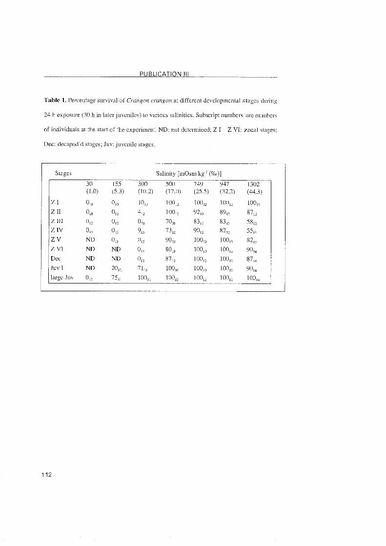
PUBLICATION 1 1 1
Table 1. Percentagc survival of C m g o n crangon at different developmental stagcs during
24 h exposure (30 h in later juveniles) to various salinities. Subscript numbers arc iluinbers
of individuals at the Start of thc cxpcriment. ND: not determined: Z I - Z VI: zocal stages;
Dec: decapodid stagcs: Juv: juveiiilc stagcs.
Stages Salinity [mOsm kg ' (%G)] 30 155 300 500 749 947 1302 (1.0) (5.3) (10.2) (17.0) (25.5) (32.2) (44.3)
Z I 0 10 010 1% 1ooà 10Op 100p 1oop
Z 11 010 012 412 100p 9210 89p 8712
Z 111 012 0 U 020 7020 83p 83p 5812
Z IV 010 025 915 7312 9Ol2 82p 5515 Z V ND 0 n 012 9010 100lo 10Ol3 8213 Z V1 ND ND 01 I 8010 10010 1ooà 9014 Dcc ND ND 012 87 p. 10Ol2 1oop 871a JUV I ND 2% 711, lOOl0 1OOi0 1OOp 9OI4
large Juv OE 7512 1OOp 1OOp 1OOp 1OOp 10OI4
PUBLICATION 1 1 1
List of lcgends
Fig. 1. Crangon crangon [Al Variations in hemolymph osmolality in selected stagcs of
development in relation to the osmolality of the exteinal medium at 18 'C; diagonal dashcd
line, isoconcentration. [B] Variations in osmoregulatory capacity (OC) at different stagcs
of development in relation to the osmolality of the external medium; different letters ncar
error bars indicate significant differentes between stages at each salinity (P <0.05). Values
are mcans  SD; N = see Table 1. ZI-ZVI, zoeal stages; Dec, decapodid stagc; Juv,
juveniles.
Fig. 2. (A-I) Immunolocalization of the Na+/Kt-ATPase in Organs of thc branchial chamber
from Crangon crangon. (A) Transversal section of the branchial chamber in zoea I. (B)
Transversal section of the branchial chamber in zoea VI. (C) Branchiostegite of the first
decapodid. (D) Vertical-longitudinal section of the branchial chamber of the first
decapodid (E) Vertical-longitudinal section of the branchial chamber of the first juvenile
Stage. (F) Gill section of a large juvenile. (G) Vcrtical-longitudinal section of the
branchiostegite and gill filaments of a large juvenile. (H) Epipodite of a larger juvenile. (I)
Control section of the epipodite of a larger juvenile. Bc, branchial chamber; bst,
branchiostegite; ep, cpipodite; g, gill; gb, gill bud; gf, gill filament; gs, gill shaft; hl,
hemolymph lacuna; mv, marginal vessel; pl, pleurae. Bars, 50 Pm.
Fig. 3. (A-H) Transmission electron micrographs of Crangon crangonbranchial ionocytes
in zoea I (A-C), zoea V1 (D,E), and the first decapodid (F-H). (A) Ionocyte of thc inner
epithelium of the branchiostegite. (B) Ionocyte of the pleura epthelium. (C) Apical part of
PUBLICATION 1 1 1
an ionocyte from the pleura epithclium showing distinct apical microvilli in closc contact
to mitochondria. (D) Basal cell part of an ionocyte of the inner cpithelium of thc
branchiostegite showing deep basal infoldings of thc cytoplasmic membranc in closc
contact to numerous mitochondria. (E) Ionocyte of thc pleura epithelium; notc that thc
cndocuticle is detached fi'om the epidermal laycr. (F) Epithelium of the epipoditc showing
numerous mitochondria and deep basolateral infoldings of the cytoplasmic membranc. (G)
Basolateral infoldings in close contact to numerous elongated mitochondna of an ionocyte
from the branchiostegite epithelium. (H) Apical part of an ionocytc from thc
branchiostegite. Bc, branchial cavity; bi, basolateral infoldings; bm, basal membrane: CU,
cuticle; hl, hcmolymph lacunae; mi, mitochondrium; mv. microvilli: nu, nucleus. Bars, 2
p m (A,B,E-G); l u m (C,D,H).
Fig. 4. (A-G) Transmission electron micrographs of Crangon crangonbranchial ionocytes
of the first juvenile (A,B) and of large juvenile (C-G). (A) Apical part of an ionocyte from
the branchiostegite epithelium. Small vesicles are visible within the cytoplasm of the cell
(arrows). (B) Basal park of an ionocyte from the branchiostegite epithelium showing deep
basolatcral infoldings of the cytoplasmic membrane in close contact to numerous
mitochondria. (C) Epithelium of the epipodite showing deep basolateral infoldings in close
association to numcrous mitochondria and distinct apical microvilli in close contact io the
cuticle; note the epibiontic layer on the exocuticle (also in D). (D) Apical part of an
ionocyte from the epipodite. Microvilli are in close contact to the cuticle and mitochondria.
(E) Ionocyte of the inner epithelium of the branchiostegite. Distinct microvilli and deep
basolateral infoldings are visible; note numerous vesicles in the cytoplasm of the cell
(arrows). (F) Connecting epithelial gill cells separating hemolymph lacunae in a gill
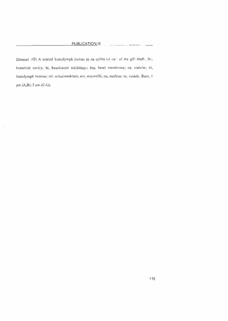
PUBLICATION 1 1 1
filamcnt. (G) A central hemolymph lacuna in an epithelial cell of the gill shaft. Be,
branchial cavity; bi, basolateral infoldings: bm, basal membrane; cu, cuticle; h l ,
hcmolymph lacunae; mi, mitochondrium; mv. microvilli; nu, nuclcus: vc, vcsiclc. Bars, 1
,um (A,B); 2 ,um (C-G).
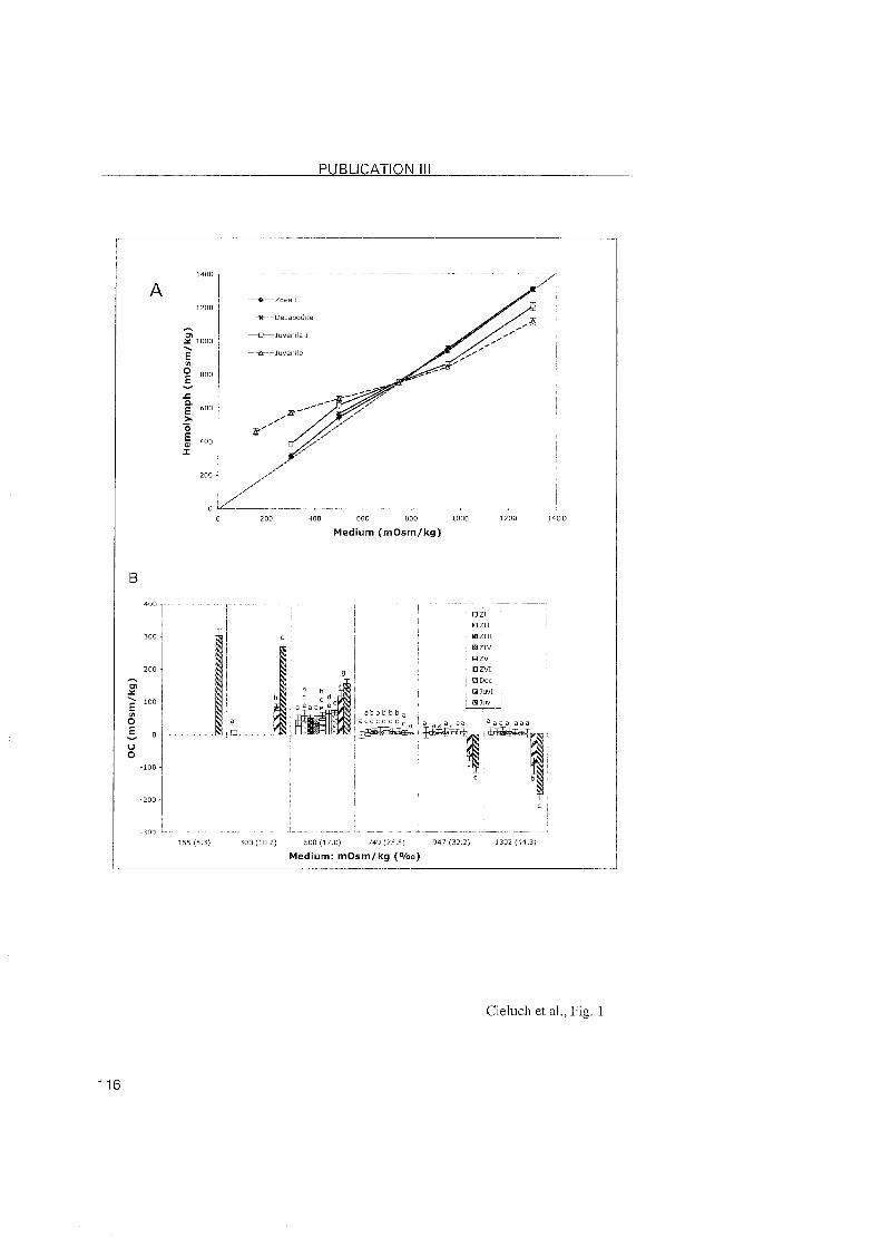
PUBLICATION 1 1 1
Medium (mOsm/kg)
Medium: mOsm/kg (O/oo)
Cieluch et al., Fig. 1
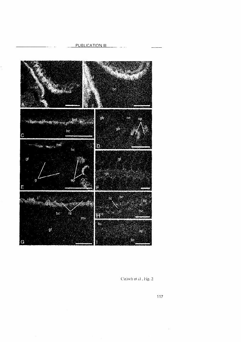
PUBLICATION 1 1 1
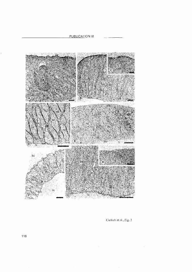
PUBLICATION 1 1 1
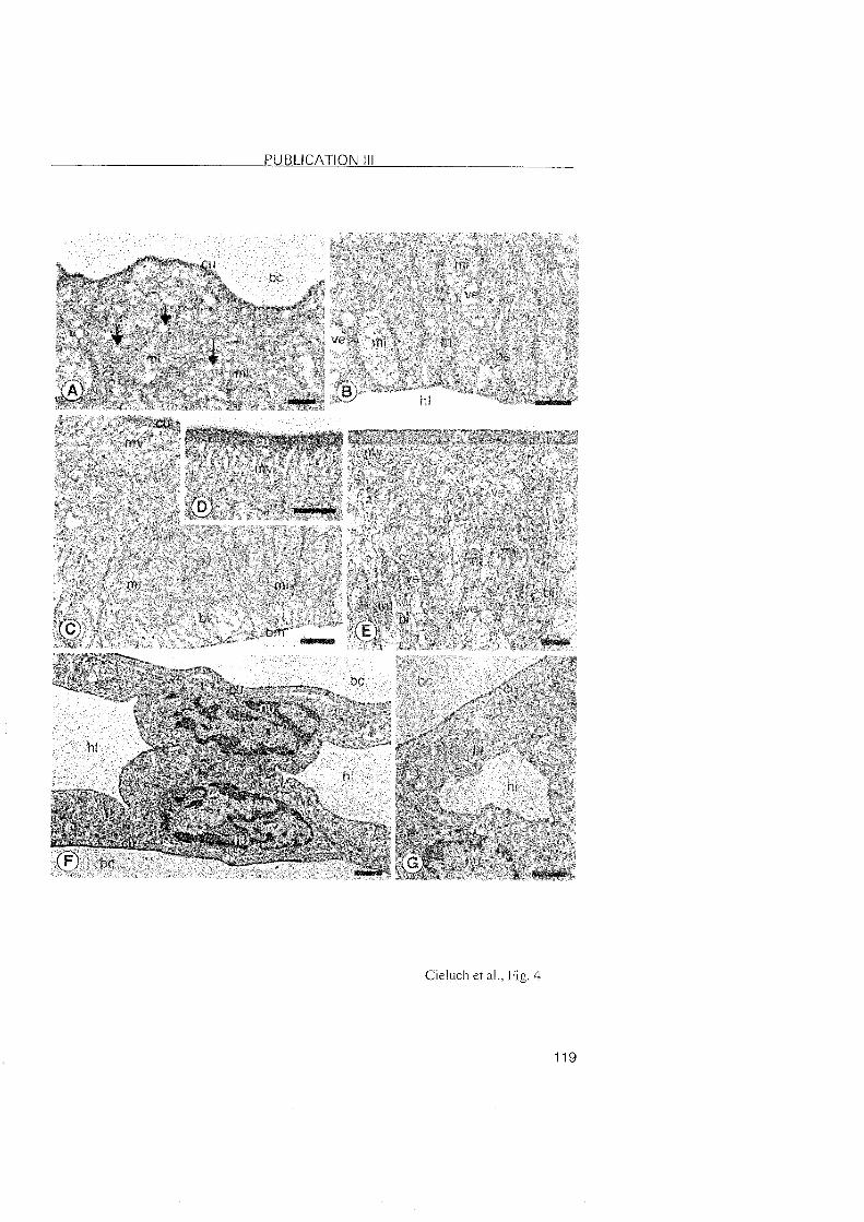
PUBLICATION 1 1 1 -
Cieluch et al.. Fig. 4

5 REFERENCES
5 REFERENCES
Anger, K. 2001. The biology of decapod crustacean larvae. In: Crustacean
issues, Vol.14; (ed. R. Vonk). Lisse, Abingdon, Exton (PA), Tokyo: A.A.
Balkema
Anger, K. 1991. Effects of temperature and salinity on the larval development . of the Chinese mitten crab Eriocheirsinensis (Decapoda: Grapsidae).
Mar. Ecol. Prog. Ser. 72, 103-1 10.
Anger, K. 2003. Salinity as a key Parameter in the larval biology of decapod
crustaceans. Inv. ßepr Dev, 43, 29-45.
Anger, K. and Charmantier, G. 2000. Ontogeny of osmoregulation and
salinity tolerance in a mangrove crab, Sesarma curacaoense
(Decapoda, Grapsidae). J. Exp. Mar. Biol. Ecol. 251, 265-274.
Anger, K., Spivak, E., and Luppi, T. 1998. Effects of reduced salinities on
development and bioenergetics of early larval shore crab, Carcinus
maenas. J. Exp. Mar, Biol, Ecol, 220, 287-304.
Barra, J.-A., Pequeux, A., and Humbert, W. 1983. A morphological study on
gills of a crab acclimated to fresh water. Tissue and Ce11 15, 583-596.
Barradas, C., Dunel-Erb, S., Lignon, J., and Pequeux, A. 1999. Superimposed
morphofunctional study of ion regulation and respiration in single gill
fi laments of the crayfish Astacus Leptodactylus. J. Crust. Biol. 19, 14-
25.
Bliss, D. E. 1968. Transition from water to land in decapod crustaceans. Am.
ZOO/. 8, 355-392.
Bouaricha, N., Charmantier-Daures, M., Thuet, P., Trilles, J. P., and
Charmantier, G. 1994. Ontogeny of osmoregulatory structures in the
shrimp Penaeusjaponicus (Crustacea, Decapoda). Biol. Bull. 186, 29-
40.
5 REFERENCES
Castilho, P. C., Martins, I. A., and Bianchini, A. 2001. Gill Na+,K'-ATPase and
osmoregulation in the estuarine crab, Chasmagnathus granulata Dana,
1851 (Decapoda, Grapsidae). J. Exp. Mar. Biol. Ecol. 256, 215-227.
Charmantier, G. 1998. Ontogeny of osmoregulation in crustaceans: a review.
Inv. Repr, Dev. 33, 177-190.
Charmantier, G. and Anger, K. 1999. Ontogeny of osmoregulation i n the
palaemonid shrimp Palaemonetes argentinus (Crustacea: Decapoda).
Mar. Ecol, Prog. Ser. 181, 125- 129.
Charmantier, G. and Charmantier-Daures, M. 1991. Ontogeny of
osmoregulation and salinity tolerance in Cancer irroratus; elements of
comparison with C. borealis (Crustacea, Decapoda). Biol. BUH. 180,
125-134.
Charmantier, G. and Charmantier-Daures, M. 2001. Ontogeny of
osmoregulation in crustaceans: the embryonic phase. Am. Zoo/. 41,
1078-1 089.
Charmantier, G., Charmantier-Daures, M., and Anger, K. 1998. Ontogeny of
osmoregulation in the grapsid crab Amarses miersii (Crustacea:
Decapoda). Mar. Ecol. Prog. Ser. 164, 285-292.
Charmantier, G., Gimenez, L., Charmantier-Daures, M., and Anger, K. 2002.
Ontogeny of osmoregulation, physiological plasticity and larval export
strategy in the grapsid crab Chasmagnathus granulata (Crustacea,
Decapoda). Mar. Ecol. Prog. Ser, 229, 185-194.
Cieluch, U. 2000. Ultrastrukturelle Untersuchung zum Einfluss von Salinitä
auf die Entwicklung von Transportepithelien bei Palaemonetes
argentinus (Crustacea, Dekapoda). Diploma thesis, Universitä
Hannover, Germany.
Cieluch, U., Anger, K., Aujoulat, F., Buchholz, F., Charmantier-Daures, M., and
Charmantier, G. 2004. Ontogeny of osmoregulatory structures and
functions in the green crab, Carcinus maenas (Crustacea, Decapoda). J.
5 REFERENCES
Cohen, A. D., Carlton, J. T., and Fountain, M. C. 1995. Introduction, dispersal
and potential impacts of the green crab Carcinus maenas in San
Francisco Bay. California. Mar. Biol. 122, 225-237.
Compere, Ph., Wanson, S., Pequeux, A., Gilles, R., and Goffinet, G. 1989.
Ultrastructural changes in the gill epithelium of the green crab
Carcinus maenas in relation to the external medium. Tissue Ce11 21,
299-318.
Criales, M. M. 1985. Untersuchungen zur Larvalentwicklung von Crangon
crangon L. und Crangon allmanni Kinahan (Decapoda, Natantia,
Caridea). Christian-Albrechts-Universität Kiel, Germany.
Criales, M. M. and Anger, K. 1986. Experimental studies on the larval
development of the shrimps Crangon crangon and C. allmanni.
Helgolände Meeresuntersuchungen 40, 241-265.
Crothers, J. H. 1967. The biology of the shore crab Carcinus maenas (L.). I.
The background-anatomy, growth and life history. Field Studies 2, 407-
434.
De Renzis, G.and Bornancin, M. 1984. Ion transport and gill ATPase. (eds.
Hoar, W. S. and Randall, D. J.), pp. 65-104. Academic Press, Orlando.
Felder, J., Felder, D., and Hand, S. 1986. Ontogeny of osmoregulation in the
estuarine ghost shrimp Callianassa jamaicense var. louisianensis
Schmitt (Decapoda, Thalassinidae). J, Exp. Mar. Biol. Ecol. 99, 91-105.
Flik, G. and Haond, C. 2000. Na+ and Ca2+ pumps in the gills, epipodites and
branchiostegites of the European lobster Homarus gammarus: effects
of dilute sea water. J. exp. Biol. 203, 213-220.
Flik, G., Verbost, P. M., Atsma, W., and Lucu, C. 1994. Calcium transport in gill
plasma membranes of the crab Carcinus maenas: evidence for carriers
driven by ATP and a Na+ gradient. J. Exp. Biol. 195, 109-122.
5 REFERENCES
Foskett, J. 1977. Osmoregulation in the larvae and adults of the grapsid crab
Sesarma reticulatum. Biol. Bull. 153, 505-562.
Gilles, R.and Pequeux, A. J. R. 1985. Ion transport in crustacean gills:
Physiological and ultrastructural approaches. In: Gilles, R., Gilles-
Baillien, M. (Eds.), Transport Processes: lono- and Osmoregulation. pp.
136-158. Springer-Verlag Berlin.
Gimenez, L. 2003. Potential effects of physiological plastic responses to
salinity On population networks of the estuarine crab Chasmagnathus
granulata. Helgol. Mar. Res. 56, 265-273.
Green, J. 1968. The Biology of Estuarine Animals. pp. 1-401 pp. London:
Sigwick and Jackson.
Grosholz, E. D. and Ruiz, G. M. 1995. Spread and potential impact o f the
recently introduced European green crab, Carcinus maenas, in central
California. Mar. Bio/. 122, 239-247.
Hagerman, L. 1971. Osmoregulation and sodium balance in Crangon vulgaris
(Fabricus)(Crustacea, Natantia) in varying salinities. Ophelia 9, 21-30.
Haond, C., Flik, G., and Charmantier, G. 1998. Confocal laser scanning and
electron microscopical studies on osmoregulatory epithelia i n the
branchial cavity of the lobster Homarus gammarus. J. exp. Biol. 201,
1817-1833.
Hebel, D. K., Jones, M. B., Moate, R. M., and Depledge, M. H. 1999. Differing
sensitivities of respiratory and osmoregulatory gill tissue of Cacinus
maenas (Crustacea: Decapoda) to water-borne copper. Mar. Biol, 133,
675-681.
Henry, R. P., Garrelts, E. E., McCarty, M. M., and Towle, D. W. 2002.
Differential induction of branchila carbonic anhydrase and
Na+/K+ATPase activity in the euryhaline crab, Carcinus maenas, in
response to low salinity exposure. J. Exp. Zoo/, 292, 595-603.
5 REFERENCES
Herborg, L.-M., Rushton, S. P., Clare, A. S., and Bentley, M. G. 2003. Spread
of the Chinese mitten crab (Eriocheir sinensis H. Milne Edwards) in
Continental Europe: analysis of a historical data set. Hydrobiologia
503, 21-28.
Holliday, C. W., Roye, D. B., and Roer, R. D. 1990. Salinity-induced changes in
branchial Na+/K+-ATPase activity and transepithelial potential
difference in the brine shrimp Artemia salina. J, exp. Biol. 151, 279-296.
Hong, S. Y. 1988. Development of epipods and gills in some pagurids and
brachyurans. J. Nat. Hist 22, 1005-1040.
Kim, C. H. and Hwang, S. G. 1995. The complete larval development of the
mitten crab Eriocheirsinensis H. Milne Edwards, 1853 (Decapoda,
Brachyura, Grapsidae) reared in the laboratory and a key to the known
zoeae of the Varuninae. Crustaceana 68, 793-81 2.
Kinne, 0. 1971. Salinity: 4.3. Animals; 4.31 Invertebrates. In: A
Comprehensive, Integrated Treatise on Life in Oceans and Coastal
Waters, vol. 1 (ed. Kinne, O.), pp. 683-1244. London: Wiley and Sons.
Lafferty, K. D. and Kuris, A. M. 1996. Biological control of marine pests.
Ecology 77, 1989-2000.
Lawson, S. L., Jones, M. B., and Moate, R. M. 1994. Structural variability and
distribution of cells in a posterior gill of Carcinus maenas (Decapoda:
Brachyura). J. Mar. Biol. Ass. U. K. 74, 771-785.
Lignot, J. H., Charmantier-Daures, M., and Charmantier, G. 1999.
Immunolocalization of Na+/K+-ATPase in the Organs of the branchial
cavity of the european lobster Homarus gammarus (Crustacea:
Decapoda). Ce11 Tissue Res. 296, 417-426.
Lignot, J.-H. and Charmantier, G. 2001. Immunolocalization of Na+,K+-ATPase
in the branchial cavity during the early development of the European
lobster Homarus gammarus (Crustacea, Decapoda). J. Histochem.
Cytochem. 49, 10 13-1023.
5 REFERENCES
Lucu, C. 1990. lonic regulatory mechanisms in crustacean gill epithelia.
Comp. Biochem, Physiol, A Physiol97, 297-306.
Lucu, C. and Devescovi, M. 1999. Osmoregulation and branchial Nai,K+-
ATPase in the lobster Homarus gammarus acclimated to dilute
seawater. J. exp. Mar. Biol, Ecol. 234, 291-304.
Lucu, C. and Towle, D. W. 2003. Na" + K+ -ATPase in gills of aquatic crustacea.
Comp. Biochem. Physiol. A Physiol. 135, 195-2 14.
Mantel, L. H.and Farmer, L. L. 1983. Osmotic and lonic Regulation. pp. 53-161.
Academic Press, Inc. New York, London.
McLusky, D., Hagerman, L., and Mitchell, P. 1982. Effect of salinity
acclimation on osmoregulation in Crangon crangon and Praunus
flexuosus. Ophelia 2 1, 89-100.
Montu, M., Anger, K., and de Bakker, C. 1996. Larval development o f the
Chinese mitten crab Eriocheir sinensis H. Milne-Edwards (Decapoda:
Grapsidae) reared in the laboratory. Helgolander Meeresunters. 50,
223-252.
Nagaraj, M. 1993. Combined effects of temperature and salinity on the zoeal
development of the green crab, Carcinus maenas (Linnaeus, 1758)
(Decapoda, Portunidae). Sci. Mar. 57, 1-8.
Neufeld, G. J., Holliday, C. W., and Pritchard, J. B. 1980. Salinity adaptation of
gill Na+/K+-ATPase in the blue crab, Callinectes sapidus. J. Exp. Zool.
21 1, 215-224.
Panning, A. 1938. The Chinese mitten crab. An. Rep. Smithsonian Inst. 361-
375.
Pkqueux, A. 1995. Osmotic regulation in crustaceans. J. Crust. Biol. 15, 1-60.
Pequeux, A.and Gilles, R. 1988. NaCI transport in gills and related structures.
Part I: Invertebrates. (ed. Gregor, R.), pp. 2-47. Springer-Verlag, Berlin.
5 REFERENCES
Peters, N. 1936. Die Verbreitung der chinesischen Wollhandkrabbe in Europa.
In: Zoologisches Staatsinstitut und Zoologisches Museum in Hamburg
(ed.) Die chinesische Wollhandkrabbe in Europa. Der Fischmarkt 415, 2-
4.
Rabalais, N. N. and Cameron, J. N. 1985. The effects of factors important i n
semi-arid environments On the early development of Uca
subcylindrica. Biol. Bull. 168, 135-1 46.
Reynolds, E. S. 1963. The use of lead citrat at high pH as an electron-opaque
stain in electron microscopy. J. Cell. Biol. 35, 313-323.
Rudnick, D. A., Hieb, K., Grimmer, K. F., and Resh, V. H. 2003. Patterns and
processes of biological invasion: the Chinese mitten crab in San
Francisco Bay. Basic and Applied Ecology 4, 249-262.
Schellenberg, A. 1928. Krebstiere oder Crustacea. II. Decapoda, Zehnfüße
Die Tierwelt Deutschlands und der angrenzenden Merresteile. Verlag
Gustav Fischer, Jena, Germany.
Schleich, C. E., Goldemberg, L. A., and Lopez Mananes, A. A. 2001. Salinity
dependent Na+-K+ATPase activity in gills of the euryhaline crab
Chasmagnathus granulata. Gen. Physiol. Biophys. 20, 255-266.
Siebers, D., Leweck, K., Markus, H., and Winkler, A. 1982. Sodium regulation
in the shore crab Carcinus maenas as related to ambient salinity. Mar,
Bio/. 69, 37-43.
Siebers, D., Winkler, A., Lucu, C., Thedens, G., and Weichart, D. 1985. Na-K-
ATPase generates an active transport potential in the gills of the
hyperregulating shore crab Carcinus maenas. Mar. Biol. 87, 185-192.
Sokal, R. R. and Rohlf, F. J. 1995. Biometry: The principles and practice of
statistics in biological research. WH Freeman and Co, San Francisco
Spanings-Pierrot, C. and Towle, D. W. 2003. Expression of Na+,K+-ATPase
mRNA in gills of the euryhaline crab Pachygrapsus marmoratus
5 REFERENCES
adapted to low and high salinities. Bull. M t Desert Island Biol. Lab 42,
44-46.
Talbot, P., Clark, W. H., and Lawrence, A. L. 1972. Light and electron
microscopic studies on osmoregulatory tissue in the developing brown
shrimp, Paneus aztecus. Tissue and Ce11 4, 271-286.
Taylor, H. H. and Greenaway, P. 1979. The structure of the gills and lungs of
the arid-zone crab, Holthuisana (Austrothelphusa) transversa
(Brachyura: Sundathelphasidae) including obsevations On arterial
vessels within the gills. J. Zool. Lond. 189, 359-884.
Taylor, H. H.and Taylor, E. W. 1992. Gills and lungs: The exchange of gases
and ions. In: In: Microscopic Anatomy of Invertebrates, Vol. 10 Decapod
Crustacea (eds. Harrison, F. W. and Humes, A. G.), pp. 203-293. Wiley-
Liss, Inc. New York.
Temming, A. and Damm, U. 2002. Life cycle of Crangon crangon i n the North
Sea: a simulation of the timming of recruitment as a function of the
seasonal temperature signal. Fish. Oceanogr. 1 1, 45-58.
Theede, H. 1969. Einige neue Aspekte bei der Osmoregulation von Carcinus
maenas. Mar. Biol. 2, 1 14-1 20.
Thiel, H. 1936. Ist die Wollhandkrabbe ein Nahrungskonkurrent unserer
Nutzfische? In: Die Chinesische Wollhandkrabbe in Europa pp. 8-1 1.
Hamburg, germany: Zoologisches Staatsinstitut und Zoologisches
Museum in Hamburg.
Thuet, P., Charmantier-Daures, M., and Charmantier, G. 1988. Relation entre
osmoregulation et activites d' ATPase Nai-K+ et d'anhydrase chez
larves et postlarves de Homarus gammarus (L.) (Crustacea: Decapoda).
J. Exp. Mar. Biol. Ecol. 115, 249-261.
Towle, D. W. 1984a. Membrane-bound ATPase in arthropod ion-transporting
tissues. Am. Zool. 24, 177-185.
5 REFERENCES
Towle, D. W. 1984b. Regulatory functions of Na+i-K+-ATPase in marine and
estuarine animals. (eds. Pequeux, A. and Bolis, L.), pp. 157-170. Berlin.
Heidelberg. New York. Tokyo: Springer Verlag.
Towle, D. W. 1981. Role of Na++K+- ATPase in ionic regulation by marine and
estuarine animals. Mar. Bio/. Lett. 2, 107-121.
Towle, D. W. and Kays, W. T. 1986. Basolateral localization of Na'/K+-ATPase
in gill epithelium of two osmoregulating crabs, Callinectes sapidus and
Carcinus maenas. J. Exp. Zoo/. 239, 31 1-318.
Watson, M. L. 1958. Staining of tissue sections with heavy metals. J. Biophys.
Biochem. Cytol. 4, 475-478.
Winkler, A., Siebers, D., and Becker, W. 1988. Osmotic and ionic regulation in
shore crabs Carcinus maenas inhabiting a tidal estuary. Helgoliinder
Meeresunters. 42, 99-1 1 1.
,,Berichte zur Polarforschung" Eine Titelübersich der Hefte 1 bis 376 (1981 - 2000) erschien zuletzt im Heft 413 der nachfolgenden Reihe ãBericht zur Polar- und Meeresforschung". Ein Verzeichnis aller Hefte beider Reihen sowie eine Zusammenstellung der Abstracts in englischer Sprache finden Sie irn Internet unter der Adresse: http://www.awi-bremerhaven.de/Resources/publications.html
Ab dem Heft-Nr. 377 erscheint die Reihe unter dem Namen: ,,Berichte zur Polar- und Meeresforschung"
Heft-Nr. 37712000 - ,,Rekrutierungsmuster ausgewähite Wattfauna nach unterschiedlich strengen Wintern" von Matthias Strasser Heft-Nr. 37812001 - ãDe Transport von Wärme Wasser und Salz in den Arktischen Ozean", von Boris Cisewski Heft-Nr. 37912001 -%Analyse hydrographischer Schnitte mit Sateilitenaltimetrie", von Martin Losch Heft-Nr. 38012001 - J3ie Expeditionen ANTARKTIS Xil l-2 des Forschungsschiffes POLARSTERN 199811999, herausgegeben von Eberhard Fahrbach und Saad Ei Naggar. Heft-Nr. 38112001 - ,,UV-Schutz- und Reparaturmechanismen bei antarktischen Diatomeen und Phaeocystis antarctica'; von Lieseiotte Riegger. Heft-Nr. 382/2001 - ,,Age determination in polar Crustacea using the autofluorescent Pigment lipofuscin", by Bodii Bluhm. Heft-Nr. 38312001 - ãZeitiich und räumlich Verteilung, Habitatspräferenze und Populationsdynamik benthischer Copepoda Harpacticoida in der Potter Cove (King George Island, Antarktis)", von Gritta Veit-Köhler Heft-Nr. 38412001 -,,Beiträg aus geophysikalischen Messungen in Dronning Maud Land, Antarktis, zur Auffindung eines optimalen Bohrpunktes fü eine Eiskerntiefbohrung", von Daniel Steinhage. Heft-Nr. 38512001 - ,,Actinium-227 als Tracer fü Advektion und Mischung in der Tiefsee", von Waiter Geibert. Heft-Nr. 38612001 -=Messung von optischen Eigenschaften troposphärische Aerosole in der Arktis" von Rolf Schumacher. Heft-Nr. 38712001 -,,Bestimmung des Ozonabbaus in der arktischen und subarktischen Stratosphäre" von Astrid Schulz. Heft-Nr. 38812001 - ,,Russian-German Cooperation SYSTEM LAPTEV SEA 2000: The Expedition LENA 2 0 0 0 , edited by Voiker Rachold and Mikhail N. Grigoriev. Heft-Nr. 38912001 -,,The Expeditions ARKTIS XVi11 and ARKTIS XVIl2 of the Research Vessel 'Poiarstern' i n 2000, edited by Gunther Krause and Ursula Schauer. Heft-Nr. 39012001 - ,,Late Quaternary ciimate variations recorded in North Atlantic deep-sea ostracodes", by Claudia Didie. Heft-Nr. 39112001 - ,,The polar and subpoiar North Atlantic during the last five glacial-intergiacial cycies", by Jan. P. Helmke. Heft-Nr. 392/2000 - ,,Geochemische Untersuchungen an hydrothermal beeinflußte Sedimenten der Bransfield Straß (Antarktis)", von Anke Dählmann Heft-Nr. 39312001 - .,The German-Russian Project On Siberian River Run-off (SIRRO): Scientific Cruise Report of the Kara- Sea Expedition 'SiRRO 2000'of RV 'Boris Petrov'and first results", edited by Ruediger Stein and Oieg Stepanets. Heft-Nr. 39412001 - ãUntersuchun der Photooxidantien Wasserstoffperoxid, Methylhydroperoxid und Formaldehyd in der Trooosohär der Antarktis", von Katia Riedei. ~, ~, Heft-Nr. 39512001 - ,,Role of benthic cnidarians in the energy transfer processes in the Southern Ocean marine ecosystem (Antarctica)", by Covadonga Orejas Saco del Valle. Heft-Nr. 39612001 - ,.Biogeochemistry of Dissolved Carbohydrates in the Arctic", by Ralph Engbrodt. Heft-Nr. 39712001 - ,,Seasonaiity of marine algae and grazers of an Antarctic rocky intertidal, with emphasis on the roie of the impet Nacilla concinna Strebe! (Gastropoda: Patellidae)", by Dohong Kim. Heft-Nr. 39812001 - ãPoiar Stratosphärenwolke und mesoskalige Dynamik an1 Poiarwirbelrand", von Marion Muiler. Heft-Nr. 39912001 - ,,North Atiantic Deep Water and Antarctic Bottom Water: Their interaction and influence on Modes of the Global Ocean Circuiation", by Hoiger Brix. Heft-Nr. 40012001 - ,,The Expeditions ANTARKTIS XVIIIl1-2 of the Research Vessel 'Polarstern' in 2000" edited by Victor Smetacek, Uirich Bathmann, Saad Ei Naggar. Heft-Nr. 40112001 - ,,Variabilitat von CH20 (Formaldehyd) - untersucht mit Hilfe der solaren Absorptionsspektroskopie und Modellen" von Torsten Albrecht. Heft-Nr. 402/2001 - ,,The Expedition ANTARKTIS XVIIl3 (EASIZ IiI) of RV 'Polarstern'in 2000, edited by Wolf E. Arntz and Thomas Brey. Heft-Nr. 40312001 - ,,MikrohabitatansprÜch benthischer Foraminiferen in Sedimenten des Südatlantiks" von Stefanie Schumacher. Heft-Nr. 40412002 - ãDi Expedition ANTARKTIS XVIil2 des Forschungsschiffes 'Polarstern' 2000, herausgegeben von Jör Thiede und Hans Oerter. Heft-Nr. 40512002 - ,,Feeding Ecoiogy of the Arctic Ice-Amphipod Gammarus wilkitzkii. Physiological, Morphological and Ecoioaical Studies". bv Carolin E. Arndt. Heft-Nr. 40612002 - ,,Radiolarienfauna im Ochotskischen Meer - eine aktuopaläontoiogisch Charakterisierung der Biozönos und Taphozönose" von Anja Nimmergut. Heft-Nr. 40712002 - ,,The Expedition ANTARKTIS XVIIil5b of the Research Vessel 'Poiarstern' in 2001, edited by Uirich Bathmann. Heft-Nr. 40812002 - ãSiediungsmuste und Wechselbeziehungen von Seepocken (Cirripedia) auf Muschelbänke (Myiiius edulis L.) im Wattenmeer", von Christian Buschbaum.

![Residuated implications derived from quasi-overlap functions ...arXiv:2002.12267v1 [cs.LO] 27 Feb 2020 Residuated implications derived from quasi-overlap functions on lattices Rui](https://static.fdocuments.fr/doc/165x107/6065d0f065c50f701a4e3e26/residuated-implications-derived-from-quasi-overlap-functions-arxiv200212267v1.jpg)