Les mycoplasmes et agents similaires (MLO) de l'homme, des...
27
COMPTE· RENDU DU CONGRÈS SUR lES MYCOPLASMES ET AGENTS SIMILAIRES (MlD) DE l' HOMME, DES ANIMAUX ET DES PLANTES 8, 9 et 10 Janvier 1913 Académie des Sciences de New· york l. GIYORD _______1 . g.fflCE DE LA RICHERCHE SCIENIIFIQUE ET TECHNIQUE OUTRE-MER _. __ ,------ ----11 CENTRE O'ADlDPODOUMÉ- CÔTE O'IVOIRE , B. P. 2D - ABiDJAN l JanYier 1973
Transcript of Les mycoplasmes et agents similaires (MLO) de l'homme, des...

COMPTE· RENDU DU CONGRÈS SUR lES
MYCOPLASMES ET AGENTS SIMILAIRES (MlD)
DE l' HOMME, DES ANIMAUX ET DES PLANTES
8, 9 et 10 Janvier 1913
Académie des Sciences de New· york
l. GIYORD
_______1
. g.fflCE DE LA RICHERCHE SCIENIIFIQUE ET TECHNIQUE OUTRE-MER
_.__ ,------ ----11CENTRE O'ADlDPODOUMÉ- CÔTE O'IVOIRE
, B. P. 2D - ABiDJAN l JanYier 1973

co~œTE RENDU DU CONGRbS SUR LES MYCOPLASMES ET AGENTS
SIMILAIRES (MLO) DE L'HOMME, DES ANIMAUX ET DES PLANTES
Académie des Sciences de New-York
8, 9 et 10 Janvier 1973
par
Louise GIVORD
Janvier 1973

COMPTE-RENDU DU CONGRES SUR LES MYCOPLASMES ET AGENTS
SIMILA IRES (MLO) DE L' HOMME, DES ANIMAUX ET DES PLA NTES
Académie des Sciences de ~ew-York
8, 9 et 10 Janvier 1973
Président du Congrès : KARL IVIARAMOROSCH, Ph. D.
Boyce Thompson Institute Yonkers N.Y.
Or~nisateur : Rear admiraI L.R. NEVILLE, USN (Ret.)
Le Docteur Karl Maramorosh a fait une courte introduction en rappelant qu1il y avait eu déjà deux congrès sur les mycoplasmes organisés par l'Académie des Sciences de New-York maisoù il n1était pas question de mycoplasmes des plantes puisqueleur découverte est plus récente.
Nous présentons tout d'abord la liste des conférenciers:
Arai, Dr. S.Banttari, Dr. E.EcBarile, Dr. M.F.Belly, Dr. R.T.Biberfeld, Dr. G.Black, Dr. F.T.Boatman, Dr. E.Bové, Dr. J.M.Bredt, Dr. W.Brock, Dr. T.D.Brunner, Dr. H"Cassell, Mr. G.H.Chanock, Dr. R.Cherry, Dr. J.D.Clyde, Dr. Vi.A., Jr.Collier, Dr. A.M.DelGiudice, Mr. R.A.Edward, Dr. D.G.ff.Fabricant, Dr. J.Fogh, Dr. J.Frederick, Mr. R"J.Freundt, Dr. E.A.Frey, Dr. M.L.Gourlay, Dr. R.N.Grabowski, Mx. M.W.Hamparian, Dr. VoVeHayflick, Dr. L.Hirumi, Dr. H.Hopps, Ms. H. E•l ida, Dr. T. T.Jensen, Dr. D.D.
Kahane, Dr. I.Kenny, Dr. G.E.Klainer, Dr. A.S.Lemcke, Dr. R.M.Liss, Mr. A.Maniloff, Dr. J.Maramorosch, Dr. K.Masover, Mr. G.K.Mclntosh, Dr. A.H.Meyer, Ms. B.C.Mo~bgab, Dr. W.J.Morowitz, Dr. H.J.Neimark, Dr. H.C.Peterson, Mx. J.F.Pollack, Drc D.Razin, Dr. S.Riggs, Mr. D.B.Rodwell, Dr. A.Rodwell, Dr. E.S.Ross, Dr. R;F.Senterfit, Dr. L.Singer, Dr. S.H.Smith, Dr. P. F•Somerson, Dr. N.L.Stone, Dr. S. S•Taylor-Robinson, Dr. D.Tully, Dr. J.G.Wallace, Mx. D.C.Wittler, Dr. R.G.Wolanski, Dr. B.S.Yedloutschnig, Mr. R.J.

2
La séance du lundi matin (8 janvier 1973) était présidéepar E.A. Freundt M.D. FAO/WHO. Reference Centre, Institute ofMedical Microbiology, University of Arhus, Denmark, en remplacement de D.G. ff. Edward M.D., State University of New-York, Downetate Medical Center, Brooklyn. N.Y., malade.
Le réswné des conférences est présenté dans l'ordrechronologique
. Titre de la séance :
CLASSIFICATION OF THE MYCOPLASMATALES
1 - PRINCIPLES OF MYCOPLASl\1A CLASSIFICATION. E.A. Freundt enremplacement do D.G. ff. Edward.
Class : MollicuteOrder : MycoplasmataleFamily 1 : Mycoplasmataceae
Genus : Mycoplasma (37 species)(1) - sterol required for growth(2) ,- sensitive to digitonin and amphoterioin B(3) - genome SiBS : 4,5 x 10⁸ daltons
Family 2 : AcholeplasmatuceaeGenus : Acholeplasma
(1) - sterol not required for growth(2) - resistant to digitonin and amphotericin B(3) - genome size : 1,0 x 10⁹ daltons
carotenoid synthesized
carotenoid not synthesized
Acholeplasma laidlawiiAcholeplasma granularum
Acholeplasma axanthum
Example of genome size
Mycoplasma pneumoniae" orale" salivarium" fermentans" hominis" gallisepticum" arthritid:i-s" T. Strain nO 27
Acholeplasma laidlawii A" laidlawii B" granularum" axanthum
• 4,8 x 10⁸ daltons•• 4,7 x 108 "•• 4,7 x 108 "•• 4,8 x 108 "•
1080 4,5 x "•• 4,9 x 108 "•0 4,4 x 108 "0
0 4,7 x 108 "•0 1 , 1 x 109 "0
• 1 ,0 x 109 Il•• 9,5 x 109 "•• 1 ,1 x 109 "•

3
2 - MOLECULAR EVOLUTIONARY STUDIES ON MYCOPLASMAS AND ACHOLEPLASMAS by Harold C. Neimark, Ph.D., Dept. of Microbiologyand Immunology, College of Medicine, S.U.N.Y., DownstateMedical Center, Brooklyn, N.Y.
Study of enzymes of energy metabolism from mycoplasmasand acholeplasmas has centered on lactate dehydrogenases(LDHs) from fermentative strains. Certain acholeplasma LDHsdiffer from mycoplasma LDHs by being activated specificallyby fructose-1, 6-diphosphate (FDP) ; FDP appears to be anallosteric activator of these enzymes. Molecular andregulatory properties of acholeplasma LDHs have been examinedin detail because the FDP activation mechanism is unusualand is known to occur only among the Lactobacillaceae.Individual acholeplasma and streptococcal LDHs were found toshare a few to several specific molccular properties ;members of the two groups differ in apparent number
. of cooperative bindingsites for act1vator. However not aIl groups of lactic acidbacteria have been examined for this property. It remainsto be determined whethar occurrence of very similar regulatory enzymes in acholeplasmas and lactic acid bacteria isa result of convergent evolution or a true evolutionaryrelationship.
3 - LIPIDS OF MYCOPLASMAS by Paul F. Smith, Ph. D., Dept. ofMicrobiology, Univ. of South Dakota, Vermillion, So. Dak.
Glycolipids and phosphoglycolipids are emerging as possible keys to explain distinctions and evolutionary origins ofmycoplasmas. Glycolipids can be considered counterparts ingram-positive bactoria and some mycoplasmas of the lipopolysaccharides of gram-negative bacteria. Identical structuresfor glycolipids have baon found for Achole~lasma laidlawiiand Streptococcus, Mycoplasma neurolyticum and Staphylococcus,M. mycoides, M. pneumoniae and the pneumococcus, the Squirestrain of mycoplasmas and Lactobacillus, and M. gallinnrumand Corynebacto~i~. Phosphoglycolipids consist of a glycerophosphoryl or a phosphatidyl radical attached to a glycosyl diglyceride. These occur as major lipids in A. laidlawiiand Thermoplasma acidophilum. Trace amounts are found instreptococci and staphylococci. The belief exists thatphosphoglycolipids are covalently attached to membraneteichoic acids in gram-positive bacteria and are not extractable with lipid solvents. In mycoplasmas these lipids areextractable because these org.anisms are daficient or devoidof high molecular weight teichoic acids. Possibly the accumulation of phosphoglycolipids in mycoplasmas marks thesite of evolutionnry progress or degeneration of cell wallbiosynthesis.

4
4 - THE BINDING OF PROTEINS AND LIPIDS Ta MYCOPLASlVIA MElVlBRANESby Samuel Razin, Dept. Clinical Microbiology, Hebrew Univ.Hadassah Medical School, Jerusalem, Israel.
n Acholeplasma laidlawii membranes bound eloctrostaticallylarge amounts of cytochrome c and lysozyme mostly to thepolar groups of membrane lipids c8using a decrense in themobility of the hydrophobic tails. Solubilized membraneproteins and serum albumin were bound to the membranes insmaller amounts mainly by hydrophobic bonds. The boundproteins retained their immunogenicity. The binding of cholesterol by A. laidlawii membranes was tested in buffer containing labelcd cholesterol and Tween 80. The data suggestthat the major portion of bound cholesterol is incorporatedinto the lipid bilayer core at a rate depandent on the degreeof fluidity of this core which is influenced by the fattyacid composition, interaction with di- or polyvalent cationsand by temperature. Complete saturation of the cholesterolbinding sites was never nchieved, indicoting thot the mechanism controlling cholesterol uptake by growing cells eitherdoes not operate or operates foutily in isolated membranes.Cholesterol concentr~tion deponds of species.
5 - THE ORIENTATION OF PROTEINS IN MEMBRANES by Itzhak Kahane,Ph. D. and Vincent T. Marchesi, MoD., Ph. D., Department ofPathology, Yale School of Medicine, New Haven, Connecticut.
Seloctive enzyme treatments have been used to study theorientation of proteins in plasma membranes of mycoplasmaand other cells. Proteins which are located on the externnlsurfaces of membranes can be selectively digested with proteases or differentially iodinated using lactoperoxidase.External proteins or exposed parts of polypeptides are identified either through their removal by the protease or theircapacity to incorporote radioactive iodide. Only two proteins (of l'\J 20) are on the externol surfnce of red bloodcolIs, one of which is a glycoprotein. This molecule isoriented at the cell surface so that part of its polype~tide
chain (tho N-Terminal segment bearing oligosaccharrides) isexposed to the cell exterior while the C-Terminal segmentis embedded in the lipid region of the membrane. Similarexperiments with mycoplasrna membranes also suggest that arelatively small number of proteins (~6 polypeptide chainsin M. pneumoniae) are located 6xternal to the lipid barrierof the membrane. One of these proteins is a glycoproteinwhich has been isolatod and pnrtially purified. Thesestudies show thot membrane proteins of these cell types aredistributed asymetricnlly at the cell surface, with thebulk of the polypeptide chains either within or internaI tothe lipid framework of the membrane.

5
6 - SEROLOGICAL REACTIONS OF rlIYCOPLASlvIAS by Ruth n. Lemcke, Ph.D.,Lister Institute of Prcvantive lliedicine, London, SW1W 8r~.
The scrological différentiation of mycoplasmas may notbe acceptable to theorctical tGxonomists bucause it fails toprovide positive data about the prop~rtios of tha organisms.In proctice, however, serology constitutes the ultimate toolfor idantifying unknown mycoplasm8B by comparison with 8Uthcntic species and for establishing that new specics havebeen isolated. Two fund2mental consider~tions pertainingto the use of serology in identification are, the mode ofpreparation of hyperimmune sera and, the type of test chosen.In regard to the former, the inclusion o~ omission of adjuvant as weIl as the amount of the illliüunogen determines therange of antigens agninst which nntibodies are formedo Inregard to choiee of test, studies with I~~. hominis showedthot antibodies active in growth-inhibition, metabolie-inhibition and indirect haemagglutinotion arc directed againstmembrane antigens whereas complement-fixing antibodios arcdireeted against both membr8nc and soluble nntigens. Moreover,in streins within the species r1. homin~s, diffcrencGs oecurin the membrons r3thcr than in the soluôle antigens. Thus,tests which involvc membr8ne ontigens rcve~l intraspeeicsdifferenccs whoreas the complement-fixation test does note
7 - SEROLOGICAL IDENTITY OF GLYCOLIPIDS FROM mYCOPLASrlIA PNEULiONIAEAND SPINACH. George E. Kenny, Deport@ont of Pathobiology,University of Washington, Seattle, Washington.
The Ill8 jor complenlGnt-fixing antigen of Iilycoylasma l?.ïc..umoniae is found in the glyccroglycolipid fraction. At eastfour glycolipid components may be rcsolvsd on thinlayerchromatogrGphy. Two of thuse components appear to have thecomposition of digalactosyl diglyceride and trigalactosyldiglyceride r~spGctively. Sincc it has bcen reported thatdigalactosyl diglyeerido from spinach cross-roacts with~nimal 8ntisera to M. Eneum~niac, digalaetosyl diglyceridewas purified from spinaeh chloroplast lipids. During thefractionation, an additional component with migration characteristics on thin-layer chromatography similar to thoseof M. ~ne~.~aG trigalaetosyl diglyceride was observed.This fraction has bcon purified and found to contain glyceroI, galactose and fatty acids. Crude lI. pneumoniae lipids,spinach digalactosyl diglyceride and spinach "trigalactosyldiglyceride ll were comparGd by complement fixation with serafrom pneumonia patients. Neorly aIl sera showed the sameantibody response when tested with spinach "trigalaetosyldiglycoridG Il 8.S they showed to crude, Mo ~nE!.umoni...§.. lipids,but only about half of the positive spee1mcns showed antibody mGasureuble with spinach digalactosyl diglycerido.

6
8 - GENOME SIZE AND LIFE CYCLE OF THE MYCOPLASMA by Harold J.Morowitz, Ph. D., Dept. of Molecular Biophysics and Biochemistry, and Douglas C. Wallace, N. Ph., Dept. of Microbiology, Yale Univ., New Haven, Conn.
Examinotion of genome sizes by renaturation kinetics andthe "Kleinschmidt ll technique haVE; substantiated tho twoMycoplasmatacoae genera, the MycoElasma with about 5x10⁸dalton (d.) genomes and the Acholeplasma with about 1.0x10⁹ d.genomes. Comparing genome sizes of the mycoplasma withthose of other prokaryotic cells except obligate intracellular parasites and with the piace size of yeast chromosomal DNA suggest a possible central role of organisms withgenomes of 5x10⁸ d. in the evolution of prokaryotic andeukaryotic cells.
The life cycle of mycoplasma has been assumed to involveelementary bodies. The elementary body hypothesis, however,is weakened because 1) microscopic studies are at the limitof resolution, 2) lack of evidence for the viability of thesmall objects seen in the microscope, and 3) inadequacy offiltration to measure the size of fluid objects such asmycoplasma. New information on mycoplasmu genome size makesit extremely unlikely that any viable unit of less than 0.33~ in diametcr exists. Hence, binary fission with or withoutfilament form2tion providGs an adequote explanation of thereproductive Dctivities of mycoplasma.
H.J. Morowit~ presented an electron micrograph of acircular molecule of DNA (Mycoplasma genus).

7
La séance du lundi après-midi (8 janvier 1973) étaitprésidée par J.G. Tully~ Ph. D. Laboratory of Microbiology,National Institutes of Health, Bethesda, Maryland.
Titre de la séance :
MYCOPLASTh~TALES - NEWER AGEWfS
9 - BIOLOGICAL AND SEROLOGICAL CHARACTERISTICS OF THE ACHOLEPLASMAS by Joseph G. Tully, Laboratory of Microbiology,National Institute of Allergy & Infectious Diseases,Bethesda, Maryland.
Acholeplasmas, as sterol-nonrequiring mycoplasmas, arereceiving renewed interest since they ara frequently isolatedfrom animaIs and have become the most prevalent mycoplasmasreported as tissue culture contaminants. The present studywas directed to sorne of the problems in identifying Acholeplasma species by conventional techniques. Strains belongingto the three classified species and approximately 15 unclassified strains were examinen in saveral biochemical and serological procedures. A. laidlawii strains varied in theirability to ferment mannos~e an~sculin, although most produced pigmented carotenoids. A. granularum and !. ~xanthum
strains each appeared to be a more homogenous group but bothwere clearly distinct from each other and from A. laidlawiiin serological tests. Most of the unclassified-strainspossessed biochemical properties similar to A. ax~nthum,
although serological tests indicated that they were not related to each other or to the three classified Acholeplasmaspecies. This information and other data suggest that thegenus Achole~~~ represents a much larger number of distinctspecies than heretofore thought.
10 - THE GENliS THERMOPLASMA by R.T. Belly, Ph. D., BoB. Bohlool,Ph. D. and T.D. Brock, Ph. D., Dept. of Bacteriology, Univ.of Wisconsin, Madison.
Thermoplasm~ ~cido~Èi~~ is a free-living mycoplasmawhich grows optlmally at 55 C and pH 2.0. The original isolate was obtained from a thermal and acidic region of a coal
~ refusepile. ThlS organism appears to be absent in naturally occur-ring thermal, acidic soils and hot springs, but it has beenreadily isolated from 20 of the 30 thermal coal refuse pilessampled. AlI isolates were similar morphologically, lackeddetectable hexosamine, grew at a pH range of 0.5 to 4~5, anda temperature range of 37 to 65 C. None of the isolatesgrew autotrophically on SO or Fe++ or heterotrophically inthe absence of yeast extract. Five isolates were studied indetail for possible strain differences. AlI of these isolatesdemonstrated reduced cytochrome peaks at 555nm. In the presence of a growth-limiting concentration of yeast extract,growth of aIl isolates was stimulated by hexoses and somedisaccharides~ but not by amine acids~ polyols, or pentoses.

8
Sucrose, glucose, mannose and fructose produce growth stimulation but no growth stimulation obtained by ribose, lactose,aspartate, glutaz1ate and glycine. Polyacrylamide gel electrophoresis of cellular proteins demonstratcd several commonbands. Immunofluorescence is being used to study serologicaldifferences, as weIl as the ecology of this organisme Someserological differences have been detected among the isolates.
Some churncteristics of Thermoplasma :
Size
Filtration
cell wall
distilled
water
S L S
Mycoplasma
0,125 - 0,5 Fpass trough
0,45 r filter
no
sensitive
11
Thermoplasma
0,3 - 0,7 fpass trough
0,45 r filter
no
resistant
lebs sensitive
11 - THE DISTRIBUTION OF T-MYCOPLASMAS WITHIN AND AMONG VARIOUSSPECIES by D. Taylor-Robinson, M.D., Clinical ResearchCentre, Harrow, Middlesex, England.
In newborn infantG T-mycoplasmas have been isolated fromthe genital and oral regions and from conjuctivae, and inadults from semen, urine, urethral, vaginal, rectal and oropharyngeal specimens. In addition, in women they have beenfound in Fallopian tubes, suprapubic urine samples, as weIlas in the blood in a case of puerperal fever and in productsof conception. The organisms have been recovered from semen,preputial sac, nose, lungs and conjuctivae of cattle, Dndfrom the oropharynx of both squirrel monkeys and cats.Semen, preputial and vaginal specimens from canine speciesalso contain theso mycoplasmas. The author has not recovered them from rabbits, hamsters, guinea pigs, mice, rats orpigs. Without reinfection the organisms may persist in manfor years ns shown by studies on subjects isolated in theAntarctic. In organ cultures there is a lack of tissue specificity ; in vivo Gourlay showed that bovine T-mycoplasmasproduced a mastitis in cattle but human strains did notePolyacrylamide electropheresis on a few strains has indicatedthat those from different animal species are closely related.However, several strains from man are serologically distinctand also distinct from sorne of those of other species. Nevertheless, cross reactions occur among strains of a speciesand between strains of different species, although it hasnot been shown that strains from different animal species areserologically idcnticalo

9
12a - THE GROWTH OF T-STRAIN MYCOPLASMAS IN MEDIlffi~ WITHOUT ADDEDUREA by Gerald K. Masover, M.S. and Leonard Hayflick, Ph.D.,Stanford University, Stanford, Calif.
It has been reported that urea must be added to mediumformulations for growth of T-strain mycoplasmas. We haveshown this not to be so. Several T-strnin mycoplasmas werepassa~ed through multiple dilutions in medium without addedurea (1) with 0.033 M putrescine, or (2) without added putrescine. Analysis of the medium detected nanomolar amountsof putrescine. No urea could be detected in the medium bya liquid chromatographic method sensitive to 10 pg/mlalthough analysis with urease detected about 15 pg(ml. Thechromatographic method also indicated that putrescine wasnot depleted. Medium in which aIl undefined componentswere dialyzed allowed growth of one of the T-strains onlyif putrescine, additional magnesium, and urea were alsoadded. The later 3 components could be replaced with agmatine or the urea with allantoin. The type strain (T-960)was passaged in medium containing dialyzed components plusputrescine and allantoin. Thus, properties of T-strainsgrown in the absence of elevated pH and (NH+) could beevaluated. The results imply a need to rea~sess the urearequirement for T-strains.
12b - BIOLOGICAL AND PHYSICAL PROPERTIES OF HilllAN T-MYCOPLASMASby Finn T. Black, M.D., Institute of Medical Microbiology.University of Aarhus, Denmark.
The serotypes I-VIII of human T-mycoplasmas were testedfor biological and physical properties. They seem toconstitute a very homogenous group, only the hemadsorptiontest could differentiate within the human T-mycoplnsmas.Other properties. however, like uroasplitting - phosphataseactivity - haemolysis of guinea pig and rabbit erythrocytessusceptibility to antimicrobial agents and digitonin together with their ability to grow at low temperature are veryuseful in the characterization of the human T-mycoplasmasas a whole.
13 - THE MOLECULAR BIOLOGY OF MYCOPLASMATALES VIRUSES by JackManiloff and Alan Liss, Univ. of Rochester, Rochester, N.Y.
About 50 isolates of mycoplasma viruses have bGen reported. Morphologically and serologically, these form twogroups: (1) most isolates are naked bullet-shaped particles,designated L1-type ; (2) two of the isolates are envelopedparticles, designated L2-type. AlI viruses contain DNA.We will only consider the biology of L1-type viruses.VIRUS CHARACTERIZATION : The L1-type viruses have differentUV inactivation rates, host ranges, one-step growth curves,and rates of inactivation with anti-L1 serum. The proteincapsid of the bullet-shaped L1-type particles has helicalsymmetry.

10
VIRUS INFECTION: These studies have been done with an L1type virus, MVL51. At MOI less than 10, virus infectionis not lytic : infected cells grow slower than uninfectedones. There is a continuaI production of virus withoutcell lysis and virus maturation appears to take place asthe viral DNA is packaged and extruded at the cell membrane.The lack of intracellular infectious particles during viralreplication was confirmed by a premature lysis experiment.VIRAL DNA INFECTION : Viral DNA added to cells is taken upand mature viruses are produced. The kinetics and stoichiometry of the DNA-cell interaction have been Gxamined.
14 - EVALUATION OF REFERENCE REA GENTS FOR IdYCOPLASMAS by E .A..
Freundt, M.D., FAO/WRO International Reference Centre forAnimal Mycoplasmas, Institute for Medical Microbiology,University of Aarhus, Denmark.
Seed and serum reagents for 29 Mycoalasma and AcholeQlasma specics or subspecies were teste. The identity ofthe seed reagents was confirmed biochemioally and serologically. The number of colony fogming units/ml varied forfrozen samples from 5x10⁴ to 10 ; it was lower and oftenunucceptable for lyophils. Potency and specificity of theserum reagents was determined by cross titrations usin~
dise growth inhibition (DGI), growth precipitation (GP),metabolic inhibition (MI), indirect hemagglutination (IRA),and complement fixation (CF) tests. Romologous titerswere usually sufficiently high in aIl or most tests. AlIsera were highly specifie as determined by DGI, GP and MI,while a rather high degree of cross-reactivity was foundin IRA and CF tests. The cross-reactivity in IRA couldbe considerably reduced when using formalinized erythrocytesinstead of fresh cells. With some reservations the reagentswere found to fulfil the requirements for internationalreference reagents.

11
La séance du Mardi matin (9 janvier 1973) était présidée par Samuel Hazin, PhoD. the Hebrew Universityp Hadassah Medical School, Jerusalem, Israëlo
Titre de la séance :
ULTRASTRUCTURE AND MORPHOLOGY
15 - MORPHOLOGY AND ULTRASTRUCTURE OF MYCOPLASMAS by EdwinBoatman, PhoDo, School of Public Health and CommunityMedicine, University of Washington, Seattle, Wu.
One outstandirlg characteristic of Mycoplasma is theirheterogeneous morphology or 3-dimensionnl shape. With morecare given to preparative procedures errors in interpretation have been reduced. In early exponential phase thebasic cell forro is coccoidal which, depending upon the species involved may remain coccoidal or become filamentous.Cellular extensions may be extensive or relatively minoreConsistent differences exist in cell size. The internaIultrastructurG of mycoplasmns consists of ribosomes, afibrillar nuclcar component and in some, specialized structures at specific locations, the whole, bounded by a doublemembrane 0 With the tl:(:-.l1iques a t hand, i t is not enough todescribe for any isolate either its morphology or ultrastructure, but both are requirod and in conjunction withestimates of cell viability throughout growtho Fixation hasindeterminable effects on both morphology and structure ;the importance of observing unfixcd organisms is obvious.The choice of fixatives remains subjective. Adequate sampling and quantitation a~e necessary particularly whensectioned material i0 observed. The aim of the morphologistis to relate structure to function and with the mycoplasmas,the task is a challenging one.E. Boatman presented a series of figures showing differentstagœof growth and change in morphology of a Mycoplasmaparticule"
16 - NATURE OF STRIATED STRUCTURES IN MYCOPLASMAS by AlanEodwell, Ph.D o, JoE. Paterson, B.V.Sc., and Eo ShirleyRodwell, Ph.D., C~S.I.R.O" Animal Health Research Labàratory, Parkville, Victoria, Australia.
A forro of Mycoplasma, designated as the rho Cp) forro, ischaracterized as a relatively rigid~ filamentous orgpnism,often with discoidal swellings, and containing an intracytoplasmic, axial fibre terminating in a characteristicstructure fig. 10 The fibre appears to be composed of parallel fibrils about 3nm diameter with structural featuresin lateral register producing a cross banding pattern afterneg.ative staining with a p~riodicity of 12 to 1405nm. Theeforro was commonly found in caprine mycoplasmas, but occurredalso in other mycoplasmas. The growth medium is importantfor selection of the p form genotype. For expression ofthe p character, a metlium of relatively high tonicity andan uhrestricted energy source is required. These conditionsare not fulfilled by some of the widely used commercialmedia. Evidence indicates that the fibre contains proteine
12
By polyacrylamide gel electrophoresis, cultures rich in rforms were shown to contain two proteins of moleculurweights approximatoly 130,000 und 28,000 daltons, whichwere absent or in low concentration in cultures without fforms.
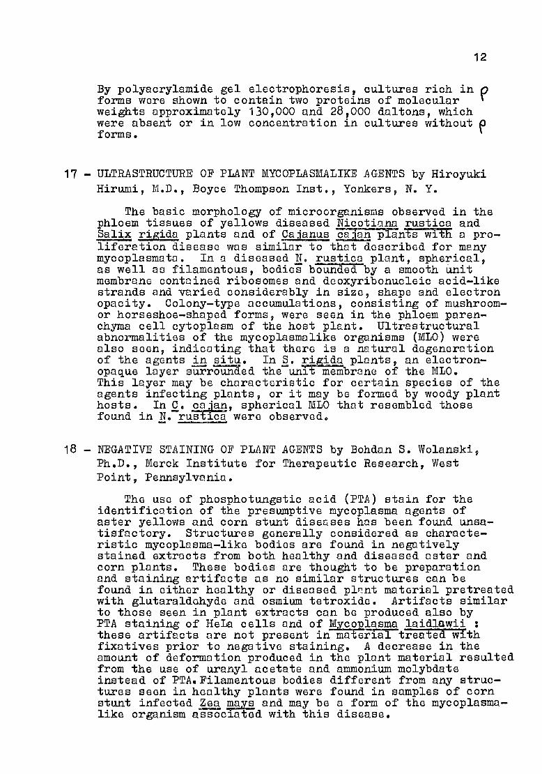
17 - ULTRASTRUCTURE OF PLANT MYCOPLASMALlKE AGENTS by HiroyukiHirumi, M.D., Boyce Thompson Inst., Yonkers, N. Y.
The basic morphology of microorganisms observed in thephloem tissues of yellows diseased Nicotianu rusticu andSalix rigida plants and of Cajanus cajun plants with a proliferation disease was similDr to that described for manymycoplasmata. In a diseased N. rustica plant, spherical,as weIl as filamentous, bodies bounded by a smooth Unitmembrane contained ribosomes and deoxyribonucleic acid-likestrands and varied considerably in size, shape and electronopacity. Colony-type accumulations, consisting of mushroomor horseshoe-shaped forms, were seen in the phloem parenchyma cell cytoplusm of the host plant. Ultrastructuralabnormalities of the mycoplasmalike orgunisms (MLO) werealso seen, indicating that there is a natural degenerationof the agents in~. In §. rigida plants, an electronopaque layer surrounded the unit membrane of the MLO.This layer may be characteristic for certain species of theagents infecting plants, or it may be formed by woody planthosts., In Q. caJan, spherical MLO that resembled thosefound ~n Ne rust~ca were observed.-
18 - NEGATIVE STAINING OF PLANT AGENTS by Bohdan S. Wolanski,Ph.D., Merck Institute for Therapeutic Research, WestPoint, Pennsylvania.
The use of phosphotungstic acid (PTA) stain for theidentification of the presumptive mycoplasma agents ofaster yellows and corn stunt diseases has been found unsatisfactory. Structures generally considered as characteristic mycoplasma-like bodies are found in negutivelystained extracts from both healthy and diseased aster andcorn plants. These bodies are thought to be preparationand staining artifacts us no similar structures can befound in either healthy or diseased pl~nt material pretreatedwith glutaraldehyde and osmium tetroxide. Artifacts similarto those seen in plant extracts can be produced also byPTA staining of HeLa cells and of MYC~laSma laidlawii :these artifacts are not present in ma~rial treated withfixatives prior to negative staining. A decrease in theamount of deformation produced in the plant material resultedfrom the use of uranyl acetate and ammonium molybdateinstead of PTA.Filamentous bodies difforent from any structures seen in healthy plants were found in sumples of cornstunt infected Zea ~ays and may be a form of the mycoplasmalike organism assoc~ated with this disease.

13
19 - SCANNING ELECTRON MICROSCOPY OF MYCOPLASrM AND MYCOPLASMALlKE AGENTS by Albert S. Klainer, MD, West VirginiaUniversity Medical Center, Morgnntown, West Virginia andJ. Dennis Pollack, Ph.D., College of Medicine, Ohio StateUniversity, Columbus, Ohio.
The morphology and the existence of n growth cycle ofMycoplasma Eneumoniae has been clearly ostablished byscanning electron microscopic studies which showed anorderly and sGquontial metamorphosis during its life cyclefrom sphericnl to filnmentous to large round forms duringthe period of growth from 8 hours to 10 days. Furtherstudies showed morphologic changes in Acholeplasma laidlawii~, !. grnnularum, A. axanthum, ~~ ggllisopticum, M. hyorhinis,and T-stroin grown in SSR-2 medium without oleic acid orShepard's U-9 medium for 1/2 to 10 days nt 37°C. Scanningelectron microscopid studies have provided unique information concerning growth-related morphologic changes ofintact fixcd org,anisms not provioualy obtainable with othertechniques. Scanning electron microscopic techniques,therefore, have become of great value in the investigationof the morphology of thesc organisms.
20 - MOTILITY OF MYCOPLASMAS by W. Bredt, Inst. f. Med. Microbiol., Johannes Gutenberg Univ., Mainz, Gcrmony.
Some Mycoplasmn species are able te glide nctivelyalong surfaces. ËXporiments were performed on M. pneumoniaestroin FH. The colIs w~re obsorved on glass surface inliquid medium with 3-5 %gelntine. Tho cells moved inirregular, mostly circular patterns. Their maximum speedwos 0.75 pm/scc. The cells appeared to stick to the glasswith a ce~tain urea of their front part. They always movedforwards with their front part, never switching to oppositedirection fig. 2. Treotment with cytochnlosin B up to100 ~g/ml failcd to inhibit the movement. PreliminaryexpoTiments on M. ggllisepticum and M. Rulmonis showed,that the cells of these species too were moving ahead witha tip-like structure. The mechonism of the movements8eems not to bo related to gliding mechunisms of animalce1ls, which are known to be inhibited by cytochalasin B.
max speed 0,75 pm/sec
irregula~mostly
circu1arDirection of movement
1! Longest distance measured 161 F . !
1------------- ------------11!
1

14
. La séance du Mardi après-midi (9 janvier 1973) étaitprésidée par Michael F. Barile, Ph.D. Mycoplasma Section, Division of Biologic standards N8tional Institutus of Health,Bethesda, Maryland.
Titre de la séance ~
MYCOPLASMA-CELL INTERACTIONS
21 - IDENTIFICATION OF MYCOPLASMA SPECIES ISOLATED FROM CONTAMINATED CELL CULTURES AND COMMERCIAL BOVINE SERA by MichaelF. Barile, PhaDe, RnA. DelGiudice, Hope S. Hopps, M.S.,M.W. Grobowski, B.S., and D.B. Riggs, Buroau of Biologics,FDA, and HEM Ressarch, Inc., Rockville, MD.
The Bureau of Biologics requires a test for the presenceof mycoplasmas in viral vaccines for hwnon use which areprepared in cell cultures. Accordingly, we have maintaindda continuing study for the past fourteen years to establishand to survey the incidence and sources of mycoplasma contaf:lination of coll cultures. Of) 6600 cell cultures examined, 1374 mycoplasmas were isolated and sero-identifiedby either the growth inhibition and/or the epi-immunofluorescence procedures. The cell culture contaminants includeseventeen distinct speciGs of mycoplnsmas. Of these, 99%are hw~nn, bovine and swine species, thus, the major sourcesof contamination originato from these three animaIs. Thesource of bovine mycoplnsma contamination of cell culturesis commercial bovine serum. Of 888 serum lots examined,285 mycoplasmus have beon isclated and 193 have been seroidentified. The serum contnminants include nt least 8distinct bovine spec~es of mycoplnsmas. The significanceof those findings will be discussed.
22 - PROBLEMS CONCERNING "NON-CULTIVABLEIl MYCOPLASMA CONTAMINANTSIN TISSUE CULTURES by Hope E. Hopps, M.S., Barbara C. Meyer,M.A., Michael F. Barile, Ph.Do and Richard A. DelGiudice,Bureau of Biologics, FDA, and HEM Resoarch, Inc., Rockville,Md.
The formation of typical colonies on agor is a major criterion for identification of mycoplasma organism~. Nevertheless, recent studie.s in this labora tory indicate thatsome mycoplasmas cannot be isolated by conventionnl techniques. A WI-38 cell culture being used for virus studieswas found to contnin a mycoplasma-like agent as determinedby observation of Giemsa-stained preparations. However, noorganisms were isolnted in standard mycoplasma broth or ag.areven after BaveraI seriaI passages. The agent grew luxuriantly in primory robbit kidney cell cultures producing almostcomplete destruct~on of tho culture in 7-10 days and attaining titers of 106 to 107 TCID50 per 0.1 ml.Identification of the organism as Myco~lasma ~YQrhinis wasachieved in this cell culture system usingfluorescent antibody and classic neutralization test procedures.

15
Growth of the organism on a~r wes eventually achieved onlyafter seriaI passage (4 times) in broth supplemented withheot-inactivated horse serum. The data suggest that incertain instances the use of cell cultures may be a usefuland important adjunct in the isolation of mycoplasmas.
23 - ORGAN CULTURE TECHNIQUES WITH MYCOPLASMAS. by Albert M.Collier, M.D. and Joel B. Baseman, Ph.D., University ofNorth Corolinn School of Medicine, Chapel Hill, N.C.
For study of mycoplasma-host cell relationship at thecellular level, tracheal orgon culture provides a means ofmaintaining living, orgonizod, diffcrentiated, respiratoryepithelium in an easily observable and manipulable environment. The pathogenesis of rcspiratory infections con beanalyzed in this syste~, including pathways of host cellparasitism and injury. Using this model, Mycoplasma Pîiumoniae has been demonstrated attachcd to epithelial cemembranes by a specialized terminal structure; the parasiteremoins extracellular, although the intercellular spacesmay be invaded. The dynamic aspects of the attachmentproccss con be studied with liquid scintillation spectrometryand radioautography using either organisms or host cellslabeled with 3H-th~~idino and -amino acids.Evidence of host coll damage accompanies orgpnism attochmentand replication, and includes ciliary dysfunction, cytopathology and exfoliation of epithelium.- Tracheal pathophysiologic changes observed were unique to M. Eneumoniae amonghuman mycoplasIDa species studied, but the techniques discussed rnay be applicable to other organ culture systems andpathogenic microorganisms.
24 - MYCOPLASMA PATHOGENICITY STUDIES ·IN ORGAN CULTURES. JamesD. Cherry, M.D. and David Taylor-Robinson, M.D., St LouisUniv., School of Med., St. Louis, Mo. and Clinical ResearchCentre, Harrow, Middlesex, England.
Chicken tracheal organ cultures and specifically thairfunctioning ciliated epithclium offer a quantitative livingsystem for the investigation of mycoplosma pathogenicityfactors •. Throe myc?~lasmas, ~. ~llisepticum, m. mycoides~. capr1 (!. capr1) and~. gpl11naru~m have been extensively studied. The rapidity of the cilia-stopping-effect(CSE) resulting from M. capri infection was directly relatedto the concentration of multiplying organisms in the culture.On the other hand, the CSE of!. ggllise~ticumwas notclosely rolatcd to dose. Medium contoin1ng tetracyclineinhibited M. ~lliseEticum. was found to be mi1dly cilio-toxic;a similar rin~ng was not notcd with M. capri. Cytadsorptiondid not appear to contribute to tne C~E of either!. caprior M. gallisepticUQ infections. M. capri was found tolibërate more poroxide thon other-mycoplasmes and this oontributed to the CSE, as the adverse affect could be partiollyreversed by tho addition of catalase to the system. Peroxidedid not contribute to the CSE of M. ~llisepticum infection.!. gallinarum (a non-pothogenic myco~asma) infection ofthe organ cultures had an inhibiting effect on the CSE of!. gallisepticum.

16
25 - MIXED MYCOPLASMA-VIRUS INFECTIONS IN CELL CULTURES byStanley H. Singer, M.D., M. F. Barile, Ph.D., und R.L.Kirschstein, M.D., Bureau of Biologics, Food and DrugAdministration, Rockville, Maryland.
Deliberate infection or covert contamination of cellcultures with mycoplasma can affect subsequent virus replication in these cultures. This effect moy rssult in eitharon increusa or decreasa in virus yield. The mechonisms ofdecreased virus yield may be due to either a partial destruction of the call cntrix by the mycoplasma or to amore complax mechanism such as the utilization by tha mycoplasma of n chemicnl substrate, such as nrginina, which isa necessary componant for replication of certain DNA viruses.One mechanism involved in increased virus yield appanrs tobe related to the ability of viruses to produca intorfaron.The presance of mycoplasmas in cell cultures decraasesinterferon production. Sinca interferon itself acts todecrease virus yield, the decrenso in interferon productioncaused by the cycoplnsmo will leud to a higher virus yield.The cellular substrats is also of importance in determiningthe effect on virus yield. For axample, using the sornevirus and nycoplasma, increased virus yiald was obtainadin a primary mouse cell culture system, whereas decreasedyield was obtained in a continuous Douse cell line.
26 - MYCOPLASMA AND ACHOLEPLASMA IN PLANTS by Arthur H.Mclntosh, Sc.D., and Karl Maramorosch, Ph.D., BoyceThompson Instituto, Yonkers, N. Y.
Experiments wero conductod to detcrmine whether or notMycoplasme and AcholaplasDu species could be racovared fromplants which hud boen exposed to these microorganisms~
Nicotiuna rustica (tobacco) and Callisto7hUS chinansis(aster) were exposed to approximataly 10 colony formingunits par ml of Mycoplasma gallisapticum and Acholaplasmalaidlawii for 45-120 min. Methods of exposure includodroots (intentionally and unintentionally damaged), cutlouves, mechanicnl abrasion of leaves and injection. ControIs consisted of plants oxposed to growth medium only.Samples of both petioles and leaves were-taken 7-8 hr following exposure to the microorganisms. A. laidlawii wasrecovered in 6 out of 15 experiments (40-%). Of these 20 %were from root exposure experimants. Tho ramoining 20-%were aqually distributed among the other c8tegorias. M.gallisepticum wns recovered in only 1 out of 11 axperiments(9 %, root exposure). No Mycoplasmatalas were recovaredfrom control plants. Isolatas wera identified by growthinhibition and icmunofluorescent tests. These observationsindicate that healthy appearing plants can incorporate animo~ mycoplasrnns and that such microorganisms are recoveroblaby cultural tachniques.
17
La séance du Bercredi matin (10 janvier 1973) étaitprésidée par Leonard Hayflick, PhoDo Stunford University, Schoolof Medicine, Stanford, California.
Titre de la séance:
ISOLATION l PATHOGENICITY AND CHEMOTHERAPY l
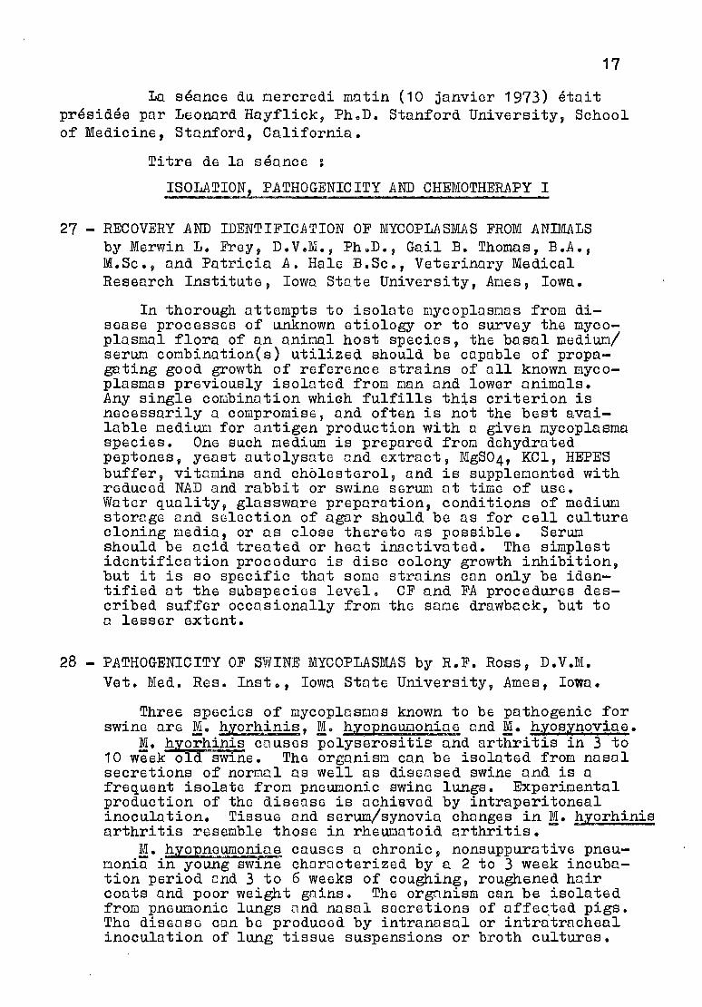
27 - RECOVERY AND IDENTIFICATION OF MYCOPLASMAS FROM ANIMALSby Merwin L. Frey, D.V.M., PhQD., Gail B. Thomas, B.A.,M.Sc., and Patricia A. Hale B.Sc., Veterinary MedicalResearch Instituts, Iowa State University, Ames, Iowa.
In thorough attompts to isolate mycoplasnas from disease processes of unknown etiology or to survey the mycoplasmal flora of an animal host specics, the basal medium/serum combination(s) utilized should be capable of propagating good growth of reference strains of aIl known mycoplasmas previously isoluted from non and lower animaIs.Any single cOQbination whiGh fulfills th~s criterion isnecessarily a compromise, and often is not the best available medium for antigen production with Q given mycoplasmaspecies. One such mediUô is prepared from dehydratedpeptones, yeast autolysute und extract, MgS04, KCl, HEPESbuffer, vitamins and cholesterol, and is supplemented withreduced NAD and rabbit or swine serum ut time of uso.Water quality, glassware preparation, conditions of mediULlstorage and selection of agar should be as for cell culturecloning media, or as close thereto as possible. SerUQshould be acid treated or heat inactivatod. The simplestidentification procedure is disc colony growth inhibition,but it is so specifie thut sorne struins can only be identified at the subspecios level. CF and FA procedures described suffer occusionally from the sune drawback, but toa lesser extent.
28 - PATHOGENICITY OF SWlNE MYCOPLASMAS by R.F. Ross, D.V.M.Veto Med. Res. Insto, Iowa State University, Ames, Iowa.
Three species of mycoplasmus known to be pathogenic forswine ure Mo hyorhinis, M. hyopneuwoniae und !. hyosynoviae.
M. hyorhinis couses polyserositis and arthritis in 3 to10 week old swine. The organism can be isolated from nasalsecretions of normal as weIl as diseQsed swine and is afrequent isolate from pneumonic swine lungs. Experimentalproduction of the diseuse is achieved by intraperitonealinoculation. Tissue and serum/synovia changes in!. hlorhinisarthritis resemble those in rheumutoid arthritis.
M. hyopneumoniae causes a chronic, nonsuppurative pneumonia in young swine choracterized by a 2 to 3 week incubation period end 3 to 6 weeks of coughing, roughened haircoats and poor weight gains. The orgnnism can be isolatedfrom pneumonic lungs and nasal secretions of affe~ted pigs.The diseuse con be produced by intranasal or intratrachealinoculation of lung tissue suspensions or broth cultures.

18
~. hyosynoviae couses on acute nonsuppurative polyarthritis in young swine over 10 weeks of age. During
the acute stage, the organisn can be isolated from arthriticand some nonarthritic joints, lymph nodGs, blood andvarious mucosal secretions; in convalescence, it persistsin the nDsopharynx and tonsils.' The disease can be produced
.in certain types of swine by intravcnous or intranasal routes.
29 THE PATHOGENICITY OF BOVINE MYCOPLASFMS by Julius FabricantV.M.D., Ph.D., Dept. of Avion Diseoses, N.Yo State VeterinaryCollege, Cornell Univ., Ithaca, N. Y•.
Mycoplnsnn mycoides var rnycoidcs in cattle is known toproduce bovine contogious pleuropneUL10nia and is also capable of causing arthritic lesions in cattle. This reviewis primarily conccrned with the occurence of other mycoplusma species in cottle and an evaluation of the evidence relating to their pothogenicity. V2rious species or serotypesof mycoplosma hove been isolated from the bovine respiratorytract (nose, trochea, lungs), the Dole reproductive tract(prepuce, sernon, seminal vesicles), the feDale reproductivetract (vugina, uterus, oviduct, abortGd fetuses), themammary gland and from joints. Existent dota clearlydemonstrntes thot nycoplasmo con couse clinical mastitis,orthritis and saninal vosiculitis in cattle. Mycoplasmacan cause less severe pneumonic and endometrial lesions.Their relationship to other bovine lesions is still notprove.q..
30 - IMF1UNOELECTROPHORETIC COMPARISON OF MYCOPLASMA lnrCOIDESISOLATED FRor.~ CATTLE AND GOATS WITH FOUR M:YCOPLASMAISOLATED FROM GOATS IN THE UNITED STATESby S.S. Stone and R.J. Yedloutschnig, Plum Island AnimalDiseuse Laboratory, Agricultural Reseurch Service, USDept. of Agriculture, Greenport, New York, and NationalAnimal Disease Laboratory, P.O. Box 70, Ames, Iowa.
Four virulent mycoplasma recently isolated from goatsin the United States of America (USA) were comparedpri~Qrily by imnunoelectrophoresis to Mycoplusma mycoidesvar capri (Von, Nigeria) and to bovine ~~ycoplasma mycoidesvar mycoides.
Using bovine, pig, and rabbit anti-M. mycoides varmycoides sero and rabbit anti-!. mycoides capri serumsevoral similor precipitin bands between the USA isolatosand the M. mxcoidGS var mycoidcs were found. Cross reactions were also observod between rabbit onti-!. mycoidesvar capri serum anâ the USA goat mycoplusma isolates.Compfement fixation tests using the same antisera showeda similar relationshipo It is concluded that these USAgout mycoplasma isolotes have un antigenic relationshipto !. mycoplosmu vor capri and to !. mycoplasma varmycoides.
19
31 - MURlNE MYCOPLASB~ RESPlRATORY DISEASES by Gail H.Cassell, M.S., J. Russell Lindsey, D.V.N., M.S., andRonald G. Overcash, D.V.M., Deptso of Comparative Medicineand Microbiology, Univo of Alabama in Birmingham,Birmingham, Ala.
Important advances have been made recently in understanding the mycoplasmal respiratory diseases of laboratory ratsand mice. AlI principal lesions of the natural "chronicrespiratory disease" in these species have been reproducedby inoculating pure cultures of Mlco~lasma pulmonis intranasally into animaIs known to be 'free of other pathogens.The experimental disease in mice has proved to be the moreconsistently reproducible model system, permitting quantitation of host response to a wide range of doses. Specifieantibodies of the G1, G2, M and A classes were detected inserum and bronchial secretions. These antibodies wereshown to be produced locally in the lung and regional nodes.Active immunization with live organisms and passive immunization with immune mouse serum have been shown to protectagainst pneumonia. The experimental disease in the rat isless weIl characterized because of poor reproducibility,particularly of lesions in the lower respiratory tract.Based on preliminary comparative studies using immunofluorescence, a possible explanation appears to be a more efficient clearance mechanism in rat lung.
32 - PATHOGENESIS STUDIES IN EXPERIThlENTAL MYCOPLASn~ DISEASEby Wallace AD Clyde, Jr., M.D. and Lewis Thomas, M.D.,Dept. Pathology, Yale Univ. Sch. Med., New Haven, Conn.
Neurologie and arthritic disease syndromes producedby Mycoplasma !lllise~ticum strain S6 in turkeys wereanalyzed by co~ation of clinical, microbiologie, pathologie,and immunofluorescence data. After intravenous inoculationbirds developed fatal encephalopathy after a dose-dependentincubation periode In disease evident at 3-7 d, but notthat at 1-2 hr, organisms were found on the endothelium orwithin the walls of cerebral arteries. Concurrently: 1)subclinical renal disease was manifest by glomerular /in jury including serum protein leakage and deposition ofimmune complexes (these changes reversed in surviving birds);and 2) organisms were found in vessels of clinicallynormal joints. After 30 d polyarthritis developed andprogressed, but the tissues became sterile after 47 d. Thearteriotropism of ~. gallisepticum represents an uniquehost-parasite relationship. Temporal-pathologie correlationssuggest that disease mediation is via products from infectedarterial foci acting upon cerebral capillaries, glomeruliand synovium where organisms may be absent. These findingshave implications for the study of human collagen-vasculardiseases whose etiology is unknown.

20
33 - RESEARCH AND DEVELOPMENT OF MYCOPLAShML VACCINES byNorman L. Somerson, Ph.D., Ohio State University Collegeof Medicine, Laurence B. Senterfit, Sc.D., Cornell University Medical College, and Vincent V. Hamparian, Ph.D.,Ohio State University College of Medicine.
A potent, formalin inactivated vaccine was preparedwith Mycoplasm~ pneumoniae for clinical trials in mah.In animaIs, this latest vaccine (designated OSU 1A) wasconsiderably more antigenic than the original prototypevaccine. Extinction potency tests in hamsters based onserologie conversion showed vaccine OSU 1A to be at leasta hundred times more potent. Improvements in the manufacturing process of vaccine OSU 1A include a better seedinoculum, an improved medium, and detachment of organismsby trypsinization and glass beads. No adverse reactionswere observed when the vaccine was administered tothirteen volunteers.
34 - EFFICACY OF INACTIVATED MYCOPLASMA PNEID10NIAE VACCINEDEMONSTRATED BY PROTECTION IN LARGE FIELD TRIALS byWilliam J. Mogabgab, M.D., Tulane University Schoolof Medicine, New Orleans, La.
lnactivated Mycoplasma pneumoniae vaccine stimulatedantibody responses that were of sufficient magnitude forprotection in most individuals and that were comparableto those produced by natural infection in amount andduration. Antibody persisted for twenty months or longer.There were no adverse local or systemic reactions ofconsequence. Protective efficacy of the vaccine wasdetermined in a controlled study in 13,892 airmen during1969-70 at Keesler Air Force Base, Mississippi wheremycoplasma pneumonia has been present for several yearswith epidemic occurrences. The protective efficacy ofthe vaccine against bronchitis caused by Myco~lasma pneumoni~ was 87 percent and against pneumonia 6 percent.Persons who developed Mycoplasma pneumoniae infectionsin spite of vaccination did not experience more severeillnesses.

21
La séance du mercredi après-midi (10 janvier 1973)était présidée par Floyd W. Denny, M.D., the University ofNorth Carolina School of Medic~ne, Chapel Hill, North Carolineen remplacement de E.A. Freund (déjà cité).
Titre de la séance:
ISOLATION, PATHOGENICITY AND CH~~OTHERAPY II
35 - PATHOGENICITY OF MYCOPLAS1MLIKE AGENTS IN PLANTS byKarl Maramorosch, Ph.D., Boyce Thompson Inst., Yonkers,N. Y.
ffiycoplasmalike organisms (MLO) induce sterility,stunting, axillary prolifer8tion, breaking of dormancyor death in over 60 different plant diseases. Coconutpalms die rapidly when affected by lethal yellowing.Aster yellows diseased plants live longer than healthyplants and may become attractive and palatable to certain insects that cannot survive on normal individuals.The mechanism of MLO action in plants is unknown butsuggestive of hormonal imbalance. Mechanical inoculation of crude plant or insect vector extracts intovectors, but not into plants, provides a sensitive MLObioassay. Systemic MLO infection has been studied byelectron microscopy of phloem sections during variousstages of disease. Mechanical clogging of vessels,as well as passage through sieve plate pores, and laterdegeneration of MLO have been observed. MLO-associatedviruses have been dotected but there is no indicationthat such viruses can overcome chronic MLO diseases.Heat therapy has provided permanent cure of certainMLO diseases, whereas tetracyclines caused temporaryremission only. Mechanical inoculation of plants withMLO, reportedly isolated and cultured on artificialmedia, has not been confirmed.
K. Maramorosch presented photographs of yellowingof several species of plants : Aster yellow, cornstunt, rice yellow dwarf, yellowing of coconut plants(occuring in Caribbonn,Jamaica, Africa), Papaya bunchyTop (cured by surgery) and Opuntia. In the case ofcorn stunt, MLO elongated particules were observed,in the vector and in the plant but rounded particlesressembling those of Acholeplasma and Mycoplasma wereobserved only in the insecte

22
36 CHARACTERIZATION OF THE MYCOPLASMA LIKE ORGANISM ASSOCIATEDWITH STUBBORN DISEASE OF CITRUS. J.M. Bové, Ph.D., Centrede Recherches de Bordeaux, INRA, Bordeaux, France.
By optical dark-field examination of log-phase brothcultures, the organism is a motile, helical filament 2-4 pmlong fig. 3. Helical shape is preserved by molybdate,but not by phosphotungstate, for negatively-stained EMexam ; and by glutaraldehyde in medium, but not in cacodylate, for fixction followed by sectioning, metal shadowing, freeze-etch, or scanning EM. Filaments are100-120 nm wide, and amplitude of helices varies from200-300 nm. In old brcth cultures or in colonial growthfrom agar, helical shape is lost. An outer layer or napon the single limiting membrane is present, to whichattaches a tailed type B bacteriophage which can also beseen developing intracellularly. Unexplained eomponentsinclude random striated structures soen by negativ8 staining and an interrupted or inconstant submembranous layerseen in sections. No cell wall nor organelles arepresent. Both strains (Morocco and California) are identical and resemble the uncultured corn stunt agent, butshow new features. Shape, motility, and presence ofclassic phage obscure taxonomie position as a mycoplasmeas suggested by other studies, but appear to warrantestablishment of a new genus to be placed later in highertaxa.
37 - FAILURE TO ISOLATE MYCOPLASb~S FROM ASTER YELLOWSDISEASED PLANTS AND LEAFHOPPERS by Leonard Hayflick,Ph.D., Stanford Univ. Sch. of Med., Sta~ord, Ca. andSumio Arai, M.D., Tohuku Univ. Sch. of Med., Sendai,Japan.
Seventeen different ager media were employed inattempts to isolate mycoplasmes from aster yellows disease and from leafhoppers that had fed on these diseasedplants. The media contained a variety of supplementsincluding coconut, soil and malt extracts ; lobsterhemolymph, glycihe, and extracts of uninfectod plants.No mycoplasmas were isolated from homogenized infectedplants or leafhoppers when placed directly on agar, influid culture, or in diphasic medium after 14 deys ofincubation and after several blind passages. Negativeresults were also obtained when the eut stems of infectedplants were imbedded directly into agar media. We conclude from these end other data that typicol mycoplasmaspecies are probably not etiologically involved in asteryellows.disease. The forms associated with this diseasehave properties not only suggestive of mycoplasmas butalso similar to the L-Phase of Bacteria, the Riekettsiaceae, the Chl~mydiaceae, some protozoa and, perhaps,might be regprded as a new group of phytopathogens.Consequently these entities should be ealled yellowsdisease agent (YDAA) until Koch's postulates arefulfilled.

23
38 - COMBlNED MYCOPLAm.1A AND VIRUS INFECTIONS IN PLANTS ANDINSECTS by Ernest E. Banttari, Ph.D., and Richard J.Zeyen, Ph.D., Department of Plant Pathology, Universityof Minnesota, St. Paul, MN. 55101.
There are nt lGast three reported cases of dual infections of plants with viruses and mycoplasmnlike organisms (MLO). One report deals with Likuben diseuse ofCitrus ponki and C. tankan infected with tristeza virusand an unidentified MLO. The second is the report ofMLO and rhabdovirus in tissues of naturally infectedCajanus Cajqa. The other report is that of Linum usitatissimum infected with oat blue dwarf viYus (OBDV) andaster yellows (AY). In Linum, AY und OBDV are phloemlimited and cause hyperplasia and obliteration of phloemêlements and hypertrophy of adjacent tissues. Extensiveleakage of a dark staining substance into fibers, cortex,and pith indicated a severe disruption of phloem fQnction. Although both disease agents werG fOQnd in thesame phloem cells, there appeared to be no qualitativeor quantitative effect of one upon the other. OBDV multiplies in immature phloem cells having a full complementof cellular constituents whereas AY apparently multipliesin more mature enucleate elementso Both disease agentsare propaêQtive in the vector, Macrost~los fascifrons.The leafhopper can acquire both disense agents fromdoubly infected plants and transmit them simultanoouslyto plant hosts.
39 - EFFECTS OF TETRACYCLINE C01TI?OUNDS ON PLANT DISEASESCAUSED BY MYCOPLASMA-LIKE AGENTS by T.T. Iida, Institutefor Plant Virus Research, Chiba, Japan.
Effects of tetracyclines to several "yellows" typediseases suspected to be caused by mycoplasma-like agentswere tested in a cooperative work by a group from 13 institutions in Japon. Mulberry dwarf, rice yellow dwarf,(Japanese) potato witches' broom (potato and tomato),(Japanese) aster yellows (tomato, carrot, potato, andcosmos), and Cryptotaenia witches' broom were tested.Tetracycline, chlortetracycline, dimethylchlortetracycline,and oxytetracycline were used in most tests. A few antibiotics other thon tetracyclines were also included insome tests. Plants were treated by dipping the roots orby spraying the leaves, usually with 10-100 ppm solutions.The treatments were made for various durntions at varioustimes befora inoculation, after inoculation, or aftersymptom appearance. The tetracyclines had distinct butonly temporary effect in causing remis sion, depending ondose, but not cure of the diseases. Other antibioticswere ineffective •• In mulberry dworf and rice yellow dwarf,infective vector leafhoppers were either administered peros or injected with tetracyclines, and tested for theirinfectivity on test plants. It resulted in prolongingthe incubation period within insect, or sometimes completesuppression of infectivity.

24
40 - ASTER YELLOWS : EFFEOT OF SULFA DRUGS by R.J. Frederick,
MS, Warner Lambort Oompany, Morris Plains, New Jersey.
Four suIfa drugs were applied individually or incombination with tetracycline-HOI to healthy and asteryellows-affected Ohina asters. Recovery from severestunting was obtained with sulfadiazine, sulfisoxazole,and sulfisomidine. Disease expression was suppressedmore and for longer periods by the combined applicationof sulfadiazine and tetracycline-HOI than by eithercompound alone. The mean tetracycline-HOI concentrations in diseased plants treated by a cut-leaf immersiontechnique decreased when the antibiotic was administeredin combinntion with sulfisomidine and sulfanilamide.Transmission by vectors of the aster yellows agentincreased or remained unchanged when the suIfa drugswere applied to infected plants. Disease transmissionefficiency of infective insects fed intermediately onhealthy plants treated with sulfadiazine and/or tetracycline-HOI was significantly lower than the transmissionefficiency of insects fed on healthy plants treatedwith sulfadiazine alone. AlI of the treated healthyplants used as intermediates for these vectors becameinfected, although expression of the disease wasdelayed.
K. Maramorosch a fait une conclusion générale en remerciant les mycoplasmatologistes "survivants" qui ont assistéà la dernière séance ; et la clôture du congrès a été faitepar le Président de llAcadémie des Sciences de New York:Kenneth W. Thompson, M.D.

.figure 1.
:>
.. figure 2 •
>
• 'figure 3.