Imagerie de Diffusion Organes Mobiles et Corps Entier
45
Imagerie de Diffusion Organes Mobiles et Corps Entier Alain Luciani, Frederic Pigneur, Emmanuel Itti, Alain Rahmouni CHU Henri Mondor, Universite Paris Est Creteil [email protected]
description
Imagerie de Diffusion Organes Mobiles et Corps Entier. Alain Luciani , Frederic Pigneur , Emmanuel Itti , Alain Rahmouni CHU Henri Mondor, Universite Paris Est Creteil [email protected]. Imagerie Diffusion Corps entier – WB-DWI. Couverture anatomique . Imagerie Fonctionnelle. - PowerPoint PPT Presentation
Transcript of Imagerie de Diffusion Organes Mobiles et Corps Entier
Imagerie de Diffusion Organes Mobiles et Corps Entier
Alain Luciani, Frederic Pigneur, Emmanuel Itti, Alain Rahmouni
CHU Henri Mondor, Universite Paris Est [email protected]

« Tout en un »
Imagerie Fonctionnelle
Couverture anatomique
Imagerie Diffusion Corps entier – WB-DWI


Comment je fais ?
Applications Cliniques
Antennes
DWI-MRI• Pré-requis technologiques et instrumentaux
Séquences

« Anatomique »
1. Plan1. Sagittal2. Coronal3. Axial
2. Séquences1. T1 WI2. T2 avec suppression
graisse
« Fonctionnelle »
1. Imagerie Diffusion Corps entier (WB-DWI-MRI)• Gradient directions ?• What about breathing ?
2. Imagerie dynamique après injection de pdc (WB- DCE-MRI)• Timing• Plans de coupes
Suppression GraisseGestion artefacts de
mouvements
Pré-requis ?Impact clinique ?
Imagerie paramétrique ?
Comment je fais ?IRM Corps entier

• Surface• Body Coil
Acquisitions parallèlesRésolution spatiale
Comment je fais ?Antennes
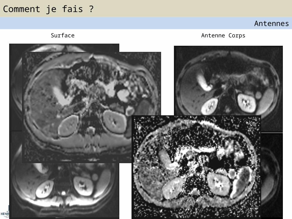
Surface Antenne Corps
Comment je fais ?Antennes

• Suppression graisse ?
FS STIR
Homogénéité Temps d’acquisition
Comment je fais ?Séquences

• Respiration – Libre + multiple Nex– Gating : Ceinture (pressure sensor)• Cycle respiratoire• Fin d’expiration => Acquisition
– Trigger :
Comment je fais ?Séquences
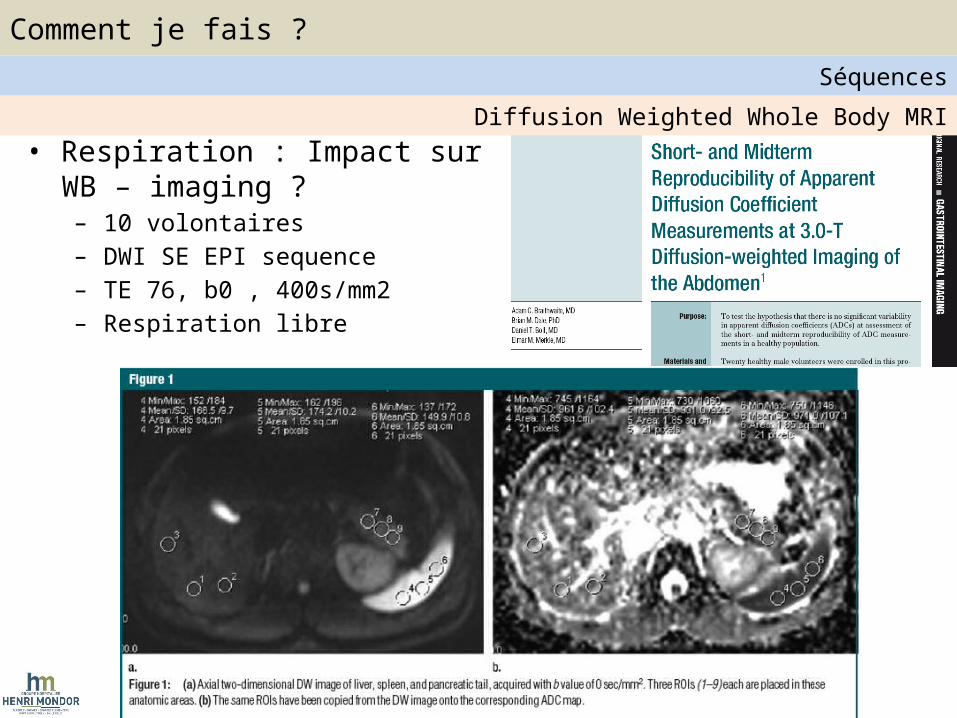
• Respiration : Impact sur WB – imaging ?– 10 volontaires – DWI SE EPI sequence– TE 76, b0 , 400s/mm2– Respiration libre
Diffusion Weighted Whole Body MRI
Comment je fais ?Séquences

• Variation intra-individuelle ADC ± 14%• Seuil de signification > 27%
Diffusion Weighted Whole Body MRI
Comment je fais ?Séquences

• Respiration : Impact sur WB-DWI ?
Diffusion Weighted Whole Body MRI
Comment je fais ?Séquences

Mêm
es p
aram
ètre
s ; N
ex=2
• Nombre de gradients de diffusion ?Diffusion Weighted Whole Body MRI
Comment je fais ?Séquences

• Choix des valeurs de b ?Diffusion Weighted Whole Body MRI
Comment je fais ?Séquences
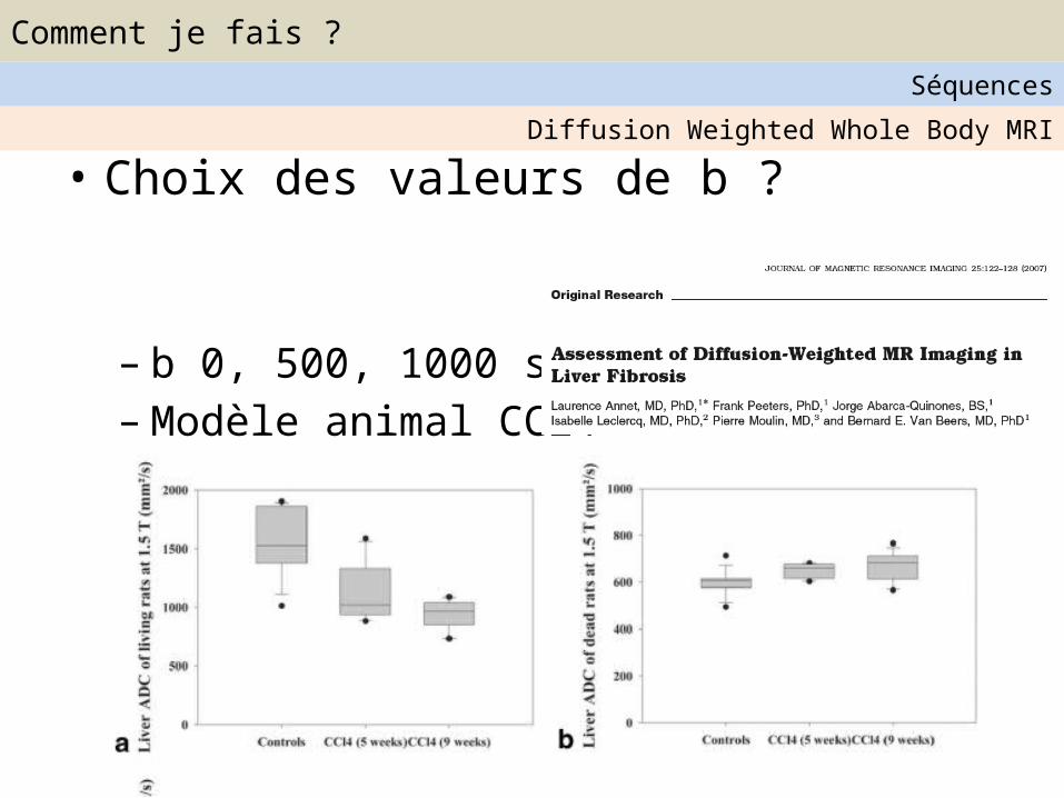
• Choix des valeurs de b ?
– b 0, 500, 1000 s/mm2
– Modèle animal CCl4
Diffusion Weighted Whole Body MRI
Comment je fais ?Séquences

• Principes de la diffusion
Composante perfusive
Diffusion moléculaire
Diffusion Weighted Whole Body MRI
Comment je fais ?Séquences
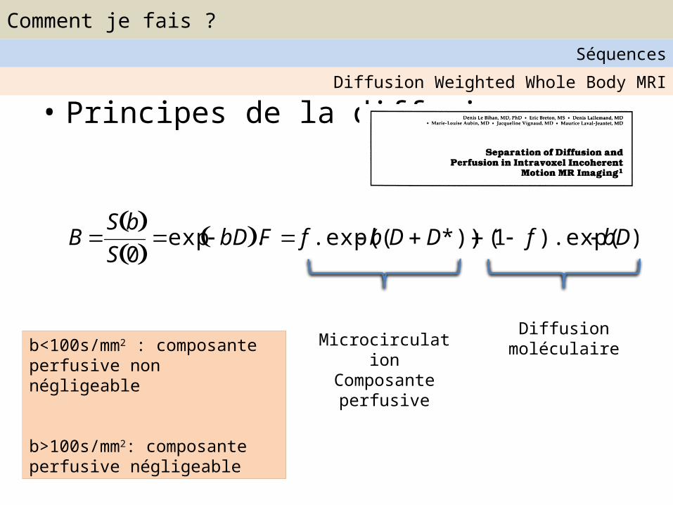
• Principes de la diffusion
BS b S 0
exp bD .F f .exp( b(DD*)) (1 f ).exp( bD)
Diffusion moléculaireMicrocirculation
Composante perfusive
b<100s/mm2 : composante perfusive non négligeable
b>100s/mm2: composante perfusive négligeable
Diffusion Weighted Whole Body MRI
Comment je fais ?Séquences

• Application cliniqueDiffusion Weighted Whole Body MRI
Comment je fais ?Séquences
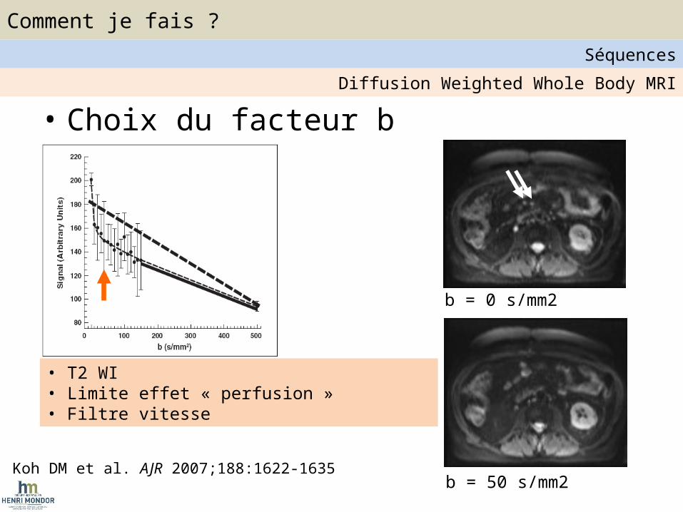
• Choix du facteur b
Koh DM et al. AJR 2007;188:1622-1635b = 50 s/mm2
b = 0 s/mm2
• T2 WI• Limite effet « perfusion »• Filtre vitesse
Diffusion Weighted Whole Body MRI
Comment je fais ?Séquences

• 1.5 T ou3 T ?– Temps d’acquisition
– Artefacts
Diffusion Weighted Whole Body MRI
Comment je fais ?Séquences

1. Antennes2. Imagerie parallèle3. Gating respiratoire4. Suppression graisse5. Injection ?6. DWI imaging :
1. TR/TE2. Direction de gradients3. Epaisseur de coupe / Gap4. Choix des valeurs de b
Messages Clés
Comment je fais ?Séquences

Temps d’acquisition : 20 – 45’1. Whole Body Scout View2. Sagittal T1 SE3. Coronal T2 TSE FS / STIR (2 à 3 Steps) (HASTE / Transverse TSE - > Lung)
4. DWI : Transverse DWI : 50,400,800s/mm2; TE=76ms
5. Transverse 2D EG T1 FS / Dixon / Water excitation 5mm
Example de protocole WB-DWI

Données de la littérature : Abondante mais hétérogène ! Instrumentation et type de
tumeurs • Staging tumeur primitive : débattu -> Instrumentation
– Antoch et al. JAMA 2003; 98 patients ; various Primary; Accuracy PET-CT (80%) > WB-MRI (52%)– Yi et al. Radiology 2008; 165 patients; NSCLC; Accuracy T staging WB MRI (86%) > PET-CT (82%)
• Screening et détection tumorale– Fischer et al. Eur Radiol 2011; 21:246-255– 68 patients staging of malignant Tumor by PET-CT (Ref Standard)– Concomitant WB-MRI (T2 + DWI b =0 / b= 700s/mm2) – Accuracy overall 82% reaching 91% for liver mets
• Métastases osseuses– Mélanome : Laurent et al. Eur J Radiol 2009; Epub
• Se IRM (DWI) = 82 % vs. 72% PET-CT• Sp IRM (DWI) = 97% vs. 92% PET-CT
– Cancer Prostate : R Venkitaraman et al. J Med Imaging Rad Oncology 2009;53:530-533• 39 patients• MRI > 99mTc
– Cancer poumon (NSCLC): Takenaka et al. J. Magn. Reson. Imaging 2009;30:298-30• 209 patients• MRI (DWI) > 99mTc and PET-CT
…………….• Hematologic Malignancies
Applications cliniques

• Takahara et al Radiat Med. 2004
Applications cliniques
WB-DWI-MRI et Lymphome

Applications cliniques
WB-DWI-MRI et Lymphome
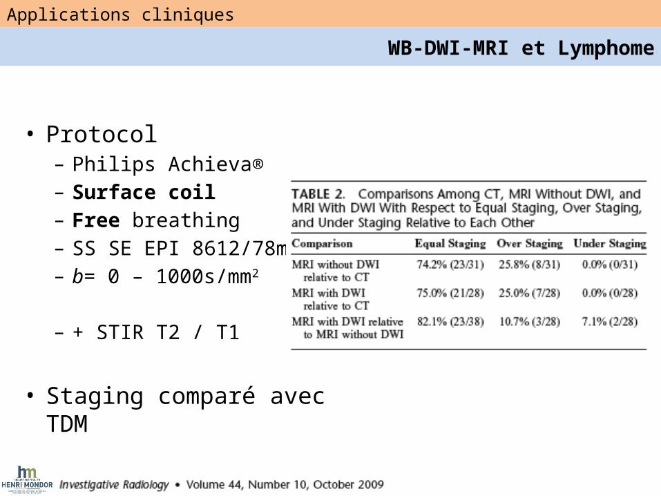
• Protocol– Philips Achieva®– Surface coil– Free breathing– SS SE EPI 8612/78ms– b= 0 – 1000s/mm2
– + STIR T2 / T1
• Staging comparé avec TDM
Applications cliniques
WB-DWI-MRI et Lymphome

• Meilleure Se / TDM
– Moelle osseuse– Atteinte extra digestive
Applications cliniques
WB-DWI-MRI et Lymphome
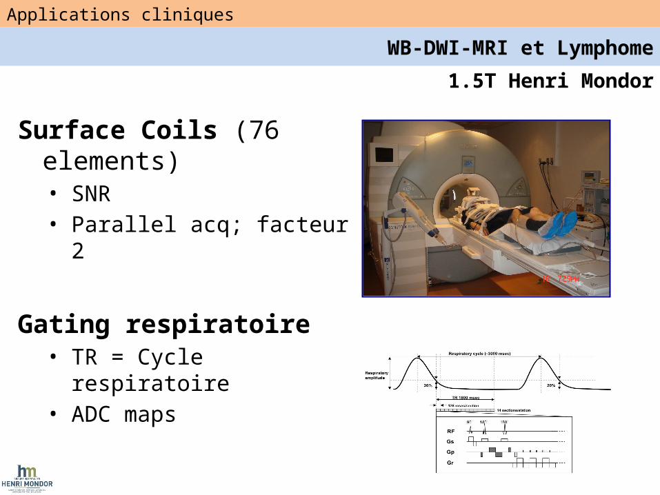
1.5T Henri Mondor
Surface Coils (76 elements)• SNR• Parallel acq; facteur 2
Gating respiratoire• TR = Cycle respiratoire• ADC maps
Applications cliniques
WB-DWI-MRI et Lymphome
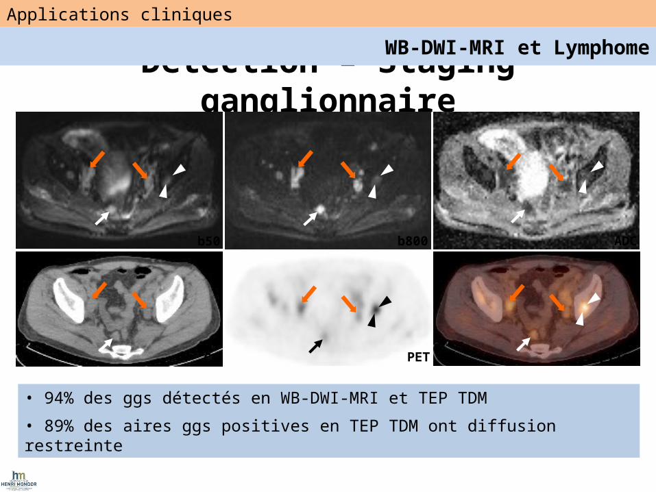
Detection – Staging ganglionnaire
CT
ADCb800b50
PET PET/CT
• 94% des ggs détectés en WB-DWI-MRI et TEP TDM
• 89% des aires ggs positives en TEP TDM ont diffusion restreinte
Applications cliniques
WB-DWI-MRI et Lymphome

• Résolution spatiale en WB-DWI =gg ou masse ?
b50 b800 ADC
Detection – Staging Ganglionnaire
Applications cliniques
WB-DWI-MRI et Lymphome

Atteinte profondeb50 b800
T2WI
Applications cliniques
WB-DWI-MRI et Lymphome
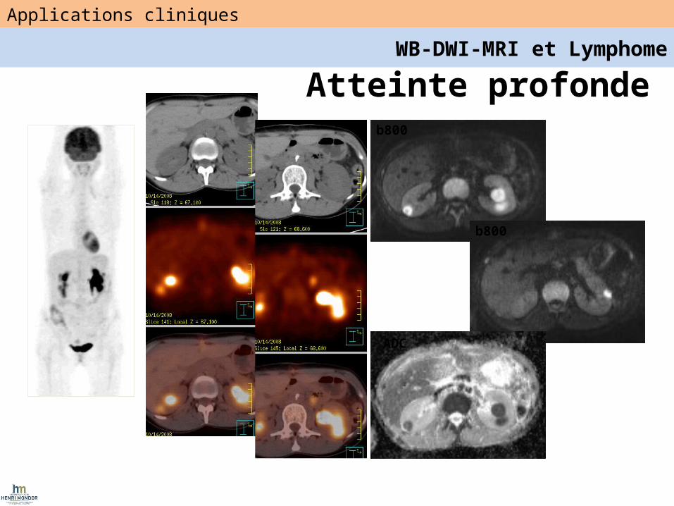
b800
b800
ADC
Atteinte profonde
Applications cliniques
WB-DWI-MRI et Lymphome

b50
b50
b800 ADC
b800 ADC
• 23 y/o patient DLBCL Mediastin
• 4 cycles de CT : Masse résiduelle 8x1cm (CRu) mais TEP TDM négatif => CR
• Pas de diffusion restreinte
Applications cliniques
WB-DWI-MRI et Lymphome
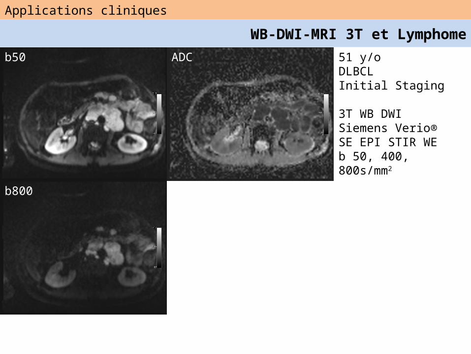
51 y/oDLBCLInitial Staging
3T WB DWISiemens Verio®SE EPI STIR WEb 50, 400, 800s/mm2
b50
b800
ADC
Applications cliniques
WB-DWI-MRI 3T et Lymphome

b50
b800
54 y/oDLBCLInitial Staging
3T WB DWISiemens Verio®SE EPIb 50, 400, 800s/mm2
Applications cliniques
WB-DWI-MRI 3T et Lymphome

54 y/oDLBCL6 cycles CT
b50
b800
Applications cliniques
WB-DWI-MRI 3T et Lymphome

Applications corps entier
54 y/oDLBCLInitial StagingStage IV (Bone)
b50 ADC mapT2 WI
Combined WB-DCE MRI and WB-DWI-MRI in Lymphoma
3D VIBE

54 y/oDLBCLInitial StagingStage IV (Bone)
b800
b50 ADC map
Applications cliniques
Approche foncitonnelle combinée
b800

54 y/oDLBCLInitial StagingStage IV (Bone)
b50
b800
ADC
Applications cliniques
WB-DWI-MRI 3T et Lymphome

Clinical Applications
Combined WB-DCE MRI and WB-DWI-MRI in Lymphoma54 y/oDLBCLAfter 6 cycles CT
b800
b50
ADC

Messages
• Instrumentation
• Instrumentation
• Instrumentation


1
2
3

Applications cliniques
• Pas encore définies ….• Standardisation ?• Sélectionner études cliniques adaptées….