Imagerie de Diffusion Corps Entier - sfrnet.org · Imagerie de Diffusion Corps Entier Alain...
42
Imagerie de Diffusion Corps Entier Alain Luciani, Frederic Pigneur, Emmanuel Itti, Alain Rahmouni CHU Henri Mondor, Université Paris Est Creteil [email protected]
Transcript of Imagerie de Diffusion Corps Entier - sfrnet.org · Imagerie de Diffusion Corps Entier Alain...
Imagerie de Diffusion Corps Entier
Alain Luciani, Frederic Pigneur, Emmanuel Itti, Alain Rahmouni
CHU Henri Mondor, Université Paris Est Creteil
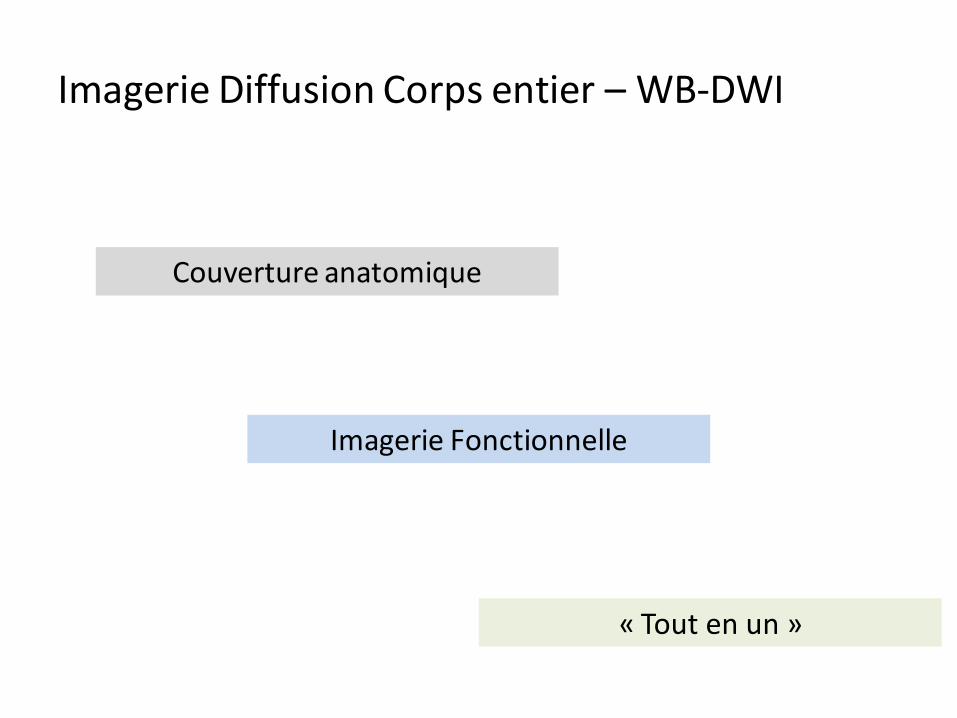
« Tout en un »
Imagerie Fonctionnelle
Couverture anatomique
Imagerie Diffusion Corps entier – WB-DWI

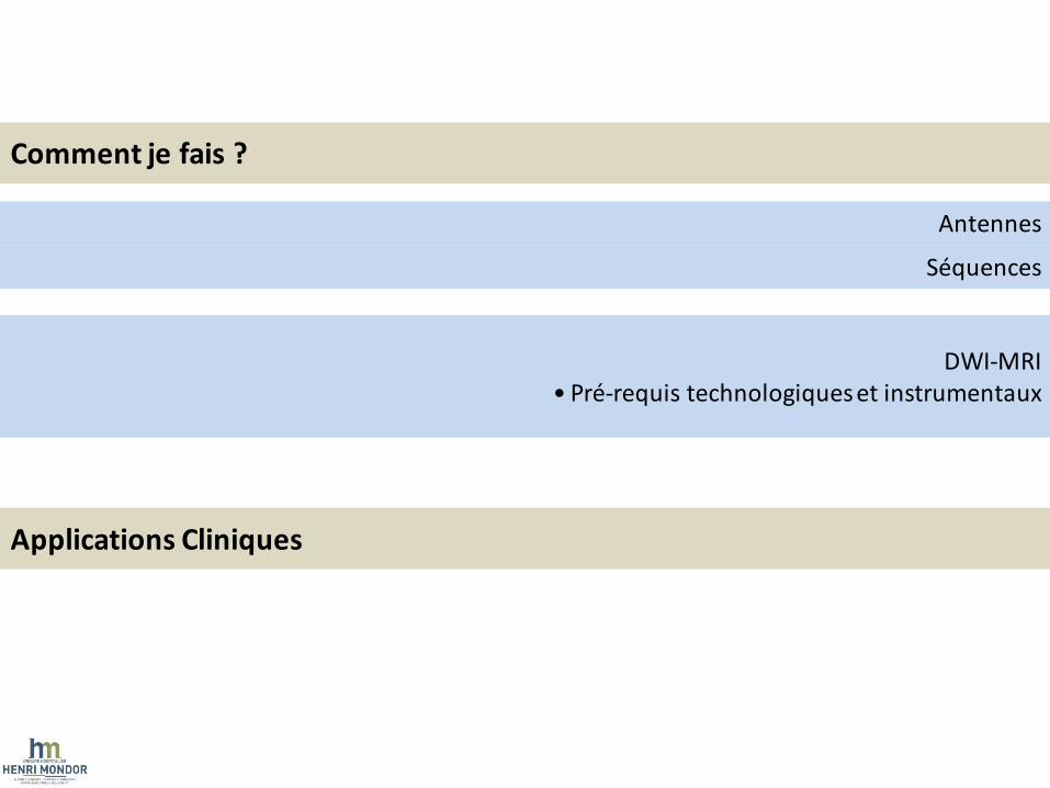
Comment je fais ?
Applications Cliniques
Antennes
DWI-MRI • Pré-requis technologiques et instrumentaux
Séquences
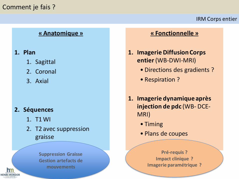
« Anatomique »
1. Plan
1. Sagittal
2. Coronal
3. Axial
2. Séquences
1. T1 WI
2. T2 avec suppression graisse
« Fonctionnelle »
1. Imagerie Diffusion Corps entier (WB-DWI-MRI)
• Directions des gradients ?
• Respiration ?
1. Imagerie dynamique après injection de pdc (WB- DCE-MRI)
• Timing
• Plans de coupes
Suppression Graisse Gestion artefacts de
mouvements
Pré-requis ? Impact clinique ?
Imagerie paramétrique ?
Comment je fais ?
IRM Corps entier
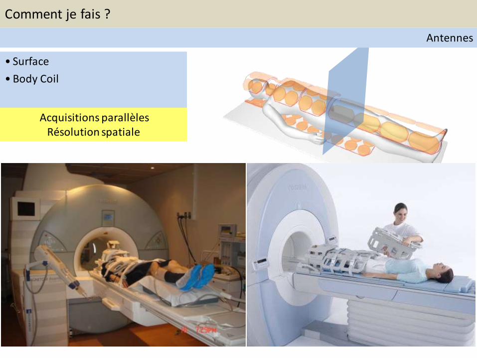
• Surface
• Body Coil
Acquisitions parallèles Résolution spatiale
Comment je fais ?
Antennes
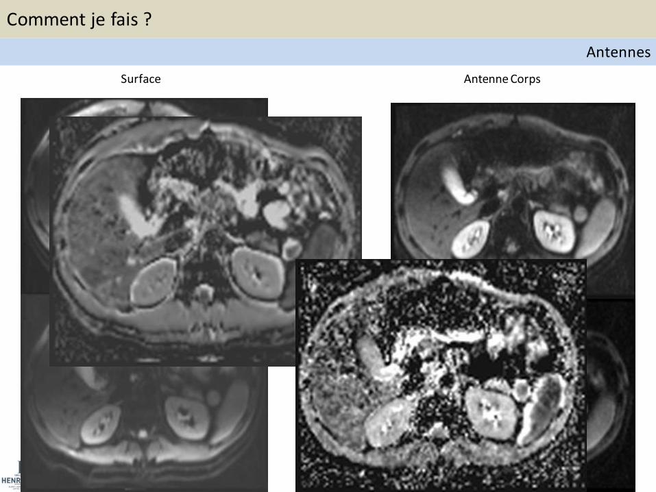
Surface Antenne Corps
Comment je fais ?
Antennes
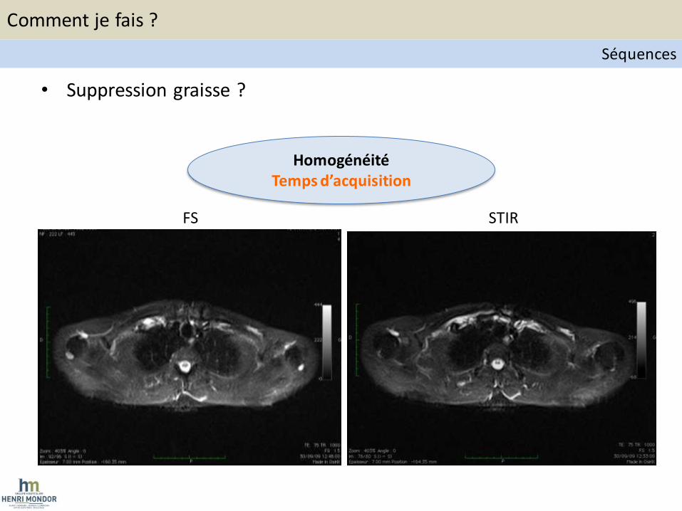
• Suppression graisse ?
FS STIR
Homogénéité Temps d’acquisition
Comment je fais ?
Séquences
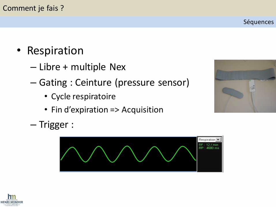
• Respiration
– Libre + multiple Nex
– Gating : Ceinture (pressure sensor)
• Cycle respiratoire
• Fin d’expiration => Acquisition
– Trigger :
Comment je fais ?
Séquences
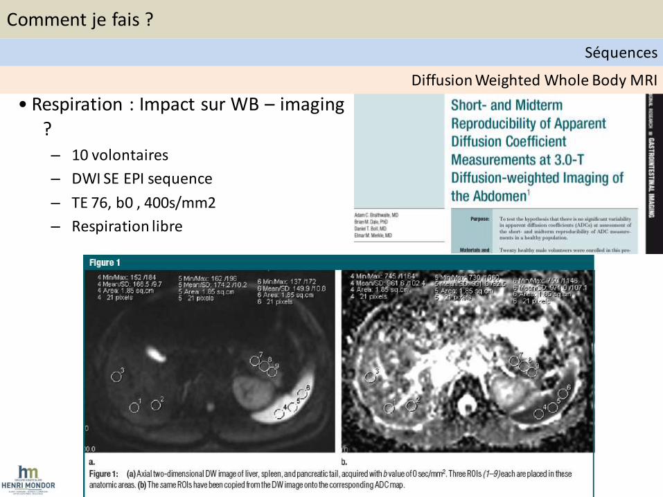
• Respiration : Impact sur WB – imaging ? – 10 volontaires
– DWI SE EPI sequence
– TE 76, b0 , 400s/mm2
– Respiration libre
Diffusion Weighted Whole Body MRI
Comment je fais ?
Séquences
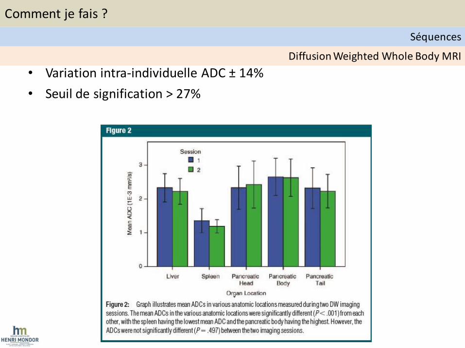
• Variation intra-individuelle ADC ± 14%
• Seuil de signification > 27%
Diffusion Weighted Whole Body MRI
Comment je fais ?
Séquences
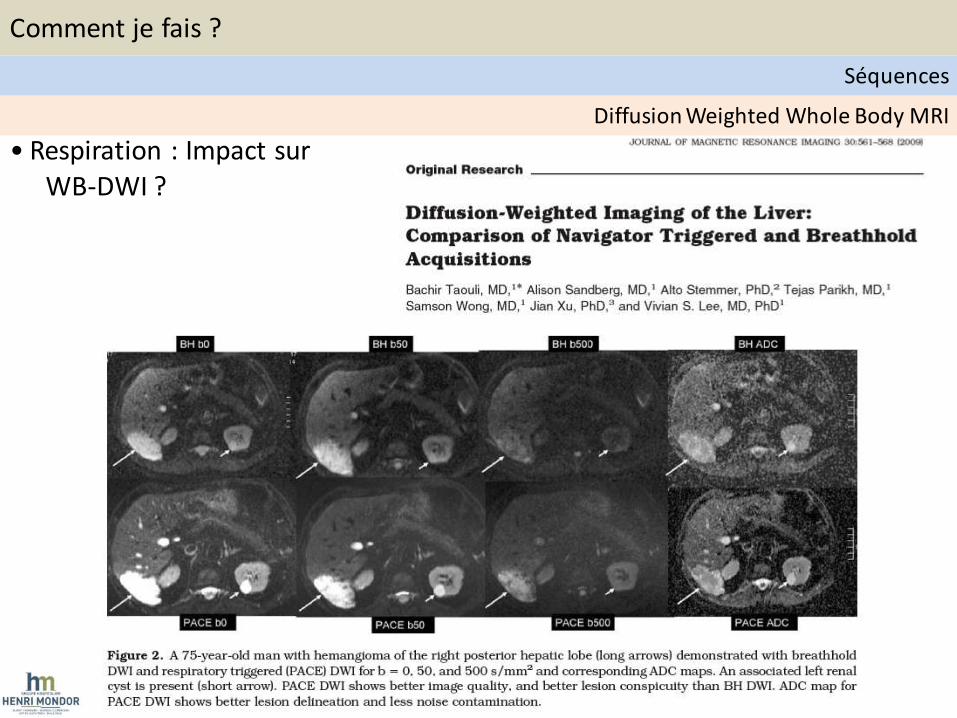
• Respiration : Impact sur WB-DWI ?
Diffusion Weighted Whole Body MRI
Comment je fais ?
Séquences
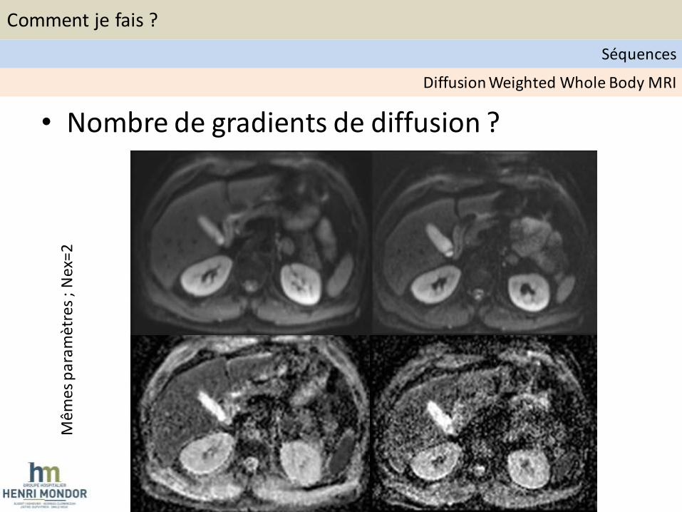
Mêm
es p
aram
ètre
s ;
Nex
=2
• Nombre de gradients de diffusion ?
Diffusion Weighted Whole Body MRI
Comment je fais ?
Séquences

• Choix des valeurs de b ?
Diffusion Weighted Whole Body MRI
Comment je fais ?
Séquences
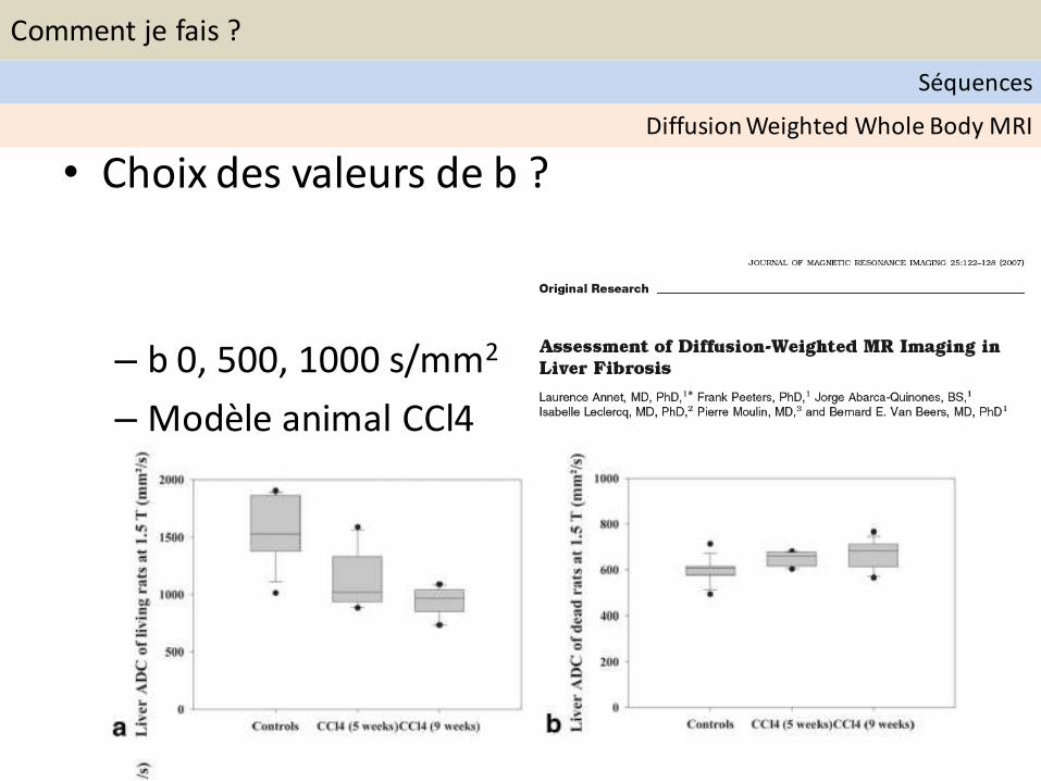
• Choix des valeurs de b ?
– b 0, 500, 1000 s/mm2
– Modèle animal CCl4
Diffusion Weighted Whole Body MRI
Comment je fais ?
Séquences

• Principes de la diffusion
Composante perfusive
Diffusion moléculaire
Diffusion Weighted Whole Body MRI
Comment je fais ?
Séquences
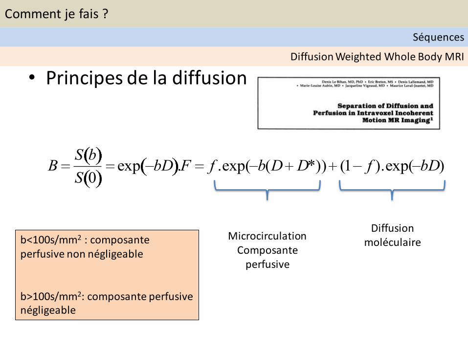
• Principes de la diffusion
BS b
S 0exp bD .F f .exp( b(D D*)) (1 f ).exp( bD)
Diffusion moléculaire
Microcirculation Composante
perfusive
b<100s/mm2 : composante perfusive non négligeable b>100s/mm2: composante perfusive négligeable
Diffusion Weighted Whole Body MRI
Comment je fais ?
Séquences
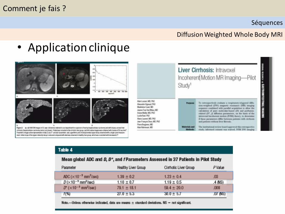
• Application clinique Diffusion Weighted Whole Body MRI
Comment je fais ?
Séquences
• IVIM – Intra Voxel Incoherent Motion - DWI
– Multiple b values
– Extraction of pure molecular diffusion
– Extraction of perfusion related diffusion
20
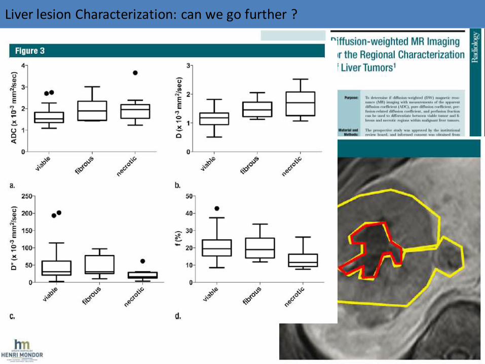
Liver lesion Characterization: can we go further ?
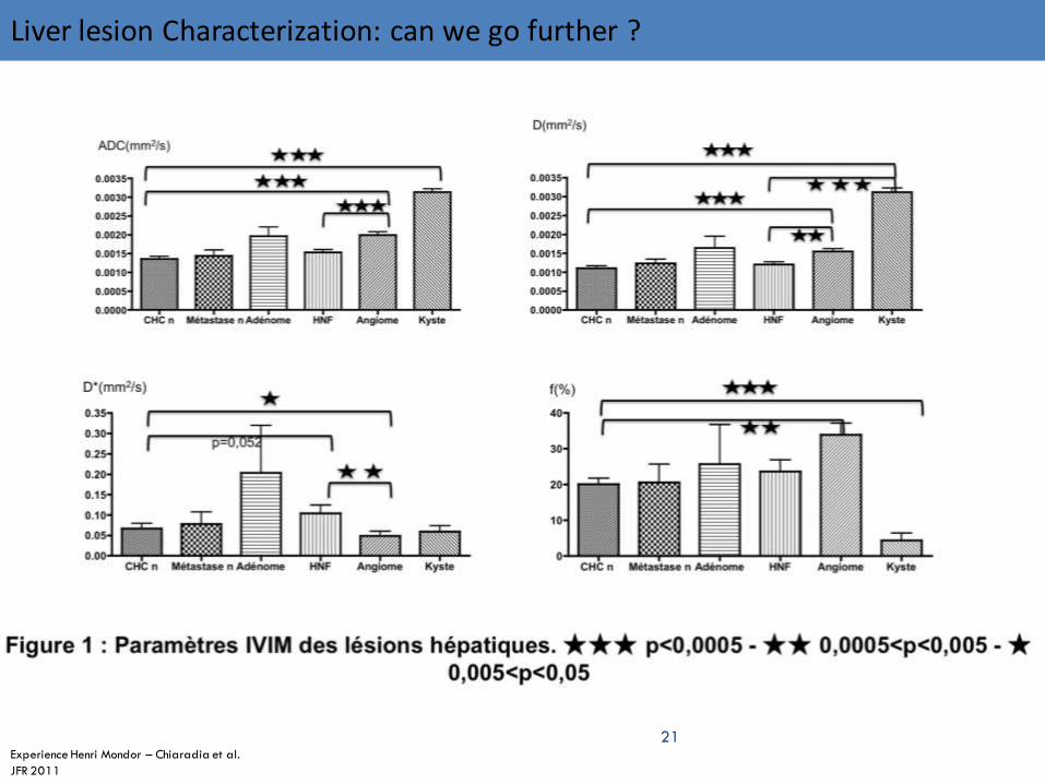
21 Experience Henri Mondor – Chiaradia et al.
JFR 2011
Liver lesion Characterization: can we go further ?
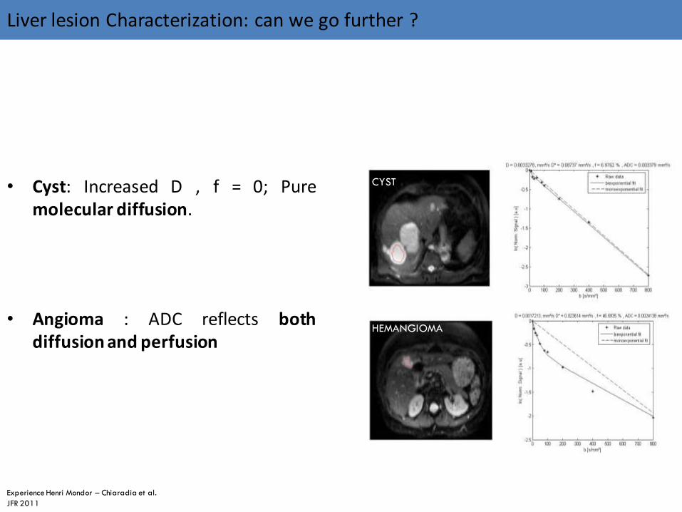
• Cyst: Increased D , f = 0; Pure molecular diffusion.
• Angioma : ADC reflects both diffusion and perfusion
CYST
HEMANGIOMA
Experience Henri Mondor – Chiaradia et al.
JFR 2011
Liver lesion Characterization: can we go further ?

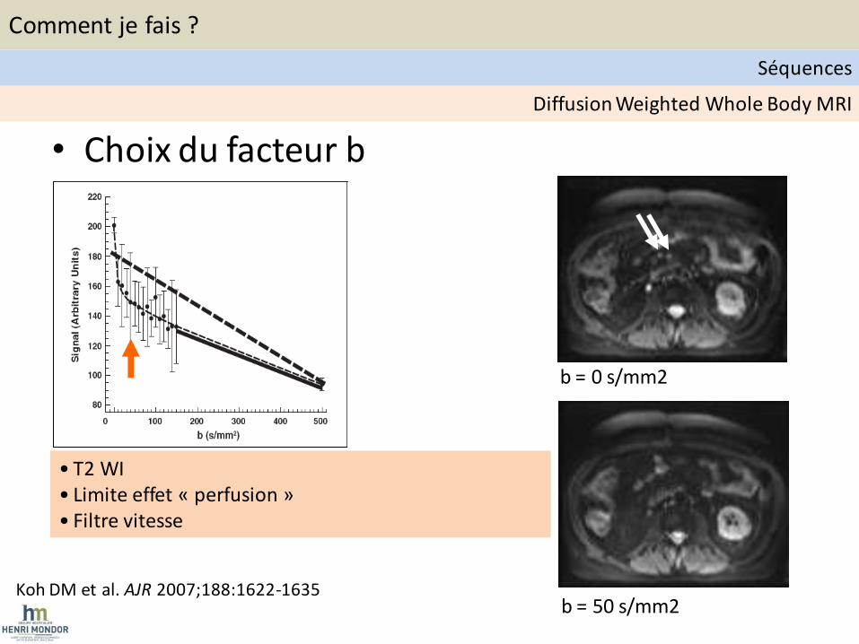
• Choix du facteur b
Koh DM et al. AJR 2007;188:1622-1635
b = 50 s/mm2
b = 0 s/mm2
• T2 WI • Limite effet « perfusion » • Filtre vitesse
Diffusion Weighted Whole Body MRI
Comment je fais ?
Séquences

• 1.5 T ou 3 T ?
– Temps d’acquisition
– Artefacts
Diffusion Weighted Whole Body MRI
Comment je fais ?
Séquences
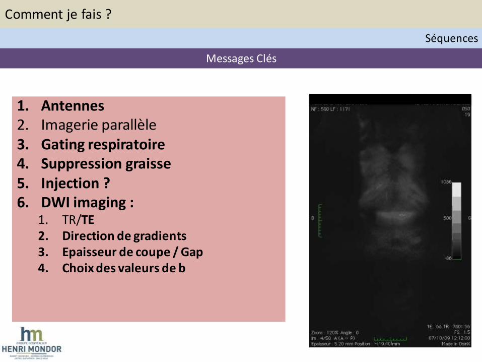
1. Antennes 2. Imagerie parallèle 3. Gating respiratoire 4. Suppression graisse 5. Injection ? 6. DWI imaging :
1. TR/TE 2. Direction de gradients 3. Epaisseur de coupe / Gap 4. Choix des valeurs de b
Messages Clés
Comment je fais ?
Séquences
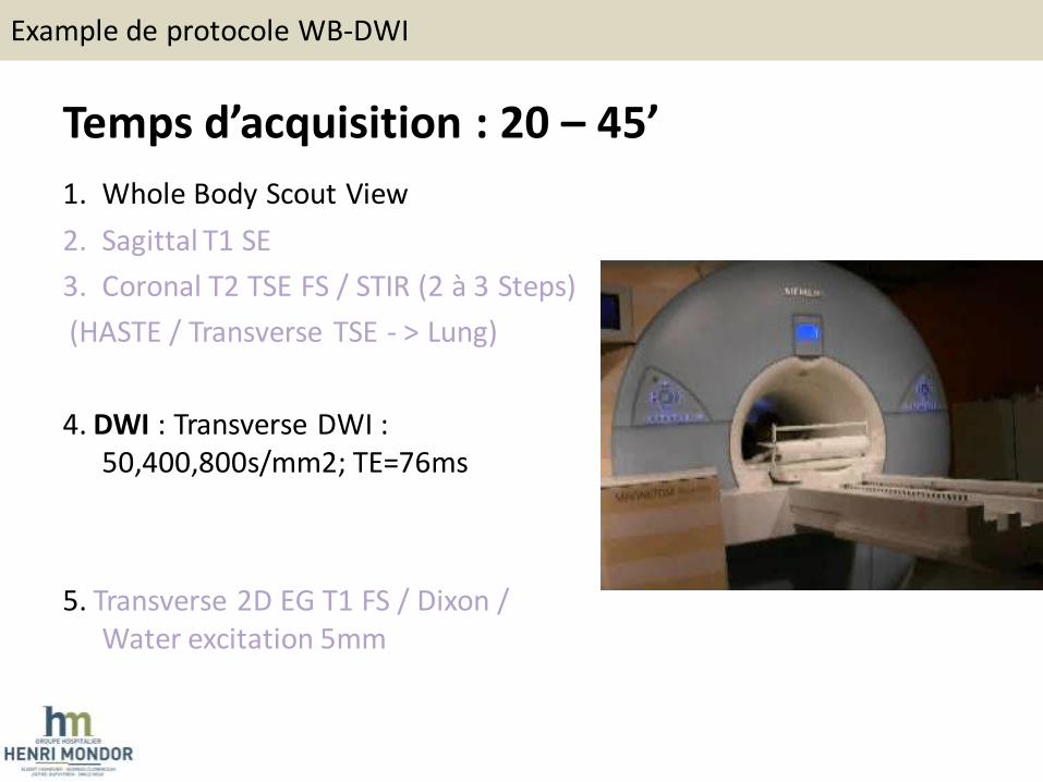
Temps d’acquisition : 20 – 45’
1. Whole Body Scout View
2. Sagittal T1 SE
3. Coronal T2 TSE FS / STIR (2 à 3 Steps)
(HASTE / Transverse TSE - > Lung)
4. DWI : Transverse DWI : 50,400,800s/mm2; TE=76ms
5. Transverse 2D EG T1 FS / Dixon / Water excitation 5mm
Example de protocole WB-DWI
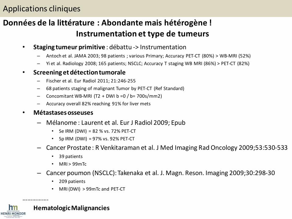
Données de la littérature : Abondante mais hétérogène ! Instrumentation et type de tumeurs
• Staging tumeur primitive : débattu -> Instrumentation – Antoch et al. JAMA 2003; 98 patients ; various Primary; Accuracy PET-CT (80%) > WB-MRI (52%)
– Yi et al. Radiology 2008; 165 patients; NSCLC; Accuracy T staging WB MRI (86%) > PET-CT (82%)
• Screening et détection tumorale – Fischer et al. Eur Radiol 2011; 21:246-255
– 68 patients staging of malignant Tumor by PET-CT (Ref Standard)
– Concomitant WB-MRI (T2 + DWI b =0 / b= 700s/mm2)
– Accuracy overall 82% reaching 91% for liver mets
• Métastases osseuses
– Mélanome : Laurent et al. Eur J Radiol 2009; Epub • Se IRM (DWI) = 82 % vs. 72% PET-CT
• Sp IRM (DWI) = 97% vs. 92% PET-CT
– Cancer Prostate : R Venkitaraman et al. J Med Imaging Rad Oncology 2009;53:530-533 • 39 patients
• MRI > 99mTc
– Cancer poumon (NSCLC): Takenaka et al. J. Magn. Reson. Imaging 2009;30:298-30 • 209 patients
• MRI (DWI) > 99mTc and PET-CT
…………….
• Hematologic Malignancies
Applications cliniques
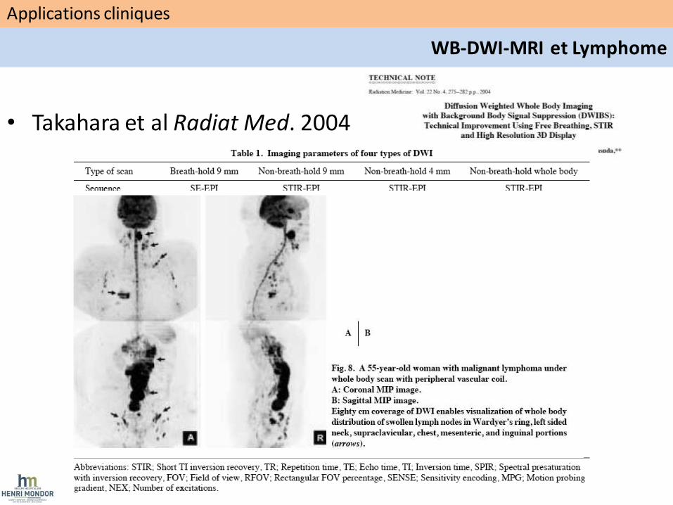
• Takahara et al Radiat Med. 2004
Applications cliniques
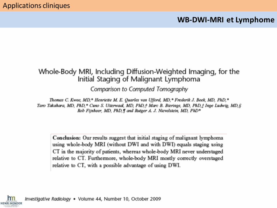
WB-DWI-MRI et Lymphome
Applications cliniques
WB-DWI-MRI et Lymphome
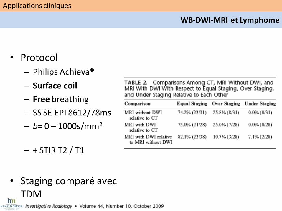
• Protocol
– Philips Achieva®
– Surface coil
– Free breathing
– SS SE EPI 8612/78ms
– b= 0 – 1000s/mm2
– + STIR T2 / T1
• Staging comparé avec TDM
Applications cliniques
WB-DWI-MRI et Lymphome
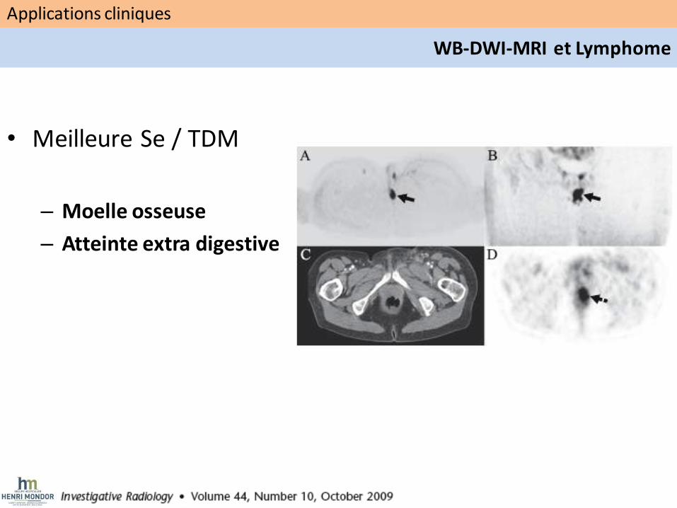
• Meilleure Se / TDM
– Moelle osseuse
– Atteinte extra digestive
Applications cliniques
WB-DWI-MRI et Lymphome
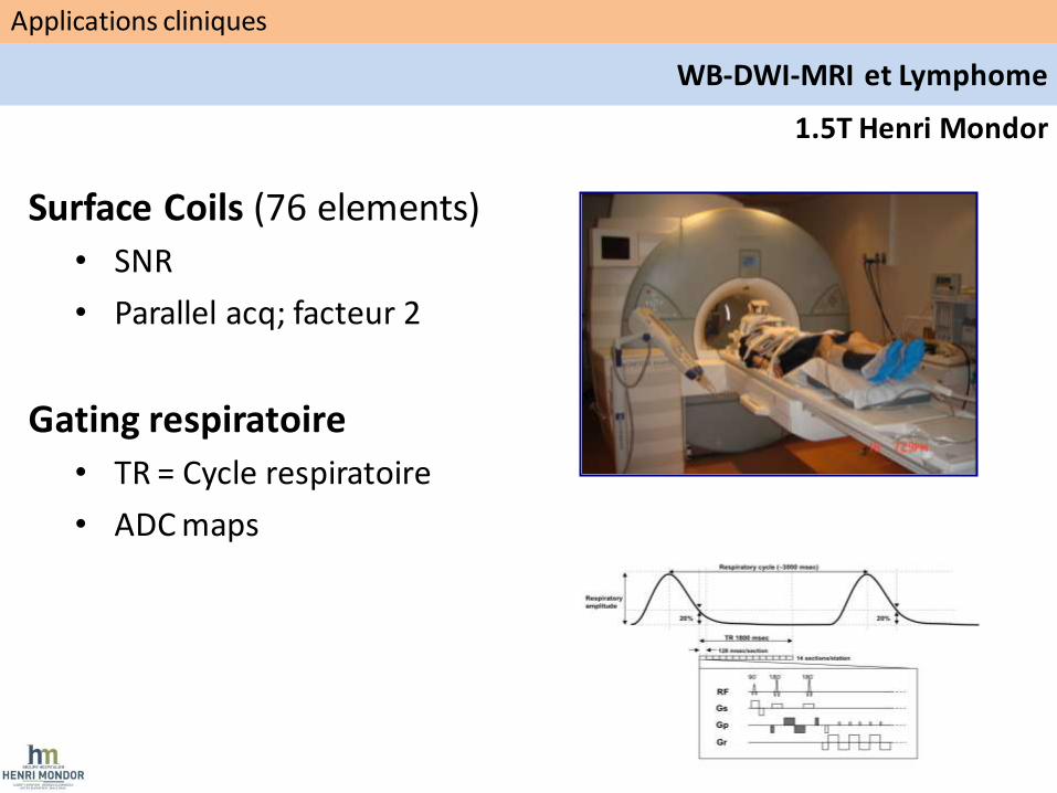
1.5T Henri Mondor
Surface Coils (76 elements)
• SNR
• Parallel acq; facteur 2
Gating respiratoire
• TR = Cycle respiratoire
• ADC maps
Applications cliniques
WB-DWI-MRI et Lymphome

Detection – Staging ganglionnaire
CT
ADC b800 b50
PET PET/CT
• 94% des ggs détectés en WB-DWI-MRI et TEP TDM
• 89% des aires ggs positives en TEP TDM ont diffusion restreinte
Applications cliniques
WB-DWI-MRI et Lymphome

• Résolution spatiale en WB-DWI =gg ou masse ?
b50 b800 ADC
Detection – Staging Ganglionnaire
Applications cliniques
WB-DWI-MRI et Lymphome
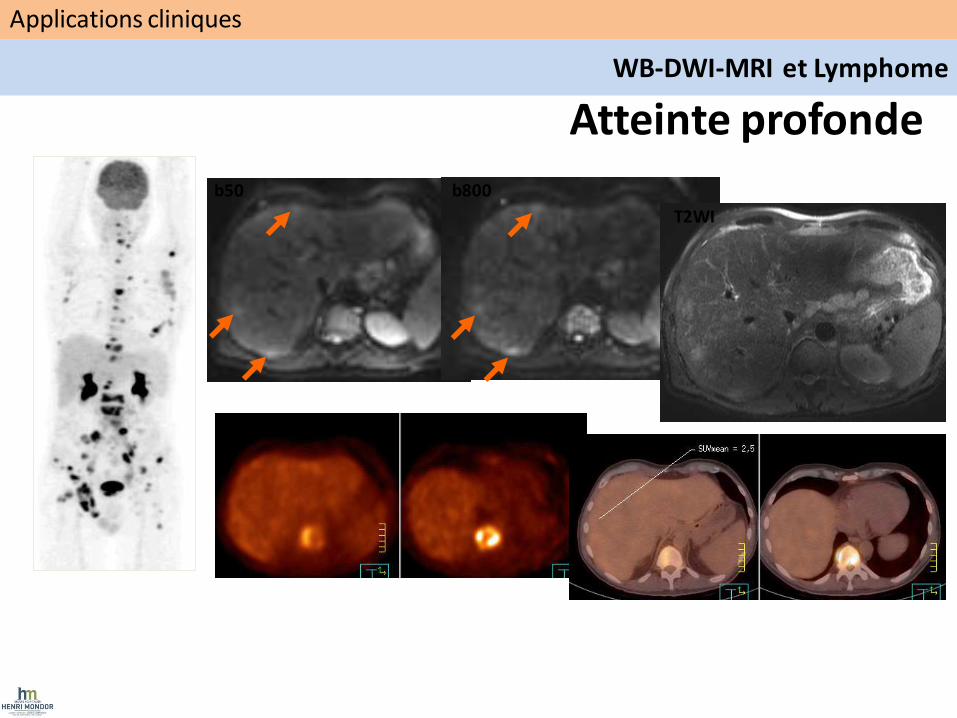
Atteinte profonde
b50 b800
T2WI
Applications cliniques
WB-DWI-MRI et Lymphome
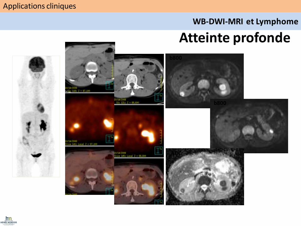
b800
b800
ADC
Atteinte profonde
Applications cliniques
WB-DWI-MRI et Lymphome
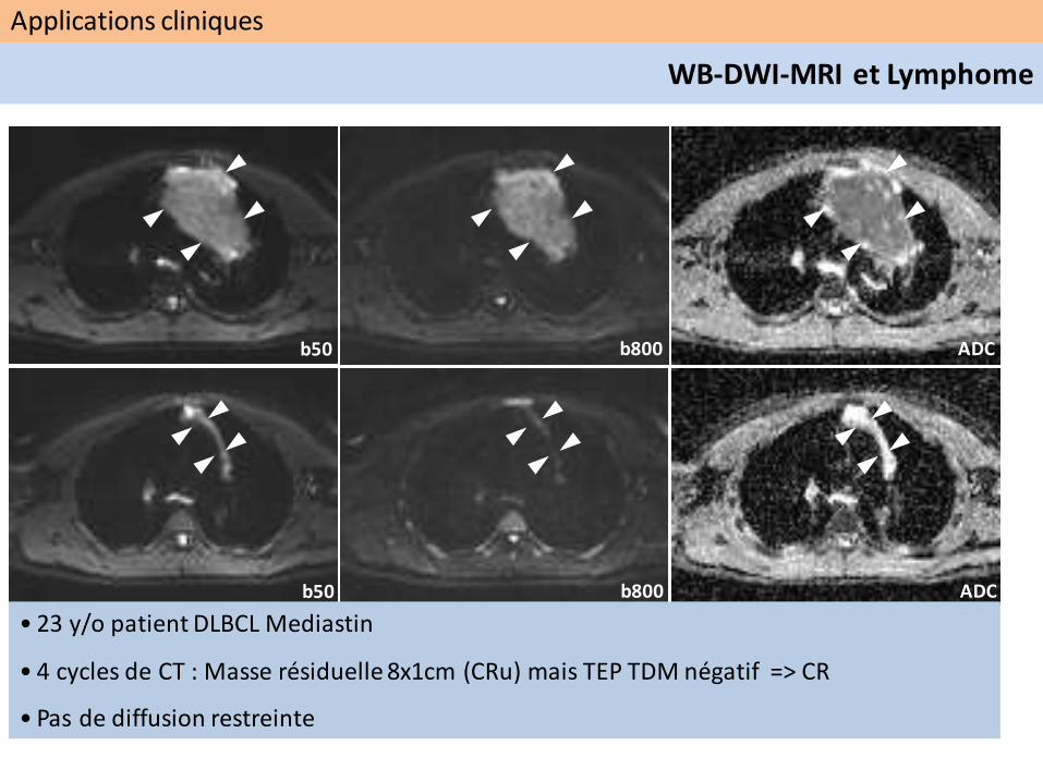
b50
b50
b800 ADC
b800 ADC
• 23 y/o patient DLBCL Mediastin
• 4 cycles de CT : Masse résiduelle 8x1cm (CRu) mais TEP TDM négatif => CR
• Pas de diffusion restreinte
Applications cliniques
WB-DWI-MRI et Lymphome
Messages
• Instrumentation
• Instrumentation
• Instrumentation
1
2
3
Applications cliniques
• Pas encore définies ….
• Standardisation ?
• Sélectionner études cliniques adaptées….
![Imagerie de la perfusion et du métabolisme cérébral Brain ...pfeifer.phas.ubc.ca/refbase/files/Payen-Annales...lés à ceux de l ’IRM de perfusion–diffusion [6].Dautres études](https://static.fdocuments.fr/doc/165x107/5ecab2e47c75fe2d781bffa2/imagerie-de-la-perfusion-et-du-mtabolisme-crbral-brain-s-ceux-de-l.jpg)


![© [C.Esnouf], [2011], INSA de Lyon, tous droits réservés 2011 SGM Auteur : ESNOUF Claude CLYM Séminaire 10 Imagerie par diffusion incohérente : HAADF (STEM)](https://static.fdocuments.fr/doc/165x107/551d9d81497959293b8bb1b8/-cesnouf-2011-insa-de-lyon-tous-droits-reserves-2011-sgm-auteur-esnouf-claude-clym-seminaire-10-imagerie-par-diffusion-incoherente-haadf-stem.jpg)