Growth delay of human bladder cancer cells by Prostate Stem Cell
9
RESEARCH ARTICLE Open Access Growth delay of human bladder cancer cells by Prostate Stem Cell Antigen downregulation is associated with activation of immune signaling pathways Emanuele Marra 1 , Paolo Uva 1 , Valentina Viti 1 , Valeria Simonelli 3 , Eugenia Dogliotti 3 , Emanuele De Rinaldis 4 , Armin Lahm 1 , Nicola La Monica 1 , Alfredo Nicosia 2 , Gennaro Ciliberto 1 , Fabio Palombo 1* Abstract Background: Prostate stem cell antigen (PSCA) is a glycosylphosphatidylinositol (GPI) anchored protein expressed not only in prostate but also in pancreas and bladder cancer as shown by immunohistochemistry and mRNA analysis. It has been targeted by monoclonal antibodies in preclinical animal models and more recently in a clinical trial in prostate cancer patients. The biological role played in tumor growth is presently unknown. In this report we have characterized the contribution of PSCA expression to tumor growth. Methods: A bladder cell line was engineered to express a doxycycline (dox) regulated shRNA against PSCA. To shed light on the PSCA biological role in tumor growth, microarray analysis was carried out as a function of PSCA expression. Expression of gene set of interest was further analyzed by qPCR Results: Down regulation of the PSCA expression was associated with reduced cell proliferation in vitro and in vivo. Mice bearing subcutaneous tumors showed a reduced tumor growth upon treatment with dox, which effectively induced shRNA against PSCA as revealed by GFP expression. Pathway analysis of deregulated genes suggests a statistical significant association between PSCA downregulation and activation of genes downstream of the IFNa/b receptor. Conclusions: These experiments established for the first time a correlation between the level of PSCA expression and tumor growth and suggest a role of PSCA in counteracting the natural immune response. Background PSCA has been discovered a decade ago and has been classified as a member of the Ly-6 family of GPI- anchored cell surface proteins [1]. It is expressed in most prostate cancer specimens, including high-grade prostatic intraepithelial neoplasia, primary androgen- dependent tumors, and hormone-refractory metastases. PSCA levels are positively correlated with Gleason grade, tumor stage, and biochemical recurrence. Its expression is also particularly elevated in bone metasta- sis. Finally, PSCA is strongly expressed in other malig- nancies, including bladder and pancreatic cancers [2-6]. Different immunotherapy approaches targeting PSCA have been tested in preclinical models including cancer vaccine, therapeutic monoclonal antibodies and antibody conjugated to toxic drugs [7-10]. More recently, a human monoclonal antibody targeting PSCA has been evaluated in a phase I clinical trial in prostate cancer patients (AACR 2006). Little information is available regarding the biological role of PSCA. Proteins belonging to the Ly6 family are involved in cell signaling events associated with thymic lymphocyte differentiation, maturation and activation. CD59, a member of this protein family, was shown to play a role in the protection against complement mediated lysis. In addition, it was found to be expressed in tumor cells where it may play a role in evading anti * Correspondence: [email protected] 1 Istituto di Ricerche Biologia Molecolare P. Angeletti (IRBM), Via Pontina Km 30,600 00040 Pomezia (Rome) Italy Marra et al. BMC Cancer 2010, 10:129 http://www.biomedcentral.com/1471-2407/10/129 © 2010 Marra et al; licensee BioMed Central Ltd. This is an Open Access article distributed under the terms of the Creative Commons Attribution License (http://creativecommons.org/licenses/by/2.0), which permits unrestricted use, distribution, and reproduction in any medium, provided the original work is properly cited.
Transcript of Growth delay of human bladder cancer cells by Prostate Stem Cell
RESEARCH ARTICLE Open Access
Growth delay of human bladder cancer cells byProstate Stem Cell Antigen downregulation isassociated with activation of immune signalingpathwaysEmanuele Marra1, Paolo Uva1, Valentina Viti1, Valeria Simonelli3, Eugenia Dogliotti3, Emanuele De Rinaldis4,Armin Lahm1, Nicola La Monica1, Alfredo Nicosia2, Gennaro Ciliberto1, Fabio Palombo1*
Abstract
Background: Prostate stem cell antigen (PSCA) is a glycosylphosphatidylinositol (GPI) anchored protein expressednot only in prostate but also in pancreas and bladder cancer as shown by immunohistochemistry and mRNAanalysis. It has been targeted by monoclonal antibodies in preclinical animal models and more recently in a clinicaltrial in prostate cancer patients. The biological role played in tumor growth is presently unknown. In this report wehave characterized the contribution of PSCA expression to tumor growth.
Methods: A bladder cell line was engineered to express a doxycycline (dox) regulated shRNA against PSCA. Toshed light on the PSCA biological role in tumor growth, microarray analysis was carried out as a function of PSCAexpression. Expression of gene set of interest was further analyzed by qPCR
Results: Down regulation of the PSCA expression was associated with reduced cell proliferation in vitro and in vivo.Mice bearing subcutaneous tumors showed a reduced tumor growth upon treatment with dox, which effectivelyinduced shRNA against PSCA as revealed by GFP expression. Pathway analysis of deregulated genes suggests astatistical significant association between PSCA downregulation and activation of genes downstream of the IFNa/breceptor.
Conclusions: These experiments established for the first time a correlation between the level of PSCA expressionand tumor growth and suggest a role of PSCA in counteracting the natural immune response.
BackgroundPSCA has been discovered a decade ago and has beenclassified as a member of the Ly-6 family of GPI-anchored cell surface proteins [1]. It is expressed inmost prostate cancer specimens, including high-gradeprostatic intraepithelial neoplasia, primary androgen-dependent tumors, and hormone-refractory metastases.PSCA levels are positively correlated with Gleasongrade, tumor stage, and biochemical recurrence. Itsexpression is also particularly elevated in bone metasta-sis. Finally, PSCA is strongly expressed in other malig-nancies, including bladder and pancreatic cancers [2-6].
Different immunotherapy approaches targeting PSCAhave been tested in preclinical models including cancervaccine, therapeutic monoclonal antibodies and antibodyconjugated to toxic drugs [7-10]. More recently, ahuman monoclonal antibody targeting PSCA has beenevaluated in a phase I clinical trial in prostate cancerpatients (AACR 2006).Little information is available regarding the biological
role of PSCA. Proteins belonging to the Ly6 family areinvolved in cell signaling events associated with thymiclymphocyte differentiation, maturation and activation.CD59, a member of this protein family, was shown toplay a role in the protection against complementmediated lysis. In addition, it was found to be expressedin tumor cells where it may play a role in evading anti
* Correspondence: [email protected] di Ricerche Biologia Molecolare P. Angeletti (IRBM), Via Pontina Km30,600 00040 Pomezia (Rome) Italy
Marra et al. BMC Cancer 2010, 10:129http://www.biomedcentral.com/1471-2407/10/129
© 2010 Marra et al; licensee BioMed Central Ltd. This is an Open Access article distributed under the terms of the Creative CommonsAttribution License (http://creativecommons.org/licenses/by/2.0), which permits unrestricted use, distribution, and reproduction inany medium, provided the original work is properly cited.
cancer immune response [11]. Deletion of PSCA genedoes not appear to interfere with normal developmentas PSCA knockout mice are viable. Additionally, cross-ing of the PSCA knockout mice with prostate tumormodels driven by large T antigen did not increase pri-mary tumor formation [12,13].Here the biological role of human PSCA was evalu-
ated using RNA interference and microarray analysis.To establish a pharmacologic control over gene expres-sion a shRNA against PSCA was identified andexpressed under the control of dox in a lentivirus sys-tem [14]. Microarray analysis was utilized to identifygenes coregulated with PSCA in tumor xenografts.
MethodsCell culture and generation of lentivirus vectorsThe 293T and SW780 cell lines were cultured in Dul-becco’s modified Eagle’s medium supplemented with10% fetal calf serum. The lentivirus system for condi-tional gene suppression with dox-inducible shRNAs uti-lized has been described previously[14]. Briefly, in theabsence of dox, tTR-KRAB repressor binds to tetO andsuppresses H1-mediated shRNA transcription, thusallowing normal expression of the cellular target gene.In the presence of 10 μg/ml of dox, tTR-KRAB cannotbind to tetO and hence shRNAs are produced, leadingto downregulation of PSCA. The green fluorescent pro-tein (GFP) cDNA contained in the shRNA vector pro-vides a monitoring device, as it is switched on by doxtreatment and GFPis expressed. All recombinant lenti-viruses were produced by transient transfection in 293Tcells. Briefly, 293T cells were cotransfected with 20 μgof pLUTHM-shPSCA3 plasmid, 15 μg of pCMV-ΔR8.91, and 5 μg of pMD2G-VSVG by calcium phos-phate precipitation. After 16 h medium was changed,and recombinant lentivirus vectors were harvested 48 hlater. FACS analysis was conducted as previouslydescribed [15].
Viability assayCell viability was monitored using the CellTiter-Blue Via-bility. The assay is based on the ability of living cells toconvert a redox dye (resazurin) into a fluorescent endproduct (resorufin); 1 × 10³ SW780-shPSCA and SW780-shControl cells +/- Dox were plated in a 96 well plate inparallel with the parental cell line SW780. Cells wereincubated at 37°c for 96 hrs, and fluorescence was subse-quently monitored using a plate-reading fluorometer.
Tumor modelsSix-week-old female CD-1 nude mice were purchasedfrom Charles River Laboratories and maintained inaccordance to Guidelines for the Care and Use ofLaboratory Animals in IRBM’s animal facility. This
study, was submitted and approved by the IRBM ethicalcommittee . Mice were injected subcutaneously (sc) inthe right flank with 2 × 10⁶ SW780-shPSCA cells resus-pended into 100 μL phosphate-buffered saline (PBS) andMatrigel (1:1). Mice received 5% sucrose only or 5%sucrose plus 0.2 mg/ml of dox for control and knock-down cohorts, respectively. All water bottles were chan-ged 3 times per week. Tumors were measured withcalipers and mice weighed twice per week. At the end ofthe dosing study, or as indicated, appropriate tumorsamples were taken.
Microarray experimentsTotal RNA from cell in culture was isolated with RNA-zolB and then dissolved in RNase-free water. 25 μg oftotal RNA was treated with DNase using the QiagenRNase-free DNase kit and RNeasy spin columns. ThenRNA was dissolved in RNase-free water to a final con-centration of 0.2 μg/μl. cRNA was generated using T7RNA polymerase on 5 μg of total RNA and labeled withCy5 or Cy3 (Cy Dye, Amersham Pharmacia Biotech).From each sample, 5 μg of labeled-RNA were co-hybri-dized with 5 μg of a reference RNA (pool of twountreated SW780 cell lines). Labeled cRNAs were frag-mented to an average size of 50-100 nucleotides byheating the samples to 60°C with 10 mM of zinc chlor-ide and then adding an hybridization buffer containing 1M NaCl, 0.5% sodium sarcosine, 50 mM MES, pH6.5,and formamide to a final concentration of 30%. Thefinal volume was 3 ml at 40°C.Samples were hybridized on a customized Agilent 44 k
array containing ~40,000 unique probes mapping to~21,000 human genes. Each sample was hybridized induplicate with fluor reversal to systematically correct fordye bias. After hybridization, slides were washed andscanned using a confocal laser scanner (Agilent Tech-nologies). The raw intensities obtained after scanningwere quantified, background-corrected and lowness nor-malized. A weighted average ratio was computed fordye-swapped pairs of hybridizations. Tumors were col-lected in RNA later and processed for RNA extractionas described previously [16]. Samples were hybridizedon a customized Affymetrix array containing ~38,000probes mapping to ~21,000 human genes [17].
Hierarchical clusteringThe microarray dataset was filtered before clustering inorder to select the 2,000 most variable probes. probeswith an absent call in more than 12 samples out of 14were removed the 2,000 probes with the higher inter-quartile range were retained for subsequent analysis.Probes were hierarchically clustered using an averagelinkage algorithm based on Pearson correlationcoefficients.
Marra et al. BMC Cancer 2010, 10:129http://www.biomedcentral.com/1471-2407/10/129
Page 2 of 9

Identification of deregulated probesDifferences in average probe expression between the dox+ (PSCA silencing) and dox-samples were computed by1-way ANOVA. Probes differentially expressed betweenthe two classes were identified based on ANOVAp-value < 0.001.
Gene set enrichment analysisGroups of genes identified by 1-way ANOVA were com-pared to a collection of annotated gene sets to identifythe functional classes that were significantly over-repre-sented. The enrichment p-values were computedaccording to the Fisher’s exact test. The gene sets wereobtained from public (Gene Ontology [18], KEGG [19],Interpro [20], Panther [21], oPOSSUM [22] and com-mercial sources (GeneGo (GeneGo Inc., St Joseph, MI,USA), Ingenuity (Ingenuity Systems Inc, MountainView, CA, USA), TRANSFAC [23].
RT-PCRMicroarray findings were confirmed by real-timereverse-transcription PCR (RT-PCR) using the averagevalue of mRNA from dox untreated SW780-shPSCAsamples as calibrator. Analysis of mRNA expression ofselected IFNa genes was performed using Applied Bio-systems expression kit following manufacturers’ instruc-tions. Expression values were computed using thecomparative CT method (ΔΔ CT) with GAPDH geneexpression value serving as normaliser.
ResultsEstablishment of cell clones with inducible shRNA againstPSCAPublished studies on PSCA have reported limitedexpression in cell lines engineered, for the most part, toexpress this protein ectopically [7,10]. Searching in amicroarray cell atlas comprising 20 cell lines of differentorigins including prostate, pancreas and bladder revealeda low frequency of PSCA expression. The few cell lineswith a detectable level of PSCA mRNA scored negativewhen expression was analyzed at protein level by FACSanalysis (data not shown), suggesting that display ofPSCA on the cell surface is uncommon. We thereforeutilized a bladder cell line, SW780, which was shown toexpress PSCA on the cell surface [8]. The cell line wasengineered to express different shRNA sequencesagainst PSCA (Table 1) or a control shRNA against luci-ferase. To achieve a pharmacological controlled expres-sion a dox inducible promoter was utilized in thecontext of a lentivirus system [14]. One out of sixshRNA against PSCA showed a clear down regulation oftarget gene expression and was therefore utilized to gen-erate SW780-shPSCA, which expresses also the tet regu-lator. Dox treatment resulted in 75% reduction of PSCA
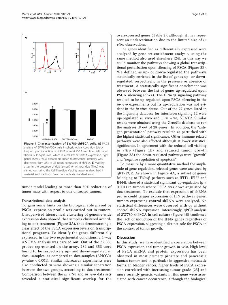
expression as measured by FACS analysis. Since PSCAis a membrane protein a further step was undertaken toimprove regulation of gene expression by sorting outcells with low PSCA expression in the presence of dox.This process led to the isolation of cells with a largerinterval of regulation mediated by dox treatment. Tocontrol that the expression of shRNA was induced upondox treatment we took advantage of GFP gene, which iscloned upstream of the shRNA and it was previouslyutilized as a marker of shRNA expression [14]. FACSanalysis showed a quantitative GFP expression only indox treated SW780-shPSCA cells and a strong reductionof PSCA expression (Figure 1A). These data suggest thatexpression of shRNA specific for PSCA can be inducedand that it leads to almost 90% downregulation of thetarget gene.
Biological effects of PSCA down-regulation in SW780 cellsTo characterize the impact of PSCA expression on cellgrowth, a viability assay was carried out. A statistical sig-nificant reduction in cell viability was observed in doxtreated cells whereas no difference was observed in con-trol transfected cells (Figure 1B).To verify the impact of PSCA expression on SW780-
shPSCA tumor growth dox treatment was either startedin vitro one week in advance or at the time of cellimplantation. A statistical significant reduction in s.c.tumor growth (p < 0.05) was observed in the cohort ofanimals treated with dox with respect to controlsincluding also SW780-shCont treated with dox(Figure 2A). Interestingly, tumors generated with cellspretreated in vitro with dox were smaller than tumorstreated with dox only in vivo, although this differencewas not statistically significant. In contrast controlshRNA did not show any statistical reduction of tumorgrowth. To confirm that shRNA against PSCA wasinduced in vivo tumors were sacrificed at day 28 andexpression of shRNA was evaluated by looking at GFPexpression. Only dox-treated mice showed GFP expres-sion in SW780-shPSCA tumors (Figure 2B). In line withthe shRNA expression, PSCA was down-regulated asshown by IHC. All together these experiments indicatethat PSCA plays a critical role in the SW780-shPSCA
Table 1 shRNA sequences against PSCA
shRNA Sequence Coding Region N
1 CATCCTAACGCAAGTCTGATT No 497-515
2 GCAAGTCTGACCATGTATGTT No 506-524
3 GGCAGATCAGCTCTAGTGATT No 585-603
4 CAAGTCTGACCATGTATGTTT No 507-525
5 GCAAGAAGAACATCACGTGTT Yes 256-274
6 GTGACACCGACTTGTGCAATT Yes 277-295
Marra et al. BMC Cancer 2010, 10:129http://www.biomedcentral.com/1471-2407/10/129
Page 3 of 9
tumor model leading to more than 50% reduction oftumor mass with respect to dox untreated tumors.
Transcriptional data analysisTo gain some hints on the biological role played byPSCA, expression profile was carried out in tumors.Unsupervised hierarchical clustering of genome-wideexpression data showed that samples clustered accord-ing to dox treatment (Figure 3A), thus demonstrating aclear effect of the PSCA expression levels on transcrip-tional programs. To identify the genes differentiallyexpressed in the two experimental conditions, a 1-wayANOVA analysis was carried out. Out of the 37,586probes represented on the array, 284 and 353 werefound to be respectively up- and down-regulated indox+ samples, as compared to dox-samples (ANOVAp-value < 0.001). Similar microarray experiments werealso conducted in vitro showing a similar separationbetween the two groups, according to dox treatment.Comparison between the in vitro and in vivo data setsrevealed a statistical significant overlap for the
overexpressed genes (Table 2), although it may repre-sent an underestimation due to the limited size of invitro observations.The genes identified as differentially expressed were
analyzed by gene set enrichment analysis, using thesame method also used elsewhere [24]. In this way wecould monitor the pathways showing a global transcrip-tional perturbation upon silencing of PSCA (Figure 3B).We defined as up- or down-regulated the pathwaysstatistically enriched in the list of genes up- or down-regulated, respectively, in the presence or absence oftreatment. A statistically significant enrichment wasobserved between the list of genes up-regulated uponPSCA silencing (dox+). The IFNa/b signaling pathwayresulted to be up-regulated upon PSCA silencing in thein-vivo experiments but its up-regulation was not evi-dent in the in vitro datase. Out of the 27 genes listed inthe Ingenuity database for interferon signaling 12 wereup-regulated in vivo and 1 in vitro, STAT2. Similarresults were obtained using the GeneGo database to runthe analyses (8 out of 28 genes). In addition, the “anti-gen presentation” pathway resulted as perturbed withthe highest statistical significance. Other immune relatedpathways were also affected although at lower statisticalsignificance. In agreement with the reduced cell viabilityin vitro (Figure 1B) and reduced tumor growth(Figure 2A) the down-regulated pathways were “growth”and “negative regulation of apoptosis”.To measure by a more quantitative method the ampli-
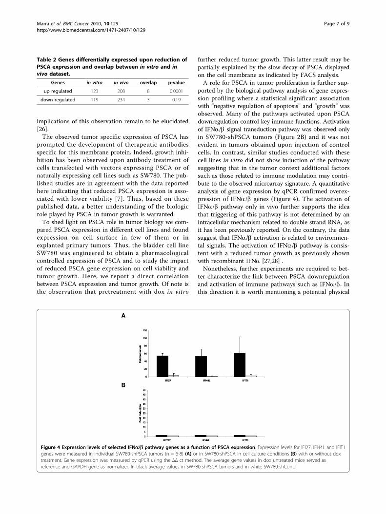
tude of gene regulation, selected genes were analyzed byqRT-PCR. As shown in Figure 4A, a subset of genesbelonging to IFNa/b pathway such as IFIT1, IFI27 andIFI44L showed a statistical significant up-regulation (p <0.001) in tumors where PSCA was down-regulated bydox treatment. To exclude that expression of shRNAper se could trigger expression of IFN pathway genes,tumors expressing control shRNA were analyzed. Nostatistical differences were observed with or withoutcontrol shRNA expression. Interestingly, qPCR analysisof SW780-shPSCA in cell culture (Figure 4B) confirmedthe lack of induction of the IFNa genes regardless ofPSCA expression, suggesting a distinct role for PSCA inthe context of tumor growth.
DiscussionIn this study, we have identified a correlation betweenPSCA expression and tumor growth in vivo. High levelof PSCA mRNA and protein expression has beenobserved in most primary prostate and pancreatichuman tumors and in particular in aggressive metastaticforms. In bladder cancer, higher levels of PSCA expres-sion correlated with increasing tumor grade [25] andmore recently genetic variants in this gene were asso-ciated with cancer occurrence, although the biological
Figure 1 Characterization of SW780-shPSCA cells. A) FACSanalysis of SW780-shPSCA cells in physiological condition (blackline) or upon induction of shRNA against PSCA (red line): left panelshows GFP expression, which is a marker of shRNA expression; rightpanel shows PSCA expression, mean fluorescence intensity wasdecreased from 333 to 35 upon expression of shRNA. B) Viabilityassay in the presence of dox (empty) or without dox (filled) wascarried out using the CellTiter-Blue Viability assay as described inmaterial and methods. Error bars indicate standard error.
Marra et al. BMC Cancer 2010, 10:129http://www.biomedcentral.com/1471-2407/10/129
Page 4 of 9
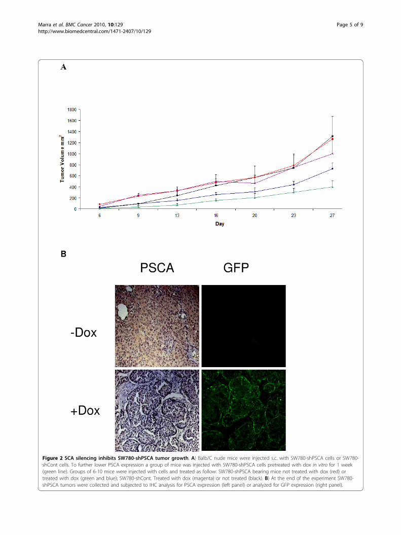
Figure 2 SCA silencing inhibits SW780-shPSCA tumor growth. A) Balb/C nude mice were injected s.c. with SW780-shPSCA cells or SW780-shCont cells. To further lower PSCA expression a group of mice was injected with SW780-shPSCA cells pretreated with dox in vitro for 1 week(green line). Groups of 6-10 mice were injected with cells and treated as follow: SW780-shPSCA bearing mice not treated with dox (red) ortreated with dox (green and blue); SW780-shCont. Treated with dox (magenta) or not treated (black). B) At the end of the experiment SW780-shPSCA tumors were collected and subjected to IHC analysis for PSCA expression (left panel) or analyzed for GFP expression (right panel).
Marra et al. BMC Cancer 2010, 10:129http://www.biomedcentral.com/1471-2407/10/129
Page 5 of 9
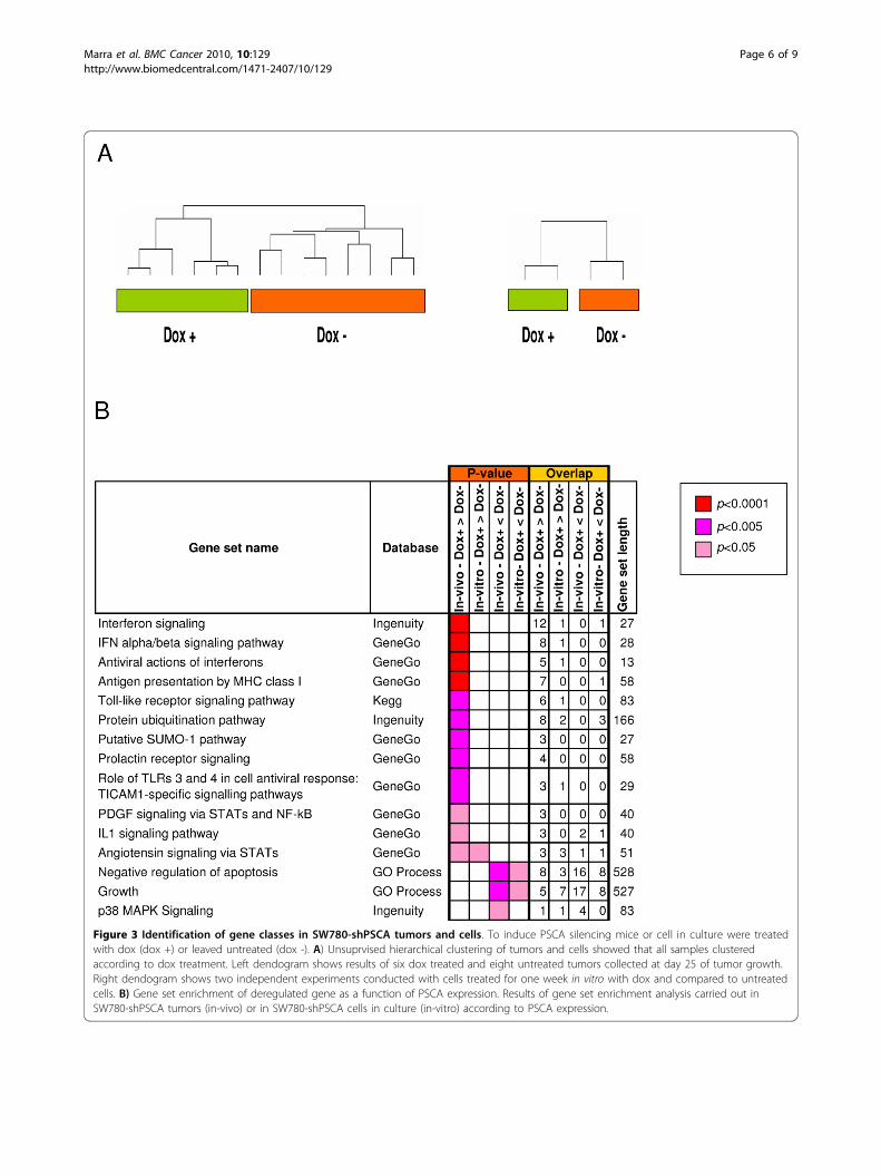
Figure 3 Identification of gene classes in SW780-shPSCA tumors and cells. To induce PSCA silencing mice or cell in culture were treatedwith dox (dox +) or leaved untreated (dox -). A) Unsuprvised hierarchical clustering of tumors and cells showed that all samples clusteredaccording to dox treatment. Left dendogram shows results of six dox treated and eight untreated tumors collected at day 25 of tumor growth.Right dendogram shows two independent experiments conducted with cells treated for one week in vitro with dox and compared to untreatedcells. B) Gene set enrichment of deregulated gene as a function of PSCA expression. Results of gene set enrichment analysis carried out inSW780-shPSCA tumors (in-vivo) or in SW780-shPSCA cells in culture (in-vitro) according to PSCA expression.
Marra et al. BMC Cancer 2010, 10:129http://www.biomedcentral.com/1471-2407/10/129
Page 6 of 9
implications of this observation remain to be elucidated[26].The observed tumor specific expression of PSCA has
prompted the development of therapeutic antibodiesspecific for this membrane protein. Indeed, growth inhi-bition has been observed upon antibody treatment ofcells transfected with vectors expressing PSCA or ofnaturally expressing cell lines such as SW780. The pub-lished studies are in agreement with the data reportedhere indicating that reduced PSCA expression is asso-ciated with lower viability [7]. Thus, based on thesepublished data, a better understanding of the biologicrole played by PSCA in tumor growth is warranted.To shed light on PSCA role in tumor biology we com-
pared PSCA expression in different cell lines and foundexpression on cell surface in few of them or inexplanted primary tumors. Thus, the bladder cell lineSW780 was engineered to obtain a pharmacologicalcontrolled expression of PSCA and to study the impactof reduced PSCA gene expression on cell viability andtumor growth. Here, we report a direct correlationbetween PSCA expression and tumor growth. Of note isthe observation that pretreatment with dox in vitro
further reduced tumor growth. This latter result may bepartially explained by the slow decay of PSCA displayedon the cell membrane as indicated by FACS analysis.A role for PSCA in tumor proliferation is further sup-
ported by the biological pathway analysis of gene expres-sion profiling where a statistical significant associationwith “negative regulation of apoptosis” and “growth” wasobserved. Many of the pathways activated upon PSCAdownregulation control key immune functions. Activationof IFNa/b signal transduction pathway was observed onlyin SW780-shPSCA tumors (Figure 2B) and it was notevident in tumors obtained upon injection of controlcells. In contrast, similar studies conducted with thesecell lines in vitro did not show induction of the pathwaysuggesting that in the tumor context additional factorssuch as those related to immune modulation may contri-bute to the observed microarray signature. A quantitativeanalysis of gene expression by qPCR confirmed overex-pression of IFNa/b genes (Figure 4). The activation ofIFNa/b pathway only in vivo further supports the ideathat triggering of this pathway is not determined by anintracellular mechanism related to double strand RNA, asit has been previously reported. On the contrary, the datasuggest that IFNa/b activation is related to environmen-tal signals. The activation of IFNa/b pathway is consis-tent with a reduced tumor growth as previously shownwith recombinant IFNa [27,28] .Nonetheless, further experiments are required to bet-
ter characterize the link between PSCA downregulationand activation of immune pathways such as IFNa/b. Inthis direction it is worth mentioning a potential physical
Table 2 Genes differentially expressed upon reduction ofPSCA expression and overlap between in vitro and invivo dataset.
Genes in vitro in vivo overlap p-value
up regulated 123 208 8 0.0001
down regulated 119 234 3 0.19
Figure 4 Expression levels of selected IFNa/b pathway genes as a function of PSCA expression. Expression levels for IFI27, IFI44L and IFIT1genes were measured in individual SW780-shPSCA tumors (n = 6-8) (A) or in SW780-shPSCA in cell culture conditions (B) with or without doxtreatment. Gene expression was measured by qPCR using the ΔΔ ct method. The average gene values in dox untreated mice served asreference and GAPDH gene as normalizer. In black average values in SW780-shPSCA tumors and in white SW780-shCont.
Marra et al. BMC Cancer 2010, 10:129http://www.biomedcentral.com/1471-2407/10/129
Page 7 of 9
interaction between PSCA and IFNa/b receptor. PSCAis a GPI-anchored protein located in lipid raft. IFNa/breceptor can be brought into this subcellular compart-ment upon interaction with its ligands [29]. Thus, PSCAmay counteract intracellular signaling exerted by theIFNa/b receptor playing a role as a defense protein.This biological role is reminiscent of the interfering roleplayed by PSCA homolog, CD59, in the complementsystem [30]. Activation of the complement system istightly regulated and among the various regulatorsCD59 has been identified as the single membrane regu-lator of the terminal membrane attack complex. More-over CD59 has been characterized as a negativeregulator of the T-cell response in mice. Although welimited our qPCR confirmation only to IFNa/b pathway,a statistical significant up regulation was observed alsofor other immune pathways such as of MHC-1 pathway.Given the pleiotropic effects of IFNa/b pathway it islikely that some pathways, including MHC-1 presenta-tion, may be affected downstream. A more in depth ana-lysis would be required to answer this question. Finallywe cannot exclude that the correlations observed onlyin tumors and not in cell culture are driven by unde-fined factors that are not captured in the expressionprofile and deserve further investigation.
ConclusionsThe current findings suggest a role of PSCA in counter-acting the natural immune response against cancer. Thisinformation could help in design more effective experi-ments in preclinical models not only with monoclonalantibodies but also with approaches based on cancervaccine [31-35]. It would be of interest to correlate anti-tumor efficacy of PSCA-targeting therapies as a functionof the natural immune response.
AcknowledgementsThe authors thank Aya Jakobovits and Jean Gudas for anti PSCA antibody,4.117. The authors also thank Brendan Leeson and Ernest Coffey from GeneExpression Laboratories - Rosetta and LAR personnel for technical support.This work was partially supported by Progetto Integrato Oncologia, Ministerodella Salute e Associazione Italiana per la Ricerca sul Cancro (AIRC).
Author details1Istituto di Ricerche Biologia Molecolare P. Angeletti (IRBM), Via Pontina Km30,600 00040 Pomezia (Rome) Italy. 2Okairos, Via dei Castelli Romani 22,00040 Pomezia (Rome) Italy. 3Istituto Superiore di Sanità (ISS), Viale ReginaElena 299, 00161 (Rome) Italy. 4Breakthrough Breast Cancer Research Unit,King’s College London, UK.
Authors’ contributionsEM generated cell lines expressing shRNA, analyzed PSCA expression byFACS analysis and performed experiments in vivo. PU performed microarrayanalysis and pathway analysis- VV generated cell lines expressing shRNA andperformed IHC on tumor samples. VS performed qPCR analysis. EDcontributed to the discussion of the data. EDR contributed in the discussionof the bioinformatics data and helped in drafting bioinformatics resultsection. AL performed the initial microarray analysis and contributed to dataanalysis. NLM, AN and GC contributed to the drafting and general discussion
of the paper. FP conceived the study, analyzed the data and wrote themanuscript. All authors read and approved the final manuscript.
Competing interestsThe authors declare that they have no competing interests.
Received: 25 November 2009 Accepted: 7 April 2010Published: 7 April 2010
References1. Reiter RE, Gu Z, Watabe T, Thomas G, Szigeti K, Davis E, Wahl M, Nisitani S,
Yamashiro J, Le Beau MM, et al: Prostate stem cell antigen: a cell surfacemarker overexpressed in prostate cancer. Proc Natl Acad Sci USA 1998,95:1735-1740.
2. Rodriguez JA, Li M, Yao Q, Chen C, Fisher WE: Gene overexpression inpancreatic adenocarcinoma: diagnostic and therapeutic implications.World J Surg 2005, 29:297-305.
3. Elsamman E, Fukumori T, Kasai T, Nakatsuji H, Nishitani MA, Toida K, Ali N,Kanayama HO: Prostate stem cell antigen predicts tumour recurrence insuperficial transitional cell carcinoma of the urinary bladder. BJU Int2006, 97:1202-1207.
4. Grubbs EG, Abdel-Wahab Z, Tyler DS, Pruitt SK: Utilizing quantitativepolymerase chain reaction to evaluate prostate stem cell antigen as atumor marker in pancreatic cancer. Ann Surg Oncol 2006, 13:1645-1654.
5. Olafsen T, Gu Z, Sherman MA, Leyton JV, Witkosky ME, Shively JE,Raubitschek AA, Morrison SL, Wu AM, Reiter RE: Targeting, imaging, andtherapy using a humanized antiprostate stem cell antigen (PSCA)antibody. J Immunother 2007, 30:396-405.
6. Tanaka M, Komatsu N, Terakawa N, Yanagimoto Y, Oka M, Sasada T, Mine T,Gouhara S, Shichijo S, Okuda S, Itoh K: Increased levels of IgG antibodiesagainst peptides of the prostate stem cell antigen in the plasma ofpancreatic cancer patients. Oncol Rep 2007, 18:161-166.
7. Gu Z, Yamashiro J, Kono E, Reiter RE: Anti-prostate stem cell antigenmonoclonal antibody 1G8 induces cell death in vitro and inhibits tumorgrowth in vivo via a Fc-independent mechanism. Cancer Res 2005,65:9495-9500.
8. Ross S, Spencer SD, Holcomb I, Tan C, Hongo J, Devaux B, Rangell L,Keller GA, Schow P, Steeves RM, et al: Prostate stem cell antigen astherapy target: tissue expression and in vivo efficacy of animmunoconjugate. Cancer Res 2002, 62:2546-2553.
9. Saffran DC, Raitano AB, Hubert RS, Witte ON, Reiter RE, Jakobovits A: Anti-PSCA mAbs inhibit tumor growth and metastasis formation and prolongthe survival of mice bearing human prostate cancer xenografts. Proc NatlAcad Sci USA 2001, 98:2658-2663.
10. Wente MN, Jain A, Kono E, Berberat PO, Giese T, Reber HA, Friess H,Buchler MW, Reiter RE, Hines OJ: Prostate stem cell antigen is a putativetarget for immunotherapy in pancreatic cancer. Pancreas 2005,31:119-125.
11. Gorter A, Meri S: Immune evasion of tumor cells using membrane-boundcomplement regulatory proteins. Immunol Today 1999, 20:576-582.
12. Jalkut MW, Reiter RE: Role of prostate stem cell antigen in prostatecancer research. Curr Opin Urol 2002, 12:401-406.
13. Moore ML, Teitell MA, Kim Y, Watabe T, Reiter RE, Witte ON, Dubey P:Deletion of PSCA increases metastasis of TRAMP-induced prostatetumors without altering primary tumor formation. Prostate 2008,68:139-151.
14. Wiznerowicz M, Trono D: Conditional suppression of cellular genes:lentivirus vector-mediated drug-inducible RNA interference. J Virol 2003,77:8957-8961.
15. Elia L, Mennuni C, Storto M, Podda S, Calvaruso F, Salucci V, Aurisicchio L,Scarito A, Ciliberto G, La Monica N, Palombo F: Genetic vaccines againstEp-CAM break tolerance to self in a limited subset of subjects: initialidentification of predictive biomarkers. Eur J Immunol 2006, 36:1337-1349.
16. D’Errico M, Rinaldis ED, Blasi MF, Viti V, Falchetti M, Calcagnile A, Sera F,Saieva C, Ottini L, Palli D, et al: Genome-wide expression profile ofsporadic gastric cancers with microsatellite instability. Eur J Cancer 2008,45(3):461-9.
17. Irizarry RA, Bolstad BM, Collin F, Cope LM, Hobbs B, Speed TP: Summariesof Affymetrix GeneChip probe level data. Nucleic Acids Res 2003, 31:e15.
18. Ashburner M, Ball CA, Blake JA, Botstein D, Butler H, Cherry JM, Davis AP,Dolinski K, Dwight SS, Eppig JT, et al: Gene ontology: tool for the
Marra et al. BMC Cancer 2010, 10:129http://www.biomedcentral.com/1471-2407/10/129
Page 8 of 9
unification of biology. The Gene Ontology Consortium. Nat Genet 2000,25:25-29.
19. Kanehisa M, Goto S: KEGG: kyoto encyclopedia of genes and genomes.Nucleic Acids Res 2000, 28:27-30.
20. Mulder NJ, Apweiler R, Attwood TK, Bairoch A, Bateman A, Binns D, Bork P,Buillard V, Cerutti L, Copley R, et al: New developments in the InterProdatabase. Nucleic Acids Res 2007, 35:D224-228.
21. Thomas PD, Campbell MJ, Kejariwal A, Mi H, Karlak B, Daverman R,Diemer K, Muruganujan A, Narechania A: PANTHER: a library of proteinfamilies and subfamilies indexed by function. Genome Res 2003,13:2129-2141.
22. Ho Sui SJ, Mortimer JR, Arenillas DJ, Brumm J, Walsh CJ, Kennedy BP,Wasserman WW: oPOSSUM: identification of over-representedtranscription factor binding sites in co-expressed genes. Nucleic Acids Res2005, 33:3154-3164.
23. Matys V, Fricke E, Geffers R, Gossling E, Haubrock M, Hehl R, Hornischer K,Karas D, Kel AE, Kel-Margoulis OV, et al: TRANSFAC: transcriptionalregulation, from patterns to profiles. Nucleic Acids Res 2003, 31:374-378.
24. Uva P, Aurisicchio L, Watters J, Loboda A, Kulkarni A, Castle J, Palombo F,Viti V, Mesiti G, Zappulli V, et al: Comparative expression pathway analysisof human and canine mammary tumors. BMC Genomics 2009, 10:135.
25. Amara N, Palapattu GS, Schrage M, Gu Z, Thomas GV, Dorey F, Said J,Reiter RE: Prostate stem cell antigen is overexpressed in humantransitional cell carcinoma. Cancer Res 2001, 61:4660-4665.
26. Wu X, Ye Y, Kiemeney LA, Sulem P, Rafnar T, Matullo G, Seminara D,Yoshida T, Saeki N, Andrew AS, et al: Genetic variation in the prostatestem cell antigen gene PSCA confers susceptibility to urinary bladdercancer. Nat Genet 2009, 41:991-995.
27. Rosebeck S, Leaman DW: Mitochondrial localization and pro-apoptoticeffects of the interferon-inducible protein ISG12a. Apoptosis 2008,13:562-572.
28. Santodonato L, Ferrantini M, Palombo F, Aurisicchio L, Delmastro P, LaMonica N, Di Marco S, Ciliberto G, Du MX, Taylor MW, Belardelli F:Antitumor activity of recombinant adenoviral vectors expressing murineIFN-alpha in mice injected with metastatic IFN-resistant tumor cells.Cancer Gene Ther 2001, 8:63-72.
29. Claudinon J, Monier MN, Lamaze C: Interfering with interferon receptorsorting and trafficking: impact on signaling. Biochimie 2007, 89:735-743.
30. Kimberley FC, Sivasankar B, Paul Morgan B: Alternative roles for CD59. MolImmunol 2007, 44:73-81.
31. Dannull J, Diener PA, Prikler L, Furstenberger G, Cerny T, Schmid U,Ackermann DK, Groettrup M: Prostate stem cell antigen is a promisingcandidate for immunotherapy of advanced prostate cancer. Cancer Res2000, 60:5522-5528.
32. Thomas-Kaskel AK, Zeiser R, Jochim R, Robbel C, Schultze-Seemann W,Waller CF, Veelken H: Vaccination of advanced prostate cancer patientswith PSCA and PSA peptide-loaded dendritic cells induces DTHresponses that correlate with superior overall survival. Int J Cancer 2006,119:2428-2434.
33. Morgenroth A, Cartellieri M, Schmitz M, Gunes S, Weigle B, Bachmann M,Abken H, Rieber EP, Temme A: Targeting of tumor cells expressing theprostate stem cell antigen (PSCA) using genetically engineered T-cells.Prostate 2007, 67:1121-1131.
34. Zhang X, Yu C, Zhao J, Fu L, Yi S, Liu S, Yu T, Chen W: Vaccination with aDNA vaccine based on human PSCA and HSP70 adjuvant enhances theantigen-specific CD8+ T-cell response and inhibits the PSCA+ tumorsgrowth in mice. J Gene Med 2007, 9:715-726.
35. Garcia-Hernandez Mde L, Gray A, Hubby B, Klinger OJ, Kast WM: Prostatestem cell antigen vaccination induces a long-term protective immuneresponse against prostate cancer in the absence of autoimmunity.Cancer Res 2008, 68:861-869.
Pre-publication historyThe pre-publication history for this paper can be accessed here: http://www.biomedcentral.com/1471-2407/10/129/prepub
doi:10.1186/1471-2407-10-129Cite this article as: Marra et al.: Growth delay of human bladder cancercells by Prostate Stem Cell Antigen downregulation is associated withactivation of immune signaling pathways. BMC Cancer 2010 10:129.
Submit your next manuscript to BioMed Centraland take full advantage of:
• Convenient online submission
• Thorough peer review
• No space constraints or color figure charges
• Immediate publication on acceptance
• Inclusion in PubMed, CAS, Scopus and Google Scholar
• Research which is freely available for redistribution
Submit your manuscript at www.biomedcentral.com/submit
Marra et al. BMC Cancer 2010, 10:129http://www.biomedcentral.com/1471-2407/10/129
Page 9 of 9