Human umbilical cord-derived mesenchymal stem cells ...€¦ · Background: To clarify the effect...
12
RESEARCH Open Access Human umbilical cord-derived mesenchymal stem cells protect against experimental colitis via CD5 + B regulatory cells Kang Chao 1,2† , Shenghong Zhang 1*† , Yun Qiu 1 , Xiaoyong Chen 3 , Xiaoran Zhang 3 , Chuang Cai 3 , Yanwen Peng 3 , Ren Mao 1 , Meirav Pevsner-Fischer 4 , Shomron Ben-horin 1 , Eran Elinav 4 , Zhirong Zeng 1 , Baili Chen 1 , Yao He 1 , Andy Peng Xiang 3* and Minhu Chen 1* Abstract Background: To clarify the effect of human umbilical cord-derived mesenchymal stem cell (hUC-MSCs) treatment on colitis and to explore the role of CD5 + B cells in MSC therapy. Methods: The trinitrobenzenesulfonic acid (TNBS)-induced colitis mouse model was used. HUC-MSCs were transferred peritoneally. Survival rates, colitis symptoms, and macroscopic and histologic scores were evaluated. CD4 + T helper (Th) cell subgroups and CD5 + regulatory B cell (Bregs) in lymphocytes were quantitated by flow cytometry. Cytokine levels were detected by ELISA and Bio-plex. CD5 + B cells were isolated for in vitro co-culture and adaptive transfer. Results: HUC-MSC treatment alleviated TNBS-induced colitis by increasing survival rates, relieving symptoms, and improving macroscopic and histologic scores. Labeled hUC-MSCs were located in the inflamed areas of colitis mice. Increases in regulatory T cells (Tregs) and CD5 + B cells and decreases in Th1 cells, Th17 cells, and several pro-inflammatory cytokines were observed with hUC-MSC treatment. After adaptive transfer, CD5 + B cells, which were located mainly in the peritoneal lavage fluid, improved TNBS-induced colitis by correcting Treg/Th1/Th17 imbalances. CD5 + B cells also inhibited T-cell proliferation and produced interleukin (IL)-10. Conclusions: HUC-MSCs protected against experimental colitis by boosting the numbers of CD5 + B cells and IL-10-producing CD5 + Bregs, and correcting Treg/Th17/Th1 imbalances. Keywords: Mesenchymal stem cells, Colitis, Crohn’ s disease, B regulatory cell, T helper cell Background Crohn’ s disease (CD) is a chronic, recurrent inflammatory disease of the gastrointestinal tract and is characterized by T-cell dysfunction, altered cytokine production, and cellu- lar inflammation. These factors ultimately lead to mucosal damage of the alimentary tract. Although the etiology of CD remains unknown, there is substantial evidence showing that a failure of the mucosal immune system plays a key role in CD, especially the imbalance between effector T cells and suppressive regulatory T cells (Tregs). This imbalance results in the expansion of self-reactive T cells and inflammation [1]. Therefore, many available ther- apies and new drugs on the pipeline target inflammation- associated pathways. However, these therapies are not effective enough, as they are mostly nonspecific and can cause multiple adverse effects. This illustrates the need for novel therapeutic approaches and specific therapies that focus on immune regulation. The restoration of immune tolerance by the re-establishment of Treg/T helper (Th) cells imbalances has been proposed as an attractive * Correspondence: [email protected]; [email protected]; [email protected] † Equal contributors 1 Division of Gastroenterology, The First Affiliated Hospital, Sun Yat-sen University, Guangzhou 510080, People’s Republic of China 3 Center for Stem Cell Biology and Tissue Engineering, The Key Laboratory for Stem Cells and Tissue Engineering, Ministry of Education, Sun Yat-Sen University, Guangzhou 510080, People’s Republic of China Full list of author information is available at the end of the article © 2016 The Author(s). Open Access This article is distributed under the terms of the Creative Commons Attribution 4.0 International License (http://creativecommons.org/licenses/by/4.0/), which permits unrestricted use, distribution, and reproduction in any medium, provided you give appropriate credit to the original author(s) and the source, provide a link to the Creative Commons license, and indicate if changes were made. The Creative Commons Public Domain Dedication waiver (http://creativecommons.org/publicdomain/zero/1.0/) applies to the data made available in this article, unless otherwise stated. Chao et al. Stem Cell Research & Therapy (2016) 7:109 DOI 10.1186/s13287-016-0376-2
Transcript of Human umbilical cord-derived mesenchymal stem cells ...€¦ · Background: To clarify the effect...

RESEARCH Open Access
Human umbilical cord-derivedmesenchymal stem cells protectagainst experimental colitis via CD5+ Bregulatory cellsKang Chao1,2†, Shenghong Zhang1*† , Yun Qiu1, Xiaoyong Chen3, Xiaoran Zhang3, Chuang Cai3, Yanwen Peng3,Ren Mao1, Meirav Pevsner-Fischer4, Shomron Ben-horin1, Eran Elinav4, Zhirong Zeng1, Baili Chen1, Yao He1,Andy Peng Xiang3* and Minhu Chen1*
Abstract
Background: To clarify the effect of human umbilical cord-derived mesenchymal stem cell (hUC-MSCs) treatmenton colitis and to explore the role of CD5+ B cells in MSC therapy.
Methods: The trinitrobenzenesulfonic acid (TNBS)-induced colitis mouse model was used. HUC-MSCs were transferredperitoneally. Survival rates, colitis symptoms, and macroscopic and histologic scores were evaluated. CD4+ T helper (Th)cell subgroups and CD5+ regulatory B cell (Bregs) in lymphocytes were quantitated by flow cytometry. Cytokine levelswere detected by ELISA and Bio-plex. CD5+ B cells were isolated for in vitro co-culture and adaptive transfer.
Results: HUC-MSC treatment alleviated TNBS-induced colitis by increasing survival rates, relieving symptoms,and improving macroscopic and histologic scores. Labeled hUC-MSCs were located in the inflamed areas ofcolitis mice. Increases in regulatory T cells (Tregs) and CD5+ B cells and decreases in Th1 cells, Th17 cells, andseveral pro-inflammatory cytokines were observed with hUC-MSC treatment. After adaptive transfer, CD5+ Bcells, which were located mainly in the peritoneal lavage fluid, improved TNBS-induced colitis by correctingTreg/Th1/Th17 imbalances. CD5+ B cells also inhibited T-cell proliferation and produced interleukin (IL)-10.
Conclusions: HUC-MSCs protected against experimental colitis by boosting the numbers of CD5+ B cells andIL-10-producing CD5+ Bregs, and correcting Treg/Th17/Th1 imbalances.
Keywords: Mesenchymal stem cells, Colitis, Crohn’s disease, B regulatory cell, T helper cell
BackgroundCrohn’s disease (CD) is a chronic, recurrent inflammatorydisease of the gastrointestinal tract and is characterized byT-cell dysfunction, altered cytokine production, and cellu-lar inflammation. These factors ultimately lead to mucosaldamage of the alimentary tract. Although the etiology
of CD remains unknown, there is substantial evidenceshowing that a failure of the mucosal immune systemplays a key role in CD, especially the imbalance betweeneffector T cells and suppressive regulatory T cells (Tregs).This imbalance results in the expansion of self-reactive Tcells and inflammation [1]. Therefore, many available ther-apies and new drugs on the pipeline target inflammation-associated pathways. However, these therapies are noteffective enough, as they are mostly nonspecific and cancause multiple adverse effects. This illustrates the need fornovel therapeutic approaches and specific therapies thatfocus on immune regulation. The restoration of immunetolerance by the re-establishment of Treg/T helper (Th)cells imbalances has been proposed as an attractive
* Correspondence: [email protected]; [email protected];[email protected]†Equal contributors1Division of Gastroenterology, The First Affiliated Hospital, Sun Yat-senUniversity, Guangzhou 510080, People’s Republic of China3Center for Stem Cell Biology and Tissue Engineering, The Key Laboratory forStem Cells and Tissue Engineering, Ministry of Education, Sun Yat-SenUniversity, Guangzhou 510080, People’s Republic of ChinaFull list of author information is available at the end of the article
© 2016 The Author(s). Open Access This article is distributed under the terms of the Creative Commons Attribution 4.0International License (http://creativecommons.org/licenses/by/4.0/), which permits unrestricted use, distribution, andreproduction in any medium, provided you give appropriate credit to the original author(s) and the source, provide a link tothe Creative Commons license, and indicate if changes were made. The Creative Commons Public Domain Dedication waiver(http://creativecommons.org/publicdomain/zero/1.0/) applies to the data made available in this article, unless otherwise stated.
Chao et al. Stem Cell Research & Therapy (2016) 7:109 DOI 10.1186/s13287-016-0376-2

therapeutic approach for CD. Stem cell therapy for CDhas attracted attention since 1993, when the first case re-port of stem cell therapy in a CD patient was published[2]. Many case series and pilot clinical trials have demon-strated the efficacy of stem cell therapy, but with muchuncertainty [3].Mesenchymal stem cells (MSCs) are mesoderm-derived,
fibroblast-like somatic cells that reside in the stroma ofsolid organs and function as precursors of nonhemato-poietic connective tissues [4]. Recent studies have shownthat MSCs are effective and safe in clinical trials ofvarious pathologies, including graft-versus-host diseases(GVHD), rheumatic diseases, and inflammatory boweldiseases [5–8]. The mechanisms involved in these trialsincluded the inhibition of T-cell proliferation, B-cellfunction, and dendritic cell maturation via the secretionof soluble factors by MSCs [9]. Apart from bonemarrow-derived MSCs (BM-MSCs), which are the mostwidely used MSCs, other major sources of humanMSCs are the umbilical cord, peripheral blood, and adi-pose tissue. Due to difficulties in obtaining sufficientautologous BM-MSCs, human MSCs obtained from theumbilical cord (hUC-MSCs) have recently emerged asan attractive alternative for cell therapy. In addition toits “immune-privileged” status and immunomodulatoryproperties, hUC-MSCs are easier to collect and expandin vitro [10, 11], thus making it a potentially promisingtool in clinical applications.Previous studies have focused on the effect of MSCs on
T cells; however, recent studies found that a new regula-tory subset, B regulatory cells (Bregs), could also play animportant role. For example, a recent study focusing onan animal model of experimental autoimmune encephalo-myelitis (EAE) found that the number of CD5+ Bregsincreased after MSC therapy [12]. Our recent studyinvolving BM-MSCs for GVHD patients also showed thisphenomenon [13]. Therefore, we conducted this study toclarify the effect of hUC-MSCs on the treatment ofexperimental colitis in mice and to also explore the role ofCD5+ B cells in hUC-MSC therapy.
MethodsCell preparationHuman umbilical cords from full-term Caesarean sectionpatients were collected upon delivery, stored in Dulbecco’smodified Eagle medium (DMEM)/F12 (1:1) culture me-dium, which was supplemented with 100 U/ml penicillinand 100 μg/ml streptomycin (GIBCO, Invitrogen Inc.,Carlsbad, CA, USA), and transferred immediately for cellisolation, according to a previously described protocol[14]. Briefly, the cord was cut into pieces that were 4–5cm long, and the vessels were pulled away to isolateWharton’s Jelly (WJ). WJ was cut into 1–2-mm3 piecesand digested with 1 mg/ml collagenase II (Millipore
Sigma, St. Louis, MO, USA) with phosphate-buffered sa-line (PBS) at 37 °C for 45 min. The digested mixture wasthen passed through a 100-μm filter (BD Biosciences,Franklin Lakes, NJ, USA) to obtain cell suspensions. Thecells were washed with PBS solution and then cultured inDMEM/F12 medium containing 10 % fetal bovine serum,2 mmol/L glutamine, 1 % nonessential amino acids,and 1 % penicillin/streptomycin (GIBCO, InvitrogenInc., Carlsbad, CA, USA) at 37 °C and 5 % CO2. Nonad-herent cells were removed by changing the mediumafter 3 days. Cells were expanded and identified accordingto the current statement of the International Society forCellular Therapy (ISCT) [15]. Briefly, a minimal set ofthree standard criteria was used as the uniform definitionof multipotent MSCs: adherence to plastic, specific surfaceantigen expression, and multipotent differentiation poten-tial. The phenotype of multipotent MSCs is defined to be,at a minimum, the cell surface co-expression of antigens(CD105, CD73, and CD90 [≥95 % positive]) and the ab-sence of hematopoietic lineage markers (CD45, CD34,CD14, CD19, and HLA-DR [≤2 % positive]). The surfacemarker was defined by the BD Stemflow hMSC AnalysisKit (BD Biosciences, Franklin Lakes, NJ, USA) containingpre-conjugated and pre-titrated cocktails of ISCT-definedpositive expression markers (CD105 PerCP-Cy™5.5/CD73APC/CD90 FITC) and negative expression markers(CD45/CD34/CD11b/CD19/HLA-DR PE). The multipo-tent differentiation potential of the isolated cells wasidentified using the Human Mesenchymal Stem CellFunctional Identification Kit (R&D, Minneapolis, MN,USA). Briefly, hUC-MSCs were seeded at 2 × 10⁴ cells/cm2
in StemXVivo Osteogenic/Adipogenic Base Media. Andafter 24 hours, the medium was replaced with adipo-genic differentiation medium to induce adipogenesis.HUC-MSCs were seeded at 4.2 × 10³ cells/cm2 inStemXVivo Osteogenic/Adipogenic Base Media. Whencells were to 50–70 % confluency, the medium was re-placed by osteogenic differentiation medium. Differen-tiation medium was replaced every 3 days, and after 3weeks cells were fixed in 10 % formalin and processedfor histochemical analysis. Adipogenic differentiationwas detected by oil red staining, and osteogenic differenti-ation was analyzed by alizarin red staining. This projectwas approved by the Human Ethics Committee of TheFirst Affiliated Hospital at Sun Yat-sen University, andwritten informed consent was obtained for umbilical cordcollections.
Induction of colitis and cell transplantationColitis was induced in specific pathogen-free maleBALB/c mice (6–8 weeks old), according to a previouslydescribed method [16]. All experiments were performedaccording to the Institutional Guidelines for the Careand Use of Laboratory Animals in Research and were
Chao et al. Stem Cell Research & Therapy (2016) 7:109 Page 2 of 12
approved by the Ethics Committee at Sun Yat-sen Univer-sity. Briefly, mice were pre-sensitized with the trinitroben-zenesulfonic acid (TNBS) pre-sensitization solution on day1. The pre-sensitization solution was prepared by mixingacetone and olive oil in a 4:1 volume ratio by rigorous vor-texing and then mixing 4 volumes of acetone/olive oil with1 volume of 5 % TNBS solution to obtain 1 % (w/v) TNBS.Control mice were treated with the pre-sensitization solu-tion without TNBS. BALB/c mice were lightly anesthetizedafter a 24-hour fast on day 8. To induce colitis, 5 %TNBS (Millipore Sigma, St. Louis, MO) in 50 % etha-nol (2.5 mg/kg TNBS) was administered intrarectally via a3.5 French (F) catheter equipped with a 1-ml syringe. Thecatheter was inserted into the rectum until the tip was ad-vanced to 4 cm proximal to the anal verge. Control micereceived 50 % ethanol alone. Passages 3–5 of hUC-MSCswere used for cell transplantation. BALB/c mice weretreated intraperitoneally either with the medium as thecontrol or with 106 hUC-MSCs/mouse 2 hours after instil-lation of TNBS.
Assessment of colitis severityAnimals were monitored for the appearance of diarrhea,body weight loss, and survival every day for a total of 14days. The baseline data were collected before instillation ofTNBS. Disease activity and histologic scores were evalu-ated as previously described [17]. For disease activity, ascore system containing percentage of weight loss, stoolconsistency, and fecal occult blood test was used [16, 17].For histopathology analysis, a colon specimen from themiddle part (1 cm to the anus to cecum) was fixed in 10 %buffered formalin phosphate and then embedded in paraf-fin. Sections were stained with hematoxylin and eosin, andinflammation was graded from 0–4 as follows, in a blindedfashion: 0, no signs of inflammation; 1, low leukocyte infil-tration; 2, moderate leukocyte infiltration; 3, high leukocyteinfiltration, moderate fibrosis, high vascular density, thick-ening of the colon wall, moderate goblet cell loss and focalloss of crypts; and 4, transmural infiltrations, massive lossof goblet cells, extensive fibrosis, and diffuse loss of crypts.Myeloperoxidase (MPO) activity was assessed by the MPOkit (Jiancheng, Nanjing, China), according to the manufac-turer’s instructions. For survival and colitis score analysis,there were 20 mice in the model and treatment groups and10 mice for the control and naïve groups. For histologicaland immunological analysis, mice were sacrificed at day 3after colitis induction, at the peak of inflammation (n = 9for each group).
In vivo imagingMSCs were traced in vivo with the Renilla Luciferase AssaySystem (Promega, Madison, WI, USA). First, the EGFP-luciferase system was conducted and transferred to MSCs.Cell transplantation was conducted as described above.
Renilla luciferase substrate was intraperitoneally injectedafter cell transfer at different time points (day 1, day 3,and day 5). Using the Xenogen IVIS Spectrum in vivo vis-ible light system (Caliper Life Sciences, Hopkinton, MA,USA), cell tracing was performed at about 10 min aftersubstrate injection.
Immunologic analysis of T- and B-cell subsets in themesenteric lymph node (MLN) and spleenColitis mice were sacrificed at day 3 after colitis induc-tion, at the peak of inflammation. Lymphocytes of MLNcells and the spleen were isolated through a 100-μmfilter (BD Biosciences, Franklin Lakes, NJ, USA). Lym-phocytes were then suspended at a density of 2 × 10⁶
cells/ml in RPMI 1640 culture medium, which was sup-plemented with 100 U/ml penicillin, 100 μg/ml strepto-mycin, 2 mmol/L glutamine, and 10 % heat-inactivatedfetal calf serum (GIBCO, Invitrogen Inc., Carlsbad, CA,USA). To identify Tregs, 2 × 10⁶ lymphocytes weresurface-labeled with phycoerythrin (PE)-labeled anti-CD4 and allophycocyanin (APC)-cyanine (Cy)7-labeledFoxp3. For Th1/Th2/Th17 cell subgroup analyses, 2 ×10⁶ cells were stimulated with 50 ng/ml phorbol myris-tate acetate and 1 mmol/L ionomycin (Millipore Sigma,St. Louis, MO, USA) for 4 hours in the presence ofmonensin (BD Biosciences, Franklin Lakes, NJ, USA).The incubator was set at 37 °C under a 5 % CO2 at-mosphere. After 4 hours, intracellular staining wasperformed with APC-labeled anti-CD4, PE-labeledanti-interleukin (IL)-4, PE-labeled anti-IL-17, andFITC-labeled anti-interferon (IFN)-γ (BD Biosciences,Franklin Lakes, NJ, USA). For CD5 cell subgroup ana-lysis, cells were surface-labeled with FITC-labeled CD5and PE-CY7-labeled CD19. Flow cytometry was carriedout using a BD FACScan (BD Biosciences, Franklin Lakes,NJ, USA), in which 300,000–500,000 events were col-lected, and lymphocytes were gated based on their for-ward and side light scatter properties. Data were analyzedusing the Gallios Flow Cytometer (Beckman Coulter, Brea,CA, USA) and Kaluza Analysis software. The propor-tion of Tregs was determined based on CD4+Foxp3.CD4+IFN-γ+, CD4+IL-4+, CD4+IL-17+, and CD5 +CD19+ cells were defined as Th1 cells, Th2 cells,Th17 cells, and CD5+ Bregs, respectively. For cytokineexpression, the serum was separated from peripheralblood collected through tail vein on day 3. Tumor ne-crosis factor (TNF)-α, IL-12, IL-6, IL-23, IL-21, IFN-γ,and IL-17A were detected by the ProcartaPlex™ Multi-plex Immunoassays kit (eBioscience, Santa Clara, CA,USA) and Bio-plex system (Bio-Rad, Hercules, CA,USA). Transforming growth factor (TGF)-β wasdetected by measured by an enzyme-linked immuno-sorbent assay (ELISA) (eBioscience, Santa Clara, CA,
Chao et al. Stem Cell Research & Therapy (2016) 7:109 Page 3 of 12

USA), according to manufacturer instructions. All as-says were performed in triplicate wells per condition ineach experiment.
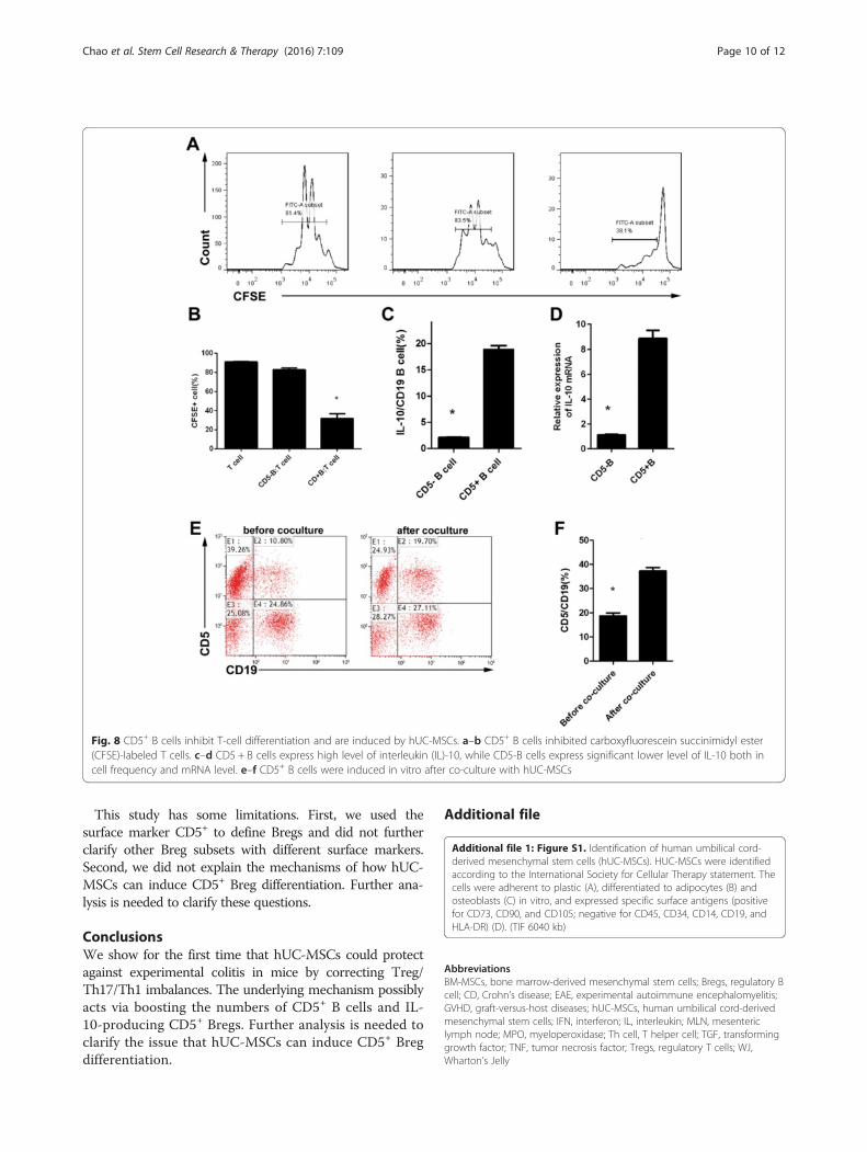
In vitro and in vivo study of CD5+ BregsCD5+ B cells have been shown to exist in the peritonealcavity [18]. Thus, CD5+ B cells were also analyzed inperitoneal lavage fluid to clarify its distribution in hUC-MSC-treated colitis mice. CD5+ B cells were isolated byflow cytometry and co-cultured with carboxyfluoresceinsuccinimidyl ester (CFSE) (Invitrogen, Inc., Carlsbad,CA, USA)-labeled T cells. Cell proliferation was then de-tected by flow cytometry (Beckman Coulter, Brea, CA,USA). For the in vivo functional study, CD5+ B cells wereisolated form spleen lymphocytes by flow cytometry. Iso-lated CD5+ B cells were transplanted through the tail veinof TNBS-induced colitis mice on day 3, in the peak of theinflammation. Disease severity and T-cell subgroups wereanalyzed according to the methods described above.
ResultsIdentification of hUC-MSCsThe hUC-MSCs showed a fibroblast-like morphology,expressed certain antigens (CD105, CD73, and CD90[≥95 % positive]), and lacked hematopoietic lineagemarkers (CD45, CD34, CD14, CD19, and HLA-DR [≤2 %positive]). After certain inducing environments of osteo-genesis and adiposis, the cells had the ability to differ-entiate multidirectionally to osteogenesis and adiposis(Additional file 1: Figure S1).
HUC-MSC therapy protected against TNBS-induced colitisTNBS-treated mice developed a severe illness, whichwas characterized by bloody diarrhea, rectal prolapse,pancolitis, and sustained weight loss. The mortality ofthe colitis model was 55 %, whereas the rates were 20 %in hUC-MSC-treated TNBS mice and 0 % in ethanoland naïve controls (Fig. 1a). Similar to ethanol controlmice, hUC-MSC-treated mice had rapidly recoveredbody weight loss (b-c) and milder inflammation. Theyalso exhibited significantly lower colitis, decreasedmacroscopic and histologic scores (Fig. 1d–f ), and lessneutrophil infiltration, as reflected by lower MPO activity(Fig. 1g). Macroscopic examination of TNBS colonsshowed hyperemia, edema, and inflammation that weresignificantly more severe than that in HUC-MSC-treatedmice (Fig. 1h, j). Histologic examination of the colonsshowed that HUC-MSC treatment reduced the TNBS-induced inflammation of the transmural area, depletion ofepithelial cells, and focal loss of crypts (Fig. 1i).
HUC-MSCs may migrate to the inflamed areasBy in vivo cell tracing, we found that hUC-MSCs accu-mulated in the peritoneal cavity of TNBS and ethanol
mice on day 1 (6 hours after colitis induction), whereasonly a few cells that were limited to the site of cell injec-tion could be found in naïve mice. At the peak of colitis,the cells still accumulated in the abdomen of TNBS micebut could not be detected in ethanol and naïve mice,thus suggesting relevance with colon inflammation. Atday 5, when recovery from colitis began, the number ofhUC-MSCs gradually decreased and could not be traced(Fig. 2). This phenomenon indicated that the MSCs maymigrate to the inflamed area and be associated with thedegree of inflammation.
HUC-MSCs altered Th cell and Treg imbalance incolitis miceWe further used flow cytometry to analyze immunologicchanges after hUC-MSC transplantation. In spleniclymphocytes, the Treg proportions were 4.31 ± 0.21 %,1.77 ± 0.32 %, 3.49 ± 1.20 %, and 5.05 ± 0.23 % in hUC-MSC-treated mice, TNBS mice, ethanol control mice,and naïve mice, respectively. Similar tendencies inMLN lymphocytes were observed among groups (Fig. 3).Furthermore, there was a significant decrease in Th1and Th17 cells in both splenic and MLN lymphocytesafter hUC-MSC therapy (Fig. 4). Th2 cells were rarelyexpressed, and no differences were observed after celltransfer. Levels of pro-inflammatory cytokines, such asTNF-α, IL-12, IL-6, IL-23, and IL-21, decreased signifi-cantly in the plasma after MSC treatment (P < 0.05). IL-17A, which is the main cytokine of Th17 cells, showed adecreased tendency (P = 0.09) (Fig. 5). IL-10 and TGF-β,which are associated with immunosuppression, were sig-nificantly higher in hUC-MSC-treated mice (P = 0.04 and0.02, respectively).
CD5+ B cells alleviated colitis in mice in vivo byregulating T-cell responsesWe found a significant increase in CD5+ B cells after celltransplantation in both splenic and MLN lymphocytes(Fig. 6), suggesting that CD5+ B cells might play a rolein immune regulation. Interestingly, CD5+ B cells mainlydistributed in the peritoneal cavity and decreased signifi-cantly in the colitis model; this was reversed by hUC-MSC therapy (Fig. 6). The above phenomenon led us tohypothesize that CD5+ B cells could regulate T-cellimbalance.To further clarify the function of CD5+ B cells, we
conducted both in vivo and in vitro studies. Adaptivetransfer of isolated CD5+ B cells had the same effect ashUC-MSC therapy (Fig. 7) and resulted in increased sur-vival, decreased disease activity, and lower macroscopicand histologic scores. Interestingly, this effect was asso-ciated with an alteration of Th/Treg balances (Fig. 7).The in vitro co-culture of hUC-MSCs and splenic lym-phocytes significantly increased the number of CD5+ B
Chao et al. Stem Cell Research & Therapy (2016) 7:109 Page 4 of 12

cells (Fig. 8). When co-cultured with CFSE-labeled Tcells, CD5+ B cells could inhibit T-cell proliferation andmay be associated with IL-10 (Fig. 8).
DiscussionIn the past decades, new drugs for CD have greatly im-proved the efficacy and quality of life of CD patients.However, even in the era of biologics, the fight against
CD is far from over, as there is still no ideal treatmentor cure. Findings from recent studies have confirmed thatimmunologic factors play a central role in the pathogen-esis of CD. We have previously found imbalances amongCD4+ T-cell subgroups in Chinese CD patients. Treg/Th1and Treg/Th17 ratios are associated with disease activityand are potential prognostic indicators for predicting CDrecurrence [19]. Thus, new treatments targeting immune
Fig. 1 Human umbilical cord-derived mesenchymal stem cell (hUC-MSC) therapy protects against TNBS-induced colitis. HUC-MSC therapy increasedthe survival rate of experimental colitis mice (a), decreased weight loss (b and c), alleviated colitis symptoms (d), and improved macroscopic (e) andhistologic (f) scores. Myeloperoxidase (MPO) activity is shown in (g), and pictures of the colon (h) with hematoxylin and eosin staining (i) of each groupare presented. The colon length which can reflect the inflammation was shown in (j). n = 20 for colitis model and treatment groups; n = 10 for modelcontrol and naïve mice; *P < 0.05 vs. MSC-treated mice. TNBS trinitrobenzenesulfonic acid
Chao et al. Stem Cell Research & Therapy (2016) 7:109 Page 5 of 12

Fig. 3 hUC-MSCs alter numbers of regulatory T cells (Tregs) in colitis mice. Lymphocytes were co-stained with anti-CD4 and anti-FoxP3 antibodiesand evaluated by flow cytometry. Tregs were defined as CD4+FoxP3+. The frequency of Tregs from the hUC-MSC-treated group was significantly lowerthan that in controls. Representative dot plots of Tregs in the spleen (a) and mesenteric lymph node (MLN) (c) of each group. Treg proportions are shownin (b) and (d). Data are presented as plots with P value. n= 9 for each group; *P< 0.05 vs. MSC-treated mice. hUC-MSC human umbilical cord-derivedmesenchymal stem cell, TNBS trinitrobenzenesulfonic acid
Fig. 2 MSCs migrate to the inflamed areas. In vivo tracing of MSCs on days 1, 3, and 5, the labeled cells were detected by the imaging system.Warmer colors indicate more accumulation of cells. MSCs mesenchymal stem cells, TNBS trinitrobenzenesulfonic acid
Chao et al. Stem Cell Research & Therapy (2016) 7:109 Page 6 of 12

imbalances over specific cytokines appear to be the besttherapeutic candidates for CD.MSC therapy has been promising in several inflamma-
tory diseases, including CD. Th1 and Treg imbalancesplay a central role [8, 17, 20–24] in CD. However, up to
now, there has only been one report concerning theeffect of hUC-MSCs on experimental colitis. This reportshowed that hUC-MSCSs could modulate Treg/Th1/Th17imbalances [14]. Similarly, we showed that hUC-MSCs in-deed alleviated experimental colitis. We also confirmed an
Fig. 4 hUC-MSCs alter T helper cell subgroups in colitis mice. Populations of Th1/Th2/Th17 cells as a proportion of total CD4+ cells wereevaluated by flow cytometry. Cells were co-stained with antibodies against CD3, CD8, interferon (IFN)-γ, interleukin (IL)-4, or IL-17 (CD4+ cells).CD3+CD8- cells were gated (a). CD4+IFN-γ+, CD4+IL-4+, and CD4+IL-17+ cells were defined as Th1, Th2, and Th17 cells, respectively. Representativedot plots are shown in panels b–c. Proportions of Th1 and Th17 cells in the four participant groups are shown in panels d–g. Data are presentedas plots with P value. n = 9 for each group; *P < 0.05 vs. MSC-treated mice. hUC-MSC human umbilical cord-derived mesenchymal stem cell, TNBStrinitrobenzenesulfonic acid
Fig. 5 Serum cytokine expression in each group. Th1 cell-related cytokines (tumor necrosis factor [TNF]-α and interleukin [IL]-12) and Th17 cell-relatedcytokines (IL-6, IL-23, and IL-21) were decreased after cell transplantation. IL-10 and transforming growth factor (TGF)-β were increasedafter cell transplantation. For IL-17A, there was a decreased tendency (P = 0.09). n = 6 for each group; *P < 0.05 vs. TNBS-treated mice. hUC-MSC humanumbilical cord-derived mesenchymal stem cell, TNBS trinitrobenzenesulfonic acid
Chao et al. Stem Cell Research & Therapy (2016) 7:109 Page 7 of 12

alteration of Treg/Th1/Th17 imbalances and cytokine pro-duction after cell transplantation. Strikingly, the number ofCD5+ B cells increased significantly after hUC-MSC ther-apy. In previous studies, Bregs showed an immunosuppres-sive role via the expression of CD5 and secretion of IL-10[25–29]. Although there are controversies concerning thedefinition and surface marker of Bregs, this subset is knownto exert immunosuppressive functions by regulating Thcells and Tregs, inducing T-cell and B-cell apoptosis, andinhibiting other immune-related cells, including CD8+ Tcells and natural killer T cells. Furthermore, Bregs are asso-ciated with autoimmune diseases, such as type I diabetes,GVHD, arthritis, and lupus [29, 30]. In colitis models, Bregscan exert anti-inflammatory effects [27, 28, 31]. In a T-cellreceptor-α-/- colitis model, B-cell depletion resulted insevere colitis, whereas the adaptive transfer of B cells allevi-ated the colitis symptoms. When CD1d was simultaneouslyknocked out, the symptoms of colitis became more severe,thereby suggesting that the B-cell subset can regulate
inflammation in the colitis models [27]. In IL-10-/- experi-mental colitis models, IL-10-expressing B cells alleviatecolitis [28]. Only two studies have reported increases in thenumbers of CD5+ B cells after BM-MSC transplantation.One study involved the EAE model and revealed an in-crease in numbers of CD5+ B cells after BM-MSC trans-plantation [12], however, this study did not further analyzethe potential mechanisms and function of CD5 + B cells.The other study from our team revealed that the CD5+ Bsubset is associated with the efficiency of BM-MSC therapyin GVHD [13]. In the present study, in vivo cell tracing re-sults show that the cells exerted their function specificallyin the inflamed areas. As CD5+ B cells mainly reside in theperitoneum, we analyzed the proportion of CD5+ B cells inperitoneal lavage fluid, splenic lymphocytes, and MLN lym-phocytes. Numbers of CD5+ B cells were significantly ele-vated after hUC-MSC transfer. We also conducted furtherin vivo and in vitro studies to verify whether MSCs canregulate CD5+ B cells to modulate the immune status of
Fig. 6 CD5+ B cells significantly increase after hUC-MSC therapy. Populations of CD5+ B cells were identified as CD5+CD19+ by flow cytometry.Representative dot plots of CD5+ B cells in the spleen (a), mesenteric lymph node (MLN) (c), and peritoneal cavity (e) are shown. Data are presented asplots with P value (b, d and f). n = 9 for each group; *P < 0.05 vs. MSC-treated mice. hUC-MSC human umbilical cord-derived mesenchymal stem cell,TNBS trinitrobenzenesulfonic acid
Chao et al. Stem Cell Research & Therapy (2016) 7:109 Page 8 of 12

the colitis model. Upon co-culture with hUC-MSCs, CD5+
B cells increased and inhibited T-cell proliferation in vitro.In addition, the adaptive transfer of CD5+ B cells into colitismice alleviated colitis in a similar manner as that achievedwith hUC-MSCs. Importantly, this effect was associatedwith the alteration of Th/Treg imbalances. CD5+ Bregs alsoinhibited T-cell proliferation in vitro. These findings suggestthat CD5+ B cells can exert a regulatory effect and protectagainst TNBS-induced colitis by regulating T-cell balances.Therefore, the effect of CD5+ B cells may be a new mech-anism of MSC therapy in CD. Furthermore, CD5+ B cellsproduced more IL-10 mRNA and protein, thus indicatingthat IL-10 may be an important factor that coordinates theimmunoregulatory effect of MSC-induced Bregs. It shouldbe emphasized that CD5 is also highly expressed onB-cell chronic lymphocytic leukemia (B-CLL). Thespecific surface marker ROR1 [32] and receptor oflysophosphatidic acid (LPA) [33, 34] are the markersof B-CLL but not normal B cell. To differentiate theimmunosuppressive B cells form B-CLL, we isolatedthe CD5+B cell from naïve, model and MSC-treatedmice and detected the expressions of LPA receptors
(LPARs) by real-time PCR. We found a very low ex-pression of LPARs (LPAR1-5) genes. Furthermore, theexpressions of LPARs in naïve, model and MSC-treated mice were similar. Therefore, we concludedthat the increased CD5 + B cell after MSCs trans-plantation were normal B cells.We did in vivo cell tracking using the luciferase
reporting system. We found that direct cell repair maybe less important than immune regulation, as the cellscould not be detected 5 days after cell transplantation.Interestingly, the cells mainly distributed in the abdo-men when transferred peritoneally in the colitis model,while they could not be tracked in naïve mice. One pos-sible reason is that the cells distributed to a wide rangeof tissues and organs in a random fashion in naïvemice, as shown in previous studies [14, 35]. But theIVIS system can track the cells when the signal is largeenough where cells were aggregated together. However,we knew very little about the destination of the cells inour analysis. Therefore, a more specific three-dimensionaloption with high resolution was needed to further localizethe cells in vivo.
Fig. 7 Adaptive transfer of CD5+ B cells alleviates TNBS-induced colitis. Sorted CD5+ B cells (a) were used for transplantation. After adaptivetransfer, the cells showed similar efficiency in colitis mice as hUC-MSCs (b–e), and this effect was associated with Treg/Th imbalances (f–i).hUC-MSC human umbilical cord-derived mesenchymal stem cell, MLN mesenteric lymph node, Th T helper, TNBS trinitrobenzenesulfonic acid
Chao et al. Stem Cell Research & Therapy (2016) 7:109 Page 9 of 12
This study has some limitations. First, we used thesurface marker CD5+ to define Bregs and did not furtherclarify other Breg subsets with different surface markers.Second, we did not explain the mechanisms of how hUC-MSCs can induce CD5+ Breg differentiation. Further ana-lysis is needed to clarify these questions.
ConclusionsWe show for the first time that hUC-MSCs could protectagainst experimental colitis in mice by correcting Treg/Th17/Th1 imbalances. The underlying mechanism possiblyacts via boosting the numbers of CD5+ B cells and IL-10-producing CD5+ Bregs. Further analysis is needed toclarify the issue that hUC-MSCs can induce CD5+ Bregdifferentiation.
Additional file
Additional file 1: Figure S1. Identification of human umbilical cord-derived mesenchymal stem cells (hUC-MSCs). HUC-MSCs were identifiedaccording to the International Society for Cellular Therapy statement. Thecells were adherent to plastic (A), differentiated to adipocytes (B) andosteoblasts (C) in vitro, and expressed specific surface antigens (positivefor CD73, CD90, and CD105; negative for CD45, CD34, CD14, CD19, andHLA-DR) (D). (TIF 6040 kb)
AbbreviationsBM-MSCs, bone marrow-derived mesenchymal stem cells; Bregs, regulatory Bcell; CD, Crohn’s disease; EAE, experimental autoimmune encephalomyelitis;GVHD, graft-versus-host diseases; hUC-MSCs, human umbilical cord-derivedmesenchymal stem cells; IFN, interferon; IL, interleukin; MLN, mesentericlymph node; MPO, myeloperoxidase; Th cell, T helper cell; TGF, transforminggrowth factor; TNF, tumor necrosis factor; Tregs, regulatory T cells; WJ,Wharton’s Jelly
Fig. 8 CD5+ B cells inhibit T-cell differentiation and are induced by hUC-MSCs. a–b CD5+ B cells inhibited carboxyfluorescein succinimidyl ester(CFSE)-labeled T cells. c–d CD5 + B cells express high level of interleukin (IL)-10, while CD5-B cells express significant lower level of IL-10 both incell frequency and mRNA level. e–f CD5+ B cells were induced in vitro after co-culture with hUC-MSCs
Chao et al. Stem Cell Research & Therapy (2016) 7:109 Page 10 of 12
AcknowledgementsWe thank Dr. Jifan Hu at Stanford University Medical School for editing themanuscript. We are also grateful to our collaborators in the multipledisciplinary teams at the First Affiliated Hospital, Sun Yat-sen University.
FundingThis study was supported by grants from the National Natural ScienceFoundation of China (81270473, 81301769, and 81470821), Guangdong Scienceand Technology (number 2014A020212128, number 2016A020214006), NaturalFund of Guangdong Province (2016A030310226), the Fundamental ResearchFunds for the Central Universities of Sun Yat-sen University (15ykpy12), and thePearl River S&T Nova Program of Guangzhou (number 201610010126).
Availability of data and materialsYes.
Authors’ contributionsKC and SHZ were responsible for study design, involved in all theexperiments, and drafted the manuscript. YQ, XYC, XRZ, CC, and YWP wereresponsible for the establishment of the animal model, cell tracing, and flowcytometry, and drafted the manuscript. RM, ZRZ, BLC, and YH played a rolein adaptive transfer, data analysis, and manuscript drafting. MPF, SHBH, andEE played a role in the analysis and interpretation of data, and revised themanuscript. APX and MHC designed the study, drafted, and revised themanuscript. All authors read and approved the manuscript.
Competing interestsThe authors declare that they have no competing interests.
Consent for publicationAll the authors have looked through the manuscript and approved thesubmission.
Ethics approval and consent to participateThis project was approved by the Human Ethics Committee of The FirstAffiliated Hospital at Sun Yat-sen University, and written informed consentwas obtained for umbilical cord collections.
Author details1Division of Gastroenterology, The First Affiliated Hospital, Sun Yat-senUniversity, Guangzhou 510080, People’s Republic of China. 2Division ofGastroenterology, The Sixth Affiliated Hospital, Sun Yat-sen University,Guangzhou 510655, People’s Republic of China. 3Center for Stem CellBiology and Tissue Engineering, The Key Laboratory for Stem Cells andTissue Engineering, Ministry of Education, Sun Yat-Sen University, Guangzhou510080, People’s Republic of China. 4Department of Immunology, WeizmannInstitute of Science, Rehovot 7610001, Israel.
Received: 7 May 2016 Revised: 13 July 2016Accepted: 26 July 2016
References1. Kaser A, Zeissig S, Blumberg RS. Inflammatory bowel disease. Annu Rev
Immunol. 2010;28:573–621.2. Drakos PE, Nagler A, Or R. Case of Crohn’s disease in bone marrow
transplantation. Am J Hematol. 1993;43:157–8.3. Salas A, Ricart E, Panes J. Cell therapies for inflammatory bowel diseases.
Expert Rev Gastroenterol Hepatol. 2009;3:321–4.4. Pittenger MF, Mackay AM, Beck SC, et al. Multilineage potential of adult
human mesenchymal stem cells. Science. 1999;284:143–7.5. Djouad F, Bouffi C, Ghannam S, et al. Mesenchymal stem cells: innovative
therapeutic tools for rheumatic diseases. Nat Rev Rheumatol. 2009;5:392–9.6. Le Blanc K, Rasmusson I, Sundberg B, et al. Treatment of severe acute graft-
versus-host disease with third party haploidentical mesenchymal stem cells.Lancet. 2004;363:1439–41.
7. Lalu MM, McIntyre L, Pugliese C, et al. Safety of cell therapy withmesenchymal stromal cells (SafeCell): a systematic review and meta-analysisof clinical trials. PLoS One. 2012;7:e47559.
8. Dryden GW. Overview of stem cell therapy for Crohn’s disease. Expert OpinBiol Ther. 2009;9:841–7.
9. Meirelles Lda S, Fontes AM, et al. Mechanisms involved in thetherapeutic properties of mesenchymal stem cells. Cytokine GrowthFactor Rev. 2009;20:419–27.
10. Troyer DL, Weiss ML. Wharton’s jelly-derived cells are a primitive stromal cellpopulation. Stem Cells. 2008;26:591–9.
11. Baksh D, Yao R, Tuan RS, et al. Comparison of proliferative and multilineagedifferentiation potential of human mesenchymal stem cells derived fromumbilical cord and bone marrow. Stem Cells. 2007;25:1384–92.
12. Guo Y, Chan KH, Lai WH, et al. Human mesenchymal stem cells upregulateCD1dCD5 (+) regulatory B cells in experimental autoimmuneencephalomyelitis. Neuroimmunomodulation. 2013;20:294–303.
13. Peng YW, Chen XY, Liu QL, et al. Mesenchymal stromal cells infusionsimprove refractory chronic graft versus host disease through an increaseof CD5+ regulatory B cells producing interleukin 10. Leukemia. 2015;93(3):636–46.
14. Lu L, Dong C, Chen X, et al. Human umbilical cord mesenchymal stem cellsameliorate mice trinitrobenzene sulfonic acid (TNBS)-induced colitis. CellTransplant. 2011;20:1395–408.
15. Dominici M, Le Blanc K, Mueller I, et al. Minimal criteria for defining multipotentmesenchymal stromal cells: The International Society for Cellular Therapyposition statement. Cytotherapy. 2006;8:315–7.
16. Wirtz S, Neufert C, Weigmann B, et al. Chemically induced mouse models ofintestinal inflammation. Nat Protoc. 2007;2:541–6.
17. Gonzalez MA, Gonzalez-Rey E, Rico L, et al. Adipose-derived mesenchymalstem cells alleviate experimental colitis by inhibiting inflammatory andautoimmune responses. Gastroenterology. 2009;136:978–89.
18. Berthelot JM, Jamin C, Amrouche K, et al. Regulatory B cells play a key rolein immune system balance. Joint Bone Spine. 2013;80:18–22.
19. Chao K, Zhong BH, Chen MH, et al. Imbalance of CD4+ T cell subgroups inCrohn’s disease and its relationship with disease activity and prognosis.J Gastroenterol Hepatol. 2014;29(10):1808–14.
20. Gonzalez-Rey E, Anderson P, Gonzalez MA, et al. Human adult stem cellsderived from adipose tissue protect against experimental colitis and sepsis.Gut. 2009;58:929–39.
21. Sanchez L, Gutierrez-Aranda I, Ligero G, et al. Enrichment of human ESC-derived multipotent mesenchymal stem cells with immunosuppressive andanti-inflammatory properties capable to protect against experimentalinflammatory bowel disease. Stem Cells. 2011;29:251–62.
22. Duijvestein M, Vos AC, Roelofs H, Wildenberg ME, et al. Autologous bonemarrow-derived mesenchymal stromal cell treatment for refractory luminalCrohn’s disease: results of a phase I study. Gut. 2010;59:1662–9.
23. Ciccocioppo R, Bernardo ME, Sgarella A, et al. Autologous bone marrow-derived mesenchymal stromal cells in the treatment of fistulising Crohn’sdisease. Gut. 2011;60:788–98.
24. Herreros MD, Garcia-Arranz M, Guadalajara H, et al. Autologous expandedadipose-derived stem cells for the treatment of complex cryptoglandularperianal fistulas: a phase III randomized clinical trial (FATT 1: fistulaAdvanced Therapy Trial 1) and long-term evaluation. Dis Colon Rectum.2012;55:762–72.
25. Yang M, Deng J, Liu Y, et al. IL-10-producing regulatory B10 cells amelioratecollagen-induced arthritis via suppressing Th17 cell generation. Am J Pathol.2012;180:2375–85.
26. Watanabe R, Ishiura N, Nakashima H, et al. Regulatory B cells (B10 cells) havea suppressive role in murine lupus: CD19 and B10 cell deficiencyexacerbates systemic autoimmunity. J Immunol. 2010;184:4801–9.
27. Mizoguchi A, Mizoguchi E, Smith RN, et al. Suppressive role of B cells in chroniccolitis of T cell receptor alpha mutant mice. J Exp Med. 1997;186:1749–56.
28. Mizoguchi AE, Mizoguchi H, Takedatsu EA, et al. Chronic intestinalinflammatory condition generates IL-10-producing regulatory B cell subsetcharacterized by CD1d upregulation. Immunity. 2002;16:219–30.
29. Mauri C, Ehrenstein MR. The ‘short’ history of regulatory B cells. TrendsImmunol. 2008;29:34–40.
30. Weber M, Stein P, Prufer S, et al. Donor and host B cell-derived IL-10contributes to suppression of graft-versus-host disease. Eur J Immunol.2014;44(6):1857–65.
31. Yanaba K, Yoshizaki A, Asano Y, et al. IL-10- producing regulatory B10 cellsinhibit intestinal injury in a mouse model. Am J Pathol.2011;178(2):735–43.
32. Mani R, Mao Y, Frissora FW, et al. Tumor antigen ROR1 targeted drugdelivery mediated selective leukemic but not normal B-cell cytotoxicity inchronic lymphocytic leukemia. Leukemia. 2015;29(2):346–55.
Chao et al. Stem Cell Research & Therapy (2016) 7:109 Page 11 of 12

33. Chiang CL, Chen SS, Lee SJ, et al. Lysophosphatidic acid induceserythropoiesis through activating lysophosphatidic acid receptor 3.Stem Cells. 2011;29(11):1763–73.
34. Kumar SA, Hu X, Brown M, Kuschak B, et al. Lysophosphatidic acid receptorexpression in chronic lymphocytic leukemia leads to cell survival mediatedthough vascular endothelial growth factor expression. Leuk Lymphoma.2009;50(12):2038–48.
35. Devine SM, Cobbs C, Jennings M, et al. Mesenchymal stem cells distributeto a wide range of tissues following systemic infusion into nonhumanprimates. Blood. 2003;101(8):2999–3001.
• We accept pre-submission inquiries
• Our selector tool helps you to find the most relevant journal
• We provide round the clock customer support
• Convenient online submission
• Thorough peer review
• Inclusion in PubMed and all major indexing services
• Maximum visibility for your research
Submit your manuscript atwww.biomedcentral.com/submit
Submit your next manuscript to BioMed Central and we will help you at every step:
Chao et al. Stem Cell Research & Therapy (2016) 7:109 Page 12 of 12
![Residuated implications derived from quasi-overlap functions ...arXiv:2002.12267v1 [cs.LO] 27 Feb 2020 Residuated implications derived from quasi-overlap functions on lattices Rui](https://static.fdocuments.fr/doc/165x107/6065d0f065c50f701a4e3e26/residuated-implications-derived-from-quasi-overlap-functions-arxiv200212267v1.jpg)