Department of Pathobiology and Population Sciences, Royal ...72 and fitness during handling and...
16
1 Long title: The occurrence of tarsal injuries in male mice of C57BL/6N substrains in multiple 2 international mouse facilities 3 4 Short title: Tarsal injury in C57BL/6N male mice 5 6 Eleanor Herbert 2 , Michelle Stewart 1 , Marie Hutchison 1 , Ann M. Flenniken 4,5 , Dawei Qu 4,5 , 7 Lauryl M. J. Nutter 4,6 , Colin McKerlie 4,6 , Liane Hobson 1 , Brenda Kick 3 , Bonnie Lyons 3 , Jean- 8 Paul Wiegand 3 , Rosalinda Doty 3 , Juan Antonio Aguilar-Pimentel 7 , Martin Hrabe de Angelis 7,8,9 , 9 Mary Dickinson 10 , John Seavitt 10 , Jacqueline K. White 3 , Cheryl L Scudamore 1 , Sara Wells 1* 10 11 12 13 Affiliations 14 1 Mary Lyon Centre, MRC Harwell Institute, Harwell Campus, Oxfordshire, OX11 0RD, UK 15 2 Department of Pathobiology and Population Sciences, Royal Veterinary College, Hertfordshire, 16 UK AL9 7TA, UK 17 3 The Jackson Laboratory, 600 Main Street, Bar Harbor, Maine 04609, USA 18 4 The Centre for Phenogenomics, Toronto, ON, Canada, M5T 3H7 19 5 Lunenfeld-Tanenbaum Research Institute, Sinai Health, 600 University Avenue, Toronto, 20 ON, Canada, M5G 1X5 21 6 The Hospital for Sick Children, 555 University Avenue, Toronto, ON, Canada, M5G 1X8 22 7 German Mouse Clinic, Institute of Experimental Genetics, Helmholtz Zentrum München, 23 Ingolstädter Landstraße 1, 85764 Neuherberg, Germany 24 8 School of Life Science Weihenstephan, Technische Universität München, Alte Akademie 8, 25 85354 Freising, Germany 26 9 German Center for Diabetes Research (DZD), Ingolstädter Landstr. 1, 85764 Neuherberg, 27 Germany 28 10 Baylor College of Medicine, One Baylor Plaza, Houston, Texas 77030, USA . CC-BY 4.0 International license preprint (which was not certified by peer review) is the author/funder. It is made available under a The copyright holder for this this version posted February 25, 2020. . https://doi.org/10.1101/2020.02.25.964254 doi: bioRxiv preprint
Transcript of Department of Pathobiology and Population Sciences, Royal ...72 and fitness during handling and...
1 Long title: The occurrence of tarsal injuries in male mice of C57BL/6N substrains in multiple
2 international mouse facilities
3
4 Short title: Tarsal injury in C57BL/6N male mice
5
6 Eleanor Herbert2, Michelle Stewart1, Marie Hutchison1, Ann M. Flenniken4,5, Dawei Qu4,5,
7 Lauryl M. J. Nutter4,6, Colin McKerlie4,6, Liane Hobson1, Brenda Kick3, Bonnie Lyons3, Jean-
8 Paul Wiegand3, Rosalinda Doty3, Juan Antonio Aguilar-Pimentel7, Martin Hrabe de Angelis7,8,9,
9 Mary Dickinson10, John Seavitt10, Jacqueline K. White3, Cheryl L Scudamore1, Sara Wells1*
10
11
12
13 Affiliations
14 1 Mary Lyon Centre, MRC Harwell Institute, Harwell Campus, Oxfordshire, OX11 0RD, UK
15 2 Department of Pathobiology and Population Sciences, Royal Veterinary College, Hertfordshire,
16 UK AL9 7TA, UK
17 3 The Jackson Laboratory, 600 Main Street, Bar Harbor, Maine 04609, USA
18 4 The Centre for Phenogenomics, Toronto, ON, Canada, M5T 3H7
19 5 Lunenfeld-Tanenbaum Research Institute, Sinai Health, 600 University Avenue, Toronto,
20 ON, Canada, M5G 1X5
21 6 The Hospital for Sick Children, 555 University Avenue, Toronto, ON, Canada, M5G 1X8
22 7German Mouse Clinic, Institute of Experimental Genetics, Helmholtz Zentrum München,
23 Ingolstädter Landstraße 1, 85764 Neuherberg, Germany
24 8School of Life Science Weihenstephan, Technische Universität München, Alte Akademie 8,
25 85354 Freising, Germany
26 9German Center for Diabetes Research (DZD), Ingolstädter Landstr. 1, 85764 Neuherberg,
27 Germany
28 10Baylor College of Medicine, One Baylor Plaza, Houston, Texas 77030, USA
.CC-BY 4.0 International licensepreprint (which was not certified by peer review) is the author/funder. It is made available under aThe copyright holder for thisthis version posted February 25, 2020. . https://doi.org/10.1101/2020.02.25.964254doi: bioRxiv preprint

29
30 * corresponding author
31 Email: [email protected]
.CC-BY 4.0 International licensepreprint (which was not certified by peer review) is the author/funder. It is made available under aThe copyright holder for thisthis version posted February 25, 2020. . https://doi.org/10.1101/2020.02.25.964254doi: bioRxiv preprint
32 Abstract
33 Dislocation in hindlimb tarsals are being observed at a low, but persistent frequency in adult
34 male mice from C57BL/6N substrains. Clinical signs included a sudden onset of mild to severe
35 unilateral or bilateral tarsal abduction, swelling, abnormal hindlimb morphology and lameness.
36 Contraction of digits and gait abnormalities were noted in multiple cases. Radiographical and
37 histological examination revealed caudal dislocation of the calcaneus and partial dislocation
38 of the calcaneoquartal (calcaneous-tarsal bone IV) joint. The detection, frequency, and cause
39 of this pathology in five large mouse production and phenotyping centres (MRC Harwell, UK;
40 The Jackson Laboratory, USA; The Centre for Phenogenomics, Canada; German Mouse
41 Clinic, Germany; Baylor College of Medicine, USA) are discussed.
42
43 Introduction
44 Inbred strains of laboratory mice are used to standardise the genetic background of mutant
45 mouse strains to reduce data variability. Produced by >20 consecutive generations of sibling
46 mating, the controlled homogeneity of inbred strains such as C57BL/6N is accompanied by
47 the fixing of spontaneous mutations in inbred genomes. Many monogenic mutations have
48 been identified in inbred mouse strains, including those causing retinal degeneration in C3H
49 strains (Schmidt, Lolley, & Racz, 1973) and age-related deafness in C57BL/6 strains
50 (Johnson, Erway, Cook, Willott, & Zheng, 1997). The characterisation of these mutations has
51 allowed their impact on individual research programs to be assessed and alternative genetic
52 backgrounds used if they interfered with the primary purpose of the studies. Conversely there
53 are reports of sporadic, low-level defects in inbred lines which are likely due to oligogenic or
54 polygenic effects and exhibit variable penetrance thus only observed or measured in a
55 proportion of the population of an inbred colony (Sundberg, Silva, Li, Cox, & King, 2004).
56 These include complex behaviours such as aggression (Miczek, Maxson, Fish, & Faccidomo,
57 2001), hyperactivity (Võikar, Kõks, Vasar, & Rauvala, 2001), morphological anomalies such
58 as sternal segment dislocation (Adissu, Medhanie, Morikawa, & White, 2015) and
.CC-BY 4.0 International licensepreprint (which was not certified by peer review) is the author/funder. It is made available under aThe copyright holder for thisthis version posted February 25, 2020. . https://doi.org/10.1101/2020.02.25.964254doi: bioRxiv preprint

59 developmental defects such as hydrocephalus (https://www.jax.org/news-and-
60 insights/2003/july/hydrocephalus-in-laboratory-mice).
61 Knowledge of the predisposition of mouse strains to such issues is not only essential for the
62 care and welfare of mice but is an important consideration in phenotyping programs. It is
63 crucial to distinguish incidental effects caused by genetic background, from outcomes arising
64 because of an experimental paradigm (e.g. genetic mutation or physiological challenge) or a
65 combination of background and paradigm together. The International Mouse Phenotyping
66 Consortium (IMPC) (www.mousephenotype.org) is generating a genetically altered (GA)
67 mouse strain carrying a null allele for each protein-coding gene in the mouse to study
68 mammalian gene function (Brown & Moore, 2013). GA strains for this programme are
69 generated on the C57BL/6N genetic background and phenotyping is performed at an early
70 adult time point (up to 17 weeks) and a late adult time point (after 12-18 months) for a subset
71 of strains. Phenotyping and husbandry protocols include the regular assessment of welfare
72 and fitness during handling and cage-changing, and motor function during phenotyping tests
73 (Rogers et al., 2001).
74 In this study, we report the recurrent observation of abnormal hindlimb morphology,
75 accompanied by lameness, in group -housed male mice of C57BL/6N substrains. This report
76 describes the nature of the injury and discusses possible aetiologies. We also provide an
77 estimate of the frequency of occurrence from five large mouse genetics centres in four different
78 countries across two continents and highlight potential consequences for projects where
79 prolonged co-housing of male C57BL/6N mice is a necessity.
80
81 Materials and Methods
82 Ethics Statement
83 Mice were examined for tarsal injury at five mouse phenotyping centres:
84 MRC Harwell: Animal studies are performed in compliance with guidelines issued by the
85 Medical Research Council (MRC) (UK) in “Responsibility in the Use of Animals for Medical
86 Research” (July 1993). The care and use of all mice in this study were in accordance with
.CC-BY 4.0 International licensepreprint (which was not certified by peer review) is the author/funder. It is made available under aThe copyright holder for thisthis version posted February 25, 2020. . https://doi.org/10.1101/2020.02.25.964254doi: bioRxiv preprint
87 UK Home Office regulations, The Animals (Scientific Procedures) Act 1986 Amendment
88 Regulations 2012 (SI 4 2012/3039), and approved by the MRC Harwell Institute Animal
89 Welfare and Ethical Review Body.
90 The Centre for Phenogenomics (TCP): All experimental procedures were approved by the
91 TCP Animal Care Committee (AUP 0279) and were conducted in accordance with the
92 guidelines of the Canadian Council on Animal Care.
93 TheJackson Laboratory (JAX): All experimental procedures were carried out under Protocols
94 14004 and 11005 approved by the JAX Institutional Animal Care and Use Committee (IACUC)
95 with NIH Office of Laboratory Animal Welfare (OLAW) assurance number D16-00170 and
96 Accreditation AAALAC #000096.
97 German Mouse Clinic (GMC): All animal experiments were carried out in accordance with
98 German legal guidelines and following the approval of the responsible animal welfare
99 authorities and the Ethics Board of the District Government of Upper Bavaria, Germany
100 (approval number 46-2016).
101 Baylor College of Medicine (BCM): Animal experiments were carried out in accordance with
102 research protocol AN-5896 and approved by the BCM Institutional Animal Care and Use
103 Committee. The Animal Welfare Assurance at BCM is approved by the Office of Laboratory
104 Animal Welfare (OLAW), and meet the requirements of the Public Health Service Policy on
105 Humane Care and Use of Laboratory Animals (assurance number D16-00475).
106
107 C57BL/6N substrains used in this study
.CC-BY 4.0 International licensepreprint (which was not certified by peer review) is the author/funder. It is made available under aThe copyright holder for thisthis version posted February 25, 2020. . https://doi.org/10.1101/2020.02.25.964254doi: bioRxiv preprint
108 Mice used at MRC Harwell were C57BL/6NTac mice purchased from originally from Taconic
109 Biosciences, USA and subsequently bred at MRC Harwell. Mice examined at The Jackson
110 Laboratory are sourced from an in-house maintained colony of C57BL/6NJ. Mice used at The
111 Centre for Phenogenomics are C57BL/6NCrl, purchased from Charles River Laboratories,
112 USA and subsequently bred at TCP. Mice at the German Mouse Clinic were C57BL/6NTac
113 purchased from Taconic Biosciences ,Germany and C57BL/6NCrl purchased from Charles
114 River Laboratories, Germany. Mice at Baylor College of Medicine were C57BL/6NJ originally
115 purchased from Charles River Laboratories, USA and subsequently bred at this facility.
116
117 Mouse housing conditions
118 Housing conditions in each institution are listed in TABLE S1 of supplementary figures.
119
120 All mice were given food and water ad libitum. Adult mice were humanely sacrificed by an
121 overdose of anaesthetic, overdose of carbon dioxide, or by cervical dislocation (according to
122 relevant national and local protocols and guidelines.
123
124 Clinical examination
125 Routine animal care and welfare checks in all facilities involved visually inspecting the mice
126 as part of a daily check and regular handling (typically no less than once every 14 days) during
127 cage changing, or during phenotyping. Mice with an abnormal gait and/or locomotor deficit
128 accompanied by abnormal hindlimb morphology and swelling or reddening of the tarsus were
129 euthanised or selected increased observation to ensure no further deterioration in the welfare
130 of the mouse.
131
132 Radiography
133 Lateral views of the affected and contralateral tarsi were taken of representative animals under
134 isoflurane anaesthesia by digital radiography at 26 kV for 3 s using a Faxitron MX‐20 digital
135 X‐ray system or a Faxitron X-Ray Model Ultrafocus 100 (both from Faxitron X‐ray Corporation,
.CC-BY 4.0 International licensepreprint (which was not certified by peer review) is the author/funder. It is made available under aThe copyright holder for thisthis version posted February 25, 2020. . https://doi.org/10.1101/2020.02.25.964254doi: bioRxiv preprint
136 Lincolnshire, IL, USA). X‐ray images were processed using the DicomWorks software
137 (http://www.dicomworks.com/).
138
139 Histopathology
140 Immediately following euthanasia, tissues from selected mice were fixed in 10% neutral
141 buffered formalin for a minimum of 24 hours. Following fixation, the hindlimbs (affected and
142 contralateral) were stripped of soft tissues, decalcified in formic acid for 96 hours, and
143 processed routinely for histopathologic evaluation. Subsequently, 4–5 μm thick, mid-sagittal
144 sections were stained with haematoxylin and eosin (H&E) for evaluation. Representative
145 images were acquired using an Olympus BX43 microscope with a Micropix Elite 5MP camera
146 and Cytocam software v1.6. All histologic evaluations were performed by a board-certified
147 veterinary pathologist.
148
149 Results
150 Clinical examination
151 A proportion of male mice of C57BL/6N substrains, housed in social groups were observed to
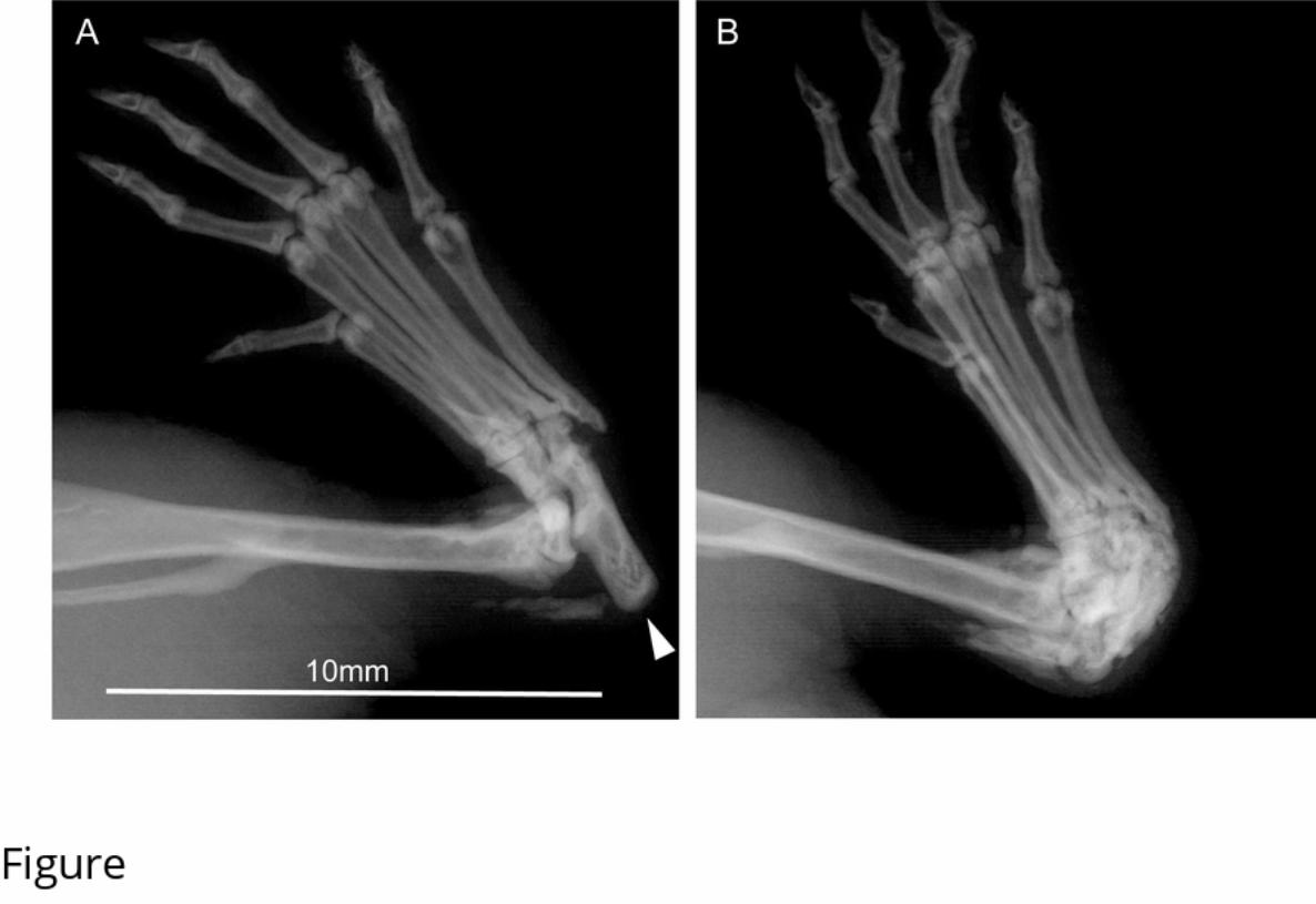
152 have clinical signs of abnormal hindlimb morphology, together with swelling or reddening of
153 the tarsus and often an abnormal gait. Similar numbers of C57BL/6N females were assessed
154 and no tarsus, hind paw, or gait abnormalities were observed. Gait abnormalities in males
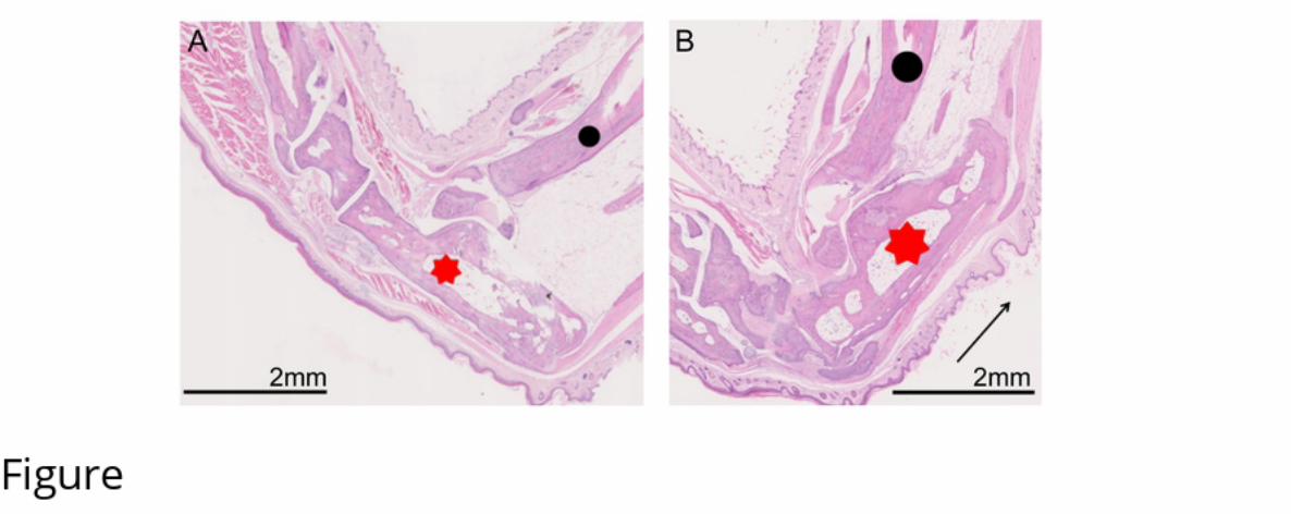
155 ranged from limping with a reduced amount of weight bearing on the affected limb to complete
156 non-weight bearing. Grossly affected tarsi showed a loss of the abrupt right angle formed from
157 the calcaneus and the calcaneal tendon, and there was variable soft tissue swelling
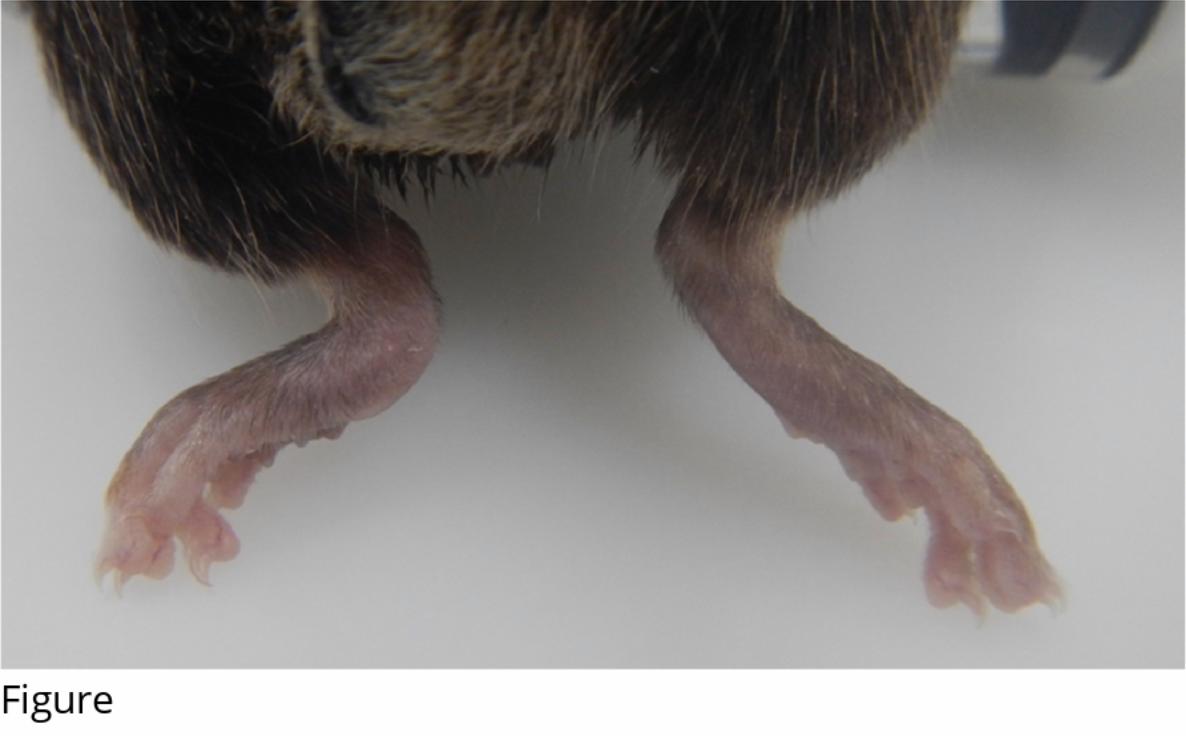
158 sometimes accompanied with redness (Figure 1). Whilst the majority of affected mice had only
159 one abnormal hind paw, 1/21 at MRC Harwell, 4/21 at TCP and 15/58 at JAX presented with
160 bilateral tarsal abnormalities.
161 Radiography confirmed caudal dislocation of the calcaneus and new periosteal bone formation
162 (Figure 2). In some animals, there was also calcification within the distal calcaneal tendon.
.CC-BY 4.0 International licensepreprint (which was not certified by peer review) is the author/funder. It is made available under aThe copyright holder for thisthis version posted February 25, 2020. . https://doi.org/10.1101/2020.02.25.964254doi: bioRxiv preprint

163 Histopathology
164 Histopathological examination identified caudo-dorsal dislocation of the calcaneus with
165 concurrent partial dislocation and hyperextension of the calcaneoquartal joint. In more chronic
166 lesions, the calcaneoquartal joint progressed to new bone formation (Figure 3). There was no
167 difference in overall dislocation of the calcaneus in acute versus chronic lesions.
168
169 Frequency and variability of occurrence
170 C57BL/6N mice bred for the IMPC late adult phenotyping programme were examined for tarsal
171 injuries at five international mouse research centres. These mice included GA strains from a
172 wide range of mutant lines examined by the IMPC as well as baseline wild type controls. Due
173 to differences in individual institutional protocol, the ages of the cohorts vary. The frequency
174 of occurrence was between 1.7% and 12.1% of male mice examined between the ages
175 indicated.
SubstrainNumber of mice (male)
Age range
(weeks)
Number affected
Earliest age
affected (weeks)
Frequency (%)
The Centre for Phenogenomics, Canada C57BL/6NCrl 235 5-59 21 20 8.9
The Jackson Laboratory, USA C57BL/6NJ 1440 4-78 58 11 4.0
MRC Harwell Institute, UK C57BL/6NTac 174 16-59 21 18 12.1
GMC Helmholtz Zentrum, Germany
C57BL/6NTac and
C57BL/6NCrl 413 4-62 7 45 1.7
Baylor College of Medicine, USA C57BL/6NJ 250 16-52 30 20 12
176
177 Husbandry, housing, and strains affected
.CC-BY 4.0 International licensepreprint (which was not certified by peer review) is the author/funder. It is made available under aThe copyright holder for thisthis version posted February 25, 2020. . https://doi.org/10.1101/2020.02.25.964254doi: bioRxiv preprint
178 Further observations were made and recorded which informed the aetiology of the incidence
179 of the tarsal injury in C57BL/6N mice.
180 Female mice: Equivalent numbers of females of the same strain which were part of
181 the same programme of work were also examined but no similar injury was reported.
182 Males in mating cages or singly-housed: No injury was observed in 584 C57BL/6Ntac
183 males in either mating cages (with one or two females) or single-housed examined
184 between the ages of 16 and 64 weeks (average age 24 weeks) at Harwell.
185
186 Discussion
187 Here we report the observation of tarsal injury in male mice with a C57BL/6N genetic
188 background occurring at five large and geographically-dispersed mouse facilities. These
189 injuries were observed in a number of different mutant strains and several wild-type substrains
190 indicating a predisposition for such lesions in mice of C57BL/6N ancestry. A similar deformity
191 has been reported in STR/ort mice with a known genetic abnormality predisposing them to
192 chronic arthropathy used as a model for studying osteoarthritis (Mason et al., 2001). In the
193 STR/ort mice, lameness and hind paw deformity also affected predominantly male mice
194 although the incidence rate was far higher and occurred from a younger age compared with
195 our observations. The radiographic findings and histopathology are consistent with an injury
196 caused by frequent high load tension from the calcaneal tendon through its insertion to the
197 calcaneous leading to a breakdown of the plantar ligaments supporting the calcaneoquartal
198 joint which are weakened in this model by a known collagen abnormality (Staines et al., 2016).
199 There is no known underlying abnormality in the C57BL/6N strains reported here and so it is
200 hypothesised that the lesion is caused by application of an abnormally high load/force through
201 the calcaneal tendon because of behavioural or husbandry practices. It should be noted that
202 all animals in this study are fed on regular maintenance or breeding diets and not on high-fat
203 or obesity inducing regimes.
.CC-BY 4.0 International licensepreprint (which was not certified by peer review) is the author/funder. It is made available under aThe copyright holder for thisthis version posted February 25, 2020. . https://doi.org/10.1101/2020.02.25.964254doi: bioRxiv preprint

204 The type of lesion we identified was restricted to group-housed males and its occurrence
205 became more prevalent as the mice aged. However, this may represent an increased
206 opportunity for this injury to occur over time, rather than an increased predisposition/weakness
207 in older males. The absence of any such injury in female mice socially-housed for the same
208 experimental purposes and for the same length of time indicates that this is a sexually
209 dimorphic effect.
210 As these injuries were observed in three different C57BL/6N substrains it is possible that this
211 genetic background is predisposed to tarsal injuries. Male C57BL/6 mice are widely reported
212 to display aggressive behaviours towards cage-mates (Lidster, Owen, Browne, & Prescott,
213 2019). Both threat (thrust and mounting) and aggressive behaviours (boxing, parrying,
214 fighting) are associated with establishing and maintaining dominance hierarchies in group
215 house male mice. Each of these behaviours involve rearing that requires repeated plantar
216 flexion of the hind paw at the tarsus, initiated by high load tension from the common calcaneal
217 tendon. Sporadic and frequent bouts of fighting have also been associated with an increased
218 mechanical load on male tibiae in C57BL/6J mice (Meakin et al., 2013), a strain related to
219 C57BL/6N. However, it is unclear whether the causal feature of the injury we observed is an
220 inherent weakness in the tarsal joint, a consequence of a behavioural characteristic of
221 C57BL/6N male mice, their interaction with the environment, or combinations of these factors.
222 Significant differences in the frequency of observation of tarsal injury between centres may
223 present any number of variances between housing and animal care regimes between the
224 facilities. Investigations into different husbandry protocols may provide insight into ways to
225 reduce occurrence in the future. Euthanasia following discovery of the injury described may
226 have substantial consequences to the study being undertaken. Disruption of an established
227 cage-group by removing and individual may lead to further perturbations in both the behaviour
228 of the existing animals or to the experimental design itself with a reduction in data collected
229 and the potential loss of statistical power.
230 In summary, this report provided a description of an injury to group-housed male C57BL/6N
231 mice observed in five different mouse centres from studies involving large numbers of animals.
.CC-BY 4.0 International licensepreprint (which was not certified by peer review) is the author/funder. It is made available under aThe copyright holder for thisthis version posted February 25, 2020. . https://doi.org/10.1101/2020.02.25.964254doi: bioRxiv preprint
232 It is likely that a similar incidence may occur undetected, or be being attributed to experimental
233 protocols, in other facilities using these substrains. The implications of these findings will be
234 study-dependent but have the potential to affect phenotyping results or cause an increase in
235 attrition for ageing studies, resulting in insufficient animals completing the studies. The
236 information that reported here should be used to assist future experimental design for
237 longitudinal studies especially those involving measurements of gait and motor skills.
238
239
240 Acknowledgements
241 We thank staff at the five facilities participating in this study for animal husbandry support,
242 necropsy, and histology services. Dr Dona Reddiar for assistance with the manuscript.
243 Funding for the IMPC studies at each centre is provided by NIH grant UM1HG006348-07S2
244 (MRC Harwell and Baylor College of Medicine), MRC grant A410 (MRC Harwell), NIH grant
245 UM1 OD023221 (TCP) and NIH UM1 OD0232221OD023222 (JAX), BMBF Infrafrontier grant
246 01KX1012 (GMC) and EU Horizon2020: IPAD-MD funding 653961 (GMC).
247
248
249 References
250 Adissu, H. A., Medhanie, G. A., Morikawa, L., & White, J. K. (2015). Right Ventricular
251 Epicardial Fibrosis in Mice With Sternal Segment Dislocation, 52(5), 967–976.
252 https://doi.org/10.1177/0300985814552108
253 Johnson, K. R., Erway, L. C., Cook, S. A., Willott, J. F., & Zheng, Q. Y. (1997). A major gene
254 affecting age-related hearing loss in C57BL/6J mice. Hearing Research, 114(1–2), 83–92.
255 https://doi.org/10.1016/S0378-5955(97)00155-X
256 Lidster, K., Owen, K., Browne, W. J., & Prescott, M. J. (2019). Cage aggression in group-
257 housed laboratory male mice: an international data crowdsourcing project. Scientific
258 Reports, 9(1), 1–12. https://doi.org/10.1038/s41598-019-51674-z
259 Meakin, L. B., Sugiyama, T., Galea, G. L., Browne, W. J., Lanyon, L. E., & Price, J. S.
.CC-BY 4.0 International licensepreprint (which was not certified by peer review) is the author/funder. It is made available under aThe copyright holder for thisthis version posted February 25, 2020. . https://doi.org/10.1101/2020.02.25.964254doi: bioRxiv preprint
260 (2013). Male mice housed in groups engage in frequent fi ghting and show a lower response
261 to additional bone loading than females or individually housed males that do not fi ght. Bone,
262 54(1), 113–117. https://doi.org/10.1016/j.bone.2013.01.029
263 Miczek, K. A., Maxson, S. C., Fish, E. W., & Faccidomo, S. (2001). Aggressive behavioral
264 phenotypes in mice. Behavioural Brain Research, 125(1–2), 167–181.
265 https://doi.org/10.1016/S0166-4328(01)00298-4
266 Schmidt, S. Y., Lolley, R. N., & Racz, E. (1973). Cyclic-nucleotide phosphodiesterase an
267 early defect in inherited retinal degeneration of C3H mice. Journal of Cell Biology, 57(1),
268 117–123. https://doi.org/10.1083/jcb.57.1.117
269 Sundberg, J. P., Silva, A. K. A., Li, A. R., Cox, A. G. A., & King, L. E. (2004). Adult-Onset
270 Alopecia Areata Is a Complex Polygenic Trait in the C3H / HeJ Mouse Model. Journal of
271 Investigative Dermatology, 123(2), 294–297. https://doi.org/10.1111/j.0022-
272 202X.2004.23222.x
273 Võikar, V., Kõks, S., Vasar, E., & Rauvala, H. (2001). Strain and gender differences in the
274 behavior of mouse lines commonly used in transgenic studies. Physiology and Behavior,
275 72(1–2), 271–281. https://doi.org/10.1016/S0031-9384(00)00405-4
276
277 Supplementary Table
278 (see excel)
279
280 Figure legends
281 Figure 1. Dorsal view of unaffected right tarsus and rounded, swollen tarsus (readers left)
282
283 Figure 2. (a) Xray image of the normal position of the calcaneus (arrow) within the tarsal joint
284 and (b) with caudo-dorsal dislocation of the calcaneus
285
286 Figure 3. (a) The unaffected tarsus with the calcaneus (red star) forming an approximate 90°
287 angle with the tibia (black circle), (b) Affected tarsus (red star) with dislocation of the calcaneus
.CC-BY 4.0 International licensepreprint (which was not certified by peer review) is the author/funder. It is made available under aThe copyright holder for thisthis version posted February 25, 2020. . https://doi.org/10.1101/2020.02.25.964254doi: bioRxiv preprint

288 caudo-dorsally to form an approximate 15° angle with the tibia (black circle). The black arrow
289 indicates the direction of movement of the calcaneus. Scale bar = 2mm.
290
.CC-BY 4.0 International licensepreprint (which was not certified by peer review) is the author/funder. It is made available under aThe copyright holder for thisthis version posted February 25, 2020. . https://doi.org/10.1101/2020.02.25.964254doi: bioRxiv preprint
.CC-BY 4.0 International licensepreprint (which was not certified by peer review) is the author/funder. It is made available under aThe copyright holder for thisthis version posted February 25, 2020. . https://doi.org/10.1101/2020.02.25.964254doi: bioRxiv preprint
.CC-BY 4.0 International licensepreprint (which was not certified by peer review) is the author/funder. It is made available under aThe copyright holder for thisthis version posted February 25, 2020. . https://doi.org/10.1101/2020.02.25.964254doi: bioRxiv preprint
.CC-BY 4.0 International licensepreprint (which was not certified by peer review) is the author/funder. It is made available under aThe copyright holder for thisthis version posted February 25, 2020. . https://doi.org/10.1101/2020.02.25.964254doi: bioRxiv preprint
![Transcription Analysis of Arabidopsis Membrane …Transcription Analysis of Arabidopsis Membrane Transporters and Hormone Pathways during Developmental and Induced Leaf Senescence1[W]](https://static.fdocuments.fr/doc/165x107/609e4ae6b5f9cd4bb26ab6d5/transcription-analysis-of-arabidopsis-membrane-transcription-analysis-of-arabidopsis.jpg)