Correction - PNASA fraction of the phosphorylated peptides was subjected to NuPAGE to ensure...
9
Protein pyrophosphorylation by inositol pyrophosphates is a posttranslational event Rashna Bhandari*, Adolfo Saiardi † , Yousef Ahmadibeni ‡ , Adele M. Snowman*, Adam C. Resnick* § , Troels Z. Kristiansen ¶ , Henrik Molina ¶ , Akhilesh Pandey ¶ , J. Kent Werner, Jr.*, Krishna R. Juluri*, Yong Xu , Glenn D. Prestwich , Keykavous Parang ‡ , and Solomon H. Snyder* , ** ††‡‡ *The Solomon H. Snyder Department of Neuroscience, Departments of **Pharmacology and Molecular Sciences, †† Psychiatry and Behavioral Sciences, and ¶ McKusick–Nathans Institute of Genetic Medicine and Department of Biological Chemistry, Johns Hopkins University School of Medicine, 725 North Wolfe Street, Baltimore, MD 21205; † Medical Research Council (MRC) Cell Biology Unit and Laboratory for Molecular Cell Biology, Department of Biochemistry and Molecular Biology, University College London, Gower Street, London WC1E 6BT, United Kingdom; ‡ Department of Biomedical and Pharmaceutical Sciences, College of Pharmacy, University of Rhode Island, Kingston, RI 02881; and Department of Medicinal Chemistry, University of Utah, 419 Wakara Way, Suite 205, Salt Lake City, UT 84108 Contributed by Solomon H. Snyder, August 3, 2007 (sent for review July 16, 2007) In a previous study, we showed that the inositol pyrophosphate diphosphoinositol pentakisphosphate (IP7 ) physiologically phosphor- ylates mammalian and yeast proteins. We now report that this phosphate transfer reflects pyrophosphorylation. Thus, proteins must be prephosphorylated by ATP to prime them for IP7 phosphor- ylation. IP7 phosphorylates synthetic phosphopeptides but not if their phosphates have been masked by methylation or pyrophosphoryla- tion. Moreover, IP 7 phosphorylated peptides are more acid-labile and more resistant to phosphatases than ATP phosphorylated peptides, indicating a different type of phosphate bond. Pyrophosphorylation may represent a novel mode of signaling to proteins. inositol polyphosphate protein phosphorylation I nositol phosphates have diverse biologic roles with the best known, inositol-1,4,5-trisphosphate (IP 3 ), releasing intracellu- lar calcium (1). Inositol pyrophosphates (also designated inositol diphosphates), exemplified by diphosphoinositol pentakisphos- phate (5-PP-I(1,2,3,4,6)P 5 , hereafter called IP 7 ) and bis- diphosphoinositol tetrakisphosphate ([PP] 2 -IP 4 , IP 8 ) (2–4) are formed by a group of three inositol hexakisphosphate (IP 6 ) kinases (IP6Ks) (5–7). IP6K and inositol pyrophosphates par- ticipate in diverse physiologic activities including endocytosis (8, 9), apoptosis (10, 11), chemotaxis (12), and telomere elongation (13, 14). Recently, a novel inositol pyrophosphate, 4/6-PP-IP 5 , synthesized by the Vip1 protein in Saccharomyces cerevisiae, was identified by York and associates (15, 16) and shown to be involved in regulating cell growth, morphology, and phosphate homeostasis in yeast. The pyrophosphate bond of IP 7 has a calculated phosphory- lation potential that equals or exceeds that of ATP, suggesting that it could serve a similar function (4, 17). We demonstrated that IP 7 physiologically phosphorylates a variety of protein targets in yeast and mammals (18). Unlike phosphorylation by ATP, IP 7 does not require separate protein kinases but directly phosphorylates its targets. We wondered how signaling by IP 7 phosphorylation is distinguished from phosphorylation by ATP. We now report that protein phosphorylation by IP 7 involves a novel modification, pyrophosphorylation, which may provide a unique mode of signaling to proteins. Results and Discussion In our earlier study, we showed that 5[ 32 P]IP 7 phosphorylates a wide range of proteins in both the yeast, S. cerevisiae, and in mammalian tissues (18). We characterized in detail phosphor- ylation of several of these proteins, such as the yeast proteins NSR1 and SRP40. To obtain large amounts of these proteins, we expressed them in Escherichia coli (Fig. 1a). Whereas these proteins purified from yeast are robustly phosphorylated by 5[ 32 P]IP 7 , the corresponding proteins purified from bacteria display no phosphorylation. What property of yeast but not bacterial extracts conveys the capacity for IP 7 phosphorylation? One distinction is that proteins in yeast may be prephosphory- lated whereas eukaryotic proteins are unlikely to be phosphor- ylated in E. coli. We examined whether yeast extracts influence IP 7 phosphorylation of proteins purified from E. coli (Fig. 1b). Although yeast extracts alone do not augment IP 7 phosphory- lation, yeast extracts plus ATP allow IP 7 phosphorylation of proteins purified from bacteria comparable to phosphorylation of the same proteins purified from yeast. Boiled yeast extracts fail to prime proteins for IP 7 phosphorylation, indicating that the critical factor may be a protein, possibly an enzyme (Fig. 1c). Several lines of evidence indicate that prephosphorylation of proteins primes them for IP 7 phosphorylation. ATP and GTP, which can donate phosphate groups, provide priming activity, whereas AMP-PNP, which is nonhydrolyzable, is inactive, as is UTP, a poor substrate for protein kinases (Fig. 1d). CK2 (formerly casein kinase-2) is one of the few protein kinases for which GTP donates phosphate to a comparable extent as ATP (19). Because GTP primes IP 7 phosphorylation as well as ATP, we examined the priming activity of CK2 on the IP 7 phosphor- ylation of NSR1 (1–50), a fragment of NSR1 which is an excellent substrate for IP 7 phosphorylation [supporting information (SI) Fig. 5] and has a strong consensus motif for CK2 phosphoryla- tion. CK2 robustly primes IP 7 phosphorylation (Fig. 1e). Fur- thermore, the IP 7 substrate NSR1 (27–50) (SI Fig. 5), can be directly phosphorylated by IP 7 when purified from E. coli that coexpress the catalytic subunit of CK2 (Fig. 1f ). If prephospho- rylation is the critical priming event for IP 7 phosphorylation, then dephosphorylation should prevent such priming. Dephos- phorylation of NSR1 (51–166) or NSR1 (1–50) by -phosphatase greatly reduces IP 7 phosphorylation (Fig. 1g). Protein kinase- mediated priming for IP 7 phosphorylation is observed for all IP 7 substrates tested, including yeast SRP40, NSR1, YGR130c, and Author contributions: R.B., A.S., Y.A., A.C.R., K.P., and S.H.S. designed research; R.B., Y.A., A.M.S., A.C.R., J.K.W., and K.R.J. performed research; T.Z.K., H.M., A.P., Y.X., and G.D.P. contributed new reagents/analytic tools; R.B., A.S., and S.H.S. analyzed data; and R.B., G.D.P., K.P., and S.H.S. wrote the paper. The authors declare no conflict of interest. Abbreviations: IP3, inositol 1,4,5-trisphosphate; IP4, inositol tetrakisphosphate; IP5, inositol pentakisphosphate; IP6, inositol hexakisphosphate; IP7, PP-IP5, diphosphoinositol pen- takisphosphate; IP8, [PP]2-IP4, bis-diphosphoinositol tetrakisphosphate; IP6K, IP6 kinase; AMP-PNP, adenyl-5-yl imidodiphosphate; DIPP, diphosphoinositol polyphosphate phos- phohydrolase. § Present address: Division of Neurosurgery, Children’s Hospital of Philadelphia, Depart- ment of Neurosurgery, University of Pennsylvania School of Medicine, Philadelphia, PA 19104. ‡‡ To whom correspondence should be addressed at: 725 North Wolfe Street, WBSB 813, Baltimore, MD 21205. E-mail: [email protected]. This article contains supporting information online at www.pnas.org/cgi/content/full/ 0707338104/DC1. © 2007 by The National Academy of Sciences of the USA www.pnas.orgcgidoi10.1073pnas.0707338104 PNAS September 25, 2007 vol. 104 no. 39 15305–15310 BIOCHEMISTRY Downloaded by guest on March 24, 2020 Downloaded by guest on March 24, 2020 Downloaded by guest on March 24, 2020 Downloaded by guest on March 24, 2020 Downloaded by guest on March 24, 2020
Transcript of Correction - PNASA fraction of the phosphorylated peptides was subjected to NuPAGE to ensure...

Protein pyrophosphorylation by inositolpyrophosphates is a posttranslational eventRashna Bhandari*, Adolfo Saiardi†, Yousef Ahmadibeni‡, Adele M. Snowman*, Adam C. Resnick*§,Troels Z. Kristiansen¶, Henrik Molina¶, Akhilesh Pandey¶, J. Kent Werner, Jr.*, Krishna R. Juluri*,Yong Xu�, Glenn D. Prestwich�, Keykavous Parang‡, and Solomon H. Snyder*,**††‡‡
*The Solomon H. Snyder Department of Neuroscience, Departments of **Pharmacology and Molecular Sciences, ††Psychiatry and Behavioral Sciences, and¶McKusick–Nathans Institute of Genetic Medicine and Department of Biological Chemistry, Johns Hopkins University School of Medicine, 725 North WolfeStreet, Baltimore, MD 21205; †Medical Research Council (MRC) Cell Biology Unit and Laboratory for Molecular Cell Biology, Department of Biochemistry andMolecular Biology, University College London, Gower Street, London WC1E 6BT, United Kingdom; ‡Department of Biomedical and Pharmaceutical Sciences,College of Pharmacy, University of Rhode Island, Kingston, RI 02881; and �Department of Medicinal Chemistry, University of Utah, 419 Wakara Way,Suite 205, Salt Lake City, UT 84108
Contributed by Solomon H. Snyder, August 3, 2007 (sent for review July 16, 2007)
In a previous study, we showed that the inositol pyrophosphatediphosphoinositol pentakisphosphate (IP7) physiologically phosphor-ylates mammalian and yeast proteins. We now report that thisphosphate transfer reflects pyrophosphorylation. Thus, proteinsmust be prephosphorylated by ATP to prime them for IP7 phosphor-ylation. IP7 phosphorylates synthetic phosphopeptides but not if theirphosphates have been masked by methylation or pyrophosphoryla-tion. Moreover, IP7 phosphorylated peptides are more acid-labile andmore resistant to phosphatases than ATP phosphorylated peptides,indicating a different type of phosphate bond. Pyrophosphorylationmay represent a novel mode of signaling to proteins.
inositol polyphosphate � protein phosphorylation
Inositol phosphates have diverse biologic roles with the bestknown, inositol-1,4,5-trisphosphate (IP3), releasing intracellu-
lar calcium (1). Inositol pyrophosphates (also designated inositoldiphosphates), exemplified by diphosphoinositol pentakisphos-phate (5-PP-I(1,2,3,4,6)P5, hereafter called IP7) and bis-diphosphoinositol tetrakisphosphate ([PP]2-IP4, IP8) (2–4) areformed by a group of three inositol hexakisphosphate (IP6)kinases (IP6Ks) (5–7). IP6K and inositol pyrophosphates par-ticipate in diverse physiologic activities including endocytosis (8,9), apoptosis (10, 11), chemotaxis (12), and telomere elongation(13, 14). Recently, a novel inositol pyrophosphate, 4/6-PP-IP5,synthesized by the Vip1 protein in Saccharomyces cerevisiae, wasidentified by York and associates (15, 16) and shown to beinvolved in regulating cell growth, morphology, and phosphatehomeostasis in yeast.
The pyrophosphate bond of IP7 has a calculated phosphory-lation potential that equals or exceeds that of ATP, suggestingthat it could serve a similar function (4, 17). We demonstratedthat IP7 physiologically phosphorylates a variety of proteintargets in yeast and mammals (18). Unlike phosphorylation byATP, IP7 does not require separate protein kinases but directlyphosphorylates its targets. We wondered how signaling by IP7phosphorylation is distinguished from phosphorylation by ATP.We now report that protein phosphorylation by IP7 involves anovel modification, pyrophosphorylation, which may provide aunique mode of signaling to proteins.
Results and DiscussionIn our earlier study, we showed that 5�[32P]IP7 phosphorylatesa wide range of proteins in both the yeast, S. cerevisiae, and inmammalian tissues (18). We characterized in detail phosphor-ylation of several of these proteins, such as the yeast proteinsNSR1 and SRP40. To obtain large amounts of these proteins, weexpressed them in Escherichia coli (Fig. 1a). Whereas theseproteins purified from yeast are robustly phosphorylated by5�[32P]IP7, the corresponding proteins purified from bacteriadisplay no phosphorylation. What property of yeast but not
bacterial extracts conveys the capacity for IP7 phosphorylation?One distinction is that proteins in yeast may be prephosphory-lated whereas eukaryotic proteins are unlikely to be phosphor-ylated in E. coli. We examined whether yeast extracts influenceIP7 phosphorylation of proteins purified from E. coli (Fig. 1b).Although yeast extracts alone do not augment IP7 phosphory-lation, yeast extracts plus ATP allow IP7 phosphorylation ofproteins purified from bacteria comparable to phosphorylationof the same proteins purified from yeast. Boiled yeast extractsfail to prime proteins for IP7 phosphorylation, indicating that thecritical factor may be a protein, possibly an enzyme (Fig. 1c).
Several lines of evidence indicate that prephosphorylation ofproteins primes them for IP7 phosphorylation. ATP and GTP,which can donate phosphate groups, provide priming activity,whereas AMP-PNP, which is nonhydrolyzable, is inactive, as isUTP, a poor substrate for protein kinases (Fig. 1d). CK2(formerly casein kinase-2) is one of the few protein kinases forwhich GTP donates phosphate to a comparable extent as ATP(19). Because GTP primes IP7 phosphorylation as well as ATP,we examined the priming activity of CK2 on the IP7 phosphor-ylation of NSR1 (1–50), a fragment of NSR1 which is an excellentsubstrate for IP7 phosphorylation [supporting information (SI)Fig. 5] and has a strong consensus motif for CK2 phosphoryla-tion. CK2 robustly primes IP7 phosphorylation (Fig. 1e). Fur-thermore, the IP7 substrate NSR1 (27–50) (SI Fig. 5), can bedirectly phosphorylated by IP7 when purified from E. coli thatcoexpress the catalytic subunit of CK2 (Fig. 1f ). If prephospho-rylation is the critical priming event for IP7 phosphorylation,then dephosphorylation should prevent such priming. Dephos-phorylation of NSR1 (51–166) or NSR1 (1–50) by �-phosphatasegreatly reduces IP7 phosphorylation (Fig. 1g). Protein kinase-mediated priming for IP7 phosphorylation is observed for all IP7substrates tested, including yeast SRP40, NSR1, YGR130c, and
Author contributions: R.B., A.S., Y.A., A.C.R., K.P., and S.H.S. designed research; R.B., Y.A.,A.M.S., A.C.R., J.K.W., and K.R.J. performed research; T.Z.K., H.M., A.P., Y.X., and G.D.P.contributed new reagents/analytic tools; R.B., A.S., and S.H.S. analyzed data; and R.B.,G.D.P., K.P., and S.H.S. wrote the paper.
The authors declare no conflict of interest.
Abbreviations: IP3, inositol 1,4,5-trisphosphate; IP4, inositol tetrakisphosphate; IP5, inositolpentakisphosphate; IP6, inositol hexakisphosphate; IP7, PP-IP5, diphosphoinositol pen-takisphosphate; IP8, [PP]2-IP4, bis-diphosphoinositol tetrakisphosphate; IP6K, IP6 kinase;AMP-PNP, adenyl-5�-yl imidodiphosphate; DIPP, diphosphoinositol polyphosphate phos-phohydrolase.
§Present address: Division of Neurosurgery, Children’s Hospital of Philadelphia, Depart-ment of Neurosurgery, University of Pennsylvania School of Medicine, Philadelphia,PA 19104.
‡‡To whom correspondence should be addressed at: 725 North Wolfe Street, WBSB 813,Baltimore, MD 21205. E-mail: [email protected].
This article contains supporting information online at www.pnas.org/cgi/content/full/0707338104/DC1.
© 2007 by The National Academy of Sciences of the USA
www.pnas.org�cgi�doi�10.1073�pnas.0707338104 PNAS � September 25, 2007 � vol. 104 � no. 39 � 15305–15310
BIO
CHEM
ISTR
Y
Dow
nloa
ded
by g
uest
on
Mar
ch 2
4, 2
020
Dow
nloa
ded
by g
uest
on
Mar
ch 2
4, 2
020
Dow
nloa
ded
by g
uest
on
Mar
ch 2
4, 2
020
Dow
nloa
ded
by g
uest
on
Mar
ch 2
4, 2
020
Dow
nloa
ded
by g
uest
on
Mar
ch 2
4, 2
020

APL6 and mammalian Nopp140, AP3�3A, and TCOF1 (datanot shown).
The requirement for priming by prephosphorylation suggeststhat either (i) ATP phosphorylation at certain sites allostericallyfacilitates IP7-mediated phosphorylation at other sites, or (ii) IP7directly phosphorylates amino acids that have been previouslyATP-phosphorylated and hence provides a pyrophosphorylationor diphosphorylation modification. Earlier, we noted that IP7phosphorylation occurs primarily at stretches of serines flankedby acidic amino acids and concluded that phosphorylation occurson the serine residues (18) (also see SI Fig. 6). In Nopp140,deletion of individual serine residues in such a stretch (aminoacid 80–100) decreases IP7 phosphorylation, implying that IP7can phosphorylate several of them (Fig. 2a). Substitution of thesingle threonine residue in this sequence has no effect on IP7phosphorylation. Because prephosphorylation by CK2 leads toIP7-mediated phosphorylation of such sites, Nopp140 fragments
containing phosphates at the CK2-preferred sites should besubstrates for IP7 phosphorylation in the absence of any otherpriming. To identify the preferred sites for CK2 phosphoryla-tion, we phosphorylated an E. coli-expressed Nopp140 fragmentcomprising amino acids 80–100 and identified the major phos-phorylation sites by mass spectrometry as serines 88 and 91 (datanot shown). We synthesized a peptide with both of these serinesprephosphorylated (Fig. 2b). This phosphopeptide is robustlyphosphorylated by 5�[32P]IP7, whereas the corresponding un-phosphorylated peptide is resistant to 5�[32P]IP7 phosphoryla-tion (Fig. 2c). Using two peptides with phosphates at serines 88and 91, respectively, we observe substantially greater phosphor-ylation at position 91 than 88 (Fig. 2c). This differential influenceon discrete sites supports the specificity of IP7-mediated phos-phorylation. Both the unphosphorylated and phosphorylatedpeptides are phosphorylated by �[32P]ATP and CK2 (Fig. 2d) onone or more of the available serine residues (data not shown). IP7
Fig. 1. IP7-mediated protein phosphorylation requires prephosphorylation by protein kinases. (a) NSR1 and SRP40 purified from E. coli or S. cerevisiae wereincubated with 5�[32P]IP7 and resolved by NuPAGE; immunoblotting with a tag-specific antibody (Left) and autoradiography to determine phosphorylation(Right). (b) GST or GST-tagged NSR1 fragment (amino acids 51–166) expressed in E. coli and immobilized on glutathione beads were preincubated with or withoutS. cerevisiae extract or ATP, washed with PBS, and then treated with 5�[32P]IP7 and resolved by NuPAGE; Coomassie brilliant blue R250 staining (Left) andautoradiography (Right). The extra phosphorylated protein bands in lanes 3–6 are yeast proteins that bound nonspecifically to glutathione beads. (c) GST-SRP40purified from E. coli was preincubated with ATP and either native S. cerevisiae extract (N) or extract that had been boiled for 5 min (B) and then treated with5�[32P]IP7 as in b. (d) GST-SRP40 purified from E. coli was preincubated with yeast extract and indicated nucleotides and then phosphorylated by 5�[32P]IP7;Coo-massie brilliant blue R250 staining (Lower) and autoradiography (Upper). (e) GST or GST-tagged NSR1 fragment (amino acids 1–50) purified from E. coli werepreincubated with or without CK2 and ATP and then treated with 5�[32P]IP7 as in b. ( f) GST-tagged NSR1 fragment (amino acids 27–50) purified from E. coli wasincubated without or with CK2 and ATP (lanes 1 and 2) or coexpressed in E. coli with the catalytic A1 subunit of human CK2 (lane 3). Purified proteins wereincubated with 5�[32P]IP7 as in b. (g) GST or GST-tagged NSR1 fragments (amino acids 1–50 and 51–166) purified from S. cerevisiae, were preincubated in theabsence or presence of �-phosphatase and then phosphorylated by 5�[32P]IP7 as in b.
15306 � www.pnas.org�cgi�doi�10.1073�pnas.0707338104 Bhandari et al.
Dow
nloa
ded
by g
uest
on
Mar
ch 2
4, 2
020

phosphorylation of the prephosphorylated but not the unphos-phorylated Nopp140 peptide is consistent with pyrophosphory-lation. This possibility is further supported by mass spectrometricanalysis showing no phosphorylation by IP7 of the unphosphor-ylated Nopp140 peptide but addition of one or two phosphatesto the prephosphorylated peptides (SI Table 1). The maximumnumber of phosphates accepted by a peptide from IP7 equals thenumber of preexisting phosphate groups on the peptide; singlephosphoserine-containing peptides accept one phosphate fromIP7, whereas the peptide containing two phosphoserine residuesaccepts one or two phosphates.
The phosphorylated Nopp140 peptide might facilitate IP7phosphorylation of nearby serines or might be pyrophosphory-lated. To distinguish those alternatives, we synthesized the samephosphopeptide in which the two phosphates have been meth-ylated to prevent pyrophosphorylation (Fig. 2b). The methylatedphosphopeptide is not phosphorylated by 5�[32P]IP7 (Fig. 2c andSI Table 1), although it can be phosphorylated by �[32P]ATP andCK2 (Fig. 2d).
The failure of the methylated phosphopeptide to be IP7-phosphorylated strongly implies that IP7 provides pyrophospho-rylation. To further test this possibility, we synthesized the sameNopp140 peptide in which both serines 88 and 91 are pyrophos-phorylated. This pyrophosphorylated peptide resists IP7 phos-phorylation, although it can be phosphorylated by CK2 (Fig. 2c and d and SI Table 1). This further supports the notion that IP7pyrophosphorylates its targets.
If IP7 phosphorylates proteins on serines in the same fashionas ATP, then the properties of the phosphoserine bond shouldbe the same with IP7 and ATP phosphorylation. By contrast, thechemical properties of a pyrophosphorylated serine should differfrom those of a conventional phosphoserine. We examined theacid sensitivity of the phospho-Nopp140 peptide after phosphor-ylation by �[32P]ATP and CK2 or 5�[32P]IP7 (Fig. 3 a and b).Phosphorylation by 5�[32P]IP7 is more labile to treatment with3 M HCl compared with phosphorylation by �[32P]ATP andCK2. Furthermore, we subjected the CK2 and ATP or IP7-phosphorylated peptides to hydrolysis in the presence of 6 MHCl to release single amino acids. Hydrolysis liberates a prom-inent peak of [32P]phosphoserine from the ATP-phosphorylatedpeptide but not from the IP7-phosphorylated peptide (Fig. 3c–e). Thus, the pyrophosphate modification on serine is more
labile to acid than the conventional phosphoserine elicited byATP-mediated phosphorylation.
In contrast to the greater lability to acid treatment, theIP7-phosphorylated peptide is more stable to enzymatic dephos-phorylation (Fig. 4a). As observed earlier (Fig. 1g), �-phospha-tase treatment before adding IP7 prevents IP7 phosphorylationof NSR1 (1–50). After IP7 phosphorylation, however, the proteinis completely resistant to �-phosphatase. �-Phosphatase greatlyreduces phosphorylation of CK2 and ATP phosphorylatedNSR1 (27–50) or Nopp140 peptide but fails to affect theIP7-phosphorylated peptides (Fig. 4 b and c). The same proteinfragments are resistant to �-phosphatase when purified fromyeast labeled with inorganic orthophosphate [32PO4]i (Fig. 4d),implying that these proteins exist within yeast cells in a pyro-phosphorylated form.
These experiments suggest that the pyrophosphoserine bondis resistant to �-phosphatase. We directly tested this possibilityusing the Nopp140 peptide containing pyrophosphorylatedserines at position 88 and 91 compared with the same peptidewith phosphorylated serines at these positions. �-Phosphataseabolishes phosphorylation of the phospho-Nopp140 peptide butdoes not affect the pyrophosphorylated Nopp140 peptide (Fig.4e and SI Table 2).
We also examined the sensitivity of the IP7-phosphorylatedNSR1 (27–50) fragment to other protein phosphatases. Thefollowing protein phosphatases are completely inactive: cal-cineurin, protein phosphatase-1, and alkaline phosphatase (SIFig. 7a). In addition, the pyrophosphatase enzymes, thermo-stable inorganic pyrophosphatase and tobacco acid pyrophos-phatase, fail to dephosphorylate the IP7-phosphorylated peptide(SI Fig. 7a). Because these enzymes hydrolyze inorganic pyro-phosphate (PPi), it is likely that they do not recognize pyro-phosphate linked to a serine residue. We also studied DIPP(diphosphoinositol polyphosphate phosphohydrolase), an en-zyme that Shears and associates (20, 21) have shown to physi-ologically dephosphorylate IP7. As expected, pretreatment of thereaction mix containing IP7 lowers phosphorylation, whereastreatment with DIPP after IP7 phosphorylation has no effect(SI Fig. 7b).
In summary, we report that IP7 pyrophosphorylates proteins.Evidence includes the requirement of prephosphorylation byATP to prime proteins for IP7 phosphorylation and the selectivephosphorylation by IP7 of synthetic phosphopeptides but not of
Fig. 2. IP7 phosphorylates a phosphoserine residue to generate pyrophosphoserine. (a) Extracts from S. cerevisiae expressing GST-tagged mouse Nopp140fragment (amino acids 80–100) WT sequence and indicated point mutants were phosphorylated by 5�[32P]IP7; autoradiography (Upper) and immunoblotting(Lower). (b) Sequences of individual peptides derived from Nopp140 (GenBank accession no. NP�941035). NOP, amino acids 76–100 of mouse Nopp140;NOP-2[pS], the same sequence as NOP except with phosphoserine at positions 88 and 91; NOP-pS 88 and NOP-pS 91, single phosphoserine residues at positions88 and 91 respectively; NOP-2[MepS], methylphosphoserine at positions 88 and 91; NOP-2[ppS], pyrophosphoserine at positions 88 and 91. (c and d) The sixsynthetic peptides in b were incubated with 5�[32P]IP7 (c) or with CK2 and �[32P]ATP (d) and resolved by NuPAGE. Coomassie G250 staining (Left) andautoradiography (Right). In each peptide lane, there are multiple bands in the 3- to 9-kDa range, corresponding to peptide monomers and multimers, generatedpossibly because of electrostatic interactions between the positively charged lysines and negatively charged acidic and phosphate-containing residues (27).Peptide dimers were also observed by MALDI-TOF analysis (data not shown).
Bhandari et al. PNAS � September 25, 2007 � vol. 104 � no. 39 � 15307
BIO
CHEM
ISTR
Y
Dow
nloa
ded
by g
uest
on
Mar
ch 2
4, 2
020
such peptides that are ‘‘blocked’’ by methylation or are alreadypyrophosphorylated. Moreover, the properties of IP7-phosphorylated peptides differ markedly from ATP-phosphor-ylated peptides in terms of acid lability, which is greater withIP7-phosphorylated peptides, and sensitivity to phosphatases,which is much less with IP7-phosphorylated peptides.
We wondered whether 4/6-PP-IP5, the novel isomer of IP7identified by York and colleagues (15), and IP8 are able tophosphorylate proteins in a manner similar to 5-PP-IP5. Radio-labeled 4/6-PP-IP5 and IP8 phosphorylate proteins in extractsobtained from S. cerevisiae (SI Fig. 8), the pattern of phosphor-ylated proteins being identical to that obtained with 5-PP-IP5.
Fig. 3. The properties of pyrophosphoserine differ from those of phosphoserine. (a and b) NOP-2[pS] peptide was phosphorylated by CK2 and �[32P]ATP (a)or by 5�[32P]IP7 (b), then treated with HCl and subjected to NuPAGE; Coomassie G250 staining (Left) and autoradiography (Right). (c) NOP-2[pS] peptide wasphosphorylated by either CK2 and �[32P]ATP or by 5�[32P]IP7 as described in SI Materials and Methods. A fraction of the phosphorylated peptides was subjectedto NuPAGE to ensure equivalent phosphorylation under both conditions; Coomassie G250 staining (Left) and autoradiography (Right). (d and e) The remainingCK2 phosphorylated peptide (d) and IP7-phosphorylated peptide (e) were hydrolyzed with 6 M HCl, and the resulting amino acids were resolved by HPLC. Datacollected during the first 40 min of the HPLC run are presented; elution profiles of the first 3 aa (Upper) and corresponding radioactivity in each 1-ml fraction(Lower). Note that high levels of 32Pi in the IP7-phosphorylated sample compared with the CK2 and ATP phosphorylated sample are due to incomplete removalof 5�[32P]IP7 from the peptide during desalting (see SI Materials and Methods).
Fig. 4. �-phosphatase does not dephosphorylate IP7-phosphorylated proteins. (a) GST-tagged NSR1 fragment (amino acids 1–50) purified from S. cerevisiaewas incubated without or with �-phosphatase and then phosphorylated by 5�[32P]IP7 (lanes 1 and 2). Alternatively, the protein was first phosphorylated by5�[32P]IP7 and then incubated without or with �-phosphatase (lanes 3 and 4). The samples were resolved by NuPAGE and autoradiographed. (b) GST-tagged NSR1fragment (amino acids 27–50) purified from S. cerevisiae was phosphorylated either by CK2 and �[32P]ATP (Left) or by 5�[32P]IP7 (Right) and then incubated withor without �-phosphatase; Coomassie G250 staining (Left) and autoradiography (Right). (c) NOP-2[pS] peptide bound to streptavidin agarose beads wasphosphorylated by either CK2 and �[32P]ATP (Left) or by 5�[32P]IP7 (Right) and then incubated with or without �-phosphatase as in b. (d) S. cerevisiae expressingGST-tagged Nopp140 fragment (amino acids 80–100) (Left) or NSR1 fragment (amino acids 27–50) (Right) were labeled with inorganic orthophosphate [32PO4]i.Purified radiolabeled proteins were incubated with or without �-phosphatase; autoradiography (Upper) and immunoblotting (Lower). (e) NOP-2[pS] peptide(Left) or NOP-2[ppS] peptide (Right) were treated with or without �-phosphatase, resolved by NuPAGE, and stained with either Coomassie G250 or Pro-QDiamond phosphoprotein gel stain, as indicated.
15308 � www.pnas.org�cgi�doi�10.1073�pnas.0707338104 Bhandari et al.
Dow
nloa
ded
by g
uest
on
Mar
ch 2
4, 2
020
Therefore, it is likely that the principal known inositol pyro-phosphates are able to pyrophosphorylate proteins.
Nonenzymatic pyrophosphorylation is thermodynamicallyfeasible. Semiempirical calculations show that, whereas the freeenthalpy (�H) of reaction for transfer of the �-phosphate fromIP7 to a simple primary alcohol is pH- and counterion-dependent, �H values at pH 6.8 can reach �38.3 kcal/mol (17).This strongly exothermic reaction equals or exceeds �H forphosphorylation by ATP. The high phosphorylation potential isattributable to the sterically and electronically crowded envi-ronment of the IP7 pyrophosphate group. The phosphoserine topyrophosphoserine conversion by IP7 will require more ad-vanced modeling, but two important clues emerge from theprotein substrates for IP7 that suggest how the 3D structures ofthe polypeptide may organize reagent and substrate to providean entropic driving force to contribute to the overall free energy(�G) for the reaction. First, the polyserine tracts must have Aspor Glu residues present for pyrophosphorylation to occur.Second, the pyrophosphorylation shows an absolute require-ment for divalent cations, with Mg2� preferred (18). Both ofthese features are reminiscent of the prenyl diphosphate syn-thases, which feature DDXXD motifs that coordinate Mg2�,which in turn activates the pyrophosphate as a leaving group(22). For serine pyrophosphorylation, one can imagine IP7phosphates organized by H-bonds within a polySer tract,whereas the IP7 pyrophosphate could be activated and targetedto a particular phosphoserine phosphate with the assistance of anAsp/Glu-chelated Mg2�.
What might be the physiologic role of pyrophosphorylation?Classic protein kinase-mediated phosphorylation modifies theconformation of proteins, enhancing or decreasing catalyticactivity, determining protein localization, or altering protein–protein interactions. Presumably pyrophosphorylation also elic-its such alterations in protein function. ATP-mediated phos-phorylation is typically regulated by dephosphorylation, whereaspyrophosphorylated proteins are resistant to known proteinphosphatases. This suggests that serine pyrophosphorylationmay be a more stable modification within the cell, even thoughit is thermodynamically more unstable compared with phospho-serine. Such a conclusion is consistent with the fact that some ofthe best IP7 targets, such as NSR1 and SRP40/Nopp140, existendogenously as abundantly phosphorylated proteins (23–25).Although it is likely that pyrophosphorylation plays a central rolein cellular processes modulated by inositol pyrophosphates, atthis time, the biological significance of this modification remainsunclear.
Materials and MethodsSynthesis of Inositol Pyrophosphate. The inositol pyrophosphateused throughout these studies was 5-PP-I(1,2,3,4,6)P5 (referredto as IP7). The synthesis and purification of radiolabeled5�[32P]IP7 was carried out as described earlier (18). By usingthese procedures, 5�[32P]IP7 was obtained at a specific activityof 60 Ci/mmol (1 Ci � 37 GBq) and used at 1–2 �Ci per reactionfor protein phosphorylation. The synthesis of 32P-labeled 4/6-PP-IP5 and IP8 is described in SI Materials and Methods. Unla-beled 5-PP-I(1,2,3,4,6)P5 was prepared by total synthesis by usingmodifications of an earlier method (26). Full experimentaldetails are provided in SI Materials and Methods.
Preparation of Recombinant Proteins. The expression and purifi-cation of all recombinant proteins are described in SI Materialsand Methods.
Protein and Peptide Phosphorylation Assays. Cell lysates were ob-tained by resuspending yeast cells in ice-cold lysis buffer [20 mMHepes (pH 6.8)/5 mM DTT/1 mM EGTA/1 mM EDTA/0.1%CHAPS, protease inhibitor mixture, and 200 mg/liter phenyl-
methanesulfonyl f luoride] and vortexing the sample in thepresence of glass beads. The homogenates were centrifuged for20 min at 15,000 � g, and supernatants were used in the assay.In experiments where lysates were used directly for phosphor-ylation, protein extracts (10–20 �g) were incubated in IP7phosphorylation buffer [25 mM Hepes (pH 7.4)/50 mM NaCl/6mM MgCl2/1 mM DTT] and 1 �Ci 5�[32P]IP7 for 15 min at 37°C.The reactions were heated at 95°C for 5 min in sample bufferbefore separation by NuPAGE (Invitrogen, Carlsbad, CA). Thegels were either stained and dried, or transferred to a PVDFmembrane. Radiolabeled proteins were detected by autoradiog-raphy. Immunoblotting using anti-Xpress (Invitrogen) or anti-GST (Sigma, St. Louis, MO) antibodies, was performed accord-ing to standard procedures.
GST- or hexahistidine-tagged proteins purified from S. cer-evisiae were used directly for phosphorylation by 5�[32P]IP7. Theprotein (1–2 �g) was incubated in the presence of 5�[32P]IP7, andthe reaction was performed as described above. Where indi-cated, the purified proteins were preincubated with �-phospha-tase (400 units; New England Biolabs, Beverly, MA) at 30°C for1 h, washed twice with PBS, and then used in a 5�[32P]IP7phosphorylation assay. GST fusion proteins purified from E. coli,were coupled to glutathione beads, and either incubated directlywith 5�[32P]IP7 as above or were first preincubated with 12 �gof extract prepared from S. cerevisiae (strain DDY1810) at 30°Cfor 1 h in IP7 phosphorylation buffer with or without 1 mM ATPor other nucleotides. Where indicated, the S. cerevisiae extractwas incubated in boiling water for 5 min, cooled, and then usedfor the preincubation with purified protein. After incubationwith yeast extract, the glutathione beads were washed twice withPBS and once with IP7 phosphorylation buffer and used forphosphorylation by 5�[32P]IP7 as described above. In some cases,proteins purified from E. coli were first preincubated with 250units of CK2 (New England Biolabs) in the supplied CK2reaction buffer, along with 1 mM ATP at 30°C for 1 h, and thenwashed and used for 5�[32P]IP7 phosphorylation. NSR1 aminoacids 27–50 purified from E. coli cells coexpressing humanCK2A1 was used directly in 5�[32P]IP7 phosphorylation assays.
Peptides were synthesized as described in SI Materials andMethods. Synthetic peptides were used for both CK2 and ATP orIP7 mediated phosphorylation assays. Peptide (3–6 �g) wasincubated with 250 units of CK2 in CK2 phosphorylation buffer,200 �M ATP, and 1 �Ci �[32P]ATP at 30°C for 15–30 min. ForIP7 phosphorylation, 3–6 �g of peptide was added to IP7phosphorylation buffer and 1–2 �Ci of 5�[32P]IP7 at 37°C for 15min, followed by incubation at 95°C for 5 min. The peptides weremixed with sample buffer and resolved on a 12% gel by using theNuPAGE system. The gels were stained by using Safe Stain(Invitrogen), dried, and used for autoradiography. Althoughpeptide phosphorylation by IP7 was routinely performed at 95°Cto maximize the extent of phosphorylation, a time-dependentincrease in phosphorylation is observed at 37°C (SI Fig. 9).
Acid-Sensitivity Assay. To determine the sensitivity of CK2 andATP or IP7-phosphorylated NOP-2[pS] peptide to treatmentwith acid, peptides were phosphorylated as described above andthen incubated with either 1 M HCl at 50°C, with 3 M HCl at25°C, or without any acid at 25°C, for 1 h. After incubation, theacid was neutralized with an appropriate volume of 10 M NaOH,and the reaction was mixed with sample buffer and subjected toNuPAGE. Phosphorylation was detected as described above.Peptide hydrolysis in the presence of 6 M HCl, followed byseparation of amino acids by HPLC, was performed as describedin SI Materials and Methods.
�-Phosphatase-Sensitivity Assay. Purified GST fusion proteins orNOP-2[pS] peptide were phosphorylated by CK2 and �[32P]ATPor by 5�[32P]IP7 as described above. After phosphorylation, the
Bhandari et al. PNAS � September 25, 2007 � vol. 104 � no. 39 � 15309
BIO
CHEM
ISTR
Y
Dow
nloa
ded
by g
uest
on
Mar
ch 2
4, 2
020
biotinylated NOP-2[pS] peptide was bound to streptavidin aga-rose beads (Sigma) and washed with PBS. Protein or peptide wasthen incubated in the presence of �-phosphatase (400 units; NewEngland Biolabs) in supplied buffer according to the manufac-turer’s instructions at 30°C for 1 h, mixed with sample buffer,resolved by NuPAGE, and subjected to autoradiography. Ex-pression and purification of 32[PO4]i-labeled GST fusion proteinsand their treatment with phosphatase is described in SI Materialsand Methods.
The Pro-Q Diamond Phosphoprotein Gel Stain (Invitrogen) wasused to determine relative levels of phosphate on the NOP-2[pS] orNOP-2[ppS] peptides before or after treatment with �-phospha-tase. NOP-2[pS] peptide (6 �g) or NOP-2[ppS] peptide (24 �g)
were incubated with 400 units of �-phosphatase for 1 h at 30°C andthen resolved by NuPAGE and stained with Pro-Q Diamond stainaccording to the manufacturer’s instructions.
We thank Philip Cole, Leslyn Hanakahi, Tamara Hendrickson, AlexHuang, Michael McMahon, Ananya Majumdar, and John Honek forhelpful discussions and Raghunand Tirumalai, Anutosh Chakraborty,Michael Koldobskiy, Makoto Hara, Katherine Sixt, and Asif Mustafa forvaluable suggestions and comments. This work was supported by U.S.Public Health Service Grant MH18501 and Research Scientist AwardDA00074 (to S.H.S.), National Institutes of Health (NIH) GrantCA106424 (to A.P.), A.S. was supported by Medical Research Councilfunding of the Cell Biology Unit, and G.D.P. thanks NIH Grant NS29632for support of synthetic work.
1. Berridge MJ, Lipp P, Bootman MD (2000) Nat Rev Mol Cell Biol 1:11–21.
2. Bennett M, Onnebo SM, Azevedo C, Saiardi A (2006) Cell Mol Life Sci 63:552–564.3. Menniti FS, Miller RN, Putney JW, Jr, Shears SB (1993) J Biol Chem
268:3850–3856.4. Stephens L, Radenberg T, Thiel U, Vogel G, Khoo KH, Dell A, Jackson TR,
Hawkins PT, Mayr GW (1993) J Biol Chem 268:4009–4015.5. Saiardi A, Erdjument-Bromage H, Snowman AM, Tempst P, Snyder SH (1999)
Curr Biol 9:1323–1326.6. Saiardi A, Nagata E, Luo HR, Snowman AM, Snyder SH (2001) J Biol Chem
276:39179–39185.7. Schell MJ, Letcher AJ, Brearley CA, Biber J, Murer H, Irvine RF (1999) FEBS
Lett 461:169–172.8. Saiardi A, Sciambi C, McCaffery JM, Wendland B, Snyder SH (2002) Proc Natl
Acad Sci USA 99:14206–14211.9. Dubois E, Scherens B, Vierendeels F, Ho MM, Messenguy F, Shears SB (2002)
J Biol Chem 277:23755–23763.10. Morrison BH, Bauer JA, Kalvakolanu DV, Lindner DJ (2001) J Biol Chem
276:24965–24970.11. Nagata E, Luo HR, Saiardi A, Bae BI, Suzuki N, Snyder SH (2005) J Biol Chem
280:1634–1640.12. Luo HR, Huang YE, Chen JC, Saiardi A, Iijima M, Ye K, Huang Y, Nagata
E, Devreotes P, Snyder SH (2003) Cell 114:559–572.
13. York SJ, Armbruster BN, Greenwell P, Petes TD, York JD (2005) J Biol Chem280:4264–4269.
14. Saiardi A, Resnick AC, Snowman AM, Wendland B, Snyder SH (2005) ProcNatl Acad Sci USA 102:1911–1914.
15. Mulugu S, Bai W, Fridy PC, Bastidas RJ, Otto JC, Dollins DE, Haystead TA,Ribeiro AA, York JD (2007) Science 316:106–109.
16. Lee YS, Mulugu S, York JD, O’Shea EK (2007) Science 316:109–112.17. Hand CE, Honek JF (2007) Bioorg Med Chem Lett 17:183–188.18. Saiardi A, Bhandari R, Resnick AC, Snowman AM, Snyder SH (2004) Science
306:2101–2105.19. Tuazon PT, Traugh JA (1991) Adv Second Messenger Phosphoprotein Res
23:123–164.20. Safrany ST, Caffrey JJ, Yang X, Bembenek ME, Moyer MB, Burkhart WA,
Shears SB (1998) EMBO J 17:6599–6607.21. Ingram SW, Safrany ST, Barnes LD (2003) Biochem J 369:519–528.22. Tarshis LC, Yan M, Poulter CD, Sacchettini JC (1994) Biochemistry 33:10871–
10877.23. Meier UT (1996) J Biol Chem 271:19376–19384.24. Meier UT, Blobel G (1992) Cell 70:127–138.25. Yan C, Melese T (1993) J Cell Biol 123:1081–1091.26. Reddy KM, Reddy KK, Falck JR (1997) Tetrahedron Lett 38:4951–
4952.27. Jackson SN, Wang HY, Yergey A, Woods AS (2006) J Proteome Res 5:122–126.
15310 � www.pnas.org�cgi�doi�10.1073�pnas.0707338104 Bhandari et al.
Dow
nloa
ded
by g
uest
on
Mar
ch 2
4, 2
020
Correction
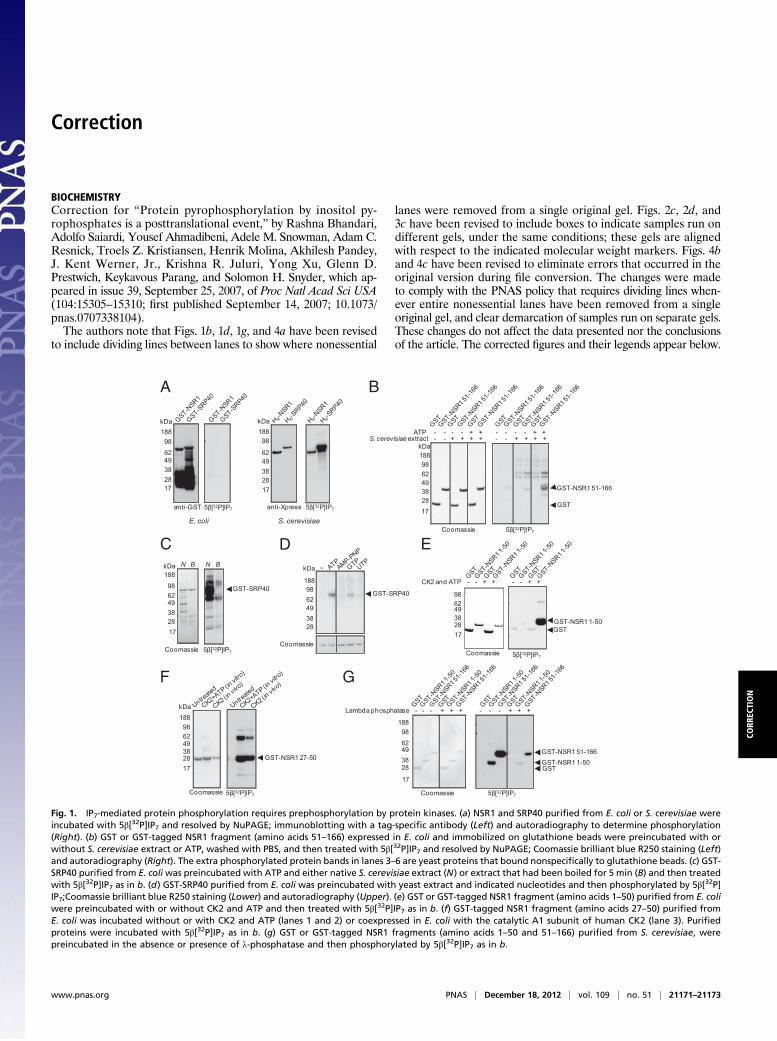
BIOCHEMISTRYCorrection for “Protein pyrophosphorylation by inositol py-rophosphates is a posttranslational event,” by Rashna Bhandari,Adolfo Saiardi, Yousef Ahmadibeni, Adele M. Snowman, Adam C.Resnick, Troels Z. Kristiansen, Henrik Molina, Akhilesh Pandey,J. Kent Werner, Jr., Krishna R. Juluri, Yong Xu, Glenn D.Prestwich, Keykavous Parang, and Solomon H. Snyder, which ap-peared in issue 39, September 25, 2007, of Proc Natl Acad Sci USA(104:15305–15310; first published September 14, 2007; 10.1073/pnas.0707338104).The authors note that Figs. 1b, 1d, 1g, and 4a have been revised
to include dividing lines between lanes to show where nonessential
lanes were removed from a single original gel. Figs. 2c, 2d, and3c have been revised to include boxes to indicate samples run ondifferent gels, under the same conditions; these gels are alignedwith respect to the indicated molecular weight markers. Figs. 4band 4c have been revised to eliminate errors that occurred in theoriginal version during file conversion. The changes were madeto comply with the PNAS policy that requires dividing lines when-ever entire nonessential lanes have been removed from a singleoriginal gel, and clear demarcation of samples run on separate gels.These changes do not affect the data presented nor the conclusionsof the article. The corrected figures and their legends appear below.
98 62 49 38
188
28 17
5β[32P]IP7Coomassie
kDa
GST-NSR1 27-50
98
62 49 38
kDa 188
28 17
anti-GST
E. coli
98
62 49 38
kDa 188
28 17
anti-Xpress
S. cerevisiae
5β[32P]IP7 5β[32P]IP7
GST-NSR1 51-166
98 62 49 38
kDa 188
28 17
GST
S. cerevisiae extract - - + + + + - - + + + +ATP - - - - + + - - - - + +
5β[32P]IP7Coomassie
GST-SRP4098 62 49 38
kDa
188
28
-
Coomassie
98 62 49 38 28 17
GSTGST-NSR1 1-50
CK2 and ATP - - + + - - + +
5β[32P]IP7Coomassie
GST-SRP40
N B N B
98 62 49 38
kDa 188
28 17
Coomassie 5β[32P]IP7
GST-NSR1 51-166GST-NSR1 1-50GST
98
62 49 38
188
28
17
Lambda phosphatase - - - + + + - - - + + +
5β[32P]IP7Coomassie
A
C
F G
D E
B
Fig. 1. IP7-mediated protein phosphorylation requires prephosphorylation by protein kinases. (a) NSR1 and SRP40 purified from E. coli or S. cerevisiae wereincubated with 5β[32P]IP7 and resolved by NuPAGE; immunoblotting with a tag-specific antibody (Left) and autoradiography to determine phosphorylation(Right). (b) GST or GST-tagged NSR1 fragment (amino acids 51–166) expressed in E. coli and immobilized on glutathione beads were preincubated with orwithout S. cerevisiae extract or ATP, washed with PBS, and then treated with 5β[32P]IP7 and resolved by NuPAGE; Coomassie brilliant blue R250 staining (Left)and autoradiography (Right). The extra phosphorylated protein bands in lanes 3–6 are yeast proteins that bound nonspecifically to glutathione beads. (c) GST-SRP40 purified from E. coliwas preincubated with ATP and either native S. cerevisiae extract (N) or extract that had been boiled for 5 min (B) and then treatedwith 5β[32P]IP7 as in b. (d) GST-SRP40 purified from E. coli was preincubated with yeast extract and indicated nucleotides and then phosphorylated by 5β[32P]IP7;Coomassie brilliant blue R250 staining (Lower) and autoradiography (Upper). (e) GST or GST-tagged NSR1 fragment (amino acids 1–50) purified from E. coliwere preincubated with or without CK2 and ATP and then treated with 5β[32P]IP7 as in b. (f) GST-tagged NSR1 fragment (amino acids 27–50) purified fromE. coli was incubated without or with CK2 and ATP (lanes 1 and 2) or coexpressed in E. coli with the catalytic A1 subunit of human CK2 (lane 3). Purifiedproteins were incubated with 5β[32P]IP7 as in b. (g) GST or GST-tagged NSR1 fragments (amino acids 1–50 and 51–166) purified from S. cerevisiae, werepreincubated in the absence or presence of λ-phosphatase and then phosphorylated by 5β[32P]IP7 as in b.
www.pnas.org PNAS | December 18, 2012 | vol. 109 | no. 51 | 21171–21173
CORR
ECTION
NOP: KKVKKETSSSDSSEDSSEEEDKAQGNOP-2[pS]: KKVKKETSSSDS[pS]ED[pS]SEEEDKAQGNOP-pS 88: KKVKKETSSSDS[pS]EDSSEEEDKAQGNOP-pS 91: KKVKKETSSSDSSED[pS]SEEEDKAQGNOP-2[MepS]: KKVKKETSSSDS[MepS]ED[MepS]SEEEDKAQGNOP-2[ppS]: KKVKKETSSSDS[ppS]ED[ppS]SEEEDKAQG
kDa 38 28 17 14
anti-GST
GST-Nopp140 80-100
38 28 17 14
14
6
3
kDa 17
Coomassie 5β[32P]IP7
peptides
14 63
kDa
17 28
CK2+γ[32P]ATPCoomassie
peptides
A
C
B
D
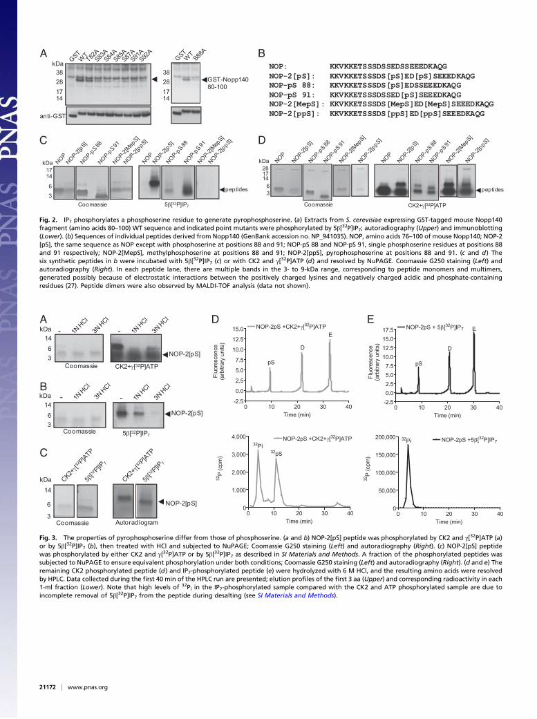
Fig. 2. IP7 phosphorylates a phosphoserine residue to generate pyrophosphoserine. (a) Extracts from S. cerevisiae expressing GST-tagged mouse Nopp140fragment (amino acids 80–100) WT sequence and indicated point mutants were phosphorylated by 5β[32P]IP7; autoradiography (Upper) and immunoblotting(Lower). (b) Sequences of individual peptides derived from Nopp140 (GenBank accession no. NP_941035). NOP, amino acids 76–100 of mouse Nopp140; NOP-2[pS], the same sequence as NOP except with phosphoserine at positions 88 and 91; NOP-pS 88 and NOP-pS 91, single phosphoserine residues at positions 88and 91 respectively; NOP-2[MepS], methylphosphoserine at positions 88 and 91; NOP-2[ppS], pyrophosphoserine at positions 88 and 91. (c and d) Thesix synthetic peptides in b were incubated with 5β[32P]IP7 (c) or with CK2 and γ[32P]ATP (d) and resolved by NuPAGE. Coomassie G250 staining (Left) andautoradiography (Right). In each peptide lane, there are multiple bands in the 3- to 9-kDa range, corresponding to peptide monomers and multimers,generated possibly because of electrostatic interactions between the positively charged lysines and negatively charged acidic and phosphate-containingresidues (27). Peptide dimers were also observed by MALDI-TOF analysis (data not shown).
-kDa 1463
-
CK2+γ[32P]ATPCoomassie
NOP-2[pS]
kDa 1463
- -
5β[32P]IP7Coomassie
NOP-2[pS]0 10 20 30 40
-2.5
0.0
2.5
5.0
7.5
10.0
12.5
15.0 NOP-2pS +CK2+γ[32P]ATP
pS
D
E
Time (min)
Fluo
resc
ence
(arb
itrar
y un
its)
0 10 20 30 400
1,000
2,000
3,000
4,000 NOP-2pS +CK2+γ[32P]ATP32Pi
32pS
Time (min)
32P
(cpm
)
0 10 20 30 40-2.50.02.55.07.5
10.012.515.017.5 NOP-2pS + 5β[32P]IP7
pS
D
E
Time (min)
Fluo
resc
ence
(arb
itrar
y un
its)
0 10 20 30 400
50,000
100,000
150,000
200,000 NOP-2pS +5β[32P]IP732Pi
Time (min)
32P
(cpm
)
14
6
3
kDa
NOP-2[pS]
AutoradiogramCoomassie
A D E
B
C
Fig. 3. The properties of pyrophosphoserine differ from those of phosphoserine. (a and b) NOP-2[pS] peptide was phosphorylated by CK2 and γ[32P]ATP (a)or by 5β[32P]IP7 (b), then treated with HCl and subjected to NuPAGE; Coomassie G250 staining (Left) and autoradiography (Right). (c) NOP-2[pS] peptidewas phosphorylated by either CK2 and γ[32P]ATP or by 5β[32P]IP7 as described in SI Materials and Methods. A fraction of the phosphorylated peptides wassubjected to NuPAGE to ensure equivalent phosphorylation under both conditions; Coomassie G250 staining (Left) and autoradiography (Right). (d and e) Theremaining CK2 phosphorylated peptide (d) and IP7-phosphorylated peptide (e) were hydrolyzed with 6 M HCl, and the resulting amino acids were resolvedby HPLC. Data collected during the first 40 min of the HPLC run are presented; elution profiles of the first 3 aa (Upper) and corresponding radioactivity in each1-ml fraction (Lower). Note that high levels of 32Pi in the IP7-phosphorylated sample compared with the CK2 and ATP phosphorylated sample are due toincomplete removal of 5β[32P]IP7 from the peptide during desalting (see SI Materials and Methods).
21172 | www.pnas.org
www.pnas.org/cgi/doi/10.1073/pnas.1218152109
Lambda phosphatase - + - +
Before IP7 After IP7
5β[32P]IP7
GST-NSR 1-50
98 62 49 38
188
28 17
Lambda phosphatase - + - +
GST-Nop140 80-100
62 49 38 28 1714
anti-GST
62 49 38 28 1714
GST-NSR127-50
anti-GST
]Spp[2-PON]Sp[2-PONLambdaphosphatase - + - + - + - +
28
17 14
6
3Coomassie Pro-Q Diamond
28
17 14
6
3Coomassie Pro-Q Diamond
NOP-2[ppS]NOP-2[pS]
1714
6
3
CK2+γ[32P]ATP
Lambdaphosphatase - + - + - + - +
Coomassie
1714
6
3
5β[32P]IP7Coomassie
NOP-2[pS]NOP-2[pS]
98 62 49 38 28 17
98 62 49 38
188
28 17
Lambda phosphatase - + - + - + - +
5β[32P]IP7CK2+γ[32P]ATP CoomassieCoomassie
GST-NSR 27-50
A
C
D E
B
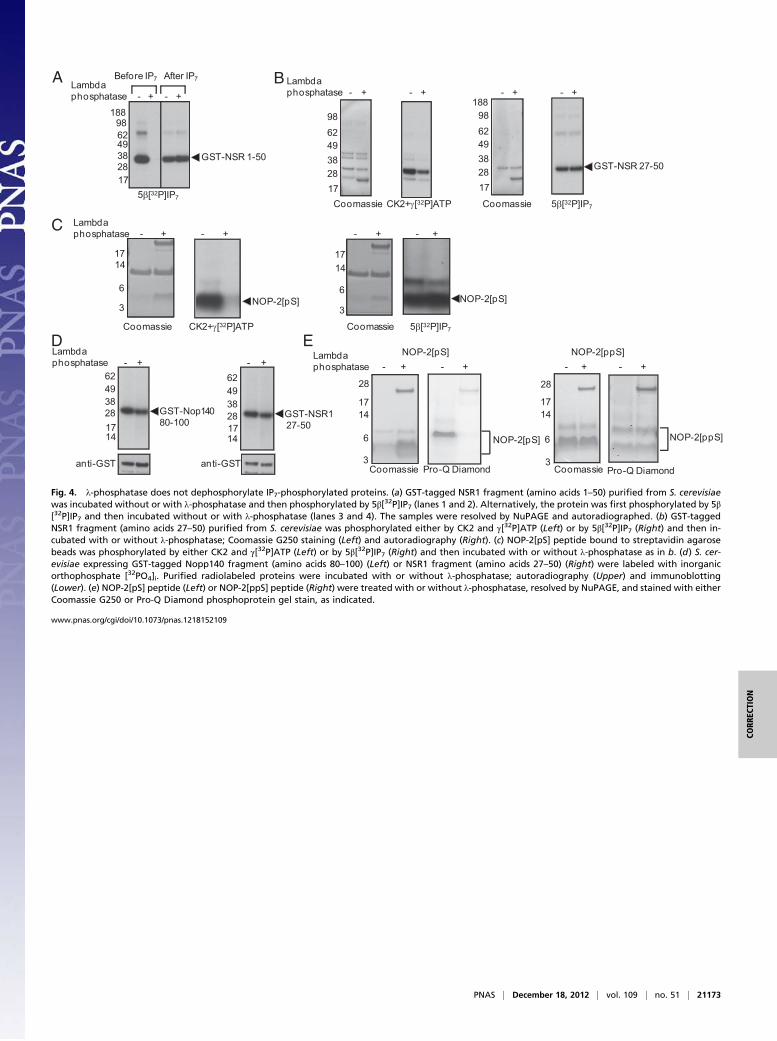
Fig. 4. λ-phosphatase does not dephosphorylate IP7-phosphorylated proteins. (a) GST-tagged NSR1 fragment (amino acids 1–50) purified from S. cerevisiaewas incubated without or with λ-phosphatase and then phosphorylated by 5β[32P]IP7 (lanes 1 and 2). Alternatively, the protein was first phosphorylated by 5β[32P]IP7 and then incubated without or with λ-phosphatase (lanes 3 and 4). The samples were resolved by NuPAGE and autoradiographed. (b) GST-taggedNSR1 fragment (amino acids 27–50) purified from S. cerevisiae was phosphorylated either by CK2 and γ[32P]ATP (Left) or by 5β[32P]IP7 (Right) and then in-cubated with or without λ-phosphatase; Coomassie G250 staining (Left) and autoradiography (Right). (c) NOP-2[pS] peptide bound to streptavidin agarosebeads was phosphorylated by either CK2 and γ[32P]ATP (Left) or by 5β[32P]IP7 (Right) and then incubated with or without λ-phosphatase as in b. (d) S. cer-evisiae expressing GST-tagged Nopp140 fragment (amino acids 80–100) (Left) or NSR1 fragment (amino acids 27–50) (Right) were labeled with inorganicorthophosphate [32PO4]i. Purified radiolabeled proteins were incubated with or without λ-phosphatase; autoradiography (Upper) and immunoblotting(Lower). (e) NOP-2[pS] peptide (Left) or NOP-2[ppS] peptide (Right) were treated with or without λ-phosphatase, resolved by NuPAGE, and stained with eitherCoomassie G250 or Pro-Q Diamond phosphoprotein gel stain, as indicated.
PNAS | December 18, 2012 | vol. 109 | no. 51 | 21173
CORR
ECTION