Contribution à l'épidémiologie de la tuberculose bovine...
175
Académie Universitaire Wallonie Europe Université de Liège Faculté de Médecine Vétérinaire Département des Maladies Infectieuses et Parasitaires Contribution à l'épidémiologie de la tuberculose bovine dans le nord de l'Equateur Contribution to the epidemiology of bovine tuberculosis in northern Ecuador Freddy PROAÑO-PEREZ Thèse présentée en vue de l‘obtention du Grade de Docteur en Sciences Vétérinaires Année académique 2011- 2012
Transcript of Contribution à l'épidémiologie de la tuberculose bovine...

Académie Universitaire Wallonie Europe
Université de Liège
Faculté de Médecine Vétérinaire
Département des Maladies Infectieuses et Parasitaires
Contribution à l'épidémiologie de la tuberculose bovine dans le nord de l'Equateur
Contribution to the epidemiology of bovine tuberculosis in northern Ecuador
Freddy PROAÑO-PEREZ
Thèse présentée en vue de l‘obtention du
Grade de Docteur en Sciences Vétérinaires
Année académique 2011- 2012
ii
Contribution to the epidemiology of bovine tuberculosis in northern Ecuador
Freddy PROAÑO-PEREZ
Promoter:
Prof. Annick Linden Department of Infectious and Parasitic Diseases
University of Liege (ULg)
Co-promoters:
Prof. Françoise Portaels Department of Microbiology, Mycobacteriology Unit
Institute of Tropical Medicine, Antwerp (ITM)
Prof. Washington Benitez-Ortiz International Centre for Zoonoses
Central University of Ecuador (UCE)
Liege 2011

iii
This thesis is dedicated to my son Emilio Alejandro and
my daughter Arianna Daniela, who are my inspiration

iv
ACKNOWLEDGEMENTS
I express my thanks and gratitude to Prof. Annick Linden, who accepted to be my promoter in
this thesis. I would like to express my thanks to Prof. Washington Benítez-Ortiz, Prof.
Françoise Portaels, Prof. Leen Rigouts and Prof. Jef Brandt, for their important scientific
guidance and support during the work in the field, manipulations in the laboratory and writing
the publications and thesis.
I wish to thanks to all the staff of the Mycobacteriology Unit of the Institute of Tropical
Medicine of Antwerp, particularly Krista Fissette, Lies Durnez, Anita van Aerde, Pim de Rijk,
Cecile Uwizeye, Juan Carlos Palomino, Anandi Martin, Miriam Eddyani, Andrea van Groll
and Fabianne Paasch for all their help and advices. I also thank to Doctor Kevin Peterson for
the correction of the English revision.
To all the staff of the International Centre for Zoonoses (CIZ-UCE), especially Maritza Celi,
Marco Coral, Julio Ortiz, Fabricio Anchatipán, Gustavo Echeverria, Ricardo Benítez, Jorge
Ron and Richar Rodríguez, thank you for your contribution during the field work, to Dafne
Urresta for her technical help, and to Lenin Ron for his support in the statistical analysis.
I am grateful to the ―Commission universitaire pour le Développement‖ (CUD), University of
Liege - Project PIC, for their financial support during the development of this work. I thank to
all cattle owners, farm and abattoir workers in the Mejia canton for their assistance and
willingness to participate in this work.
Finally, I express my profound gratitude to my family, particularly to my parents, to my son
Emilio and my daughter Arianna, for being my support and stimulation to perform this work.

v
TABLE OF CONTENTS
ACKNOWLEDGEMENTS .................................................................................................. iv
TABLE OF CONTENTS........................................................................................................v
SUMMARY ........................................................................................................................ vii
RÉSUMÉ ...............................................................................................................................x
RESUMEN .........................................................................................................................xiv
LIST OF ABBREVIATIONS ........................................................................................... xviii
CHAPTER 1 – INTRODUCTION..........................................................................................1
1.1 History of bovine tuberculosis ..................................................................................2
1.2 Taxonomy and characteristics of the Mycobacterium bovis .......................................4
1.2.1 Characteristics of M. bovis ................................................................................6
1.2.2 M. bovis genome ...............................................................................................7
1.3 Pathogenesis . ...........................................................................................................9
1.4 Epidemiology ......................................................................................................... 10
1.4.1 Prevalence of BTB in cattle ............................................................................. 10
1.4.2 Transmision of BTB among cattle ................................................................... 14
1.4.3 Prevalence and transmission of BTB among non-bovine species ..................... 17
1.4.4 Prevalence and transmission of BTB among humans ....................................... 20
1.5 Diagnosis ............................................................................................................... 22
1.5.1 Ante-mortem (in vivo) diagnosis ...................................................................... 22
1.5.2 Post-mortem inspection ................................................................................... 29
1.5.3 Laboratory diagnostic tools ............................................................................. 33
1.6 Control of BTB ...................................................................................................... 46
1.7 Eradication ............................................................................................................. 49
CHAPTER 2 – OBJECTIVES .............................................................................................. 51
CHAPTER 3 – RESULTS .................................................................................................... 53

vi
3.1 Preliminary observations on Mycobacterium spp. in dairy cattle in Ecuador ........... 54
3.2 Comparative intradermal tuberculin test in dairy cattle in the north of Ecuador and
risk factors associated with bovine tuberculosis ...................................................... 61
3.3 Post-mortem examination and laboratory based analysis for the diagnosis of bovine
tuberculosis among dairy cattle in Ecuador ............................................................. 69
3.4 Single Mycobacterium bovis strain prevailing among dairy cattle in northern
Ecuador .................................................................................................................. 78
3.5 Risk analysis for infection of Mycobacterium spp. in a human population in
Ecuador .................................................................................................................. 85
3.6 Review article: Situation of bovine tuberculosis in Ecuador ................................... 102
CHAPTER 4 – DISCUSSION AND PERSPECTIVES ....................................................... 124
CHAPTER 5 – SYNOPSIS ................................................................................................ 135
CHAPTER 6 – REFERENCES .......................................................................................... 142

SUMMARY
In Ecuador, as in other Latin American countries, bovine tuberculosis (BTB) is a major
concern in the dairy cattle industry, with economic and public health importance. The increase
of peri-urban dairy production associated with the absence of BTB control program in
Ecuador are major risk factors in the context of tuberculosis. Furthermore, official data related
to the disease are scarce and there is no national veterinary reference laboratory for
mycobacteria in Ecuador. The present work was undertaken in this context. The main
objectives were to provide data on the epidemiologic situation of BTB among dairy cattle in
northern Ecuador, and to compare the performances of the standard diagnostic tools used to
identify M. bovis.
In a first step, a skin test survey was performed in 2003 in the Mejia canton, the major dairy
cattle production region of Ecuador. Randomly selected 1012 cattle from 59 dairy herds,
classified according to herd size, were tested for tuberculosis by the single intradermal
tuberculin test (SITT) and the comparative intradermal tuberculin test (CITT). Results
demonstrated 7.95 % of tuberculin skin-reactors in large herds versus 4.24 % in medium and
0.30 % in small herds. These first results underlined the importance of BTB in dairy cattle in
the Mejia canton with herd size as a probable risk factor.
A more detailed field investigation was performed in 2007 and 2008 in the same region. A
total of 2022 cattle from 20 dairy herds were studied. In 2007, each animal was tested using
CITT and a follow-up test was performed in the same herds in 2008. The true annual

viii
Summary
incidence was 1.70 %. Analysis of CITT results showed significant differences between
medium and large herds with a BTB prevalence of 0.27 % and 0.57 % in medium-sized herds
compared with 8.63 % and 8.43 % in large herds. Herd size was identified as a significant
risk factor, confirming the results of the first study. The number of skin test-positive cases
also increased significantly with age, contacts with other animal species and introduction of
new cattle into the herd.
A third study was carried out in 1390 slaughtered cattle over a two years period. Classical
diagnostic tools were compared to detect infected cattle. A total of 33 animals with gross
suspected lesions were detected representing 2.3 % and 2.4 % of animals inspected in 2007
and 2008, respectively. From these suspected cases, acid-fast bacilli (direct microscopy) were
identified in 33% (11/33) and suggestive microscopic lesions (histopathology) in 27.3 %
(9/33) of the cases. Cultures and PCR yielded 36.4 % (12/33) and 27.3 % (9/33) of positives,
respectively. Overall, the combined use of microscopy, histopathology, PCR and culture
identified infected cattle (at least one positive test) in 51.5 % (17/33) of the suspected
animals. Compared to culture, other laboratory procedures displayed a sensitivity of 56.5 %
(PCR) and 43.5 % (microscopy and histopathology) and a specificity of 94.4 % (PCR and
microscopy) and 97.2 % (histopathology). These results underline that reliable post-mortem
laboratory testing either requires the combination of several tests or necessitates the
development of improved tools with better performance characteristics. It should be noted
that lesions were mainly observed in thoracic lymph nodes suggesting that the respiratory
tract is the main route of transmission in dairy cattle inspected at slaughterhouse.
In a subsequent step, spoligotyping was used to compare M. bovis strains. All strains (n = 23)
isolated from slaughtered cattle from Mejia canton presented the same spoligotype (SB0980).

ix
Summary
Such homogeneity could be related to the relatively small study area. A more extensive
molecular study, at a national level, could be helpful to assess the strain diversity of M. bovis
in cattle populations in Ecuador.
In countries where BTB controls are lacking, M. bovis is also of public health concern. The
last study was undertaken to evaluate the prevalence of Mycobacterium spp. in risk
populations by tuberculin skin test (TST). The study was conducted on 157 people (farm and
slaughterhouse workers) and TST revealed a high prevalence of reactors (29 %). The main
risk factors associated with positive human reactors were consumption of raw milk or
handmade cheese. Campaign informations are needed to make rural populations aware of the
zoonotic risk related to M. bovis.
In conclusion, data presented in this thesis confirm the importance of BTB in dairy cattle in
northern Ecuador and highlight the need for a national BTB control program. Ideally, this
program should include (1) a reference laboratory for BTB and the training of qualified
veterinarians; (2) mandatory skin testing and culling of reactors with official compensations;
(3) detailed post-mortem inspection with laboratory-based diagnosis associated with a trace-
back system and (4) information campaigns related to the zoonotic risk of M. bovis in rural
areas.

RÉSUMÉ
En Equateur, comme dans d‘autres pays d‘Amérique Latine, la tuberculose bovine reste un
problème important tant au niveau de la santé animale que de la santé humaine. L‘OMS
considère comme prioritaire les programmes de lutte contre la tuberculose en Amérique
latine. La spéculation laitière dans les zones péri-urbaines associée à l‘absence de
programmes de contrôle officiels de la tuberculose bovine représentent des facteurs de risque
majeurs en Equateur. Il existe très peu de données sur l‘importance de la tuberculose bovine
dans les élevages laitiers des régions andines et, jusqu‘à présent, aucun laboratoire n‘était
compétent pour le diagnostic des mycobactéries d‘origine animale. C‘est dans ce contexte que
ce projet a été initié. Les objectifs de cette thèse étaient (i) de fournir des données sur la
situation épidémiologique de la tuberculose bovine dans les élevages laitiers du nord de
l‘Equateur et, (ii) de comparer les performances des outils de diagnostic classiques utilisés
pour identifier Mycobacterium bovis (M. bovis).
Une première étude a été réalisée dans le canton de Mejia en 2003. Les campagnes de
tuberculination (intradermo-tuberculination simple [IDS] et double [IDD]) ont concerné plus
de 1.000 bovins provenant de 59 troupeaux classés en 3 catégories selon leur taille : catégorie
1 ( > 70 têtes) ; catégorie 2 (entre 25 et 70 têtes) et catégorie 3 (< 25 têtes). Au sein des
troupeaux de grande taille, 7.95 % de bovins réagissaient positivement aux tests cutanés alors
qu‘en catégorie 2 et 3, respectivement 4.24 % et 0.30 % des bovins réagissaient positivement.
Cette première enquête a permis de mettre en évidence la présence de M. bovis dans les
élevages laitiers du canton de Mejia avec une prévalence plus importante dans les troupeaux
de grande taille.

xi
Résumé
Une enquête plus détaillée a été réalisée en 2007 et 2008 dans la même région. Au total, plus
de 2.000 bovins issus de 20 troupeaux ont été investigués. En 2007, chaque animal a été testé
(IDD) et un suivi a été réalisé dans les mêmes troupeaux l‘année suivante. L‘incidence
annuelle réelle était de 1.70 %. Au cours des 2 années, des différences significatives ont été
mises en évidence entre les prévalences calculées dans les troupeaux de taille moyenne (0.27
% et 0.57 %) et celles calculées dans les troupeaux de grande taille (8.63 % et 8.43 %). La
taille du troupeau a clairement été identifiée comme un facteur de risque pour la tuberculose
bovine, de même que l‘âge des bovins, le contact éventuel avec d‘autres espèces animales et
l‘introduction de nouveaux bovins dans l‘exploitation.
Une troisième étude a été menée dans un abattoir du canton de Mejia sur 1390 bovins abattus
sur une période de deux ans. Au total, 33 bovins ont été détectés avec des lésions
macroscopiques suspectes, soit 2.3 % et 2.4 % des animaux inspectés en 2007 et 2008. Parmi
ces 33 cas suspects, des bacilles acido-résistants ont été mis en évidence dans 33% des cas
(microscopie directe) et des lésions microscopiques suggestives dans 27 % des cas
(histopathologie). La mise en culture et la PCR ont mis en évidence la présence de M. bovis
dans 36 % et 27 % des cas suspects, respectivement. L‘utilisation combinée de ces 4 outils de
diagnostic a permis de confirmer que 51 % des animaux suspects étaient positifs pour M.
bovis. Comparés à la mise en culture, considérée comme la technique de référence pour la
détection de M. bovis, les autres outils de diagnostic présentaient une sensibilité de 56 %
(PCR) et 43 % (microscopie et histopathologie) et une spécificité de 94 % (PCR et
microscopie) et 97 % (histopathologie). Ces résultats soulignent que plusieurs outils doivent
être utilisés en parallèle pour le diagnostic post mortem de la tuberculose bovine. Les lésions
de tuberculose étant principalement localisées au niveau des ganglions thoraciques, la

xii
Résumé
transmission de M. bovis par voie aérogène semble la plus plausible pour les bovins inclus
dans cette enquête.
Pour compléter cette troisième étude, une approche par spoligotypage a été mise en œuvre.
Toutes les souches (n = 23) isolées à l‘abattoir présentaient le même profil (spoligotype
SB0980). Une étude d‘envergure devrait être envisagée afin de déterminer la diversité des
souches de M. bovis présentes en Equateur.
Dans les pays qui ne possèdent pas de politique de contrôle de la tuberculose bovine dans les
élevages, M. bovis peut représenter un problème de santé publique. La cinquième étude avait
pour objectif de déterminer la prévalence de Mycobacterium spp dans des populations
humaines ciblées. Des tests cutanés ont été réalisés sur 157 personnes appartenant à deux
groupes à risque (personnel d‘abattoir et techniciens d‘élevage). Au total, 29 % des personnes
testées présentaient une réaction positive au test cutané. Les facteurs de risque majeurs
associés aux résultats cutanés positifs étaient la consommation de lait cru et de fromages
confectionnés à la main. Des campagnes d‘information en zone rurale sont nécessaires pour
conscientiser la population au risque zoonotique de la tuberculose bovine.
En conclusion, les données présentées dans ce manuscrit soulignent l‘importance de M. bovis
dans les élevages de bovins laitiers dans le canton de Mejia. Une politique de contrôle de la
tuberculose bovine devrait être envisagée à l‘échelle nationale. Idéalement, ce programme
devrait inclure (1) un laboratoire de référence pour les mycobactéries et des formations
spécialisées pour les inspecteurs vétérinaires; (2) une procédure obligatoire de détection-
abattage des bovins infectés, avec compensation financière; (3) des procédures d‘inspection
post mortem standardisées et associées à des analyses de laboratoire et un système de

xiii
Résumé
traçabilité et enfin, (4) des campagnes d‘information en zones rurales relatives au risque
zoonotique associé à M. bovis.

RESUMEN
En Ecuador, como en otros países latinoamericanos, la tuberculosis bovina (TBB) es un
problema importante en la industria lechera, con implicaciones económicas y en la salud
pública. El aumento de la producción lechera en áreas periurbanas asociada a la ausencia de
un programa de control de TBB en Ecuador son factores de riesgo importantes en el contexto
de la TBB. Complementariamente, los datos oficiales relacionados con la enfermedad son
escasos, y no existe en Ecuador un laboratorio veterinario de referencia nacional para el
estudio de micobacterias; con este contexto el presente trabajo fue planificado. Los objetivos
principales fueron proporcionar información de la situación epidemiológica de la TBB en el
ganado lechero del norte del Ecuador, y comparar el funcionamiento de las herramientas
estándar de diagnóstico utilizadas para identificar M. bovis.
En un primer paso, en el año 2003 se desarrollo un estudio en el cantón de Mejía, una de las
principales regiones lecheras del norte del país, utilizando pruebas de tuberculinización.
Aleatoriamente se seleccionó 1012 bovinos provenientes de 59 fincas lecheras, clasificados
según el tamaño del hato, estos animales fueron sometidos a las pruebas de tuberculinización
intradérmica simple (TIS) y tuberculinización intradérmica comparativa (TIC). Los resultados
fueron 7.95% de reactores positivos en hatos grandes, en contraste con 4.24% en medianos y
0.30% en pequeños hatos. Estos primeros resultados evidencian la importancia de la TBB en
el ganado lecheros del cantón de Mejía e identifican al tamaño del hato como un factor de
riesgo.

xv
Resumen
Posteriormente, se realizó una investigación más detallada en la misma región durante los
años 2007 y 2008, en un total de 2022 bovinos provenientes de 20 hatos lecheros. En el 2007,
cada animal fue diagnosticado a través de la prueba de TIC y un año después se realizó un
seguimiento utilizando la misma prueba en el mismo grupo de animales; la incidencia real
anual fue calculada en 1.70%. El análisis de los resultados de las pruebas de TIC demostraron
una diferencia significativa entre hatos lecheros medianos y grandes, con una prevalencia de
TBB de 0.27% y 0.57% en hatos medianos, comparados con el 8.63% y 8.43% en hatos
grandes para los años 2007 y 2008, respectivamente. El tamaño del hato fue identificado
como un factor de riesgo significativo, confirmando los resultados del primer estudio. El
número de reactores positivos a la prueba TIC también aumentó considerablemente con la
edad, el contacto con otras especies de animales y con la introducción de nuevos animales en
el hato.
Un tercer estudio fue realizado en 1390 bovinos sacrificados durante un período de dos años,
en el se comparó de las pruebas de diagnóstico clásicas para detectar ganado infectado. Un
total de 33 bovinos con lesiones evidentes (sospechosas) fueron detectados, representando un
2.3% y 2.4% de los animales examinados en 2007 y 2008, respectivamente. Del total de casos
sospechosos, solamente fueron identificados bacilos acido-resistentes (microscopia directa) en
33% (11/33) de los casos, y a través de las lesiones microscópicas sugestivas a TBB
(histopatología) en 27.3% (9/33). El cultivo in vitro y PCR detectaron el 36.4% de animales
positivos (12/33) y el 27.3% (9/33), respectivamente. Al utilizar de manera combinada todas
las pruebas para la identificación de animales infectados (microscopia, histopatología, PCR y
cultivo in vitro), en los cuales por lo menos una prueba era positiva, se obtuvo el 51.5%
(17/33) de bovinos infectados del total de animales sospechosos. Comparando el cultivo in
vitro con otras pruebas de laboratorio, se calculó la sensibilidad (Se) y especificidad (Sp) de

xvi
Resumen
las otras pruebas, la PCR tuvo una Se de 56.5%, la microscopia e histopatología 43.5%;
mientras que la Sp para la PCR y microscopia fue de 94.4% y 97.2% para la histopatología.
Estos resultados demuestran que un diagnóstico post-mortem confiable requiere de la
combinación de varias pruebas de laboratorio, es necesario el desarrollo y mejoramiento de
herramientas de diagnóstico que brinden mejores características de funcionamiento. Tomando
en cuenta que las lesiones macroscópicas fueron observadas principalmente en ganglios
linfáticos torácicos, se sugiere que la vía respiratoria es la principal ruta para la transmisión de
la TBB en ganado lechero examinado en el camal del cantón Mejía.
En un paso adelante, se utilizó el spoligotyping para comparar las cepas de M. bovis. Todas
las cepas aisladas (n = 23) del ganado sacrificado del cantón de Mejía presentaron el mismo
spoligotipo (SB0980); tal homogeneidad podía estar relacionada con un área de estudio
relativamente pequeña. Un estudio de epidemiologia molecular más extenso, a un nivel
nacional, podía ser provechoso para determinar la diversidad de las cepas de M. bovis en la
población bovina del Ecuador. En países donde no existe control de la TBB, M. bovis es
también de interés de la salud pública.
El último estudio fue planificado para evaluar la prevalencia de Mycobacterium spp. en la
población humana en riesgo a través de la prueba de tuberculinización. El estudio fue
conducido en 157 personas (trabajadores de finca y camal), la prueba demostró una alta
prevalencia de reactores (29%). Los principales factores de riesgo asociados a la reactividad
en humanos fueron el consumo de leche cruda y queso artesanal. Con estos resultados se hace
necesaria la implementación de campañas de información, para difundir en las poblaciones
rurales el riesgo zoonótico relacionado con M. bovis.

xvii
Resumen
En conclusión, los datos presentados en esta tesis confirman la importancia de la TBB en el
ganado lechero del norte del Ecuador y destacan la necesidad de un programa nacional de
control de la TBB. Preferentemente, este programa debe incluir (1) la existencia de un
laboratorio de referencia para la TBB y el entrenamiento de veterinarios calificados; (2)
pruebas tuberculínicas obligatorias y compensación oficial por animales reactores; (3)
inspección post-mortem detallada con diagnóstico de laboratorio asociados a un sistema del
trazabilidad de animales tuberculosos, y (4) campañas de información relacionadas con el
riesgo zoonótico de M. bovis en zonas rurales.

xviii
LIST OF ABBREVIATIONS
AD: Anno Domini
AFB: Acid fast bacilli
BCG: Bacille Calmette-Guérin
bp: Base pair
BTB: Bovine tuberculosis
CD4+: Cluster of differentiation 4 glicoprotein
CDS: CoDing Sequence
CFSPH: The Center for Food Security and Public Health
CITT: Comparative intradermal tuberculin test
DNA: Desoxyribonucleic acid
DR: Direct repeat
ELISA: Enzyme-linked immunosorbent assay
ESAT-6: 6 kDa early secretory antigenic target
FAO: Food and Agriculture Organization
FDA: Food and Drug Administration
GC: Guanine - cytosine
G+C: Guanine plus cytosine
GM-CSF: Granulocyte monocyte colony stimulating factor
HIV: Human immunodeficiency virus
γ-IFN: Gamma interferon

xix
IL: Interleukin
INPAAZ: Pan American Institute for Food Protection and Zoonoses
IS: Insertion sequence
IU: International Units
MGIT: Mycobacteria Growth Indicator Tube
MIF: Migration inhibition factor
MIRU: Mycobacterial interspersed repetitive units
MOTT: Mycobacteria other than tuberculosis
NTM: Non-tuberculous mycobacteria
OIE: Office International des Epizooties
OR: Odds ratio
OT: Old tuberculin
PAHO: Pan American Health Organization
PANTA: Polymyxin B, amphotericin B, nalidixic acid, trimethoprim, azlocillin
PCR: Polymerase chain reaction
PGRS: Polymorphic guanine-cytosine rich sequences
PPD: Purified protein derivative
PZA: Pyrazinamide
rDNA: Ribosomal desoxyribonucleic acid
RFLP: Restriction fragment length polymorphism
RNA: Ribonucleic acid
rRNA: Ribosomal ribonucleic acid
Se: Sensitivity
SITT: Simple intradermal tuberculin test
Sp: Specificity

xx
ST: Spoligotype
TB: Tuberculosis
TCH: Thiophene-2-carboxylic acid hydrazide
Th1: T helper 1 lymphocyte
TST: Tuberculin skin test
UK: United Kingdom
UV: Ultraviolet
VNTR: Variable number tandem repeats
WHO: World Health Organization
ZN: Ziehl Neelsen

CHAPTER 1 – INTRODUCTION

2
Introduction
1 INTRODUCTION
1.1 History of bovine tuberculosis
BTB among cattle was first described in 14 A.D. by Columella in Northern Italy (Wood et al.,
1994). However, the earliest documentation of a macroscopically recognized TB infection
was carried out in a fossil from an extinct Bison (Bison antiquus), dated 17.000 years ago.
According to this study, bovids from North America were the vectors that transported the
causative agent of BTB. DNA-based identification of this M. tuberculosis-complex strain
showed 72.7% similarity with the current M. bovis (Rothschild et al., 2001). Initially, it has
been suggested that M. tuberculosis arose from M. bovis at the time of the domestication of
cattle, approximately 10 to 15,000 years ago. This was based on the observation of the wide
host range of M. bovis, infecting wild and domesticated mammals as well as man, whereas M.
tuberculosis appears to be restricted mostly to humans (Hewinson et al., 2006). However,
recent DNA analyses contradict this hypothesis, the systematic analysis of polymorphisms in
a large panel of strains indicate that M. canettii is likely the ancestral specie of the M.
tuberculosis complex (Thoen & Barletta, 2006).
In 1865, a French military doctor, Jean Antoine Villemin, demonstrated that ―consumption‖
or tuberculosis (TB) was an infectious disease from humans and cattle. He made experiments
inoculating laboratory rabbits with material from infected humans and cattle, and published
his results in the treatise ―Etudes sur la Tuberculosis” (Studies on Tuberculosis), which
describes the transmission of TB from humans to rabbits, from cattle to rabbits, and from
rabbits to rabbits. He suggested that a specific microorganism could be the cause of the
disease that could be transmitted from human to cattle (Villemin, 1868).

3
Introduction
In 1882, Robert Koch demonstrated that TB was caused by tubercle bacilli and performed
cutaneous inoculations in guinea pigs. He developed a staining method to idenfity acid-fast-
bacilli and cultured them on solid medium. Koch also isolated a factor from culture filtrates
which halted the growth of TB bacilli, known as tuberculin and now described as Koch‘s old
tuberculin (OT). This was inoculated and used to diagnose TB in man since 1890. It was the
first clear demonstation of delayed hypersensivity or cell-mediated immunity. The technique
was adapted by veterinarians and used for testing cattle mainly through the febrile reaction in
tuberculous cows. (Daniel, 2005; Koch, 1906).
In 1898, an American researcher, Theobald Smith, observed differences between tubercle
bacilli of bovine and human origin, and named them as the human and bovine variants of the
tubercle bacillus, eventhough he warned against the assumption that these strains were limited
to their respective hosts (Smith, 1898; Grange, 2001). However, only since 1911, the Royal
Commission named by the British Government had firmly established that the human
population was susceptible to BTB and laid the fundations of the test-slaughter policy for
BTB eradication (Anonymous, 1911).
In 1921, two French bacteriologists: Albert Calmette and Camille Guérin (veterinarian)
investigated the intestinal route of TB infection. Calmette and Guérin began to grow M. bovis
in a beef bile-glycerine medium, with continuous replanting of the culture in this medium, and
thus lowering the virulence of M. bovis strain. This attenuated strain was called ―Bacille-
Calmette-Guérin‖ (BCG). Exhaustive testing of BCG showed its safety and effectiveness in
protecting young animals against TB. In 1924, the vaccination of newborn infants with BCG
began in France to protect against severe forms of TB, but its effect did not prevent

4
Introduction
pulmonary TB in adults. The BCG vaccine was spread worldwide and it is still widely used
today in developing countries (Hawgood, 2007; Oettinger et al., 1999).
1.2 Taxonomy and characteristics of the Mycobacterium bovis
M. bovis is one of the members of M. tuberculosis complex. Current taxonomy recognizes 8
members in this group i.e., M. tuberculosis, M. bovis, M. bovis BCG, M. caprae, M.
africanum, M. pinnipedii, M. microti and M. cannettii (Wayne & Kubica, 1986; van
Soolingen et al., 1997; Pfyffer et al., 1998; Cousins et al., 2003; Aranaz et al., 2003).
The genus Mycobacterium is the unique genus of the family Mycobacteriaceae, order
Actinomycetales, class Actinomycetes. Apart of the M. tuberculosis-complex the genus
Mycobacterium comprises M. leprae, M. ulcerans and more than 100 species of non-
tuberculous mycobacteria (NTM) or mycobacteria other than tuberculosis (MOTT). The latter
are distributed in four groups according to the Runyon classification, i.e., group I = slow
growers – photochromogen, group II = slow growers – scotochromogen, group III = slow
growers – nonchromogen, and group IV = rapid growers (Wayne & Kubica, 1986).
Table 1: Members of the Mycobacterium tuberculosis complex and hosts
Members Main hosts
M. tuberculosis Human TB
M. bovis, M. bovis BCG Cattle and human TB
M. africanum Human TB in Africa
M. tuberculosis M. canettii Human TB
Complex M. microti TB in small rodents
M. caprae TB in goats
M. pinnipedii TB in seals

5
Introduction
Tubercle bacilli are aerobic and predominantly rod-shaped, 0.3 to 0.5µm wide and with
variable length (1.5 – 3.5 µm). Spores, flagella and capsules are absent. It is an acid-fast
bacillus, an important characteristic that has been taken advantage for Ziehl Neelsen staining.
The bacillus resists decolourization with acid-alcohol after a first red stain with carbol
fuchsin, and can be easily detected when using a methylene blue counterstain (Biberstein &
Hirsh, 1999). Although cytochemically Gram-positive, mycobacteria do not take up the dyes
of the Gram-stain because the cell walls are rich in lipids and mycolic acids (Quinn et al.,
1999).
Tubercle bacilli are rather resistant to disinfectants and require long contact times for its
inactivation. They survive exposure to alkali or acids for 15 to 30 minutes, conditions
commonly used to decontaminate diagnostic specimens (Portaels et al., 2001). They resist
drying and survive for long periods in soil, but are fairly heat sensitive and killed by sunlight,
ultraviolet irradiation and pasteurization (Biberstein & Hirsh, 1999).
Mycobacteria are most closely related to the genus Nocardia and Rhodococcus and all three
genuses have a similar cell wall type (Quinn et al., 1999). The genus Mycobacterium has a
unique cell wall formed by abound lipids, which have a low permeability. These lipids
account for acid fastness, as well as pathogenic and immunologic properties. The wall
presents a double layer of petidoglycan and arabinogalactan surrounded by long chain
mycolic acids (Figure 1). Mycosides, phospholipids and sulpholipids apparently protect
tubercle bacilli against phagocytic killing and allow intracellular and extracellular
multiplication to continue. The cell wall is the interface between the bacillus and host; many

6
Introduction
pathogenic genes are encoding cell wall structures and secreted proteins (Hewinson et al.,
2006).
Figure 1. Structure of the mycobacterial cell wall.
Mycobacteria produce a thick mycolate-rich outer covering which functions as an
exceptionally efficient barrier.
Doc Kaiser's Microbiology Home Page Copyright © Gary E. Kaiser. All Rights Reserved.
Updated: July, 2008
1.2.1 Characteristics of M. bovis
Although M. bovis is an obligate intracellular pathogen, experimental studies have shown that
it can survive under specific conditions. The survival rates are different depending on the

7
Introduction
availability of nutrients, and climatic conditions i.e., humidity, exposure to sunlight and
temperature (Morris et al., 1994). M bovis can survive in faeces, blood, and urine from 150 to
332 days at 12 to 24°C, when shielded from direct sunlight, particularly in cold, dark and
moist conditions (Genov, 1965). Nevertheless, in natural pastures M. bovis remained viable
and virulent for at least 7 weeks during the English summer (Maddock, 1933). Under artificial
conditions, M. bovis can be cultured from stored samples after two years, which were keeped
at -20ºC in tubes containing Dubos broth supplemented with PANTA to inhibit the growth of
contaminants and to preserve the mycobacteria (Proaño-Perez et al., 2009). In some dairy
products (i.e., cheese), tubercle bacilli can survive for up to 14 days after processing
(Gallagher & Jenkins, 1998).
M. bovis strains display ―dysgonic‖ growth on media containing glycerol. Therefore, the
bacillus requires pyruvate to be added to media where glycerol is the sole carbon source. This
reflects a defect in the metabolism of glycerol, due to a mutation in the gene for the pyruvate
kinase enzyme, which catalyses the final step in the glycolysis (Keating et al., 2005). M. bovis
is intrinsically resistant to pyrazinamide (PZA), due to the lack of enzyme pyrazinamidase,
which converts the PZA to the toxic pyrazinoic acid (Konno et al., 1967). Consequently, PZA
cannot be used as a drug for TB treatment caused by M. bovis.
1.2.2 M. bovis genome
The completion of a number of mycobacterial genome sequences can allow the development
of antigen mining techniques that rapidly identify M. bovis-specific genes. The genome
contains approximately 4000 genes, encoding different properties, potential virulence factors
and antigens (Hewinson et al., 2006). Cell wall components and secreted proteins show the
8
Introduction
greatest variation compared with M. tuberculosis and M. leprae, indicating their important
role in host-bacillus interactions or immune evasion. These variations were the key to the
evolution of M. bovis (Garnier et al., 2003).
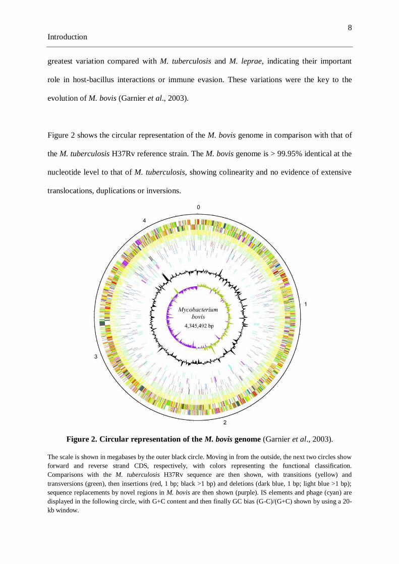
Figure 2 shows the circular representation of the M. bovis genome in comparison with that of
the M. tuberculosis H37Rv reference strain. The M. bovis genome is > 99.95% identical at the
nucleotide level to that of M. tuberculosis, showing colinearity and no evidence of extensive
translocations, duplications or inversions.
Figure 2. Circular representation of the M. bovis genome (Garnier et al., 2003).
The scale is shown in megabases by the outer black circle. Moving in from the outside, the next two circles show
forward and reverse strand CDS, respectively, with colors representing the functional classification.
Comparisons with the M. tuberculosis H37Rv sequence are then shown, with transitions (yellow) and
transversions (green), then insertions (red, 1 bp; black >1 bp) and deletions (dark blue, 1 bp; light blue >1 bp);
sequence replacements by novel regions in M. bovis are then shown (purple). IS elements and phage (cyan) are
displayed in the following circle, with G+C content and then finally GC bias (G-C)/(G+C) shown by using a 20-
kb window.

9
Introduction
1.3 Pathogenesis of M. bovis.
Pathogenesis is the process or mechanism of disease. In the case of BTB this process
comprises several elements: sources and routes of infection, intrinsic properties of the
bacillus, and interaction with the host and immune responses. Both humoral and cell-mediated
immune responses can be induced after mycobacterial infection. However, the cell-mediated
immune system has the most significant role in protective immunity against M. bovis (Neill et
al., 1994).
Tubercle bacilli do not produce toxins and their pathological effects result essentially from the
strong antigenicity of their cell walls (Gallagher & Jenkins, 1998). Virulence of M. bovis
appears to reside in the lipids of the cell wall, i.e., mycosides, phospholipids and sulpholipids,
which are also thought to protect the tubercle bacilli against phagocytosis. In the first week
after infection, cell-mediated immune reactions begin to modify the host response. Infected
macrophages secrete IL-12, a cytokine responsible for stimulating CD4+TH1 lymphocytes to
produce gamma interferon (IFN-), granulocyte monocyte colony stimulating factor (GM-
CSF), and migration inhibition factor (MIF), which attract and activate macrophages.
Activated macrophages acquire the capacity to kill some mycobacteria, and their efficiency
depends on the adequacy of the immune response and the virulence of bacteria. The
aggregation of macrophages contributes to the formation of a tubercle (Biberstein & Hirsh,
1999; Quinn et al., 1999).
The primary lung lesion is a bronchopneumonic focus which may progress rapidly or may
remain quiescent for many years (Morris et al., 1994). Once cell-mediated immunity is
established, lymphatic spread is retarded but contiguous extension via the erosion of bronchi,

10
Introduction
blood vessels or viscera to new areas, and T-lymphocyte-mediated reactions cause tissue
damage (Quinn et al., 1999). Immature and aged individuals often develop more severe
lesions than mature animals. Zebu cattle are more resistant than European breeds (Biberstein
& Hirsh, 1999).
1.4 Epidemiology of bovine tuberculosis
Bovine tuberculosis (BTB), caused by Mycobacterium bovis is a worldwide distributed
disease, with economic and public health importance. In humans, BTB is being considered as
a re-emergent zoonosis related to different situations: the persistence of the infection in
livestock, the maintenance of wildlife reservoirs, the appearance of strains resistant to the
main anti-TB drugs, and/or the spread of the human immunodeficiency virus (Cosivi et al.,
1998; Abalos & Retamal, 2004). In developing countries from Africa, Asia, Latin America
and the Caribbean, persistence or increase of BTB among cattle is caused by the increasing
production in the dairy industry, intensification of dairy herds, and the lack of appropriate
measures to control the disease (Suazo et al., 2003).
1.4.1 Prevalence of BTB in cattle
In many industrialised countries, BTB has been controlled or eradicated by application of
tests and slaughter programmes. Nevertheless, sporadic cases are still found among cattle in
some of these countries like the United Kingdom (UK) and Ireland, where the presence of M.
bovis in wildlife reservoirs seems to be the main cause of persistence of BTB (Phillips et al.,
2003). Several countries have been officially classified as tuberculosis-free i.e., Australia,
Iceland, Denmark, Sweden, Norway, Finland, Austria, Switzerland, Luxembourg, Latvia,

11
Introduction
Slovakia, Lithuania, Estonia, the Czech Republic, Singapore, Jamaica, Barbados and Israel
(CFSPH, 2007). Figure 3 shows the distribution of BTB among cattle in the world in 2010,
classified according to the occurrence of the disease.
Figure 3. Distribution of bovine tuberculosis among cattle based on data obtained
between January and June 2010. (OIE, 2010)
In Latin America, the presence of BTB among cattle is not well quantified (de Kantor &
Ritacco, 2006). Out of thirty four Latin American and Caribbean countries, twelve reported
BTB as sporadic or with low ocurrence, seven have reported BTB as an enzootic disease, and
only in one country (Dominican Republic) a high occurrence was noticed. On the other hand,
BTB was absent in twelve countries and data were lacking in the remaining two. Control
measures based on the test-and-slaughter policy have been established in only 12 countries in
the region that consider BTB as a notifiable disease. In the remaining 22 nations, the disease
is only partly or not at all controlled (Cosivi et al., 1998).
12
Introduction
About 70% of the cattle population in Latin America and the Cariabbean (261 million) is held
in areas where M. bovis-infection rates are higher then 1%. Ecuador, together with Haiti,
Guatemala, Argentina, Bolivia, Brazil, Chile, Peru and Guyana, are considered to belong to a
group of countries with a relatively high prevalence or no officially reported information (de
Kantor & Ritacco, 2006).
In Mexico, like in other developing countries the ocurrence of BTB is much higher in dairy
than in beef cattle. The prevalence of BTB in beef cattle has been estimated at 2.9%
(Zendejas-Martinez et al., 2008), while in dairy cattle it was approximately 16%. The high
prevalence observed in this study, is thought to be caused by the lack of farmers‘ participation
in the eradication measures (Perez-Guerrero et al., 2008). The disease is affecting dairy cattle
from circumscribed areas. In Queretaro, 17% of gross TB lesions where found among 1201
carcasses (Milián-Suazo et al., 2000). According to Zendejas-Martinez et al., (2008), at least
two reasons can explain this observation, i.e., management practices and food supply. Dairy
cattle are maintained under intensive production conditions, with large numbers of animals
kept per unit area, which increases the risk of disease transmission by the close proximity
between cattle. Dairy herds are mainly kept close to urban areas. On the contrary, beef cattle
are raised in dry coastal areas, with a limited number of cattle per unit area, conditions
reducing disease transmission and survival of the bacillus.
In Argentina, since a test and culling policy to control and eradicate BTB was put in force in
1999, 7713 farms have been officially declared free of infection, of which 6767 are dairy
farms. This measure has decreased the prevalence of TB in cattle and swine. Nowadays, the
national rates of BTB in cattle and swine detected by the observation of macroscopic lesions
in abattoirs have been reduced from 6.7% and 8.4% in 1969, to 0.9% and 0.4% in 2008,

13
Introduction
respectively (Torres, 2009). TB in swine has been controlled by farming improvements
including slaughter at an earlier age and avoiding the practice of feeding animals with non-
pasteurised dairy products (Perez et al., 2002). At slaughterhouses, animals with lesions are
confirmed by laboratory diagnosis, followed by tracebacking the origin of infected animals
and tuberculin tests are applied in the herds (Torres, 2009).
In Brazil, an average of 7.1% of herds were infected with BTB as estimated during a
tuberculin testing survey in 1998, ranging from 2.8% in the Central West to 58.3% in the
Northen regions. TB lesions have been observed in 0.07% of 9.5 millon cattle slaughtered
between 1993 and 1997 in Minas Gerais (de Kantor & Ritacco, 2006); while in Mato Grosso
do Sul, only 0.2% of lesions suggestive of BTB were detected by veterinary inspection (Pires
de Araujo et al., 2005). Nevertheless, a high BTB prevalence was reported in a dairy area
close to Rio de Janeiro, whith 12.7% skin test reactors (Lilenbaum et al., 1999).
In Colombia, no data on the prevalence of BTB are publically available. Tuberculin skin
testing and culling of all positive animals is the official preventive measure in place, but BTB
is not yet under control. (Romero et al., 1999).
In Ecuador, the national prevalence of BTB is unknown and the scarce information available
is restricted to tuberculin testing surveys performed in only some areas of the country. In
absence of a national BTB control program is difficult to organize the data, apply sanitary
regulations or propose measures to control the disease, and to estimate its effects. The
expansion of the dairy industry caused by the high demand for milk and byproducts, the
intensification of the farms, an increase of the cattle population, the presence of M. bovis and
the lack of control measures are influencing the raise in BTB prevalence in Ecuador. In 1977,

14
Introduction
the first survey conducted to identify the prevalence of BTB on dairy cattle showed a low
occurrence of 0.33% (Acosta & Parreño, 1977), whereas in 2003, the prevalence increased to
1.22% (Aleman et al., 2003) in the same area (Tungurahua province). In 2008, the true annual
incidence was estimated at 1.70% and the true prevalence at 7.13%, in medium and large
herds from the Mejia canton, an important dairy region in the north of Ecuador. In addition,
2.24% of cattle were diagnosed at slaughtering, and the risk factors of BTB among dairy
cattle in this region were identified, namely herd size, age, contacts with other animal species,
and introduction of new cattle (Proaño-Perez et al., 2006; Proaño-Perez et al., 2009).
1.4.2 Transmision of BTB among cattle
Cattle to cattle transmission mainly occurs by the respiratory route (80-90%), whereas the
digestive tract is an important route of transmission between species (Morris et al., 1994;
Goodchild & Clifton-Hadley, 2001). According to Gil & Samartino (2001), cattle are resistant
to M. avium infections and more yet to M. tuberculosis; however, the presence of M. avium
can influence the skin tests in exposed cattle.
In dairy farms, the intensive management system can increase the rate of cattle to cattle
transmission in the herd, and the head to head proximity during feeding or drinking can
facilite the spread of the bacillus (Goodchild & Clifton-Hadley, 2001). Mycobacteria can be
excreted in sputum, milk, urine, or faeces and contaminate the environment, leading to
infections of animals and man (Phillips et al., 2003). In aerogenous infections, bacteria are
inhaled in droplets nuclei of 2 to 5 µm in diameter which have the ability to reach the alveolar
spaces of the lungs; the bacteria contained in the droplet are ingested by macrophages. Larger
particles (or nuclei) are trapped in the upper airways and are removed by means of the

15
Introduction
mucocialliary apparatus, after which they are swallowed (Cousins et al., 2004). Infection
acquired through ingestion of M. bovis is more likely to result in non-pulmonary forms of the
disease (Cosivi et al., 1998).
Cattle remain asymptomatic for some months or, in some cases, infection can be latent for
years before to become active. Commonly the disease appears when the balance between the
guest and the infectious agent is disrupted during periods of stress or in old cattle (Acha &
Szyfres, 2001). The period of latency preceeds the period in which animals can be infectious
to others, during this period animals do not react to skin tests; the length of the unresponsive
period can vary between 8 and 65 days (Goodchild & Clifton-Hadley, 2001). It was estimated
that in the absence of control measures, one infectious animal infects 2.2 cattle per year (in
average) in an Argentinean dairy farm with semi-extensive farming conditions. This rate was
calculated taking into account that BTB is a chronic disease using a mean incubation period
of 24 months (Perez et al., 2002).
Vertical transmission can also occur in infected females. The uterus may serve as a portal for
foetal infection and surviving calves commonly develop liver and spleen lesions (Biberstein
& Hirsh, 1999). Nevertheless, only 1% of calves from tuberculous cows can be congenitally
infected (Morris et al., 1994). Another possibility for infection from cow to calf is by
ingestion of colostrum or milk contaminated with M. bovis (Zanini et al., 1998). The large
doses needed for infection by oral route in cattle explains the low infectivity of M. bovis-
contaminated faeces (Morris et al., 1994). However, the risk of infection by this way may be
important in developing countries where control measures are not effective (Humblet et al.,
2009).

16
Introduction
Human beings with open TB due to M. bovis can also infect cattle. These infections will occur
principally by respiratory route but there have been reports of farmers with genitourinary TB
due to M. bovis infecting cattle by urinating in cowsheds (Grange, 2001). This may constitute
a risk in developing countries, where people have no sanitary facilities and often urinate on
pastures (Ayele et al., 2004).
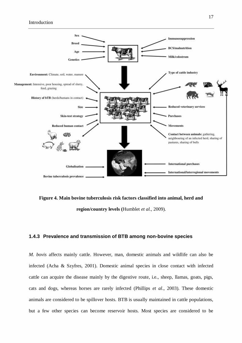
Transmission of BTB can be influenced by several factors, with a three-level classification:
individual, herd and region/country level (Figure 4). The situation in developed and
developing countries might be different in terms of risk of BTB, i.e., farming practices,
presence of wildlife, stocking density, pasturing systems and contact between animals
(Humblet et al., 2009). Thus, in developing countries the main risk factors are related to the
intensification of the farms, and the lack of measures to control the disease.
In general, contamination of feed and pasture is not an important source of transmission of M.
bovis in cattle, because survival times of infective doses of organisms on fomites are
relatively short under real conditions and animals are not commonly exposed to a dose high
enough to be infective by the oral route (Morris et al., 1994). In natural infections occurring
via the respiratory tract, M. bovis can be detected in nasal mucus from around 80 – 100 days
after infection (Neill et al., 1994).
17
Introduction
Figure 4. Main bovine tuberculosis risk factors classified into animal, herd and
region/country levels (Humblet et al., 2009).
1.4.3 Prevalence and transmission of BTB among non-bovine species
M. bovis affects mainly cattle. However, man, domestic animals and wildlife can also be
infected (Acha & Szyfres, 2001). Domestic animal species in close contact with infected
cattle can acquire the disease mainly by the digestive route, i.e., sheep, llamas, goats, pigs,
cats and dogs, whereas horses are rarely infected (Phillips et al., 2003). These domestic
animals are considered to be spillover hosts. BTB is usually maintained in cattle populations,
but a few other species can become reservoir hosts. Most species are considered to be

18
Introduction
spillover hosts. Populations of spillover hosts do not maintain M. bovis indefinitely in the
absence of maintenance hosts, but may transmit the infection between their members or to
other species for a time. Some spillover hosts can become maintenance hosts if their
population density is high (Morris et al., 1994; CFSPH, 2007).
In sheep, experimental studies conducted by Chaussé (1913) showed that the effective dose to
cause the disease by respiratory route is no more than 5 and probably as few as one bacillus
would produce a lesion in the lung, whereas 13 million bacilli would not always infect sheep
by the oral route.
In pigs, the occurrence of BTB can increase with age. In this specie, the main route of
transmission is the digestive tract; mainly by consumption of milk or dairy products, kitchen
and abattoir scraps, and excreta from tuberculous cattle (Acha & Szyfres, 2001). Although
transmission between swines can occur, it is considered to be epidemiologically no relevant
(Morris et al., 1994). In farms with a high prevalence of BTB, up to 50% of the cats may be
infected (CFSPH, 2007).
M. bovis infection in wildlife and feral species is a potential source of infection for livestock
and a threat to protected and endangered species (Aranaz et al., 2004). In some countries, TB
in cattle could be a readily controllable disease in absence of some wildlife reservoir (Morris
et al., 1994). In 22% of the countries, BTB has been detected in wildlife species, according to
the report from the Office International des Epizooties (OIE) (Livingstone, 2000). In Europe
and North America, some wild animals can frequently be affected. Known maintenance hosts
are red deer (Cervus elaphus), roe deer (Capreolus capreolus), fallow deer (Dama dama),
wild boar (Sus scrofa) and iberian lynx (Lynx pardina) in Spain, badger (Meles meles) in the

19
Introduction
UK and Ireland, brush-tailed possum (Trichosurus vulpecula) and ferret (Mustela furo) in
New Zealand, deer (Axis axis) in the United States, bison (Bison bison) in Canada and
ungulates in other countries. In Africa, infections have been reported in lion (Panthera leo),
cheetah (Acynonyx jubatus), and other wildlife species (Aranaz et al., 2004; Whipple &
Palmer, 2000). The maintenance wildlife hosts are characterized by their ability to produce
temporally continuous but spatially very patchy distribution of infection in their own
population. The route of transmission varies between species and can be observed in Figure 5.
The vast majority of spread within wildlife is by the airborne route (Morris et al., 1994). Also
among badgers, transmission appears to be mainly by respiratory route, and often it occurs as
a result of fighting between males (Morris et al., 1994). Cattle may be infected by badgers
through direct or indirect (namely urine, faeces and pus) transmission.
Figure 5. Routes of bovine tuberculosis transmission
CATTLE INFECTED
HEALTHY CATTLE
DIFFERENT HOSTS HUMANS
TRANSMISSION 80 – 90% BY RESPIRATORY ROUTE
OCCASIONALLY REINFECTION BY RESPIRATORY ROUTE
DIGESTIVE ROUTE & RESPIRATORY ROUTE (less frequent)
DIGESTIVE ROUTE & RESPIRATORY ROUTE (less frequent)

20
Introduction
1.4.4 Prevalence and transmission of BTB among humans
In humans, TB induced by M. bovis or M. tuberculosis is not clinically different (Cosivi et al.,
1998). In the progressive stage of disease, the following symptoms can become evident:
subfebrile temperatures, night sweats, lymphadenopathy, cough, fatigue, loss of appetite and
erythema on the extremities (Krauss et al., 2003). In immunodepressed patients, pneumonia
lymphadenopathy or disseminated disease can be observed (Acha & Szyfres, 2001).
The main risk factors involved in BTB infections are consumption of contaminated milk or
dairy products, and close physical contact between humans and potentially infected animals
(Acha & Szyfres, 2001). People who work in close contact with potentially infected animals,
such as the dairy farm and abattoir workers and also veterinarians run a high risk (Krauss et
al., 2003). Although consumption of meat from tuberculous cattle presents a theoretical risk,
the main hazard comes mainly from cooking and preparation methods (Shakespeare, 2002).
In countries where BTB is not controlled, most of the human cases occur in young people as a
result of unpasteurized milk consumption, which is an important source of TB due to M. bovis
for humans. In these patients, cervical lymphoadenitis, intestinal and skin lesions are
frequently observed (Gil & Samartino, 2001). The respiratory disease in human population at
risk may be acquired by inhaling droplet nuclei produced during cough spray from infected
cattle (Grange, 2001).
Many laboratories do not distinguish between M. bovis and M. tuberculosis infections, and the
documented cases on zoonotic TB are scare, and even more the cases of TB due to M. bovis
as a result of human to human transmission leading to disease occurs very rarely.
Nevertheless, in 1994 this kind of infection was reported in The Netherlands (van Soolingen
21
Introduction
et al., 1994). Although in general it would be very difficult to prove the association because
usually many years elapse between infection and development of bacteriologically positive
disease (Grange, 2001).
In Latin America and the Caribbean, epidemiological data related to TB are scarce, mainly
because the diagnosis of human TB is generally limited to sputum smear examination by
Zielh Neelsen stain, which cannot differenciate among the various species of mycobacteria.
Furthermore, samples are cultured mainly on Löwenstein-Jensen medium containing glycerol,
which inhibits growth of M. bovis (Zumarraga et al., 1999; Abalos & Retamal, 2004).
According to de Kantor & Ritacco (2006), 21 million people in 15 Latin American and
Caribbean countries are living in close contact with cattle, including meat processing and
veterinary services workers, cattle breeders and farmers. In addition, relatively high
incidences of human TB cases (30 to100 per 100000 inhabitants) are reported in those
countries. It has been estimated that an annual incidence of 7000 new TB cases due to M.
bovis occurs in this region.
In Argentina, M. bovis was identified in 2.3% of 4243 isolates from TB patients. Most
zoonotic TB cases in the region were associated with infection via the respiratory tract, and
65% of the cases occurred among persons at risk due to close contact with animals. Milk
pasteurisation and veterinary control in slaughterhouses have improved significantly here
during the last decade. These measures are effective to decrease the risk of oral transmission
but slaughterhouse and farm workers remain at risk of aerosol-borne pulmonary disease
(Torres, 2009; de Kantor & Ritacco, 2006).
22
Introduction
In Mexico, it has been demonstrated that M. bovis plays an important role in human TB
(Zendejas-Martinez et al., 2008). Human infection occurred mainly by the oral route,
specifically by the consumption of cheese. M. bovis has been isolated in cheese, representing
a high risk for public health. It has been calculated that 30 to 40% of the milk used to make
cheese and other dairy products is sold raw (unpasteurized) (Perez-Guerrero et al., 2008). In
San Diego-USA, close to the border with Mexico, approximately 7% of the culture-positive
TB patients had disease caused by M. bovis during the last decade; 90% of this group had
Hispanic origin (LoBue, 2006).
In Ecuador, the real occurrence of human TB from animal origin is most probably
underdiagnosed because the identification of the etiological agent is not performed as a
routine procedure. Diagnosis of human TB is mostly restricted to direct microscopy, and
Stonebrink medium - needed to grow M. bovis - is only used rarely. According to the National
Reference Laboratory of infectious diseases, only two cases of TB due to M. bovis have been
confirmed by culture (de Kantor et al., 2008). In 2007, a tuberculin skin testing survey
conducted in 157 farm and slaughterhouse workers (Benítez-Capistros, 2007), showed 29% of
positive reactors with a significant association among the positive-TST and the habit of
drinking raw milk (p < 0.00), as well as with the consumption of cheese (p = 0.003).
1.5 Diagnosis
1.5.1 Ante-mortem (in vivo) diagnosis
1.5.1.1 Clinical diagnosis
Diagnosis of BTB based on clinical signs alone is not possible, because most of cattle do not
present symptoms before an advanced stage of the disease, and these clinical signs are not
23
Introduction
specific. The severity of the disease varies with the dose of infectious organisms and
individual immunity. In the late stage, some signs may be suggestive such as gradual loss of
condition, emaciation, low–grade fluctuating fever, weakness and inappetence. Occasionally,
when the retropharyngeal nodes are enlarged, cattle present dysphagia. In case of advanced
pulmonary disease, a chronic soft cough is present, which is worse in the morning, during
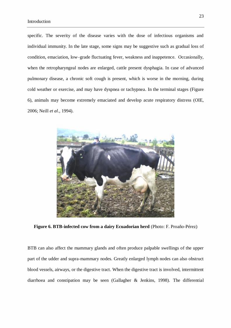
cold weather or exercise, and may have dyspnea or tachypnea. In the terminal stages (Figure
6), animals may become extremely emaciated and develop acute respiratory distress (OIE,
2006; Neill et al., 1994).
Figure 6. BTB-infected cow from a dairy Ecuadorian herd (Photo: F. Proaño-Pérez)
BTB can also affect the mammary glands and often produce palpable swellings of the upper
part of the udder and supra-mammary nodes. Greatly enlarged lymph nodes can also obstruct
blood vessels, airways, or the digestive tract. When the digestive tract is involved, intermittent
diarrhoea and constipation may be seen (Gallagher & Jenkins, 1998). The differential
24
Introduction
diagnosis of BTB includes contagious bovine pleuropneumonia, Pasteurella or
Corynebacterium pyogenes pneumonia, and aspiration pneumonia.
In maintenance hosts other than cattle, the severity of the disease varies with the species
(Neill et al., 1994). Brush-tailed possums are highly susceptible to BTB, and usually it causes
a fulminating pulmonary disease. In the final stage, they become disoriented, can not climb,
and sometimes can be seen wandering about in daylight. In badgers, on the contrary, BTB
does not frequently develop visible lesions and animals can survive for many years with the
bacilli. In symptomatic badgers, BTB is primarily a respiratory disease (Morris et al., 1994).
1.5.1.2 The tuberculin test
In human and veterinary medicine, intradermal tuberculinization or the tuberculin test is the
oldest and the most widely used in vivo diagnostic test. Tuberculin tests have been used for
the diagnosis of BTB in cattle for more than 100 years, and it is still widely used in the field
due to its ease of application on a large scale in livestock and low cost. Eventhough its
limitations have been recognized for many years, the tuberculinization has been the key
procedure in national test and slaughter programmes for BTB and its use has facilitated the
eradication of the disease in many regions of the world. Furthermore, it has provided most of
the available data on tuberculosis (Acha & Szyfres, 2001; Mongham et al., 1994).
Consequently, rigorous application of tuberculin testing and culling reacting cattle resulted in
the control of M. bovis infections; in the United States, it allowed to control BTB in domestic
livestock but without complete eradication (Whipple & Palmer, 2000).

25
Introduction
This diagnostic method depends upon the cell-mediated immunological response of the host
during the development of the disease (Adams, 2001). Wax D and various tuberculoproteins
induce a delayed hypersensitivity reaction detected in the tuberculin test (Quinn et al., 1999).
Primary TB can be diagnosed only by the development of a positive skin test (Krauss et al.,
2003). The standard procedure used for routine diagnosis of BTB by tuberculin test consists
of reading the skin reaction to an intradermally injected purified protein derivative (PPD)
(Shakespeare, 2002). Since 1975, PPD production switched to use M. bovis AN5, a strain
originally isolated in England and used nowadays worldwide for bovine PPD production. The
genome analysis showed that M. bovis AN5 has not suffered extensive gene deletion events
and it can be used for the detection of infection by M. bovis strains that are currently prevalent
(Hewinson et al., 2006).
Sensitivity of tuberculin tests has been reported in ranges from 77 to 95%, and specificity
from 96 to 99% (Mongham et al., 1994). The cutoff points can influence the sensitivity and
specificity of the skin tests i.e, if the cut-off is lowered, more infected animals are detected
but more non-infected animals are identified as false positives as well (Mongham et al., 1994;
Demelash et al., 2009).
The tuberculin test depends on several factors including high-quality reagents as well as the
immunological status of the animal. False-positive reactions are partly explained by allergies
to the simultaneous use of avian tuberculin, hypersensitivity to NTM, Corynebacterium,
Fasciola hepatica, and Nocardia species (Sreevatsan et al., 2000). False-negative responses
occur in animals in the early or in the late stages of the disease if anergy develops due to
excess of antigen or poor immune responses. Nonspecific factors such as malnutrition, stress
or recent calved, are alternative causes of anergy. Desensitization may be observed if time

26
Introduction
intervals between tuberculinizations are too short or if the procedure is not respected
(injection of insufficient tuberculin, use of tuberculins of reduced potency). The variability
among observers is also a source of errors (Biberstein & Hirsh, 1999). BCG vaccination
sensitises animals to antigens present in the bovine PPD and interfers with the tuberculin skin
test diagnosis in BCG vaccinated animals (Buddle et al., 1999).
1.5.1.2.1 Simple intradermal tuberculin test (SITT)
The SITT is a screening skin test mostly used in animals to identify possible reactors to BTB,
which is based on the dermal reaction obtained in animals that have had a previous contact
with the bacillus. In cattle, the skin areas used to perform this test are the skin of the neck or
in the caudal, vulvar or anal fold (Figure 7). The selected area needs to be cleaned, and the
tickness of the skin measured with callipers. This procedure is carried out before the injection
and when reading the result after 72 hours (+/- 6 hours). A dose of 0.1 mL of bovine PPD
needs to be injected intradermally, and should not be lower than 2000 International Units
(IU). The interpretation of the SITT considers a positive reaction if a swelling of 5 mm or
more at the side of the injection developed in 72 hours. This suggests a past or present
infection. An inconclusive reaction is considered if the increase in skin-fold thickness is
between 3 and 4mm, whereas for a negative reaction, the increase is less than 3mm without
clinical signs. Animals with positive or inconclusive reactions may be subjected to a further
SITT or confirmatory skin test after a period of minimum 42 days. In some areas, this
procedure is conducted after only 60 days (Mongham et al., 1994; OIE, 2006). It should be
kept in mind that the skin of the neck is much more sensitive than the skin of the caudal fold.
For this reason, the former site is always used in a second test to discard the false positives by
the first tuberculinization.
27
Introduction
1.5.1.2.2 Comparative intradermal tuberculin test (CITT)
The CITT is a common skin test used mainly in suspected and positive reactors identified by
the SITT to confirm BTB. It is used mainly to differenciate between animals infected with M.
bovis and those sensitised to tuberculin due to exposure to NTM or related genus. The CITT
is based on the simultaneous use of bovine and avian PPDs (PPD-B & PPD-A). This
procedure is applied in the middle third of the neck, in this area two different spots need to be
shaved and cleaned at least 12 cm apart (Figure 8). The tickness of the skin is measured with
callipers before the injection of the antigens: 0.1 mL (25 000 IU/mL) of PPD-A and 0.1 mL
(20 000 IU/mL) of PPD-B. 72 hours after PPD injections, skin thicknesses are measured again
to identify positive reactors.
A positive reaction to CITT is defined as a relative increase in skin thickness at the injection
site for PPD-bovine of at least 4 mm greater than the increase in skin thickness at the injection
site for PDD-avian. The reaction is inconclusive if no clinical signs are observed and if the
increase of the skin thickness is more than 2 mm and less than 4 mm. A negative reaction is
considered if only limited swelling is developed with an increase of no more than 2 mm and
without clinical signs. (OIE, 2008; Grooms & Molesworth, 2000).

28
Introduction
Figure 7. Simple intradermal tuberculin test (SITT)
(Photo: F. Proaño-Pérez)
Figure 8. Comparative intradermal tuberculin test (CITT)
(Photo: F. Proaño-Pérez)

29
Introduction
1.5.2 Post-mortem inspection
1.5.2.1 Necropsy procedure
The necropsy procedure performed after slaughtering of animals allows for the detection of
gross lesions suggestive of BTB, even in apparently healthy cattle. Veterinary inspection is
established as a routine procedure in most of the slaughterhouses in developing countries. In
the later stages of an eradication campaign and when the prevalence of BTB is low, the
detection of infected animals is mainly restricted to the routine slaughterhouse inspection
(Corner, 1994).
When detailed necropsy is conducted on reactor cattle, different tissues need to be sampled
(Table 2). In order to identify gross lesions compatible with BTB, careful examination needs
to be carried out. Although this method is used to identify infected animals, it can be affected
by the technique used and the anatomical sites examined (Corner, 1994). According to
Demelash et al. (2009), the sensitivity of the necropsy has been calculated in 95%, when as
few as 6 pairs of thoracic lymph nodes, lungs, and mesenteric lymph nodes are inspected. The
sensitivity of the necropsy procedure depends also largely on the time and diligence of the
inspectors during the examination of the carcasses (Corner, 1994).
Necropsy can fail to detect infected animals in an early stage of infection or if observation and
sampling are not complete. In addition, not all M. bovis-infected cattle develop visible gross
lesions (Whipple et al., 1996). Lesions due to an infection with non-tuberculous mycobacteria
(NTM) can be easily mistaken with BTB lesions (Oloya et al., 2007). Therefore, it is
important to confirm the presence of M. bovis by the use of available laboratory methods.

30
Introduction
Table 2. Tissues to be examined for the presence of macroscopic lesions in cattle
reacting to a tuberculin test.
Regions Tissues
Head mandibular lymph nodes*
parotid lymph nodes*
medial retropharyngeal lymph nodes*
lateral retropharyngeal (atlantal) lymph nodes*
Tonsils
Thorax mediastinal lymph nodes**
tracheobronchial lymph nodes*
bronchial lymph node***
Lungs
Abdomen Liver
hepatic lymph node
Spleen
mesenteric lymph nodes (along the entire length of the gastro-intestinal tract)
Kidneys
Carcasse caudal cervical (prescapular) lymph nodes*
subiliac (prefemoral) lymph nodes*
internal iliac lymph nodes*
medial iliac lymph nodes*
lateral iliac lymph nodes*
gluteal (ischiatic) lymph nodes*
sacral lymph nodes*
superficial inguinal (supramammary or scrotal) lymph nodes*
udder or scrotal contents and seminal vesicles
* = left and right
** = anterior and posterior
*** = cranial and medial
(From Corner et al., 1990)
31
Introduction
1.5.2.2 Macroscopic lesions
BTB is characterized by the development of granulomas or tubercles where the bacilli are
localized. When M. bovis is transmitted in a new host, a primary lesion or focus of infection is
established, which depends on the interaction between host and pathogen. This primary lesion,
together with the lesion in the regional lymph node is called the ―primary complex‖ (Neill et
al., 1994). The granulomas are usually yellowish and either caseous, caseo-calcareous or
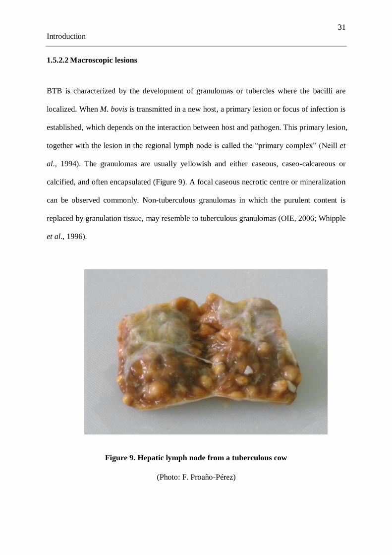
calcified, and often encapsulated (Figure 9). A focal caseous necrotic centre or mineralization
can be observed commonly. Non-tuberculous granulomas in which the purulent content is
replaced by granulation tissue, may resemble to tuberculous granulomas (OIE, 2006; Whipple
et al., 1996).
Figure 9. Hepatic lymph node from a tuberculous cow
(Photo: F. Proaño-Pérez)

32
Introduction
Figure 10. Mediastinal lymph node from a tuberculous cow, showing a yellowish caseous
granuloma (Photo: F. Proaño-Pérez)
Figure 11. Lung from a tuberculous cow showing calcified granulomas
(Photo: F. Proaño-Pérez)

33
Introduction
BTB infection in cattle mainly occurs by inhalation of M. bovis in droplet nuclei. Thus, the
primary complex is usually found in the lungs and associated lymph nodes. In cattle,
granulomas are located mainly in the lymph nodes from head and thorax; however, they are
also common in the lungs, spleen, liver, and the surfaces of body cavities (Figures 10 and 11).
In disseminated cases, multiple small granulomas may be found in numerous organs (Cousins
et al., 2004). Lymph node lesions may be absent in cases of chronic tuberculosis pneumonia.
According to Neill et al. (1994), 57% of cattle present lesions confined to the bronchial and/or
mediastinal lymph nodes, 23% in the head only (retropharyngeal and submaxillary lymph
nodes) and a lower occurrence of 3.2% in mesenteric lymph nodes. However, it is important
to consider that the distribution of lesions can vary depending on several factors, i.e., route of
transmission, infecting dose, incubation period before examination, virulence, individual
immune response, age or breed (Corner, 1994; Neill et al., 1994; Whipple et al., 1996). In
some species such as deer, the lesions tend to resemble abscesses rather than typical tubercles.
Some tubercles are small enough to be missed by the naked eye, unless the tissue is sectioned
(CFSPH, 2007).
1.5.3 Laboratory diagnostic tools
M. bovis can be identified by various laboratory methods. Samples collected from suspicious
animals are commonly used to confirm the presence of M bovis by applying microscopy,
histopathology, in vitro culture, PCR, and blood testing. They must be treated as risk/hazard
group III to prevent human infections during their processing (OIE, 2006).
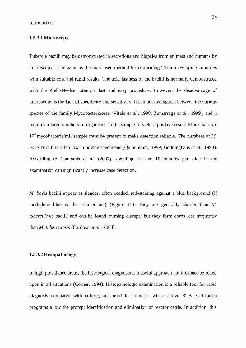
34
Introduction
1.5.3.1 Microscopy
Tubercle bacilli may be demonstrated in secretions and biopsies from animals and humans by
microscopy. It remains as the most used method for confirming TB in developing countries
with suitable cost and rapid results. The acid fastness of the bacilli is normally demonstrated
with the Ziehl-Neelsen stain, a fast and easy procedure. However, the disadvantage of
microscopy is the lack of specificity and sensitivity. It can not distinguish between the various
species of the family Mycobacteriaceae (Vitale et al., 1998; Zumarraga et al., 1999), and it
requires a large numbers of organisms in the sample to yield a positive result. More than 5 x
104
mycobacteria/mL sample must be present to make detection reliable. The numbers of M.
bovis bacilli is often low in bovine specimens (Quinn et al., 1999; Boddinghaus et al., 1998).
According to Cambanis et al. (2007), spending at least 10 minutes per slide in the
examination can significantly increase case detection.
M. bovis bacilli appear as slender, often beaded, red-staining against a blue background (if
methylene blue is the counterstain) (Figure 12). They are generally shorter than M.
tuberculosis bacilli and can be found forming clumps, but they form cords less frequently
than M. tuberculosis (Cardoso et al., 2004).
1.5.3.2 Histopathology
In high prevalence areas, the histological diagnosis is a useful approach but it cannot be relied
upon in all situations (Corner, 1994). Histopathologic examination is a reliable tool for rapid
diagnosis compared with culture, and used in countries where active BTB eradication
programs allow the prompt identification and elimination of reactor cattle. In addition, this

35
Introduction
method permits identification of typical mycobacterial lesions and its differentiation from
other causes (Varello et al., 2008).
Figure 12. Mycobacterium bovis bacilli in a smear stained with Ziehl Neelsen
(magnification 1000x) (Photo: F. Proaño-Pérez)
Figure 13. Macrophage in a bronchial lymph node specimen stained with Ziehl Neelsen
(Photo: D. Desmecht & F. Proaño-Pérez)
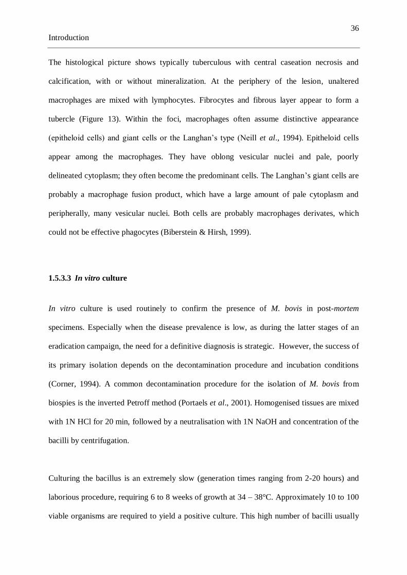
36
Introduction
The histological picture shows typically tuberculous with central caseation necrosis and
calcification, with or without mineralization. At the periphery of the lesion, unaltered
macrophages are mixed with lymphocytes. Fibrocytes and fibrous layer appear to form a
tubercle (Figure 13). Within the foci, macrophages often assume distinctive appearance
(epitheloid cells) and giant cells or the Langhan‘s type (Neill et al., 1994). Epitheloid cells
appear among the macrophages. They have oblong vesicular nuclei and pale, poorly
delineated cytoplasm; they often become the predominant cells. The Langhan‘s giant cells are
probably a macrophage fusion product, which have a large amount of pale cytoplasm and
peripherally, many vesicular nuclei. Both cells are probably macrophages derivates, which
could not be effective phagocytes (Biberstein & Hirsh, 1999).
1.5.3.3 In vitro culture
In vitro culture is used routinely to confirm the presence of M. bovis in post-mortem
specimens. Especially when the disease prevalence is low, as during the latter stages of an
eradication campaign, the need for a definitive diagnosis is strategic. However, the success of
its primary isolation depends on the decontamination procedure and incubation conditions
(Corner, 1994). A common decontamination procedure for the isolation of M. bovis from
biospies is the inverted Petroff method (Portaels et al., 2001). Homogenised tissues are mixed
with 1N HCl for 20 min, followed by a neutralisation with 1N NaOH and concentration of the
bacilli by centrifugation.
Culturing the bacillus is an extremely slow (generation times ranging from 2-20 hours) and
laborious procedure, requiring 6 to 8 weeks of growth at 34 – 38°C. Approximately 10 to 100
viable organisms are required to yield a positive culture. This high number of bacilli usually

37
Introduction
occurs only in advanced stages of the disease. For in vitro culture of M. bovis, Stonebrink or
Coletsos medium are widely used. They are adapted Lowenstein-Jensen media without
glycerol and with addition of sodium pyruvate (Figures 14 and 15). The presence of glycerol
inhibits the growth of M. bovis. Biochemical and enzymatic tests can be applied for the in
vitro identification (Table 3). M. bovis is characterized by rough colony morphology, absence
of niacin production, nitrate reduction, catalase and pyrazinamidase negative activity,
presence of urease activity, and susceptibility to thiophene-2-carboxylic acid hydrazide and p-
animo-salicylic acid (Biberstein & Hirsh, 1999; Gallagher & Jenkins, 1998).
Table 3. In vitro tests for some significant mycobacteria
species
Runyon g
roup
Gro
wth
wit
hin
7 d
ays
Inhib
itio
n b
y g
lyce
rol
Colo
nia
l m
orp
holo
gy *
Pig
men
tati
on *
*
Nia
cin p
roduct
ion
Tole
rence
of
5%
NaC
l
Dea
min
atio
n o
f P
ZN
Nit
rate
red
uct
ion
Ure
ase
pro
duct
ion
M. bovis neg pos S/R n neg neg neg neg v
M. tuberculosis neg neg R n pos neg pos pos v
M. avium intracellulare III neg neg S/R n neg neg pos neg neg
M. ulcerans III neg neg R n neg neg neg neg v
M. simiae I neg neg S p v neg pos neg pos
M. kansasii I neg neg SR p neg neg neg pos v
M. marinum I neg neg S/SR p -(+) neg pos neg pos
M. scrofulaceum II neg neg S s neg neg v neg v
M. xenopi III neg neg S n(s) neg neg v neg neg
M. chelonae IV pos neg S/R n v +(-) pos neg pos
M. fortuitum IV pos neg S/R n v pos pos pos pos
* R = rough, S = smooth, and SR = intermediate
** p = photochromogenic (pigment produced only if culture is exposed to light)
s = scotochromogenic (pigment produced in the light and in the dark)
n = non-chromogenic (no pigment produced)
v = variable reactions, -(+) = majority negative, +(-) = majority positive
(From Quinn et al., 1999)

38
Introduction
Figure 14. Colonies of M. bovis on Stonebrink medium after 8 weeks of incubation
(Photo: F. Proaño-Pérez)
Figure 15. Colonies of M. bovis on Stonebrink medium after 24 weeks of incubation
(Photo: F. Proaño-Pérez)

39
Introduction
Corner & Nicolacopoulos (1988) recommended the use of Stonebrink, Middlebrook 7H11 or
tuberculosis bovine blood agar as a media of choice for the primary isolation of M. bovis. In
addition, liquid media with a radiometric detection technique can be used i.e., BACTEC®
system (Middlebrook et al., 1977). This method is based on a radiolabelled palmitic acid on
liquid medium, which allows a rapid detection of growing bacilli that produce radiolabeled
CO2. The disadvantage of the BACTEC culture is that it can be performed only in a high level
laboratory, that it is labor intensive and poses a risk of exposure to radiation. Nowadays, a
novel culture method has been evaluated as a suitable alternative for the replacement of the
BACTEC 460 radiometric system, the BACTEC Mycobacteria Growth Indicator Tube
(MGIT) system, which rapid, and based on a non-radiometric fluorescent detection system
(Hines et al., 2006). However, again the automated version requires expensive equipment and
media.
1.5.3.4 Imnunological tests
Blood-based immunological assays can be used to diagnose BTB, especially in zoo animals
and wildlife. The available tests are the lymphocyte proliferation and gamma-interferon
assays, both measuring cellular immunity. The lymphocyte proliferation test is not commonly
used in cattle, but may be useful in wildlife and zoo animals, as they must be captured only
once, rather than twice for the tuberculin test. The enzyme-linked immunosorbent assay
(ELISA) may complement tests of cellular immunity in anergic cattle. There is no universally
accepted diagnostic blood test for BTB in cattle or other animals (OIE, 2006; Thoen & Ebel,
2006).
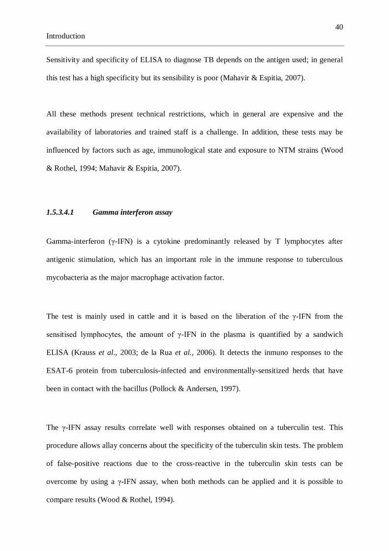
40
Introduction
Sensitivity and specificity of ELISA to diagnose TB depends on the antigen used; in general
this test has a high specificity but its sensibility is poor (Mahavir & Espitia, 2007).
All these methods present technical restrictions, which in general are expensive and the
availability of laboratories and trained staff is a challenge. In addition, these tests may be
influenced by factors such as age, immunological state and exposure to NTM strains (Wood
& Rothel, 1994; Mahavir & Espitia, 2007).
1.5.3.4.1 Gamma interferon assay
Gamma-interferon (γ-IFN) is a cytokine predominantly released by T lymphocytes after
antigenic stimulation, which has an important role in the immune response to tuberculous
mycobacteria as the major macrophage activation factor.
The test is mainly used in cattle and it is based on the liberation of the γ-IFN from the
sensitised lymphocytes, the amount of γ-IFN in the plasma is quantified by a sandwich
ELISA (Krauss et al., 2003; de la Rua et al., 2006). It detects the inmuno responses to the
ESAT-6 protein from tuberculosis-infected and environmentally-sensitized herds that have
been in contact with the bacillus (Pollock & Andersen, 1997).
The γ-IFN assay results correlate well with responses obtained on a tuberculin test. This
procedure allows allay concerns about the specificity of the tuberculin skin tests. The problem
of false-positive reactions due to the cross-reactive in the tuberculin skin tests can be
overcome by using a γ-IFN assay, when both methods can be applied and it is possible to
compare results (Wood & Rothel, 1994).

41
Introduction
Blood samples for the γ-IFN test must be transported to the laboratory promptly, as this test
must be started within 24 to 30 hours after blood collection. However, the main disadvantage
is the high cost (i.e., aprox. 50 USD) of the γ-IFN limiting its use especially in developing
countries (Thoen & Ebel, 2006).
1.5.3.5 Polymerase chain reaction (PCR)
Differentiation of the causative Mycobacterium species usually requires in vitro growth of the
organism, which is time consuming. Alternatively, PCR has been developed with
considerable success (Shakespeare, 2002). PCR has been successfully applied to identify M.
tuberculosis-complex DNA directly in samples from animals and humans like sputum, blood,
milk, nasal swabs and tissues. The advantage of this method versus culturing is its rapidity of
detection and ability to detect non-viable bacilli. PCR significantly reduces the time to
confirm diagnosis in suspected cases (Boddinghaus et al., 1990). However, its relative high
cost is considered to be prohibitive for extensive routine diagnosis (i.e., aprox 40 $USD).
Therefore, PCR is mostly restricted for research purposes in developing countries. The
detection limit reported by PCR-based methods can vary between 1 and 100 bacilli present in
the sample (Zumarraga et al., 2005).
The primers most commonly used in M. bovis PCR-based assays target IS6110, an insertion
sequence (IS) that usually has more than 10 copies in the genome of M. tuberculosis, while
M. bovis contains in general a single copy (Thierry et al., 1990). Boddinghaus and others
(1990) have developed a sensitive and rapid technique based on the amplification of nucleic
acid sequences from 16S rRNA gene, a gene present in all prokaryotes. The rRNA is an
essential constituent of bacterial ribosomes, with highly conserved sequences but also a

42
Introduction
considerable amount of variability. It is present in large copy numbers (103 to 10
4 molecules
per cell) (Bates, 1979). This technique is performed by using specific primers directed to the
16S rRNA gene and allows identifying organisms belonging to the genus Mycobacterium and
members of the M. tuberculosis complex.
The 500-bp genomic fragment is conserved among M. bovis strains and is used on PCR-based
assay to identify BTB (Rodriguez et al., 1999). This method is able to detect down to 10 fg
DNA, which corresponds roughly to two bacilli and can be used directly on biological
samples such as milk making this a good assay for the rapid and specific diagnosis of M.
bovis and can be used as an epidemiological tool (Rodriguez et al., 1995). The first studies on
direct detection of M. bovis from bovine tissues by the use of PCR were reported in 1995
(Wards et al., 1995; Liébana et al., 1995).
The complex structure and impermeability of the cell walls of mycobacteria complicate the
extraction of DNA and/or RNA (Boddinghaus et al., 1990). In addition, the low numbers of
AFB in tissues from BTB-infected cattle suggest that a sensitive extraction procedure is
required for the PCR (Roring et al., 2000). The sensitivity of the PCR can also be affected by
the presence of inhibitors in the samples (Mangiapan et al., 1996; Cardoso et al., 2009).
Besides, extraction of mycobacterial DNA from tissue specimens is more difficult than from
sputa (Wards et al., 1995). Therefore, the choice of DNA extraction method is a key factor
determining the efficacity of diagnosis by PCR. Mangiapan and colleagues (1996) have
developed a usefull extraction approach to improve the sensitivity of detection, based on the
use of biotinylated primers that permit the specific capture of mycobacterial DNA prior to
amplification.
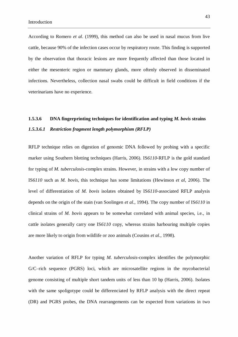
43
Introduction
According to Romero et al. (1999), this method can also be used in nasal mucus from live
cattle, because 90% of the infection cases occur by respiratory route. This finding is supported
by the observation that thoracic lesions are more frequently affected than those located in
either the mesenteric region or mammary glands, more oftenly observed in disseminated
infections. Nevertheless, collection nasal swabs could be difficult in field conditions if the
veterinarians have no experience.
1.5.3.6 DNA fingerprinting techniques for identification and typing M. bovis strains
1.5.3.6.1 Restriction fragment length polymorphism (RFLP)
RFLP technique relies on digestion of genomic DNA followed by probing with a specific
marker using Southern blotting techniques (Harris, 2006). IS6110-RFLP is the gold standard
for typing of M. tuberculosis-complex strains. However, in strains with a low copy number of
IS6110 such as M. bovis, this technique has some limitations (Hewinson et al, 2006). The
level of differentiation of M. bovis isolates obtained by IS6110-associated RFLP analysis
depends on the origin of the stain (van Soolingen et al., 1994). The copy number of IS6110 in
clinical strains of M. bovis appears to be somewhat correlated with animal species, i.e., in
cattle isolates generally carry one IS6110 copy, whereas strains harbouring multiple copies
are more likely to origin from wildlife or zoo animals (Cousins et al., 1998).
Another variation of RFLP for typing M. tuberculosis-complex identifies the polymorphic
G/C–rich sequence (PGRS) loci, which are microsatellite regions in the mycobacterial
genome consisting of multiple short tandem units of less than 10 bp (Harris, 2006). Isolates
with the same spoligotype could be differenciated by RFLP analysis with the direct repeat
(DR) and PGRS probes, the DNA rearrangements can be expected from variations in two

44
Introduction
different repetitive DNA sequences. Consequently, a combination of two methods
(spoligotyping, and typing with RFLP analysis with the DR and PGRS probes) would be
necessary for the differenciation of M. bovis for a detailed epidemiological study (Zumarraga
et al., 1999).
1.5.3.6.2 Spoligotyping
Spacer oligonucleotide typing (Spoligotyping) is a usefull molecular typing approach
enabling to differentiate M. bovis strains, a distinction which is often difficult to make by
traditional methods (Kamerbeek et al., 1997). Spoligotyping is a simple, rapid and robust
method for simultaneous detection and typing of M. tuberculosis complex, it is possible to
carry out directly from tissue specimens (i.e., microscopy positive) without the need to culture
the bacillus or to purify DNA (Roring et al., 2000).
This method is based on PCR amplification of a high polymorphic DR locus within the
genome of the M. tuberculosis complex, which contains DR sequences interspersed with
variable spacer sequences. Each DR is 36 bp long and spacer length varies from 35 to 41 bp.
The DR locus is a preferential site for insertion of a copy of the IS6110 element. The
interpretation of the test result is based on the presence or absence of specific spacer region
sequences between two DR regions, using labelled primers directed to this region followed by
reversed line blot hybridization. The test format includes oligonucleotides allowing detection
of 43 spacer sequences. The absence of spacers 3, 9, 16, and 39-43 is a characteristic of all M.
bovis strains (Kamerbeek et al., 1997).

45
Introduction
Since it is relatively fast, it is in particular useful in low-incidence communities or when the
study of an outbreak requires urgent differentiation (van Helden, 1999). A second-generation
of the spoligotyping assay detects the presence of the 43 traditional spacers plus 51 new
additional spacers oligonucleotides; however, it was detected a redundancy analysis and only
40 essential spacer oligonucleotides of the 94-spacer sequences were selected. This technique
was performed in Netherlands and described by van der Zanden et al. (2002), which is used
whenever extended discrimination is required (i.e., for low-copy-number IS6110 strains).
1.5.3.6.3 Variable number tandem repeats (VNTRs)
A more recent PCR-based typing method based on 12 loci containing variable number tandem
repeats (VNTRs)-genotyping of genetic elements named mycobacterial interspersed repetitive
units (MIRUs). M. tuberculosis complex members have 41 different MIRUs ramdomly
located throughout their genomes (Supply et al., 2000; Roring et al., 2002). Recently, an
optimized set of 24 loci was defined, including a highly discriminatory subset of 15 loci for
specific first-line epidemiological investigation (Supply et al., 2006).
MIRU-VNTR includes a PCR amplification using primers specific for the flanking regions of
the MIRUs, and studies the variability in number of repeats of these minisatelites in specific
loci of the M. tuberculosis complex genome. VNTR-typing for M. bovis targets minimum six
loci, whereas Spoligotyping only records variation at a single locus (Frothingham and
Meeker-O‘Connell, 1998; Supply et al., 2000).
The VNTRs can be used for analysis of the global genetic diversity of M. tuberculosis
complex strains at different levels of evolutionary, divergence opening the way for global

46
Introduction
epidemiological surveillance of tuberculosis (Supply et al., 2001) or ranging from pedigree to
evolucionary distant phylogenetic relationships (Hewinson et al., 2006). Certain MIRU-
VNTR markers may be useful for larger population-based studies, whereas others may be
more appropriate for outbreak analysis (Harris, 2006). This is the epidemiological typing
method of choice for the near future and has 100% reproducible and its use is being evaluated
to accept it as diagnostic tool (Kremer et al., 2005).
1.6 Control of BTB
The control of BTB is important not only because its human health impact but also because of
the resulting economic losses (OIE, 2006). In most countries, TB is a notifiable disease in
humans and animals, and strict controls have to be exerted on affected herds. Control
measures usually include early diagnosis with tuberculin test followed by slaughtering of
infected animals.
The basic strategies required for control and elimination of BTB in Latin America are well
known and well defined by the World Organization for Animal Health. Regural testing and
removal of infected animals at intervals of less than a year, in combination with apropiate
hygiene measures and restrictions to prevent introduction of infected animals could eliminate
the infection from herds (Morris et al., 1994). However, because of financial constraints,
scarcity of trained professionals, lack of political will, as well as the underestimation of the
importance of this zoonotic disease in both animals and public health sectors by national
governments, these control measures are not applied or are applied inadequately in most
developing countries (Cosivi et al., 1998).
47
Introduction
In Latin America, a ―Plan of Action for the eradication of BTB in the Americas‖, has been
developed by the Pan American Health Organization (PAHO), in cooperation with the Pan
American Institute for Food Protection and Zoonoses (INPAAZ) and the Food and
Agriculture Organization (FAO). It aims to eradicate BTB from countries with more
advanced national programs (Cosivi et al., 1998).
In developed countries, control programs have been conducted with success and some
countries are free of BTB. However, one of the greatest threats to any control program in
domestic ruminants is wildlife reservoirs that cannot be controlled and can re-introduce the
disease in livestock (Biet et al., 2005). It is now recognised that wildlife reservoirs play a
significant role in the persistence of M. bovis in cattle and make the eradication of the disease
difficult (Corner, 2006), especially when domestic and wildlife species share the same habitat
(Gallagher & Jenkins, 1998). The control programs are difficult if (1) BTB may persist in a
wild population (maintenance hosts) without presence of infected cattle and if (2) there is a
risk of contact between these wild populations and cattle. However, the only control measure
available for wildlife hosts is removal of infected animals (Morris et al., 1994).
BTB distribution might facilitate the work of the national control programme by identification
of areas of higher or lower BTB transmission risk, complemented with environmental model
that summarises conditions i.e., climate, topography, soils and land cover, under M. bovis is
expected to be absent, versus potentially present. These environmental characteristics can be
useful to study BTB transmission on spatial scales. In Mexico, the national BTB control
campaign focused on involving farmers by means of certification of farm as BTB-free after 3
consecutive negative tuberculin test results; however, outbreaks in certified BTB free herds

48
Introduction
raise the question if M. bovis can survive undetected and that environmental factors are
involved (Zendejas-Martinez et al., 2008).
M bovis can enter into a herd from outside via cattle by two major routes, i.e., though
purchase of infected cattle and from neighbouring farms by contiguous spread. According to
Goodchild & Clifton-Hadley (2001), an adequate biosecurity program can reduce spread
between herds, and improvements in management measures i.e., adequate ventilation, reduce
of group sizes and hygienic measures can reduce the transmission within herds. Additionally,
a complementary strategy need to be implemented to minimize the number of movements
from infected to non-infected herds or areas, using the animal or herd identification to control
the movements. However, if the infection persists under such cicunstances, it is entirely due
to management procedures allowing infected animals to remain in herds and to move between
herds (Morris et al., 1994; Phillips et al., 2003).
To ensure the effectivity during the identification of bovine reactors, the production,
standarization and quality control of purified protein derives is urgently required for use in
control and eradication campaigns in order to assure reability of reagents and comparability of
data on tuberculin testing (Kantor & Ritacco, 2006). Once positive BTB cattle is found, they
need to be eliminated through of an official compensation for culled animals; nevertheless, if
a political will is not established, this can represents a serious drawback that could jeopardize
the control program. The first step is motivate farmers to cooperate, and convince them that
progress is achievable, the farmer can be the leadership in programs to ensure that the farming
community contributes fully to the design of control measures and their enforcement (Morris
et al., 1994).
49
Introduction
In human population, pasteurization of milk and vaccination with bacillus Calmette-Guerin
(BCG) prevents the infection, especially in children (Lee et al., 2004). In Latin America, the
milk is pasteurized but the quality control of this process is not always complete and reliable
and its coverage is low in some areas (Ritacco et al., 2006). Vaccination is focused on infants
and tuberculin-negative individuals anticipating exposure (Acha & Szyfres, 2001).
1.7 Eradication
Once a control BTB program is implemented with success, the eradication plan could begin.
Infected herds will be quarantined, and animals that have been in contact with reactors need to
be traced. Only test-and-slaughter techniques are guaranteed to eradicate tuberculosis from
domesticated animals. Nevertheless, the occurrence of M. bovis in wildlife reservoir hosts
complicates eradication efforts. Where there is interplay between infection in wildlife and
domestic animals, eradication of the disease becomes impractical (Morris et al., 1994).
Slaughter surveillance with tracing of infected animals may be a more efficient use of
resources, when the eradication is nearly complete. Sanitation and disinfection may reduce the
spread of the agent within the herd; rodent control may also be advisable on affected farms.
Barriers can be used around hay storage areas to prevent wildlife access. In addition,
biosecurity measures on farms decrease interactions between wildlife and domesticated
animals (Collins, 2006).
The BCG vaccine has been used in calves, but this practice is inappropriate in countries
attempting to eradicate TB, because it interferes with the interpretation of the tuberculin test
and other immunological tests to detect infected animals (Suazo et al., 2003; OIE, 2006).

50
Introduction
Effective bovine tuberculosis vaccines are not currently available for cattle; the new vaccines
are being developed and tested, particularly for wildlife reservoirs (Buddle et al., 2006).

CHAPTER 2 – OBJECTIVES

52
Objectives
General objectives
The main objectives of this thesis were to provide data on the epidemiologic situation of
bovine tuberculosis (BTB) among dairy cattle in northern Ecuador and to compare the
performances of the standard diagnostic tools used to identify M. bovis.
Specific objectives:
1. To evaluate the situation of BTB, in northern Ecuador (Mejia canton), among small,
medium and large farms by using the single tuberculin caudal test and the comparative
tuberculin test, and to isolate M. bovis from dairy cattle in this region.
2. To determine the prevalence and annual incidence of BTB in dairy cattle in this canton
at individual and herd levels and to describe the associated risk factors.
3. To compare currently available laboratory tools to diagnose BTB and to characterize
the distribution of lesions from slaughtered cattle as a marker for the possible route of
transmission.
4. To perform a molecular characterization of M. bovis using spoligotyping and to
compare the strains isolated from the Mejia canton.
5. To determine the zoonotic impact of BTB through a preliminary study in risk
populations (farm and slaughterhouse workers) by tuberculin skin test.

CHAPTER 3 – RESULTS

54
Study 1
3.1 Preliminary observations on Mycobacterium spp. in dairy cattle in Ecuador
FREDDY PROAÑO-PEREZ, LEEN RIGOUTS, JEF BRANDT, PIERRE DORNY, JORGE
RON, MARIA-AUGUSTA CHAVEZ, RICHAR RODRIGUEZ, KRISTA FISSETTE,
ANITA VAN AERDE, FRANÇOISE PORTAELS, AND WASHINGTON BENITEZ-
ORTIZ
American Journal of Tropical Medicine and Hygiene. 2006 Aug;75(2):318-323.

55
Study 1
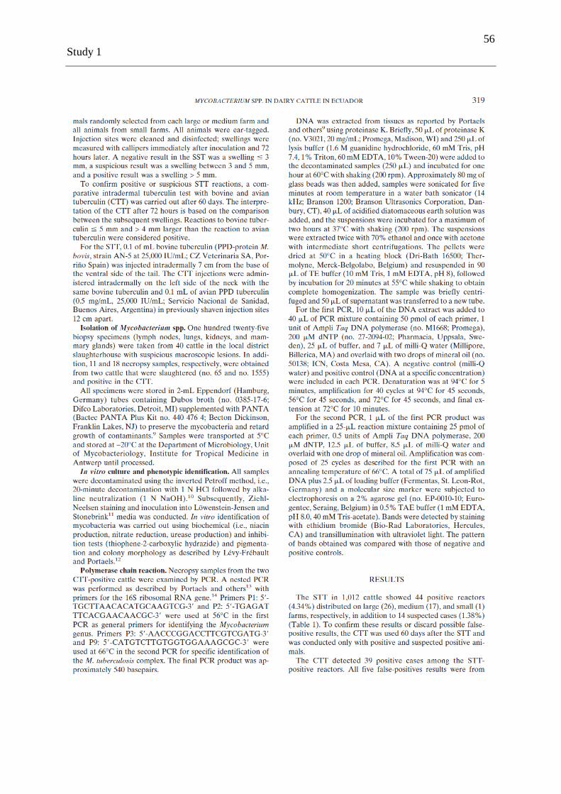
56
Study 1
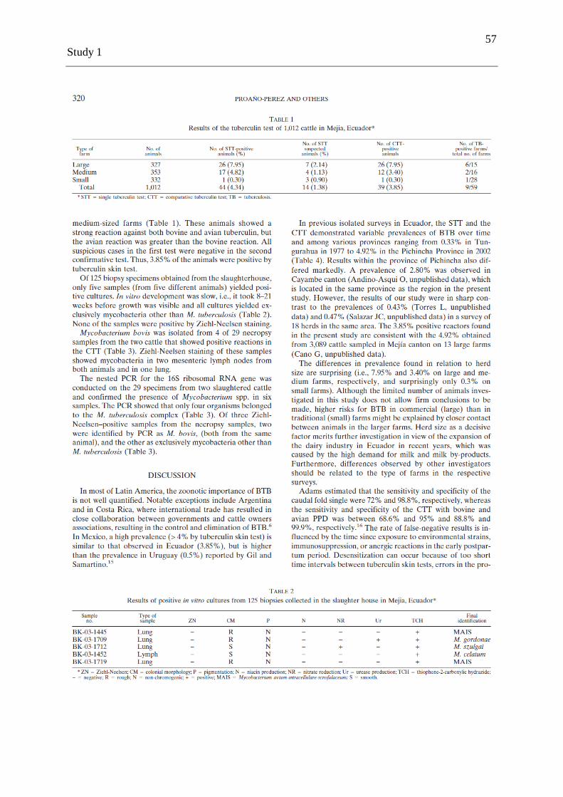
57
Study 1

58
Study 1

59
Study 1

60
Study 1

61
Study 2
3.2 Comparative intradermal tuberculin test in dairy cattle in the north of Ecuador and risk factors associated with bovine tuberculosis
PROAÑO-PEREZ FREDDY, BENITEZ-ORTIZ WASHINGTON, CELI-ERAZO
MARITZA, RON-GARRIDO LENIN, BENITEZ-CAPISTROS RICARDO, PORTAELS
FRANÇOISE, RIGOUTS LEEN AND LINDEN ANNICK.
American Journal of Tropical Medicine and Hygiene. 2009 Dec;81(6):1103-1109.

62
Study 2

63
Study 2

64
Study 2
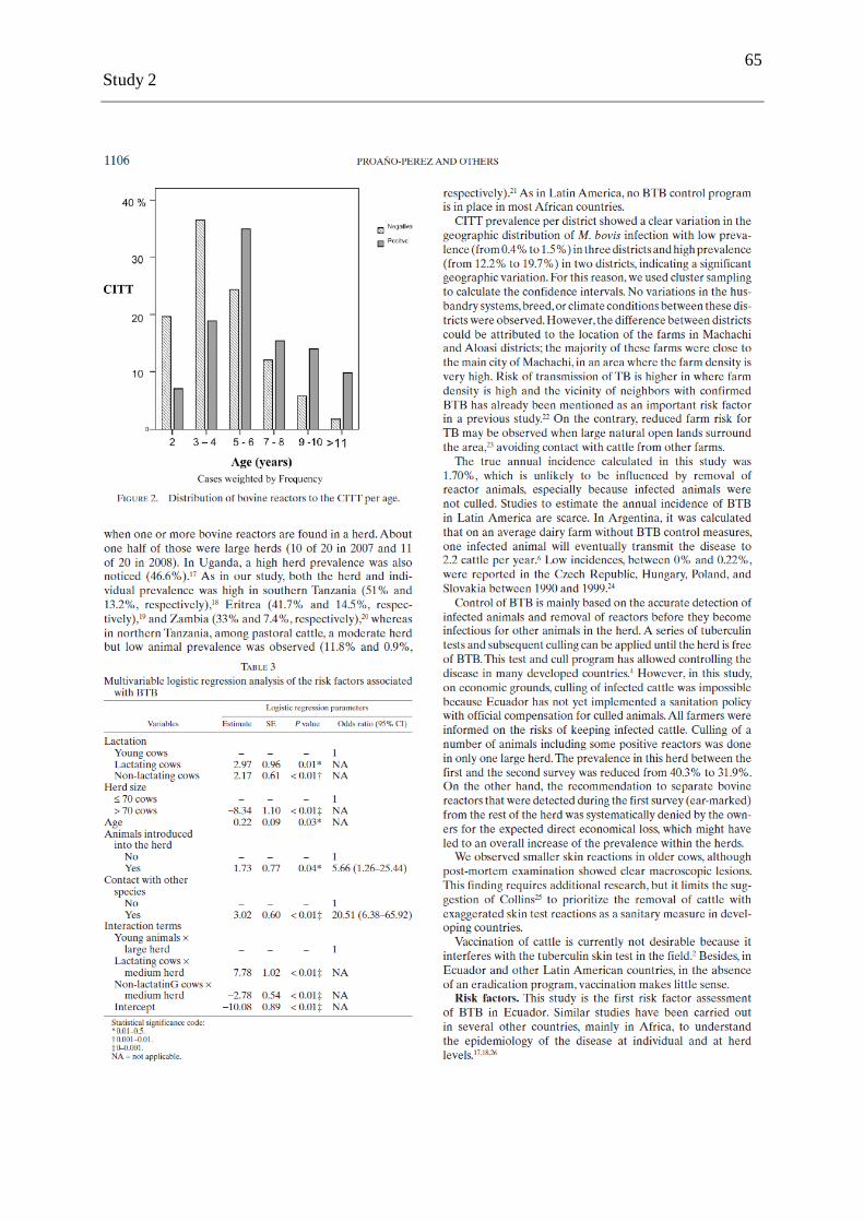
65
Study 2
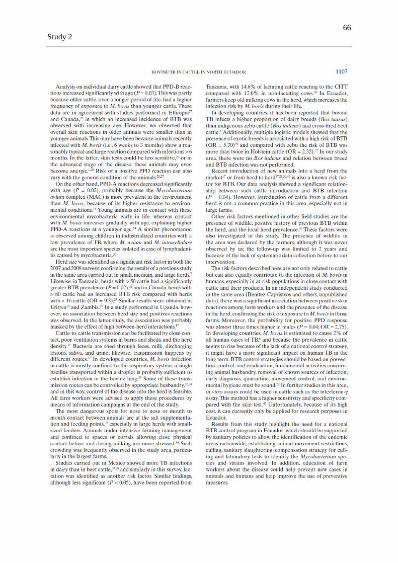
66
Study 2

67
Study 2

68
Study 2

69
Study 3
3.3 Post-mortem examination and laboratory based analysis for the diagnosis of bovine tuberculosis among dairy cattle in Ecuador
PROANO-PEREZ FREDDY, BENITEZ-ORTIZ WASHINGTON, DESMECHT DANIEL,
URRESTA DAFNE, CORAL MARCO, ORTIZ JULIO, RON LENIN, PORTAELS
FRANÇOISE, RIGOUTS LEEN & LINDEN ANNICK.
Journal of Preventive and Veterinary Medicine. 2011 Aug;101:65-72.

70
Study 3
71
Study 3

72
Study 3

73
Study 3

74
Study 3

75
Study 3

76
Study 3

77
Study 3

78
Study 4
3.4 Single Mycobacterium bovis strain prevailing among dairy cattle in northern Ecuador
PROANO-PEREZ FREDDY, BENITEZ-ORTIZ WASHINGTON, PORTAELS
FRANÇOISE, RIGOUTS LEEN AND LINDEN ANNICK.
Journal of Preventive and Veterinary Medicine (submitted)

79
Study 4
Single Mycobacterium bovis strain prevailing among dairy cattle in northern Ecuador.
Freddy Proaño-Pérez 1-2
, Washington Benitez-Ortiz1-3
, Françoise Portaels5,
Leen Rigouts 5-6
, Annick Linden2
1
International Centre for Zoonoses, Central University of Ecuador, Quito, Ecuador; 2 Department of Infectious and Parasitic Diseases,
Faculty of Veterinary Medicine, University of Liege, Belgium; 3
Faculty of Veterinary Medicine, Central University of Ecuador, Quito,
Ecuador; 4
Department of Morphology and Pathology, Faculty of Veterinary Medicine, University of Liege, Belgium; 5 Mycobacteriology
Unit, Department of Microbiology, Institute of Tropical Medicine, Antwerp, Belgium; 6
Department of Pharmaceutical, Veterinary and
Biomedical Sciences, University of Antwerp, Antwerp, Belgium.
Abstract
Spoligotyping has been used for molecular identification of Mycobacterium bovis strains in
Ecuadorian dairy cattle. From 1390 slaughtered cattle, it was possible to isolate 23 M. bovis
strains with the same spoligotype named SB0980, according to the M. bovis molecular typing
database. The homogeneity of the strains could be related to the relatively small study area;
therefore it is necessary extent molecular epidemiology studies in all provinces of Ecuador.
Key words: Spoligotyping / Mycobacterium bovis / dairy cattle / Ecuador
Introduction
Even though national prevalence data remains unknown, isolated studies have shown that
bovine tuberculosis (BTB) is still very prevalent among dairy cattle in Ecuador (Proaño-Pérez
et al., 2006; Proaño-Pérez et al., 2009). There is no national BTB control program in place,
because the disease has not been declared as an animal or public health importance.
Consequently, molecular identification of prevailing M. bovis strains has not been performed,
and thus data on M. bovis strain diversity are lacking in Ecuador. Spoligotyping is an
important tool for molecular epidemiology studies on BTB (Müller et al., 2008; Milian-Suazo
et al., 2008; Ameni et al., 2007; Asiimwe et al., 2009). This DNA-fingerprinting technique is
based on the strain-dependent hybridization patterns of amplified DNA reacting with multiple
spacer oligonucleotides specific for the direct repeat region of M. tuberculosis-complex
(Kamerbeek et al., 1997).
The aim of this study was to type strains causing BTB among dairy cattle in the Mejia canton,
the major dairy cattle production area in northern Ecuador.

80
Study 4
Materials and methods
Veterinary inspection was carried out in 1390 slaughtered dairy cattle at the abattoir of
Machachi located in the Mejia canton (manuscript for publication). Detailed postmortem
examination was performed in 687 and 703 cattle, during the period from February to March
in the years 2007 and 2008, respectively. The lungs, liver, spleen, kidney, mammary gland,
mandibular, retropharyngeal, tracheobronchial, mediastinal, hepatic, mesenteric, and
supramammary lymph nodes were inspected. Individual data (age, breed, sex, name of the
owner, origin of the animal, and lesions found) were recorded for each animal. All cattle
originating from areas outside the Mejia canton were excluded for the prevalence study.
Samples were stored in a cool box, and transported the same day towards the Laboratory of
Microbiology at the International Centre for Zoonoses, in Quito. Approximately 5 grams of
tissue was stored at -20ºC in 2mL Eppendorf®
tubes containing Dubos broth (No. 0385-17-6,
Difco Laboratories, Detroit, USA) supplemented with PANTA (Bactec PANTA plus kit No.
440 476 4; Becton Dickinson, New Jersey, USA) to inhibit the growth of contaminants and to
preserve the mycobacteria. Samples were cut and homogenized in a sterile mortar with 2mL
of sterile phosphate buffered saline. In vitro culture was carried out after decontamination
using the reversed Petroff method (Portaels et al., 2001), and inoculation on Stonebrink media
containing 0.5% sodium pyruvate.
Spoligotyping was conducted as described by Kamerbeek, et al. (1997). Identification of the
obtained profiles was carried out by the use of the M. bovis molecular typing database
(www.mbovis.org).
Results and discussion
Twenty-three isolates from 17 different animals were identified as M. bovis, typically lacking
spacers 3, 9, 16 and 37 - 42 (Kamerbeek et al. 1997). All isolates showed the same
spoligotype (Figure 1), named SB0980 according to the M. bovis molecular typing database.
The strain was found in 7 different farms located in 5 districts of the canton (Figure 2) i.e.,
Machachi (2 farms), Aloasi (2 farms), Tambillo (1 farms), Aloag (1 farm) and Chaupi (1
farm).

81
Study 4
Figure 1. Spoligotype patterns of 9 M. bovis isolates from dairy cattle in the Mejia
canton in Ecuador as compared to the M. tuberculosis H37Rv and M. bovis BCG
reference strains.
Mtb = M. tuberculosis H37Rv, Mb = M. bovis BCG
Lanes 1-9: M. bovis isolates from dairy cattle; Lane 10 Mtb = M. tuberculosis H37Rv; Lane
11 Mb = M. bovis BCG
The low polymorphism in our study could be related to the relatively small study area, a
common source of infection for the studied cattle population, the fact that no new life cattle
was imported in this region, and/or the absence of a wild life source infecting cattle. This
finding of a single strain prevailing among cattle differs from data obtained in Brazil and
Mexico, where a higher diversity was observed: 12 spoligotypes among 163 isolates from 2
states in Brazil, and 33 spoligotypes among 50 isolates obtained in 8 states from Mexico
(Zanini et al., 2005; Milian-Suazo et al., 2008). The higher genetic diversity observed in
Brazil and Mexico might be a consequence of the unrestricted movement of cattle between
geographic areas within the country, and the fact that BTB is endemic in those countries
(Milian-Suazo et al., 2008). As this is the first study of its kind in Ecuador, we can not
confirm whether this clonal spread is representative for the entire country or whether different
or more spoligotypes are present in the rest of the country.

82
Study 4
Figure 2. Distribution of M. bovis spoligotype SB0980 isolated from dairy cattle in the
Mejia canton
Nevertheless, this data contribute to understand the origin of the disease in this dairy region.
This particular strain has been reported in the Republic of Ireland (www.mbovis.org) and in
Texas, USA (Milian-Suazo et al., 2008). It is very similar to spoligotype SB0140 carrying
spacer 13 in addition, and found in cattle from the UK, Republic of Ireland, France, Australia,
Argentina, Venezuela, Mexico, and Iran (Cousins et al., 1998, Tadayon et al., 2008). This
data suggest that most probably M. bovis was introduced along with the importation of
European breeds, mainly Holstein Friesian cattle in the middle of the 20th century, when the
dairy sector started to develop in Ecuador. M. bovis spoligotypes have been reported to
change in vitro in a timescale of approximately 60 years (Frang et al., 1998). Thus, the
homogeneity of isolates found in our study reflects a short timeframe of events, suggesting
that BTB appeared here from after the 1950s onwards. Although our spoligotype differs from

83
Study 4
all 12 spoligotypes found in the South-east of Brazil, it has also been suggested that BTB was
introduced independently in that country along with cattle imported from Europe (Zanini et
al., 2005).
Nowadays, around 90% of dairy cattle in the Mejia canton are Holstein Frisian, and it is
known that European breeds are less resistant to BTB than Zebu (Omer et al., 2001; Ameni et
al., 2007). Our observations coupled with the fact that movement of cattle between farms is
very limited, and no Zebu cattle were found positive - even in farms with positive Holstein
Frisian cattle- support this data.
Conclusion
As only one M. bovis spoligotype was identified, specific routes of intra-cattle transmission
could not be documented in this study area and period. Continued surveillance of BTB among
cattle in all provinces of Ecuador, supported by spoligotyping data to properly identify the
causative agent and type individual strains, is needed to confirm our findings and to document
possible geographical and chronological heterogeneity.
Acknowledgements
The authors gratefully acknowledge the financial support from ‗Commission Universitaire
pour le Développement‘ (CUD), University of Liege - Project PIC. We also thank Gladys
Agno and Cécile Uwizeye for their collaboration during the laboratory work at the Institute of
Tropical Medicine in Antwerp, Belgium.
References
1. Ameni, G., Aseffa, A., Engers, H., Young, D., Gordon, S., Hewinson, G., Vordemeier,
M., 2007. High prevalence and increased severity of pathology of bovine tuberculosis
in Holsteins compared to zebu breeds under field cattle husbandry in Central Ethiopia.
Clin Vaccine Immunol. 14, 1356-1361.
2. Asiimwe, B.B., Asiimwe, J., Kallenius, G., Ashaba, F.K., Ghebremichael, S., Joloba,
M., Koivula, T., 2009. Molecular characterisation of Mycobacterium bovis isolates
from cattle carcases at a city slaughterhouse in Uganda. Vet Rec. 164, 655-658.
3. Cousins, D., Williams, S., Liébana, E., Aranaz, A., Bunschoten, A., Van Embden, J.,
Ellis, T., 1998. Evaluation of four DNA typing techniques in epidemiological
investigations of bovine tuberculosis. J Clin Microbiol. 36, 168-178.
84
Study 4
4. Fang, Z., Morrison, N., Watt, B., Doig, C., Forbes, K.J., 1998. IS6110 transposition
and evolutionary scenario of the direct repeat locus in a group of closely related
Mycobacterium tuberculosis strains. J Bacteriol. 180, 2102-2109.
5. Kamerbeek, J., Schouls, L., Kolk, A., Van Agterveld, M., Van Soolingen, D., Kuijper,
S., Bunschoten, A., Molhuizen, H., Shaw, R., Goyal, M., Van Embden, J., 1997.
Simultaneous detection and strain differentiation of Mycobacterium tuberculosis for
diagnosis and epidemiology. J Clin Microbiol. 35, 907-914.
6. Milian-Suazo, F., Harris, B., Arriaga-Díaz, C., Romero-Torres, C., Stuber, T.,
Alvarez-Ojeda, G., Morales-Loredo, A., Pérez-Soria, M., Payeur, J.B., 2008.
Molecular epidemiology of Mycobacterium bovis: usefulness in international trade.
Prev Vet Med. 87, 261-271.
7. Müller, B., Steiner, B., Bonfoh, B., Fané, A., Smith, N.H., Zinsstag, J., 2008.
Molecular characterisation of Mycobacterium bovis isolated from cattle slaughtered at
the Bamako abattoir in Mali. BMC Veterinary Research. 4, 26.
8. Omer, M.K., Skjerve, E., Woldehiwet, Z., Holstad, G., 2001. A cross-sectional study
of bovine tuberculosis in dairy farms in Asmara, Eritrea. Trop Anim Health Prod. 33,
295-303.
9. Palomino, J.C., Portaels, F., 1998. Effects of decontamination methods and culture
conditions on viability of Mycobacterium ulcerans in the BACTEC System. J Clin
Microbiol. 36, 402-408.
10. Proaño-Pérez, F., Benítez-Ortiz, W., Celi-Erazo, M., Ron-Garrido, L., Benítez-
Capistros, R., Portaels, F., Rigouts, L., Linden , A., 2009. Comparative intradermal
tuberculin test in dairy cattle in the north of Ecuador and risk factors associated with
bovine tuberculosis. Am J Trop Med Hyg. 81, 1103-1109.
11. Proaño-Perez, F., Rigouts, L., Brandt, J., Dorny, P., Ron, J., Chavez, M., Rodriguez,
R., Fissette, K., Van Aerde, A., Portaels, F., Benitez-Ortiz, W., 2006. Preliminary
observations on Mycobacterium spp. in dairy cattle in Ecuador. Am J Trop Med Hyg.
75, 318-323.
12. Tadayon, K., Mosavari, N., Sadeghi, F., Forbes, K.J., 2008. Mycobacterium bovis
infection in Holstein Friesian cattle, Iran. Emerg Infect Dis. 14, 1919-1921.
13. Zanini, M.S., Moreira, E.C., Salas, C.E., Lopes, M.T., Barouni, A.S., Roxo, E., Telles,
M. A., Zumarraga, M.J., 2005. Molecular typing of Mycobacterium bovis isolates
from south-east Brazil by spoligotyping and RFLP. J Vet Med B. 52, 129-133.

85
Study 5
3.5 Risk analysis for infection of Mycobacterium spp. in a human population in Ecuador
BENÍTEZ-CAPISTROS RICARDO, PROAÑO-PÉREZ FREDDY, CELI-ERAZO
MARITZA, ANCHATIPÁN-TAPIA FABRICIO, RON-GARRIDO LENIN,
LINDEN ANNICK AND BENÍTEZ-ORTÍZ WASHINGTON
In preparation

86
Study 5
Risk analysis for infection of Mycobacterium spp. in a human population in Ecuador.
Benítez-Capistros, R.,1-2
Proaño-Pérez, F.,1-4
Ron-Garrido, L.,1 Celi-Erazo, M.,
1
Linden, A.,4 and Benítez-Ortíz, W.
1-5
1 International Center of Zoonosis (CIZ), Central University of Ecuador, Quito, Ecuador;
2 School of Biological Sciences, Pontificia Catholic
University of Ecuador (PUCE), Quito, Ecuador; 3 School of Chemistry and Pharmacy, Central University of Ecuador, Quito, Ecuador;
4
Faculty of Veterinary Medicine, University of Liège (ULg), Liège, Belgium; 5
Faculty of Veterinary Medicine, Central University of
Ecuador, Quito, Ecuador.
Abstract
Tuberculosis (TB) in humans is mainly caused by Mycobacterium tuberculosis. However, the
pathogenic agent of bovine TB, Mycobacterium bovis, can also cause the disease in human
beings (zoonotic TB). This study was carried out in 157 people, conformed by farm workers
and slaughterhouse workers from Mejia canton, considered an important dairy area in
Ecuador located in Pichincha Province. The purpose of this research was to evaluate the
prevalence of Mycobacterium spp. in risk populations by tuberculin skin test (TST) and to
analyze the risk factors associated with the transmission of the disease. The results establish a
significant association among the positivity of TST to masculine gender (P=0,026),
consumption of milk coming from farms (P=0,030), consuming raw milk (P =0,000), and
consumption of elaborated cheeses made by hand (P=0,003). Consumption of raw milk gave
an odds ratio of 4,65 [ C.I.= 2,01-10,75 at 95%]. The prevalence of Mycobacterium spp. was
estimated in 29% with [C.I. = 22-36% at 95%] for this population.
Key words: bovine tuberculosis, Ecuador, Mycobacterium bovis, purified protein derivative
(PPD), zoonoses.
Introduction
Although Mycobacterium tuberculosis is mainly the causal agent of human tuberculosis (TB),
Mycobacterium bovis, the etiologic agent of bovine tuberculosis, can also be responsible for
the disease in human beings, which makes these bacteria an important zoonotic species
(Cosivi et al., 1998, Taylor et al., 2007). Current taxonomy recognizes 8 members inside the
Mycobacterium tuberculosis Complex: M. tuberculosis, M. bovis, M. bovis BCG, M. caprae,
M. africanum, M. pinnipedii, M. microti and M. cannettii (van Soolingen et al., 1997; Cousins

87
Study 5
et al., 2003; Aranaz et al., 2003; Mostowy et al., 2005; Pavlik et al., 2005; Taylor et al.,
2007).
Success of the tubercle bacilli as a pathogen is due to its ability to persist in the host during
long periods of time and to cause the disease overcoming the immune response of the host
(Flynn and Chan, 2001). Tubercle bacillus survives well in not pasteurized milk and its sub
products. It has been reported that dairy products like yogurt and cheese, manufactured from
milk coming from infected animals can store the bacillus more than 14 days after the
elaboration of these products and in butter can remain contaminated more than 100 days
(Kleeberg (1984) mentioned by Gallagher and Jenkins, 1998).
TB caused by M. tuberculosis or M. bovis is clinically, radiologic and pathologically
indistinguishable (Wedlock et al., 2002). Human-human infection with M. bovis is usually
considered as a rare event, however the high prevalence of HIV combined with the presence
of several risk factors could change the epidemiology of the disease in the human guest
human-human transmission and be more common (Ayele et al., 2004).
Human infection with M. bovis can occur by inhalation of droplets coming from infected
animals as the result of the animal‘s excretion but they can also be produced during the
handling of slaughtered animals with tuberculous lesions (Neill et al., 1989); another
possibility for infection is the ingestion of not pasteurized milk coming from sick animals.
Thereby, people that work in animal husbandry, slaughterhouse workers, veterinarians and
people in close contact with possible infected animals are those that could be in a higher risk
to acquire M. bovis infection (Cousins and Dawson, 1999).
Immune response against mycobacteria infection is mainly cellular, the delayed type
hypersensitivity reaction (DTH) stimulated by the antigens present in the bacteria,
traditionally has been quantified by the TST (Reece et al., 2005). This test has turned out to
be an advantageous method and low cost to determinate the cellular immune response toward
the purified protein derivative (PPD) obtained from mycobacteria (Reichman, 1976; Katial et
al., 2001). Identification of infected people with the tuberculous bacillus, especially in
epidemic studies has been carried out by this test (Reece et al., 2005).

88
Study 5
In Ecuador as well as in other countries of the region, the rate of human incidence of TB has
regularized and it even has begun to descend lightly in the last years (WHO, 2007), as a result
of the implementation and consolidation of DOTS strategy (Directly Observed Treatment
Short Course) especially in provinces with a higher human density (MSP, 2007). In 2005, the
Ministry of Public Health (MSP) reported 3854 cases of pulmonary TB with a positive
baciloscopy (BK+), 401 cases of extra-pulmonary TB and 1259 deaths caused by TB at a
national level (MSP, 2006). Studies concerning human TB caused by M. bovis do not exist in
Ecuador; the Ministry of Public Health, Ministry of Agriculture or the Ecuadorian Service of
Animal Sanity (SESA) have never notified any case.
The objective of this study was to identify Mycobacterium spp. in human populations at risk
in Mejia canton in Ecuador through immunologic test (TST) and determine its prevalence; in
addition, a survey was applied to evaluate the risk factors involved in the possible
transmission of the disease among humans.
Materials and Methods
Study population
The study group was formed by 157 people belonging to 9 farms (n=92) and a slaughterhouse
(n=65) from Mejia canton in Pichincha province located in the north region of Ecuador. To
select the participants to form this group were taking into account the followings aspects: 1)
Patients under 15 years were excluded of the study to avoid confusions caused by the
vaccination with BCG. 2) Patients do not report chronic diseases as hypertension, diabetes,
HIV/AIDS, recent history of vaccination against flu and smallpox and immunosuppressor
treatments.
Tuberculin skin test (TST)
After the agreement of each patient, TST was performed administrating the antigen (PPD)
injected intradermally in the middle half surface of the right forearm, 2 IU of PPD RT-23
(Statens Serum Institute, Copenhagen, Denmark, lot nº 685461.7), equivalent to 5 IU of PPD-
S contained in 0,1 mL of solution. The test was considered successful when it was observed
an elevation of at least 6 to 10 mm occurred in the skin after the injection.
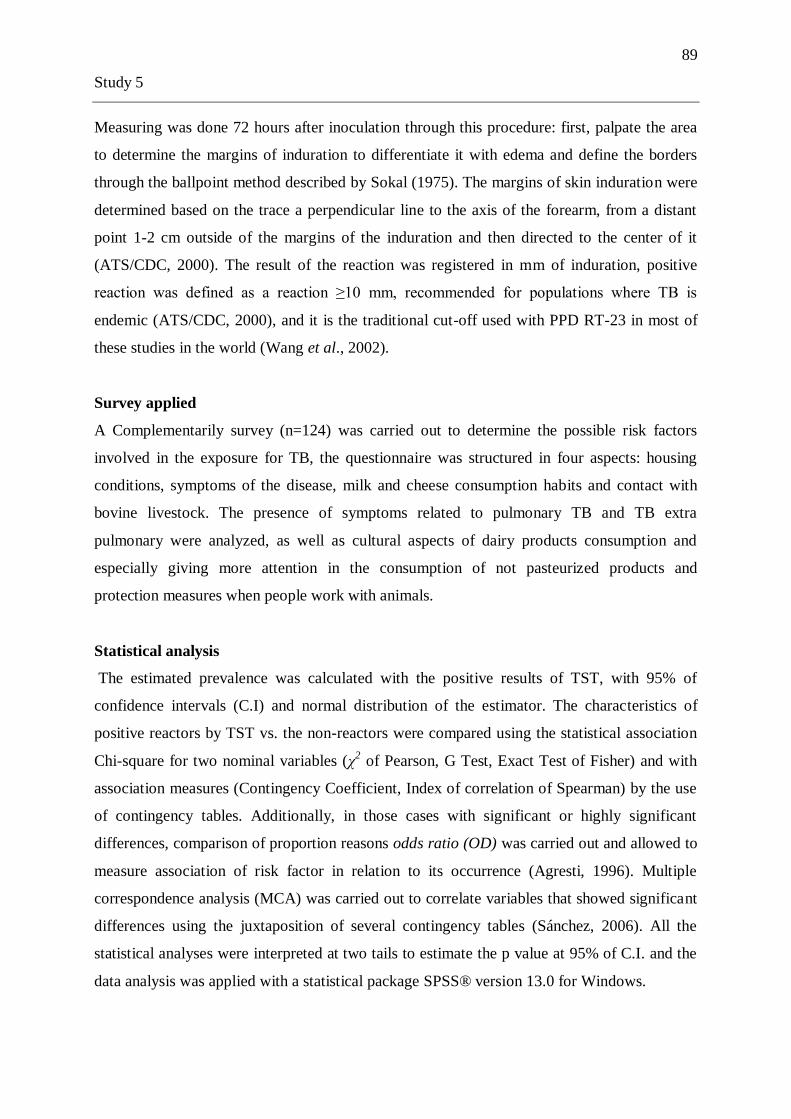
89
Study 5
Measuring was done 72 hours after inoculation through this procedure: first, palpate the area
to determine the margins of induration to differentiate it with edema and define the borders
through the ballpoint method described by Sokal (1975). The margins of skin induration were
determined based on the trace a perpendicular line to the axis of the forearm, from a distant
point 1-2 cm outside of the margins of the induration and then directed to the center of it
(ATS/CDC, 2000). The result of the reaction was registered in mm of induration, positive
reaction was defined as a reaction ≥10 mm, recommended for populations where TB is
endemic (ATS/CDC, 2000), and it is the traditional cut-off used with PPD RT-23 in most of
these studies in the world (Wang et al., 2002).
Survey applied
A Complementarily survey (n=124) was carried out to determine the possible risk factors
involved in the exposure for TB, the questionnaire was structured in four aspects: housing
conditions, symptoms of the disease, milk and cheese consumption habits and contact with
bovine livestock. The presence of symptoms related to pulmonary TB and TB extra
pulmonary were analyzed, as well as cultural aspects of dairy products consumption and
especially giving more attention in the consumption of not pasteurized products and
protection measures when people work with animals.
Statistical analysis
The estimated prevalence was calculated with the positive results of TST, with 95% of
confidence intervals (C.I) and normal distribution of the estimator. The characteristics of
positive reactors by TST vs. the non-reactors were compared using the statistical association
Chi-square for two nominal variables (χ2 of Pearson, G Test, Exact Test of Fisher) and with
association measures (Contingency Coefficient, Index of correlation of Spearman) by the use
of contingency tables. Additionally, in those cases with significant or highly significant
differences, comparison of proportion reasons odds ratio (OD) was carried out and allowed to
measure association of risk factor in relation to its occurrence (Agresti, 1996). Multiple
correspondence analysis (MCA) was carried out to correlate variables that showed significant
differences using the juxtaposition of several contingency tables (Sánchez, 2006). All the
statistical analyses were interpreted at two tails to estimate the p value at 95% of C.I. and the
data analysis was applied with a statistical package SPSS® version 13.0 for Windows.

90
Study 5
Results
Study population
Total studied population (n=157) was distributed in 58,6 % farm workers and 41,4%
slaughterhouse employees, with an average age of 37,6 years old (y) [minimum 15y; maxim
79y] standard deviation (SD): ± 13,1y. The mean time working with animals was 10,4y with a
SD: ± 10,8y. 42,0 % of people inhabited in Machachi city, 27,0 % lived in the farms and
31,0% came from towns near to their working place. Table 1 shows the distribution of the
study population where the TST was carried out.
Survey prior the TST
Of the 157 any patient manifested to have been diagnosed with TB as well as neither to have
been related and/or to know cases of people with active TB and 97,5% mentioned they had
never went through TST previously, 145 patients (92,4%) had been vaccinated with BCG, and
from this percentage 55,8% received a second vaccine as reinforcement. Likewise, 33 patients
(21,0%) had undergo to HIV/AIDS test, all of them with a negative result.
Tuberculin Skin Test (TST)
From the total 157 patients tested by TST, 152 (96,8%) returned after 72 hours for the reading
of the reaction in the skin, the average reaction was 5,3 mm with a SD: ± 6,1 mm. The results
of TST revealed 108 cases (71,1%) with a negative reaction (< 10 mm) and 44 cases with a
positive reaction (≥10 mm), meaning an estimated prevalence of 29,0% with C.I. = [22,0-
36,0]. Inside of the positive reactors group the average age was 40,46y with a SD: ± 13,05y
and the average size of the reaction to the tuberculin was 13,80 mm with a SD: ± 3,012 mm.
Survey
In 124 patients (79,0 %) a survey related to the risk factors was applied and these data were
analyzed using the statistical analyses of association.
Related symptoms
11,9% of the study group manifested to have frequent cough and 11,1% to have presented
cough at some time during more than 15 days. With regard to other symptoms related to the
TB it was found that 15,0% manifested night perspiration, 15,9% loss of weight, 17,5%
stomach pains, 3,2% frequent diarrheas; 23,0% renal pains and 8,7% urination malaise.

91
Study 5
Consumption habits of dairy products and bio-security
88,9 % of the patients consume milk, from this percentage 34,8 % had consumed or they are
still consuming raw milk, while 65,2% manifested that they do not ingest raw milk or have
always boiled it. The precedence of the milk is 40,2% from farm properties directly, 10,7% of
consumed milk came from small supermarkets in that area, 16,1% consume milk coming
from their own cows and 37,0% declared to only buy milk in small supermarkets, being
assumed that this milk was subjected to the pasteurization process. In respect to the
consumption of cheese we found that 7,1% did not consume this product and, on the contrary,
the great majority (92,9%) did. We found that the provenance of cheese for consumption
mainly came from artisans (47,0%) as well as of stores (47,0%) and 6,0% manufactures their
own cheese and in this case, 89,7% declared to use raw milk for the elaboration of the cheese
while 10,3% only used pasteurized milk for its elaboration. In relation with the biosafety
measures 5,6% manifested to use a mask when they work with livestock and 42% to use
gloves.
Association analysis among variables
Several contingency tables were performed to determine if some relationship existed among
the different variables in study. There was not significant association (p>0,05) among TST
results and patients‘ age, kind of work, working time and BCG vaccination (Table 2).
However, it was found a significant association (p=0,026) among gender (masculine or
feminine) and positive or negative reaction to TST, with a degree of correlation among both
variables also significant (p=0,021) which was given by a value of Spearman correlation of -
0,187 which indicates that, according to the order of the categories, the fact of being woman
is associated with a smaller number of positive results, the odds ratio for this risk factor
among both variables was 2,52 with C.I. = [5,61-1,131] what indicates that between the
comparison of both genders men have more than double probability to be positive by TST
regarding with respect to women in this population.
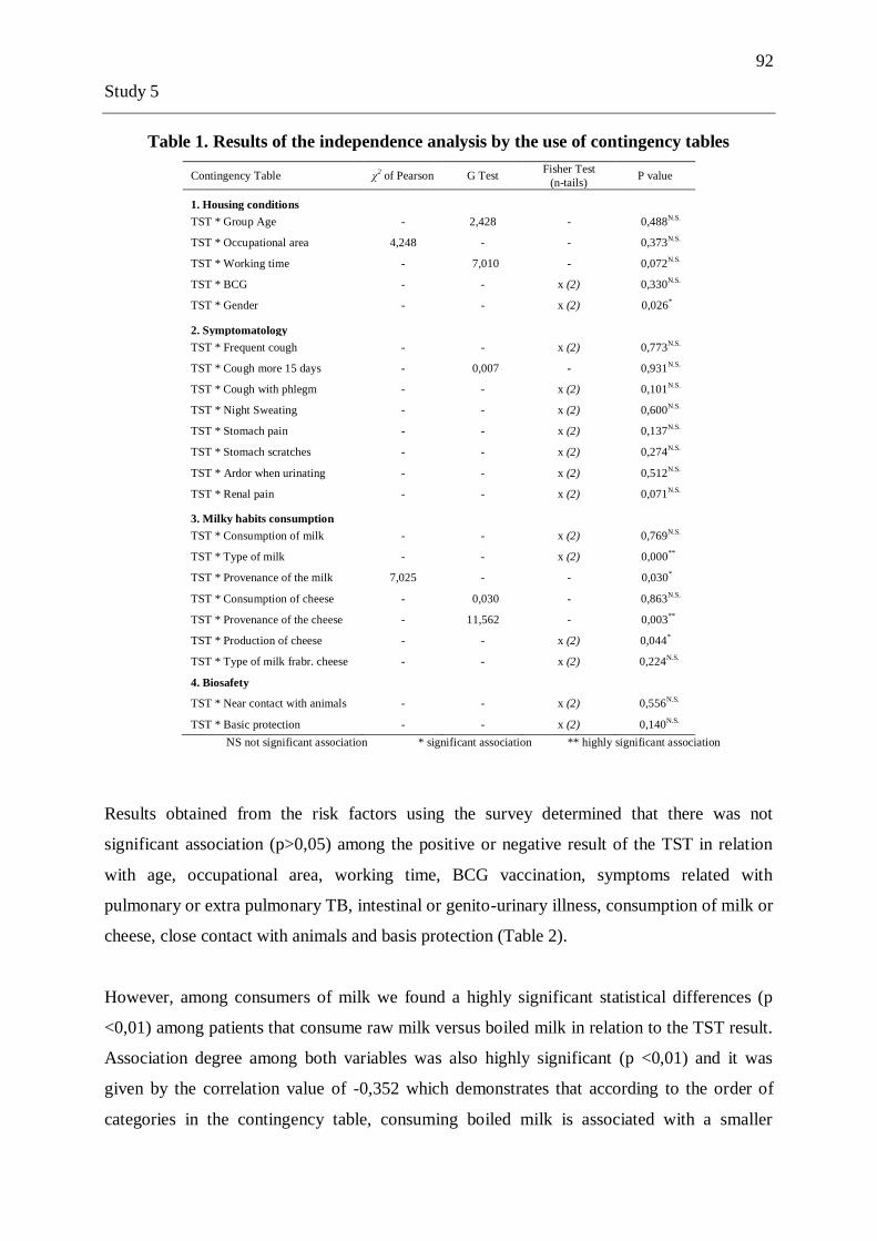
92
Study 5
Table 1. Results of the independence analysis by the use of contingency tables
Contingency Table χ2 of Pearson G Test
Fisher Test
(n-tails) P value
1. Housing conditions
TST * Group Age - 2,428 - 0,488N.S.
TST * Occupational area 4,248 - - 0,373N.S.
TST * Working time - 7,010 - 0,072N.S.
TST * BCG - - x (2) 0,330N.S.
TST * Gender - - x (2) 0,026*
2. Symptomatology
TST * Frequent cough - - x (2) 0,773N.S.
TST * Cough more 15 days - 0,007 - 0,931N.S.
TST * Cough with phlegm - - x (2) 0,101N.S.
TST * Night Sweating - - x (2) 0,600N.S.
TST * Stomach pain - - x (2) 0,137N.S.
TST * Stomach scratches - - x (2) 0,274N.S.
TST * Ardor when urinating - - x (2) 0,512N.S.
TST * Renal pain - - x (2) 0,071N.S.
3. Milky habits consumption
TST * Consumption of milk - - x (2) 0,769N.S.
TST * Type of milk - - x (2) 0,000**
TST * Provenance of the milk 7,025 - - 0,030*
TST * Consumption of cheese - 0,030 - 0,863N.S.
TST * Provenance of the cheese - 11,562 - 0,003**
TST * Production of cheese - - x (2) 0,044*
TST * Type of milk frabr. cheese - - x (2) 0,224N.S.
4. Biosafety
TST * Near contact with animals - - x (2) 0,556N.S.
TST * Basic protection - - x (2) 0,140N.S.
NS not significant association * significant association ** highly significant association
Results obtained from the risk factors using the survey determined that there was not
significant association (p>0,05) among the positive or negative result of the TST in relation
with age, occupational area, working time, BCG vaccination, symptoms related with
pulmonary or extra pulmonary TB, intestinal or genito-urinary illness, consumption of milk or
cheese, close contact with animals and basis protection (Table 2).
However, among consumers of milk we found a highly significant statistical differences (p
<0,01) among patients that consume raw milk versus boiled milk in relation to the TST result.
Association degree among both variables was also highly significant (p <0,01) and it was
given by the correlation value of -0,352 which demonstrates that according to the order of
categories in the contingency table, consuming boiled milk is associated with a smaller

93
Study 5
number of positive results to the TST. The odds ratio value of risk relationship among both
variables was established in 4,65 with C.I. = [2,01-10,75], people who drink raw milk have
more than four times the probability to be positive by TST related with those drink boiled
milk.
The provenance of the milk had significant differences (p=0,030) with the TST result, the
association degree among both variables was significant (p=0,030) and it was given by a
contingency coefficient of 0,245, meaning that if the milk is less treated (pasteurization) a
bigger number of positive cases will be found in the TST (Table 2).
A highly significant association (p<0,01) was found among the provenance of cheese and
TST result, the association degree showed significant differences (p=0,018) and value of
Spearman correlation of -0,218 what it means that better quality of milk to make cheese
represents a higher number of negative cases. Likewise, there were significant differences
(p=0,044) among people who manufacture cheese and those that do not make it with the TST
result, the association degree among both variables was significant (p=0,028) obtaining a
value of Spearman correlation of 0,198 which indicates that manufacturing cheese is
associated with a bigger number of positive results (Table 2).
Multiple Correspondences Analysis
The MCA or multiple correspondences analysis allowed us to compare more than two
variables of nominal response. Four contingency tables were performed using the results with
significant differences to independence analyses in relation to TST result analyzed with:
consumption of raw or boiled milk, provenance of milk and cheese. We fulfilled the condition
that the rows of all the tables correspond to the same individuals and the columns to different
variables with their respective classifications. Figure 1 allows identify the relationship
between the variables, it was observed that patients with positive result (≥10 mm) mainly
were related with consumption of raw milk, consumption of milk coming from farms,
consuming milk from their own cows and buy handmade cheeses.
In contrast, patients that had a negative result to the TST (<10 mm) were related with the
consumption of boiled milk, milk and cheese bought it in small supermarkets and when they

94
Study 5
10-1-2-3
Dimension 1
1,0
0,5
0,0
-0,5
-1,0
Dim
en
sio
n 2
TST_Positivo
TST_Negativo
Sin consumo
Q_Tienda
Q_Propio
Q_Artesanos
Sin consumo
Hacienda
Tienda
Propias_vacas
Sin consumo
L_Hervida
L_Cruda
Resultado_TST
Proced_Queso
Proced_Leche
Cruda_Hervida
Variable Principal Normalization.
Joint Plot of Category Points
manufacture them homemade. Finally, the group of people that doesn't consume milk and/or
cheese didn't have any relationship with the analyzed variables.
Figure 1. Quantification Plot of the categories using Multiple Correspondences Analysis
Discussion
The results obtained in this study demonstrate that the analyzed population is in risk; from
157 farm and slaughterhouse workers, 44 were positive reactors to tuberculin skin test
representing almost one third of the study population (29,0%), these findings were correlated
with the information obtained by the use of a survey. In Ecuador an important sub-
registration of TB is calculated as a consequence of the detection and report of the pulmonary
symptomatic cases exclusively. This situation has generated that the Control Program for
Tuberculosis of the PHO/WHO considers Ecuador like a country of high
prevalence/incidence, with rates 2 to 3 times superiors to those reported at a National level
and situate Ecuador among the 10 countries with higher load of TB (MSP, 2006).
Additionally, the number of human cases generated by M. bovis and which is the importance
of this pathogen in our context is unknown.

95
Study 5
Tuberculin Skin Test (TST)
This is the first study in Ecuador that investigated the relationship among positivity to TST
and exposure to Mycobacterium spp. in a risk human population exposed to bovine infected
cattle and their dairy products. It was used a reference cut off of 10 mm, which is
recommended for populations in risk to determinate the positive cases for tuberculin reaction
and estimate the prevalence infection with pathogenic mycobacteria (ATS/CDC, 2000).
Although the data don't suggest that close physical contact with bovines is a risk factor to
have a positive result by TST, the estimated high percentage of positive cases found in farm
and slaughterhouse workers could reflect: (1) possible previous exposure to tuberculous
animals (2) transmission of M. bovis from livestock toward the humans or (3) simply,
previous contact with human TB cases (M. tuberculosis); in contrast to this aspect, all the
interviewed did not manifested to have been related or did not know cases of people with
active TB.
The significant statistical differences found in relation to the gender with a higher probability
that men have a positive reaction to the tuberculin can be related according to Cousins &
Dawson (1999), because men are more exposed to mycobacteria in one or another way when
they work with livestock, representing a bigger occupational risk.
Reliability of TST can be a controversial thematic to discuss, false positive and negative
results can be caused by intrinsic variability of this test such as dosage variations in the
tuberculin, administration technique, lecture variability and anergy (Norval et al., 2004). In
order to avoid vials by these remarks it was used an uniform protocol based on rigorous
administration of equals quantity of antigen (0,1 mL), maintenance of cold chain to avoid the
degradation of the tuberculin, identical technique for lecture, interpretation and this
procedure must be carried out by the same person. A couple of studies report that 4,0% of the
reactor cases may be the probability of cross reactions generated by non tuberculous
mycobacteria (NTM) reported by Rupp et al. (1994) and Grabau et al. (1995).
Risk factors analysis
It is very difficult to determine with accuracy the transmission routes involved in the exposure
to M. bovis, mainly due to the association of this pathogen with ingestion of infected milk or
dairy products, as well as with the infection by respiratory route at the moment of breathing

96
Study 5
contaminated aerosols (LoBue et al., 2004). The results previously presented show that there
was not correlation between a positive TST and symptoms related with pulmonary disease
(air transmission), neither with symptoms related to extra pulmonary disease which mostly
could be associated with consumption of milk and cheese coming from animals with BTB.
Possible transmission of M. bovis from livestock toward humans has been reported in other
studies, consumption of not pasteurized milk coming from bovines with TB is a risk factor
involved in the infection (Winthrop et al., 2005; Remacha et al., 2006). The findings in this
study suggest a high correspondence among consumption of milk coming from farms, raw
milk and handmade cheese associated with a positive TST, on the contrary a negative TST is
associated with consumption of boiled milk coming from the farms and with consumption of
pasteurized milk and cheese from formal trades. In consequence, consumption of not
pasteurized milk and their dairy products would constitute a risk factor for the infection and
transmission of the zoonotic TB and probably contributed to the positive reactions observed
in the TST.
In Ecuador there is not information about the distribution, epidemic patterns and dynamics of
transmission of this important zoonosis and whose pathogen agent is M. bovis. In a study
carried out by Proaño-Pérez et al. (2006) an estimated prevalence of 3,85% for BTB in Mejia
canton was reported, this data confirms the high risk that exists for the transmission of the
bacteria (air borne/or digestive) among sick animals with or without symptoms toward
workers in close contact with them, this study did not show statistical differences in relation
to this aspect. Nevertheless other studies have demonstrated this possibility as an inducible
factor to development the disease in humans (Ameni et al., 2003; Biet et al., 2005; Evans et
al., 2007) especially when they have an attenuation of the immune system which can appear
frequently due to deficiencies in health and nutrition conditions most commonly found in
rural inhabitants.
In consequence, the main problem remains in the cultural habits from farm workers
consuming raw milk that comes from sick animals, could be infecting themselves without
knowing it and if by any chance they become inmunosuppressed, the bacteria inside their
bodies (latent infection) could pass to an active state and cause disease. On the other hand, the
milk coming from the livestock with TB can be sold by individual people or artisans that own

97
Study 5
small factories to elaborate cheese and which do not complete the pasteurization process, and
the pathogen can be distributed among consumers, making viable the infection with M. bovis
with can survive more than 14 days in cheese and more than 100 days in butter (Kleeberg
(1984) mentioned by Gallagher and Jenkins, 1998).
In slaughterhouse workers the survey demonstrated that they have different consumption
habits of dairy products in relation to farm workers due to easy access that slaughterhouse
workers have to shops and supermarkets inside the trade in the Machachi city, situation that
was not presented in farm workers. This would be the reason to justify those people could be
able to acquire pasteurized dairy products. Wilkins et al. (2003) have reported that the risk of
M. bovis infection in slaughterhouse workers is given by the lack of hygiene inside the
buildings, informal market that exists, absence of veterinary inspection, illiteracy of the
disease and deficient use of gloves, all these aspects can facilitate the cutaneous infection
particularly when workers cut themselves with knives during slice in sick animals.
The findings support the necessity to evaluate and quantify the impacts of the zoonotic TB in
order to be able to outline to the sanitary services and different departments that are
responsible of human and animal health to develop a control program especially in areas
where the presence of the disease is significant. In Ecuador the lack of political will, the
existence of an informal trade which does not fulfill appropriate sanitary rules and permits
informal sell of dairy products without any control is a big problem, although the big dairy
companies pasteurize most of the national production there is still a percentage that evades
this process, this study showed 47% of the people buy fresh handmade cheese elaborated with
raw milk at the.
Acknowledgements
We would like to thank all the staff from the International Centre of Zoonoses for their
support and help during the different activities carried out in this study, the Project PIC from
the University of Liege for funding this work. We would also like to thank the livestock and
slaughterhouse workers in Mejia canton.

98
Study 5
References:
Agresti, A. 1996. An introduction to categorical data analysis. Wiley-Interscience Publication,
New York, USA.
Ameni, G., Amenu, K., Tibbo, M. 2003. Bovine tuberculosis: prevalence and risk factor
assessment in cattle and cattle owners in Wuchale-Jida District, Central Ethiopia. The
International Journal of Aplplied Research in Veterinary Medicine 1: s/p. En internet
http://www.jarvm.com/articles/Vol1Iss1/AMENIJVM.htm.
American Thoracic Society (ATS) and Centre for Disease Control and Prevention (CDC).
2000. Diagnostic standards and classification of tuberculosis in adults and children. American
Journal of Respiratory and Critical Care Medicine 161: 1376-1395.
Aranaz, A., Cousins, D., Mateos, A., Domínguez, L. 2003. Elevation of Mycobacterium
tuberculosis subsp. caprae Aranaz et al. 1999 to species rank as Mycobacterium caprae
comb. nov., sp. nov. International Journal of Systematic and Evolutionary Microbiology 53:
1785-1789.
Ayele, W.Y., Neill, S.D., Zinsstag, J., Weiss, M.G., Pavlik, I. 2004. Bovine tuberculosis: an
old disease but a new threat to Africa. International Journal of Tuberculosis and Lung Disease
8: 924-937.
Barrera, L. 2007. Chapter 3: The basics of clinical bacteriology. En: Tuberculosis 2007: From
Basic science to patient care (J.C. Palomino, S.C. Leão, V. Ritacco, eds) pp. 93-112.
Disponible en internet en www.TuberculosisTextbook.com
Biet, F., Boschiroli, M.L., Thorel, M.F., Guilloteau, L.A. 2005. Zoonotic aspects of
Mycobacterium bovis and Mycobacterium avium-intracellulare complex (MAC). Veterinary
Research 36: 411-436.
Cosivi, O., Grange, J.M., Daborn, C.J., Raviglione, M.C., Fujikura, T., Cousins, D.,
Robinson, R.A., Huchzermeyer, H.F.A.K., de Kantor, I., Meslin, F.X. 1998. Zoonotic
Tuberculosis due to Mycobacterium bovis in Developing Countries. Emerging Infectious
Diseases 4: 59-70.
Cousins, D.V., Bastida, R., Cataldi, A., Quse, V., Redrobe, S., Dow, S., Duignan, P., Murray,
A., Dupont, C., Ahmed, N., Collins, D.M., Butler, W.R., Dawson, D., Rodríguez, D.,
Loureiro, J., Romano, M.I., Alito, A., Zumarraga, M., Bernardelli, A. 2003. Tuberculosis in
seals caused by a novel member of the Mycobacterium tuberculosis complex: Mycobacterium
pinnipedii sp. nov. International Journal of Systematic and Evolutionary Microbiology 53:
1305-1314.

99
Study 5
Cousins, D.V., Dawson, D.J. 1999. Tuberculosis due to Mycobacterium bovis in the
Australian population: cases recorded during 1970-1994. The International Journal of
Tuberculosis and Lung Disease 3: 715-721.
Evans, J.T., Smith, E.G., Banerjee, A., Smith, R.M., Dale, J., Innes, J.A., Hunt, D., Tweddell,
A., Wood, A., Anderson, C., Hewinson, R.G., Smith, N.H., Hawkey, P., Sonnenberg, P. 2007.
Cluster of human tuberculosis caused by Mycobacterium bovis: evidence for person-to-person
transmission in the UK. Lancet 369: 1270-1276.
Flynn, J.L., Chan, J. 2001. Immunology of tuberculosis. Annual Review of Immunology 19:
93-129.
Gallagher, J., Jenkins, P.A. 1998. Mycobacterial diseases. En: Zoonoses, Biology, Clinical
Practice and Public Health Control (S.R. Palmer, L. Soulsby, D. Simpson, eds) pp. 155-164.
Oxford University Press, Oxford, USA.
Grabau, J.C., DiFerdinando, G.T., Novick, L.F. 1995. False positive tuberculin skin test
results. Public Health Reports 110: 703-706.
Katial, R.K., Hershey, J., Purohit-Seth, T., Belisle, J.T., Brennan, P.J., Spencer, J.S., Engler,
R.J.M. 2001. Cell-mediated immune response to tuberculosis antigens: comparison of skin
testing and measurement of in vitro gamma interferon production in whole-blood culture.
Clinical and Diagnostic Laboratory Immunology 8: 339-345.
LoBue, P.A., Betancourt, W., Cowan, L., Seli, L., Peter, C., Moser, K.S. 2004. Identification
of a familial cluster of pulmonary Mycobacterium bovis disease. The International Journal of
Tuberculosis and Lung Disease 8: 1142-1146.
Ministerio de Salud Pública del Ecuador (MSP). 2007. Informe de gestión del programa de
control de tuberculosis Ecuador 2005-2006. El Fondo Mundial de lucha contra el SIDA, la
tuberculosis y la malaria, Quito, Ecuador.
Ministerio de Salud Pública del Ecuador (MSP). 2006. Situación de la salud del Ecuador.
Imprenta Zenitram, Quito, Ecuador.
Mostowy, S., Inwald, J., Gordon, S., Martin, C., Warren, R., Kremer, K., Cousins, D., Behr,
M.A. 2005. Revisiting the evolution of Mycobacterium bovis. Journal of Bacteriology 187:
6386-6395.
Neill, S.D., Hanna, J., O´Brien, J.J., McCracken, R.M. 1989. Transmission of tuberculosis
from experimentally infected cattle to in-contact calves. The Veterinary Record 124:269-271.
Norval, P.Y., Roustit, C., San, K.K. 2004. From tuberculin to prevalence survey in Cambodia.
The International Journal of Tuberculosis and Lung Disease 8: 299-305.

100
Study 5
Pavlik, I., Trcka, I., Parmova, I., Svobodova, J., Melicharek, I., Nagy, G., Cvetnic, Z.,
Ocepek, M., Pate, M., Lipiec, M. 2005. Detection of bovine and human tuberculosis in cattle
and other animals in six Central European countries during the years 2000-2004. Veterinary
Medicine Czech 50: 291-299.
Proaño-Pérez, F., Rigouts, L., Brandt, J., Dorny, P., Ron, J., Chávez, M.A., Rodríguez, R.,
Fissette, K., Van Aerde, A., Portaels, F., Benítez-Ortíz, W. 2006. Preliminary observations on
Mycobacterium spp. in dairy cattle in Ecuador. The American Journal of Tropical Medicine
and Hygiene 75: 318-323.
Reece, S.T., Stride, N., Ovendale, P., Reed, S.G., Campos-Neto, A. 2005. Skin test performed
with highly purified Mycobacterium tuberculosis recombinant protein triggers tuberculin
shock in infected guinea pigs. Infection and Immunity 73: 3301-3306.
Reichman, L.B. 1976. Tuberculin skin testing: the state of the art. Chest 76: 764-770.
Remacha, M.A., Parra, M.I., Esteban, A. 2006. Pulmonary tuberculosis due to Mycobacterium
bovis in León. The International Journal of Tuberculosis and Lung Disease 10: 349-350.
Rupp, M.E.A., Schultz, W., Davis, J.C. 1994. Discordance between tuberculin skin test results
with two commercial purified protein derivative preparations. The Journal of Infectious
Diseases 169: 1174-1175.
Sánchez, J. 2006. Introducción a la estadística no paramétrica y al análisis multivariado.
Quality Print, Quito, Ecuador.
Smith, I. 2003. Mycobacterium tuberculosis pathogenesis and molecular determinants of
virulence. Clinical Microbiology Reviews 16: 463-496.
Sokal, J.E. 1975. Measurment of delayed skin test responses. The New England Journal of
Medicine 293: 501-502.
31.Taylor, G.M., Murphy, E., Hopkins, R., Rutland, P., Chistov, Y. 2007. First report of
Mycobacterium bovis DNA in human remains from the iron age. Microbiology 153: 1243-
1249.
Van Soolingen, D.,Hoogenboezem, T., de Haas, P.E.W., Hermans, P.W.M., Koedam, M.A.,
Teppema, K.S., Brennan, P.J., Besra, G.S., Portaels, F., Top, J., Schouls, L.M.., van Embden,
J.D.A. 1997. A novel pathogenic taxon of the Mycobacterium tuberculosis complex, canetti:
characterization of an exceptional isolate from Africa. International Journal of Systematic
Bacteriology 47: 1236-1245.
Wang, L., Turner, M.O., Elwood, R.K., Schulzer, M., FitzGerald, J.M. 2002. A meta analysis
of the effect of bacilli Calmette Guerin vaccination on tuberculin skin test measurements.
Thorax 57: 804-809.

101
Study 5
Wedlock, D.N., Skinner, M.A., de Lisle, G.W., Buddle, B.M. 2002. Control of
Mycobacterium bovis infection and the risk to human populations. Microbes and Infection 4:
471-480.
Wilkins, M.J., Bartlett, P.C., Frawley, B., O´Brien, D.J., Miller, C.E., Boulton, M.L. 2003.
Mycobacterium bovis (bovine TB) exposure as a recreational risk for hunters: results of a
Michigan hunter survey, 2001. The International Journal of Tuberculosis and Lung Disease 7:
1001-1009.
Winthrop, K.L., Scott, J., Brown, D., Jay, M.T., Rios, R., Mase, S., Richardson, D.,
Edmonson, A., MacLean, M., Flood, J. 2005. Investigation of human contacts: a
Mycobacterium bovis outbreak among cattle at a California dairy. The International Journal of
Tuberculosis and Lung Disease 9: 809-813.
World Health Organization (WHO). 2007. Global tuberculosis control: surveillance, planning,
financing: WHO Report 2007. Geneve, Switzerland.

102
Study 6
3.6 Review article: Situation of bovine tuberculosis in Ecuador
PROANO-PEREZ FREDDY, BENITEZ-ORTIZ WASHINGTON, PORTAELS
FRANÇOISE, RIGOUTS LEEN AND LINDEN ANNICK.
Pan American Journal of Public Health. 2011 Sept;30(3):000-00.

103
Study 6
Temas de actualidad / Current topics
Situation of
bovine tuberculosis in
Ecuador
Freddy Proaño-Pérez,1-2
Washington Benítez-Ortiz,1-3
Françoise Portaels,4 Leen Rigouts,
4-5 and Annick Linden
2
Suggested citation: Proaño-Pérez F, Benítez-Ortiz W, Portaels F, Rigouts L, Linden A.
Situation of bovine tuberculosis in Ecuador. Rev Panam Salud Pública. 2011; 30(3):000-00.
SYNOPSIS
Bovine tuberculosis (BTB) is a chronic and contagious disease that affects domestic
animals, wildlife, and humans. Caused by Mycobacterium bovis, BTB causes major
economic losses and poses a serious constraint to the international trade of animal products.
Moreover, in developing countries where BTB controls are lacking, M. bovis is a public
health concern. In most developing countries, the prevalence of BTB in livestock is unknown
because the information is either not reported or not available. In Ecuador, there is no
national BTB control program.
This article reviews the BTB situation in different geographic areas of Ecuador by
examining exhaustive data reflecting tuberculin testing surveys, as well as slaughterhouse
surveillance studies, from 1972 – 2008. In Ecuador, the dairy industry‘s expansion (pre-
empted by the high demand for milk and its byproducts), intensified efforts to increase the
cattle population, the presence of M. bovis, and a lack of BTB controls has caused a rise in
BTB prevalence, and subsequently a growing push for the implementation of a national BTB
control program.
Key words: Mycobacterium bovis; zoonoses; cattle diseases; tuberculosis, bovine;
communicable diseases, emerging; Ecuador.
1 International Center for Zoonoses, Central University of Ecuador, Quito, Ecuador. 2 Department of Infectious and Parasitic Diseases, Faculty of Veterinary Medicine, University of Liege, Belgium. 3 Faculty of Veterinary Medicine, Central University of Ecuador, Quito, Ecuador.
4 Mycobacteriology Unit, Department of Microbiology, Institute of Tropical Medicine of Antwerp, Belgium.
5 Department of Pharmaceutical, Veterinary and Biomedical Sciences, University of Antwerp, Antwerp, Belgium.
104
Study 6
Bovine tuberculosis (BTB) is a chronic infectious disease caused by Mycobacterium
bovis. This disease mainly affects cattle, but can also be found in other domestic and wild
animals, and occasionally, in humans (1). The World Organization for Animal Health (OIE)
considers BTB to be an important zoonotic disease with a socioeconomic and public health
impact that affects the international trade of livestock and animal products (2).
The prevalence of BTB in developing countries remains largely unknown. According
to a study conducted in 2006, Ecuador is among a group of Latin American countries
assumed to have a relatively high prevalence of BTB, but a lack of reporting (3). Other
countries in the group are Argentina, Bolivia, Brazil, Chile, Guatemala, Guyana, Haiti, and
Peru. In 1998, it was estimated that for 24% of the Latin American bovine population, control
measures for BTB were incomplete or inexistent (4). This absence of control represents a
high risk for the rural inhabitants living in direct contact with the animals (5). Of
approximately 374 million cattle in Latin America and the Caribbean, 70% are held in areas
where M. bovis infection rates among cattle exceed 1% (3).
In humans, the proportion of tuberculosis induced by M. bovis is relatively low
compared to M. tuberculosis. But in recent years, M. bovis tuberculosis in humans has
become increasingly prevalent where human populations are confronted with poverty,
malnutrition, human immunodeficiency virus (HIV), and reduced access to health care (6).
Transmission through the consumption of unpasteurized milk and dairy products from
infected cattle occurs mostly among the general public, whereas exposure through airborne
infection remains highest among farmers, veterinarians, and slaughterhouse workers (6).
According to study published in 1998, M. bovis could be responsible for more than 2 % of the
total pulmonary tuberculosis (TB) cases and 9.4 % of extrapulmonary forms among humans
in Latin America, a considerable number. In addition, another study showed that in
Argentina, 2 % of human TB cases have been recorded as being caused by M. bovis (3).
Given the appearance of new cases in recent years, BTB in humans has been
designated a reemerging disease in developed countries. This reemergence is most likely the
result of an increase in world population augmented by the movement of people and animals,
plus environmental changes, the crossing of the interspecies barrier, and changes in the
management of livestock production (4, 7, 8).

105
Study 6
The total economic losses due to BTB are underestimated because its impact on
public health has not been thoroughly evaluated (9). Estimates of economic loss are limited to
animal health issues and are based on weight loss, decreased milk production, lowering of the
reproductive rate, mortality, and condemnation of carcasses (10). In Argentina, the National
Health and Food Safety Service (Servicio Nacional de Sanidad y Calidad Agroalimentaria -
SENASA) estimates that in 1995 the annual losses due to BTB were approximately US$ 63
million (4).
This article aims to provide information on the current situation of BTB in Ecuador
based on available data from studies applying tuberculin skin-testing at farms, veterinary
inspections at slaughterhouses, and laboratory-based diagnosis. The data presented in this
article were obtained from the college libraries serving schools of veterinary medicine
throughout the country. Most of the surveys were a component of veterinary students‘ theses.
In Ecuador, there is only one group of researchers working on BTB, which explains the
limited number of published studies.
BTB PREVALENCE
BTB cases in Ecuador are not documented nor quantified clearly for several reasons:
lack of proper recording of positive cases, limited use of diagnostic tests, and insufficient
veterinary inspection in most slaughterhouses, but mainly because BTB is not a notifiable
disease. Furthermore, it is not unlikely that in Ecuador—as may be the case in other
countries—BTB is not reported due to a lack of trust between farmers and health officials (5).
Currently, control measures related to relocation and transportation of animals focus solely
on Foot and Mouth Disease. Furthermore, results of veterinary inspection are not recorded
properly in slaughterhouses, jeopardizing the ―traceability‖ of positive BTB cases observed
during veterinary inspection.
The prevalence of BTB can be influenced by several factors at the individual, herd,
and/or provincial/country level (11). For instance, livestock husbandry varies by herd size,
farm size, and type of cattle industry, i.e., for dairy or meat production. Factors such as these
can affect the occurrence of BTB disease by increasing or decreasing close contact between
animals. In Ecuador, the cattle population is not equally distributed throughout the country:

106
Study 6
the provinces of Azuay and Pichincha have dense livestock populations predominantly
focused on dairy production, while Santo Domingo and Manabí provinces have important
meat industries (12), this distribution influence on the occurrence of BTB. The scarce
information that is available in Ecuador is mostly based on tuberculin skin-testing performed
by students at the schools of veterinary medicine within the various universities of Ecuador
(Figure 1).
Tuberculin skin test surveys
The sensitivity of skin testing can be affected by the potency and dose of tuberculin
administered, the post-infection interval, desensitization, deliberate interference, postpartum
immunosuppression, variation in the interpretation by the observer, and contact with
environmental non-tuberculous mycobacteria (NTM) (13).
Surveys performed in Ecuador using the single and comparative intradermal
tuberculin test (SITT and CITT, respectively) have demonstrated a variable prevalence of
BTB. Briefly, the tuberculin skin test is based on the use of bovine purified protein derivative
AN5 strain (PPD-B) 20 000 IU/mL and avian purified protein derivative D4 ER strain (PPD-
A) 25 000 IU/mL, the test can be applied in the caudal fold of the tail (SITT) or in the middle
third of the neck (CITT); the thicknesses of the skin is measured with calipers 72 hours
before and after the injection of 0.1 mL PPD-A and PPD-B antigens. The interpretation is
based on the reaction observed (14, 15). In most of the studies performed in Ecuador, the
PPD antigens used were not reported.
Table 1 presents an overview of the tuberculin-testing surveys conducted in dairy
cattle in Ecuador (16 – 21). In 1977, the first survey detected a low (0.3%) prevalence among
dairy cattle in the Tungurahua province located in the middle of the country (16). A second
study conducted in 2003 in the same area showed a 1.2% increase in the disease (17). In both
studies, SITT was applied in all animals and CITT was used to confirm all positive and
suspected cases detected. Data on the herd size and origin of the PPD reagents were not
presented in these studies.

107
Study 6
In contrast, in the province of Imbabura, a survey conducted in 2001 showed a high
prevalence of BTB (7.3%) (18). However, 2 years later, it showed a lower prevalence (2.4%)
(19). In the province of Carchi, located north of the Imbabura province at the border with
Colombia, the prevalence observed in 2001 (2.1%) was comparable to that of 2002 (1.7%)
(18, 20). In general, the prevalence found in the latter two provinces was higher than that of
Tungurahua. This was probably due to the smaller cattle population and lower animal density
of Tungurahua. Plus, Tungurahua is an area more focused on agriculture than animal
production (12).
The majority of the surveys, however, were performed in the province of Pichincha,
specifically in the cantons of Mejia and Cayambe, the two most important dairy areas, located
near the capital city, Quito. In 2001, a prevalence of 2.8% was observed in the Cayambe
canton (18). This finding was in sharp contrast to a prevalence of 0.5% reported a year later
(21). Worth noting is that the first study sampled only two farms; therefore, the BTB
prevalence data is questionable.
Studies performed in the Mejia canton showed a high occurrence of the disease in
dairy cattle. In 2002, a study (22) observed 4.9% of skin reactors, which was confirmed by
the prevalence of 3.9% found by another survey applied in the same area, but to different
herds (14). The two studies used similar diagnostic methods, but sampling procedures
differed. In the latter study, 22 cattle were randomly selected per herd from farms that were
small (1 – 25 cattle), medium (25 – 70 cattle), and large ( > 70 cattle). The diagnostic test
used was CZV Bovine Tuberculin PPD® (CZ Veterinaria S.A., Porriño, Spain). The
difference found related to herd size was highly significant: 7.9% in large herds; 3.4% in the
medium; and surprisingly, only 0.3% in small herds (14). Recent information from CITT
analysis, using Bovituber® PPD (Synbiotics Corporation, a subsidiary of Pfizer Incorporated,
Lyon-France), of 20 dairy herds in the Mejia canton showed an annual incidence of 1.7%,
with a true prevalence at 7.1%. Herd size was identified as a main risk, as were age (P =
0.03), contact with other animal species (P < 0.01), and introducing new animals into the
herd (P = 0.04) (15).

108
Study 6
The expansion of the dairy industry in Ecuador—resulting from a growing population
and its high demand for milk and by-products—meant closer contact among animals. This
increased the risk of transmitting M. bovis bacilli (23).
Breed-related differences have been reported (1), with BTB infecting a higher
proportion of dairy breeds (Bos taurus), than indigenous zebu cattle (Bos indicus) and
crossbred beef cattle. Additionally, the use of the European breeds (e.g., the Holstein
Friesian) to improve milk production through artificial insemination, resulted in higher BTB
susceptibility (24), indirectly increasing the probability of BTB infections. Beef cattle
husbandry systems differ from dairy management and have a decreased risk of infection due
to less contact between animals (25). Meat production farms in Ecuador often have expansive
pastures and most are located in tropical areas. Table 2 summarizes all available studies
conducted on beef cattle in Ecuador (26–36).
All surveys showed a low number of skin reactors, except for two studies in the
province of Guayas that showed prevalences of 3.4% and 5.6%. This high percentage can be
attributed to the fact that in both studies only SITT was applied and positive reactors were not
confirmed by CITT. Some of the reactions, therefore, could have been caused by contact with
environmental NTM, especially in young cattle (13, 37).
Veterinary inspection
In geographic areas with high BTB, its prevalence can be estimated by the proportion
of macroscopic tuberculous lesions detected during post-mortem examination (followed by
the rejection of carcasses and viscera from these animals), if a reliable system exists (9).
Indeed, programs based on slaughterhouse surveillance are only effective if they use a
reliable traceability system for tracing-back to the herd of origin. The distribution and
development of lesions depend on the route of transmission (38) and can vary, although most
often located in thoracic lymph nodes due to the infection by respiratory route (39).
Therefore, exhaustive veterinary control needs to be established to identify affected animals,
i.e., palpation and inspection of lungs, liver, spleen, kidney, mammary gland, and associated
lymph nodes (39). Vigilance is recommended, however, since not all infected cattle exhibit
gross lesions. On the other hand, detailed meat inspection allows the identification of lesions

109
Study 6
in apparently healthy animals, which increases the number of detected animals and avoids the
consumption of BTB-infected cattle (40).
Although veterinary inspection at slaughterhouses potentially constitutes a good
method for identifying the presence of TB, it is not routinely implemented in Ecuador and
only a few studies on the topic have been published. Those 14 studies are shown in Table 3
(40–52). The studies that took place in 1972 – 2003 were conducted in the provinces located
in the tropical areas where meat production is the priority; later studies were performed
mainly on dairy cattle in the highlands. The first study, conducted in 1972, entailed veterinary
inspection at the slaughterhouse of Guayaquil to identify pigs with BTB (41). A low
prevalence of 0.2% was found. More recently, a high proportion of affected dairy cattle were
observed in 2007 (2.3%) and 2008 (2.2%) in the highlands of Ecuador, specifically the Mejia
canton of the Pichincha province (40). This study applied various laboratory-based diagnostic
methods, in addition to the macroscopic investigation, and the presence of M. bovis was
confirmed (40).
In the Ecuador‘s dairy areas, 90% of the cattle in the highlands are Holstein Friesian,
whereas in the tropical areas, the main breeds used for milk production are Jersey, Brown
Swiss, and crossbreeds. The differences in BTB prevalence among cattle breeds and
geographic areas present in Ecuador, were also found in the slaughterhouses of Argentina (9),
Brazil (53, 54), and Mexico (55, 56).
Laboratory diagnosis
Laboratory-based techniques are important for improving diagnosis of mycobacteria
and identifying the species. In the slaughterhouse survey in the Mejia canton, all positive
cultures obtained from tissue samples of tuberculous-like lesions were identified (40). Apart
from M. bovis, NTM were also identified, i.e., M. gordonae, M. szulgai, M. celatum, and M.
avium-intracellulare-scrofulaceum; however their clinical relevance was not further
investigated. In addition, polymerase chain reaction (PCR) based on 16S rRNA (ribosomal
ribonucleic acid) confirmed the presence of M. bovis and NTM in lungs and lymph nodes
(14).

110
Study 6
DNA-fingerprinting analysis allowed identifying different M. bovis strains. All M.
bovis isolates from the Mejia canton surveys displayed the same spoligotype (SB0980)
(Proaño-Pérez et al., submitted for publication. Contact the corresponding author for more
information).
The homogeneity of the isolates found in this study could reflect a short timeframe of
events. M. bovis spoligotypes have been reported to change in vitro in approximately 60 years
(57), which could suggest that BTB was introduced into this dairy area together with new,
European cattle breeds in the mid-Twentieth century.
ZOONOTIC TB
M. bovis infection in humans can occur through inhalation of infectious droplets from
a live or slaughtered animal or by consumption of unpasteurized dairy or meat products from
infected animals (58, 59). People working in animal husbandry, slaughterhouse workers,
veterinarians, and people in close contact with possibly-infected animals are at a higher risk
for M. bovis infection (60). In Argentina, a 2.4% prevalence of zoonotic TB has been
reported, with a high association with type of work performed (9). Lack of hygiene, existence
of informal trade, absence of veterinary inspection, ignorance of the disease, and deficient use
of gloves have been reported as risk factors among slaughterhouse workers (61).
In Ecuador, human cases of TB detected in public and private hospitals are reported to
the Ministry of Public Health, which is responsible for the human TB control program and
provides free treatment. In 2009, the Ministry of Public Health reported 3 317 microscopy-
positive new cases of pulmonary TB, 584 cases of extra-pulmonary TB, and 613 deaths
caused by the disease nationwide (62). According to the World Health Organization (WHO),
Ecuador reported 4 703 cases of TB in 2009 (63).
The national reference laboratory, ―Leopoldo Izquieta Pérez Institute‖ (LIP),
(Guayaquil, Ecuador) identified only two M. bovis isolates based on their growth and/or
morphological characteristics (in 1998–2005): one from the lymph node of a girl 1 year of
age, and the other from a pulmonary specimen of a boy 3 years of age (64). However, it is
important to take into account that M. bovis requires special conditions to grow in vitro. The

111
Study 6
appropriate medium to facilitate growth of the bacilli (65) should contain sodium pyruvate,
but no glycerol, e.g., Stonebrink medium. The latter medium is not routinely used in the LIP
laboratory. Secondly, a number of human M. bovis infections might be misdiagnosed because
the procedure required for differentiating between M. bovis and the other members of the M.
tuberculosis-complex is not routinely performed.
In 2007, a study was conducted on 157 farm and slaughterhouse workers from the
Mejia canton to evaluate TB prevalence by tuberculin skin testing (TST). A positive reaction
was seen in 29%. In addition, the risk factors associated with disease transmission among this
population were studied. A significant association was documented between TST positivity
and masculine gender (P = 0.02; odds ratio [OR] = 2.5) and consumption of raw milk (P <
0.00) or cheese made by hand/homemade (P = 0.003) (66). These findings clearly show that
cultural habits play a major role in risk behavior; consumption of raw milk is considered
healthier and is widespread among this population group.
ECONOMIC LOSSES FROM BTB
The national economic losses due to BTB cannot be accurately calculated because
standardized data is lacking, and the data that does exist is simply an estimate of BTB
prevalence. However, the economic losses in the Mejia canton are estimated at approximately
US$ 460 000 per year. This estimation is based on the true prevalence determined in 2008 by
CITT (15) and official data on the cattle population for this canton: approximately 55 000
head of cattle distributed among 2 722 herds (12). Three important factors were considered in
estimating economic losses caused by affected cattle: decreased milk production (13%),
weight loss (36%), and decreased reproductive rate (12%) (10). Nevertheless, the
condemnation of carcasses was not taken into account for this calculation because the BTB-
suspected cattle were not always entirely destroyed due to a lack of sanitary controls in some
slaughterhouses.
BTB CONTROL PROGRAM
Today, the quality of dairy products is a high priority for the global dairy industry, as
it is for Ecuador‘s. In Ecuador, the Ministry of Agriculture and Livestock food safety

112
Study 6
department focuses on the quality of animal products. The private dairy sector pays more to
dairy farmers who have earned a health certificate, awarded by the Ministry of Agriculture
and Livestock, declaring their herd to be free of brucellosis and BTB. In theory, this status
should be granted only after two consecutive negative results, meaning disease is not found
among the herd, at two 6-monthly controls. In addition, the certification should be granted
only by the Ministry of Agriculture and Livestock. However, this is not always the case in
practice. Private laboratories offer tuberculin testing for cattle, but the tests are generally not
administered by qualified veterinarians. Plus, the quality of PPD is not tested, as there is no
reference laboratory for animal diseases to carry out quality control.
In general, BTB control programs are based on the early diagnosis and rapid
elimination of positive CITT reactors in farms and at trade. Complementary strategies are
recommended, i.e., epidemiological surveillance, proper veterinary supervision at
slaughterhouses and cattle markets, and adequate control of cattle movement (67). In Spain,
such a policy has been implemented since 1965, resulting in a reduction of BTB prevalence
among cattle (68).
In Ecuador, there is no national BTB control program implemented yet. A national
policy should be established with an obligatory CITT and culling of positive reactors.
However, the implementation of such a national program has economic and logistic
limitations. The total cost of a control program cannot be financed by the government alone;
it should be shared with the private sector. Financial support should cover all related
expenses, i.e., education and public awareness, incentives for farmers to improve the health
status of their herds (through the price of the milk), regular CITT, compensation for culling,
surveillance monitoring, and research.
NEXT STEPS
In conclusion, the data presented by this article confirm the presence of BTB in beef
and dairy cattle in Ecuador, and justify the implementation of a national health policy to
control the disease. A national control program is urgently needed to avoid the spread of the
BTB, and is an important step toward promoting international trade of animals and their
products. Moreover, these measures would reduce the zoonotic impact of the disease among

113
Study 6
the population, especially in high-risk groups living in BTB-prevalent areas of the country.
The situation in Ecuador may be similar to that of other countries in the Region of the
Americas; thus the information present here could be used in the planning of control and
eradication programs elsewhere.
Acknowledgements. The authors gratefully acknowledge the financial support provided by
the Commission Universitaire pour le Développement, University of Liege (Liege –
Belgium), Project PIC (Programme interuniversitaire ciblé).
REFERENCES
1. Acha P, Szyfres B. Zoonoses and communicable diseases common to man and animals.
In: Bacterioses and mycoses. 3rd
ed. Washington D.C.: Pan American Health
Organization; 2001. Pp. 283-299. (Scientific and Technical Publication 580).
2. Office International des Epizooties. Código sanitario para los animales terrestres. Vol 2.
Available from: http://www.oie.int/es/normas-internacionales/codigo-terrestre/acceso-en-
linea/. Accessed on 12 April 2011.
3. de Kantor IN, Ritacco V. An update on bovine tuberculosis programmes in Latin
American and Caribbean countries. Vet Microbiol. 2006;112(2-4):111-8.
4. Cosivi O, Grange JM, Daborn CJ, Raviglione MC, Fujikura T, Cousins D, et al. Zoonotic
tuberculosis due to Mycobacterium bovis in developing countries. Emerg Infect Dis.
1998;4(1):59-70.
5. Gil A, Samartino L. Zoonosis en los sistemas de producción animal de las áreas urbanas y
periurbanas de América Latina. Rome: FAO; 2001. Pp. 16 – 22. (Livestock Policy
Discussion Paper No. 2).
6. Michel AL, Müller B, van Helden PD. Mycobacterium bovis at animal-human interface:
A problem or not? Vet Microbiol. 2010;140(3-4):371-81.
7. Brown C. Emerging diseases of animals: an overview. In: Emerging diseases of animals.
Brown C, Bolin C, eds. Washington DC: ASM Press; 2000. Pp. 1-12.
8. Abalos P, Retamal P. Tuberculosis: ¿una zoonosis re-emergente? Rev Sci Tech Off int
Epiz. 2004;23(2):583-94.

114
Study 6
9. Latini O, Canal AM, Ferrara ME, Sequeira MD, Sequeira G, Bagnaroli R, et al.
Confiabilidad en la determinación de prevalencia de infección por Mycobacterium bovis
en ganado bovino por decomisos en frigoríficos. Arch Med Vet. 1997;29(2):197-204.
10. Suazo FM, Escalera AM, Torres RM. A review of M. bovis BCG protection against TB in
cattle and other animals species. Prev Vet Med. 2003;58(1-2):1-13.
11. Humblet MF, Boschiroli ML, Saegerman C. Classification of worldwide bovine
tuberculosis risk factors in cattle: a stratified approach. Vet Res. 2009;40(5):50.
12. Servicio de Informática y Censo Agropecuario. Tercer Censo Nacional Agropecuario,
2000. Available from: http://www.sica.gov.ec/censo/docs/nacionales/tabla6.htm.
Accessed on 2 May 2008.
13. Monaghan ML, Doherty ML, Collins JD, Kazda JF, Quinn PJ. The tuberculin test. Vet
Microbiol. 1994;40(1-2):111-24.
14. Proaño-Perez F, Rigouts L, Brandt J, Dorny P, Ron J, Chavez M, et al. Preliminary
observations on Mycobacterium spp in dairy cattle in Ecuador. Am J Trop Med Hyg.
2006;75(2):318-23.
15. Proaño-Pérez F, Benítez-Ortiz W, Celi-Erazo M, Ron-Garrido L, Benítez-Capistros R,
Portaels F, et al. Comparative intradermal tuberculin test in dairy cattle in the north of
Ecuador and risk factors associated with bovine tuberculosis. Am J Trop Med Hyg.
2009;81(6):1103-9.
16. Acosta C, Parreño B. Incidencia de tuberculosis bovina en veinte haciendas del cantón
Píllaro. [Thesis]. Quito: Facultad de Medicina Veterinaria, Universidad Central del
Ecuador; 1977.
17. Alemán R, Cajilema C, Jaramillo F. Diagnóstico de Tuberculosis Bovina mediante la
prueba intradérmica única en hatos lecheros de la provincia de Tungurahua. [Thesis].
Quito: Facultad de Medicina Veterinaria, Universidad Central del Ecuador; 2003.
18. Andino-Ashqui O. Diagnóstico de Tuberculosis bovina mediante la prueba intradérmica
única en hatos lecheros de la Sierra Ecuatoriana. [Thesis]. Quito: Facultad de Medicina
Veterinaria, Universidad Central del Ecuador; 2001.
19. Bedón T, Verdesoto S. Diagnóstico de tuberculosis bovina mediante la prueba
intradérmica única en hatos lecheros de la provincia de Imbabura. [Thesis]. Quito:
Facultad de Medicina Veterinaria, Universidad Central del Ecuador; 2003.
20. Burbano R, León J. Diagnóstico de la Tuberculosis bovina mediante la prueba
intradérmica única en hatos lecheros de los cantones Espejo, Montufar, Tulcán y Huaca

115
Study 6
en la provincia del Carchi. [Thesis]. Quito: Facultad de Medicina Veterinaria,
Universidad Central del Ecuador; 2002.
21. Salazar JC, Cevallos C. Diagnóstico de la Tuberculosis bovina mediante la prueba
intradérmica única en hatos lecheros del cantón Cayambe en la provincia del Pichincha.
[Thesis]. Quito: Facultad de Medicina Veterinaria, Universidad Central del Ecuador;
2002.
22. Cano G, Chulde P. Diagnóstico de la Tuberculosis bovina mediante la prueba
intradérmica única en hatos lecheros del cantón Mejía en la provincia de Pichincha.
[Thesis]. Quito: Facultad de Medicina Veterinaria, Universidad Central del Ecuador;
2002.
23. Goodchild AV, Clifton-Hadley RS. Cattle-to-cattle transmission of Mycobacterium bovis.
Tuberculosis (Edinb). 2001;81(1-2):23-41.
24. Ameni G, Aseffa A, Engers H, Young D, Gordon, S, Hewinson G, et al. High prevalence
and increased severity of pathology of bovine tuberculosis in Holsteins compared to zebu
breeds under field cattle husbandry in Central Ethiopia. Clin Vaccine Immunol.
2007;14(10):1356-61.
25. Phillips CJ, Foster CR, Morris PA, Teverson R. The transmission of Mycobacterium
bovis infection to cattle. Res Vet Sci. 2003;74(1):1-15.
26. Cañizares R. Diagnóstico de la tuberculosis bovina utilizando el derivado protéico
purificado (PPD) en algunos hatos del cantón Guayaquil. [Thesis end of studies].
Guayaquil: Facultad de Medicina Veterinaria, Universidad Agraria del Ecuador; 1977.
27. Maretti A. Prevalencia de tuberculosis en el ganado bovino en la isla San Cristóbal,
Provincia de Galápagos. [Thesis end of studies]. Machala: Facultad de Agronomía y
Veterinaria, Universidad Técnica de Machala; 1981.
28. Lojan F. Prevalencia de tuberculosis en hembras bovinas adultas del Cantón Santa Rosa,
provincia de El Oro. [Thesis end of studies]. Machala: Facultad de Agronomía y
Veterinaria, Universidad Técnica de Machala; 1982.
29. Aguirre E. Prevalencia de tuberculosis en el ganado bovino en cantón Pasaje, provincia
de El Oro. [Thesis end of studies]. Machala: Facultad de Agronomía y Veterinaria,
Universidad Técnica de Machala; 1984.
30. Torres L, Burbano D, Rivadeneira E. Diagnóstico de tuberculosis bovina mediante la
prueba intradérmica única de tuberculina PPD en la Hacienda San Antonio. [Thesis end
of studies]. Quito: Facultad de Medicina Veterinaria, Universidad Central del Ecuador;
1996.

116
Study 6
31. Gutiérrez M. Determinación de tuberculosis en hatos ganaderos del cantón el Triunfo,
provincia del Guayas. [Thesis end of studies]. Guayaquil: Facultad de Medicina
Veterinaria, Universidad Agraria del Ecuador; 1997.
32. Muñoz M. Determinación de tuberculosis bovina en la parroquia Quinsaloma del cantón
Ventanas. [Thesis end of studies]. Guayaquil: Facultad de Medicina Veterinaria,
Universidad Agraria del Ecuador; 1998.
33. Villamar J. Prevalencia de tuberculosis bovina en hatos del cantón Paján, provincia de
Manabí. [Thesis end of studies]. Guayaquil: Facultad de Medicina Veterinaria,
Universidad Agraria del Ecuador; 2000.
34. Moncada F. Determinación de la prevalencia de tuberculosis bovina en hatos lecheros del
cantón Naranjal. [Thesis end of studies]. Guayaquil: Facultad de Medicina Veterinaria,
Universidad Agraria del Ecuador; 2003.
35. Avellán E. Determinación de tuberculosis bovina en hatos lecheros del cantón Bucay,
provincia del Guayas. [Thesis end of studies]. Guayaquil: Facultad de Medicina
Veterinaria, Universidad Agraria del Ecuador; 2003.
36. Arévalo C, Zamora D. Diagnóstico de la Tuberculosis bovina mediante la prueba
intradérmica única en hatos lecheros en Santo Domingo de los Colorados. [Thesis end of
studies]. Quito: Facultad de Medicina Veterinaria, Universidad Central del Ecuador;
2004.
37. Biet F, Boschiroli ML, Thorel MF, Guilloteau L. Zoonotic aspects of Mycobacterium
bovis and Mycobacterium avium-intracellulare complex (MAC). Vet Res.
2005;36(3):411-36.
38. Neill SD, Pollock JM, Bryson DB, Hanna J. Pathogenesis of Mycobacterium bovis
infection in cattle. Vet Microbiol. 1994;40(1-2):41-52.
39. Whipple DL, Bolin CA, Miller JM. Distribution of lesions in cattle infected with
Mycobacterium bovis. J Vet Diagn Invest. 1996;8(3):351-4.
40. Proaño-Pérez F, Benítez-Ortiz W, Desmecht D, Coral M, Ortiz J, Ron L, et al. Post-
mortem examination and laboratory-based analysis for the diagnosis of bovine
tuberculosis among dairy cattle in Ecuador. Prev Vet Med. 2011;101(1-2):65-72.
41. Villacís E. Investigación de tuberculosis porcina por estudio anatomopatológico e
histológico en porcinos sacrificados en el matadero municipal de Guayaquil. [Thesis].
Guayaquil: Facultad de Agronomía y Veterinaria, Universidad de Guayaquil; 1972.

117
Study 6
42. Mata M. Determinación del índice de bovinos tuberculosos que se sacrifican en el
matadero municipal de la cuidad de Guayaquil. [Thesis end of studies]. Guayaquil:
Facultad de Agronomía y Veterinaria, Universidad de Guayaquil; 1973.
43. Cruz N. Tuberculosis en porcinos sacrificados en el matadero municipal de la cuidad de
Machala. [Thesis end of studies]. Machala: Facultad de Agronomía y Veterinaria,
Universidad Técnica de Machala; 1985.
44. Haz J. Diagnóstico post-mortem de tuberculosis en porcinos despostados en el camal
municipal de Vinces. [Thesis end of studies]. Guayaquil: Facultad de Agronomía y
Veterinaria, Universidad de Guayaquil; 1987.
45. Cueto N, Suárez M. Porcentaje de lesiones tuberculosas encontradas en animales de
abasto a nivel de matadero y su verificación con diagnostico bacteriológico. [Thesis end
of studies]. Guayaquil: Facultad de Medicina Veterinaria, Universidad Agraria del
Ecuador; 1993.
46. Andrade Y. Prevalencia de tuberculosis en caprinos despostados en el camal municipal
de Guayaquil. [Thesis end of studies]. Guayaquil: Facultad de Medicina Veterinaria,
Universidad Agraria del Ecuador; 2000.
47. Coloma L. Determinación de tuberculosis en bovinos sacrificados en el matadero
municipal de Santo Domingo de los Colorados. [Thesis end of studies]. Guayaquil:
Facultad de Medicina Veterinaria, Universidad Agraria del Ecuador; 2000.
48. Villón W. Prevalencia de tuberculosis bovina a nivel del matadero municipal de
Guayaquil. [Thesis end of studies]. Guayaquil: Facultad de Medicina Veterinaria,
Universidad Agraria del Ecuador; 2002.
49. Torres D. Determinación de la prevalencia de tuberculosis en bovinos sacrificados en el
camal municipal del cantón Pasaje. [Thesis end of studies]. Machala: Facultad de
Agronomía y Veterinaria, Universidad Técnica de Machala; 2002.
50. Jaramillo B. Presencia de tuberculosis bovina en animales sacrificados en el camal
municipal de Santa Rosa. [Thesis end of studies]. Machala: Facultad de Agronomía y
Veterinaria, Universidad Técnica de Machala; 2002.
51. Jáuregui D. Determinación de tuberculosis en bovinos sacrificados en el camal de
Colimes, por medio de lesiones anatomopatológicas. [Thesis end of studies]. Guayaquil:
Facultad de Medicina Veterinaria, Universidad Agraria del Ecuador; 2003.
52. Mera M. Diagnóstico post-mortem de tuberculosis en bovinos faenados en el camal
municipal del cantón Quinindé, Provincia de Esmeraldas. [Thesis end of studies].
Guayaquil: Facultad de Medicina Veterinaria, Universidad Agraria del Ecuador; 2003.

118
Study 6
53. Nassar AF, Miyashiro S, Oliveira CG, Pacheco WA, Ogata RA. Isolation and
identification of bovine tuberculosis in a Brazilian herd (São Paulo). Mem Inst Oswaldo
Cruz. 2007;102(5):639-42.
54. Pires de Araújo C, Leite CQ, Andrade de Prince KA, Jorge K, Osório AL.
Mycobacterium bovis identification by a molecular method from post-mortem inspected
cattle obtained in abattoirs of Mato Grosso do Sul, Brazil. Mem Inst Oswaldo Cruz.
2005;100(7):749-52.
55. Milian-Suazo F, Salman MD, Ramirez C, Payeur JB, Rhyan JC, Santillan M.
Identification of tuberculosis in cattle slaughtered in Mexico. Am J Vet Res. 2000;
61(1):86-9.
56. Brown WH, Hernández de Anda J. Tuberculosis in adult beef cattle of Mexican origin
shipped direct-to-slaughter into Texas. J Am Vet Med Assoc. 1998;212(4):557-9.
57. Fang Z, Morrison N, Watt B, Doig C, Forbes KJ. IS6110 transposition and evolutionary
scenario of the direct repeat locus in a group of closely related Mycobacterium
tuberculosis strains. J Bacteriol. 1998;180(8):2102-9.
58. Grange JM. Mycobacterium bovis infection in human beings. Tuberculosis (Edinb).
2001;81(1-2):71-7.
59. LoBue P. Public health significance of Mycobacterium bovis. In: Thoen C, Steele J,
Gilsdorf M, eds. Mycobacterium bovis infection in animals and humans. 2nd
ed. Iowa:
Blackwell Publishing Press; 2006. Pp. 6- 12.
60. Cousins DV, Dawson DJ. Tuberculosis due to Mycobacterium bovis in the Australian
population: cases recorded during 1970-1994. Int J Tuberc Lung Dis. 1999;3(8):715-21.
61. Wilkins M, Bartlett P, Frawley B, O‘Brien D, Miller C, Boulton M. Mycobacterium bovis
(bovine TB) exposure as a recreational risk for hunters: results of a Michigan Hunter
Survey, 2001. Int J Tuberc Lung Dis. 2003;7(10):1001-9.
62. Ministerio de Salud Pública del Ecuador. Informe de gestión del programa de Control de
tuberculosis en Ecuador 2005-2009. Quito: MSP; 2010.
63. World Health Organization. Global tuberculosis control: surveillance, planning,
financing. Geneva: WHO; 2010. Available from:
http://www.who.int/tb/publications/global_report/2010/en/index.html. Accessed on 7
April 2011.
64. de Kantor IN, Ambroggi M, Poggi S, Morcillo N, Da Silva Telles MA, Osório Ribeiro M,
et al. Human Mycobacterium bovis infection in ten Latin American countries.
Tuberculosis (Edinb). 2008;88(4):35-65.
119
Study 6
65. Parks LC. Microbiological Media. Second edition. New York: CRC Press; 1997. Pp.
774–6.
66. Benítez-Capistros R. Prevalencia de Mycobacterium spp. en poblaciones en riesgo del
cantón Mejía, Pichincha - Ecuador. [Thesis]. Quito: Facultad de Biología, Pontifica
Universidad Católica del Ecuador; 2007.
67. Collins JD. Tuberculosis in cattle: strategic planning for the future. Vet Microbiol.
2006;112(2-4):369-81.
68. Aranaz A, Liebana E, Mateos A, Dominguez L, Vidal D, Domingo M, et al. Spacer
oligonucleotide typing of Mycobacterium bovis strains from cattle and other animals: a
tool for studying epidemiology of tuberculosis. J Clin Microbiol. 1996;34(11):2734-40.

120
Study 6
TABLE 1. Apparent prevalence of bovine tuberculosis in dairy cattle according to available studies, Ecuador, 1977 – 2008
Reference Year Location Test Herd
sizea
No. of Positive cattle / (%)
Province Canton used Herds No. of animals
Acosta & Parreño (16) 1977 Tungurahua Píllaro SITTb & CITT
c NA
d 20 7 / 2 132 0.33
Andino-Ashqui (18) 2001
Pichincha Cayambe
SITT & CITT
large 2 5 / 178 2.81
Imbabura Otavalo large 1 24 / 329 7.29
Carchi Espejo large 2 11 / 516 2.13
Salazar & Cevallos (20) 2002 Pichincha Cayambe SITT & CITT NA 26 14 / 3 006 0.47
Cano & Chulde (21) 2002 Pichincha Mejía SITT & CITT large 13 152 / 3 089 4.92
Burbano & Léon (19) 2002 Carchi Espejo, Tulcán
SITT & CITT NA NA 52 / 3 011 1.73 Montufar, Huaca
Bedón & Verdesoto (45) 2003 Imbabura Ibarra, Otavalo SITT & CITT large 13 73 / 3 005 2.43
Alemán, et al. (17) 2003 Tungurahua Pillaro, Mocha SITT & CITT NA 24 49 / 4 012 1.22
Proaño-Pérez, et al. (14) 2006 Pichincha Mejía SITT & CITT large 15 26 / 327 7.95
Proaño-Pérez, et al. (15) 2007 Pichincha Mejía CITT large 13 142 / 1 644 8.63
Proaño-Pérez, et al. (15) 2008 Pichincha Mejía CITT large 13 122 / 1 446 8.43
a > 70 bovines
b Simple intradermal tuberculin test
c Comparative intradermal tuberculin test
d Not available.

121
Study 6
TABLE 2. Apparent prevalence of bovine tuberculosis in meat production cattle according to available studies, Ecuador, 1977 –
2004
Reference Year
Location Test Herd
sizea
No. of Positive cattle /
(%) Province Canton used Herds
No. of animals
(%)
Cañizares (26) 1977 Guayas Guayaquil SITTb NA
c NA 33 / 975 3.38
Maretti (27) 1981 Galapagos San Cristobal SITT NA NA 0 / 1 000 0.00
Lojan (28) 1982 El Oro Santa Rosa SITT NA NA 4 / 1 465 0.27
Aguirre (29) 1984 El Oro Pasaje SITT NA NA 0 / 500 0.00
Torres, et al. (30) 1996 Santo Domingo Santo Domingo SITT large 1 21 / 4 888 0.43
Gutierrez (31) 1997 Guayas El Triunfo SITT NA NA 0 / 300 0.00
Muñoz (32) 1998 Los Rios Ventanas SITT NA NA 0 / 320 0.00
Villamar (33) 2000 Manabí Paján SITT NA NA 0 / 300 0.00
Moncada (34) 2003 Guayas Naranjal SITT NA NA 14 / 250 5.60
Avellan (35) 2003 Guayas Bucay SITT NA NA 2 / 200 1.00
Arevalo & Zamora (36) 2004 Santo Domingo Santo Domingo
SITT &
CITTd large 37 20 / 4 029 0.50
a > 70 bovines
b Simple intradermal tuberculin test
c Not available.
d Comparative intradermal tuberculin test

122
Study 6
TABLE 3. Veterinary inspection performed on dairy and meat production animals to identify bovine tuberculosis in slaughtered
animals, Ecuador, 1972 - 2008
Reference Year
Location Specie Positive cattle / %
Province Canton slaughtered No. of animals
Villacis (41) 1972 Guayas Guayaquil porcine 19 / 10 739 0.18
Mata (42) 1973 Guayas Guayaquil bovine 16 / 3 425 0.47
Cruz (43) 1985 El Oro Machala porcine 0 / 1 500 0.00
Haz (59) 1987 Los Rios Vinces porcine 1 / 6 420 0.02
Cueto & Suárez (45) 1993 Guayas Guayaquil bovine 28 / 23 029 0.12
Andrade (46) 2000 Guayas Guayaquil goats 2 / 550 0.36
Coloma (47) 2000 Santo Domingo Santo Domingo bovine 6 / 2778 0.21
Villón (48) 2002 Guayas Guayaquil bovine 80 / 4200 1.90
Torres (49) 2002 El Oro Pasaje bovine 0 / 1395 0.00
Jaramillo (50) 2002 El Oro Santa Rosa bovine 0 / 1047 0.00
Jauregui (51) 2003 Guayas Colimes bovine 0 / 283 0.00
Mera (52) 2003 Esmeraldas Quinindé bovine 0 / 400 0.00
Proaño-Perez, et al. (40) 2007 Pichincha Mejía bovine 16 / 687 2.33
Proaño-Perez, et al. (40) 2008 Pichincha Mejía bovine 17 / 703 2.24

123
Study 6
FIGURE 1. Studies conducted using tuberculin skin tests and veterinary inspections to
determine prevalence of bovine tuberculosis, Ecuador, 1972 - 2008
.

CHAPTER 4 – DISCUSSION AND PERSPECTIVES

125
Discussion and perspectives
In Ecuador, as in other Latin American countries, BTB is a major concern in the dairy cattle
industry, with economic and public health importance. The increase of peri-urban dairy
production associated with the absence of active BTB control program in Ecuador are major
risk factors in the context of BTB. Moreover, official information related to the epidemiologic
situation of BTB in Ecuador is lacking and there is no national veterinary reference
laboratory. The present work was undertaken in this context. The objectives were to provide
data on the epidemiologic situation of BTB among dairy cattle in northern Ecuador and start
up the first national diagnostic laboratory for M. bovis. The research was focused on the
prevalence and incidence of BTB among dairy cattle herds in the Mejia canton, including an
analysis of risk factors, post-mortem examination, laboratory-based diagnostic, molecular
epidemiology, and zoonotic implications.
Official data related to BTB in Ecuador are scarce. Currently, there is no national BTB
control program based on test and slaughter, notification and systematic surveillance in
slaughterhouses. According to de Kantor & Ritacco (2006), Ecuador is in the group of Latin
American countries with a supposed relatively high prevalence.
So, in the first step, it was important to determine the prevalence of BTB in northern Ecuador.
We conducted a first survey in 1012 cattle from 59 dairy herds (small, medium and large
farms) from the Mejia canton, which is one of the most important dairy regions in the country.
The single intradermal tuberculin test (SITT) and the comparative intradermal tuberculin test
(CITT) were applied to identify reactor cattle. Results demonstrated 7.95 % of tuberculin
skin-reactors in large herds versus 4.24% in medium and 0.30% in small herds. This higher
prevalence of BTB in large herds can be attributed mainly to the closer contact between
animals in these farms. In large dairy herds, cattle-to-cattle transmission can be facilitated by
126
Discussion and perspectives
poor ventilation systems in barns and sheds (Goodchild et al., 2001), and also by sharing
small sized feeders. Additionally, cattle with stressful conditions related to increased density
in small corrals can be more susceptible to BTB infection with M. bovis (Parra et al., 2005).
These conditions were frequently observed in the Ecuadorian large herds.
During the first study as a skin test survey performed in the Mejia canton, M. bovis was
isolated for the first time from cattle in Ecuador. Although some skin-testing studies were
performed since 1977 on dairy and meat production cattle (Acosta & Parreño, 1977;
Cañizares, 1977), M. bovis was never isolated nor identified. This can be attributed to the fact
that this bacillus requires selective media and needs at least 6 to 8 weeks to grow in vitro.
With the background obtained from the first study, we planned a second survey in the same
canton including 2022 cattle from 20 medium and large herds. The aim was to determine the
real prevalence and the annual incidence of BTB by using CITT during a two years study.
The results showed that the true annual incidence was 1.70%, and the true prevalence 7.41 %
and 7.13% in 2007 and 2008, respectively; the apparent prevalence in large herds was 8.43%
for the year 2008, suggesting an increase of the disease (i.e. the apparent prevalence in large
herds was 7.95% in 2006, and 8.43% in 2008). These data underline the importance to apply
test-and-slaughter schemes to avoid the spread of BTB in the area (Collins, 2006), and to
evaluate herd cattle rather than individual animals.
The threshold for a positive tuberculin skin test was the internationally accepted one of
≥4mm, however it has been argued that optimal thresholds may vary by setting (i.e., by breed
and specific PPD) (Streiner & Cairney, 2007). In Ethiopia, the true prevalence observed at a
threshold of >2mm was 19.6% compared to 18,5% when using a threshold of >4mm,

127
Discussion and perspectives
resulting in increased sensitivity without a loss of specificity according to this study (Ameni
et al., 2008). We therefore next set out to evaluate the specific cut-offs according to the
conditions in Ecuador and taking into account factors that could interfere in the reactions such
as technique, management and origin of PPDs, and presence of NTM.
In the second study, possible risk factors for BTB were also investigated. The CITT results
were compared with individual (i.e. lactation, age) and herd-level (i.e. herd size, new cattle
introduction and contact with other species of animals) risk factors through a multivariate
logistic regression. Herd size was identified as a significant BTB risk factor in dairy cattle,
confirming the results of the first study. Large herds with more than 70 cattle had a higher
prevalence of infection than medium herds (P < 0.01). The number of reactors also increased
significantly with age (P = 0.03). In addition, lactating cows presented a higher risk to
develop a positive PPD reaction in large herds than in medium herds (OR = 1.75). This result
highlights the need to improve the management in large herds especially during the lactation
period. According to Kazwala et al., (2001), stressful conditions associated with high milk
production have to be considered. Other risk factors such as contact with other animal species
(carnivores, small ruminants or pigs) were also identified. It is very common to find different
species of domestic animals living in close contact in the farming systems in Ecuador.
Contact with wildlife is an important risk factor for BTB recognized in many studies,
however in our survey information obtained from farmers was inadequate to assess this risk
factor; this could be addressed in a future study.
While the results of these studies show the scope of BTB in Ecuador and suggest husbandry
strategies that could diminish it, the risks from BTB are not recognized by cattle owners, farm

128
Discussion and perspectives
and slaughterhouse workers. Information campaigns in rural areas will be key components in
the overall strategy for the control of BTB in Ecuador.
In the third paper, we evaluated the different routes of transmission of M. bovis in cattle,
drawing inferences from the patterns of lesions in slaughtered animals (Neill et al., 1994).
Moreover, we sampled lesions and compared available BTB diagnostic tools.
Veterinary inspection was carried out in the local abattoir of the Mejia canton. The detection
of macroscopic lesions should normally be performed by the veterinarian inspectors. However
in Ecuador, this procedure is not systematically followed because the veterinarians are not
trained in it and also because it requires additional time in the process of slaughter. In our
survey, detailed veterinary inspection was performed and all suggestive lesions were sampled.
Visible lesions were mainly observed in thoracic lymph nodes suggesting that the respiratory
tract is the main route of transmission in dairy cattle from this area. In developed countries,
M. bovis infection in cattle is mostly confined to the respiratory system, where a single
bacillus transported within a droplet is probably sufficient to establish infection in the lung
(Menzies et al., 2000). The transmission by respiratory route could be reduced by appropriate
management, proper ventilation in closed areas, and reduction of close contact between
animals (Phillips et al., 2003; Goodchild et al., 2001).
Although slaughterhouse inspection is essential in BTB control programs, visible lesions are
not present in all infected animals and infections with NTM can be mistaken for BTB lesions
(Corner, 1994; Whipple et al., 1996; Asseged et al., 2004; Teklul et al., 2004). Therefore, in
vivo tests in farms (i.e., CITT) associated with post-mortem inspection at the slaugherthouse
could improve the detection rate of infected cattle. Only an official control program could

129
Discussion and perspectives
promote this practice as a routine procedure during slaughtering, and by this way detect and
reject carcasses from affected animals. In a program based on slaughterhouse inspection, trace
back of tuberculous animals to herds of origin is crucial. Its success is highly dependent on an
effective animal identification system such as individual ear tags. This system is not yet
implemented in Ecuador.
To evaluate the diagnostic laboratory tools currently available to identify M. bovis, we
analyzed the samples collected at the abattoir during veterinary inspection. The tissue samples
were examined for the presence of mycobacteria by microscopy, histopathology, in vitro
culture and PCR.
Culture remains the gold standard method for confirmation of BTB infection. The diagnostic
accuracy of a test is primarily defined by its sensitivity (Se) and specificity (Sp). Compared
to culture, the three other diagnostic tools presented a lower Se. Even if culture is the gold
standard, the Se of detection by culture can be reduced due to the decontamination procedure,
storage conditions, and number of freeze-thaw cycles of the samples, which could affect the
viability of the mycobacteria (Portaels et al., 1988). The Se of PCR can also be influenced by
e.g. insufficient bacillary load or inhibitors present in the samples (Mangiapan et al., 1996;
Cardoso et al., 2009), whereas the Se of microscopy and histopathology can be strongly
influenced by the technique used and performance of the technician (Demelash et al., 2009).
A survey kappa analysis of the laboratory tests revealed a high concordance between culture
and PCR (κ= 0.61), a moderate concordance between culture and histopathology (κ= 0.49),
culture and microscopy (κ= 0.44), PCR and microscopy (κ= 0.47), and PCR and
histopathology (κ= 0.44). Nevertheless, we observed a low concordance between microscopy

130
Discussion and perspectives
and histopathology (κ= 0.20). Most positive samples by culture were confirmed by PCR,
although not always the same samples were positive in all tests which hightlights the
importance of applying more than one test to confirm the presence of M. bovis. These results
showed that more sensitive tests need to be developed for BTB control/eradication programs.
To complete the study, spoligotyping was used to compare the strains of M. bovis isolated
from the Mejia canton. This technique was performed in the Mycobacteriology Unit of the
Institute of Tropical Medicine, Antwerp, Belgium. Surprisingly, all M. bovis isolates showed
the same spoligotype named SB0980 according to the M. bovis molecular typing database
(www.mbovis.org). It lacked spacers 3,9,16 and 39 to 43. The absence of these spacers is
characteristic of M. bovis isolates from South America and Spain (Zumarraga et al., 1999).
High clustering rates, i.e. high numbers of isolates showing identical fingerprints, has been
described as an indication of active transmission (van Soolingen et al., 1994). The identical
spoligotypes encounterd in Latin American and European countries suggest relative recent
transmission of these strains. This would coincide with the import of several European cattle
breeds in Latin American countries since the 19th
century. The same strain found in different
countries can reflect the active trade of livestock between these regions (Zumarraga et al.,
1999). Only systematic typing of all M. bovis isolates from the country, would allow for
proper identification of the causative agent and demonstration of geographical and
chronological heterogeneity of strains playing a role in BTB in Ecuador.
Human public health can also be affected by BTB, especially in rural inhabitants who live in
direct contact with infected cattle. The zoonotic impact of BTB on public health was one of
the principal reasons for the earlier introduction of direct inspection methods in meat hygiene
control (veterinary inspection) and the pasteurization of milk (Collins, 2006). Nevertheless,

131
Discussion and perspectives
although the pasteurization method has reduced the transmission of M. bovis from cattle to
humans, the increased incidence in cattle makes exposure of human populations more likely
(Cornejo et al., 1998). In addition, wildlife may be a source of infection, particularly in
countries where bush-meat is eaten.
It is estimated that 24% of the Latin American bovine population resides in areas devoid of
proper BTB control programs (Gil & Samartino, 2001). In Argentina, two percent of human
TB cases have been recorded as caused by M. bovis (de Kantor & Ritacco, 2006). To evaluate
the situation in human populations from the Mejia canton, we performed a survey among
farmers and abattoir workers. Tuberculin skin testing revealed a high prevalence of reactors
(29%), indicating a very high exposure risk in this population. Most of them were living in a
farm with at least one bovine CITT reactor. The main risk factors associated with positive
human reactors were consumption of raw milk coming directly from the same farm where
they work or by cheese manufactured in a traditional manner. However, we did not isolate
any M. bovis strain in slaughterhouse or farm workers. The presence of M. bovis in tissues
from infected cattle represents a high risk in abattoir workers as they may be exposed to the
bacteria during slaughtering. These animals should be slaughtered following strict safety
procedures to avoid human infection. In the meat industry, airborne infection is frequent
among slaughterhouse and farm workers (Thoen et al., 2006).
Human TB caused by M. tuberculosis or M. bovis is clinically, radiologic and pathologically
indistinguishable (Wedlock et al., 2002). Human-human infection with M. bovis is usually
considered as a rare event. However, the high prevalence of HIV combined with the presence
of several risk factors could change the epidemiology of the disease, increasing the human-
human transmission (Ayele et al., 2004). In Ecuador, the estimation of the number of human

132
Discussion and perspectives
cases due to M. bovis is unknown. Only two human M. bovis cases were reported by the
Nacional Laboratory of Hygiene ―Leopoldo Izquieta Pérez‖. The finding was a result of a
study performed on strains stored in the period from 1998 to 2005, which identified M. bovis
in two children: one from a lymph node and the other from a pulmonary specimen (de Kantor
et al., 2008).
Control programs of human TB (regardless of the causative agent) are primarily based on
clinical diagnosis supported by X-Ray analysis and/or laboratory-based methods. It is known
that M. bovis may be involved more frequently in extra-pulmonary disease. In developing
countries however, the frequency and involvement of M. bovis in non-pulmonary TB is not
quantified because of the limited laboratory facilities for culture and identification of tubercle
bacilli (Cosivi et al., 1998). The in vivo diagnosis (tuberculin skin test) is an important
procedure in the control of the disease that needs to be complemented with available
laboratory tools. Treatment of human TB is done following standard short course
chemotherapy, which has been attempted with a 95% cure rate for drug-sensitive bacilli. In
developing countries, the Bacille-Calmette-Guerin (BCG) vaccine has been used in children
to protect against severe disease, especially in areas with a high prevalence of TB. Although it
is considered to have moderate efficacy, BCG vaccine is the only licensed vaccine against TB
to use in humans.
While treatment has been studied in cattle, it is not considered as rational approach as test and
slaughter programs. Vaccination can be used in cattle to reduce the spread of infection in
cases where the incidence of TB is high and when eradication program cannot be organised.
In this case, vaccination is used as a temporary measure. Although in most countries
vaccination is not applied in cattle because lack of efficacy and also because BCG vaccines

133
Discussion and perspectives
compromise the interpretation of the tuberculin skin test (Mongham et al., 1994). However,
BCG vaccines can be used in wildlife reservoirs to reduce the infection (Amanfu W., 2006),
this procedure is important to apply because M. bovis in wildlife may complicate
control/eradication programs of BTB in cattle (Thoen, 1997), especially in developed
countries.
M. bovis is a serious constraint in the international trade of animals and their products, and
causes major economic losses to livestock. In Ecuador, dairy cattle management leads to
diseases such as BTB due to the stress induced by intensive milk production and the
prolonged lifetimes of dairy cows. The main factors considered to raise the prevalence of
BTB are the expansion of the dairy industry caused by the high demand of milk and
byproducts, the intensification of the farms increasing the cattle population, the presence of
M. bovis in livestock and the lack of control measures. In Iran, in a relative short time, M.
bovis has emerged as a major cause of cattle illness and economic important loss, caused by
increased host susceptibility, changes in farming practices (i.e., intensification), and frequent
escape of M. bovis from skin test-slaughter-scheme (Tadayon et al., 2008). As other countries
face similar risks, implementation of nation-wide BTB control programs is urgently needed.
The control of BTB has been included in the Millenium Development Goals; this activity will
be reached through the Veterinary Public Health team of the Food and Agriculture
Organization (FAO). The main actions to take into account to achieve this goal could involve:
(1) to increase biosecurity measures on farms to avoid BTB infections and improve the animal
health, (2) to implement or improve animal testing by increasing the sensitivity and specificity
of diagnostic test available, and (3) to develop a wildlife surveillance network (Humblet et al.,
2009). Nowadays, the concept ―One World, one Health‖ is being adopted to control several

134
Discussion and perspectives
zoonoses, which establishes a more interdisciplinary and cross-sectoral approach to prevent
epidemic or epizootic diseases. Within this concept, medical doctors and veterinarians can
join forces to fight against BTB control and its eradication.
In some Latin American countries control programs are being implemented. In Argentina, the
control program is based on a close cooperation among animal health services, farmer
organizations, veterinary associations, veterinary schools, and milk processing plants. One
serious drawback remains: no official compensation for culled animals. This represents an
important problem that could jeopardize the program (de Kantor & Ritacco, 2006). Taking
into account these experiences, a proposition for a national control program in Ecuador could
be based on the following topics: (1) to train and qualify veterinarians to perform early in vivo
diagnosis (i.e., CITT) and detailed post-mortem inspection, (2) to create a reference laboratory
for BTB for detection and identification of the bacillus and stimulate investigation of the
disease, (3) to regularly perform skin testing and culling of reactors in combination with
control movement, (4) to improve animal identification and record keeping systems in a
centralized database, (5) to establish a trace-back system to identify the source of infection for
animals detected at slaughter, (6) to develop an official compensation for culled animals
through financial cooperation of the public and private sectors, and (7) to implement an
education campaign with emphasis on populations at risk.
Finally, by applying this holistic approach, BTB could be under control in Ecuador; first in
prevalent regions and then at a national level. However, without strengthened control
measures M. bovis is unlikely to disappear in Ecuador.

CHAPTER 5 – SYNOPSIS

136
Synopsis
Bovine tuberculosis (BTB) is a chronic infectious disease with worldwide distribution that
mainly affects cattle. However, it can also be present in humans, domestic animals and
wildlife. BTB is considered as an important disease with socio-economic and public health
importance. BTB significantly affects the international trade of animals and their products,
and causes major economic losses to those directly or indirectly involved in that trade. In
developing countries from Latin America and Africa, BTB is underestimated due to the lack
of reporting and active surveillance for the disease. The prevalence and epidemiology of BTB
in Ecuador have remained unknown, because the disease has not been declared yet as animal
and public health importance. The aims of this thesis were to provide data on the epidemiologic
situation of BTB among dairy cattle in northern Ecuador, to perform laboratory-based analyses to
identify the bacillus, to evaluate risk factors and provide the basic tools to create a control
program for BTB in Ecuador. Our results are presented as five original studies and one
review.
(i) Preliminary observations on Mycobacterium spp. in dairy cattle in Ecuador.
American Journal of Tropical Medicine and Hygiene, 2006, 75(2):318–323
Freddy Proaño-Pérez, Leen Rigouts, Jef Brandt, Pierre Dorny, Jorge Ron, Maria-Augusta Chavez, Richar Rodriguez, Krista Fissette, Anita
Van Aerde, Françoise Portaels and Washington Benitez-Ortiz
This study aimed to evaluate the situation of bovine tuberculosis in Mejia canton, a major
dairy cattle production region in Ecuador. Randomly selected cattle (1012 from 59 farms),
classified according to herd size, were tested by the single tuberculin test (STT). Sixty days
later, positive reactors were tested again by the comparative tuberculin test (CTT). In
addition, tissue samples from 2 STT-CTT-positive reactors detected on a farm were
necropsied in a local slaughterhouse and analyzed bacteriologically. On average, 4.24% cattle

137
Synopsis
reacted positive by STT; 3.85% positives were detected by CTT, the highest number (7.95%)
in large herds vs. 3.4% in medium and 0.3% in small herds. At necropsy, Mycobacterium
bovis was isolated in mesenteric lymph nodes and the lungs of one animal. 16S rRNA-gen-
based PCR confirmed culture results and allowed to differentiate mycobacteria other than
tuberculosis (MOTT). This study confirms the importance of tuberculosis in Ecuadorian
dairy cattle with herd-size likely to be a crucial parameter in the prevalence of the disease.
The implementation of a national control program is necessary and should be based on the
detection of positive cattle by in vivo tests such as STT in combination with CTT, which need
to be applied as a routine procedure to detect tuberculous cattle, followed by culling.
(ii) Comparative intradermal tuberculin test in dairy cattle in the north of Ecuador and
risk factors associated with bovine tuberculosis
American Journal of Tropical Medicine and Hygiene, 2009, 81(6):1103-1109
Freddy Proaño-Pérez, Washington Benitez-Ortiz, Maritza Celi-Erazo, Lenin Ron-Garrido, Ricardo Benitez-Capistros, Françoise Portaels,
Leen Rigouts and Annick Linden
We investigated the prevalence of bovine tuberculosis (BTB) in Mejia canton, the major dairy
cattle production area in northern Ecuador. Twenty dairy herds comprising 2022 cattle were
selected. In 2007 each animal was tested using the comparative intradermal tuberculin test
(CITT). In 2008 a follow-up test was performed in the same herds. The true annual incidence
was 1.70%, and the true prevalence was 7.41% and 7.13% in 2007 and 2008, respectively.
The prevalence was 0.27% and 0.57% in medium-sized herds in 2007 and 2008 respectively
compared to 8.63% and 8.43% in large herds (P < 0.01). The number of skin-test-positive
cases also increased significantly with age (P = 0.03), contacts with other species of animals
(P < 0.01) and introduction of new cattle (P = 0.04). Herd prevalence was 55% in 2007 and

138
Synopsis
65% in 2008. The true annual incidence showed that the disease is increasing because control
measures are not yet established. The risk factor analysis detected the main reasons involved
in the spread of BTB, only an improved livestock husbandry could avoid these factors and
reduce the occurrence of M. bovis infections. This study demonstrates the poor knowledge of
cattle farmers about this zoonosis, and the necessity for a national BTB-control program in
Ecuador.
(iii) Post-mortem examination and laboratory-based analysis for the diagnosis of bovine
tuberculosis among dairy cattle in Ecuador.
Journal of Preventive and Veterinary Medicine, 2011, 101(1-2):65-72
Freddy Proaño-Pérez, Washington Benitez-Ortiz, Daniel Desmecht, Marco Coral, Julio Ortiz, Lenin Ron, Françoise Portaels, Leen Rigouts,
Annick Linden
Veterinary inspection in slaughterhouses allows for the detection of macroscopic lesions
reminiscent of bovine tuberculosis, but the presence of Mycobacterium bovis must be
confirmed by laboratory methods. This study aimed at comparing the performances of the
standard diagnostic tools used to identify M. bovis in tissue specimens sampled from
suspicious animals. During a two years period, 1390 cattle were inspected at the Machachi
abattoir in the Mejia canton – Ecuador. A total of 33 animals with granulomatous lesions were
detected, representing 2.33% (16/687) and 2.42% (17/703) animals examined in 2007 and
2008, respectively. Ninety-four tissue specimens were sampled and screened for the presence
of mycobacteria. Acid-fast bacilli were identified in one third of the suspicious cattle (11/33)
and suggestive microscopic lesions in 27.3% (9/33) of the samples examined by direct
microscopy and histopathology, respectively. Culturing on Stonebrink medium and 16S-
rRNA-based polymerase chain reaction (PCR) yielded 36.4% (12/33) and 27.3% (9/33) of

139
Synopsis
positives, respectively. Compared to culture, other diagnostic procedures displayed a lower
sensitivity, with 56.5% for PCR, and 43.5% for direct microscopy and histopathology;
however, the specificity was higher (94.4% for PCR and microscopy, and 97.2% for
histopathology). We conclude that reliable post-mortem laboratory testing either requires the
combination of a set of available diagnostic tools or necessitates the development of improved
new-generation tools with better sensitivity and specificity characteristics.
(iv) Single Mycobacterium bovis strain prevailing among dairy cattle in northern
Ecuador.
Journal of Preventive and Veterinary Medicine, 2011, submitted
Freddy Proaño-Pérez, Washington Benitez-Ortiz, Françoise Portaels, Leen Rigouts, Annick Linden
Spoligotyping has been used as an important tool for molecular epidemiology studies on BTB
in several countries. In our study, a single M. bovis strain prevailing among cattle was
demonstrated (spoligotype SB0980).This low polymorphism could be related to the relatively
small study area, a common source of infection for the studied cattle population, the fact that
no new live cattle was imported in this region and/or the absence of a wildlife source infecting
cattle. Therefore, it is necessary to perform molecular epidemiology studies in the rest of the
country to identify if more spoligotypes are present and establish geographical distribution
and chronological heterogeneity.

140
Synopsis
(v) Risk analysis for infection of Mycobacterium spp. in a human population in Ecuador.
In preparation
Benítez-Capistros Ricardo, Proaño-Pérez Freddy, Celi-Erazo Maritza, Anchatipán-Tapia Fabricio, Ron-Garrido Lenin, Linden Annick, and
Benítez-Ortíz Washington
Tuberculosis (TB) in humans is mainly caused by Mycobacterium tuberculosis. However, the
pathogenic agent of bovine TB, Mycobacterium bovis, can also cause the disease in human
beings (zoonotic TB). This study was carried out in 157 people, conformed by farm workers
and slaughterhouse workers from Mejia canton, considered an important dairy area in
Ecuador located in Pichincha Province. The purpose of this research was to evaluate the
prevalence of Mycobacterium spp. in risk populations by tuberculin skin test (TST) and to
analyze the risk factors associated with the transmission of the disease. The results establish a
significant association among the positivity of TST to masculine gender (P=0,026),
consumption of milk coming from farms (P=0,030), consuming raw milk (P=0,000), and
consumption of elaborated cheeses made by hand (P=0,003). Consumption of raw milk gave
an odds ratio of 4,65 [C.I.= 2,01-10,75 at 0,95]. The prevalence to Mycobacterium spp. was
estimated in 29% [C.I.= 22-36% at 0,95] for this population. In this study the tuberculin skin
test results had a high correlation with consumption of raw milk; however, we did not isolate
any M. bovis strain in slaughterhouse or farm workers. Human specimens require culture and
identification of the isolated mycobacterial species in order to apply the accurate treatment.
Although introduction of milk pasteurization and meat hygiene control has reduced cases of
BTB from cattle to human beings, in rural areas from Ecuador these procedures are not
always implemented in rural areas from Ecuador. The estimation of the number of human
cases due to M. bovis is unknown in the country and further studies are needed to evaluate the
real situation.
141
Synopsis
(vi) Review article: Situation of bovine tuberculosis in Ecuador
Pan American Journal of Public Health, 2011, 30(3):000-00.
Freddy Proaño-Pérez, Washington Benitez-Ortiz, Françoise Portaels, Leen Rigouts, Annick Linden
Bovine tuberculosis (BTB) is a chronic and contagious disease caused by Mycobacterium
bovis that affects domestic animals, wildlife and humans. M. bovis is a serious constraint in
the international trade of animal products, and causes major economic losses to those making
their living from livestock. Moreover, in developing countries where BTB controls are
lacking, M. bovis is also of public health concern. In most developing countries, the
prevalence of BTB in livestock is unknown because the information is either not reported or
not available. In Ecuador, there is neither national BTB control nor elimination programs.
This article reviews the situation of BTB in different regions of Ecuador, by presenting
exhaustive data from both tuberculin testing surveys and slaughterhouses surveillance studies
since 1972 till 2008. The expansion of peri-urban dairy industry caused by the high demand
of milk and byproducts, the intensification of the farms increasing the cattle population, the
presence of M. bovis and the lack of control caused a raise in the prevalence of BTB in
northern Ecuador, and calls for the implementation of a nation-wide BTB control program.
The program planned should combine in vivo skin testing before slaughter to identify early
infections with M. bovis, with extended post-mortem inspection including laboratory-based
diagnosis. It will only be possible to avoid the spread of the disease and to be able to export
and trade products from animal origin if control measures proposed in this thesis are
implemented.

CHAPTER 6 – REFERENCES

143
References
Abalos P. & Retamal P. 2004. Tuberculosis: ¿una zoonosis re-emergente?. Revue scientifique
et technique (International Office of Epizootics) 23:583-594.
Acha P. & Szyfres B. 2001. Zoonoses and communicable diseases common to man and
animals. Bacterioses and Mycoses. Third edition, Pan American Health Organization.
Scientific and technical Publication No. 580. Washington D.C., p 283-299.
Acosta C. & Parreño B. 1977. Incidencia de tuberculosis bovina en veinte haciendas del catón
Píllaro. Tesis Doctoral en Medicina Veterinaria y Zootecnia, Universidad Central del
Ecuador. Quito.
Adams L.G. 2001. In vivo and in vitro diagnosis of Mycobacterium bovis infection. Revue
scientifique et technique Office International des Epizooties 20: 304-324.
Amanfu W. 2006. The situation of tuberculosis and tuberculosis control in animals of
economic interest. Tuberculosis 86:330-335.
Ameni G., Hewinson G., Aseffa A., Young D., Vordermeier, M. 2008. Appraisal of
Interpretation Criteria for the Comparative Intradermal Tuberculin Test for Diagnosis of
Tuberculosis in Cattle in Central Ethiopia. Clinical and Vaccine Immunology 15:1272–1276.
Anonymous. 1911. Royal Commission on Tuberculosis. Final Report. London, HMSO.
Andino-Ashqui O. 2001. Diagnóstico de Tuberculosis bovina mediante la prueba intradérmica
única en hatos lecheros de la Sierra Ecuatoriana. Tesis Doctoral en Medicina Veterinaria y
Zootecnia, Universidad Central del Ecuador. Quito, p. 51.
Aguilar E., 2004. Estadísticas de la enfermedades respiratorias crónicas. Vigilancia
Epidemiológica Ministerio de Salud Pública. Quito.
Aranaz A., De Juan L., Montero C., Sánchez C., Galka M., Delso C., Álvarez J., Romero B.,
Bezos J., Vela A., Briones V., Mateos A., Domínguez, L. 2004. Bovine
Tuberculosis(Mycobacterium bovis) in Wildlife in Spain. Journal of Clinical Microbiology
42: 2602-2608.
Aranaz A., Cousins D., Mateos A., Domínguez L. 2003. Elevation of Mycobacterium
tuberculosis subsp. caprae Aranaz et al. 1999 to species rank as Mycobacterium caprae
comb. nov., sp. nov. International Journal of Systematic and Evolutionary Microbiology
53:1785-1789.
Aranaz A., Liebana E., Gomez-Mampaso E., Galan JC., Cousins D., Ortega A., Blazquez J.,
Baquero F., Mateos A., Suarez G., Dominguez L. 1999. Mycobacterium tuberculosis subsp.
caprae subsp. nov.: a taxonomic study of a new member of the Mycobacterium tuberculosis
complex isolated from goats in Spain. Internacional Journal of Systematic Bacteriology 49:
1263-1273.
Aranaz A., Liebana E., Mateos A., Dominguez L., Vidal D., Domingo M., Gonzolez O.,
Rodriguez-Ferri EF., Bunschoten AE., Van Embden JD., Cousins D. 1996. Spacer
144
References
oligonucleotide typing of Mycobacterium bovis strains from cattle and other animals: a tool
for studying epidemiology of tuberculosis. Journal of Clinical Microbiology 34:2734-40.
Asseged B., Woldesenbet Z., Yimer E., Lemma E. 2004. Evaluation of abattoir inspection for
the diagnosis of Mycobacterium bovis infection in cattle at Addis Ababa abattoir. Tropical
Animal Health Production. 36:537-546.
Ayele W.Y., Neill S.D., Zinsstag J., Weiss M.G., Pavlik I. 2004. Bovine tuberculosis: an old
disease but a new threat to Africa. Int J Tuberc Lung Dis. 8:924-937.
Bates J.H. 1979. Diagnosis of tuberculosis. Chest. 76:757-763.
Benenson A. 1997. Manual para el control de las enfermedades transmisibles. Informe
oficial de la asociación de estado de la Salud Pública. OPS/OMS, Pan American Health
Organization. Scientific and technical Publication No. 564. Washington D.C.
Benítez-Capistros R. 2007. Prevalencia de Mycobacterium spp. en poblaciones en riesgo del
cantón Mejía, Pichincha, Ecuador. Tesis de fin de estudios en ciencias biológicas. Pontifica
Universidad Católica del Ecuador. Quito. p. 112.
Biberstein E.L. & Hirsh D.C. 1999. Mycobacterium species: the agents of animal
tuberculosis. In: Veterinary Microbiology. Hirsh D.C. & Zee Y.C., editors. Blackwell Science
Press, Malden, p. 158 – 164.
Biet F., Boschiroli M.L., Thorel M.F., Guilloteau L. 2005. Zoonotic aspects of
Mycobacterium bovis and Mycobacterium avium-intracellulare complex (MAC). Veterinary
Research. 36:411–436.
Boddinghaus B., Rogall T., Flohr T., Blocker H., Bottger E.C. 1990. Detection and
identification of mycobacteria by amplification of rRNA. Journal of Clinical Microbiology
28: 1751-1759.
Buddle B.M., Parlane N.A., Keen D.L., Aldwell F.E., Pollock J.M., Lightbody K., Andersen
P. 1999. Differentiation between Mycobacterium bovis BCG-vaccinated and M. bovis-
infected cattle by using recombinant mycobacterial antigens. Clin Diagn Lab Immunol. 6:1-5.
Cambanis A., Ramsay A., Wirkom V., Tata E., Cuevas L.E. 2007. Investing time in
microscopy: an opportunity to optimize smear-based case detection of tuberculosis. Int J
Tuberc Lung Dis. 11:40-45.
Cano G. & Chulde P. 2002. Diagnóstico de la Tuberculosis bovina mediante la prueba
intradérmica única en hatos lecheros del cantón Mejía en la provincia de Pichincha. Tesis
Doctoral en Medicina Veterinaria y Zootecnia, Universidad Central del Ecuador. Quito.
Cañizares R. 1977. Diagnóstico de la tuberculosis bovina utilizando el derivado protéico
purificado (PPD) en algunos hatos del cantón Guayaquil. Tesis doctoral, Guayaquil,
Universidad Agraria del Ecuador, Facultad de Medicina Veterinaria y Zootecnia.
145
References
Cardoso M.A., Cardoso R.F., Hirata R.D., Hirata M.H., Leite C.Q., Santos A.C., Siqueira
V.L., Okano W., Rocha N.S., Lonardoni M.V. 2009. Direct Detection of Mycobacterium
bovis in Bovine Lymph Nodes by PCR. Zoonoses Public Health. 56:465-470.
Cardoso S., Martin A., Mejia G., Palomino J.C., Robledo J., Telles M., Portales, F. 2004.
Identification of M. tuberculosis complex and differentiation of species belonging to the
complex. In: Practical Handbook for the phenotypic and genotypic identification of
Mycobacteria. First edition Anwerp – Belgium. p 41.
The Center for Food Security and Public Health (CFSPH). 2007. Bovine tuberculosis.
http://www.cfsph.iastate.edu/Factsheets/pdfs/bovine_tuberculosis.pdf Accessed on July 1,
2009.
Chaussé P. 1913. Des méthodes à employer pour réaliser la tuberculose expérimentale par
inhalation. Bull. Soc. Met. Vet. 31:267-274. Veterinary Microbiology. 112:303-312.
Collins J.D., 2006. Tuberculosis in cattle: strategic planning for the future. Vet Microbiol. 112:369-381.
Cornejo B.J., Sahagún-Ruiz A., Suárez-Güemes F., Thornton C.G., Ficht T.A., Adams L.G.
1998. Comparison of C18-carboxypropylbetaine and glass bead DNA extraction methods for
detection of Mycobacterium bovis in bovine milk samples and analysis of samples by PCR.
Appl Environ Microbiol. 64:3099-3101.
Corner L.A. 2006. The role of wild animal populations in the epidemiology of tuberculosis in
domestic animals: How to assess the risk. Veterinary Microbiology. 112:303-312.
Corner LA. 1994. Post mortem diagnosis of Mycobacterium bovis infection in cattle.
Veterinary Microbiology. 40:53-63.
Corner L.A.., Melville L., McCubbin K., Small K.J., McCormick B.S., Wood P.R., Rothel
J.S. 1990. Efficiency of inspection procedures for the detection of tuberculous lesions in
cattle. Aust Vet J. 67:389-392.
Corner L.A. & Nicolacopoulos C. 1988. Comparison of media used for the primary isolation
of Mycobacterium bovis by veterinary and medical diagnostic laboratories. Aust Vet J.
65:202-205.
Cosivi O., Grange J.M., Daborn C.J., Raviglione M.C., Fujikura T., Cousins D., Robinson
R.A., Huchzermeyer H.F., de Kantor I., Meslin F.X. 1998. Zoonotic Tuberculosis due to
Mycobacterium bovis in Developing Countries. Emerging Infectious Diseases 4:59-70.
Cousins D.V., Huchzermeyer H.F.K.A., Griffin J.F.T., Bruckner G.K., Van Rensburg I.B.J.,
Kriek N.P.J. 2004. Tuberculosis. Coetzer J.A.W., Tustin R.C., editors. In: Infectious diseases
of livestock. Second edition. New York, Oxford University press, p 1973-1989.
Cousins D.V., Bastida R., Cataldi A., Quse V., Redrobe S., Dow S., Duignan P., Murray A.,
Dupont C., Ahmed N., Collins D.M., Butler W.R., Dawson D., Rodríguez D., Loureiro J.,
Romano M.I., Alito A., Zumarraga M., Bernardelli A. 2003. Tuberculosis in seals caused by a

146
References
novel member of the Mycobacterium tuberculosis complex: Mycobacterium pinnipedii sp.
nov. International Journal of Systematic and Evolutionary Microbiology 53:1305-1314.
Cousins D., Williams S., Liébana E., Aranaz A., Bunschoten A., Van Embden J., Ellis T.
1998. Evaluation of four DNA typing techniques in epidemiological investigations of bovine
tuberculosis. J Clin Microbiol. 36:168-178.
Cueto N. & Suárez M. 1993. Porcentaje de lesiones tuberculosas encontradas en animales de
abasto a nivel de matadero y su verificación con diagnostico bacteriológico. Tesis doctoral,
Guayaquil, Universidad Agraria del Ecuador, Facultad de Medicina Veterinaria y Zootecnia.
Daniel T.M. 2005. Robert Koch and the pathogenesis of tuberculosis. Int J Tuberc Lung Dis
11:1181-1182.
de Kantor I.N., Ambroggi M., Poggi S., Morcillo N., Da Silva Telles M.A., Osório Ribeiro
M., Garzón Torres M.C., Llerena Polo C., Ribón W., García V., Kuffo D., Asencios L.,
Vásquez Campos LM., Rivas C., de Waard J.H. 2008. Human Mycobacterium bovis infection
in ten Latin American countries. Tuberculosis (Edinb). 88:35-65.
de Kantor I.N. & Ritacco V. 2006. An update on bovine tuberculosis programmes in Latin
American and Caribbean countries. Veterinary Microbiology 112:111-118.
de la Rua-Domenech R., Goodchild A.T., Vordermeier H.M., Hewinson R.G., Christiansen
K.H., Clifton-Hadley R.S. 2006. Ante mortem diagnosis of tuberculosis in cattle: a review of
the tuberculin tests, gamma-interferon assay and other ancillary diagnostic techniques. Res
Vet Sci. 81:190-210.
Demelash B., Inangolet F., Oloya J., Asseged B., Badaso M., Yilkal A., Skjerve E.. 2009.
Prevalence of bovine tuberculosis in Ethiopian slaughter cattle based on post-mortem
examination. Trop Anim Health Prod. 41:755-765.
Gallagher J. & Jenkins P.A. 1998. Mycobacterial disease. In: Zoonoses, biology, clinical
practice and public health control. Palmer S.R., Soulsby, L. & Simpson D., Editors. Oxford
University Press, Oxford, p. 155–164.
Garnier T., Eiglmeier K., Camus J.C., Medina N., Mansoor H., Pryor M., Duthoy S., Grondin
S., Lacroix C., Monsempe C., Simon S., Harris B., Atkin R., Doggett J., Mayes R., Keating
L., Wheeler P.R., Parkhill J., Barrell B.G., Cole S.T., Gordon S.V., Hewinson R.G. 2003. The
complete genome sequence of Mycobacterium bovis. Proc Natl Acad Sci U S A. 100:7877-
7882.
Genov I. 1965. The effect of certain physical and chemical agents on Mycobacterium
tuberculosis. Vet. Med. Nauki. 2:97-107.
Gil A. & Samartino L. 2001. Zoonosis en los sistemas de producción animal de las áreas
urbanas y periurbanas de América Latina. Livestock Policy Discussion Paper No. 2, FAO.
Roma, p. 16 – 22.
147
References
Goodchild A.V. & Clifton-Hadley R.S. 2001. Cattle-to-cattle transmission of Mycobacterium
bovis. Tuberculosis (Edinb). 81:23-41.
Grange J.M. 2001. Mycobacterium bovis infection in human beings. Tuberculosis (Edinb). 81:71-77.
Grooms D. & Molesworth J. 2000. The Comparative Cervical Tuberculin Test. Michigan
State University Extension. Bovine TB Notes. Extension Buletin E-2731, New, September.
Available at: http://cvm.msu.edu/extension/docs/TB/E-2731.pdf Accessed October 10, 2008.
Harris N.B. 2006. Molecular Techniques: Applications in Epidemiologic Studies. In:
Mycobacterium bovis infection in animals and humans. Thoen C., Steele J & Gilsdorf M.
Editors. Blackwell publishing, Second edition, Iowa USA, p. 54-62.
Hawgood B.J. 2007. Albert Calmette (1863-1933) and Camille Guérin (1872-1961): the C
and G of BCG vaccine. J Med Biogr. 5:139-146.
Hewinson R.G., Vordermeier H.M., Smith N.H., Gordon S.V. 2006. Recent advances in our
knowledge of Mycobacterium bovis: a feeling for the organism. Vet Microbiol. 112:127-139.
Hines N., Payeur J.B., Hoffman L.J. 2006. Comparison of the recovery of Mycobacterium
bovis isolates using the BACTEC MGIT 960 system, BACTEC 460 system, and Middlebrook
7H10 and 7H11 solid media. J Vet Diagn Invest. 18:243-250.
Humblet M.F., Boschiroli M.L., Saegerman C. 2009. Classification of worldwide bovine
tuberculosis risk factors in cattle: a stratified approach. Vet Res. 40:50.
Kamerbeek J., Schouls L, Kolk A., Van Agterveld M., Van Soolingen D., Kuijper S.,
Bunschoten A., Molhuizen H., Shaw, R., Goyal M., Van Embden J. 1997. Simultaneous
detection and strain differentiation of Mycobacterium tuberculosis for diagnosis and
epidemiology. Journal of Clinical Microbiology 35: 907-914.
Kazwala R.R., Kambarage D.M., Daborn C.J., Nyange J., Jiwa S.F., Sharp J.M. 2001. Risk
factors associated with the occurrence of bovine tuberculosis in cattle in the Southern
Highlands of Tanzania. Vet Res Commun. 25:609-614.
Keating L.A., Wheeler P.R., Mansoor H., Inwald J.K., Dale J., Hewinson R.G., Gordon S.V.
2005. The pyruvate requirement of some members of the Mycobacterium tuberculosis
complex is due to an inactive pyruvate kinase: implications for in vivo growth. Mol
Microbiol. 56:163-174.
Koch R. 1906. The Novel Lecture on how the fight against tuberculosis now stands. Lancet
1:1449-1451.
Konno K., Feldmann F.M., McDermott W. 1967. Pyrazinamide susceptibility and amidase
activity of tubercle bacilli. Am. Rev. Respir. Dis. 95:461-469.

148
References
Krauss H., Weber A., Appel M., Enders B., Isenberg H., Schiefer G., Slenczka W., Von
Graevenitz A., Zahner, H. 2003. Zoonoses infectious diseases transmisible from animals to
humans. Third edition. ASM Press. Washington D.C., p. 210 – 216.
Kremer K., Arnold C., Cataldi A., Gutierrez M.C., Haas W.H., Panaiotov S., Skuce R.A.,
Supply P., van der Zanden A.G., van Soolingen D. 2005. Discriminatory power and
reproducibility of novel DNA typing methods for Mycobacterium tuberculosis complex
strains. Jounal of Clinical Microbiology 43:5628-38.
Latini O., Canal A.M., Ferrara M.E., Sequeira M.D., Sequeira G., Bagnaroli R., Torres P.
1997. Confiabilidad en la determinación de prevalencia de infección por Mycobacterium
bovis en ganado bovino por decomisos en frigoríficos. Archivos de Medicine Veterinaria 29.
Lee J., Choi K., Olin M.R., Cho S.N., Molitor T.W. 2004. Gammadelta T cells in immunity
induced by Mycobacterium bovis bacillus Calmette-Guerin vaccination. Infection and
Immunity 72:1504-1511.
Liébana E., Aranaz A., Mateos A., Vilafranca M., Gomez-Mampaso E., Tercero J.C.,
Alemany J., Suarez G., Domingo M., Dominguez L. 1995. Simple and rapid detection of
Mycobacterium tuberculosis complex organisms in bovine tissue samples by PCR. J Clin
Microbiol. 33:33-36.
Lilenbaum W., Ribeiro E.R., Souza G.N., Moreira E.C., Fonseca L.S., Ferreira M.A.,
Schettini J. 1999. Evaluation of an ELISA-PPD for the diagnosis of bovine tuberculosis in
field trials in Brazil. Res Vet Sci. 66:191-195.
Livingstone P.G. 2000. Avances en materia de diagnóstico, control y erradicación de la
tuberculosis bovina (Mycobacterium bovis) en animales domésticos y salvajes. 68th Sesión
General, Comité International. Office International des Epizooties, Paris, France.
LoBue P. 2006. Public health significance of Mycobacterium bovis. In: Mycobacterium bovis
infection in animals and humans. Thoen, C., Steele, J. & Gilsdorf, M., editors. Second edition.
Blackwell Publishing Press, p. 6- 12.
Maddock E.C.G. 1933. Studies on the survival time of the bovine tubercle bacillus in soil, soil
and dung, in dung and on grass, with experiments on the preliminary treatment of infected
organic matter and the cultivation of the organism. Journal Hygiene. 33:103-117.
Mahavir S. & Espitia C. 2007. Immunological Diagnosis of Tuberculosis. In: Tuberculosis.
Palomino J.C., Leao S. & Ritacco V., editors. First edition. p. 425-437.
Mangiapan G., Vokurka M., Schouls L., Cadranel J., Lecossier D., van Embden J., Hance A.J.
1996. Sequence capture-PCR improves detection of mycobacterial DNA in clinical
specimens. J Clin Microbiol. 34:1209-1215.
Menzies FD & Neill SD, 2000. Cattle-to-cattle transmission of bovine tuberculosis.
Veterinary Journal. 160:92-106.

149
References
Middlebrook G., Reggiardo Z., Tigertt W.D. 1977. Automatable radiometric detection of
growth of Mycobacterium tuberculosis in selective media. The American Review of
Respiratory Disease 115: 1066-1069.
Milián-Suazo F., Sánchez L.M., Toledo P., Ramírez C., Santillán M.A. 2000. Descriptive
study of human and bovine tuberculosis in Querétaro, México. Rev Latinoam Microbiol. 42:13-19.
Monaghan M.L., Doherty M.L., Collins J.D., Kazda J.F., Quinn P.J. 1994. The tuberculin test.
Veterinary Microbiology 40: 111-124.
Morris R.S., Pfeiffer D.U., Jackson R. 1994. The epidemiology of Mycobacterium bovis
infections. Vet Microbiol. 40:153-177.
Neill S.D., Pollock J.M., Bryson D.B., Hanna J. 1994. Pathogenesis of Mycobacterium bovis
infection in cattle. Veterinary Microbiology 40:41–52.
Oettinger T., Jørgensen M., Ladefoged A., Hasløv K., Andersen P. 1999. Development of the
Mycobacterium bovis BCG vaccine: review of the historical and biochemical evidence for a
genealogical tree. Tubercle and Lung Disease 79:243–250.
Office International des Epizooties (OIE). 2010. Código sanitario para los animales terrestres.
Volumen 2. Paris, France. Available from: http://www.oie.int/es/normas-
internacionales/codigo-terrestre/acceso-en-linea/ Accessed 10 Mayo 2011.
Office International des Epizooties (OIE). 2006. Bovine Tuberculosis. In: Manual of
Diagnostic Tests and Vaccines for Terrestrial Animals. 5th Edition. Paris, France.
O'Reilly L.M. & Daborn C.J. 1995. The epidemiology of Mycobacterium bovis infections in
animals and man: a review. Tubercle and Lung Disease 76: 1-46.
Parra A., Larrasa J., García A., Alonso J.M., de Mendoza J.H. 2005. Molecular epidemiology
of bovine tuberculosis in wild animals in Spain: a first approach to risk factor analysis.
Veterinary Microbiology. 110:293-300.
Palomino J.C. & Portaels F. 1998. Effects of decontamination methods and culture
conditions on viability of Mycobacterium ulcerans in the BACTEC System. J Clin
Microbiol. 36: 402-408.
Pérez A.M., Ward M.P., Charmandarián A., Ritacco V. 2002. Simulation model of within-
herd transmission of bovine tuberculosis in Argentine dairy herds. Prev Vet Med. 54:361-372.
Pérez-Guerrero L., Milián-Suazo F., Arriaga-Díaz C., Romero-Torres C., Escartín-Chávez M.
2008. Molecular epidemiology of cattle and human tuberculosis in Mexico. Salud Pública de
México. 50:286-291.
Pfyffer G.E., Auckenthaler R., van Embden J.D., van Soolingen D. 1998. Mycobacterium
canettii, the smooth variant of M. tuberculosis, isolated from a Swiss patient exposed in
Africa. Emerg. Infect. Disease 4:631-634.

150
References
Phillips C.J., Foster C.R., Morris P.A., Teverson R. 2003. The transmission of
Mycobacterium bovis infection to cattle. Research in Veterinary Science 74:1-15.
Pollock J.M. & Andersen P. 1997. Predominant recognition of the ESAT-6 protein in the first
phase of infection with Mycobacterium bovis in cattle. Infect Immunology. 65:2587-2592.
Portaels F., Johnson R.C., Meyers W.M., 2001. Buruli Ulcer. Diagnosis of Mycobacterium
ulcerans. A Manual for Health Care Providers. Geneva: World Health Organization.
WHO/CDS/GBUI/2001.
Portaels F., Fissette K., De Ridder K., Macedo P.M., De Muynck A., Silva M.T. 1988. Effects
of freezing and thawing on the viability and the ultrastructure of in vivo grown mycobacteria.
Int J Lepr Other Mycobact Dis. 56:580-587.
Proaño-Pérez F., Benítez-Ortiz W., Desmecht D., Coral M., Ortiz J., Ron L., Portaels F.,
Rigouts L., Linden A. 2011. Post-mortem examination and laboratory-based analysis for the
diagnosis of bovine tuberculosis among dairy cattle in Ecuador. Preventive Veterinary
Medicine. 101:65-72.
Proaño-Pérez F., Benítez-Ortiz W., Celi M., Ron L., Benítez-Capistros R., Portaels F.,
Rigouts L., Linden A. 2009. Comparative intradermal tuberculin test in dairy cattle in the
north of Ecuador and risk factors associated with bovine tuberculosis. American Journal of
Tropical Medicine and Hygiene. 81:1103-1109.
Proaño-Perez, F., Rigouts, L., Brandt, J., Dorny, P., Ron, J., Chavez, M., Rodriguez, R.,
Fissette, K., Van Aerde, A., Portaels, F., Benitez-Ortiz, W. 2006. Preliminary observations on
Mycobacterium spp. in dairy cattle in Ecuador. American Journal of Tropical Medicine and
Hygiene 75:318-23.
Quinn P.J., Carter M.E., Markey B., Carter G.R. 1999. Clinical Veterinary Microbiology.
First edition, Mosby Press. London. p 156-169.
Rigouts L., Maregeya B., Traore H., Collart J.P., Fissette K., Portaels F. 1996. Use of DNA
restriction fragment typing in the differentiation of Mycobacterium tuberculosis complex
isolates from animals and humans in Burundi. Tubercle and Lung Disease 77: 264-268.
Ritacco V., Sequeira M.D., Kantor I.N. 2006. Human tuberculosis caused by Mycobacterium
bovis in Latin America and the Caribbean. In: Mycobacterium bovis infection in animals and
humans. Thoen C., Steele J & Gilsdorf M. Editors. Blackwell publishing, Second edition,
Iowa USA, p. 13-17.
Rodríguez J.G., Fissanoti J.C., Del Portillo P., Patarroyo M.E., Romano M.I., Cataldi A.
1999. Amplification of a 500-base-pair fragment from cultured isolates of Mycobacterium
bovis. J Clin Microbiol. 37:2330-2332.
Rodriguez J.G., Mejía G.A., Del Portillo P., Patarroyo M.E., Murillo L.A. 1995. Species-
specific identification of Mycobacterium bovis by PCR. Microbiology. 141:2131-2138.
151
References
Romero R.E., Garzón D.L., Mejía G.A., Monroy W., Patarroyo M.E., Murillo L.A. 1999.
Identification of Mycobacterium bovis in bovine clinical samples by PCR species-specific
primers. Can J Vet Res. 63:101-106.
Roring S., Hughes M.S., Skuce R.A., Neill S.D. 2000. Simultaneous detection and strain
differentiation of Mycobacterium bovis directly from bovine tissue specimens by
spoligotyping. Veterinary Microbiology 74:227-36.
Rothschild BM, Martin LD, Lev G, Bercovier H, Bar-Gal GK, Greenblatt C, Donoghue H,
Spigelman M, Brittain D. 2001. Mycobacterium tuberculosis complex DNA from an extinct
bison dated 17,000 years before the present. Clin Infect Dis. 33:305-311.
Salazar J.C. & Cevallos C. 2002. Diagnóstico de la Tuberculosis bovina mediante la prueba
intradérmica única en hatos lecheros del cantón Cayambe en la provincia del Pichincha. Tesis
Doctoral en Medicina Veterinaria y Zootecnia, Universidad Central del Ecuador. Quito.
Shakespeare M. 2002. Zoonoses. First Edition. Pharmaceutical Press, Great Britain, p. 98 –
101.
SICA, Servicio de informática y Censo agropecuario del Ministerio de Agricultura y
ganadería del Ecuador. 2000. Tercer Censo Nacional Agropecuario.
http://www.sica.gov.ec/censo/docs/nacionales/tabla6.htm
Smith T. 1898. A comparative study of bovine tubercle bacilli and of human bacilli from
sputum. J Exp Med. 3:451-511.
Sreevatsan S., Bookout J.B., Ringpis F., Perumaalla V.S., Ficht T.A., Adams L.G., Hagius
S.D., Elzer P.H., Bricker B.J., Kumar G.K., Rajasekhar M., Isloor S. & Barathur R.R. 2000. A
multiplex approach to molecular detection of Brucella abortus and/or Mycobacterium bovis
infection in cattle. Journal of Clinical Microbiology 38: 2602-2610.
Streiner D. & Cairney J. 2007. What‘s under the ROC? An introduction to receiver operating
characteristics curves. Can. Journal Psychiatry. 52:121-128.
Suazo F.M., Escalera A.M., Torres R.M. 2003. A review of M. bovis BCG protection against
TB in cattle and other animal species. Preventive Veterinary Medicine 58: 1-13.
Supply P., Allix C., Lesjean S., Cardoso-Oelemann M., Rusch-Gerdes S., Willery E., Savine
E., Haas P., van Deutekom H., Roring S., Bifani P., Kurepina N., Kreiswirth B., Sola C.,
Rastogi N., Vatin V., Gutierrez M.C., Fauville M., Niemann S., Skuce R., Kremer K., Locht
C. & van Soolingen, D. 2006. Proposal for Standardization of Optimized Mycobacterial
Interspersed Repetitive Unit–Variable-Number Tandem Repeat Typing of Mycobacterium
tuberculosis. Journal of Clinical Microbiology 44: 4498-4510.
Supply P., Lesjean S., Savine E., Kremer K., Van Soolingen D. & Locht C. 2001. Automated
high-throughput genotyping for study of global epidemiology of Mycobacterium tuberculosis
based on mycobacterial interspersed repetitive units. Journal of Clinical Microbiology 39:
3563-3571.
152
References
Supply P., Mazars E., Lesjean S., Vincent V., Gicquel B., Locht C. 2000. Variable human
minisatellite-like regions in the Mycobacterium tuberculosis genome. Mol Microbiol. 36:762-
771.
Tadayon K., Mosavari N., Sadeghi F., Forbes K.J. 2008. Mycobacterium bovis infection in
Holstein Friesian cattle, Iran. Emerg Infect Dis. 14:1919-1921.
Thierry D., Brisson-Noël A., Vincent-Lévy-Frébault V., Nguyen S., Guesdon J.L., Gicquel B.
1990. Characterization of a Mycobacterium tuberculosis insertion sequence, IS6110, and its
application in diagnosis. J Clin Microbiol. 28:2668-2673.
Thoen C.O. & Barletta R.G. 2006. Pathogenesis of Mycobacterium bovis: Mycobacterium
bovis infection in animals and humans. Thoen C., Steele J & Gilsdorf M. Editors. Blackwell
publishing, Second edition, Iowa USA, p. 18-33.
Thoen C.O., Lobue P, de Kantor I. 2006. The importance of Mycobacterium bovis as a
zoonosis. Vet Microbiol. 112:339-345.
Thoen C.O. & Ebel E.D. 2006. Diagnostic Tests for Bovine Tuberculosis. In: Mycobacterium
bovis infection in animals and humans. Thoen C., Steele J & Gilsdorf M. Editors. Blackwell
publishing, Second edition, Iowa USA, p. 49-53.
Thoen C.O. 1997. Tuberculosis in wildlife. In: Proceedings of the animal tuberculosis in
Africa and Middle East. Berrada J., Bouchriti N., Bouslikhane M. Editors. Rabat-Morocco,
p.161-164.
Torres, P. 2008. Situacion de la tuberculosis bovina en la Republica Argentina. In: Programa
Nacional de Control y Erradicacion de la tuberculosis, SENASA (Servicio Nacional de
Sanidad Animal), Secretaria de Agricultura, Buenos Aires.
Teklul A, Asseged B, Yimer E, Gebeyehu M, Woldesenbet Z. 2004. Tuberculous lesions not
detected by routine abattoir inspection: the experience of the Hossana municipal abattoir,
southern Ethiopia. Rev Sci Tech. 23:957-964.
Van Helden P. 1999. Molecular epidemiology human tuberculosis. In: Mycobacteria,
molecular biology and virulence. Ratledge, C. & Dale, J., editors. Blackwell Science Press,
Great Britain, p. 110-122.
van der Zanden A.G., Kremer K., Schouls L.M., Caimi K., Cataldi A., Hulleman A.,
Nagelkerke N.J., van Soolingen D. 2002. Improvement of differentiation and interpretability
of spoligotyping for Mycobacterium tuberculosis complex isolates by introduction of new
spacer oligonucleotides. Journal of Clinical Microbiology 40:4628-39.
van Soolingen D., Hoogenboezem T., De Haas P.E., Hermans P.W., Koedam M.A., Teppema
K.S., Brennan P.J., Besra G.S., Portaels F., Top J., Schouls L.M., Van Embden J.D. 1997. A
novel pathogenic taxon of the Mycobacterium tuberculosis complex, Canetti: characterization
of an exceptional isolate from Africa. International Journal of Systematic Bacteriology 47:
1236-1245.

153
References
van Soolingen D., de Haas PE, Haagsma J., Eger T., Hermans P.W., Ritacco V., Alito A., van
Embden J.D. 1994. Use of various genetic markers in differentiation of Mycobacterium bovis
strains from animals and humans and for studying epidemiology of bovine tuberculosis.
Journal of Clinical Microbiology 32:2425-33.
Varello K., Pezzolato M., Mascarino D., Ingravalle F., Caramelli M., Bozzetta E. 2008.
Comparison of histologic techniques for the diagnosis of bovine tuberculosis in the
framework of eradication programs. J Vet Diagn Invest. 20:164-169.
Villemin J.A. 1868. Études Expérimentales et Cliniques sur Tuberculose. Paris, Bailliere et
Fils.
Vitale F., Capra G., Maxia L., Reale S., Vesco G., Caracappa S. 1998. Detection of
Mycobacterium tuberculosis complex in cattle by PCR using milk, lymph node aspirates, and
nasal swabs. Journal of Clinical Microbiology 36: 1050-1055.
Wards B.J., Collins D.M., de Lisle G.W. 1995. Detection of Mycobacterium bovis in tissues
by polymerase chain reaction. Vet Microbiol. 43:227-240
Wayne L.G. & Kubica G.P. 1986. The mycobacteria. In: Bergey‘s manual of systematic
bacteriology Vol 2., Sneath P.H.A., Holt J.G., Editors. Baltimore, Williams and Wilkins. p.
1435-1457.
Wedlock D.N., Skinner M.A., de Lisle G.W., Buddle B.M. 2002. Control of Mycobacterium
bovis infection and the risk to human populations. Microbes and Infection 4:471-480.
Whipple D.L. & Palmer M.V. 2000. Reemergence of tuberculosis in animals in Unites States.
In: The Emerging diseases of animals. Browndand C. & Bolin C., editors. ASM Press,
Washington D.C., p. 281-299.
Whipple D.L., Bolin C.A., Miller J.M. 1996. Distribution of lesions in cattle infected with
Mycobacterium bovis. J Vet Diagn Invest. 8:351-354.
Wood P.R. & Rothel J.S. 1994. In vitro immunodiagnostic assays for bovine tuberculosis.
Veterinary Microbiology. 40:125-135.
Wood P., Monaghan M., Rothel J. 1994. Preface. Veterinary Microbiology 40:vii.
Zanini M.S., Moreira E.C., Lopes M.T., Mota P., Salas C.E. 1998. Detection of
Mycobacterium bovis in milk by polymerase chain reaction. Zentralbl Veterinarmed B. 45:473-479.
Zendejas-Martínez H., Townsend Peterson A., Milián-Suazo F. 2008. Coarse-scale spatial and
ecological analysis of tuberculosis in cattle: an investigation in Jalisco, Mexico. Geospatial
Health. 3:29-38.
Zumárraga M.J., Martin C., Samper S., Alito A., Latini O., Bigi F., Roxo E., Cicuta M.E.,
Errico F., Ramos M.C., Cataldi A., van Soolingen D., Romano M.I. 1999. Usefulness of

154
References
spoligotyping in molecular epidemiology of Mycobacterium bovis-related infections in South
America. J Clin Microbiol. 37:296-303.
Zumárraga M.J., Meikle V., Bernardelli A., Abdala A., Tarabla H., Romano M.I., Cataldi A.
2005. Use of touch-down polymerase chain reaction to enhance the sensitivity of
Mycobacterium bovis detection. J Vet Diagn Invest. 17:232-238.