Composition of Royal Jelly (RJ) and Its Anti-Androgenic E ...
15
antioxidants Article Composition of Royal Jelly (RJ) and Its Anti-Androgenic Effect on Reproductive Parameters in a Polycystic Ovarian Syndrome (PCOS) Animal Model Norhamidar Ab Hamid 1 , Ainul Bahiyah Abu Bakar 1 , Anani Aila Mat Zain 2 , Nik Hazlina Nik Hussain 3 , Zaidatul Akmal Othman 1,4 , Zaida Zakaria 1 and Mahaneem Mohamed 1,5, * 1 Department of Physiology, School of Medical Sciences, Universiti Sains Malaysia, Kubang Kerian 16150, Malaysia; [email protected] (N.A.H.); [email protected] (A.B.A.B.); [email protected] (Z.A.O.); [email protected] (Z.Z.) 2 Department of Pathology, School of Medical Sciences, Universiti Sains Malaysia, Kubang Kerian 16150, Malaysia; [email protected] 3 Unit of Women’s Health Development, School of Medical Sciences, Universiti Sains Malaysia, Kubang Kerian 16150, Malaysia; [email protected] 4 Unit of Physiology, Faculty of Medicine, Universiti Sultan Zainal Abidin, Kuala Terengganu 20400, Malaysia 5 Unit of Integrative Medicine, School of Medical Sciences, Universiti Sains Malaysia, Kubang Kerian 16150, Malaysia * Correspondence: [email protected]; Tel.: +60-9767-6158 Received: 1 May 2020; Accepted: 5 June 2020; Published: 7 June 2020 Abstract: Royal jelly (RJ) has been shown to contribute its positive effects upon imbalance in the reproductive system. However, it remains unknown as to whether RJ has an anti-androgenic effect on reproductive parameters in a polycystic ovarian syndrome (PCOS) animal model. Composition of RJ was assessed by phytochemical screening and the LC–MS method. Forty immature female rats (3 weeks, 40–50 g) were randomly divided into five groups (n = 8 per group), i.e., control, testosterone (T), T+100RJ (100 mg/kg/day), T+200RJ (200 mg/kg/day RJ), and T+400RJ (400 mg/kg/day RJ) groups. Hyperandrogenism was induced by daily subcutaneous injection of T propionate for 3 weeks, followed by oral RJ for 4 weeks. The T+200RJ group had a significantly higher follicle-stimulating hormone level, and significantly lower luteinizing hormone, testosterone, and estradiol levels in comparison to the T group. Malondialdehyde level and glutathione peroxidase activity were significantly lower, while total antioxidant capacity level was significantly higher in the T+200RJ group compared to the T group. Histologically, the T+200RJ group showed recovery of various stages of ovarian follicular development. RJ at 200 mg/kg/day for 4 weeks significantly improved reproductive parameters in PCOS rats partly due to its anti-androgenic effect through antioxidant action and probably due to modulation on estrogenic activity, which needs further study to evaluate its exact mechanism of action. Keywords: polycystic ovary syndrome; royal jelly; immature rats; antioxidant; LC–MS 1. Introduction Polycystic ovarian syndrome (PCOS) is the most common female endocrine disorder that affects women at active reproductive age. The prevalence of PCOS was estimated at about 12.6% [1] in Malaysia and at 5–16% for women of different ethnicities worldwide [2]. The Rotterdam Consensus Criteria for diagnosis of PCOS depends upon the presence of at least two of the following symptoms: Antioxidants 2020, 9, 499; doi:10.3390/antiox9060499 www.mdpi.com/journal/antioxidants
Transcript of Composition of Royal Jelly (RJ) and Its Anti-Androgenic E ...
antioxidants
Article
Composition of Royal Jelly (RJ) and ItsAnti-Androgenic Effect on Reproductive Parametersin a Polycystic Ovarian Syndrome (PCOS)Animal Model
Norhamidar Ab Hamid 1, Ainul Bahiyah Abu Bakar 1 , Anani Aila Mat Zain 2,Nik Hazlina Nik Hussain 3, Zaidatul Akmal Othman 1,4 , Zaida Zakaria 1 andMahaneem Mohamed 1,5,*
1 Department of Physiology, School of Medical Sciences, Universiti Sains Malaysia,Kubang Kerian 16150, Malaysia; [email protected] (N.A.H.); [email protected] (A.B.A.B.);[email protected] (Z.A.O.); [email protected] (Z.Z.)
2 Department of Pathology, School of Medical Sciences, Universiti Sains Malaysia,Kubang Kerian 16150, Malaysia; [email protected]
3 Unit of Women’s Health Development, School of Medical Sciences, Universiti Sains Malaysia,Kubang Kerian 16150, Malaysia; [email protected]
4 Unit of Physiology, Faculty of Medicine, Universiti Sultan Zainal Abidin, Kuala Terengganu 20400, Malaysia5 Unit of Integrative Medicine, School of Medical Sciences, Universiti Sains Malaysia,
Kubang Kerian 16150, Malaysia* Correspondence: [email protected]; Tel.: +60-9767-6158
Received: 1 May 2020; Accepted: 5 June 2020; Published: 7 June 2020�����������������
Abstract: Royal jelly (RJ) has been shown to contribute its positive effects upon imbalance in thereproductive system. However, it remains unknown as to whether RJ has an anti-androgenic effecton reproductive parameters in a polycystic ovarian syndrome (PCOS) animal model. Composition ofRJ was assessed by phytochemical screening and the LC–MS method. Forty immature female rats(3 weeks, 40–50 g) were randomly divided into five groups (n = 8 per group), i.e., control, testosterone(T), T+100RJ (100 mg/kg/day), T+200RJ (200 mg/kg/day RJ), and T+400RJ (400 mg/kg/day RJ) groups.Hyperandrogenism was induced by daily subcutaneous injection of T propionate for 3 weeks, followedby oral RJ for 4 weeks. The T+200RJ group had a significantly higher follicle-stimulating hormonelevel, and significantly lower luteinizing hormone, testosterone, and estradiol levels in comparison tothe T group. Malondialdehyde level and glutathione peroxidase activity were significantly lower,while total antioxidant capacity level was significantly higher in the T+200RJ group compared to theT group. Histologically, the T+200RJ group showed recovery of various stages of ovarian folliculardevelopment. RJ at 200 mg/kg/day for 4 weeks significantly improved reproductive parametersin PCOS rats partly due to its anti-androgenic effect through antioxidant action and probably dueto modulation on estrogenic activity, which needs further study to evaluate its exact mechanismof action.
Keywords: polycystic ovary syndrome; royal jelly; immature rats; antioxidant; LC–MS
1. Introduction
Polycystic ovarian syndrome (PCOS) is the most common female endocrine disorder that affectswomen at active reproductive age. The prevalence of PCOS was estimated at about 12.6% [1] inMalaysia and at 5–16% for women of different ethnicities worldwide [2]. The Rotterdam ConsensusCriteria for diagnosis of PCOS depends upon the presence of at least two of the following symptoms:
Antioxidants 2020, 9, 499; doi:10.3390/antiox9060499 www.mdpi.com/journal/antioxidants
Antioxidants 2020, 9, 499 2 of 15
oligo-/or anovulation (ovulatory dysfunction), features of hyperandrogenism, and polycystic ovaries [3].Hyperandrogenism, or surplus of circulating androgens, is one of the most important characteristicsof polycystic ovary syndrome (PCOS) which results in disturbance of ovarian function and femalefertility. The excess androgen levels can modify gonadotropin-induced progesterone and estrogensynthesis in the follicles [4], consequently leading to physical and physiological disturbance such ashirsutism, menstrual abnormality, infertility, and glucose intolerance. Studies also have reported thatoxidative stress (OS) is closely linked to pathological features of PCOS, and individuals with PCOShave imbalance in antioxidant status [5]. Conventional hormonal replacement therapy for PCOSpatients seems to produce undesirable side effects such as breast tenderness, vaginal spotting, andhypertension [6]. Therefore, it is crucial to provide and explore new alternative therapies to retardhyperandrogenism with minimal side effects.
RJ (royal jelly) or bee’s milk is produced by the hypopharyngeal glands of the worker honeybeesand is composed of a yellowish-white acidic secretion with a slightly pungent odor and taste [7].It is considered a vital product among honeybee production with high biological and nutritionalproperties. Generally, it consists of water (60–70%), proteins (9–18%), sugars (7–18%), lipids (3–8%),minerals, vitamins, and essential amino acids [8]. It possesses some biological properties such asantibacterial [9], anti-cancer [10], immunomodulatory [11], and antioxidant [9,12,13] properties. It alsoimproves menopausal symptoms [14], increases fertility [15], improves reproductive performance, andreduces OS status in male rabbits [16]. Studies have shown that intake of RJ increases progesteronehormone [17] and pregnancy rate [18] in female sheep. RJ has also been shown to modulateestrogenic activity by interaction with estrogen receptors (ERs) and to enhance mRNA expression ofestrogen-responsive genes [19]. 10-Hydroxy-trans-2-decenoic acid (10-HDA), 10-hydroxydecanoic acid(HDAA), trans-2-decenoic acid, and 24-methylenecholesterol [20] are compounds that are present in RJand have been identified as the most effective ligand-binding assays for ERs. To date, no study hasbeen performed on the possible anti-androgenic effects of RJ on reproductive parameters in a PCOSanimal model. Therefore, in the current study, we evaluated various doses of RJ for any effects onhormonal profile, estrus cycle, oxidant-antioxidant status, and ovarian histology, in relation to thepresence of its phytochemical and bioactive compounds.
2. Materials and Methods
2.1. Royal Jelly
RJ from honeybee (Apis mellifera) was purchased from a local beekeeper in Johor, Malaysia. It wascollected during the period of February to June (dry season) and stored at −80 ◦C. On the basis of thebody surface area normalization method, the dose of RJ (100 mg/kg in rat) for the animal study wasrelative to the amount of fresh RJ traditionally consumed in humans, which is 1 g/kg body weight [21].Therefore, the doses used in the present study were 100 mg/kg (as a low dose), 200 mg/kg (as a mediumdose), and 400 mg/kg (as a high dose). RJ was freshly prepared by suspending it in distilled water tomake 0.5 mL suspension, and it was then administered to the rats by oral gavage.
2.2. Phytochemical Screening and Liquid Chromatography–Mass Spectrometry (LC–MS) Analysis
Phytochemical screening test was performed using previous standardized methods to evaluatethe presence for alkaloids, phenols [22], flavonoids [23], glycosides, resins, saponins, tannins,terpenoids, and xanthoproteins [24]. Liquid chromatography–mass-spectrometry (LC–MS) analysiswas performed to analyze the presence of non-volatile phytochemical compounds. It was performedusing Finnegan Surveyor plus HPLC instrument (Shimadzu-Hitachi, Kyoto, Japan) equipped with adiode array detection (DAD) (Shimadzu, Tokyo, Japan) and coupled to a MS (Shimadzu, Tokyo, Japan).The chromatographic system consisted of quaternary pump, auto-sampler, degasser, photodiode-arraydetector, and automatic thermostatic column compartment. A total of 10 mg of RJ was filtered througha 0.2 µm nylon membrane at a rate of 1 mL/min, then 200 µL/min was split out to the mass spectrometer.
Antioxidants 2020, 9, 499 3 of 15
The experiment was performed using helium as collision gas at an energy rate of 25–40 eV. The peakmass spectra in mass per charge ions (m/z) were acquired for the possible compound names in therange of 200–600 nm and identification of compounds was performed by using ReSPect (RIKEN MSnspectral database for phytochemicals, Yokohama, Japan).
2.3. Animal Experimental Design
Forty immature female Sprague Dawley rats (40–50 g; 3 weeks old) were acquired from theAnimal Research and Service Centre (ARASC), Universiti Sains Malaysia. Adaptation was allowed forall rats for at least 4 days in laboratory prior to the experiment. The rats were kept under ambienttemperature (22 ± 2 ◦C) with 12:12 h light/dark cycle, water was provided ad libitum, and they werefed a commercial rat diet (Gold Coin Sdn. Bhd., Selangor, Malaysia). The rats were divided by a simplerandomization into control (normal healthy rats; n = 8) and testosterone (T)-treated (n = 32) groups.Hyperandrogenism was induced in the T-treated group by daily subcutaneous (SC) injection of Tpropionate in olive oil (10 mg/kg) for 3 weeks while the control group received an equivalent amountof SC olive oil [25]. After 3 weeks, the animals in each group were daily treated for 4 weeks as follows:
(a) Control group: given SC olive oil (10 mg/kg bodyweight) and oral distilled water (0.5 mL);(b) T group: given SC testosterone and oral distilled water;(c) T+100RJ group: given SC testosterone and oral 100 mg/kg RJ;(d) T+200RJ group: given SC testosterone and oral 200 mg/kg RJ;(e) T+400RJ group: given SC testosterone and oral 400 mg/kg RJ.
At the end of experimental period, the animals were fasted overnight and anesthetized withintraperitoneal 90 mg/kg ketamine and 5 mg/kg xylazine during diestrus phase. Rats were sacrificed andblood was collected via posterior vena cava for determination of reproductive hormones. The ovary wasdissected out for determination of oxidant/antioxidant status and histological assessment. The animalexperiment was conducted in accordance with the Guide for the Care and Use of Laboratory Animalsof Universiti Sains Malaysia, and this study was approved by the Animal Ethic Committee, UniversitiSains Malaysia (USM/Animal Ethics Approval, 2013 (90) (505)).
2.4. Measurement of Reproductive Hormones
Blood sample was centrifuged, and the separated serum was used to determine reproductivehormones (testosterone (T), estradiol (E2), luteinizing hormone (LH), and follicle-stimulating-hormone(FSH)) levels using commercially available kits (Cloud clone Corp., Houstan, TX, USA, and Qayee-BioLife Science Co. LTD., Shanghai, China).
2.5. Evaluation of Estrus Cycle
During the treatment, estrus cycle was assessed daily by vaginal smear for 4 weeks everymorning between 9.00 a.m. and 10.00 a.m. A small amount of 0.9% normal saline was introducedinto the entrance of vaginal canal and gently flushed. The suspension was placed on a glass slideusing a blunt-ended glass dropper tip to assess the presence of leucocytes, cornified epithelial cells,and nucleated epithelial cells. The slide was closed with cover slip and examined at 10× and 40×magnifications under a light microscope (Leica DM750, LEICA, Wetzlar, Germany). The evaluation ofestrus cycle was determined on the basis of the proportion of the cells [26].
2.6. Measurement of Oxidant-Antioxidant Status
The right ovary was removed, washed in ice-cold normal saline, and immediately homogenizedwith phosphate-buffered saline. After being centrifuged (Avanti J-HC, Beckman Coulter,Brea, CA, USA) at 4000 rpm for 15 min, the supernatant was collected and stored at −80 ◦C untilfurther use for analysis of oxidant/antioxidant status (malondialdehyde (MDA), total antioxidant
Antioxidants 2020, 9, 499 4 of 15
capacity (TAC), superoxide dismutase (SOD), glutathione peroxidase (GPx), and catalase (CAT)) usingcommercially available kits (Northwest Life Sciences, Vancouver, WA, USA, and BioAssay Systems(EnzyChrom), San Francisco, CA, USA).
2.7. Histology of Ovary
The left ovary was carefully dissected and fixed in 10% formalin. The ovary was divided into smallparts, processed, and stained with hematoxylin and eosin (H&E). Ovarian histological assessmentwas based on the number of cystic follicles (more than 12 cysts were considered as abnormal) and thepresence of primary follicles, secondary follicles, and corpora lutea. The section was examined underan image analyzer (Olympus, Tokyo, Japan) at 40×magnification.
2.8. Statistical Analysis
All data obtained were analyzed using IBM SPSS version 22 (IBM Corp., Armonk, NY, USA).Distribution and variance of numerical data were evaluated by using whisker-box plot and Levene’stest, respectively. Data with normal distribution and homogenous variance were examined usingone-way analysis of variance (ANOVA) followed by Tukey’s post-hoc test, and are presented as mean(standard error of mean, SEM). Categorical data was analyzed using Pearson’s chi-squared or Fisher’sexact tests, and are presented as percentages. A value of p < 0.05 was considered statistically significant.
3. Results
3.1. Phytochemical Screening and Liquid Chromatography–Mass Spectrometry (LC–MS) Analysis
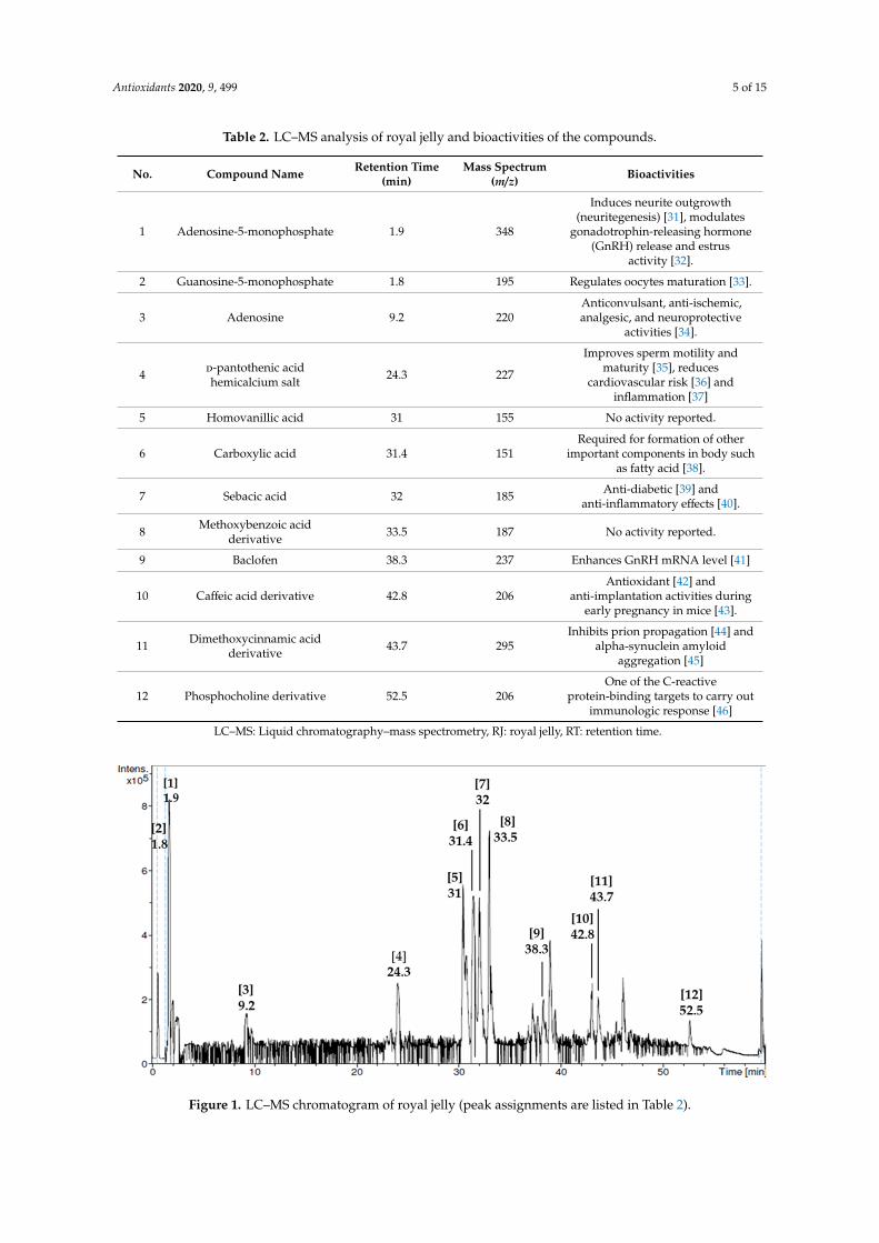
Phytochemical screening test revealed all nine phytochemical compounds found in RJ (Table 1)together with their reported bioactivities. From LC–MS analysis, we identified 12 non-volatilephytochemical compounds in RJ, and their reported biological activities are presented in Table 2.The compound with the highest mass spectrum was adenosine-5-monophosphate (348 m/z), whilesebacic acid (185 m/z) had the lowest mass spectrum. The chromatogram and peak assignments of thecompounds are shown in Figure 1.
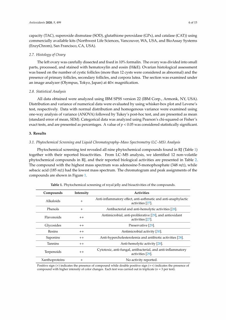
Table 1. Phytochemical screening of royal jelly and bioactivities of the compounds.
Compounds Intensity Activities
Alkaloids +Anti-inflammatory effect, anti-asthmatic and anti-anaphylactic
activities [27].
Phenols + Antibacterial and anti-hemolytic activities [28].
Flavonoids ++Antimicrobial, anti-proliferative [29], and antioxidant
activities [27].
Glycosides ++ Preservative [29].
Resins ++ Antimicrobial activity [30].
Saponins ++ Anti-hypercholesterolemia and antibiotic activities [28].
Tannins ++ Anti-hemolytic activity [28].
Terpenoids ++Cytotoxic, anti-fungal, antibacterial, and anti-inflammatory
activities [29].
Xanthoproteins + No activity reported.
Positive sign (+) indicates the presence of compound while double positive sign (++) indicates the presence ofcompound with higher intensity of color changes. Each test was carried out in triplicate (n = 3 per test).
Antioxidants 2020, 9, 499 5 of 15
Table 2. LC–MS analysis of royal jelly and bioactivities of the compounds.
No. Compound Name Retention Time(min)
Mass Spectrum(m/z) Bioactivities
1 Adenosine-5-monophosphate 1.9 348
Induces neurite outgrowth(neuritegenesis) [31], modulates
gonadotrophin-releasing hormone(GnRH) release and estrus
activity [32].
2 Guanosine-5-monophosphate 1.8 195 Regulates oocytes maturation [33].
3 Adenosine 9.2 220Anticonvulsant, anti-ischemic,analgesic, and neuroprotective
activities [34].
4 d-pantothenic acidhemicalcium salt 24.3 227
Improves sperm motility andmaturity [35], reduces
cardiovascular risk [36] andinflammation [37]
5 Homovanillic acid 31 155 No activity reported.
6 Carboxylic acid 31.4 151Required for formation of other
important components in body suchas fatty acid [38].
7 Sebacic acid 32 185 Anti-diabetic [39] andanti-inflammatory effects [40].
8 Methoxybenzoic acidderivative 33.5 187 No activity reported.
9 Baclofen 38.3 237 Enhances GnRH mRNA level [41]
10 Caffeic acid derivative 42.8 206Antioxidant [42] and
anti-implantation activities duringearly pregnancy in mice [43].
11 Dimethoxycinnamic acidderivative 43.7 295
Inhibits prion propagation [44] andalpha-synuclein amyloid
aggregation [45]
12 Phosphocholine derivative 52.5 206One of the C-reactive
protein-binding targets to carry outimmunologic response [46]
LC–MS: Liquid chromatography–mass spectrometry, RJ: royal jelly, RT: retention time.
Antioxidants 2020, 9, x FOR PEER REVIEW 5 of 15
Table 2. LC–MS analysis of royal jelly and bioactivities of the compounds.
No. Compound Name Retention
Time (min)
Mass Spectrum
(m/z) Bioactivities
1 Adenosine-5-
monophosphate 1.9 348
Induces neurite outgrowth (neuritegenesis) [31], modulates gonadotrophin-releasing
hormone (GnRH) release and estrus activity [32].
2 Guanosine-5-
monophosphate 1.8 195 Regulates oocytes maturation [33].
3 Adenosine 9.2 220 Anticonvulsant, anti-ischemic, analgesic,
and neuroprotective activities [34].
4 D-pantothenic acid hemicalcium salt
24.3 227 Improves sperm motility and maturity [35],
reduces cardiovascular risk [36] and inflammation [37]
5 Homovanillic acid 31 155 No activity reported.
6 Carboxylic acid 31.4 151 Required for formation of other important components in body such as fatty acid [38].
7 Sebacic acid 32 185 Anti-diabetic [39] and anti-inflammatory
effects [40].
8 Methoxybenzoic acid
derivative 33.5 187 No activity reported.
9 Baclofen 38.3 237 Enhances GnRH mRNA level [41]
10 Caffeic acid derivative 42.8 206 Antioxidant [42] and anti-implantation
activities during early pregnancy in mice [43].
11 Dimethoxycinnamic
acid derivative 43.7 295
Inhibits prion propagation [44] and alpha-synuclein amyloid aggregation [45]
12 Phosphocholine
derivative 52.5 206
One of the C-reactive protein-binding targets to carry out immunologic response
[46] LC–MS: Liquid chromatography–mass spectrometry, RJ: royal jelly, RT: retention time.
Figure 1. LC–MS chromatogram of royal jelly (peak assignments are listed in Table 2).
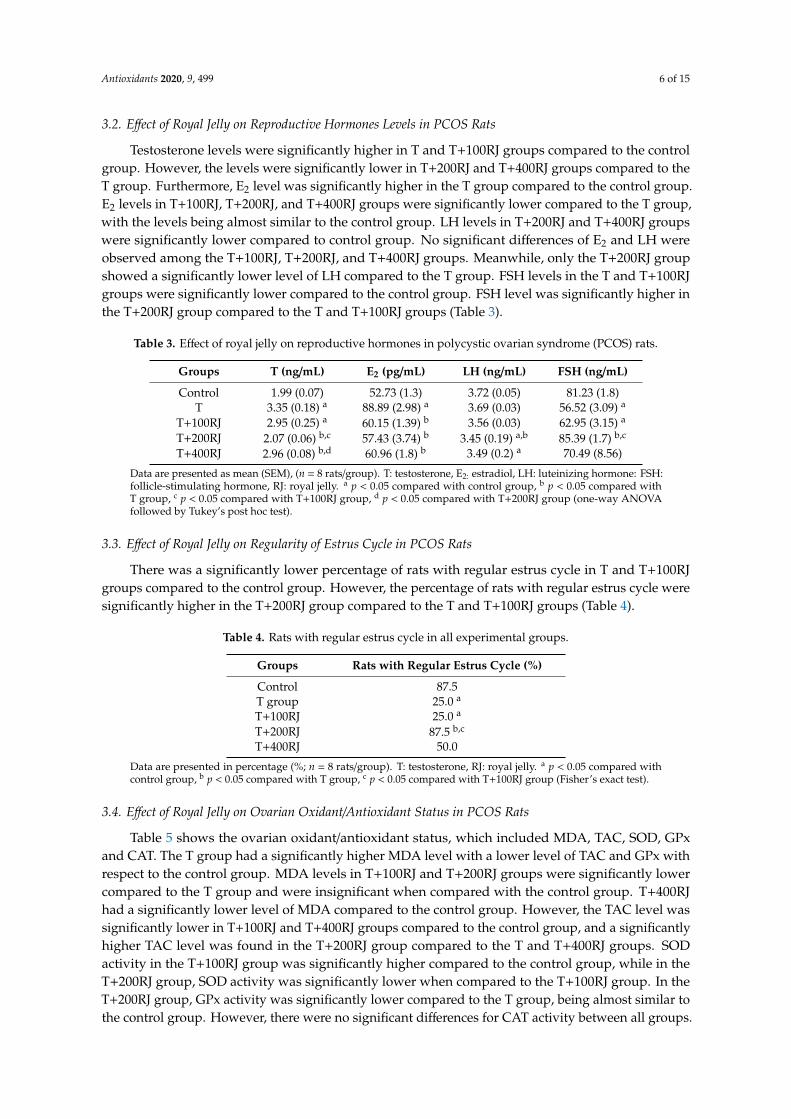
3.2. Effect of Royal Jelly on Reproductive Hormones Levels in PCOS Rats
Testosterone levels were significantly higher in T and T+100RJ groups compared to the control group. However, the levels were significantly lower in T+200RJ and T+400RJ groups compared to the
Figure 1. LC–MS chromatogram of royal jelly (peak assignments are listed in Table 2).
Antioxidants 2020, 9, 499 6 of 15
3.2. Effect of Royal Jelly on Reproductive Hormones Levels in PCOS Rats
Testosterone levels were significantly higher in T and T+100RJ groups compared to the controlgroup. However, the levels were significantly lower in T+200RJ and T+400RJ groups compared to theT group. Furthermore, E2 level was significantly higher in the T group compared to the control group.E2 levels in T+100RJ, T+200RJ, and T+400RJ groups were significantly lower compared to the T group,with the levels being almost similar to the control group. LH levels in T+200RJ and T+400RJ groupswere significantly lower compared to control group. No significant differences of E2 and LH wereobserved among the T+100RJ, T+200RJ, and T+400RJ groups. Meanwhile, only the T+200RJ groupshowed a significantly lower level of LH compared to the T group. FSH levels in the T and T+100RJgroups were significantly lower compared to the control group. FSH level was significantly higher inthe T+200RJ group compared to the T and T+100RJ groups (Table 3).
Table 3. Effect of royal jelly on reproductive hormones in polycystic ovarian syndrome (PCOS) rats.
Groups T (ng/mL) E2 (pg/mL) LH (ng/mL) FSH (ng/mL)
Control 1.99 (0.07) 52.73 (1.3) 3.72 (0.05) 81.23 (1.8)T 3.35 (0.18) a 88.89 (2.98) a 3.69 (0.03) 56.52 (3.09) a
T+100RJ 2.95 (0.25) a 60.15 (1.39) b 3.56 (0.03) 62.95 (3.15) a
T+200RJ 2.07 (0.06) b,c 57.43 (3.74) b 3.45 (0.19) a,b 85.39 (1.7) b,c
T+400RJ 2.96 (0.08) b,d 60.96 (1.8) b 3.49 (0.2) a 70.49 (8.56)
Data are presented as mean (SEM), (n = 8 rats/group). T: testosterone, E2: estradiol, LH: luteinizing hormone: FSH:follicle-stimulating hormone, RJ: royal jelly. a p < 0.05 compared with control group, b p < 0.05 compared withT group, c p < 0.05 compared with T+100RJ group, d p < 0.05 compared with T+200RJ group (one-way ANOVAfollowed by Tukey’s post hoc test).
3.3. Effect of Royal Jelly on Regularity of Estrus Cycle in PCOS Rats
There was a significantly lower percentage of rats with regular estrus cycle in T and T+100RJgroups compared to the control group. However, the percentage of rats with regular estrus cycle weresignificantly higher in the T+200RJ group compared to the T and T+100RJ groups (Table 4).
Table 4. Rats with regular estrus cycle in all experimental groups.
Groups Rats with Regular Estrus Cycle (%)
Control 87.5T group 25.0 a
T+100RJ 25.0 a
T+200RJ 87.5 b,c
T+400RJ 50.0
Data are presented in percentage (%; n = 8 rats/group). T: testosterone, RJ: royal jelly. a p < 0.05 compared withcontrol group, b p < 0.05 compared with T group, c p < 0.05 compared with T+100RJ group (Fisher’s exact test).
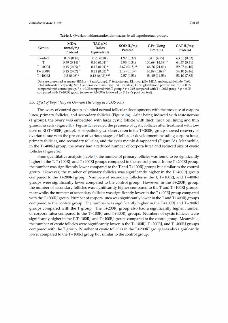
3.4. Effect of Royal Jelly on Ovarian Oxidant/Antioxidant Status in PCOS Rats
Table 5 shows the ovarian oxidant/antioxidant status, which included MDA, TAC, SOD, GPxand CAT. The T group had a significantly higher MDA level with a lower level of TAC and GPx withrespect to the control group. MDA levels in T+100RJ and T+200RJ groups were significantly lowercompared to the T group and were insignificant when compared with the control group. T+400RJhad a significantly lower level of MDA compared to the control group. However, the TAC level wassignificantly lower in T+100RJ and T+400RJ groups compared to the control group, and a significantlyhigher TAC level was found in the T+200RJ group compared to the T and T+400RJ groups. SODactivity in the T+100RJ group was significantly higher compared to the control group, while in theT+200RJ group, SOD activity was significantly lower when compared to the T+100RJ group. In theT+200RJ group, GPx activity was significantly lower compared to the T group, being almost similar tothe control group. However, there were no significant differences for CAT activity between all groups.
Antioxidants 2020, 9, 499 7 of 15
Table 5. Ovarian oxidant/antioxidant status in all experimental groups.
GroupMDA
(nmol/mgProtein)
TAC µMTrolox
Equivalents
SOD (U/mgProtein)
GPx (U/mgProtein)
CAT (U/mgProtein)
Control 0.09 (0.18) 0.25 (0.01) 1.92 (0.32) 34.1 (4.75) 63.61 (0.63)T 0.39 (0.14) a 0.10 (0.01) a 2.93 (0.34) 100.69 (14.39) a 64.47 (0.41)
T+100RJ 0.15 (0.05) b 0.12 (0.01) a 3.67 (0.15) a 66.76 (21.81) 59.07 (4.16)T+ 200RJ 0.15 (0.07) b 0.21 (0.03) b 2.19 (0.15) c 40.09 (5.89) b 54.10 (4.46)T+400RJ 0.3 (0.06) a 0.12 (0.03) a,d 2.57 (0.53) 54.15 (14.53) 53.10 (7.83)
Data are presented as mean (SEM; n = 8 rats/group). T: testosterone, RJ: royal jelly, MDA: malondealdehyde, TAC:total antioxidant capacity, SOD: superoxide dismutase, CAT: catalase, GPx: glutathione peroxidase. a p < 0.05compared with control group, b p < 0.05 compared with T group, c p < 0.05 compared with T+100RJ group, d p < 0.05compared with T+200RJ group (one-way ANOVA followed by Tukey’s post hoc test).
3.5. Effect of Royal Jelly on Ovarian Histology in PCOS Rats
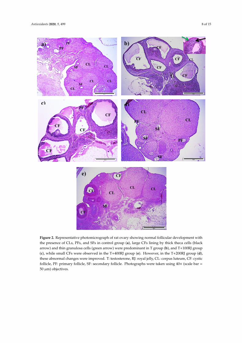
The ovary of control group exhibited normal follicular developments with the presence of corporalutea, primary follicles, and secondary follicles (Figure 2a). After being induced with testosterone(T group), the ovary was embedded with large cystic follicle with thick theca cell lining and thingranulosa cells (Figure 2b). Figure 2c revealed the presence of cystic follicles after treatment with lowdose of RJ (T+100RJ group). Histopathological observation in the T+200RJ group showed recovery ofovarian tissue with the presence of various stages of follicular development including corpora lutea,primary follicles, and secondary follicles, and the cysts mainly disappeared (Figure 2d). Meanwhile,in the T+400RJ group, the ovary had a reduced number of corpora lutea and reduced size of cysticfollicles (Figure 2e).
From quantitative analysis (Table 6), the number of primary follicles was found to be significantlyhigher in the T, T+100RJ, and T+400RJ groups compared to the control group. In the T+200RJ group,the number was significantly lower compared to the T and T+100RJ groups but similar to the controlgroup. However, the number of primary follicles was significantly higher in the T+400RJ groupcompared to the T+200RJ group. Numbers of secondary follicles in the T, T+100RJ, and T+400RJgroups were significantly lower compared to the control group. However, in the T+200RJ group,the number of secondary follicles was significantly higher compared to the T and T+100RJ groups;meanwhile, the number of secondary follicles was significantly lower in the T+400RJ group comparedwith the T+200RJ group. Number of corpora lutea was significantly lower in the T and T+400RJ groupscompared to the control group. The number was significantly higher in the T+100RJ and T+200RJgroups compared with the T group. The T+200RJ group also had a significantly higher numberof corpora lutea compared to the T+100RJ and T+400RJ groups. Numbers of cystic follicles weresignificantly higher in the T, T+100RJ, and T+400RJ groups compared to the control group. Meanwhile,the number of cystic follicles were significantly lower in the T+100RJ, T+200RJ, and T+400RJ groupscompared with the T group. Number of cystic follicles in the T+200RJ group was also significantlylower compared to the T+100RJ group but similar to the control group.
Antioxidants 2020, 9, 499 8 of 15
Antioxidants 2020, 9, x FOR PEER REVIEW 8 of 15
Figure 2. Representative photomicrograph of rat ovary showing normal follicular development with the presence of CLs, PFs, and SFs in control group (a), large CFs lining by thick theca cells (black arrow) and thin granulosa cells (green arrow) were predominant in T group (b), and T+100RJ group (c), while small CFs were observed in the T+400RJ group (e). However, in the T+200RJ group (d), these abnormal changes were improved. T: testosterone, RJ: royal jelly, CL: corpus luteum, CF: cystic follicle, PF: primary follicle, SF: secondary follicle. Photographs were taken using 40X (scale bar = 50 µm) objectives.
From quantitative analysis (Table 6), the number of primary follicles was found to be significantly higher in the T, T+100RJ, and T+400RJ groups compared to the control group. In the T+200RJ group, the number was significantly lower compared to the T and T+100RJ groups but similar to the control group. However, the number of primary follicles was significantly higher in the T+400RJ group compared to the T+200RJ group. Numbers of secondary follicles in the T, T+100RJ, and T+400RJ groups were significantly lower compared to the control group. However, in the T+200RJ group, the number of secondary follicles was significantly higher compared to the T and T+100RJ groups; meanwhile, the number of secondary follicles was significantly lower in the T+400RJ group compared with the T+200RJ group. Number of corpora lutea was significantly lower in the T
CL
CL CL
CL CL
PF
SF
SF
PF a)
CF
PF c)
CF
CF
CF
PF
CL
CL PF
SF
SF
SF
d)
CL CL
CL CF
CF
CF SF
e)
CL CF
CF
CF
CF
CF
b)
Figure 2. Representative photomicrograph of rat ovary showing normal follicular development withthe presence of CLs, PFs, and SFs in control group (a), large CFs lining by thick theca cells (blackarrow) and thin granulosa cells (green arrow) were predominant in T group (b), and T+100RJ group(c), while small CFs were observed in the T+400RJ group (e). However, in the T+200RJ group (d),these abnormal changes were improved. T: testosterone, RJ: royal jelly, CL: corpus luteum, CF: cysticfollicle, PF: primary follicle, SF: secondary follicle. Photographs were taken using 40× (scale bar =
50 µm) objectives.

Antioxidants 2020, 9, 499 9 of 15
Table 6. The number of primary and secondary follicles, number of corpora lutea, and number of cysticfollicles in all experimental groups.
Variables No. of PrimaryFollicles
No. of SecondaryFollicles
No. of CorporaLutea
No. of CysticFollicles
Control 3.25 (0.37) 7.88 (0.8) 6.64 (0.93) 1.13 (0.3)T 7.25 (0.53) a 2.25 (0.36) a 3.13 (0.35) a 9.25 (0.77) a
T+100RJ 5.88 (0.40) a 2.38 (0.42) a 4.75 (0.59) b 5.50 (0.42) a,b
T+200RJ 3.25 (0.37) b,c 7.00 (0.57) b,c 8.75 (0.36) b,c 3.00 (0.423) b,c
T+400RJ 6.00 (0.33) a,d 2.13 (0.3) a,d 3.75 (0.45) a,d 4.38 (0.26) a,b
Data are presented as mean (SEM; n = 8 rats/group). T: testosterone, RJ: royal jelly. a p < 0.05 compared with controlgroup, b p < 0.05 compared with T group, c p < 0.05 compared with T+100RJ group, d p < 0.05 compared withT+200RJ group (one-way ANOVA followed by Tukey’s post hoc test).
4. Discussion
In the present study, we determined composition and anti-androgenic effect of RJ in a PCOSanimal model. From our phytochemical screening test, we found that RJ used in the present studyhad flavonoids and phenols that were consistent with other studies performed on RJ from Jordan [47]and China [48]. Further analysis using LC–MS showed that it had compounds that have antioxidantproperties such as caffeic acid derivatives [42] and anti-inflammatory properties such as sebacicacid [40]. To our knowledge, this is the first study to show the LC–MS profile of RJ.
In our animal experimental study, T propionate was injected into the rats to inducehyperandrogenism, which is one of the characteristics of PCOS. The significantly higher levelof testosterone in the T group compared to the control group showed a successful inductionof hyperandrogenism. This finding is consistent with previous studies that have reported onthe high level of testosterone in rats after being induced by T propionate [49], letrozole [50],and dehydroepiandrosterone [51]. Another animal study in prenatally androgenized adult femaleoffspring showed significantly increased testosterone levels when compared to control rats [52]. In aclinical study, PCOS patients exhibited a higher level of total testosterone as compared to normalwomen [53]. On the other hand, T level was significantly lower in T+200RJ and T+400RJ groupscompared to the T group, which is consistent with another study [12]. This might suggest that RJ at200 and 400 mg/kg is able to lower T level in this PCOS animal model.
The present study also showed a significantly higher E2 level in the T group compared to thecontrol group, which is similar with other studies. For example, significant increase of E2 level wasalso found in PCOS mouse ovary after treatment with dehydroepiandrosterone [54] and in PCOSpatients [55] compared to control group, which might contribute to unfavorable conditions for thedevelopment of follicles. It is possible that high E2 level was attributed to the concomitant high levelof testosterone, as it can be converted to form E2. Interestingly, E2 level was significantly lower inall groups treated with RJ compared to the T group, which is consistent with other studies usingother natural products such as bee venom in E2-valerate-induced rats [56] and Commiphora wightiiin dehydroepiandrosterone-induced PCOS rats [51]. The low E2 levels found in the present studymight have been caused by concomitant low levels of testosterone. Furthermore, the non-significantdifferences in E2 levels among T+100RJ, T+200RJ, and T+400RJ groups might suggest that the effect ofRJ is not dose-dependent.
LH level in the T group was not significantly different from the control group, suggesting that thisPCOS animal model did not have an effect on LH level. However, LH level is significantly higher inPCOS women than normal women [57] and it occurs in about 60% of women with PCOS [58]. ElevatedLH level results in high production of androgen by theca cells in ovaries [59]. Therefore, the normallevel of LH in T group might suggest that the high T level is not attributable to LH level but may bedue to high synthesis of T, which needs further study. However, LH level was significantly lower inthe T+200RJ group compared to the control group, which is in agreement with a previous study inwhich administration of myoinositol, an insulin sensitizer, in PCOS patients reduced LH level and

Antioxidants 2020, 9, 499 10 of 15
LH/FSH ratio as well as improved menstrual cycle [60]. In contrast, in another study, LH level wassignificantly increased in infertile men treated with RJ [61]. Furthermore, in a study of male rats,supplementation of ofloxacin concomitant with RJ led to elevated level of LH [62]. Hence, the low LHlevel in rats treated with RJ in this animal model of the present study may suggest a low secretionof LH by anterior pituitary which needs further study. FSH level in T group that was significantlylower compared to control group, which is similar with other studies in which serum FSH level waslow in androgen-sterilized female rats compared with normal rats [63]. The low FSH level in the Tgroup might have been due to its concomitant high E2 level that could lead to low FSH secretion bythe anterior pituitary. However, FSH level was significantly higher in the T+200RJ group compared toT group which could be attributed to its concomitant low E2 level.
The T group had a significantly lower percentage of regular estrus cycle compared to the controlgroup, suggesting successful induction of hyperandrogenism that induced an irregular estrus cycle.This is in accordance with other studies that administered testosterone in prenatal period [52] and inimmature female rats, resulting in irregular estrus cycle [25]. The irregular estrus cycle is suggesteddue to the significantly high T, high E2, and low FSH levels found in the T group. This is supportedby another study where the irregular estrus cycle in androgenized transgenic mice exhibited analteration in hypothalamic–pituitary–gonadal axis function [64]. However, in the T+200RJ group, itspercentage of regular estrus cycle was significantly higher than T group, which is in agreement withother previous studies [17,65]. The improved estrus cycle in the T+200RJ group could be explainedby the improvement in its T, E2, LH, and FSH levels. Furthermore, this finding might indicate thatadministration of RJ has the capability to modulate estrogenic activity that could ameliorate theimpaired reproductive function in PCOS. In addition, the estrogenic property might also be attributedby the presence of 10-HDA and HDAA in RJ, which have been reported to have estrogenic activity [66].
In the present study, the ovarian MDA level, a marker of lipid peroxidation, was significantlyhigher in the T group compared to the control group, which is in agreement with a clinical study inwhich MDA level was significantly higher in PCOS patients compared to control patients, suggestingthe presence of OS in PCOS [67]. Hyperandrogenemia is suggested to be the reason for higher MDAlevel or imbalance in oxidant/antioxidant status, even though the exact mechanism is not clearlyunderstood. The significantly lower ovarian MDA levels in the T+100RJ and T+200RJ groups mightsuggest the ability of RJ to ameliorate OS in rat ovary, which corresponds to other studies [68,69].
Ovarian TAC level was significantly lower in the T group compared with the control group,which is consistent with another study that reported decreased serum TAC level in women withPCOS [67] and low serum TAC level in E2-vealerate-induced PCOS rats [70]. This low level of TACmight explain the high ovarian MDA level in the T group. However, ovarian TAC level in the T+200RJgroup was significantly higher compared to the T group, which might be attributable to the lowerovarian MDA level found in the T+200RJ group. This finding is similar to other studies, whereby RJaugmented the TAC level on paclitaxel-induced cardiotoxicity in rats [71] and in patients with insulinresistance type 2 diabetes mellitus [72]. In the present study, activity of ovarian GPx in the T groupwas significantly higher compared to the control group, which could be a result of up-regulation orincreased synthesis of GPx as a compensatory mechanism to overcome OS. This finding is consistentwith a previous study that reported increased GPx activity in PCOS patients [53]. However, the activityof GPx was significantly decreased in the T+200RJ group, which might suggest the ability of RJ at adose of 200 mg/kg to reduce the increased activity of GPx in this PCOS animal model. For ovarian CATactivity, no significant difference was found between T and control groups, which is similar to otherstudy whereby there was no difference for CAT activity in PCOS patients as compared to controls [73].There were no changes of CAT activities in T+100RJ, T+200RJ, and T+400RJ groups, which mightsuggest that RJ has no effect on CAT activity. However, the increased SOD activity in the T+100RJ groupmight suggest that RJ at a dose of 100 mg/kg possibly could up-regulate or increase the synthesis ofSOD, which requires further study. The significant changes observed on oxidant/antioxidant markersin rat from the T+200RJ group might indicate that RJ at the dose of 200 mg/kg exhibits an optimal

Antioxidants 2020, 9, 499 11 of 15
antioxidant property that counteracts OS in this PCOS animal model. The antioxidant property ofRJ has also been reported in a study, as RJ has a protective oxidative effect against cisplatin-inducedtesticular damage in rats [12], as well as in yeast Saccharomyces cerevisiae cells, in which it could reduceintracellular oxidation [74]. The improved oxidant/antioxidant status could also be attributed to somephytochemical compounds found in RJ from the LC–MS analysis that have antioxidant properties suchas caffeic acid [42] and dimethoxycinnamic acid [75] derivatives.
Histologically, the T group had a significantly higher number of primary and cystic follicles, aswell as a significantly lower number of secondary follicles and corpora lutea compared to the controlgroup, suggesting the establishment of PCOS characteristics in this animal model. A higher numberof primary follicles may suggest the presence of a high number of retarded primary follicles thatdid not develop into secondary follicles and corpora lutea, as supported by the concomitant lownumbers of secondary follicles and corpora lutea in the T group. It is suggested that elevated androgenconcentration stimulates follicular growth by increasing FSH receptor expression, leading to formationof multiple follicles. However, at a low FSH level, the growth of follicles is restricted, which in turnleads to formation of atretic and cystic follicles [76]. It has been suggested that androgen also modifiesthe response of follicles in the ovary to gonadotropins, giving rise to the changes found in polycysticovaries [76].
Interestingly, all these ovarian histological changes were significantly improved in the T+200RJgroup compared to the T group, although there were improvements for a number of corpora lutea inthe T+100RJ group and a number of cystic follicles in the T+100RJ and T+400RJ groups, suggestingthat RJ at 200 mg/kg/day is an optimum dose for the improvement of ovarian histology in this PCOSanimal model. Furthermore, this histological finding may also explain the improved estrus cycleand reproductive hormonal levels found in the T+200RJ group. Studies have also speculated thatthe increase in OS has led to anovulation in terms of reduced granulosa cell luteinization and oocytematuration [77]. Therefore, it is plausible to suggest that this antioxidant effect of RJ possibly couldexplain the improved ovarian histology and function, as well as the estrus cycle in the T+200RJ group.We would also like to speculate that the beneficial ovarian changes found in the present study mighthave been due to the action of royalactin, a 57 kDa protein that is present in RJ, which can stimulatenormal ovarian development in queen bees [78]. In addition, the improvement in the ovary could alsobe attributed to its phytochemical compounds such as 10-HDA and HDAA found in RJ, which canmodulate estrogenic activity [66].
5. Conclusions
RJ at the dose of 200 mg/kg for 4 weeks significantly improved reproductive hormone levels(T, E2, FSH, LH), estrus cycle regularity, ovarian oxidant-antioxidant status (MDA, TAC, GPx), andovarian histology in a PCOS animal model. These effects could be attributed partly to the combinedanti-androgenic effect of RJ, which possess phytochemical and bioactive antioxidant compounds, andrequires further study to determine its exact mechanism of action.
Author Contributions: Conceptualization, M.M.; data curation, N.A.H.; formal analysis, N.A.H.; fundingacquisition, M.M.; investigation, N.A.H.; methodology, A.B.A.B., N.H.N.H., and M.M.; project administration,M.M.; validation, A.B.A.B., A.A.M.Z., N.H.N.H., and M.M.; writing—original draft, N.A.H.; writing—reviewand editing, A.B.A.B., Z.A.O., Z.Z., and M.M. All authors have read and agreed to the published version ofthe manuscript.
Funding: This study was supported by the Universiti Sains Malaysia Short Term Grant (304/PPSP/61312107).
Acknowledgments: Authors hereby acknowledge the technical assistance rendered by Aminah Che Romli andNormawati Ahmad (Physiology Laboratory, USM) during the laboratory stage of this work.
Conflicts of Interest: The authors report no conflict of interest. The authors alone are responsible for the contentand writing of this article.
Antioxidants 2020, 9, 499 12 of 15
References
1. Dashti, S.; Abdul Hamid, H.; Mohamad Saini, S.; Shah Abu Bakar, A.; Sabri, N.A.I.; Ismail, M.; Esfehani, A.J.Prevalence of Polycyctic ovary syndrome among Malaysian female university staff. J. Midwifery Reprod. Health2019, 7, 1560–1568.
2. Ding, T.; Hardiman, P.J.; Petersen, I.; Wang, F.F.; Qu, F.; Baio, G. The prevalence of polycystic ovary syndromein reproductive-aged women of different ethnicity: A systematic review and meta-analysis. Oncotarget 2017,8, 96351–96358. [CrossRef] [PubMed]
3. Rotterdam ESHRE/ASRM-Sponsored PCOS Consensus Workshop Group. Revised 2003 consensus ondiagnostic criteria and long-term health risks related to polycystic ovary syndrome. Fertil. Steril. 2004, 81,19–25. [CrossRef] [PubMed]
4. Wachs, D.S.; Coffler, M.S.; Malcom, P.J.; Shimasaki, S.; Chang, R.J. Increased androgen responseto follicle-stimulating hormone administration in women with polycystic ovary syndrome. J. Clin.Endocrinol. Metab. 2008, 93, 1827–1833. [CrossRef]
5. Zhang, R.; Liu, H.; Bai, H.; Zhang, Y.; Liu, Q.; Guan, L.; Fan, P. Oxidative stress status in Chinese women withdifferent clinical phenotypes of polycystic ovary syndrome. Clin. Endocrinol. 2017, 86, 88–96. [CrossRef]
6. Vitek, W.; Alur, S.; Hoeger, K.M. Off-label drug use in the treatment of polycystic ovary syndrome. Fertil. Steril.2015, 103, 605–611. [CrossRef]
7. Ramadan, M.F.; Al-Ghamdi, A. Bioactive compounds and health-promoting properties of royal jelly: A review.J. Funct. Foods 2012, 4, 39–52. [CrossRef]
8. Sabatini, A.G.; Marcazzan, G.L.; Caboni, M.F.; Bogdanov, S.; Almeida-Muradian, L.B.D. Quality andstandardisation of royal jelly. J. Apiproduct. Apimedical. Sci. 2009, 1, 1–6. [CrossRef]
9. Park, M.J.; Kim, B.Y.; Park, H.G.; Deng, Y.; Yoon, H.J.; Choi, Y.S.; Lee, K.S.; Jin, B.R. Major royal jelly protein 2acts as an antimicrobial agent and antioxidant in royal jelly. J. Asia Pac. Entomol. 2019, 22, 684–689. [CrossRef]
10. Shirzad, M.; Kordyazdi, R.; Shahinfard, N.; Nikokar, M. Does royal jelly affect tumor cells? J. Herbmed. Pharm.2013, 2, 45–48.
11. Gasic, S.; Vucevic, D.; Vasilijic, S.; Antunovic, M.; Chinou, I.; Colic, M. Evaluation of the immunomodulatoryactivities of royal jelly components in vitro. Immunopharmacol. Immunotoxicol. 2007, 29, 521–536. [CrossRef][PubMed]
12. Silici, S.; Ekmekcioglu, O.; Kanbur, M.; Deniz, K. The protective effect of royal jelly against cisplatin-inducedrenal oxidative stress in rats. World J. Urol. 2011, 29, 127–132. [CrossRef] [PubMed]
13. Petelin, A.; Kenig, S.; Kopinc, R.; Dezelak, M.; Bizjak, M.C.; Praznikar, Z.J. Effects of royal jelly administrationon lipid profile, satiety, inflammation, and antioxidant capacity in asymptomatic overweight adults.Evid. Based Complement. Altern. Med. 2019, 2019, 4969720. [CrossRef] [PubMed]
14. Sharif, S.N.; Darsareh, F. Effect of royal jelly on menopausal symptoms: A randomized placebo-controlledclinical trial. Complement. Clin. Pr. 2019, 37, 47–50. [CrossRef]
15. Elnagar, S.A. Royal jelly counteracts bucks’ “summer infertility”. Anim. Reprod. Sci. 2010, 121, 174–180.[CrossRef]
16. El-Hanoun, A.M.; Elkomy, A.E.; Fares, W.A.; Shahien, E. Impact of royal jelly to improve reproductiveperformance of male rabbits under hot summer conditions. World Rabbit Sci. 2014, 22, 241–248. [CrossRef]
17. Husein, M.Q.; Kridli, R.T. Reproductive responses following royal jelly treatment administered orally orintramuscularly into progesterone-treated Awassi ewes. Anim. Reprod. Sci. 2002, 74, 45–53. [CrossRef]
18. Kridli, R.T.; Husein, M.Q.; Humphrey, W.D. Effect of royal jelly and GnRH on estrus synchronization andpregnancy rate in ewes using intravaginal sponges. Small Rumin. Res. 2003, 49, 25–30. [CrossRef]
19. Mishima, S.; Suzuki, K.-M.; Isohama, Y.; Kuratsu, N.; Araki, Y.; Inoue, M.; Miyata, T. Royal jelly has estrogeniceffects in vitro and in vivo. J. Ethnopharmacol. 2005, 101, 215–220. [CrossRef]
20. Suzuki, K.M.; Isohama, Y.; Maruyama, H.; Yamada, Y.; Narita, Y.; Ohta, S.; Araki, Y.; Miyata, T. Estrogenicactivities of fatty acids and a sterol isolated from royal jelly. Evid. Based Complement. Altern. Med. 2008, 5,295–302. [CrossRef]
21. Reagen-Shaw, S.; Nihal, M.; Ahmad, N. Dose translation from animal to human studies revisited. Faseb. J.2008, 22, 659–661. [CrossRef] [PubMed]
22. Raaman, N. Phytochemical Techniques; New India Publishing Agency: Pitam Pura, New Delhi, India, 2006.
Antioxidants 2020, 9, 499 13 of 15
23. Ajayi, G.O.; Olagunju, J.A.; Ademuyiwa, O.; Martins, O.C. Gas chromatography-mass spectrometry analysisand phytochemical screening of ethanolic root extract of Plumbago Zeylanica, Linn. J. Med. Plants Res. 2011,5, 1756–1761.
24. Ganesh, S.; Vennila, J.J. Phytochemical analysis of Acanthus Ilicifolius and Avicennia officinalis by GC-MS.Res. J. Phytochem. 2011, 5, 60–65. [CrossRef]
25. Abdulghani, M.; Hussin, A.H.; Sulaiman, S.A.; Chan, K.L. The ameliorative effects of Eurycoma longifoliaJack on testosterone-induced reproductive disorders in female rats. Reprod. Biol. 2012, 12, 247–255. [CrossRef]
26. Marcondes, F.K.; Bianchi, F.J.; Tanno, A.P. Determination of the estrous cycle phases of rats: Some helpfulconsiderations. Braz. J. Biol. 2002, 62, 609–614. [CrossRef] [PubMed]
27. Antonisamy, J.M.; Aparna, J.S.; Jeeva, S.; Sukumaran, S.; Anantham, B. Preliminary phytochemical studieson the methanolic flower extracts of some selected medicinal plants from India. Asian Pac. J. Trop. Biomed.2012, 2, S79–S82. [CrossRef]
28. Rao, M.R.K.; Kumar, M.H.; Amutha, A.; Prabhu, K.; Chatterjee, B.; Selva Kumar, S. Phytochemical analysisand antioxidant efficacy of the resin of Bombax ceiba (Salmali). Int. J. Pharm. Sci. Rev. Res. 2015, 30, 335–339.
29. Ramalakshmi, S.; Muthuchelian, K. Analysis of bioactive constituents from the ethanolic leaf extract ofTabebuia rosea (Bertol.) DC by gas chromatography-mass spectrometry. Int. J. ChemTech Res. 2011, 3,1054–1059.
30. Termentzi, A.; Fokialakis, N.; Leandros Skaltsounis, A. Natural resins and bioactive natural products thereofas potential anitimicrobial agents. Curr. Pharm. Des. 2011, 17, 1267–1290. [CrossRef]
31. Hattori, N.; Nomoto, H.; Fukumitsu, H.; Mishima, S.; Furukawa, S. AMP N1 -Oxide, a unique compound ofroyal jelly, induces neurite outgrowth from PC12 cells via signaling by protein kinase A independent of thatby mitogen-activated protein kinase. Evid. Based Complement. Altern. Med. 2010, 7, 63–68. [CrossRef]
32. Coyral-Castel, S.; Tosca, L.; Ferreira, G.; Jeanpierre, E.; Rame, C.; Lomet, D.; Caraty, A.; Monget, P.;Chabrolle, C.; Dupont, J. The effect of AMP-activated kinase activation on gonadotrophin-releasing hormonesecretion in GT1-7 cells and its potential role in hypothalamic regulation of the oestrous cyclicity in rats.J. Neuroendocr. 2008, 20, 335–346. [CrossRef]
33. Ratner, A. Effects of follicle stimulating hormone and luteinizing hormone upon cyclic AMP and cyclic GMPlevels in rat ovaries in vitro. Endocrinology 1976, 99, 1496–1500. [CrossRef] [PubMed]
34. Daval, J.-L.; Nehlig, A.; Nicolas, F. Physiological and pharmacological properties of adenosine: Therapeuticimplications. Life Sci. 1991, 49, 1435–1453. [CrossRef]
35. Kuo, Y.M.; Hayflick, S.J.; Gitschier, J. Deprivation of pantothenic acid elicits a movement disorder andazoospermia in a mouse model of pantothenate kinase-associated neurodegeneration. J. Inherit. Metab. Dis.2007, 30, 310–317. [CrossRef] [PubMed]
36. Rumberger, J.A.; Napolitano, J.; Azumano, I.; Kamiya, T.; Evans, M. Pantethine, a derivative of vitaminB5 used as a nutritional supplement, favorably alters low-density lipoprotein cholesterol metabolism inlow- to moderate-cardiovascular risk North American subjects: A triple-blinded placebo and diet-controlledinvestigation. Nutr. Res. 2011, 31, 608–615.
37. Jung, S.; Kim, M.K.; Choi, B.Y. The long-term relationship between dietary pantothenic acid (vitamin B5)intake and C-reactive protein concentration in adults aged 40 years and older. Nutr. Metab. Cardiovasc. Dis.2017, 27, 806–816. [CrossRef]
38. Layden, B.T.; Angueira, A.R.; Brodsky, M.; Durai, V.; Lower, W.L., Jr. Short chain fatty acids and theirreceptors: New metabolic targets. Transl. Res. 2013, 161, 131–140. [CrossRef]
39. Iaconelli, A.; Gastaldelli, A.; Chiellini, C.; Gniuli, D.; Favuzzi, A.; Binnert, C.; Mace, K.; Mingrone, G. Effect oforal sebacic acid on postprandial glycemia, insulinemia, and glucose rate of appearance in type 2 diabetes.Diabetes Care 2010, 33, 2327–2332. [CrossRef]
40. Chen, Y.-F.; Wang, K.; Zhang, Y.-Z.; Zheng, Y.-F.; Hu, F.-L. In vitro anti-inflammatory effects of three fattyacids from royal jelly. Mediat. Inflamm. 2016, 2016, 3583684. [CrossRef]
41. Cho, B.N.; Kim, K. Differential effect of baclofen on hypothalamic GnRH and pituitary LHβ gene expressionin steroid-treated rats. Mol. Cells 1997, 7, 605–609.
42. Nakajima, Y.; Tsuruma, K.; Shimazawa, M.; Mishima, S.; Hara, H. Comparison of bee products based onassays of antioxidant capacities. BMC Complement. Altern. Med. 2009, 9, 1–9. [CrossRef] [PubMed]

Antioxidants 2020, 9, 499 14 of 15
43. Liu, Y.; Qiu, S.; Wang, L.; Zhang, N.; Shi, Y.; Zhou, H.; Liu, X.; Shao, L.; Liu, X.; Chen, J.; et al. Reproductiveand developmental toxicity study of caffeic acid in mice. Food Chem. Toxicol. 2019, 123, 106–112. [CrossRef][PubMed]
44. Zanyatkin, I.; Stroylova, Y.; Tishina, S.; Stroylov, V.; Melnikova, A.; Haertle, T.; Muronetz, V. Inhibition ofprion propagation by 3,4-dimethoxycinnamic acid. Phytother. Res. 2017, 31, 1046–1055. [CrossRef] [PubMed]
45. Medvedeva, M.; Barinova, K.; Melnikova, A.; Semenyuk, P.; Kolmogorov, V.; Gorelkin, P.; Erofeev, A.;Muronetz, V. Naturally occuring cinnamic acid derivatives prevent amyloid transformation of alpha-synuclein.Biochimie 2020, 170, 128–139. [CrossRef]
46. Thompson, D.; Pepys, M.B.; Wood, S.P. The physiological structure of human C-reactive protein and itscomplex with phosphocholine. Structure 1999, 7, 169–177. [CrossRef]
47. Nabas, Z.; Haddadin, M.S.Y.; Haddadin, J.; Nazer, I.K. Chemical composition of royal jelly and effects ofsynbiotic with two different locally isolated probiotic strains on antioxidant activities. Pol. J. Food Nutr. Sci.2014, 64, 171–180. [CrossRef]
48. Nagai, T.; Inoue, R. Preparation and the functional properties of water extract and alkaline extract of royaljelly. Food Chem. 2004, 84, 181–186. [CrossRef]
49. Beloosesky, R.; Gold, R.; Almog, B.; Sasson, R.; Dantes, A.; Land-Bracha, A.; Hirsh, L.; Itskovitz-Eldor, J.;Lessing, J.B.; Homburg, R.; et al. Induction of polycystic ovary by testosterone in immature female rats:Modulation of apoptosis and attenuation of glucose/insulin ratio. Int. J. Mol. Med. 2004, 14, 207–215.[CrossRef]
50. Pandey, V.; Shukla, R.; Krishna, A.; Tripathi, Y.B. Effect of combined treatment of modern and herbalsupplement in the management of letrozole induced polycystic ovary syndrome. J. Endocrinol. Diabetes 2017,4, 1–6.
51. Kavitha, A.; Babu, A.N.; Kumar, S.M.; Kiran, S.V. Evaluation of effects of Commiphora wightii indehydroepiandrosterone (DHEA) induced polystic ovary syndrome (PCOS) in rats. PharmaTutor 2016, 4,47–55.
52. Tehrani, F.R.; Noroozzadeh, M.; Zahediasl, S.; Piryaei, A.; Azizi, F. Introducing a rat model of prenatalandrogen-induced polycystic ovary syndrome in adulthood. Exp. Physiol. 2014, 99, 792–801. [CrossRef][PubMed]
53. Kandasamy, S.; Sivagamasundari, R.I.; Bupathy, A.; Sethubathy, S.; Gobal, V. Evaluation of insulin resistanceand oxidative stress in obese patients with polycystic ovary syndrome. Int. J. Appl. Biol. Pharm. Technol.2010, 1, 391–398.
54. Yaba, A.; Demir, N. The mechanism of MTOR (mammalian target of rapamycin) in a mouse model ofpolycystic ovary syndrome (PCOS). J. Ovarian Res. 2012, 5, 1–12. [CrossRef] [PubMed]
55. Bartolone, L.; Smedile, G.; Arcoraci, V.; Trimarchi, F.; Benvenga, S. Extremely high levels of estradiol andtestosterone in a case of polycystic ovarian syndrome. Hormone and clinical similarities with the phenotypeof the α estrogen receptor null mice. J. Endocrinol. Investig. 2000, 23, 467–472. [CrossRef]
56. Karimzadeh, L.; Nabiuni, M.; Sheikholeslami, A.; Irian, S. Bee venom treatment reduced C-reactive proteinand improved follicle quality in a rat model of estradiol valerate-induced polycystic ovarian syndrome.J. Venom. Anim. Toxins Incl. Trop. Dis. 2012, 18, 384–392. [CrossRef]
57. Taylor, A.E.; McCourt, B.; Martin, K.A.; Anderson, E.J.; Adams, J.M.; Schoenfeld, D.; Hall, J.E. Determinantsof abnormal gonadotropin secretion in clinically defined women with polycystic ovary syndrome. J. Clin.Endocrinol. Metab. 1997, 82, 2248–2256. [CrossRef]
58. Laven, J.S.E.; Imani, B.; Eijkemans, M.J.C.; Fauser, B.C. New approach to polycystic ovary syndrome andother forms of anovulatory infertility. Obs. Gynecol. Surv. 2002, 57, 755–767. [CrossRef]
59. Ehrmann, D.A. Polycystic Ovary Syndrome. N. Engl. J. Med. 2005, 352, 1223–1236. [CrossRef]60. Genazzani, A.D.; Lanzoni, C.; Ricchieri, F.; Jasonni, V.M. Myo-inositol administration positively affects
hyperinsulinemia and hormonal parameters in overweight patients with polycystic ovary syndrome.Gynecol. Endocrinol. 2008, 24, 139–144. [CrossRef]
61. Al-Sanafi, A.E.; Mohssin, S.A.; Abdulla, S.M. Effect of royal jelly on male infertility. Thi-Qar Med. J. 2007, 1,1–12.
62. Manas, G.E.; Najafi, G. Protective effects of royal jelly on the histomorphologic, oxidative stress and spermparameters in ofloxacin treated rat. Comp. Clin. Path. 2017, 26, 1111–1115. [CrossRef]
Antioxidants 2020, 9, 499 15 of 15
63. Sun, F.; Yu, J. The effect of a special herbal tea on obesity and anovulation in androgen-sterilized rats. Proc. Soc.Exp. Biol. Med. 2000, 223, 295–301. [CrossRef] [PubMed]
64. Sullivan, S.D.; Moenter, S.M. Prenatal androgens alter GABAergic drive to gonadotropin-releasing hormoneneurons: Implications for a common fertility disorder. Proc. Natl. Acad. Sci. USA 2004, 101, 7129–7134.[CrossRef] [PubMed]
65. Bhuvaneshwari, S.; Poornima, R.; Averal, H.I. Detection of polycystic ovary syndrome and its treatment withPergulariadaemia in rat models. IOSR J. Pharm. 2015, 5, 42–49.
66. Hamid, N.A.; Abu Bakar, A.B.; Mohamed, M. Phytochemical analysis and GC-MS profile of royal jelly fromselected areas in Malaysia. Malays. Appl. Biol. 2018, 47, 101–107.
67. Maleedhu, P.; Vijayabhaskar, M.; Rao, P.; Kodumuri, P. Antioxidant status in women with polycystic ovarysyndrome. J. Med. Health Sci. 2014, 3, 91–96.
68. Ghanbari, E.; Nejati, V.; Najafi, G.; Khazaei, M.; Babaei, M. Study on the effect of royal jelly on reproductiveparameters in streptozotocin-induced diabetic rats. Int. J. Fertil. Steril. 2015, 9, 113–120.
69. Najafi, G.; Nejati, V.; Jalali, A.S.; Zahmatkesh, E. Protective role of royal jelly in oxymetholone-inducedoxidative injury in mouse testis. Iran. J. Toxicol. 2014, 8, 1073–1080.
70. Ghasemzadeh, A.; Farzadi, L.; Khaki, A.; Ahmadi, S.K. Effect of Allium cepa seeds ethanolic extract onexperimental polycystic ovary syndrome (PCOS) apoptosis induced by estradiol-valerate. Life Sci. J. 2013,10, 170–175.
71. Malekinejad, H.; Ahsan, S.; Delkhosh-Kasmaie, F.; Cheraghi, H.; Rezaei-Golmisheh, A.; Janbaz-Acyabar, H.Cardioprotective effect of royal jelly on paclitaxel-induced cardio-toxicity in rats. Iran. J. Basic Med. Sci. 2016,19, 221–227.
72. Shidfar, F.; Jazayeri, S.; Mousavi, S.N.; Malek, M.; Hosseini, A.F.; Khoshpey, B. Does supplementation withroyal jelly improve oxidative stress and insulin resistance in type 2 diabetic patients? Iran. J. Public Health2015, 44, 797–803. [PubMed]
73. Kurdoglu, Z.; Ozkol, H.; Tuluce, Y.; Koyuncu, I. Oxidative status and its relation with insulin resistance inyoung non-obese women with polycystic ovary syndrome. J. Endocrinol. Investig. 2012, 35, 317–321.
74. Jamnik, P.; Goranovic, D.; Raspor, P. Antioxidative action of royal jelly in the yeast cell. Exp. Gerontol. 2007,42, 594–600. [CrossRef]
75. Gómez-Caravaca, A.M.; Gómez-Romero, M.; Arráez-Román, D.; Segura-Carretero, A.; Fernández-Gutiérrez, A.Advances in the analysis of phenolic compounds in products derived from bees. J. Pharm. Biomed. Anal.2006, 41, 1220–1234. [CrossRef] [PubMed]
76. Gleicher, N.; Weghofer, A.; Barad, D.H. The role of androgens in follicle maturation and ovulation induction:Friend or foe of infertility treatment? Reprod. Biol. Endocrinol. 2011, 9, 1–12. [CrossRef]
77. Agarwal, A.; Gupta, S.; Sharma, R. Oxidative stress and its implications in female infertility—A clinician’sperspective. Reprod. Biomed. Online 2005, 11, 641–650. [CrossRef]
78. Kamakura, M. Royalactin induces queen differentiation in honeybees. Nature 2011, 473, 478–483. [CrossRef][PubMed]
© 2020 by the authors. Licensee MDPI, Basel, Switzerland. This article is an open accessarticle distributed under the terms and conditions of the Creative Commons Attribution(CC BY) license (http://creativecommons.org/licenses/by/4.0/).