Chlamydomonas reinhardtii PsbS Protein Is … reinhardtii PsbS Protein Is Functional and Accumulates...
14
Chlamydomonas reinhardtii PsbS Protein Is Functional and Accumulates Rapidly and Transiently under High Light 1 Tania Tibiletti, Pascaline Auroy, Gilles Peltier, and Stefano Caffarri* Aix Marseille Université (AMU), Commissariat à l’Energie Atomique (CEA), Centre National de la Recherche Scientifique (CNRS), UMR 7265 Biologie Végétale et Microbiologie Environnementales, Bioscience and Biotechnology Institute of Aix-Marseille (BIAM), Laboratoire de Génétique et Biophysique des Plantes, 13009 Marseille, France (T.T., S.C.); and Aix Marseille Université (AMU), Commissariat à l’Energie Atomique (CEA), Centre National de la Recherche Scientifique (CNRS), UMR 7265 Biologie Végétale et Microbiologie Environnementales, Bioscience and Biotechnology Institute of Aix-Marseille (BIAM), Laboratoire de Bioénergétique et Biotechnologie des Bactéries et Microalgues, 13108 St. Paul Les Durance, France (P.A., G.P.) Photosynthetic organisms must respond to excess light in order to avoid photo-oxidative stress. In plants and green algae the fastest response to high light is non-photochemical quenching (NPQ), a process that allows the safe dissipation of the excess energy as heat. This phenomenon is triggered by the low luminal pH generated by photosynthetic electron transport. In vascular plants the main sensor of the low pH is the PsbS protein, while in the green alga Chlamydomonas reinhardtii LhcSR proteins appear to be exclusively responsible for this role. Interestingly, Chlamydomonas also possesses two PsbS genes, but so far the PsbS protein has not been detected and its biological function is unknown. Here, we reinvestigated the kinetics of gene expression and PsbS and LhcSR3 accumulation in Chlamydomonas during high light stress. We found that, unlike LhcSR3, PsbS accumulates very rapidly but only transiently. In order to determine the role of PsbS in NPQ and photoprotection in Chlamydomonas, we generated transplastomic strains expressing the algal or the Arabidopsis psbS gene optimized for plastid expression. Both PsbS proteins showed the ability to increase NPQ in Chlamydomonas wild-type and npq4 (lacking LhcSR3) backgrounds, but no clear photoprotection activity was observed. Quantification of PsbS and LhcSR3 in vivo indicates that PsbS is much less abundant than LhcSR3 during high light stress. Moreover, LhcSR3, unlike PsbS, also accumulates during other stress conditions. The possible role of PsbS in photoprotection is discussed. Photosynthetic organisms can be frequently exposed to light intensities that surpass the photosynthetic electron transport capacity. In these conditions, the excess absorbed energy can be transferred from excited chlorophylls in the triplet state ( 3 Chls*) to molecular O 2 and cause the production of harmful reactive oxygen species (ROS). In plants and green algae most of the light is absorbed by membrane proteins of the Light harvesting complex (Lhc) family (Jansson, 1999; Ballottari et al., 2012; Buchel, 2015), which are associ- ated with photosystem I and II (Dekker and Boekema, 2005; Caffarri et al., 2014) and participate in photo- protection (Duffy and Ruban, 2015). The fastest photoprotective mechanisms are acti- vated to harmlessly dissipate excess absorbed energy as heat in a process called non-photochemical quenching of the energy (NPQ). NPQ is a complex and multifac- torial process that has been deeply investigated in re- cent years. Although the molecular mechanisms are only partially understood and still debated (Holt et al., 2005; Ruban et al., 2007; Bode et al., 2009; Holzwarth et al., 2009; Kruger et al., 2014), several key actors in- volved in NPQ have been discovered in plants and green algae (see Duffy and Ruban [2015] and Goss and Lepetit [2015] for recent reviews). The fastest component of NPQ, called qE, is triggered by a low luminal pH that is detected by low pH-responsive proteins. So far, three proteins have been shown to have a major role in detecting and transducing the low pH signal: the low pH activated violaxanthin de-epoxidase enzyme (VDE; Arnoux et al., 2009) that catalyses the conversion of the xanthophyll violaxanthin into zeaxanthin, a pigment involved in NPQ and other photoprotection mechanisms (Demmig-Adams, 1990; Havaux and Niyogi, 1999); the pigment-binding LhcSR proteins that belong to the Lhc family and are very likely the active site of quenching after protonation of C-terminal residues (Peers et al., 2009; 1 This work was supported by the French National Research Agency Grant ANR-12-JSV8-0001-01 and the A*MIDEX project (no. ANR-11-IDEX-0001-02). * Address correspondence to [email protected]. The author responsible for distribution of materials integral to the findings presented in this article in accordance with the policy de- scribed in the Instructions for Authors (www.plantphysiol.org) is: Stefano Caffarri ([email protected]). S.C. conceived the research; T.T. performed most of the experi- ments with some contribution by S.C.; A.P. provided technical help for chloroplast transformation; S.C. and T.T. analyzed the data; S.C. wrote the article, and T.T. complemented the writing; G.P. discussed the results and revised the manuscript. www.plantphysiol.org/cgi/doi/10.1104/pp.16.00572 Plant Physiology Ò , August 2016, Vol. 171, pp. 2717–2730, www.plantphysiol.org Ó 2016 American Society of Plant Biologists. All Rights Reserved. 2717 www.plantphysiol.org on June 6, 2019 - Published by Downloaded from Copyright © 2016 American Society of Plant Biologists. All rights reserved.
-
Upload
truongduong -
Category
Documents
-
view
216 -
download
0
Transcript of Chlamydomonas reinhardtii PsbS Protein Is … reinhardtii PsbS Protein Is Functional and Accumulates...

Chlamydomonas reinhardtii PsbS Protein IsFunctional and Accumulates Rapidly andTransiently under High Light1
Tania Tibiletti, Pascaline Auroy, Gilles Peltier, and Stefano Caffarri*
Aix Marseille Université (AMU), Commissariat à l’Energie Atomique (CEA), Centre National de la RechercheScientifique (CNRS), UMR 7265 Biologie Végétale et Microbiologie Environnementales, Bioscience andBiotechnology Institute of Aix-Marseille (BIAM), Laboratoire de Génétique et Biophysique des Plantes, 13009Marseille, France (T.T., S.C.); and Aix Marseille Université (AMU), Commissariat à l’Energie Atomique (CEA),Centre National de la Recherche Scientifique (CNRS), UMR 7265 Biologie Végétale et MicrobiologieEnvironnementales, Bioscience and Biotechnology Institute of Aix-Marseille (BIAM), Laboratoire deBioénergétique et Biotechnologie des Bactéries et Microalgues, 13108 St. Paul Les Durance, France (P.A., G.P.)
Photosynthetic organisms must respond to excess light in order to avoid photo-oxidative stress. In plants and green algae thefastest response to high light is non-photochemical quenching (NPQ), a process that allows the safe dissipation of the excessenergy as heat. This phenomenon is triggered by the low luminal pH generated by photosynthetic electron transport. In vascularplants the main sensor of the low pH is the PsbS protein, while in the green alga Chlamydomonas reinhardtii LhcSR proteinsappear to be exclusively responsible for this role. Interestingly, Chlamydomonas also possesses two PsbS genes, but so far the PsbSprotein has not been detected and its biological function is unknown. Here, we reinvestigated the kinetics of gene expression andPsbS and LhcSR3 accumulation in Chlamydomonas during high light stress. We found that, unlike LhcSR3, PsbS accumulates veryrapidly but only transiently. In order to determine the role of PsbS in NPQ and photoprotection in Chlamydomonas, we generatedtransplastomic strains expressing the algal or the Arabidopsis psbS gene optimized for plastid expression. Both PsbS proteinsshowed the ability to increase NPQ in Chlamydomonas wild-type and npq4 (lacking LhcSR3) backgrounds, but no clearphotoprotection activity was observed. Quantification of PsbS and LhcSR3 in vivo indicates that PsbS is much less abundantthan LhcSR3 during high light stress. Moreover, LhcSR3, unlike PsbS, also accumulates during other stress conditions. Thepossible role of PsbS in photoprotection is discussed.
Photosynthetic organisms can be frequently exposedto light intensities that surpass the photosyntheticelectron transport capacity. In these conditions, theexcess absorbed energy can be transferred from excitedchlorophylls in the triplet state (3Chls*) to molecular O2and cause the production of harmful reactive oxygenspecies (ROS). In plants and green algae most of thelight is absorbed by membrane proteins of the Lightharvesting complex (Lhc) family (Jansson, 1999;Ballottari et al., 2012; Buchel, 2015), which are associ-ated with photosystem I and II (Dekker and Boekema,
2005; Caffarri et al., 2014) and participate in photo-protection (Duffy and Ruban, 2015).
The fastest photoprotective mechanisms are acti-vated to harmlessly dissipate excess absorbed energy asheat in a process called non-photochemical quenchingof the energy (NPQ). NPQ is a complex and multifac-torial process that has been deeply investigated in re-cent years. Although the molecular mechanisms areonly partially understood and still debated (Holt et al.,2005; Ruban et al., 2007; Bode et al., 2009; Holzwarthet al., 2009; Kruger et al., 2014), several key actors in-volved in NPQ have been discovered in plants andgreen algae (see Duffy and Ruban [2015] and Goss andLepetit [2015] for recent reviews).
The fastest component ofNPQ, called qE, is triggeredbya low luminal pH that is detected by low pH-responsiveproteins. So far, three proteins have been shown to have amajor role in detecting and transducing the lowpH signal:the low pH activated violaxanthin de-epoxidase enzyme(VDE; Arnoux et al., 2009) that catalyses the conversion ofthe xanthophyll violaxanthin into zeaxanthin, a pigmentinvolved in NPQ and other photoprotection mechanisms(Demmig-Adams, 1990; Havaux and Niyogi, 1999); thepigment-binding LhcSR proteins that belong to the Lhcfamily and are very likely the active site of quenching afterprotonation of C-terminal residues (Peers et al., 2009;
1 This work was supported by the French National ResearchAgency Grant ANR-12-JSV8-0001-01 and the A*MIDEX project (no.ANR-11-IDEX-0001-02).
* Address correspondence to [email protected] author responsible for distribution of materials integral to the
findings presented in this article in accordance with the policy de-scribed in the Instructions for Authors (www.plantphysiol.org) is:Stefano Caffarri ([email protected]).
S.C. conceived the research; T.T. performed most of the experi-ments with some contribution by S.C.; A.P. provided technical helpfor chloroplast transformation; S.C. and T.T. analyzed the data; S.C.wrote the article, and T.T. complemented the writing; G.P. discussedthe results and revised the manuscript.
www.plantphysiol.org/cgi/doi/10.1104/pp.16.00572
Plant Physiology�, August 2016, Vol. 171, pp. 2717–2730, www.plantphysiol.org � 2016 American Society of Plant Biologists. All Rights Reserved. 2717 www.plantphysiol.orgon June 6, 2019 - Published by Downloaded from
Copyright © 2016 American Society of Plant Biologists. All rights reserved.

Bonente et al., 2011; Liguori et al., 2013); and the PsbSprotein that is protonated on two luminal Glu residues (Liet al., 2000; Li et al., 2004) and is proposed to subsequentlypromote the reorganization of the photosynthetic appa-ratus in order to create quenching sites elsewhere (Bassiand Caffarri, 2000; Bonente et al., 2008a; Johnson et al.,2011). It should be noted that efficient energy quenchingalso requires the presence of Lhc proteins, but this does notrequire a specific Lhc isoform (Andersson et al., 2001, 2003;de Bianchi et al., 2008, 2011). Our present investigation isfocused on PsbS andLhcSR3 expression inChlamydomonasreinhardtii and the functionality of PsbS in the green alga.
PsbS belongs to the Lhc supergene family, but it hasthe unique property of being the only member withfour transmembrane helices instead of the more com-mon three-helix organization (Kim et al., 1992). Becauseno pigment binding has been detected either by bio-chemical analysis (Bonente et al., 2008a) or in in therecent crystal structure (Fan et al., 2015), it is very likelythat PsbS is not the quenching site during NPQ.Therefore, it has been suggested that PsbS promotes areorganization of the photosynthetic membranes inorder to induce the formation of a quenching site in theLhc antenna system (Bassi and Caffarri, 2000; Betterleet al., 2009; Johnson et al., 2011).
PsbS genes are present in organisms of the greenlineage (plants and green algae) but are absent in otheroxygenic photosynthetic organisms not belonging tothis lineage (Buchel, 2015). The protein has beendetected both in vascular (Tracheophytes) and non-vascular (Bryophytes) plants, as well in the closest algalrelatives to plants (Charophytes) and in multicellulargreen algae (Chlorophytes; Bonente et al., 2008b).However, previous immunoblot analyses aimed at in-vestigating the accumulation of PsbS in unicellulargreen algae such as Chlamydomonas reinhardtii showedthat the protein did not appear to accumulate underseveral different growth conditions (Bonente et al.,2008b). Nevertheless, the presence of two highly ho-mologous psbS genes in Chlamydomonas that code forproteins that are well conserved with respect to plantPsbS proteins (Bonente et al., 2008b) would suggest thatit is unlikely that these genes are pseudogenes. How-ever, it has been proposed that the Chlamydomonas PsbSdoes not play or plays only a minor role in NPQ(Bonente et al., 2008b). Consistent with this idea, a ge-netic screen based on NPQ induction capacity resultedin the isolation of amutant (called npq4) with a very lowlevel of NPQ (Peers et al., 2009). The mutation knockedout the LhcSR3 protein, which since the discovery of itsrole in NPQ, has been intensively studied. In Chlamy-domonas, LhcSR proteins are encoded by three genes:lhcSR1, lhcSR3.1 and 3.2, the last two coding for iden-tical proteins that are both knocked out in the npq4mutant. Interestingly, LhcSR3 plays a predominant rolein NPQ induction (Peers et al., 2009). LhcSR3 is ho-mologous to the Lhc proteins: it has a similar structurewith three transmembrane helices and it is also able tobind Chls and Cars (Bonente et al., 2011). It has beensuggested that LhcSR3 itself is the energy quencher due
to its pigment binding ability, the unusual fluorescenceproperties that show very short lifetime components,and its pH sensitivity (Bonente et al., 2011; Liguori et al.,2013; Tokutsu and Minagawa, 2013). A recent investi-gation suggests the LhcSR is localized both in granamembranes and stroma lamellae in the moss Phys-comitrella patens (Pinnola et al., 2015), and investigationsonChlamydomonas suggest that LhcSR3 can bind both toPSI or PSII depending on illumination conditions(Allorent et al., 2013; Bergner et al., 2015). In contrast topsbS, lhcSR genes are widespread in a many photo-synthetic organisms, including green algae, brownalgae,diatoms, nonvascular plants, and even some vascularplants (Buchel, 2015). In the moss Physcomitrella patens,LhcSR-dependent and the PsbS-dependentNPQ operateindependently and additively (Alboresi et al., 2008).
Even if PsbS proteins in Chlamydomonas have notbeen detected in previous analyses, the presence ofconserved psbS genes, their expression under nitrogendeprivation conditions (Miller et al., 2010), and the ex-pression of the genes during a dark to light shift asfound in a very recent report (Zones et al., 2015) suggesta role for PsbS in the alga. Thus, in this work, we havereinvestigated the expression and accumulation of PsbSin Chlamydomonas during light stress and other stressconditions in order to determine whether this proteincould play a role inNPQunder particular conditions andcompared our results with those obtained on LhcSR3.
We found that PsbS rapidly and transiently accu-mulates during light stress, and that contrasts withLhcSR3 that accumulates over a much longer period. Inorder to demonstrate a role of the algal PsbS protein inNPQ, we created Chlamydomonas strains constitutivelyexpressing the algal or the Arabidopsis psbS genes. Boththe overexpressed PsbS proteins showed the ability toincrease NPQ in Chlamydomonas.
A comparison with expression/accumulation ofLhcSR suggests that LhcSR is more abundant and isinvolved in photoprotection under several stress con-ditions, while PsbS seems to respond very rapidly andtransiently only to high light stress and has a minor rolein photoprotection in Chlamydomonas.
RESULTS
Analysis of PsbS and LhcSR3 Accumulation inChlamydomonas reinhardtii in High Light
In previous investigations (Bonente et al., 2008b), PsbSwas not detected upon high light acclimation. Therefore,we performed a new analysis of the kinetics PsbS accu-mulation during high light treatment by sampling cells atseveral time points starting from 30 min until 32 h afterthe switch from normal light (45 mmol photons m22 s21)to high light (Fig. 1; Supplemental Fig. S1). Experimentswere performed in liquid minimal medium at 1200 mmolphotons m22 s21 with four dilutions of the medium inorder to avoid autoshading by the growing cells (Fig. 1)and were also repeated without dilutions at threedifferent light intensities (400, 800, and 1200 mmol
2718 Plant Physiol. Vol. 171, 2016
Tibiletti et al.
www.plantphysiol.orgon June 6, 2019 - Published by Downloaded from Copyright © 2016 American Society of Plant Biologists. All rights reserved.

photons m22 s21; Supplemental Fig. S1). In all cases, thekinetics of PsbS induction were similar (Fig. 1;Supplemental Fig. S1): PsbS, undetectable in low lightacclimated cells, showed a rapid increase and was al-ready detectable after 30min under high light reaching amaximum level after around 4 to 8 h. PsbS then decreasedand was almost undetectable at 32 h. The decline in PsbSwas independent of the increase in cell density. Themaximal PsbS quantity appeared dependent on the lightintensity (Supplemental Fig. S1). Interestingly, kinetics inwild-type (4A+) and npq4 cells were practically identicaldespite the absence of LhcSR3 in the npq4 mutant.In parallel, we also investigated the accumulation of
the LhcSR3 protein, which has a key role in NPQ induc-tion in Chlamydomonas (Peers et al., 2009). LhcSR3 is alsoinduced by high light (Fig. 1; Supplemental Fig. S1), aspreviously shown (Peers et al., 2009; Bonente et al., 2011;Allorent et al., 2013). However, in contrast to PsbS, themaximum intensity was reached later (after 8 h) and ac-cumulation remained rather stable and high until the endof our experiments (32 h). Moreover, LhcSR3 was detec-ted at two positions in immunoblot assays, correspond-ing to the phosphorylated and unphosporylated proteins(Bonente et al., 2011), and the ratio of P-LhcSR3 over non-P-LhcSR3 increased during the stress period (Fig. 1).It should be noted that in a previous work (Bonente
et al., 2008b), the antibody used to check PsbS accu-mulation was raised against a recombinant barley PsbSprotein. For this work, we used a new antibody raisedagainst a synthetic peptide from the Chlamydomonas
PsbS sequence that is also well conserved in the Ara-bidopsis PsbS (see “Materials and Methods”). The newantibody, despite some unspecific cross-reactivity, isable to detect the Chlamydomonas PsbS protein (Figs.1 and 5) as well as the Arabidopsis one (SupplementalFig. S2). Several immunoblot assays confirmed theability of this antibody to correctly recognize the PsbSband: (1) the recombinant Chlamydomonas PsbS protein isdetected (Supplemental Fig. S2A); (2) comparison of granamembranes from Arabidopsis wild-type and npq4(koPsbS) plants showed a specific band at the MW ofPsbS in the wild-type sample absent in the mutant(Supplemental Fig. S2A); (3) the putative PsbS bands dis-appear in samples from high light treated Chlamydomonascells and Arabidopsis membranes in a competition assaywhere the antibody solution was preincubated with anexcess of recombinant Chlamydomonas protein before theimmunoblot analysis (Supplemental Fig. S2B); (4) PsbSwas detected by mass spectrometry only in high lightstressed thylakoids of Chlamydomonas (Supplemental Fig.S2C; Supplemental Table S2); (5) comparison betweenPsbS-overexpressor strains (see below) and high lighttreated cells showed that the transiently appearing banddetected on stressed cells has exactly the same position asoverexpressed PsbS (Fig. 5B).
Analysis of the Expression of psbS and lhcSR Genes
Quantitative real-time PCR experiments were per-formed on the same samples used for kinetic analysis ofPsbS and LhcSR3 protein accumulation. For this anal-ysis, we designed primers able to distinguish betweenthe two psbS genes (JGI v. 5.5 locus Cre01.g016600 ispsbS1 and locus Cre01.g016750 is psbS2; Zones et al.,2015), as well the two lhcSR3 genes (lhcSR3.1 andlhcSR3.2) and the lhcSR1 gene (Maruyama et al., 2014).
As shown in Figure 2, the results are in good agree-ment with the those obtained by immunoblot analysis onproteins (Fig. 1; Supplemental Fig. S1); the induction ofpsbS gene expression is extremely fast and transient, witha maximum in the very first part of the low light to highlight transition. A similar behavior was found for lhcSRgenes, but delayed compared to psbS and in agreementwith the later appearance of the LhcSR3 protein.
Both psbS genes are expressed, as previously found(Bonente et al., 2008b; Zones et al., 2015), as well alllhcSR genes. However, there are two major differencesbetween the analysis on mRNA and on protein: (1) themaximum accumulation of mRNA occurs earlier thanthe maximum protein accumulation; and (2) while bothpsbS mRNA and PsbS protein decrease during thestress, this is not the case for lhcSR3 RNAs and LhcSR3proteins, since lhcSR3 gene expression decreases sig-nificantly before the end of the 32 h kinetic experiment,while the protein remains abundant.
Overexpression of PsbS in Chlamydomonas
In order to test the functionality of the PsbS protein,we created overexpressor strains that we compared
Figure 1. Kinetics of PsbS accumulation in Chlamydomonas wild-type(CC124 and 4A+) and npq4 cells during high light treatment. Cellsgrown inminimalmediumwere exposed to 1200mmolphotonsm22 s21 for32 h, and aliquots were harvested at different times as indicated. Twomicrograms (in Chls) of total membranes were analyzed by SDS-PAGEand by immunoblot assays using antiPsbS and antiLhcSR3 antibodies.PsbS (arrow) showed a transient accumulation in all three genotypeswith a peak around 4–8 h. LhcSR3 showed a slightly delayed inductionand a strong accumulation until the end of the treatment. The position ofphosphorylated LhcSR3 (P-LhcSR3) and non-phosphorylated LhcSR3(LhcSR3) are indicated in the wild-type (CC124) immunoblot as ex-ample. Dilutions (dil) of the medium were done in order to avoid cellautoshading. Similar kinetics experiments at different light intensitiesand without dilutions are presented in Supplemental Figure S1. TheCoomassie stained gels are shown as protein loading control.
Plant Physiol. Vol. 171, 2016 2719
Characterization of PsbS in Chlamydomonas
www.plantphysiol.orgon June 6, 2019 - Published by Downloaded from Copyright © 2016 American Society of Plant Biologists. All rights reserved.

with wild-type cells that do not contain PsbS whengrown under moderate light (Fig. 1).
Because nuclear transformation in Chlamydomonasis not straightforward and a previous assay to over-express PsbS by nuclear transformation was un-successful (Bonente et al., 2008b), we generatedPsbS-overexpressing strains by means of chloroplasttransformation. We used two different constructs: (1)oePsbs.Cr, to create strains overexpressing the fulllength PsbS protein of Chlamydomonas (since the maturesequence is not known) and (2) oePsbS.At, to createstrains overexpressing the mature PsbS protein ofArabidopsis. We transformed both wild-type (CC124)and npq4 (koLhcSR3, 4A+ background) cells, and thestrains obtained are here after indicated as oePsbS.
Cr(wt)/oePsbS.At(wt) and oePsbS.Cr(npq4)/oePsbS.At(npq4), where “Cr” and “At” indicated the presenceof the Chlamydomonas or the Arabidopsis PsbS and thestrain transformed is indicated in brackets. We used thenpq4 (4A+) strain since it is the original koLhcSR3 mu-tant (Peers et al., 2009), and the wild type (CC124) as it isa very common strain used for genetic screening andphysiological studies (Proschold et al., 2005). It shouldbe noted that, even if we cannot directly compareoePsbS(npq4) to oePsbS(wt) cells, each oePsbS strain iscompared with its own non-transformed control (WT-CC124 or npq4). In the case of the oePsbS(npq4) strains,for NPQmeasurements (see next section), they are alsocompared with the wild type with the same geneticbackground (4A+).
Figure 2. Kinetics of psbS and lhcSR expression during high light stress. Expression of the two psbS genes and the three lhcSRgenes (lhcSR1, lhcSR3.2, and lhcSR3.2) was analyzed by quantitative real-time PCR on cells grown on minimal medium. In asimilar manner to the immunoblot experiments, psbS expression was induced rapidly and transiently after the switch to high lightin all three genotypes. LhcSR genes were also induced by high light, but the maximum expression was slightly delayed ascompared to psbS, similar to the immunoblot results. Averages6 SE are shown (threemeasurements from two biological replicatesfor wild-type [CC124] and npq4 cells; two replicates from one biological sample for wild-type [4A+] cells). Data are normalizedto one for the maximum value obtained for a given transcript. A gene encoding the cytosolic 40S small ribosomal subunit RACK1,also known as Chlamydomonas G protein b-subunit-like polypeptide (CBLP), and the gene IDA5 were used as a control.
2720 Plant Physiol. Vol. 171, 2016
Tibiletti et al.
www.plantphysiol.orgon June 6, 2019 - Published by Downloaded from Copyright © 2016 American Society of Plant Biologists. All rights reserved.

Immunoblot analysis performed on thylakoid mem-branes showed that both algal and plant PsbS accu-mulated in transplastomic Chlamydomonas strains (Fig.3). Note also (Fig. 5B) that the full length Chlamydomo-nas PsbS protein overexpressed in the chloroplast iscorrectly processed, because the overexpressed proteinis detected at a Mr identical to that of the nuclear-expressed PsbS in high light.Similar PsbS accumulation levels were obtained for
each construction/genotype, except for two oePsbS.Cr(wt) strains, where one line showed low expressionand another high expression (oePsbS.Cr9Wt no. 1 andno. 2, respectively; Fig. 3). This was useful for correlatingPsbS dose with the NPQ phenotype as described below.
Chlorophyll Fluorescence Properties of oePsbSChlamydomonas Strains
Twelve independent PsbS overexpressing lines[3 oePsbS.Cr(wt); 3 oePsbS.At(wt); 4 oePsbS.Cr(npq4);2 oePsbS.At(npq4)]were tested forNPQ.Cells grownon asolid minimal medium at moderate light (45 mmolphotonsm22 s21)were exposed to 800mmolphotonsm22 s21
of actinic light. NPQ induction was higher in all theoePsbS lines as comparedwith the respective control lines(WT-CC124 or npq4; Fig. 4 for an average of the lines withthe same construction/genotype and Supplemental Fig.S3A for each independent line tested).Both the Chlamydomonas and the Arabidopsis PsbS
were able to increase NPQ in the overexpressing
strains, indicating that both algal and plant PsbS pro-teins are functional in Chlamydomonas.
The NPQ kinetics (Fig. 4B) showed a rapid inductionin the first minute (likely due to a fast and elevatedDpHformed at the beginning of the actinic light phase),followed by a decrease and then a second increase inNPQ levels after a few minutes.
Unexpectedly, we found differences in NPQ kineticsbetween strains in the CC124 and in the 4A+ back-grounds. In CC124 cells, both the plant and the algal PsbSoverexpressors showed similar kinetics, and the maxi-mum NPQ was much higher than the NPQ in the wild-type (CC124) control, and theNPQwas largely reversibleduring the first 30 s of the dark phase. In the 4A+ back-ground, Chlamydomonas PsbS was more efficient than theArabidopsis PsbS, the NPQ at the end of the actinic lightphase was significantly higher in oePsbS (npq4) strainsthan in wild-type (4A+) and npq4 control cells, but thedifference was smaller than that between oePsbS(wt)lines and itswild-type (CC124) control and theNPQdidnotshow reversibility. The NPQ measurements on liquid cul-tures with a classical PAM instrument (Supplemental Fig.S3B) are in accordance with measurements performed byvideo-imaging on cultures grown on solid medium.Moreover, in this case, we found reversibility of NPQ atthe end of the actinic light for the oePsbS(npq4) strains,but not for npq4 and wild-type (4A+) cells despite someaccumulation of LhcSR1 (Supplemental Fig. S4B). How-ever, LhcSR1 accumulation was similar in all strains.
The level of NPQ showed a correlation with PsbSabundance, at least in the case of the Chlamydomonas
Figure 3. PsbS overexpression in Chlamydomonas.PsbS overexpressing strains were created by chloro-plast transformation using the Chlamydomonas psbSsequence (oePsbS.Cr) or the Arabidopsis psbS(oePsbS.At). Overexpression strains were made bothin the wild-type (CC124) background (top) and in thenpq4 (4A+) background (bottom). Immunoblot anal-ysis on total cell extracts (2 mg in Chls) from inde-pendent transformants confirmed the presence of therecombinant PsbS proteins, absent in wild-type andnpq4 cells grown under normal light (NL) on mini-mal medium. The strains oePsbS.Cr(wt)#1 and oePsbS.Cr(wt)#2 showed the lowest and highest PsbS content,respectively (results obtained on three biological repli-cates). Coomassie stained gels are shown as proteinloading controls.
Plant Physiol. Vol. 171, 2016 2721
Characterization of PsbS in Chlamydomonas
www.plantphysiol.orgon June 6, 2019 - Published by Downloaded from Copyright © 2016 American Society of Plant Biologists. All rights reserved.

PsbS where we obtained strains with different expres-sion levels. oePsbS.Cr(wt) no. 1, which accumulates lowamount of PsbS, (Fig. 3), showed a lower NPQ capa-bility than oePsbS.Cr(wt) no. 2 that accumulates highamounts of PsbS (Fig. 4A; Supplemental Fig. S3A). Thisis similar to the situation in Arabidopsis, where PsbSabundance is also correlated to NPQ levels (Li et al.,2002a, 2002b).
It should be noted the increase of NPQ capability inoePsbS strains is very unlikely due to some majorchange in the organization of the photosynthetic com-plexes. Biochemical characterization of the strainsshowed that Chl a/b ratios of oePsbS and control strainsare very similar, as well as the content of proteins rep-resentative of major photosynthetic complexes andprotein involved in cyclic electron transfer, except for
Figure 4. Fluorescence phenotypes of PsbS overexpressing strains. A, Wild type (CC124), wild type (4A+), npq4, overexpressingstrains of the Arabidopsis, and the Chlamydomonas PsbS in the npq4 (4A+) background [oePsbS.At(npq4) and oePsbS.Cr(npq4)respectively], and overexpressing strains of the Arabidopsis and the Chlamydomonas PsbS in the wild-type (CC124) background[oePsbS.At(wt) and oePsbS.Cr(wt), respectively] were plated on minimal medium, grown at ; 45 mmol photons m22 s21 andimaged with a fluorescence camera. The NPQ levels on a representative plate after 6 min under actinic light (800 mmol photonsm22 s21) are indicated in false colors. All oePsbS lines showed a higher NPQ capacity as compared to their control. B, The fullNPQ kinetics of the same strains as in A are shown. The values are the average 6 SD for oePsbS lines with the same back-grounds/construction calculated from five replicate (n = 5) plates arranged as in A (three spots of each independent oePsbS strainper plate; six spots for wild-type and npq4 strains per plate). The kinetics of the single independent oePsbS strains are shown inSupplemental Figure S3. C, Maximum quantum yields (Fv/Fm) calculated over four replicate plates as in B.
2722 Plant Physiol. Vol. 171, 2016
Tibiletti et al.
www.plantphysiol.orgon June 6, 2019 - Published by Downloaded from Copyright © 2016 American Society of Plant Biologists. All rights reserved.
PGRL1 that was less abundant in oePsbS.At(wt) cells(Supplemental Fig. S4).In conclusion, results from oePsbS lines strongly
support that both the plant and the algal PsbS arefunctional and able to increase NPQ capability inChlamydomonas cells.
In Vivo Quantification of PsbS and LhcSR
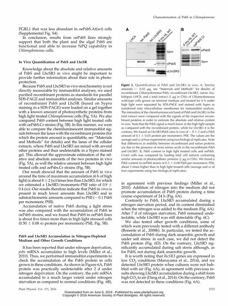
Knowledge about the absolute and relative amountsof PsbS and LhcSR3 in vivo might be important toprovide further information about their role in photo-protection.Because PsbS and LhcSR3 in vivo stoichiometry is not
directly measurable by immunoblot analyses, we usedpurified recombinant proteins as standards for parallelSDS-PAGE and immunoblot analyses. Similar amountsof recombinant PsbS and LhcSR (based on Syprostaining in a SDS-PAGE) were loaded on a gel togetherwith a known amount of photosynthetic proteins fromhigh light treated Chlamydomonas cells (Fig. 5A). We alsocompared PsbS content between high light treated cellswith oePsbS.Cr strains (Fig. 5B). In this manner, we wereable to compare the chemiluminescent immunoblot sig-nals between the laneswith the recombinant proteins (forwhich the protein amount is quantifiable, see “Materialsand Methods” for details) and the lanes of the cellularextracts, where PsbS and LhcSR3 are mixed with severalother proteins and thus undetectable in a Sypro stainedgel. This allowed the approximate estimation of the rel-ative and absolute amounts of the two proteins in vivo(Fig. 5A), as well the relative amount between high lighttreated cells and oePsbs.Cr strains (Fig. 5B).Our result showed that the amount of PsbS in vivo
around the time of maximum accumulation (6 h of highlight) is about 86 2 (SD) times less than LhcSR3, forwhichwe estimated a LhcSR3/monomeric-PSII ratio of 0.9 60.4 (SD). Our results therefore indicate that PsbS in vivo ispresent in much lower amounts than LhcSR3 and insubstoichiometric amounts compared to PSII (; 0.1 PsbSper monomeric PSII).Accumulation of native PsbS during a light stress
was also compared with the accumulation of PsbS inoePsbS strains, and we found that PsbS in oePsbS linesis about five times more than in high light stressed cells(0.50 6 0.08 SD protein per monomeric-PSII, Fig. 5B).
PsbS and LhcSR3 Accumulation in Nitrogen-DepletedMedium and Other Growth Conditions
It has been reported that under nitrogen deprivation,psbs mRNA accumulates to high levels (Miller et al.,2010). Thus, we performed immunoblot experiments tocheck the accumulation of the PsbS protein in cellsgrown in these conditions. As shown in Figure 6A, PsbSprotein was practically undetectable after 2 d undernitrogen deprivation. On the contrary, the psbs mRNAaccumulated to a much larger extent under nitrogenstarvation as compared to normal conditions (Fig. 6B),
in agreement with previous findings (Miller et al.,2010). Addition of nitrogen into the medium did notpromote accumulation of PsbS protein during a timecourse experiment of 24 h (Fig. 6A).
Contrarily to PsbS, LhcSR3 accumulated during anitrogen starvation period, and its content diminishedwhen the nitrogen was added to the medium (Fig. 6A).After 7 d of nitrogen starvation, PsbS remained unde-tectable, while LhcSR3 was still detectable (Fig. 6C).
We also tested other growth conditions, some ofwhich were previously tested with a different antibody(Bonente et al., 2008b). In particular, we tested the ac-cumulation of PsbS during dark anaerobic growth andunder salt stress: in each case, we did not detect thePsbS protein (Fig. 6D). On the contrary, LhcSR3 sig-nificantly accumulated during salt stress although, asfor PsbS, not during dark anaerobic growth.
It is worth noting that lhcSR3 genes are expressed inlow CO2 conditions (Maruyama et al., 2014), and wedetected LhcSR3 protein when cultures were not bub-bled with air (Fig. 6A), in agreement with previous re-sults showing LhcSR3 accumulation during a shift formhighCO2 to air (Dang et al., 2014). On the contrary, PsbSwas not detected in these conditions (Fig. 6A).
Figure 5. Quantification of PsbS and LhcSR3 in vivo. A, Similaramounts (; 0.03 mg, see “Materials and Methods” for details) ofrecombinant Chlamydomonas PsbS, recombinant LhcSR3, native Ara-bidopsis LHCII, and a total extract (2 mg in Chls) of Chlamydomonaswild-type cells grown on minimal medium and treated for 6 h underhigh light were separated by SDS-PAGE and stained with Sypro ortransferred onto nitrocellulose membranes for immunoblot analysis.The intensities of the chemiluminescent band of PsbS and LhcSR3 in thetotal extract were compared with the signals of the respective recom-binant proteins in order to estimate the absolute and relative contentin vivo. Note that the PsbS signal is much lower in the high light sampleas compared with the recombinant protein, while for LhcSR3 it is thecontrary. We found an LhcSR3/PsbS ratio in vivo of; 86 2 and a PsbSamount of 0.1 6 0.05 protein per monomeric PSII. The values are theaverage and SD of four experiments using two biological replicates. Notethat differences in mobility between recombinant and native proteinsare due to the presence of more amino acids in the recombinant PsbSand LhcSR3. B, PsbS content in high light treated cells and oePsbS.Cr(wt) cells was compared by loading total cell extracts containingsimilar amounts of photosynthetic proteins (2 mg in Chls). We found aPsbS content in oePsbS strains of 0.5 6 0.08 PsbS per monomeric PSII,about 5 times higher than in high light treated cells (average and SD offour experiments using two biological replicates).
Plant Physiol. Vol. 171, 2016 2723
Characterization of PsbS in Chlamydomonas
www.plantphysiol.orgon June 6, 2019 - Published by Downloaded from Copyright © 2016 American Society of Plant Biologists. All rights reserved.
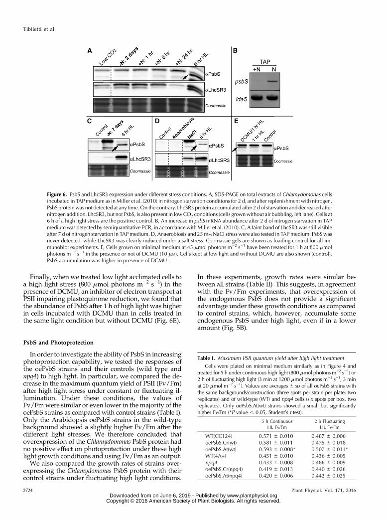
Finally, when we treated low light acclimated cells toa high light stress (800 mmol photons m22 s21) in thepresence of DCMU, an inhibitor of electron transport atPSII impairing plastoquinone reduction, we found thatthe abundance of PsbS after 1 h of high light was higherin cells incubated with DCMU than in cells treated inthe same light condition but without DCMU (Fig. 6E).
PsbS and Photoprotection
In order to investigate the ability of PsbS in increasingphotoprotection capability, we tested the responses ofthe oePsbS strains and their controls (wild type andnpq4) to high light. In particular, we compared the de-crease in the maximum quantum yield of PSII (Fv/Fm)after high light stress under constant or fluctuating il-lumination. Under these conditions, the values ofFv/Fmwere similar or even lower in the majority of theoePsbS strains as compared with control strains (Table I).Only the Arabidopsis oePsbS strains in the wild-typebackground showed a slightly higher Fv/Fm after thedifferent light stresses. We therefore concluded thatoverexpression of the Chlamydomonas PsbS protein hadno positive effect on photoprotection under these highlight growth conditions and using Fv/Fm as an output.
We also compared the growth rates of strains over-expressing the Chlamydomonas PsbS protein with theircontrol strains under fluctuating high light conditions.
In these experiments, growth rates were similar be-tween all strains (Table II). This suggests, in agreementwith the Fv/Fm experiments, that overexpression ofthe endogenous PsbS does not provide a significantadvantage under these growth conditions as comparedto control strains, which, however, accumulate someendogenous PsbS under high light, even if in a loweramount (Fig. 5B).
Figure 6. PsbS and LhcSR3 expression under different stress conditions. A, SDS-PAGE on total extracts of Chlamydomonas cellsincubated in TAPmedium as inMiller et al. (2010) in nitrogen starvation conditions for 2 d, and after replenishment with nitrogen.PsbS proteinwas not detected at any time.On the contrary, LhcSR3 protein accumulated after 2 d of starvation and decreased afternitrogen addition. LhcSR3, but not PsbS, is also present in lowCO2 conditions (cells grownwithout air bubbling, left lane). Cells at6 h of a high light stress are the positive control. B, An increase in psbSmRNA abundance after 2 d of nitrogen starvation in TAPmediumwas detected by semiquantitative PCR, in accordancewithMiller et al. (2010). C, A faint band of LhcSR3was still visibleafter 7 d of nitrogen starvation in TAPmedium. D, Anaerobiosis and 25mMNaCl stress were also tested in TAPmedium: PsbS wasnever detected, while LhcSR3 was clearly induced under a salt stress. Coomassie gels are shown as loading control for all im-munoblot experiments. E, Cells grown on minimal medium at 45 mmol photons m22 s21 have been treated for 1 h at 800 mmolphotons m22 s21 in the presence or not of DCMU (10 mM). Cells kept at low light and without DCMU are also shown (control).PsbS accumulation was higher in presence of DCMU.
Table I. Maximum PSII quantum yield after high light treatment
Cells were plated on minimal medium similarly as in Figure 4 andtreated for 5 h under continuous high light (800 mmol photons m22 s21) or2 h of fluctuating high light (3 min at 1200 mmol photons m22 s21, 3 minat 20 mmol m22 s21). Values are averages 6 SD of all oePsbS strains withthe same backgrounds/construction (three spots per strain per plate; tworeplicates) and of wild-type (WT) and npq4 cells (six spots per box, tworeplicates). Only oePsbS.At(wt) strains showed a small but significantlyhigher Fv/Fm (*P value , 0.05, Student’s t test).
5 h Continuous
HL Fv/Fm
2 h Fluctuating
HL Fv/Fm
WT(CC124) 0.571 6 0.010 0.487 6 0.006oePsbS.Cr(wt) 0.581 6 0.011 0.475 6 0.018oePsbS.At(wt) 0.593 6 0.008* 0.507 6 0.011*WT(4A+) 0.451 6 0.010 0.436 6 0.005npq4 0.433 6 0.008 0.486 6 0.009oePsbS.Cr(npq4) 0.419 6 0.013 0.440 6 0.026oePsbS.At(npq4) 0.420 6 0.006 0.442 6 0.025
2724 Plant Physiol. Vol. 171, 2016
Tibiletti et al.
www.plantphysiol.orgon June 6, 2019 - Published by Downloaded from Copyright © 2016 American Society of Plant Biologists. All rights reserved.

DISCUSSION
PsbS Accumulates Transiently and Is Functionalin Chlamydomonas
So far, the role of PsbS in Chlamydomonas has beenelusive. On one hand, PsbS was not detected underseveral growth conditions (Bonente et al., 2008b), andthe npq4 mutant (koLhcSR3) suggests that almost allalgal NPQ capability depends on LhcSR3 (Peers et al.,2009). However, on the other hand, the Chlamydomonasgenome contains two psbS genes that are conservedwith one another and with the plant psbS (Merchantet al., 2007), and some investigations have shown thatpsbS RNA accumulates in particular conditions (Milleret al., 2010; Zones et al., 2015).Thus, we (re)investigated PsbS gene expression and
protein accumulation in Chlamydomonas. Here, we showthat the PsbS protein accumulates rapidly and transientlyduring high light stress, peaking around 4/8 h of thetreatment (Fig. 1; Supplemental Fig. S1); PsbS accumu-lation and the expression of both psbS genes follow asimilar but slightly delayed trend duringhigh light stress;psbS gene expression kinetics are independent of thepresence of LhcSR3 and the high light intensity (at least inthe conditions we tested, which all saturate photosyn-thesis), but PsbS protein amounts seem dependent on thehigh light intensity (Supplemental Fig. S1).LhcSR3 accumulation was delayed as compared to
PsbS and the amount remained elevated during the en-tire 32 h light stress. Moreover, the ratio of the phos-phorylated LhcSR3 over the nonphosphorylated LhcSR3(upper and lower band, respectively, in Fig. 1 andSupplemental Fig. S1; Bonente et al., 2011) increasedduring the light stress. Phosphorylation of LhcSR3 couldbe involved in the localization of the protein (Allorentet al., 2013; Bergner et al., 2015). Minor and major LHCIIantenna can be phosphorylated in Chlamydomonas tomove from PSII to PSI during state transition (Iwai et al.,2008; Rochaix, 2014), and this could also be the case forLhcSR3. Although clear evidence showing interactionsbetween LhcSR3 and PSI are still lacking in Chlamydo-monas, in the moss Physcomitrella patens, LhcSR proteinshave been shown to interact and protect both photosys-tems (Pinnola et al., 2015).The results showing a delay betweenmaximum gene
expression and maximum protein accumulation can be
explained by a relatively slow turnover of the proteins,which thus accumulate during the first hours when theRNA is still abundant. However, in the case of LhcSR3,the turnover appears to be particularly slow, since theaccumulation of the protein (Fig. 1) continues for sev-eral hours after the maximum gene expression (Fig. 2).Alternatively, the relatively low abundant lhcSR3transcripts in the late stage of the high light periodcould be very efficiently transcribed.
PsbS in Photoprotection
Our results suggest that PsbS expression is veryrapidly activated in response to light stress (30 min areenough to detect PsbS accumulation), but that its roleseems to be only transient, in contrast to the situation inplants and mosses. Instead, LhcSR3 might have a muchmore important role in acclimation to high light, andpossibly some change at the level of photosystemswould require phosphorylation of LhcSR3 in a latestage of the light stress. Therefore, in addition to theobserved temporal separation between PsbS andLhcSR3 expression, it is reasonable to think that the twoproteins are differentially localized within the thyla-koid membranes as recently observed in Physcomitrellapatens (Pinnola et al., 2015). Physcomitrella is the firstorganismwhere both PsbS and LhcSR have been foundand for which KO mutants have been created (Alboresiet al., 2010). Interestingly, while in Chlamydomonas,PsbS and LhcSR are stress-inducible proteins that ac-cumulate transiently during stress conditions; in plants,PsbS is usually detected under non-stress conditions;and in Physcomitrella, both PsbS and LhcSR proteins canbe detected in low light grown plants (Gerotto et al.,2011). This phenomenon occurs as well in the multi-cellular green alga Ulva linza (Zhang et al., 2013). Thismight support the idea of Allorent et al. (2013) pro-posing that an inducible and less efficient NPQ systemin Chlamydomonas is compensated by a large statetransition capacity.
In order to investigate the ability of the Chlamydo-monas PsbS to induce NPQ and increase photo-protection, we created PsbS overexpressor strains bychloroplast transformation. We also generated strainsaccumulating the Arabidopsis PsbS protein to deter-mine whether the plant protein is functional in
Table II. Growth rates of oePsbS.Cr strains versus their control strains in minimal medium
Growth rates at low light (45 mmol photons m22 s21) and fluctuating high light (3 min at 1200 mmolphotons m22 s21, 3 min at 45 mmol photons m22 s21) were not significantly different between wild-type(WT) and oePsbS strains. Values are averages 6 SD of three biological replicates (wild-type background) ortwo biological replicates (npq4 background) using two different oePsbS.Cr strains and three flasks perstrain per replicate.
Low Light Doubling Time (Days) Fluct. High Light Doubling Time (Days)
WT(CC124) 0.450 6 0.018 1.369 6 0.053oePsbS.Cr(wt) 0.436 6 0.026 1.403 6 0.054npq4 0.444 6 0.000 1.382 6 0.029oePsbS.Cr(npq4) 0.420 6 0.046 1.447 6 0.206
Plant Physiol. Vol. 171, 2016 2725
Characterization of PsbS in Chlamydomonas
www.plantphysiol.orgon June 6, 2019 - Published by Downloaded from Copyright © 2016 American Society of Plant Biologists. All rights reserved.

Chlamydomonas cells. OePsbS lines, which constitu-tively accumulate PsbS, were compared with wild-typeand npq4 cells grown in conditions where PsbS is un-detectable (low light) and LhcSR3 has no or negligibleaccumulation (low light and high CO2/air bubbledcultures) (Figs. 1 and 6; Supplemental Fig. S4B). Asvisible in Figure 4 and Supplemental Figure S3, alloePsbS lines showed a significantly higher NPQ ca-pacity as comparedwith their respective controls. In thecase of oePsbS lines in the wild-type (CC124) back-ground, the NPQ was rapidly reversible in the dark, asit is the case in plants, due to the rapid neutralizationof the low luminal pH that is necessary for activat-ing PsbS. NPQ measurements on liquid cultures(Supplemental Fig. S3B) showed reversibility also forthe oePsbS strains in the npq4 (4A+) background, butnot for wild-type (4A+) and npq4 controls. The fact thatwild-type strains and npq4 cells did not show a signif-icant NPQ reversibility at the end of the actinic light isin agreement with fact that LhcSR3 has no or negligibleaccumulation in the growth conditions used (Figs.1 and 6; Supplemental Fig. S4B). However, we noticedthat LhcSR1was accumulated in our growth conditions(Supplemental Fig. S4B), but since the amount is similarin oePsbS and control strains, and considering the lessimportant role of LhcSR1 in NPQ (Peers et al., 2009),NPQ differences between oePsbS(wt) strains and theircontrols almost certainly have to be ascribed to the ac-tivity of PsbS.
In both genotypes (CC124 and 4A+), PsbS-dependentNPQ seems superimposed on another phenomenon thatis detected as NPQ and seems stronger in the 4A+ geno-type. This could be state transitions, as investigated inhigh light treated cells ofChlamydomonas (Allorent et al.,2013). Considering the different NPQ responses ofoePsbS strains in the two genetic backgrounds, it islikely that significant photosynthetic differences existbetween the two genotypes, but investigation of this isbeyond the scope of our present research.
The fact that the heterologous expression of theArabidopsis PsbS had a similar effect as the Chlamydo-monas PsbS (Fig. 3) suggests that the mechanism ofPsbS, which is still debated, is at least partially con-served between plants and the green algae despitesome differences in photosystem II organization be-tween the organisms. In particular, LHCII proteins inChlamydomonas have biochemical/spectroscopic prop-erties similar to those of plants (Natali and Croce, 2015),but they cannot be classified in the Lhcb1-3 isoforms asin plants (Jansson, 1999; Elrad and Grossman, 2004). InChlamydomonas, the CP24 subunit, which is next to theLHCII-M trimer (Dekker and Boekema, 2005) and thathas been suggested to detach from PSII during NPQinduction in a PsbS-dependent phenomenon (Betterleet al., 2009), is lacking and at its place a third LHCIItrimer is bound to PSII (Tokutsu et al., 2012; Drop et al.,2014). Even if it is possible that PsbS in Chlamydomonasinteracts somehow differently with PSII than in plants,we found that PsbS accumulation has a positive corre-lation with NPQ capacity as in plants (Li et al., 2002a,
2002b), further supporting a conserved PsbS-dependentNPQ mechanism.
Investigation of the absolute and relative PsbS andLhcSR3 stoichiometry in vivo indicated that the level ofPsbS is sub-stoichiometric as compared to PSII (wefound about 0.1 PsbS per monomeric PSII). Even at itsmaximum expression (;6 h of high light, Fig. 1) PsbSaccumulates to much lower levels than LhcSR3, beingabout eight times less (it should be noted that our es-timation of LhcSR3 amount is higher than the value of;0.2 previously proposed (Bonente et al., 2011, possi-bly due to the different approach or growth conditions).Since the mode of action of PsbS is still not well un-derstood, and since PSII particles are densely packedwithin the grana, it might be possible that one PsbS perten PSII is enough to efficiently protect the photosyn-thetic apparatus. However, PsbS in Arabidopsis accu-mulates at higher and likely stoichiometric amountswith PSII (see, for instance, 2D gels in Li et al. [2004] andCaffarri et al. [2009]) where PsbS is present in amountscomparable to those of monomeric Lhc proteins). Dueto the fact that PsbS in algae and plants likely works in asimilar manner and that there is a correlation betweenthe amount of PsbS and NPQ level, we suggest that thelow amount of PsbS in vivo might contribute to only asmall part of the NPQ capacity of Chlamydomonas.
Our tests to determine whether PsbS overexpressionaffects photoprotection did not reveal a significantdifference in oePsbS strains versus control strains. Itshould be noted, however, that the control strains arealso able to accumulate the endogenous PsbS (Fig. 1)and LhcSR1 (and LhcSR3 in the wild-type background),which could attenuate the differences between controland oePsbS strains. It should be also noted that theChlamydomonas npq4 mutant lacking LhcSR3 and un-able to significantly increase NPQ capability whengrown at high light (Peers et al., 2009) also did not showsignificant differences for either Fv/Fm or growth inhigh light as comparedwith wild-type cells (Peers et al.,2009). Similarly, Arabidopsis mutants lacking PsbS alsoshow relatively small differences in comparison withwild-type plants under high light conditions, due to theenhancement of other photoprotective mechanisms(Golan et al., 2006). It is likely that photoprotectionstrategies are partially redundant and, thus, that otherphotosynthetic controls such as state transitions(Allorent et al., 2013) and cyclic electron transport(Chaux et al., 2015; Saroussi et al., 2016), which seemparticularly important in Chlamydomonas photo-protection, could mask the photoprotection ability ofPsbS in laboratory conditions. Thus, PsbS may haveroles in increasing fitness in the simultaneous presenceof high light and other natural stress conditions.
PsbS Accumulates in Response to Light; LhcSR3Accumulates under Several Stress Conditions
We determined PsbS accumulation under differentstresses to obtain more information about the possiblerole of PsbS under unfavorable growth conditions. As
2726 Plant Physiol. Vol. 171, 2016
Tibiletti et al.
www.plantphysiol.orgon June 6, 2019 - Published by Downloaded from Copyright © 2016 American Society of Plant Biologists. All rights reserved.
previously found (Miller et al., 2010), we detected psbSgene transcription under nitrogen starvation (Fig. 6).However, we did not detect protein accumulation, aresult that suggests the existence of some control onmRNA translation. Besides the absence of the PsbSprotein under nitrogen deprivation conditions, we didnot detect PsbS after acclimation to any of the otherstresses tested, including low CO2, high salt conditions,and anaerobiosis. On the contrary, LhcSR3 was detec-ted after acclimation to low CO2, in agreement withprevious investigations (Dang et al., 2014; Maruyamaet al., 2014) and also in response to high salt andnitrogen starvation (and its quantity decreased after theaddition of nitrogen in the medium; Fig. 6). In contrastto anaerobiosis, these three stresses lead to productionof reactive oxygen species that could be involved in theactivation of the lhcSR genes. In summary, after accli-mation to various stresses, only LhcSR was found incells, but not PsbS, indicating that psbS gene expressionis under the control of a signal appearing very rapidlyin the cells after a transition to high light. Notably, highlight specifically provokes a rapid and significant pro-duction of singlet oxygen (1O2*; Flors et al., 2006) due toenergy transfer from triplet excited chlorophylls tooxygen, especially at the level of PSII. We cannot ex-clude a role of the redox state of the plastoquinone (PQ)on PsbS accumulation. However, this possibility is lesslikely since the PQ redox state is mainly involved inoptimizing photosynthesis under not saturating lightconditions, such as during state transitions, or duringlong-term acclimation (Fey et al., 2005; Adamiec et al.,2008). Moreover, our result (Fig. 6E) showing anincrease of PsbS accumulation during high light treat-ment in presence of DCMU, which blocks plastoqui-none reduction, further supports a minor role of the PQredox state in psbS gene expression in the first phase of alight stress. In later stages of stress or in the deficiency ofthe final electron acceptor CO2, other reactive oxygenspecies, such as superoxide (O2
2) and hydrogen per-oxide (H2O2) formed at PSI by the Mehler reaction, orthe redox state of the chloroplast, may be more im-portant in triggering a change in the genetic expressionnecessary for acclimation to the stress. We thereforeargue that the rapid accumulation of PsbS is likelytriggered by a 1O2* derived signal and is important for afast and transient need of energy dissipation at the levelof PSII. This action would be performed in parallel withLhcSR or even earlier, due to the more rapid appear-ance of PsbS. In later stages of the stress, LhcSR, but notPsbS, would have a major role in photoprotection ofboth photosystems.
CONCLUSION
We investigated the expression of two key proteins inNPQ induction, PsbS and LhcSR. We showed thatChlamydomonas psbS gene transcripts and the PsbSprotein accumulate very rapidly and transiently duringhigh light stress. We demonstrate that the endogenousPsbS protein is able to activate NPQ in Chlamydomonas,
as well as the Arabidopsis PsbS, suggesting a conservedmechanism of action. PsbS accumulation in Chlamydo-monas seems strongly dependent on high light stress,while LhcSR3 accumulation is detected under severaldifferent stress conditions that can lead to general oxi-dative stress even under moderate light. We alsoshowed that PsbS in vivo stoichiometry is significantlylower than that of LhcSR3. We cannot exclude thatother stress conditions (not tested) are able to inducePsbS, but our results, together with those indicatingthat LhcSR3 accumulation can explain almost all fastinduced NPQ in high light acclimated Chlamydomonascells (Peers et al., 2009), suggest that in ChlamydomonasPsbS plays a less important role in the protection of thephotosynthetic apparatus than in plants. However, theconservation of functional PsbS proteins in the greenalga and their very rapid accumulation (faster thanLhcSR3) indicate that PsbS must be important for thefitness of Chlamydomonas in natural conditions during atransient response to high light.
MATERIALS AND METHODS
Strains and Culture Conditions
The Chlamydomonas reinhardtii wild-type strain CC124 (mt+ nit1 nit2), the4A+ wild type and the mutant npq4 (Peers et al., 2009) were grown at 22°Cunder continuous illumination in a PercivalAR-41L3 incubator (CLFPlantClimatics,Germany). Batch cultures were grown on a rotary shaker in Erlenmeyer flasks(75 mL) under 45 mmol photons m22 s21. TAP or minimal medium (E. Harris,The Chlamydomonas Sourcebook) were used. For high light treatments, cellswere cultured in 1 L flasks on a rotary shaker and supplied with continuousfiltered air bubbling generated by an aquarium pump. For light stress, illumi-nation was supplied by two white LED panels (Photon System Instruments,Czech Republic) placed radially along the flasks to reach an intensity up to 1,200mmol photons m22 s21. Sampling for immunoblot and qRT-PCR were per-formed after 30 min, 1 h, 2 h, 4 h, 8 h, 14 h, and 32 h. A volume of 12 mL of cellswas harvested by centrifugation (3,500 g, 10 min, 4°C), supernatant was re-moved, and the pellet was immediately frozen in liquid nitrogen. Cell pelletwas stored at 280°C until used.
Nitrogen depletion experimentswere carried out as described byMiller et al.(2010) at 100 mmol m22 s21. Anaerobic conditions were created in the dark asdescribed by Bonente et al. (2008b). Sodium chloride stress was created growingcells in TAP with the addition of 25 mM NaCl for 3 d under 45 mmol photonsm22s21. Prolonged nitrogen stress was created maintaining cells without anitrogen source for 7 d under 45mmol photonsm22 s21. A nitrogen replenishingexperiment was carried out as described by Miller et al. (2010). After 2 d ofnitrogen depletion, NH4Cl was added to the medium and sampling was per-formed after 1 h, 6 h, and 24 h.
DCMU experiment was performed on cells grown on minimal medium at45 mmol photons m22 s21. The cell culture was split in two flasks before highlight treatment and DCMU (10 mM) was added to one flask, and then cells wereexposed to 800 mmol photons m22 s21 for 1 h
Fluctuating light conditions for Fv/Fm analysis (3 min at 1,200 mmol m22 s21 +3 min at 20 mmol m22 s21) were created in the Fluorcam FC 800-O using the blueled actinic panel (Photon System Instruments). Fluctuating light conditions forgrowth rate experiments (3 min at 1,200 mmol m22 s21 + 3min at 45mmolm22 s21)were created with a white Fytoled system equipped with a LC100 controller(Photon System Instrument, Czech Republic). Growth rates were calculated on thechlorophyll absorption in the red and corrected for the light scattering (two mea-surements per day, at least 2 d for each light conditions).
Overexpression of PsbS in C. reinhardtii
Synthetic psbS genes from C. reinhardtii and Arabidopsis were designed tooptimize the chloroplast codon usage of C. reinhardtii. We used the maturesequence for the Arabidopsis PsbS and the full sequence in the case of
Plant Physiol. Vol. 171, 2016 2727
Characterization of PsbS in Chlamydomonas
www.plantphysiol.orgon June 6, 2019 - Published by Downloaded from Copyright © 2016 American Society of Plant Biologists. All rights reserved.

Chlamydomonas PsbS (the mature Chlamydomonas PsbS is not known; however,the PsbS precursor is correctly processed inside the chloroplast, Fig. 5B). Notethat the two psbS genes fromC. reinhardtii encode for identical proteins (Bonenteet al., 2008b), except for one conservative Ser ⁄Thr substitution at position 28,which is very likely in the signal peptide sequence. The synthetic genes wereintegrated in the pLM21 vector using the BglII restriction site using theIn-Fusion kit (Clontech). The pLM21 vector was originated from the IR-intvector from Michelet et al. (2011), designed for gene insertion into the chloro-plast genome by homologous recombination. The IR-int vector was digested byClaI and PmlI to excise VapA and its promoter. A PCR was performed on atpB-int vector to amplify the psaA promoter and 59UTR using Advantage HD(Clontech) using the following primers: 59-ATGGAGCTGTACCACGTT-TAAACAGATCTCCATGGATTTCTCCTTATAA-39 and 59-CGGATCC-GATATCGATAAGCTTTCTTAATTCAAC-39. These primers added BglII andPmeI as multicloning sites. Then, the PCR product was cloned by In-fusion kit(Clontech) between ClaI and PmlI within the IR-int vector.
In the pLM21-CpPSBSCr and in the pLM21-CpPSBSAt, both psbs genes areunder the control of the psaA 59untranslated region (UTR) promoter region andthe rbcL 39UTR region, while the aadA gene (spectinomycin resistance) is undercontrol of atpA 59UTR promoter region and the rbcL 39UTR region. For plastidtransformation experiments, cells were grown in TAP medium at 45 mmolphotons m22 s21 until reaching 2.5 3 106 cells mL21 and then plated ontominimal medium agar plate containing 50 mg mL21 spectinomycin. Biolistictransformation and DNA extraction for genotyping were performed as de-scribed in Baltz et al. (2014). The chloroplast DNA loading control has beendone on 16S using fw primer 59-TCCATGGAGAGTTTGATCCTG-39 and revprimer 59-TCCTCTCAGACCAGCTACTGC-39, Tm: 50°C. Homoplasmy wasverified by PCR (forward primer 59-CGTTATTAGCCTTTCGTCGCT-39 andreverse primer 59-CCGAAACGGTGGTTATTCCAGGCC-39) by the lack ofamplification of the insertion region in the chloroplast genome, since it is bro-ken. The presence of PSBSCr or PSBSAt was verified using specific primers(PSBSAt fw primer 59-GGTACATCAGGTGGTATTGG-39, PSBSAt rev primer59-CCTAATTGAGCTAAACGACC-39, PSBSCr fw primer 59-TCAAAAACA-GAAGTTGAACG-39, PSBSCr rev primer 59-CGAATTGTGCTAATGCACCT-39). PCR was performed using the Taq‘Ozime Purple Mix.
Thylakoid Membranes, Total Membranes, and TotalProtein Purification
Thylakoid membranes were isolated as described by Bonente et al. (2008b).Total membranes were prepared breaking 10–20 mL of harvested cells in thebuffer containing 25mMHEPES (pH 7.5), 5 mMMgCl2, 0.3 M Suc, and a proteaseinhibitor cocktail (EasyPack EDTA free, Roche) using glass beads. Glass beadsand unbroken cells were removed by centrifugation (600 g, 2min), and from thesupernatant fraction, membraneswere pelleted by centrifugation at 12,000 g for5 min. Membranes were suspended and stored in 5 mM HEPES-KOH (pH 7.5),10 mM EDTA and 50% glycerol and stored at280°C. For total protein extraction, avolume of 10–20mL of exponential grown culturewas harvested by centrifugation(3,500 g, 10 min) and suspended in SDS-PAGE sample buffer (62.5 mM Tris [pH6.8], 10% glycerol, 5% SDS, 5% b-mercaptoethanol) containing a protease inhibitorcocktail (EasyPack EDTA free, Roche). An amount of 200 mg of glass beads (425–600 mm) were then added, and cells were broken using vigorous vortexing. Glassbeads and cell debris were removed by centrifugation (21,000 g, 10 min), and thechlorophyll amount was quantified using a fitting procedure of the absorptionspectrum after acetone extraction (Croce et al., 2002).
Immunoblot
About 1.5–2 mg of chlorophyll were loaded on 12% (w/v) SDS-PAGE with6 M urea and transferred onto a nitrocellulose membrane. Immublotting anal-yses were performed using an antibody raised in rabbit against the syntheticpeptide (C)-TGKGALAQFDIETG, corresponding to the first part of the secondluminal loop of the Chlamydomonas PsbS. This sequence has been chosen for thegood predicted antigenicity and for the strong conservation with the first ho-molog loop of Chlamydomonas PsbS and with the corresponding loops of Ara-bidopsis PsbS, in order to have a wide cross-reactivity (see also SupplementalFig. S2). aLhcSR3, aLhcSR1, aPsbO, aPsaC, and aPetA antibodies were pur-chased from Agrisera. aPGRL1 and aNDA2 are described in Tolleter et al.(2011) and Desplats et al. (2009), respectively. aPsbC was raised in rabbitagainst a recombinant peptide corresponding to the loop E. An antirabbit IgGperoxidase-conjugated antibody (CellSignaling) and the Fusion Fx7 (VilberLourmat) was used for detection.
Peptide Competition Assay
For the competition assay, 7.5 mg of PsbS antibody was incubated with therecombinant His-tagged PsbS of C. reinhardtii (25 mg of protein) (Bonenteet al., 2008b) in 15 mL of PBS buffer containing 2% (w/v) milk powder and0.25% (v/v) TritonX-100 for 1 h at room temperature on a rotary shaker. Thissolution was used to hybridize a membrane as described in the Immunoblotsection.
Quantification of PsbS and LhcSR3
Similar visual amount of recombinant PsbS (Bonente et al., 2008b), recom-binant LhcSR3 (Bonente et al., 2011) and native LHCII were loaded on aSDS-PAGE together with 2 mg (in Chls) of a total extract of high light treatedChlamydomonas wild-type cells and oePsbS.Cr cells. SDS-PAGE were eitherstained with Sypro or transferred to nitrocellulose membranes for immunoblotanalysis with the antiPsbS and antiLhcSR3 antibodies. Amount of recombinantPsbS and LhcSR3 were calculated by densitometry of the Sypro stained SDS-PAGE and comparison with the signal of LHCII, for which 0.03 mg of apo-protein were loaded (calculated on the Chls absorption, where 1 ODQy/cm isequal to 40 mg/ml of apoprotein). The values obtained by densitometry werealso corrected for the number of basic residues in each protein since Sypro ismainly bound by His, Arg and Lys residues. Apoprotein amounts in vivo wereobtained by densitometry and comparison of the ECL signals between lanescontaining the recombinant proteins and lanes containing total cell extracts. Forabsolute quantification, the same assumption as in (Bonente et al., 2011) aboutPSII/PSI ratio (1.18) and photosystem antenna size (PSI = 240 Chls, PSII =222 Chls) were considered.
RNA Extraction, Synthesis of cDNA, and Semi-quantitative and Quantitative RT-PCR Analysis
A volume of 12 mL of pelleted cells was used for RNA extraction using theTRIreagent and subjected to DNase treatment with the RNAase-free DNase I(Thermo scientific). A total of 250 ng of DNA-free RNAwas used for the cDNAsynthesis using PrimeScript RT reagent kit (Perfect Real Time) from Takara inaccordance to the manufacturer’s instructions. Primers for quantitative real-time (qRT)-PCR are listed in Supplemental Table S1. Each reaction containedthe master mix, 200 ng of each primer, and cDNA corresponding to 12.5 nginput RNA in the reverse transcriptase reaction. Real-time (RT)-PCR was per-formed on the light Cycler Instrument (CFX96 Real-Time PCR Detection Sys-tem, Bio Rad) using SYBR Green I as a fluorescent dye. The reaction conditionswere as follow: 95°C for 3 min, followed by cycles of 95°C for 10 s, 60°C for 10 s,and 72°C for 30 s up to a total of 44 cycles. Controls without template or reversetranscriptase were included. A gene encoding the cytosolic 40S small ribosomalsubunit RACK1 also known as Chlamydomonas G protein b-subunit-like poly-peptide (CBLP) and the gene IDA5 were used as a control. Data analysis wasperformed using Bio-Rad CFX Manager software. Gene expression ratios werenormalized to controls using the 2-DCt. For semiquantitative RT-PCR, cDNAwas synthetize using the Superscript III Reverse transcriptase (Invitrogen)following manufacture’s instruction. PCR cycle conditions were 3 min of initialdenaturation at 94°C, following 34 cycles of 30 s of denaturation, 30 s ofannealing at 60°C and 3 min of elongation at 72°C. Final elongation wasperformed for 10 min at 72°C. Primers were the same used in (Miller et al.,2010).
Fluorescence Measurements
Fluorescence measurements on cells grown on solid minimal medium havebeen performed using an imaging fluorometer (Fluorcam FC 800-O; PhotonSystem Instruments). An amount of cells corresponding to about 1.5 mg ofchlorophyll were spotted onto minimal medium agar plate and let grow for5-7 d at 45 mmol photons m22 s21 before measurements. Cells were dark-acclimated for at least 30 min and then subjected to NPQ measurement with800 mmol photons m22 s21 of blue light. Fluorescence measurements on cellsgrown on liquid minimal medium have been performed with a DUAL-PAM-100 (DUAL-PAM/F, Walz) on dark adapted cultures concentrated ; 0.5 mgChls/ml. Actinic light was provided by a red LED set at 1200 mmol photonsm22 s21. Sedimentation was avoided by using a miniature magnetic stirrer.NPQwas calculated as (Fm - Fm’)/Fm’where Fm is the maximum fluorescenceof dark-adapted tissues and Fm’ is the maximum fluorescence of light adaptedtissues.Maximumquantumyield of PSII (Fv/Fm)was calculated as (Fm-Fo)/Fm.
2728 Plant Physiol. Vol. 171, 2016
Tibiletti et al.
www.plantphysiol.orgon June 6, 2019 - Published by Downloaded from Copyright © 2016 American Society of Plant Biologists. All rights reserved.
Supplemental Data
The following supplemental materials are available.
Supplemental Figure S1. Kinetics of PsbS accumulation in ChlamydomonasWT(4A+) and npq4 cells during a high light treatment at three differentlight intensities.
Supplemental Figure S2. Test of the antibody against PsbS.
Supplemental Figure S3. NPQ of the individual oePsbS strains and onliquid cultures.
Supplemental Figure S4. Biochemical characterization of the oePsbS andcontrol strains.
Supplemental Materials and Methods S1. Mass spectrometry analysis.
Supplemental Table S1. Primers used for the quantitative real-time PCRexperiments.
Supplemental Table S2. Mass spectrometry results on Chlamydomonas thy-lakoids from low light and high light treated cells.
ACKNOWLEDGMENTS
Christophe Laloi, Ben Field, and Patrice Crete at the LGBP are thanked fortechnical help and fruitful discussion on qRT-PCR. Ben Field is further thankedfor manuscript revision. Sabrina Lignon, Rémy Puppo, Pascal Mansuelle, andRégine Lebrun, from the Proteomic Platform of the Mediterranean Institute ofMicrobiology, Marseille Proteomique IBiSA labeled, are thanked for mass spec-trometry analyses. Roberto Bassi is thanked for the LhcSR3 recombinant pro-tein. Riot Claire at the LB3M is thanked for technical assistance in chloroplasttransformation.
Received April 7, 2016; accepted June 18, 2016; published June 21, 2016.
LITERATURE CITED
Adamiec M, Drath M, Jackowski G (2008) Redox state of plastoquinonepool regulates expression of Arabidopsis thaliana genes in response toelevated irradiance. Acta Biochim Pol 55: 161–173
Alboresi A, Caffarri S, Nogue F, Bassi R, Morosinotto T (2008) In silicoand biochemical analysis of Physcomitrella patens photosynthetic an-tenna: identification of subunits which evolved upon land adaptation.PLoS One 3: e2033
Alboresi A, Gerotto C, Giacometti GM, Bassi R, Morosinotto T (2010)Physcomitrella patens mutants affected on heat dissipation clarify theevolution of photoprotection mechanisms upon land colonization. ProcNatl Acad Sci USA 107: 11128–11133
Allorent G, Tokutsu R, Roach T, Peers G, Cardol P, Girard-Bascou J,Seigneurin-Berny D, Petroutsos D, Kuntz M, Breyton C, Franck F,Wollman FA, et al (2013) A dual strategy to cope with high light inChlamydomonas reinhardtii. Plant Cell 25: 545–557
Andersson J, Walters RG, Horton P, Jansson S (2001) Antisense inhibitionof the photosynthetic antenna proteins CP29 and CP26: implications forthe mechanism of protective energy dissipation. Plant Cell 13: 1193–1204
Andersson J, Wentworth M, Walters RG, Howard CA, Ruban AV, HortonP, Jansson S (2003) Absence of the Lhcb1 and Lhcb2 proteins of thelight-harvesting complex of photosystem II - effects on photosynthesis,grana stacking and fitness. Plant J 35: 350–361
Arnoux P, Morosinotto T, Saga G, Bassi R, Pignol D (2009) A structuralbasis for the pH-dependent xanthophyll cycle in Arabidopsis thaliana.Plant Cell 21: 2036–2044
Ballottari M, Girardon J, Dall’osto L, Bassi R (2012) Evolution and func-tional properties of photosystem II light harvesting complexes in eu-karyotes. Biochim Biophys Acta 1817: 143–157
Baltz A, Dang KV, Beyly A, Auroy P, Richaud P, Cournac L, Peltier G(2014) Plastidial expression of type II NAD(P)H dehydrogenase in-creases the reducing state of plastoquinones and hydrogen photopro-duction rate by the indirect pathway in Chlamydomonas reinhardtii. PlantPhysiol 165: 1344–1352
Bassi R, Caffarri S (2000) Lhc proteins and the regulation of photosyntheticlight harvesting function by xanthophylls. Photosynth Res 64: 243–256
Bergner SV, Scholz M, Trompelt K, Barth J, Gäbelein P, Steinbeck J, XueH, Clowez S, Fucile G, Goldschmidt-Clermont M, Fufezan C, HipplerM (2015) STATE TRANSITION7-dependent phosphorylation is modu-lated by changing environmental conditions, and its absence triggersremodeling of photosynthetic protein complexes. Plant Physiol 168: 615–634
Betterle N, Ballottari M, Zorzan S, de Bianchi S, Cazzaniga S, Dall’osto L,Morosinotto T, Bassi R (2009) Light-induced dissociation of an antennahetero-oligomer is needed for non-photochemical quenching induction.J Biol Chem 284: 15255–15266
Bode S, Quentmeier CC, Liao PN, Hafi N, Barros T, Wilk L, Bittner F,Walla PJ (2009) On the regulation of photosynthesis by excitonic inter-actions between carotenoids and chlorophylls. Proc Natl Acad Sci USA106: 12311–12316
Bonente G, Formighieri C, Mantelli M, Catalanotti C, Giuliano G,Morosinotto T, Bassi R (2011) Mutagenesis and phenotypic selection asa strategy toward domestication of Chlamydomonas reinhardtii strains forimproved performance in photobioreactors. Photosynth Res 108: 107–120
Bonente G, Howes BD, Caffarri S, Smulevich G, Bassi R (2008a) Inter-actions between the photosystem II subunit PsbS and xanthophyllsstudied in vivo and in vitro. J Biol Chem 283: 8434–8445
Bonente G, Passarini F, Cazzaniga S, Mancone C, Buia MC, Tripodi M,Bassi R, Caffarri S (2008b) The occurrence of the psbS gene product inChlamydomonas reinhardtii and in other photosynthetic organisms and itscorrelation with energy quenching. Photochem Photobiol 84: 1359–1370
Büchel C (2015) Evolution and function of light harvesting proteins. J PlantPhysiol 172: 62–75
Caffarri S, Kouril R, Kereïche S, Boekema EJ, Croce R (2009) Functionalarchitecture of higher plant photosystem II supercomplexes. EMBO J 28:3052–3063
Caffarri S, Tibiletti T, Jennings RC, Santabarbara S (2014) A comparisonbetween plant photosystem I and photosystem II architecture andfunctioning. Curr Protein Pept Sci 15: 296–331
Chaux F, Peltier G, Johnson X (2015) A security network in PSI photo-protection: regulation of photosynthetic control, NPQ and O2 photore-duction by cyclic electron flow. Front Plant Sci 6: 875
Croce R, Canino G, Ros F, Bassi R (2002) Chromophore organization in thehigher-plant photosystem II antenna protein CP26. Biochemistry 41:7334–7343
Dang KV, Plet J, Tolleter D, Jokel M, Cuiné S, Carrier P, Auroy P,Richaud P, Johnson X, Alric J, Allahverdiyeva Y, Peltier G (2014)Combined increases in mitochondrial cooperation and oxygen photore-duction compensate for deficiency in cyclic electron flow in Chlamydomonasreinhardtii. Plant Cell 26: 3036–3050
de Bianchi S, Betterle N, Kouril R, Cazzaniga S, Boekema E, Bassi R,Dall’Osto L (2011) Arabidopsis mutants deleted in the light-harvestingprotein Lhcb4 have a disrupted photosystem II macrostructure and aredefective in photoprotection. Plant Cell 23: 2659–2679
de Bianchi S, Dall’Osto L, Tognon G, Morosinotto T, Bassi R (2008) Minorantenna proteins CP24 and CP26 affect the interactions between pho-tosystem II subunits and the electron transport rate in grana membranesof Arabidopsis. Plant Cell 20: 1012–1028
Dekker JP, Boekema EJ (2005) Supramolecular organization of thylakoidmembrane proteins in green plants. Biochim Biophys Acta 1706: 12–39
Demmig-Adams B (1990) Carotenoids and photoprotection in plants: Arole for the xanthophyll zeaxanthin. Biochim Biophys Acta 1020: 1–24
Desplats C, Mus F, Cuiné S, Billon E, Cournac L, Peltier G (2009) Char-acterization of Nda2, a plastoquinone-reducing type II NAD(P)H dehydro-genase in chlamydomonas chloroplasts. J Biol Chem 284: 4148–4157
Drop B, Webber-Birungi M, Yadav SK, Filipowicz-Szymanska A, FusettiF, Boekema EJ, Croce R (2014) Light-harvesting complex II (LHCII) andits supramolecular organization in Chlamydomonas reinhardtii. BiochimBiophys Acta 1837: 63–72
Duffy CD, Ruban AV (2015) Dissipative pathways in the photosystem-IIantenna in plants. J Photochem Photobiol B 152(Pt B): 215–226
Elrad D, Grossman AR (2004) A genome’s-eye view of the light-harvestingpolypeptides of Chlamydomonas reinhardtii. Curr Genet 45: 61–75
Fan M, Li M, Liu Z, Cao P, Pan X, Zhang H, Zhao X, Zhang J, Chang W(2015) Crystal structures of the PsbS protein essential for photo-protection in plants. Nat Struct Mol Biol 22: 729–735
Fey V, Wagner R, Bräutigam K, Pfannschmidt T (2005) Photosyntheticredox control of nuclear gene expression. J Exp Bot 56: 1491–1498
Plant Physiol. Vol. 171, 2016 2729
Characterization of PsbS in Chlamydomonas
www.plantphysiol.orgon June 6, 2019 - Published by Downloaded from Copyright © 2016 American Society of Plant Biologists. All rights reserved.

Flors C, Fryer MJ, Waring J, Reeder B, Bechtold U, Mullineaux PM,Nonell S, Wilson MT, Baker NR (2006) Imaging the production ofsinglet oxygen in vivo using a new fluorescent sensor, Singlet OxygenSensor Green. J Exp Bot 57: 1725–1734
Gerotto C, Alboresi A, Giacometti GM, Bassi R, Morosinotto T (2011)Role of PSBS and LHCSR in Physcomitrella patens acclimation to highlight and low temperature. Plant Cell Environ 34: 922–932
Golan T, Müller-Moulé P, Niyogi KK (2006) Photoprotection mutants ofArabidopsis thaliana acclimate to high light by increasing photosynthesisand specific antioxidants. Plant Cell Environ 29: 879–887
Goss R, Lepetit B (2015) Biodiversity of NPQ. J Plant Physiol 172: 13–32Havaux M, Niyogi KK (1999) The violaxanthin cycle protects plants from
photooxidative damage by more than one mechanism. Proc Natl AcadSci USA 96: 8762–8767
Holt NE, Zigmantas D, Valkunas L, Li XP, Niyogi KK, Fleming GR (2005)Carotenoid cation formation and the regulation of photosynthetic lightharvesting. Science 307: 433–436
Holzwarth AR, Miloslavina Y, Nilkens M, Jahns P (2009) Identification oftwo quenching sites active in the regulation of photosynthetic light-harvesting studied by time-resolved fluorescence. Chem Phys Lett 483:262–267
Iwai M, Takahashi Y, Minagawa J (2008) Molecular remodeling of pho-tosystem II during state transitions in Chlamydomonas reinhardtii. PlantCell 20: 2177–2189
Jansson S (1999) A guide to the Lhc genes and their relatives in Arabi-dopsis. Trends Plant Sci 4: 236–240
Johnson MP, Goral TK, Duffy CD, Brain AP, Mullineaux CW, Ruban AV(2011) Photoprotective energy dissipation involves the reorganization ofphotosystem II light-harvesting complexes in the grana membranes ofspinach chloroplasts. Plant Cell 23: 1468–1479
Kim S, Sandusky P, Bowlby NR, Aebersold R, Green BR, Vlahakis S,Yocum CF, Pichersky E (1992) Characterization of a spinach psbS cDNAencoding the 22 kDa protein of photosystem II. FEBS Lett 314: 67–71
Krüger TP, Ilioaia C, Johnson MP, Ruban AV, van Grondelle R (2014)Disentangling the low-energy states of the major light-harvesting com-plex of plants and their role in photoprotection. Biochim Biophys Acta1837: 1027–1038
Li XP, Björkman O, Shih C, Grossman AR, Rosenquist M, Jansson S,Niyogi KK (2000) A pigment-binding protein essential for regulation ofphotosynthetic light harvesting. Nature 403: 391–395
Li XP, Gilmore AM, Caffarri S, Bassi R, Golan T, Kramer D, Niyogi KK(2004) Regulation of photosynthetic light harvesting involves intra-thylakoid lumen pH sensing by the PsbS protein. J Biol Chem 279:22866–22874
Li XP, Gilmore AM, Niyogi KK (2002a) Molecular and global time-resolved analysis of a psbS gene dosage effect on pH- and xanthophyllcycle-dependent nonphotochemical quenching in photosystem II. J BiolChem 277: 33590–33597
Li XP, Muller-Moule P, Gilmore AM, Niyogi KK (2002b) PsbS-dependentenhancement of feedback de-excitation protects photosystem II fromphotoinhibition. Proc Natl Acad Sci USA 99: 15222–15227
Liguori N, Roy LM, Opacic M, Durand G, Croce R (2013) Regulation oflight harvesting in the green alga Chlamydomonas reinhardtii: theC-terminus of LHCSR is the knob of a dimmer switch. J Am Chem Soc135: 18339–18342
Maruyama S, Tokutsu R, Minagawa J (2014) Transcriptional regulation ofthe stress-responsive light harvesting complex genes in Chlamydomo-nas reinhardtii. Plant Cell Physiol 55: 1304–1310
Merchant SS, Prochnik SE, Vallon O, Harris EH, Karpowicz SJ, WitmanGB, Terry A, Salamov A, Fritz-Laylin LK, Maréchal-Drouard L,Marshall WF, Qu LH, et al (2007) The Chlamydomonas genome revealsthe evolution of key animal and plant functions. Science 318: 245–250
Michelet L, Lefebvre-Legendre L, Burr SE, Rochaix JD, Goldschmidt-Clermont M (2011) Enhanced chloroplast transgene expression in anuclear mutant of Chlamydomonas. Plant Biotechnol J 9: 565–574
Miller R, Wu G, Deshpande RR, Vieler A, Gärtner K, Li X, Moellering ER,Zäuner S, Cornish AJ, Liu B, Bullard B, Sears BB, et al (2010) Changes intranscript abundance in Chlamydomonas reinhardtii following nitrogen depri-vation predict diversion of metabolism. Plant Physiol 154: 1737–1752
Natali A, Croce R (2015) Characterization of the major light-harvestingcomplexes (LHCBM) of the green alga Chlamydomonas reinhardtii.PLoS One 10: e0119211
Peers G, Truong TB, Ostendorf E, Busch A, Elrad D, Grossman AR,Hippler M, Niyogi KK (2009) An ancient light-harvesting protein iscritical for the regulation of algal photosynthesis. Nature 462: 518–521
Pinnola A, Cazzaniga S, Alboresi A, Nevo R, Levin-Zaidman S, Reich Z,Bassi R (2015) Light-harvesting complex stress-related proteins catalyzeexcess energy dissipation in both photosystems of Physcomitrella patens.Plant Cell 27: 3213–3227
Pröschold T, Harris EH, Coleman AW (2005) Portrait of a species: Chla-mydomonas reinhardtii. Genetics 170: 1601–1610
Rochaix JD (2014) Regulation and dynamics of the light-harvesting system.Annu Rev Plant Biol 65: 287–309
Ruban AV, Berera R, Ilioaia C, van Stokkum IHM, Kennis JTM, PascalAA, van Amerongen H, Robert B, Horton P, van Grondelle R (2007)Identification of a mechanism of photoprotective energy dissipation inhigher plants. Nature 450: 575–578
Saroussi SI, Wittkopp TM, Grossman AR (2016) The type II NADPHdehydrogenase facilitates cyclic electron flow, energy-dependentquenching, and chlororespiration metabolism during acclimatiation ofChlamydomonas reinhardtii to nitrogen deprivation. Plant Physiol 170:1975–1988
Tokutsu R, Kato N, Bui KH, Ishikawa T, Minagawa J (2012) Revisiting thesupramolecular organization of photosystem II in Chlamydomonasreinhardtii. J Biol Chem 287: 31574–31581
Tokutsu R, Minagawa J (2013) Energy-dissipative supercomplex of pho-tosystem II associated with LHCSR3 in Chlamydomonas reinhardtii. ProcNatl Acad Sci USA 110: 10016–10021
Tolleter D, Ghysels B, Alric J, Petroutsos D, Tolstygina I, Krawietz D,Happe T, Auroy P, Adriano JM, Beyly A, Cuiné S, Plet J, et al (2011)Control of hydrogen photoproduction by the proton gradient generatedby cyclic electron flow in Chlamydomonas reinhardtii. Plant Cell 23:2619–2630
Zhang X, Ye N, Mou S, Xu D, Fan X (2013) Occurrence of the PsbS andLhcSR products in the green alga Ulva linza and their correlation withexcitation pressure. Plant Physiol Biochem 70: 336–341
Zones JM, Blaby IK, Merchant SS, Umen JG (2015) High-resolution pro-filing of a synchronized diurnal transcriptome from Chlamydomonasreinhardtii reveals continuous cell and metabolic differentiation. PlantCell 27: 2743–2769
2730 Plant Physiol. Vol. 171, 2016
Tibiletti et al.
www.plantphysiol.orgon June 6, 2019 - Published by Downloaded from Copyright © 2016 American Society of Plant Biologists. All rights reserved.