Cœur et Vaisseaux Claire Vinel [email protected] U.E 24: Adaptations physiologiques À lexercice.
Cellules souches hépatiques et biothérapies
M.Daujat-Chavanieu
Inserm 1183
Institute for Regenerative Medicine and Biotherapy, IRMB-Montpellier
Diplôme d’Université Médecine Régénératrice «Thérapie Cellulaire et Génique »
Master Biologie Santé, parcours BIOTIN et Médecine Expérimentale et Régénératrice 2018-2019

• Liver structure and function • Liver regeneration & hepatic stem cells • Biotherapy • Stem cells
• Tools • Cell sources • Differentiation and use in biotherapy
• Moving towards tissue engineering
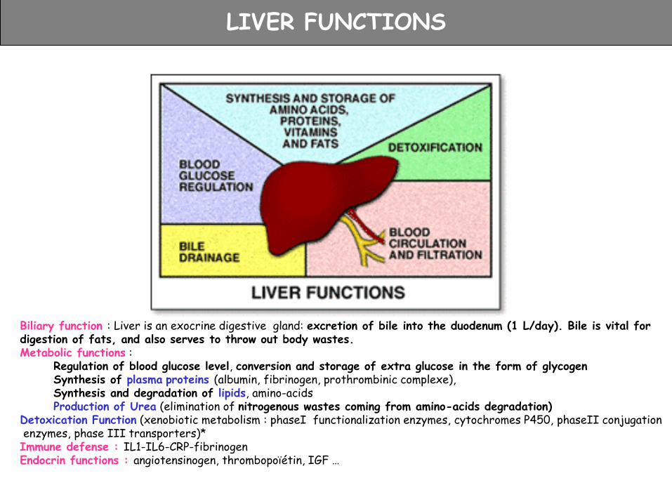
LIVER FUNCTIONS
Biliary function : Liver is an exocrine digestive gland: excretion of bile into the duodenum (1 L/day). Bile is vital for digestion of fats, and also serves to throw out body wastes. Metabolic functions :
Regulation of blood glucose level, conversion and storage of extra glucose in the form of glycogen Synthesis of plasma proteins (albumin, fibrinogen, prothrombinic complexe), Synthesis and degradation of lipids, amino-acids Production of Urea (elimination of nitrogenous wastes coming from amino-acids degradation)
Detoxication Function (xenobiotic metabolism : phaseI functionalization enzymes, cytochromes P450, phaseII conjugation enzymes, phase III transporters)* Immune defense : IL1-IL6-CRP-fibrinogen Endocrin functions : angiotensinogen, thrombopoïétin, IGF …
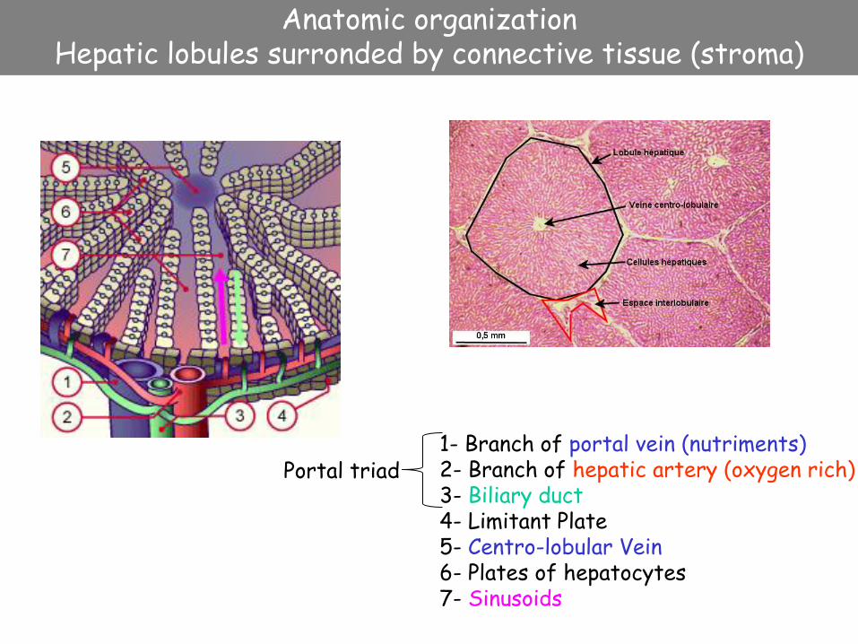
Anatomic organization Hepatic lobules surronded by connective tissue (stroma)
1- Branch of portal vein (nutriments) 2- Branch of hepatic artery (oxygen rich) 3- Biliary duct 4- Limitant Plate 5- Centro-lobular Vein 6- Plates of hepatocytes 7- Sinusoids
Portal triad

Cellular cooperation
1 – Stellate cells (storage of vitA, Lipids, synthesis of extra cellular matrix) (3-8%) 2 – Sinusoid lumen 3 – Disse space 4 – Biliary Canaliculus 5 – Hepatocytes (60-65%) 6 – Tight junctions 7 - Kupffer cells (resident macrophages, degradation of red cells) (8-12%) 8 – Discontinuous endothelium (sinusoidal cells) (10-20%)

Zone 1 Zone 2 Zone 3 PP
PV
CD45
Diameter of fenestrae
Vit A
Size of phagocytosis Cytokines
Number of fenestrae per cells
Col I, III, laminin, vimentin Col IV, VI, sulfated proteoglycans
Ureagenesis CPS1
Glutamine synthesis GLUL
Drug metabolism CAR, AhR, POR, CYP1A2, CYP2E1
Gluconeogenesis PEPCK, G6Pase
Glycolysis GK
Glucose metabolism
Ammonia detoxification
Drug detoxification
Functional zonation
Cholesterol metabolism Bile acid synthesis CYP7A1, CYP8B1, CYP27A1
• Liver structure and function • Liver regeneration & hepatic stem cells • Biotherapy • Stem cells
• Tools • Cell sources • Differentiation and use in biotherapy
• Moving towards tissue engineering
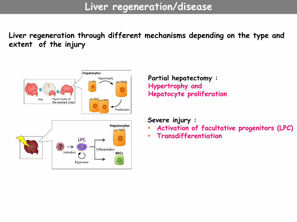
Partial hepatectomy : Hypertrophy and Hepatocyte proliferation
Severe injury : Activation of facultative progenitors (LPC) Transdifferentiation
Liver regeneration through different mechanisms depending on the type and extent of the injury
Liver regeneration/disease
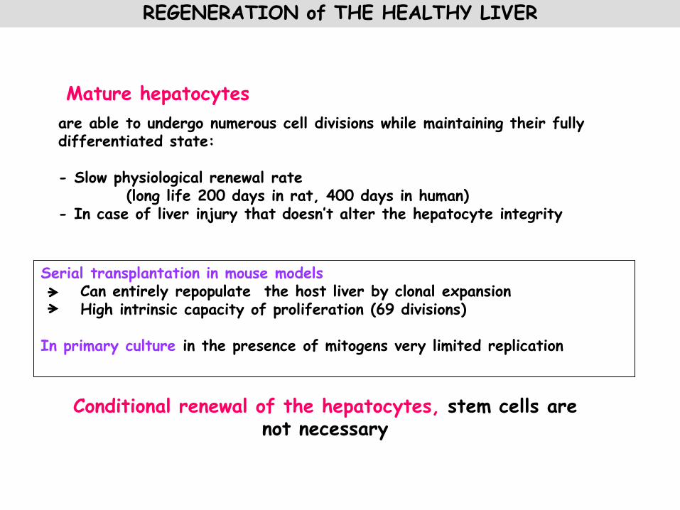
are able to undergo numerous cell divisions while maintaining their fully differentiated state: - Slow physiological renewal rate (long life 200 days in rat, 400 days in human) - In case of liver injury that doesn’t alter the hepatocyte integrity
Conditional renewal of the hepatocytes, stem cells are not necessary
Mature hepatocytes
Serial transplantation in mouse models Can entirely repopulate the host liver by clonal expansion High intrinsic capacity of proliferation (69 divisions) In primary culture in the presence of mitogens very limited replication
REGENERATION of THE HEALTHY LIVER

Partial hepatectomy (2/3) of rat liver-->hepatocytes (and other cell types) proliferate until liver mass is restored (2weeks). During this process, tissue mass and function are regulated.
Partial Hepatectomy
Gut-derived factors, such as lipopolysaccharide (LPS), are upregulated after liver injury or hepatectomy activate Kupffer and stellate cells tumour necrosis factor (TNF) and interleukin (IL)-6, HGF, TNFa , TGFb …
Taub 2004 Nature Reviews Molecular Cell Biology

(Fausto N. 2006 Hepatol)
Network of signals from different cell types to drive cell cycle progression EX Kupffer cells are a major contributing source of Wnt secretion necessary for β‐catenin activation during regeneration (Yang, J. Hepatology 2014)
Initiation elicited by cytokines, progression by growth factors and metabolic signals, termination by TGF and Hippo signallings
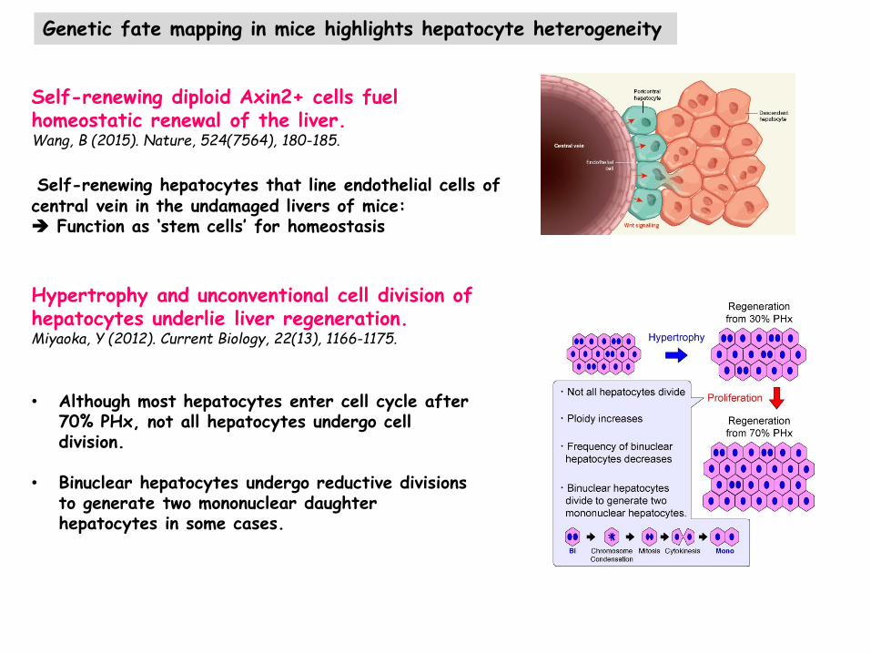
Hypertrophy and unconventional cell division of hepatocytes underlie liver regeneration. Miyaoka, Y (2012). Current Biology, 22(13), 1166-1175.
• Although most hepatocytes enter cell cycle after
70% PHx, not all hepatocytes undergo cell division.
• Binuclear hepatocytes undergo reductive divisions to generate two mononuclear daughter hepatocytes in some cases.
Self-renewing diploid Axin2+ cells fuel homeostatic renewal of the liver. Wang, B (2015). Nature, 524(7564), 180-185.
Self-renewing hepatocytes that line endothelial cells of central vein in the undamaged livers of mice: Function as ‘stem cells’ for homeostasis
Genetic fate mapping in mice highlights hepatocyte heterogeneity

REGENERATION of PATHOLOGICAL/DISEASED LIVER
1- “Transdifferentiation” : Cellular plasticity of the liver epithelial cells - Mouse hepatocytes in culture dedifferentiate and express biliary cell markers (CD49F, Ck19). However depending on the age of culture or induction, they can re-express the hepatocyte function (Fougère-Deschatrette, Stem Cells 2006)
- In vivo transdifferentiation of hepatocytes into cholangiocytes following bile duct ligature and biliary toxicity (Michalopoulos, Hepatology 2005) or in vitro transdifferentiation of gallblader cells into hepatocytes (Kuver R,AmJ Physiol Gastrointest Liver Physio, 2007)
hepatocyte cholangiocyte
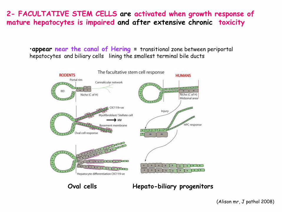
2- FACULTATIVE STEM CELLS are activated when growth response of mature hepatocytes is impaired and after extensive chronic toxicity
•appear near the canal of Hering = transitional zone between periportal hepatocytes and biliary cells lining the smallest terminal bile ducts
(Alison mr, J pathol 2008)
Oval cells Hepato-biliary progenitors

Observed in models of hepatic carcinogenesis/toxicity induced by combinations of - A chemical injury such as Galactosamine D, acetyl aminofluorene AAF, dipine , CCl4 , allyl alcohol, choline deficient diet supplemented with ethionine - And partial hepatectomy (regeneration stimuli)
OVAL CELLS Rat/mouse
* Heterogenous population of small oval cells, with high ratio Nucleus/cytoplasm appearing in the periportal region of hepatic lobule Depending on the injury: * Ductular reaction (biliary ducts without distinctive lumen developing from a portal biliary duct, and extending through the parenchyma) * Peri-ductular

equivalent to rat oval cell response (proliferation of hepato-biliary progenitors (Liver progenitor cells LPC), formation of ductules with the presence of transitional small hepatocytes) Roskams et al. Semin Liver Dis., 2004
Massive destruction of the liver resulting from toxical or viral injury (fulminant hepatic failure) Chronic cholestatic diseases Hepatitis and alcoholic cirrhosis Chronic virus C hepatitis
Human
DUCTULAR REACTIONS
healthy mild reaction pronounced reaction CK19
Correlation between the degree of inflammation and stage of chronic disease and LPC number
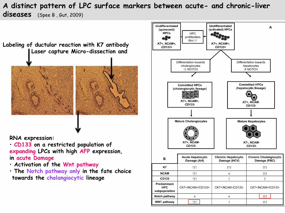
Labeling of ductular reaction with K7 antibody Laser capture Micro-dissection and
RNA expression: • CD133 on a restricted population of expanding LPCs with high AFP expression, in acute Damage • Activation of the Wnt pathway • The Notch pathway only in the fate choice towards the cholangiocytic lineage
A distinct pattern of LPC surface markers between acute- and chronic-liver diseases (Spee B , Gut, 2009)

Share common markers with hepatoblasts (albumin, alpha-fetoprotein, OV-6, CK7, CK19) hematopoïetic (CD34, c-kit) and mesenchymal stem cells
PHENOTYPE of LPC (OVAL CELLS/ HEPATO-BILIARY PROGENITORS)
EpCAM, LGR5 clonal expansion and bi-differentiation
Markers may differ depending on species
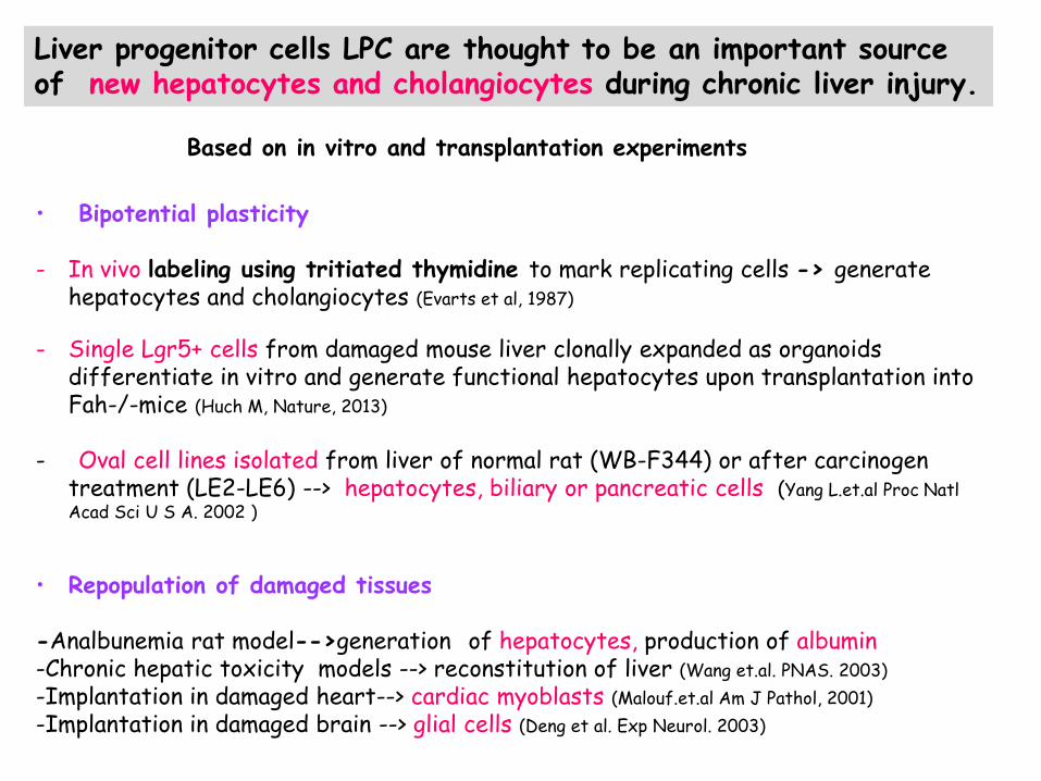
• Bipotential plasticity - In vivo labeling using tritiated thymidine to mark replicating cells -> generate
hepatocytes and cholangiocytes (Evarts et al, 1987)
- Single Lgr5+ cells from damaged mouse liver clonally expanded as organoids
differentiate in vitro and generate functional hepatocytes upon transplantation into Fah-/-mice (Huch M, Nature, 2013)
- Oval cell lines isolated from liver of normal rat (WB-F344) or after carcinogen
treatment (LE2-LE6) --> hepatocytes, biliary or pancreatic cells (Yang L.et.al Proc Natl Acad Sci U S A. 2002 )
• Repopulation of damaged tissues
-Analbunemia rat model-->generation of hepatocytes, production of albumin -Chronic hepatic toxicity models --> reconstitution of liver (Wang et.al. PNAS. 2003)
-Implantation in damaged heart--> cardiac myoblasts (Malouf.et.al Am J Pathol, 2001)
-Implantation in damaged brain --> glial cells (Deng et al. Exp Neurol. 2003)
Liver progenitor cells LPC are thought to be an important source of new hepatocytes and cholangiocytes during chronic liver injury.
Based on in vitro and transplantation experiments
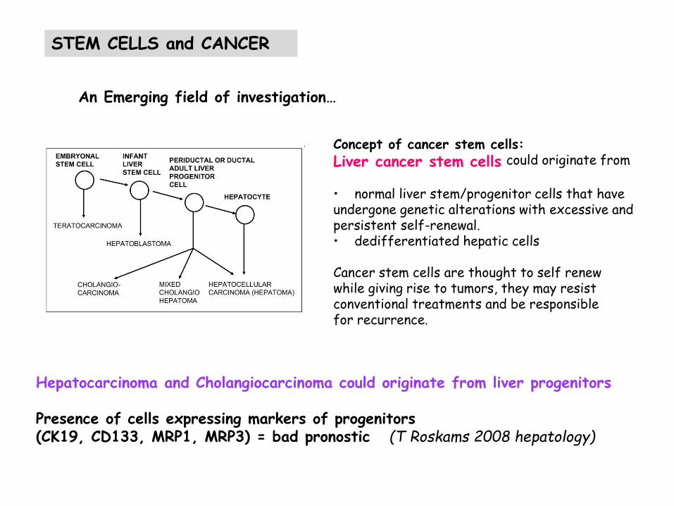
Hepatocarcinoma and Cholangiocarcinoma could originate from liver progenitors Presence of cells expressing markers of progenitors (CK19, CD133, MRP1, MRP3) = bad pronostic (T Roskams 2008 hepatology)
STEM CELLS and CANCER
An Emerging field of investigation…
Concept of cancer stem cells:
Liver cancer stem cells could originate from
• normal liver stem/progenitor cells that have undergone genetic alterations with excessive and persistent self-renewal. • dedifferentiated hepatic cells Cancer stem cells are thought to self renew while giving rise to tumors, they may resist conventional treatments and be responsible for recurrence.

Interaction with cellular and extracellular microenvironment for self-renewal, activation and differentiation.
LPC NICHE regulates THE FATE
Location in the canal of Hering in portal space (close to hepatocytes, cholangiocytes, stellate/myofibroblast…, blood flow rich in oxygen and nutrients, specific elements of ECM)
A timeline representing the stages of oval cell activation (activation, proliferation, migration, and differentiation) and the involved factors. (Erker L Stem cell res 2008)
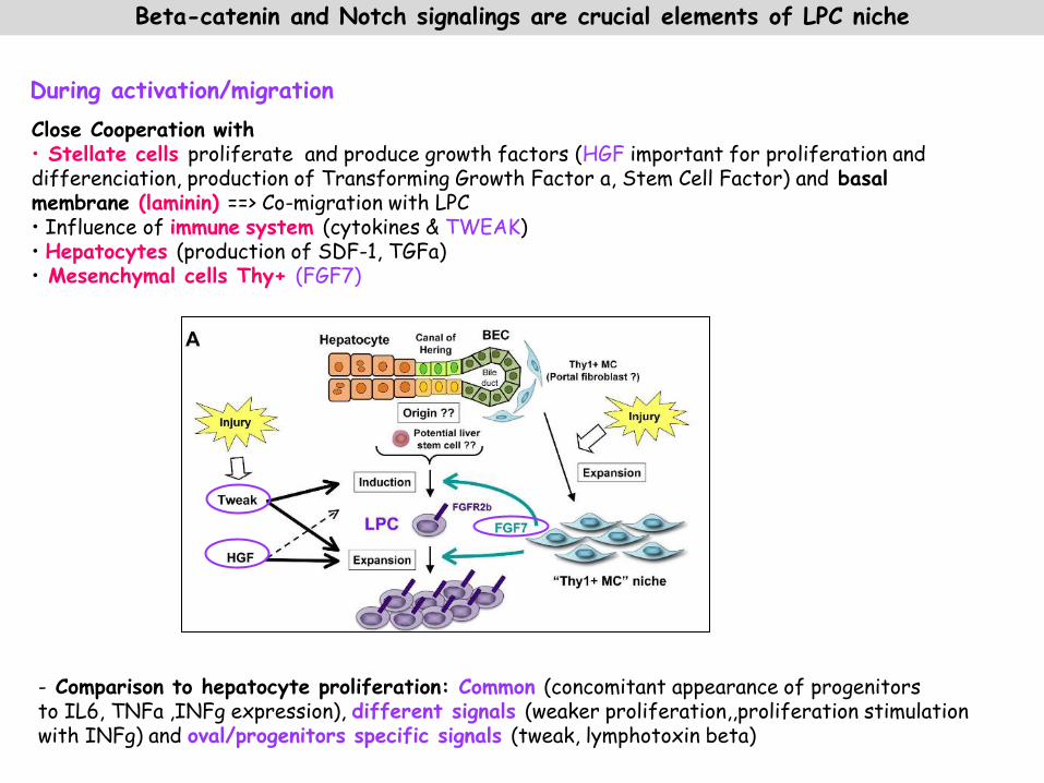
Close Cooperation with • Stellate cells proliferate and produce growth factors (HGF important for proliferation and differenciation, production of Transforming Growth Factor a, Stem Cell Factor) and basal membrane (laminin) ==> Co-migration with LPC • Influence of immune system (cytokines & TWEAK) • Hepatocytes (production of SDF-1, TGFa) • Mesenchymal cells Thy+ (FGF7)
During activation/migration
Beta-catenin and Notch signalings are crucial elements of LPC niche
- Comparison to hepatocyte proliferation: Common (concomitant appearance of progenitors to IL6, TNFa ,INFg expression), different signals (weaker proliferation,,proliferation stimulation with INFg) and oval/progenitors specific signals (tweak, lymphotoxin beta)
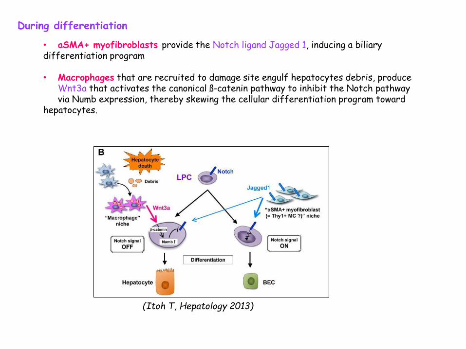
During differentiation
• aSMA+ myofibroblasts provide the Notch ligand Jagged 1, inducing a biliary differentiation program • Macrophages that are recruited to damage site engulf hepatocytes debris, produce
Wnt3a that activates the canonical ß-catenin pathway to inhibit the Notch pathway via Numb expression, thereby skewing the cellular differentiation program toward
hepatocytes.
(Itoh T, Hepatology 2013)
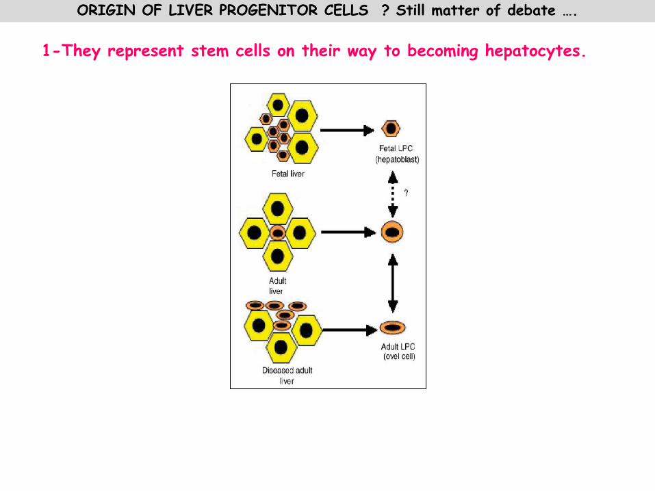
ORIGIN OF LIVER PROGENITOR CELLS ? Still matter of debate …. 1-They represent stem cells on their way to becoming hepatocytes.

• Common embryonic origin of pancreatic, biliary cells and hepatocytes, • lineage tracing • Ex vivo isolation and characterization of multipotent biliary tree stem cells • In vivo transplantation of these cells (Cardinale V, Nature Reviews Gastroenterology and Hepatology 2012)
Existence of dormant or fetal stem cells in niches …
• Canal of Hering and bile ductules
• Peribiliary glands (stem cell niches that contribute to tissue maintenance and to ongoing organogenesis of both liver and pancreas throughout life )
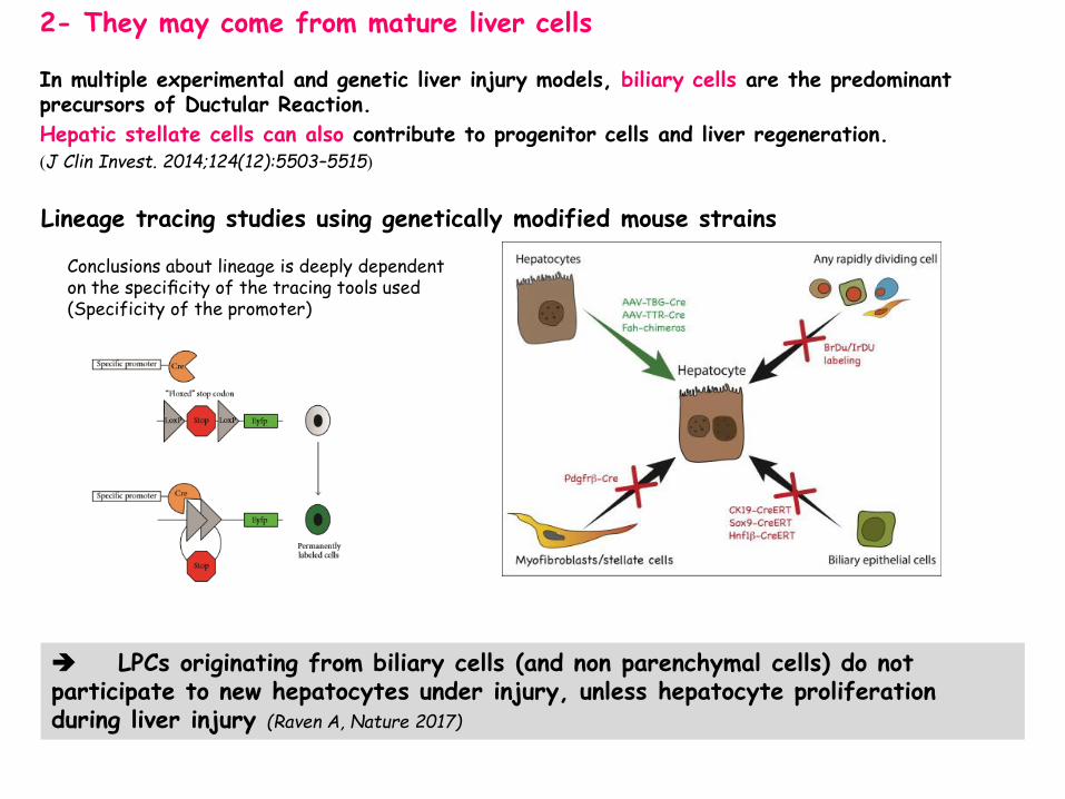
Lineage tracing studies using genetically modified mouse strains
Conclusions about lineage is deeply dependent on the specificity of the tracing tools used (Specificity of the promoter)
2- They may come from mature liver cells
In multiple experimental and genetic liver injury models, biliary cells are the predominant precursors of Ductular Reaction.
Hepatic stellate cells can also contribute to progenitor cells and liver regeneration. (J Clin Invest. 2014;124(12):5503–5515)
LPCs originating from biliary cells (and non parenchymal cells) do not participate to new hepatocytes under injury, unless hepatocyte proliferation during liver injury (Raven A, Nature 2017)

Hepatocyte-derived proliferative
ducts (hep-PDs)
Biliary-derived proliferative
ducts (bil-PDs)
Bil-PDs>hep-PDs
mTomato Hep
Bipotential Adult Liver Progenitors Are Derived from Chronically Injured Mature Hepatocytes (Tarlow, Stem Cell Stem Cell. 2014)
In vitro functionality mesenchymal phenotype. Better ability to differentiate to hepatocyte phenotype In vivo functionality hep-PDs retain the ability to revert to hepatocytes upon cessation of the injury and after serial transplantation with an efficiency much more higher (60%) than bil-PD (1%) Conversion of human hepatocytes into biliary-like proliferative ductal cells
Pre-existing population of periportal hepatocytes, in healthy livers and expressing - low amounts of Sox9 and normal level of HNF4, - undergo extensive proliferation and replenish liver mass after chronic hepatocyte-depleting injuries - do not give rise to hepatocarcinoma
Hybrid Periportal Hepatocytes Regenerate the Injured Liver without Giving Rise to Cancer (Font-Burgada J. 2015, Cell.)
Overall, several studies call the very concept of an adult liver stem cell into question (Rodrigo-Torres et al., 2014, Schaub et al., 2014, Tarlow et al., 2014 and Yanger et al., 2014)

• The liver regenerative response is highly complex and involves crosstalk between numerous resident and recruited cells.
• There are several theories to questions remain
about how instrumental each cell type is to the different phases of liver regeneration.
• Moreover, the idea of hepatocyte plasticity and heterogeneity suggests that these cells or a subset of cells have the capacity to become progenitor cells and assist with regeneration
CONCLUSION
Lukacs-Kornek Veronika , J Hepatol 2016 Zarett KS, vol 524, Nature 2015
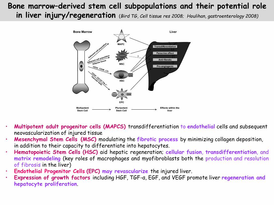
• Multipotent adult progenitor cells (MAPCS) transdifferentiation to endothelial cells and subsequent neovascularization of injured tissue
• Mesenchymal Stem Cells (MSC) modulating the fibrotic process by minimizing collagen deposition, in addition to their capacity to differentiate into hepatocytes.
• Hematopoietic Stem Cells (HSC) aid hepatic regeneration; cellular fusion, transdifferentiation, and matrix remodeling (key roles of macrophages and myofibroblasts both the production and resolution of fibrosis in the liver)
• Endothelial Progenitor Cells (EPC) may revascularize the injured liver. • Expression of growth factors including HGF, TGF-α, EGF, and VEGF promote liver regeneration and
hepatocyte proliferation.
Bone marrow–derived stem cell subpopulations and their potential role in liver injury/regeneration (Bird TG, Cell tissue res 2008; Houlihan, gastroenterology 2008)

• Liver structure and function • Liver regeneration & hepatic stem cells • Biotherapy • Stem cells
• Tools • Cell sources • Differentiation and use in biotherapy
• Moving towards tissue engineering
FOIE
Virus
Alcohol
Toxics
Drugs
Genetic
deficiency
LIVER TRANSPLANTATION
BIOTHERAPY
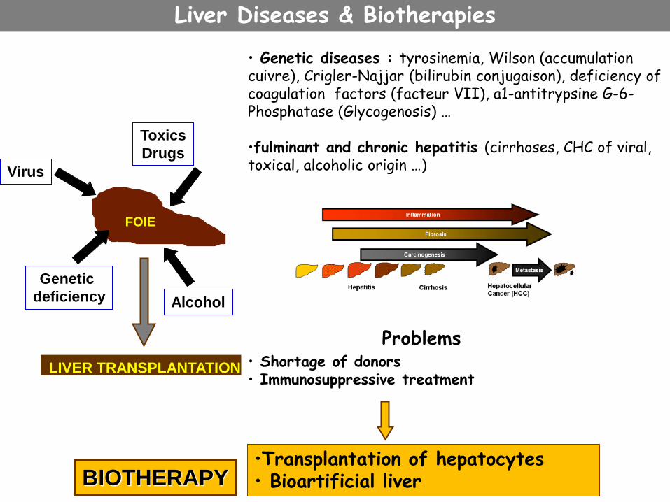
• Genetic diseases : tyrosinemia, Wilson (accumulation cuivre), Crigler-Najjar (bilirubin conjugaison), deficiency of coagulation factors (facteur VII), a1-antitrypsine G-6-Phosphatase (Glycogenosis) … •fulminant and chronic hepatitis (cirrhoses, CHC of viral, toxical, alcoholic origin …)
Problems • Shortage of donors • Immunosuppressive treatment
•Transplantation of hepatocytes • Bioartificial liver
Liver Diseases & Biotherapies

Transplantation of hepatocytes
Theorical Avantages: -Less rejection (no endothelial cells), immunosppression is needed -Can be cultured but do not proliferate and “dedifferentiation” while adapting to culture -Genetic Manipulation for deficiency correction, for Immortalisation use of a « suicide» gene -Bank of Cryopreserved hepatocytes loss of cellular viability, poor quality of hepatocytes
isolated from human donor’s liver rejected for transplantation
Implantation site and grafting: Heterotopic sites (spleen, peritoneal when encapsulated) or intra hepatic delivery, via portal
vein .
Biodistribution, hemodynamic problems if portal hypertension occurs, IBMIR (instant blood-mediated inflammatory reaction = activation coagulation and complement systems)
low cell survival and homing (migration and proper integration into the hepatocyte plates), proliferation?? Insufficient long-term efficiency
Results in human: (Dahwan, Nat rev gastroenterology & Hepatology 2010) • Genetic disease: Partial correction of Criggler-Najar syndrome, Glycogen storage type 1a (transplantation of 1% mass liver), Factor VII deficiency (temporaly corrected), Urea cycle disorder • Acute hepatic failure: Full recovery or allowed to wait for a donor liver.
• Chronic hepatic Insuffisancy : 3 patients with cirrhosis transplanted=encephalopathy
deseappered and anurie was corrected.

Extra-corporal bio-artificial liver
Hemodialysis against hepatocytes
Bio-reactors seeded with pig hepatocytes or human hepatoma cell lines
Phase I/II clinical trials on patients with fulminant hepatitis: safe,improvement of biological hepatic parameters and intra-cranal pressure • But evaluation is difficult because of the low number of patients (& liver
transplantation) • Important issues such as cost, cell availability, maintenance of cell viability and
functionality throughout treatment, • Controversy over cell used : transmission of virus or tumoral cell passage in blood
circulation
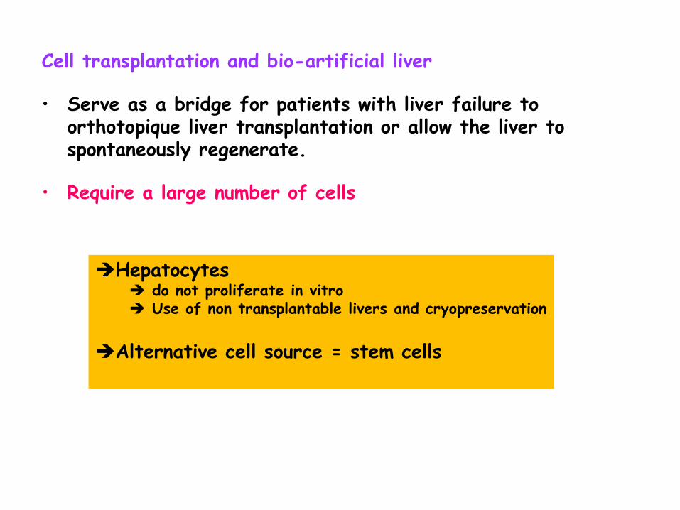
Cell transplantation and bio-artificial liver • Serve as a bridge for patients with liver failure to
orthotopique liver transplantation or allow the liver to spontaneously regenerate.
• Require a large number of cells
Hepatocytes do not proliferate in vitro Use of non transplantable livers and cryopreservation
Alternative cell source = stem cells

• Liver structure and function • Liver regeneration & hepatic stem cells • Biotherapy • Stem cells
• Tools • Cell sources • Differentiation and use in biotherapy
• Moving towards tissue engineering
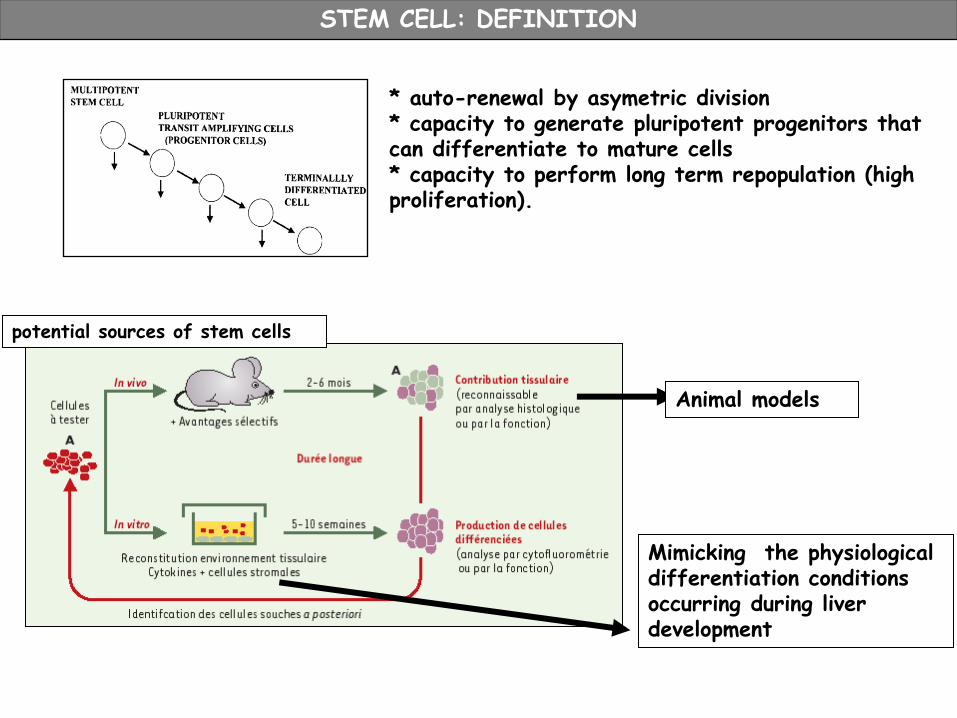
STEM CELL: DEFINITION
* auto-renewal by asymetric division * capacity to generate pluripotent progenitors that can differentiate to mature cells * capacity to perform long term repopulation (high proliferation).
Mimicking the physiological differentiation conditions occurring during liver development
Animal models
potential sources of stem cells

ANIMAL MODELS for TRANSPLANTATION
Principle: a selective advantage is brought to the transplanted cells to favor repopulation
Survival selective advantage -Resistance to Fas induced apoptosis: Bcl2 gene expression, either in transplanted cells or directly in the liver using gene therapy (in vivo transduction of a small number of hepatocytes) -uPA-Alb Model Constitutive expression of urokinase gene in the liver: chronic cytolysis/permanent regeneration -Fumarylacetoacetate hydrolase FAH-/- mouse model Terminal enzyme of tyrosine metabolism toxic metabolites accumulation, can be shunted by NTBC Progressive liver and kidney damage if NTBC is removed from diet Growth selective advantage -Inhibition of hepatocyte proliferation with alkylating agents or high irradiation dose
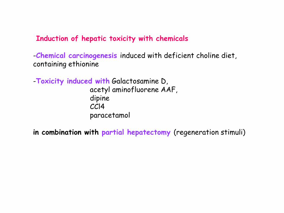
Induction of hepatic toxicity with chemicals -Chemical carcinogenesis induced with deficient choline diet, containing ethionine -Toxicity induced with Galactosamine D, acetyl aminofluorene AAF, dipine CCl4 paracetamol in combination with partial hepatectomy (regeneration stimuli)

TRACKING THE TRANSPLANTED CELLS
CELL FUSION
Detection of fusion events: -co-localisation of host and donor markers in the same cell -co-expression of host and donor genes in the same cell -genetic labelling of donor cells: conditional GFP expression will be activated in fused cells
1- Transplantation of cells from a male donor XY into a femelle host XX : detection of Y chromosome in the differentiated cells (FISH) 2- Transplantation into host bearing a protein/enzyme deficiency (albumin, DPPIV…) 3- Detection of specific epitopes (HLA, different haplotypes, species specificity) 4- Genetic (beta-galactosidase, GFP, luc …) metabolic (non specific: esterase , membrane labelling) physical (fluorescent nano particules, ferric beads…) labelling 5- Differentiation : definition of the hepatocyte phenotype expression of several tissue-specific markers, integration in the liver architecture, restored liver functions

Mesoderm
Endoderm
Ectoderm
Fetal
Hepatoblast
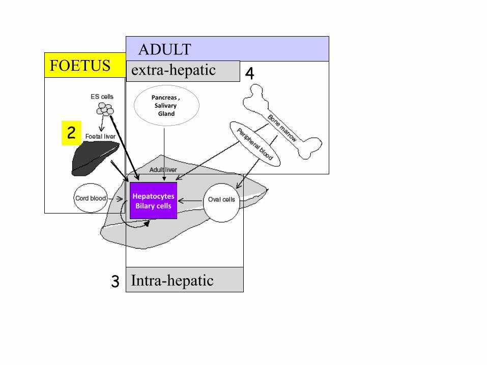
Stem cells sources
Embryonic
stem cells Blastocyst Pluripotent SC:
3 germ layers
And homeostasie/
regeneration
Multipotent fetal SC:
several related tissues
Adult SC:
tissue restricted
Progenitors
Mature cells
F
E
T
U
S
A
D
U
L
T
E
M
B
R
Y
O
iPS induced
Plutipotent
cells
plasticity Sox2,Oct4,Klf4,c/myc
(Yamanaka, Thomson)
R
E
P
R
O
G
R
A
M
A
T
I
O
N Mesenchymal SC Liver SC
Pancreas , Salivary
Gland
FOETUS ADULT
Intra-hepatic
extra-hepatic
Hepatocytes Bilary cells
2
3
4

HCFU-C Suzuki A, et.al 2000 Hepatology. 2002 Semin Cell Dev Biol. 2003 cell transpl.
Isolation of diploid cells from murine fetal liver, clonal analysis •Self-renewal and high in vitro proliferation, •Multipotency: •in vitro differentiation into hepatocytes (HGF) maturation in presence of OSM, biliary duct structure (Matrigel), •in vivo differentiation into hepatocytes, biliary, intestinal and pancreatic cells
Mouse

Transplantation Souris uPA-SCID 3-8 weeks 20%:
In vivo differentiation - Hepatocytes and Cholangiocytes - Zonal expression
Isolation : from ED14 liver with “plate and wait“ culture strategy, serum, IGFI, EGF, Insulin No hepatic markers expressed In vitro reversible differentiation, : Dex+AMPc or aggregates hepatocyte Matrigel +HGF cholangiocyte
BMEL Bipotential mouse embryonic liver stem cell lines Strick-Marchand H and Weiss MC, Hepatology. 2002, Semin Liver Dis. 2003, PNAS . 2004
Mouse
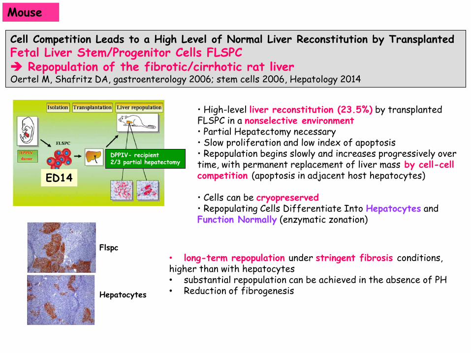
Cell Competition Leads to a High Level of Normal Liver Reconstitution by Transplanted Fetal Liver Stem/Progenitor Cells FLSPC Repopulation of the fibrotic/cirrhotic rat liver Oertel M, Shafritz DA, gastroenterology 2006; stem cells 2006, Hepatology 2014
• High-level liver reconstitution (23.5%) by transplanted FLSPC in a nonselective environment • Partial Hepatectomy necessary • Slow proliferation and low index of apoptosis • Repopulation begins slowly and increases progressively over time, with permanent replacement of liver mass by cell-cell competition (apoptosis in adjacent host hepatocytes)
• Cells can be cryopreserved • Repopulating Cells Differentiate Into Hepatocytes and Function Normally (enzymatic zonation)
ED14
DPPIV- recipient 2/3 partial hepatectomy
Mouse
• long-term repopulation under stringent fibrosis conditions, higher than with hepatocytes • substantial repopulation can be achieved in the absence of PH • Reduction of fibrogenesis
Flspc Hepatocytes

Repopulation of athymic Mouse Liver by Cryopreserved Early Human Fetal Hepatoblast Mahieu-Caputo Hum Gene Ther 2004
• In vitro : High capacity of proliferation Tolerate cryopreservation • Transplantation into mice without liver damage
Human hepatoblasts (10-13 weeks) - Proliferate and engraft (1-10%) - Functional (Albumin secretion) - Acquisition of «adult markers» - Without any selection pressure
• Transplantation into mouse model of familial intrahepatic cholestasis (Mdr2-/-) partial restoration of the function • Transplantation into galactosamineD mouse model hepatocytes(alb,AAT,CD26)
(Nowak et al gut 2005 )
Human
Conclusions : Fetal liver is an interesting cell source (proliferation, differentiation, cryopreservation) But their use in cell therapy is not realistic
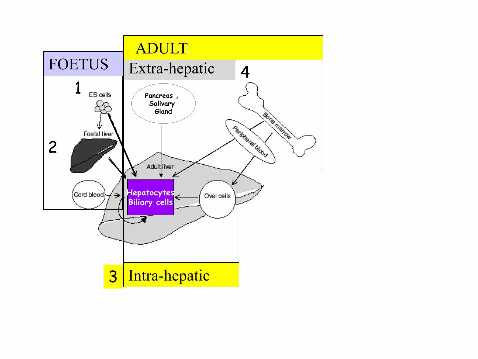
Pancreas , Salivary Gland
FOETUS ADULT
Intra-hepatic
Extra-hepatic
Hepatocytes Biliary cells
1
2
3
4

• In vitro Differentiation and Comparison to Human hepatocytes in primary culture Cocktails of HGF, FGF, Dex, ITS , matrigel for 21days
• Isolation and amplification of Non Parenchymal Epithelial cells NPE • Cryopreservation
Differentiation of Non Parenchymal Epithelial cells isolated from adult human livers
Non Parenchymal
Fraction
Perfusion
dissociation
HNF4a
A sub population differentiate into « hepatocyte-like » cells
Albumin
1-antitrypsin
Fibrinogen
D0 D24 Plasma protein
secretion
Hepatic markers
D24
Albumin HNF4a
• In vivo Differentiation NOD/SCID mice in regeneration: • Secretion of human albumin in mouse serum • Expression of human albumin and CYP3A4 in liver
Non diseased livers
Duret C, Stem Cells, 2007, Methods Mol Biol. 2010
BUT Hepatobiliary and mixed Fetal /Adult phenotype Poor expression of detox enzymes
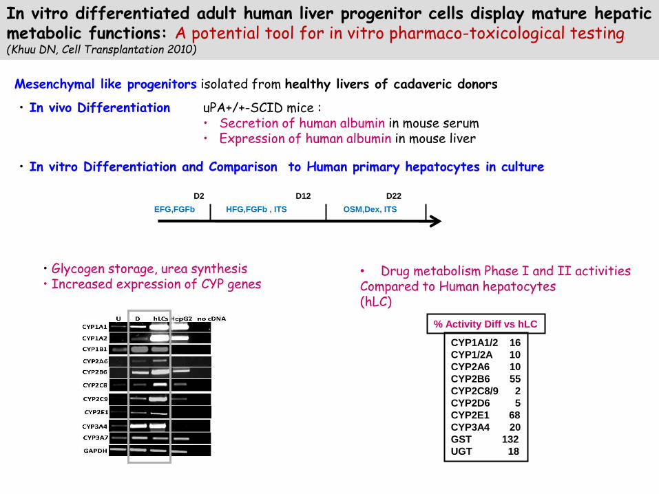
In vitro differentiated adult human liver progenitor cells display mature hepatic metabolic functions: A potential tool for in vitro pharmaco-toxicological testing (Khuu DN, Cell Transplantation 2010)
Mesenchymal like progenitors isolated from healthy livers of cadaveric donors
• Glycogen storage, urea synthesis • Increased expression of CYP genes
EFG,FGFb HFG,FGFb , ITS OSM,Dex, ITS
D2 D12 D22
• In vitro Differentiation and Comparison to Human primary hepatocytes in culture
• In vivo Differentiation uPA+/+-SCID mice : • Secretion of human albumin in mouse serum • Expression of human albumin in mouse liver
CYP1A1/2 16
CYP1/2A 10
CYP2A6 10
CYP2B6 55
CYP2C8/9 2
CYP2D6 5
CYP2E1 68
CYP3A4 20
GST 132
UGT 18
% Activity Diff vs hLC
• Drug metabolism Phase I and II activities Compared to Human hepatocytes (hLC)
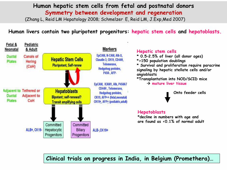
Hepatic stem cells * 0.5–2.5% of liver (all donor ages) *>150 population doublings * Survival and proliferation require paracrine signaling by hepatic stellate cells and/or angioblasts *Transplantation into NOD/SCID mice mature liver tissue
Onto feeder cells
Hepatoblasts *decline in numbers with age and are found as <0.1% of normal adult
Human hepatic stem cells from fetal and postnatal donors Symmetry between development and regeneration
(Zhang L, Reid LM Hepatology 2008; Schmelzer E, Reid LM, J.Exp,Med 2007)
Human livers contain two pluripotent progenitors: hepatic stem cells and hepatoblasts.
Clinical trials on progress in India, in Belgium (Promethera)…

Pancreas , Salivary Gland
FOETUS ADULT
Intra-hepatic
Extra-hepatic
Hpatocytes Biliary cells
1
2
3
4
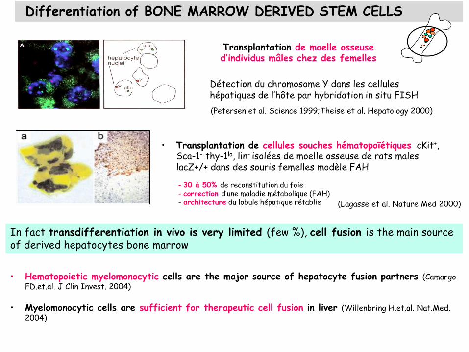
(Petersen et al. Science 1999;Theise et al. Hepatology 2000)
Transplantation de moelle osseuse d’individus mâles chez des femelles
Détection du chromosome Y dans les cellules hépatiques de l’hôte par hybridation in situ FISH
Differentiation of BONE MARROW DERIVED STEM CELLS
- 30 à 50% de reconstitution du foie - correction d’une maladie métabolique (FAH) - architecture du lobule hépatique rétablie
• Transplantation de cellules souches hématopoïétiques cKit+, Sca-1+ thy-1lo, lin- isolées de moelle osseuse de rats males lacZ+/+ dans des souris femelles modèle FAH
(Lagasse et al. Nature Med 2000)
In fact transdifferentiation in vivo is very limited (few %), cell fusion is the main source of derived hepatocytes bone marrow
• Hematopoietic myelomonocytic cells are the major source of hepatocyte fusion partners (Camargo FD.et.al. J Clin Invest. 2004)
• Myelomonocytic cells are sufficient for therapeutic cell fusion in liver (Willenbring H.et.al. Nat.Med.
2004)
After differentiation store glycogen, synthesize urea,
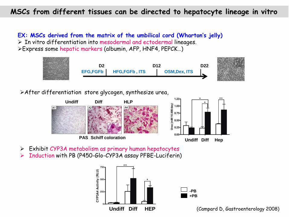
MSCs from different tissues can be directed to hepatocyte lineage in vitro
EX: MSCs derived from the matrix of the umbilical cord (Wharton’s jelly) In vitro differentiation into mesodermal and ectodermal lineages. Express some hepatic markers (albumin, AFP, HNF4, PEPCK…)
EFG,FGFb HFG,FGFb , ITS OSM,Dex, ITS
D2 D12 D22
Undiff Diff Hep
Undiff Diff HEP
-PB
+PB
PAS Schiff coloration
Exhibit CYP3A metabolism as primary human hepatocytes Induction with PB (P450-Glo-CYP3A assay PFBE-Luciferin)
Undiff Diff HLP
(Campard D, Gastroenterology 2008)

Mesenchymal stem cells isolated from bone marrow, adipose tissue (Sato et al , blood 2005; Aurich Gut 2007; Baba J Hepatol 2004…)
and liver biotherapy…
MSC properties
More than 31 clinical trials in progress for Chronic liver diseases
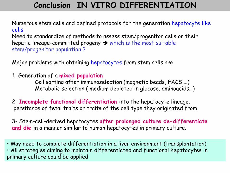
Numerous stem cells and defined protocols for the generation hepatocyte like cells Need to standardize of methods to assess stem/progenitor cells or their hepatic lineage-committed progeny which is the most suitable stem/progenitor population ? Major problems with obtaining hepatocytes from stem cells are 1- Generation of a mixed population Cell sorting after immunoselection (magnetic beads, FACS …) Metabolic selection ( medium depleted in glucose, aminoacids…) 2- Incomplete functional differentiation into the hepatocyte lineage. persitance of fetal traits or traits of the cell type they originated from. 3- Stem-cell-derived hepatocytes after prolonged culture de-differentiate and die in a manner similar to human hepatocytes in primary culture. • May need to complete differentiation in a liver environment (transplantation)
• All strategies aiming to maintain differentiated and functional hepatocytes in primary culture could be applied
Conclusion IN VITRO DIFFERENTIATION

CONCLUSION Stem cells and liver regeneration
Different requirements for different Liver pathologies
Different stem cell types for regenerative medicine
Studies on New strategies to stimulate liver regeneration and reduce liver scarring Possible tailored cell therapy options for the major types of liver disease
(Forbes SJ, J Hepatol, 2011)

• Liver structure and function • Liver regeneration & hepatic stem cells • Biotherapy • Stem cells
• Tools • Cell sources • Differentiation and use in biotherapy
• Moving towards tissue engineering

Improvement of terminal differentiation and function
Enforced expression of master regulators of hepatocyte differentiation or function ( transcription factors, nuclear receptors, miRNA, antagomirs )
Epigenetic influence
Reconstitution of the hepatic niche
Dynamic patterns of histone methylation are associated with ontogenic expression of the Cyp3a genes during mouse liver maturation ( Li Y, Faseb J 2009)
Heterotypic cell culture and 3D organisation
Perfusion in bioreactor
And/or
Hepatic Differentiation and Maturation of Human Embryonic Stem Cells Cultured in a Perfused Three-Dimensional Bioreactor Sivertsson L, 2013 stem cell & dev

Tissue engineering
Components Advanced technologies - Nanotechnology - Microfluidic
Tissue engineering evolved from the field of biomaterials development and refers to the practice of combining scaffolds, cells, and biologically active molecules into functional tissues Aims to assemble functional constructs that restore, maintain, or improve damaged tissues or whole organs.
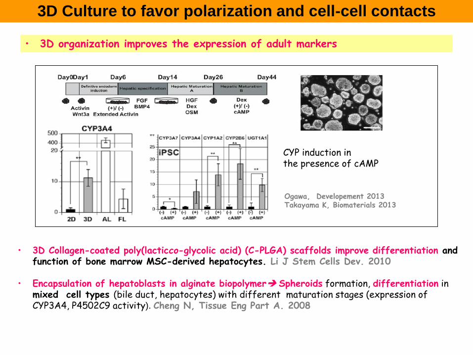
3D Culture to favor polarization and cell-cell contacts
• 3D organization improves the expression of adult markers
Ogawa, Developement 2013 Takayama K, Biomaterials 2013
CYP induction in the presence of cAMP
• 3D Collagen-coated poly(lacticco-glycolic acid) (C-PLGA) scaffolds improve differentiation and function of bone marrow MSC-derived hepatocytes. Li J Stem Cells Dev. 2010
• Encapsulation of hepatoblasts in alginate biopolymer Spheroids formation, differentiation in
mixed cell types (bile duct, hepatocytes) with different maturation stages (expression of CYP3A4, P4502C9 activity). Cheng N, Tissue Eng Part A. 2008

Chen AA, PNAS. 2011
Growth of blood vessels (red) enables implanted HEALs to grow and function in the mouse.
Humanized mice with ectopic artificial liver tissues for drug testing In the future : construct an implantable artificial liver as an alternative to whole-organ transplantation for people with liver failure.
-Coculture of primary hepatocytes and stromal fibroblasts on collagen-coated plates for 7–10 days -Encapsulation with liver endothelial cells in PEG-DA scaffolds derivatized with adhesion peptides -Implantation in the subcutaneous or peritoneal cavity of nude mice
Co-culture and Scaffolds

Controlled organization of different cell types in 3D
Interpenetrating cell-cell contacts are essential to improve cell functionality
Micropatterning : Multi-compartmental placement dictates hepatic tissue function
InVERT molding for scalable control of tissue Microarchitecture,
Synthetic matrix
Micropatterned Tissue (Human/rat hepatocytes with fibroblasts) implanted in the intraperitoneal space of nude mice
Optimal cellular architectures that sustained hepatic functions for at least 4 weeks after transplantation
Stevens HR,2013 , Nature comm

L’impression 3D : les dispositifs médicaux ou prothèses sur mesure ont été les premières applications de cette nouvelle technologie.
La Bio-impression a pour objectif de concevoir des tissus fonctionnels dans le but de créer : -Dans l’immédiat, des modèles prédictifs reproduisant la physiologie de tissus humains sains ou de tissus pathologiques -D’ici 3 à 5 ans, des tissus individualisés, réalisés à partir des cellules du patient, pour sélectionner in vitro les traitements et développer des solutions thérapeutiques personnalisées. -D’ici 7 à 10 ans, des tissus/organes complexes implantables pour la médecine régénératrice. Ear cartilage reconstruction with
integrated tissue–organ printer (ITOP)
3D Bio-imprinting
Kang HW, Nature biotech 2016
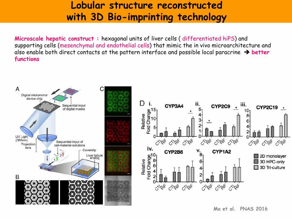
Lobular structure reconstructed with 3D Bio-imprinting technology
Ma et al. PNAS 2016
Microscale hepatic construct : hexagonal units of liver cells ( differentiated hiPS) and supporting cells (mesenchymal and endothelial cells) that mimic the in vivo microarchitecture and also enable both direct contacts at the pattern interface and possible local paracrine better functions
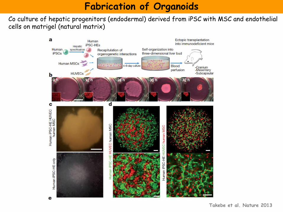
Fabrication of Organoids
Takebe et al. Nature 2013
Co culture of hepatic progenitors (endodermal) derived from iPSC with MSC and endothelial cells on matrigel (natural matrix)
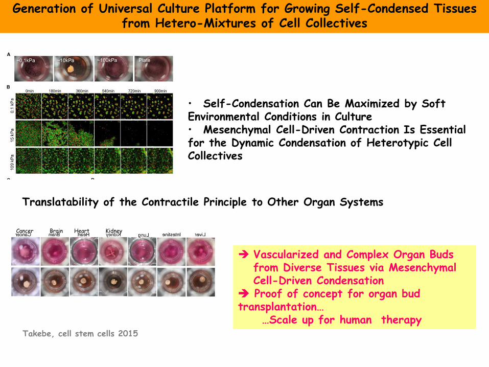
Translatability of the Contractile Principle to Other Organ Systems
• Self-Condensation Can Be Maximized by Soft Environmental Conditions in Culture • Mesenchymal Cell-Driven Contraction Is Essential for the Dynamic Condensation of Heterotypic Cell Collectives
Generation of Universal Culture Platform for Growing Self-Condensed Tissues from Hetero-Mixtures of Cell Collectives
Vascularized and Complex Organ Buds from Diverse Tissues via Mesenchymal Cell-Driven Condensation
Proof of concept for organ bud transplantation… …Scale up for human therapy
Takebe, cell stem cells 2015
Cancer Brain Heart Kidney
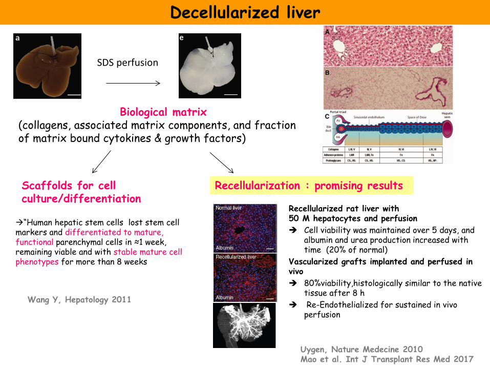
Decellularized liver
“Human hepatic stem cells lost stem cell markers and differentiated to mature, functional parenchymal cells in ≈1 week, remaining viable and with stable mature cell phenotypes for more than 8 weeks
SDS perfusion
Biological matrix (collagens, associated matrix components, and fraction of matrix bound cytokines & growth factors)
Scaffolds for cell culture/differentiation
Recellularization : promising results
Recellularized rat liver with 50 M hepatocytes and perfusion
Cell viability was maintained over 5 days, and albumin and urea production increased with time (20% of normal)
Vascularized grafts implanted and perfused in vivo
80%viability,histologically similar to the native tissue after 8 h
Re-Endothelialized for sustained in vivo perfusion
Wang Y, Hepatology 2011
Uygen, Nature Medecine 2010 Mao et al. Int J Transplant Res Med 2017
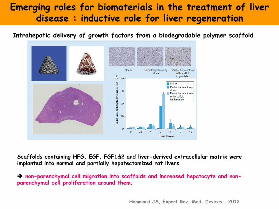
Scaffolds containing HFG, EGF, FGF1&2 and liver-derived extracellular matrix were implanted into normal and partially hepatectomized rat livers non-parenchymal cell migration into scaffolds and increased hepatocyte and non-parenchymal cell proliferation around them.
Emerging roles for biomaterials in the treatment of liver disease : inductive role for liver regeneration
Hammond JS, Expert Rev. Med. Devices , 2012
Intrahepatic delivery of growth factors from a biodegradable polymer scaffold
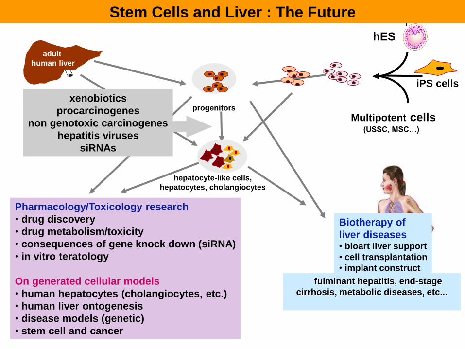
adult
human liver
hES
progenitors
hepatocyte-like cells,
hepatocytes, cholangiocytes
Multipotent cells (USSC, MSC…)
fulminant hepatitis, end-stage
cirrhosis, metabolic diseases, etc...
Biotherapy of
liver diseases • bioart liver support
• cell transplantation
• implant construct
Stem Cells and Liver : The Future
Pharmacology/Toxicology research
• drug discovery
• drug metabolism/toxicity
• consequences of gene knock down (siRNA)
• in vitro teratology
On generated cellular models
• human hepatocytes (cholangiocytes, etc.)
• human liver ontogenesis
• disease models (genetic)
• stem cell and cancer
xenobiotics
procarcinogenes
non genotoxic carcinogenes
hepatitis viruses
siRNAs
iPS cells