AMP - Journal of Cell Science
11
Redox implications of AMPK-mediated signal transduction beyond energetic clues Simone Cardaci 1, *, Giuseppe Filomeni 1,2, * ,` and Maria Rosa Ciriolo 1,2,` 1 Department of Biology, University of Rome ‘‘Tor Vergata’, Via della Ricerca Scientifica, 00133 Rome, Italy 2 IRCCS San Raffaele Pisana, Via di Val Cannuta, 00166 Rome, Italy *These authors contributed equally to this work ` Authors for correspondence ([email protected]; [email protected]) Journal of Cell Science 125, 1–11 ß 2012. Published by The Company of Biologists Ltd doi: 10.1242/jcs.095216 Summary Since the discovery of AMP-dependent protein kinase (AMPK), its fundamental role in regulating metabolic pathways and the molecular mechanism underlying the regulation of its activity by adenine nucleotides has been widely studied. AMPK is not only an energy-responsive enzyme, but it also senses redox signals. This review aims at recapitulating the recent lines of evidence that demonstrate the responsiveness of this kinase to metabolic and nitroxidative imbalance, thus providing new insights into the intimate networks of redox-based signals upstream of AMPK. In particular, we discuss its well-recognized activation downstream of mitochondrial dysfunction, debate the recent findings that AMPK is directly targeted by pro-oxidant species, and question alternative redox pathways that allow AMPK to be included into the large class of redox-sensing proteins. The possible therapeutic implications of the role of AMPK in redox-associated pathologies, such as cancer and neurodegeneration, are also discussed in light of recent advances that suggest a role for AMPK in the tuning of redox-dependent processes, such as apoptosis and autophagy. Key words: ROS, Apoptosis, Autophagy, Nitric oxide Introduction Living organisms derive energy from the breakdown of the molecular bonds of nutrients, and then convert it into the more usable forms of ATP and NADPH (see Box 1). Cell homeostasis depends on the maintenance of ATP levels, whose hydrolysis can be coupled to synthesis reactions, transport across membranes and other endoergonic processes. For this reason, cells actively synthesize ATP and have evolved molecular mechanisms aimed at counteracting even slight decreases of ATP (Hardie, 2011). The mutual regulation of the ATP-regenerating and ATP-consuming processes, however, does not depend on the concentration of this single metabolite, but is in response to the energy balance of the cell (Atkinson, 1970). Indeed, the concentration of ATP, which has been estimated to range from 1 to 10 mM, can be subjected to marked variations. Therefore, the absolute intracellular ATP concentration tightly depends on the concentration of the other adenylates (adenylate pool) and should be considered as a function of the adenylate energy charge, which, in accordance to the definition provided by Atkinson in 1968, is expressed as ([ATP] + 0.5[ADP])/([ATP] + [ADP] + [AMP]) and ranges from 0 to 1 (Atkinson, 1968). The reversible adenylate-kinase-catalyzed reaction (2[ADP] « [ATP] + [AMP]) is sufficiently fast in cells to maintain the reactants at their equilibrium concentrations. Under resting conditions, in which the adenylate energy charge is close to 1 – implying that the adenylate pool size is approximately equal to the ATP concentration (Hardie et al., 2011) – adenylate kinase catalyzes the reaction in favor of ADP synthesis, thereby maintaining very low AMP concentrations (approximately one or two orders of magnitude lower than those of ADP or ATP, respectively). Under conditions that raise ATP utilization, or upon metabolic stress that interferes with ATP synthesis, adenylate kinase shifts the above reaction towards the regeneration of ATP to increase its concentration and availability. This represents a valuable tool to rapidly restore ATP concentrations with a significant increase in AMP levels. Physiologically, the relative changes in AMP concentrations are much greater than those occurring to ATP (Ataullakhanov and Vitvitsky, 2002), thereby making the ratio between AMP and ATP the most reliable indicator of the cellular energetic state (Fogarty and Hardie, 2010; Kahn et al., 2005). AMP-dependent protein kinase (AMPK) senses even small changes in AMP concentrations, and represents the principal metabolic gatekeeper of the cell (Hardie, 2011). Upon energetic imbalance, intracellular concentrations of AMP increase, thus promoting AMPK activation (Fig. 1). Activated AMPK stimulates catabolic pathways (e.g. glycolysis and fatty acid oxidation) and, concomitantly, inhibits the rate of anabolic reactions (protein, cholesterol, triglyceride and fatty acid synthesis) to restore the correct adenylate energy charge (Hardie, 2011). Although the binding of AMP remains the principal event triggering the activation of AMPK, recent findings have demonstrated that ADP also has a role in this process (Oakhill et al., 2011; Xiao et al., 2011), because it contributes to inhibit phosphatase-mediated dephosphorylation of AMPK (see below), for instance by protein phosphatase2 Ca (PP2Ca) (Fig. 1). On the basis of these results, Oakhill and co-workers suggested that AMPK is a direct adenylate-charge-sensing protein kinase instead of a specifically AMP-activated kinase (Oakhill et al., 2011). Commentary 1 Journal of Cell Science JCS online publication date 22 May 2012
Transcript of AMP - Journal of Cell Science
Redox implications of AMPK-mediated signaltransduction beyond energetic clues
Simone Cardaci1,*, Giuseppe Filomeni1,2,*,` and Maria Rosa Ciriolo1,2,`
1Department of Biology, University of Rome ‘‘Tor Vergata’’, Via della Ricerca Scientifica, 00133 Rome, Italy2IRCCS San Raffaele Pisana, Via di Val Cannuta, 00166 Rome, Italy
*These authors contributed equally to this work`Authors for correspondence ([email protected]; [email protected])
Journal of Cell Science 125, 1–11� 2012. Published by The Company of Biologists Ltddoi: 10.1242/jcs.095216
SummarySince the discovery of AMP-dependent protein kinase (AMPK), its fundamental role in regulating metabolic pathways and the
molecular mechanism underlying the regulation of its activity by adenine nucleotides has been widely studied. AMPK is not only anenergy-responsive enzyme, but it also senses redox signals. This review aims at recapitulating the recent lines of evidence thatdemonstrate the responsiveness of this kinase to metabolic and nitroxidative imbalance, thus providing new insights into the intimate
networks of redox-based signals upstream of AMPK. In particular, we discuss its well-recognized activation downstream ofmitochondrial dysfunction, debate the recent findings that AMPK is directly targeted by pro-oxidant species, and question alternativeredox pathways that allow AMPK to be included into the large class of redox-sensing proteins. The possible therapeutic implications ofthe role of AMPK in redox-associated pathologies, such as cancer and neurodegeneration, are also discussed in light of recent advances
that suggest a role for AMPK in the tuning of redox-dependent processes, such as apoptosis and autophagy.
Key words: ROS, Apoptosis, Autophagy, Nitric oxide
IntroductionLiving organisms derive energy from the breakdown of the
molecular bonds of nutrients, and then convert it into the more
usable forms of ATP and NADPH (see Box 1). Cell homeostasis
depends on the maintenance of ATP levels, whose hydrolysis can
be coupled to synthesis reactions, transport across membranes and
other endoergonic processes. For this reason, cells actively
synthesize ATP and have evolved molecular mechanisms aimed
at counteracting even slight decreases of ATP (Hardie, 2011). The
mutual regulation of the ATP-regenerating and ATP-consuming
processes, however, does not depend on the concentration of this
single metabolite, but is in response to the energy balance of the
cell (Atkinson, 1970). Indeed, the concentration of ATP, which has
been estimated to range from 1 to 10 mM, can be subjected to
marked variations. Therefore, the absolute intracellular ATP
concentration tightly depends on the concentration of the other
adenylates (adenylate pool) and should be considered as a function
of the adenylate energy charge, which, in accordance to the
definition provided by Atkinson in 1968, is expressed as ([ATP] +
0.5[ADP])/([ATP] + [ADP] + [AMP]) and ranges from 0 to 1
(Atkinson, 1968).
The reversible adenylate-kinase-catalyzed reaction (2[ADP] «[ATP] + [AMP]) is sufficiently fast in cells to maintain the
reactants at their equilibrium concentrations. Under resting
conditions, in which the adenylate energy charge is close to 1 –
implying that the adenylate pool size is approximately equal to the
ATP concentration (Hardie et al., 2011) – adenylate kinase
catalyzes the reaction in favor of ADP synthesis, thereby
maintaining very low AMP concentrations (approximately one
or two orders of magnitude lower than those of ADP or ATP,
respectively). Under conditions that raise ATP utilization, or upon
metabolic stress that interferes with ATP synthesis, adenylate
kinase shifts the above reaction towards the regeneration of ATP to
increase its concentration and availability. This represents a
valuable tool to rapidly restore ATP concentrations with a
significant increase in AMP levels. Physiologically, the relative
changes in AMP concentrations are much greater than those
occurring to ATP (Ataullakhanov and Vitvitsky, 2002), thereby
making the ratio between AMP and ATP the most reliable
indicator of the cellular energetic state (Fogarty and Hardie, 2010;
Kahn et al., 2005).
AMP-dependent protein kinase (AMPK) senses even small
changes in AMP concentrations, and represents the principal
metabolic gatekeeper of the cell (Hardie, 2011). Upon energetic
imbalance, intracellular concentrations of AMP increase, thus
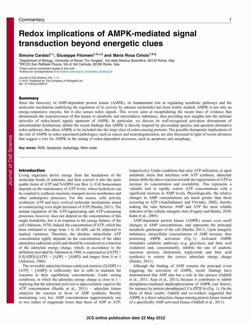
promoting AMPK activation (Fig. 1). Activated AMPK
stimulates catabolic pathways (e.g. glycolysis and fatty acid
oxidation) and, concomitantly, inhibits the rate of anabolic
reactions (protein, cholesterol, triglyceride and fatty acid
synthesis) to restore the correct adenylate energy charge
(Hardie, 2011).
Although the binding of AMP remains the principal event
triggering the activation of AMPK, recent findings have
demonstrated that ADP also has a role in this process (Oakhill
et al., 2011; Xiao et al., 2011), because it contributes to inhibit
phosphatase-mediated dephosphorylation of AMPK (see below),
for instance by protein phosphatase2 Ca (PP2Ca) (Fig. 1). On the
basis of these results, Oakhill and co-workers suggested that
AMPK is a direct adenylate-charge-sensing protein kinase instead
of a specifically AMP-activated kinase (Oakhill et al., 2011).
Commentary 1
Journ
alof
Cell
Scie
nce
JCS online publication date 22 May 2012

In addition to the complex regulation of AMPK by the
adenylate pool, recently, AMPK has been reported to sense and
respond to pro-oxidant conditions that are induced by reactive
oxygen or nitrogen species (ROS and RNS, respectively), thereby
activating a number of cellular processes related to the
modulation of cell viability, such as apoptosis and autophagy
(Poels et al., 2009). This additional feature makes AMPK a
versatile molecular effector in transducing both metabolic and
oxidative stimuli into phosphorylation signals that widely
regulates cell response.
In this Commentary, we give a general overview of the
metabolic regulation of AMPK and provide evidence supporting
a possible redox-based modulation of its activity. We also
highlight how the intracellular redox imbalance could regulate
AMPK activity, independent of the adenylate energy charge
status and attempt to evaluate the implications of this alternative
activation pathway for the induction of processes governing cell
viability and death. We then discuss the impact that apoptosis and
autophagy, induced by redox and metabolic stimuli, can have on
pathological states (neurodegeneration and cancer), in which
AMPK is thought to have a role, and discuss how AMPK-
dependent pathophysiological processes can be driven by both
metabolic and redox signaling networks.
Molecular aspects and metabolic role of AMPK
Metabolic control in higher organisms is subjected to both short-
and long-term variations in nutrients. Nucleotide-based molecules
(e.g. NAD, NADP, ATP) control the activity of many enzymes
through feedback loops, resulting in rapid changes along metabolic
fluxes. Although several energy-regulated proteins contain binding
Box 1. Interconnections between intracellular metabolic flux and redox pathways
Cells require a constant supply of energy to sustain fundamental biological processes. In heterotrophic organisms this energy derives from the
oxidation of nutrients, whose chemical bond breakdown allows electrons to be stored in more useful sinks: NADH and NADPH. NADH is mainly
generated in the tricarboxylic acid (TCA) cycle and provides the electrons needed for the mitochondrial generation of ATP through oxidative
phosphorylation (OXPHOS) (see figure, bottom left). By contrast, the reduction of NADP+ to NADPH mainly occurs in the oxidative branch of the
pentose phosphate pathway, which takes place in the cytosol (see figure, top middle). ATP and NADPH provide the energy and the reducing
equivalents, respectively, to drive the majority of endoergonic processes. However, besides sustaining biosynthetic reactions, ATP and NADPH also
meet the needs of all aerobic organisms to maintain their intracellular redox state. This control is warranted for keeping the balance between the
generation of reactive oxygen species (ROS) – mainly produced by mitochondria – and reactive nitrogen species (RNS), and their elimination
through the synergistic action of the antioxidant enzymes and the thiol-containing antioxidants, such as glutathione (GSH) and thioredoxin (Trx).
Indeed, the GSH and Trx redox systems require ATP as an indispensable factor for their neo-synthesis and NADPH as a source of reductants for
their regeneration (see figure, center). Disruption of redox homeostasis results in the onset of conditions known as ‘nitroxidative stress’, which is
characterized by the accumulation of nitroxidative damage to cellular biomolecules. G6PDH, glucose-6-phosphate dehydrogenase; GCL, glutamate
cysteine ligase; GR, glutathione reductase; GS, glutathione synthetase; GSH, reduced glutathione; GSSG, glutathione disulfide; HK, hexokinase;
NO., nitric oxide; NO–2/3, nitrites and nitrates; NOS, nitric oxide synthase; PHI, phosphohexose isomerase; RNS, reactive nitrogen species; ROS,
reactive oxygen species; Trx(SH)2, reduced thioredoxin; Trx(S-S), oxidized thioredoxin; TrxR, thioredoxin reductase.
Glucose
Fattyacids
Aminoacids
TCACycle
Glucose-6P++NADP
NADPH
GSSG, Trx(S-S) GSH, Trx(SH)2
H2O, NO–ROS
G6PDH
Reduction(GR, TrxR)
Fructose-6P
PHI
OXPHOS
HK
ADP
ATP++NAD
NADH
Ribulose-5P
NORNS
Nitroxidativestress
Neo-synthesis(GCL/GS, ribosomes)
Pi
Pyruvate
NOS
2/3
Journal of Cell Science 125 (0)2
Journ
alof
Cell
Scie
nce

sites for adenine ribonucleotides, almost all eukaryotes rely upon
AMPK to sense and respond to changes in the adenylate energy
charge (Ghillebert et al. 2011).
AMPK structure and regulation
AMPK is a heterotrimer serine threonine kinase that is composed
of a catalytic a-subunit, and regulatory b- and c-subunits (Hardie,
2007). The a subunit contains the N-terminal serine threonine
kinase domain that also includes the Thr172 residue, whose
phosphorylation by upstream kinases is required for AMPK
activation (Fig. 1), an autoinhibitory domain that is needed to
reduce kinase activity in the absence of AMP (Crute et al., 1998;
Chen, L. et al., 2009), and a C-terminal globular domain, which
binds the b- and c-subunits.
The b-subunit contains a central carbohydrate-binding
molecule (CBM), which allows AMPK to bind glycogen.
Although this does not appear to affect AMPK activity
(Polekhina et al., 2003), recent results indicate that diverse
glycogen mimics act as inhibitors of AMPK, thereby suggesting a
further role of AMPK as a glycogen sensor (McBride et al.,
2009). The b-subunit has been also indicated to be co-
translationally myristoylated at Gly2 residues (Mitchelhill et al.,
1997) and it is essential for phosphorylation of the a-subunit at
Thr172 (Fig. 1) (Oakhill et al., 2011).
The c-subunit contains four tandem sequence repeats termed
the CBS motif (from cystathionine b-synthase, where they were
originally found), which function in pairs to provide four
potential nucleotide binding sites. Two of these bind either
OH
γ
αβ
T172
G2
AATP1
23
4AMP
ATP
O
γ
αβ
T172
G2
AMPADPAMP
O
γ
αβT172
G2
AMPAMP
ATP
AMP
ADPAD
P
AMP
ATPAMP
ADPADP
3PO2–
ADP
ATP
PO4
2 – AMPATP
ADPAMP
AMP
ATPATP
AMP
LKB1CaMKKβ
3PO2–
ADP AMPAMP
ATPATP
+
+
+
AKAK
AMP
PP2Cα
3PO2–
ADPAMP
slevel PTA fo noitar o t seR
Phosphorylation
imbalance
ADP
A
B
C
grenE ygy
Fig. 1. Model of structure and regulation of AMPK. AMPK is a heterotrimer comprising an a-subunit that contains the catalytic residue (T172), b- and c-
subunits that have a regulatory role. The b-subunit is myristoylated at G2 residue (orange aliphatic chain), whereas the c-subunit contains four nucleotide binding
sites, two of which bind AMP, ADP or ATP reversibly (site 1 and site 3), one containing a non-exchangeable AMP moiety (site 4), and the last one remaining
unoccupied (site 2). In conditions of high adenylate energy charge (green area of the circle), adenylate kinase (AK) catalyzes the reaction 2ADP « ATP + AMP in
favor of ADP synthesis (top left). AMPK is bound to ATP and inactive (A) owing to the binding of the myristoyl group to a putative myristoyl binding site within
the a-subunit, which renders it inaccessible to being modified. Upon energetic imbalance, AK shifts this reaction generating ATP and AMP (top right).
However, the relative changes in AMP are much greater than those occurring to ATP, because it has been estimated that a 10% variation of ATP levels results in a
more than eightfold change in AMP concentrations. In these conditions, adenylate energy charge decreases (yellow area of the circle), AMP replaces ATP in the c-
subunit (the precise site and mechanism responsible for this are not known) and triggers a myristoyl switch, allowing T172 to be phosphorylated (addition of
phosphate group, PO32–) by liver kinase B1 (LKB1) or Ca2+-calmodulin-dependent kinase kinase b (CaMKKb) (B). Maintenance of low adenylate charge
conditions (red area of the circle) establishes a high ratio between AMP and ATP (bottom right) and further allows activation of AMPK by inducing allosteric
effect (up to threefold increase in AMPK activity with respect to the phosphorylated form of the protein), and preventing T172 from being dephosphorylated (gray
box around PO32–) (C). Recent observations indicate that ADP binds to site 1 with the same efficiency as AMP, and that this step is fundamental to prevent
phosphatase-mediated dephosphorylation of T172. It has been shown that the half maximal activation of AMPK occurs at an adenylate charge of 0.91
[corresponding to 1678 mM ATP, 284 mM ADP and 38 mM AMP] (see Oakhill et al., 2011), therefore it is reasonable to hypothesize that the contribution of ADP
to retain the activated phosphorylation state of AMPK, at least physiologically, is greater than that of AMP, owing to its higher concentration. Conversely, no
competition between AMP and ADP has been observed for site 3, whose binding to AMP alone underlies its allosteric effect. Induction of ATP synthesis
downstream of AMPK-mediated signaling (bottom left) restores ATP levels, increases adenylate charge (green area of the circle) and allows AMP and ADP to be
replaced with ATP. AMPK is then dephosphorylated by protein phosphatase 2Ca (PP2Ca) and inactivated.
AMPK in redox-metabolic signaling routes 3
Journ
alof
Cell
Scie
nce

AMP or ATP reversibly (site 1 and site 3), one contains a non-exchangeable, tightly bound AMP moiety (site 4), and the last
one (site 2) remains unoccupied (Xiao et al., 2007; Scott et al.,2004). Recent observations indicate that ADP shows the samebinding constant of AMP for site 3, which is the site that, whenoccupied, confers protection against dephosphorylation. As the
concentration of free ADP is between 10- to 400-fold higher thanthat of AMP and their binding constants are similar, ADP will bemore successful in competing for site 3 with ATP than AMP.
Conversely, no competition between AMP and ADP has beenobserved for site 1, whose binding to AMP alone underlies itsfurther role of allosteric activator of AMPK (up to threefold
increase in AMPK activity with respect to the phosphorylatedform of the protein).
Biochemical and genetic analyses have revealed that the liverkinase B1 (LKB1) is the major AMPK-phosphorylating kinase
under condition of energy stress across metazoans (Hardie, 2007).However, the observation that cells lacking functional LKB1, suchas HeLa cells, are still able to phosphorylate and activate AMPK
led to the identification of Ca2+-calmodulin-dependent kinasekinase b (CaMKKb) and transforming growth factor-b activatedkinase 1 (TAK1) as further protein kinases involved in its
activation (Hawley et al., 2005; Herrero-Martın et al., 2009).
Metabolic targets
The concerted regulation of energy homeostasis by AMPK is
accomplished by phosphorylation events that inhibit the activity ofproteins involved in anabolic pathways and concomitantly activatethose implicated in catabolic routes; these include biosynthetic
enzymes, transporters and ion channels (Hardie, 2007). The firstroutes discovered, where AMPK acts as metabolic gatekeeperunder energy-limiting conditions, were the reduction of fatty
acid and cholesterol synthesis by phosphorylation-mediatedinactivation of acetyl-CoA carboxylase 2 (ACC2) and 3-hydroxy-3-methylglutaryl-coenzyme A reductase, respectively
(Winder and Hardie, 1996), and the stimulation of glucoseuptake and oxidation by inducing the plasma membranetranslocation of glucose transporter 4 (GLUT4) (Kurth-Kraczeket al., 1999). Later, it became clear that metabolic control is
achieved by multiple mechanisms and occurs through both short-and long-term effects. For instance, AMPK-mediated activation ofglucose uptake can occur either quickly, through the translocation
of GLUT proteins to the plasma membrane (Kurth-Kraczek et al.,1999) and by direct activation of GLUT proteins located at the cellmembrane (Barnes et al., 2002), or, as a long-term response
through the enhanced transcription of genes encoding GLUTs,probably through the engagement of the myocyte enhancer factor-2 (Zheng et al., 2001). Similarly, the inhibitory phosphorylation ofACC2 accounts for the short-term regulation of fatty acid
catabolism (Merrill et al., 1997). The long-term effect of AMPKon fatty acid oxidation is exerted by an increase of mitochondrialoxidative capacity that is achieved by increasing mitochondrial
biogenesis (Zong et al., 2002; Jager et al., 2007). It is worthwhilementioning that the stimulation of fatty acid catabolism is alsoinduced by inhibition of the lipogenic isoform 1 of ACC (ACC1),
and by downregulation of lipogenic transcription factors, suchas sterol regulatory element-binding protein-1 (SREBP1) andcarbohydrate response element-binding protein, (ChREBP), which
induce the expression of lipogenic and glycolytic genes, includingthose encoding ACC1, fatty acid synthase and pyruvate kinase(Zhou et al., 2001; Kawaguchi et al., 2002).
Overall, activation of AMPK on glucose and lipid metabolismresults in a reduction of the plasma levels of glucose, fatty acids
and triglycerides (Steinberg and Kemp, 2009). The observationthat an AMPK-mediated decrease of plasma glucose occursindependently of insulin has rendered AMPK an attractive target
for anti-diabetic drugs, because it allows the bypass of defects ininsulin-stimulated glucose uptake in insulin-resistant individuals(Long and Zierath, 2006; Yu et al., 2010). Similarly, becauseinsulin insensitivity and hyperlipidaemia are two components of
the metabolic syndrome, AMPK-activating drugs could bebeneficial for patients that are affected by obesity (Winderet al., 1999), for which only one anti-obesity medication, orlistat,
is currently approved by the US Food and Drug Administrationfor long-term use.
Activation of AMPK by nitroxidative stressBesides the canonical role of AMPK as a metabolic regulator, it
has been recently proposed that pro-oxidant conditions as aconsequence of reduced nutrient availability or hypoxia, or theselective inhibition of the mitochondrial electron transport chain,also play a pivotal role in the stimulation of AMPK activity
(Hardie, 2007), as discussed below.
Regulation of AMPK by ROS
Reactive sulfhydryls are cysteine residues that, owing to theparticular chemical environment, can exist in their deprotonated
form at physiological pH. In their thiolate form, cysteines caneasily react with partially reduced species, such as H2O2 or nitricoxide (NO), to form S-hydroxylated or S-nitrosylated derivatives,
respectively, which can next generate more stable, but stillreversible, disulfide adducts (Fig. 2). These modifications canalter structure and function of specific proteins that arecommonly named redox-sensing proteins. A decade ago, Choi
and co-workers proposed that AMPK activation coupled withincreased levels of H2O2; however, they did not refer to anypossible cysteine oxidation, but suggested that the ability of H2O2
to activate AMPK was indirect, and occurred as a consequence ofdecreased intracellular levels of ATP (Choi et al., 2001). Inagreement, a recent study has demonstrated that overexpression
of the R531G mutant of the AMPK c2-subunit, which is nolonger able respond to AMP or ATP, completely abolishesAMPK-mediated phosphorylative signaling when the cells weresubjected to oxidative stress, indicating that AMPK activation is
completely independent of redox modifications and depends onlyon an intracellular rise in AMP levels (Hawley et al., 2010).
Although the energy imbalance that derives from pro-oxidantconditions is probably the main means by which increased ROSgeneration induces the activation of AMPK in vivo, it has beendemonstrated that under moderate hypoxic conditions, AMPK
activation is elicited in a ROS-dependent manner, but withoutappreciable changes in the adenylate pool (Emerling et al., 2009).This was confirmed by pre-treating cells with antioxidants, which
dampens AMPK activity without altering the ratio between ATPand AMP. Although these results give strength to the hypothesisof a direct redox regulation of AMPK, it is worthwhile noting that
overexpression of the R531G mutant of the AMPK c2-subunit tovalidate the study has not been done. Moreover, no evidence thatLKB1 and PP2Ca remain unaffected by the antioxidant treatment
has been provided; and the low adenylate energy charge arguesthe possibility that AMPK activity could be not directlymodulated by intracellular redox state. Zmijewski and
Journal of Cell Science 125 (0)4
Journ
alof
Cell
Scie
nce

co-workers suggest that exposure to H2O2 results in the oxidation
of cysteine residues of the a- and b-subunits of AMPK, which
appears to be sufficient to increase its kinase activity (Zmijewski
et al., 2010). They proposed that mutation of Cys299 of AMPK
decreases its H2O2-mediated activation, whereas mutation of
Cys304 entirely prevents it, suggesting that these two thiols are
involved in the redox control of AMPK activity. In accordance
with these observations, oxidative modifications of AMPK
induce an allosteric rearrangement of the AMPK abcheterotrimer, thereby facilitating AMP-mediated activation of
the kinase domain (Fig. 3). Nevertheless, also in this case, no
measurement of adenylate activity was provided, leaving the
discussion open for a possible redox dependency of AMPK.
Regulation of AMPK by RNS
Similar to ROS, NO and RNS deriving from it such as
peroxynitrite (ONOO–) have been implicated in AMPK
activation (see below). RNS-dependent stimulation of
phosphorylation by AMPK is mainly related to the ability of
NO to modulate the mitochondrial electron transport chain, and
to negatively affect mitochondrial ATP production (Piantadosi,
2011). Indeed, nanomolar levels of NO are able to induce a
selective and reversible inhibition of cytochrome c oxidase
through the formation of a nitrosyl–heme complex containing
the reduced form of the cytochrome a3, whereas higher
concentrations of RNS can induce S-nitrosylation of cysteines,
or nitration of tyrosine residues of virtually all mitochondrial
complexes (Almeida et al., 2005; Moncada and Bolanos, 2006).
Growing evidence of the past few years also points to the ability
of NO to activate AMPK independently of mitochondrial failure.
For example, Zhang and colleagues demonstrated that NO acts as
an endogenous activator of AMPK in endothelial cells by
increasing Ca2+ levels through the activation of soluble guanylyl
cyclase, the canonical target of NO. NO-induced Ca2+ increase
elicits CaMKKb-mediated AMPK activation (Zhang et al., 2008).
This signaling pathway might be predominant under resting
conditions, because it allows regulation of physiological functions
related to vascular homeostasis (Zhang et al., 2008). Conversely, in
pathological circumstances characterized by overproduction of
ONOO–, it has been reported that ONOO– activates protein kinase
Cf (PKCf) (Xie et al., 2006), which, in turn, phosphorylates
LKB1, leading to the activation of AMPK and the energy-restoring
pathways downstream (Zou et al., 2004; An et al., 2007).
Interestingly, the endothelial isoform of NO synthase (eNOS) is
activated by AMPK through the phosphorylation of Ser1177 (Chen
et al., 1999), indicating that AMPK is also able to regulate RNS
levels by inducing NO production. Therefore, an increase in
AMPK activity under different physiological and pathological
conditions (e.g. stimulation of vascular endothelial growth factor)
can lead to an increase in NO synthesis by eNOS (Reihill et al.,
2007; Levine et al., 2007), which, in turn, could further increase
its production through the AMPK positive-feedback loop.
Importantly, in Nos3–/– mice, the AMPK-activating drug
metformin (Calvert et al., 2008) has no effect on AMPK in
endothelial cells, emphasizing the role of endogenous NO in
activating AMPK and its subsequent consequences on bioenergetic
metabolism (Fig. 3).
S-nitrosylation as a possible regulatory process of
AMPK activity
The impact of NO on cellular metabolism is not only regulated at
the level of NO synthesis, but also through removal of its
SH
S
H2H2O2 SOH
SNONO
SSGGSH
2 GSH GPx
2 H2O + GSSG
GSH
GSNO
GSNORNH3 + GSSG
Thiol Thiolate
S-hydroxylatedderivative
S-nitrosylatedderivative
S-glutathionylatedderivative
SH
GSH
GSH
NADH
Fig. 2. Reversible nitroxidative modifications of protein thiols. Reactive thiols are cysteine residues, which because of the particular chemical environment, can
exist in their deprotonated form at physiological pH. In their thiolate form, cysteines can easily react with partially reduced species, such as H2O2 or NO to form S-
hydroxylated or S-nitrosylated derivatives, respectively. Such oxidized protein thiols are unstable and tend to react with reduced glutathione (GSH), or with other thiols
(e.g. free cysteine or protein sulphydryls, not indicated in the figure), which leads to the formation of a disulfide bridge and the generation of a more stable, but still
reversible, S-glutathionylated adduct (shown on the right). Hydrogen peroxide and NO can also react directly with GSH to become reduced, but without affecting the
redox state of protein thiols. GSH-dependent H2O2 reduction is catalyzed by glutathione peroxidase (GPx) with the concomitant production of glutathione disulfide
(GSSG). Conversely, NO spontaneously reacts with GSH to form S-nitrosoglutathione (GSNO). S-nitrosoglutathione is in equilibrium with protein nitrosothiols
through trans-nitrosylation reactions, and is fully reduced to NH3 and GSSG through the NADH-dependent reaction catalyzed by GSNO reductase (GSNOR). GPx and
GSNOR both indirectly impact the total level of the reversibly oxidized proteins by directly regulating the concentration of H2O2 and GSNO.
AMPK in redox-metabolic signaling routes 5
Journ
alof
Cell
Scie
nce
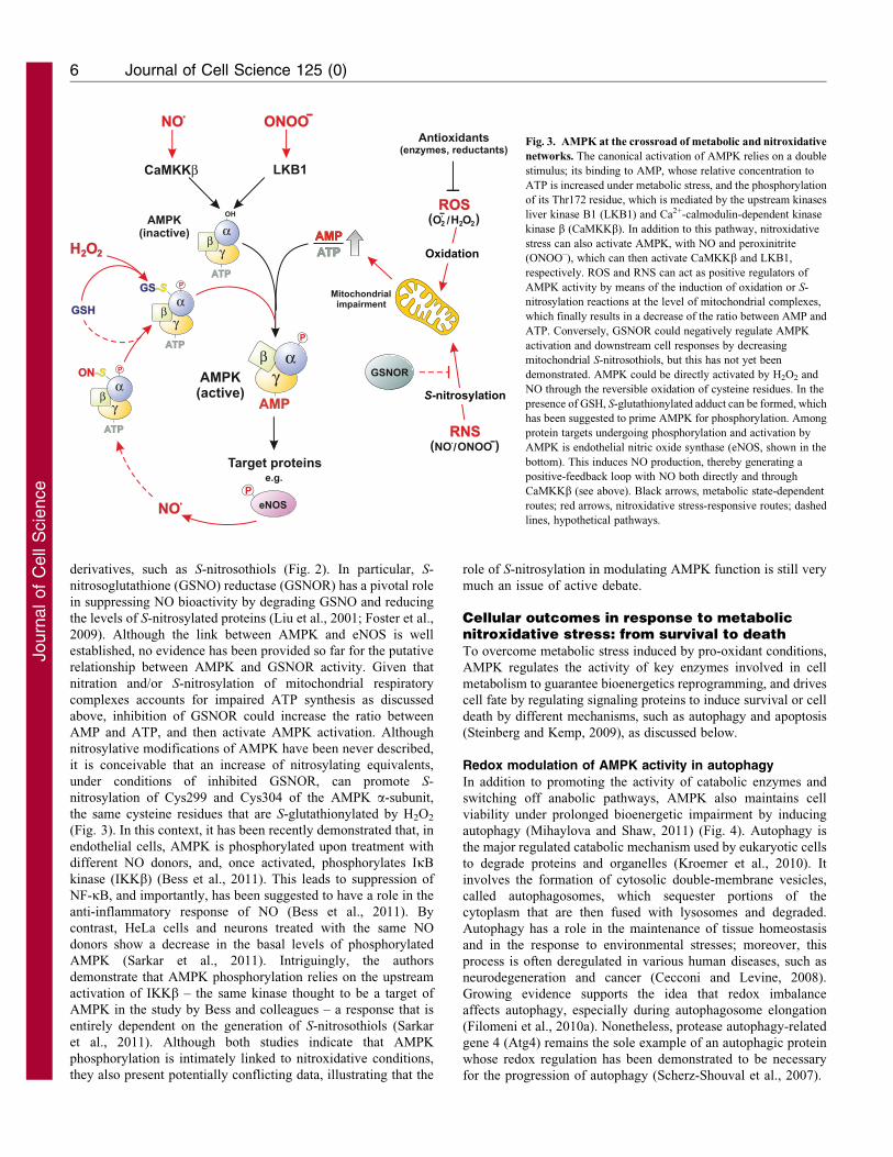
derivatives, such as S-nitrosothiols (Fig. 2). In particular, S-
nitrosoglutathione (GSNO) reductase (GSNOR) has a pivotal role
in suppressing NO bioactivity by degrading GSNO and reducing
the levels of S-nitrosylated proteins (Liu et al., 2001; Foster et al.,
2009). Although the link between AMPK and eNOS is well
established, no evidence has been provided so far for the putative
relationship between AMPK and GSNOR activity. Given that
nitration and/or S-nitrosylation of mitochondrial respiratory
complexes accounts for impaired ATP synthesis as discussed
above, inhibition of GSNOR could increase the ratio between
AMP and ATP, and then activate AMPK activation. Although
nitrosylative modifications of AMPK have been never described,
it is conceivable that an increase of nitrosylating equivalents,
under conditions of inhibited GSNOR, can promote S-
nitrosylation of Cys299 and Cys304 of the AMPK a-subunit,
the same cysteine residues that are S-glutathionylated by H2O2
(Fig. 3). In this context, it has been recently demonstrated that, in
endothelial cells, AMPK is phosphorylated upon treatment with
different NO donors, and, once activated, phosphorylates IkB
kinase (IKKb) (Bess et al., 2011). This leads to suppression of
NF-kB, and importantly, has been suggested to have a role in the
anti-inflammatory response of NO (Bess et al., 2011). By
contrast, HeLa cells and neurons treated with the same NO
donors show a decrease in the basal levels of phosphorylated
AMPK (Sarkar et al., 2011). Intriguingly, the authors
demonstrate that AMPK phosphorylation relies on the upstream
activation of IKKb – the same kinase thought to be a target of
AMPK in the study by Bess and colleagues – a response that is
entirely dependent on the generation of S-nitrosothiols (Sarkar
et al., 2011). Although both studies indicate that AMPK
phosphorylation is intimately linked to nitroxidative conditions,
they also present potentially conflicting data, illustrating that the
role of S-nitrosylation in modulating AMPK function is still very
much an issue of active debate.
Cellular outcomes in response to metabolicnitroxidative stress: from survival to deathTo overcome metabolic stress induced by pro-oxidant conditions,
AMPK regulates the activity of key enzymes involved in cell
metabolism to guarantee bioenergetics reprogramming, and drives
cell fate by regulating signaling proteins to induce survival or cell
death by different mechanisms, such as autophagy and apoptosis
(Steinberg and Kemp, 2009), as discussed below.
Redox modulation of AMPK activity in autophagy
In addition to promoting the activity of catabolic enzymes and
switching off anabolic pathways, AMPK also maintains cell
viability under prolonged bioenergetic impairment by inducing
autophagy (Mihaylova and Shaw, 2011) (Fig. 4). Autophagy is
the major regulated catabolic mechanism used by eukaryotic cells
to degrade proteins and organelles (Kroemer et al., 2010). It
involves the formation of cytosolic double-membrane vesicles,
called autophagosomes, which sequester portions of the
cytoplasm that are then fused with lysosomes and degraded.
Autophagy has a role in the maintenance of tissue homeostasis
and in the response to environmental stresses; moreover, this
process is often deregulated in various human diseases, such as
neurodegeneration and cancer (Cecconi and Levine, 2008).
Growing evidence supports the idea that redox imbalance
affects autophagy, especially during autophagosome elongation
(Filomeni et al., 2010a). Nonetheless, protease autophagy-related
gene 4 (Atg4) remains the sole example of an autophagic protein
whose redox regulation has been demonstrated to be necessary
for the progression of autophagy (Scherz-Shouval et al., 2007).
AMPATP
AMPK(inactive)
OH O2H2O2-( (/ROS
γ
AMP
ATP
LKB1
αβ
P
γαβ
CaMKKβ
AMPK(active)
ONOONO -( ( /RNS
Mitochondrialimpairment
O2H2
ONOO-NO
GSH
P
γATP
αβ
ON-S
eNOSP
NO
P
γATP
αβ
GS-S
Oxidation
S-nitrosylation
GSNOR
Antioxidants(enzymes, reductants)
AMPATP
Target proteinse.g.
Fig. 3. AMPK at the crossroad of metabolic and nitroxidative
networks. The canonical activation of AMPK relies on a double
stimulus; its binding to AMP, whose relative concentration to
ATP is increased under metabolic stress, and the phosphorylation
of its Thr172 residue, which is mediated by the upstream kinases
liver kinase B1 (LKB1) and Ca2+-calmodulin-dependent kinase
kinase b (CaMKKb). In addition to this pathway, nitroxidative
stress can also activate AMPK, with NO and peroxinitrite
(ONOO–), which can then activate CaMKKb and LKB1,
respectively. ROS and RNS can act as positive regulators of
AMPK activity by means of the induction of oxidation or S-
nitrosylation reactions at the level of mitochondrial complexes,
which finally results in a decrease of the ratio between AMP and
ATP. Conversely, GSNOR could negatively regulate AMPK
activation and downstream cell responses by decreasing
mitochondrial S-nitrosothiols, but this has not yet been
demonstrated. AMPK could be directly activated by H2O2 and
NO through the reversible oxidation of cysteine residues. In the
presence of GSH, S-glutathionylated adduct can be formed, which
has been suggested to prime AMPK for phosphorylation. Among
protein targets undergoing phosphorylation and activation by
AMPK is endothelial nitric oxide synthase (eNOS, shown in the
bottom). This induces NO production, thereby generating a
positive-feedback loop with NO both directly and through
CaMKKb (see above). Black arrows, metabolic state-dependent
routes; red arrows, nitroxidative stress-responsive routes; dashed
lines, hypothetical pathways.
Journal of Cell Science 125 (0)6
Journ
alof
Cell
Scie
nce
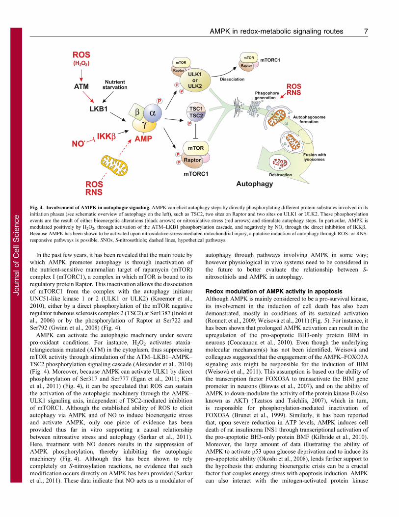
In the past few years, it has been revealed that the main route by
which AMPK promotes autophagy is through inactivation of
the nutrient-sensitive mammalian target of rapamycin (mTOR)
complex I (mTORC1), a complex in which mTOR is bound to its
regulatory protein Raptor. This inactivation allows the dissociation
of mTORC1 from the complex with the autophagy initiator
UNC51-like kinase 1 or 2 (ULK1 or ULK2) (Kroemer et al.,
2010), either by a direct phosphorylation of the mTOR negative
regulator tuberous sclerosis complex 2 (TSC2) at Ser1387 (Inoki et
al., 2006) or by the phosphorylation of Raptor at Ser722 and
Ser792 (Gwinn et al., 2008) (Fig. 4).
AMPK can activate the autophagic machinery under severe
pro-oxidant conditions. For instance, H2O2 activates ataxia-
telangiectasia mutated (ATM) in the cytoplasm, thus suppressing
mTOR activity through stimulation of the ATM–LKB1–AMPK–
TSC2 phosphorylation signaling cascade (Alexander et al., 2010)
(Fig. 4). Moreover, because AMPK can activate ULK1 by direct
phosphorylation of Ser317 and Ser777 (Egan et al., 2011; Kim
et al., 2011) (Fig. 4), it can be speculated that ROS can sustain
the activation of the autophagic machinery through the AMPK–
ULK1 signaling axis, independent of TSC2-mediated inhibition
of mTORC1. Although the established ability of ROS to elicit
autophagy via AMPK and of NO to induce bioenergetic stress
and activate AMPK, only one piece of evidence has been
provided thus far in vitro supporting a causal relationship
between nitrosative stress and autophagy (Sarkar et al., 2011).
Here, treatment with NO donors results in the suppression of
AMPK phosphorylation, thereby inhibiting the autophagic
machinery (Fig. 4). Although this has been shown to rely
completely on S-nitrosylation reactions, no evidence that such
modification occurs directly on AMPK has been provided (Sarkar
et al., 2011). These data indicate that NO acts as a modulator of
autophagy through pathways involving AMPK in some way;
however physiological in vivo systems need to be considered in
the future to better evaluate the relationship between S-
nitrosothiols and AMPK in autophagy.
Redox modulation of AMPK activity in apoptosis
Although AMPK is mainly considered to be a pro-survival kinase,
its involvement in the induction of cell death has also been
demonstrated, mostly in conditions of its sustained activation
(Ronnett et al., 2009; Weisova et al., 2011) (Fig. 5). For instance, it
has been shown that prolonged AMPK activation can result in the
upregulation of the pro-apoptotic BH3-only protein BIM in
neurons (Concannon et al., 2010). Even though the underlying
molecular mechanism(s) has not been identified, Weisova and
colleagues suggested that the engagement of the AMPK–FOXO3A
signaling axis might be responsible for the induction of BIM
(Weisova et al., 2011). This assumption is based on the ability of
the transcription factor FOXO3A to transactivate the BIM gene
promoter in neurons (Biswas et al., 2007), and on the ability of
AMPK to down-modulate the activity of the protein kinase B (also
known as AKT) (Tzatsos and Tsichlis, 2007), which in turn,
is responsible for phosphorylation-mediated inactivation of
FOXO3A (Brunet et al., 1999). Similarly, it has been reported
that, upon severe reduction in ATP levels, AMPK induces cell
death of rat insulinoma INS1 through transcriptional activation of
the pro-apoptotic BH3-only protein BMF (Kilbride et al., 2010).
Moreover, the large amount of data illustrating the ability of
AMPK to activate p53 upon glucose deprivation and to induce its
pro-apoptotic ability (Okoshi et al., 2008), lends further support to
the hypothesis that enduring bioenergetic crisis can be a crucial
factor that couples energy stress with apoptosis induction. AMPK
can also interact with the mitogen-activated protein kinase
AAMP
P
γαβ
P
TSC1TSC2
P
P
mTORC1
ULK1or
ULK2
mTOR
Raptor
mTORC1
Destruction
Fusion withlysosomes
Autophagosomeformation
Phagophoregeneration
P
O )2(H2
P
LKB1
ATM
ROS
Dissociation
mTOR
Raptor
mTOR
Raptor
Nutrientstarvation ROS
RNS
NO
ROSRNS
Autophagy
IKKβ
Fig. 4. Involvement of AMPK in autophagic signaling. AMPK can elicit autophagy steps by directly phosphorylating different protein substrates involved in its
initiation phases (see schematic overview of autophagy on the left), such as TSC2, two sites on Raptor and two sites on ULK1 or ULK2. These phosphorylation
events are the result of either bioenergetic alterations (black arrows) or nitroxidative stress (red arrows) and stimulate autophagy steps. In particular, AMPK is
modulated positively by H2O2, through activation of the ATM–LKB1 phosphorylation cascade, and negatively by NO, through the direct inhibition of IKKb.
Because AMPK has been shown to be activated upon nitroxidative-stress-mediated mitochondrial injury, a putative induction of autophagy through ROS- or RNS-
responsive pathways is possible. SNOs, S-nitrosothiols; dashed lines, hypothetical pathways.
AMPK in redox-metabolic signaling routes 7
Journ
alof
Cell
Scie
nce
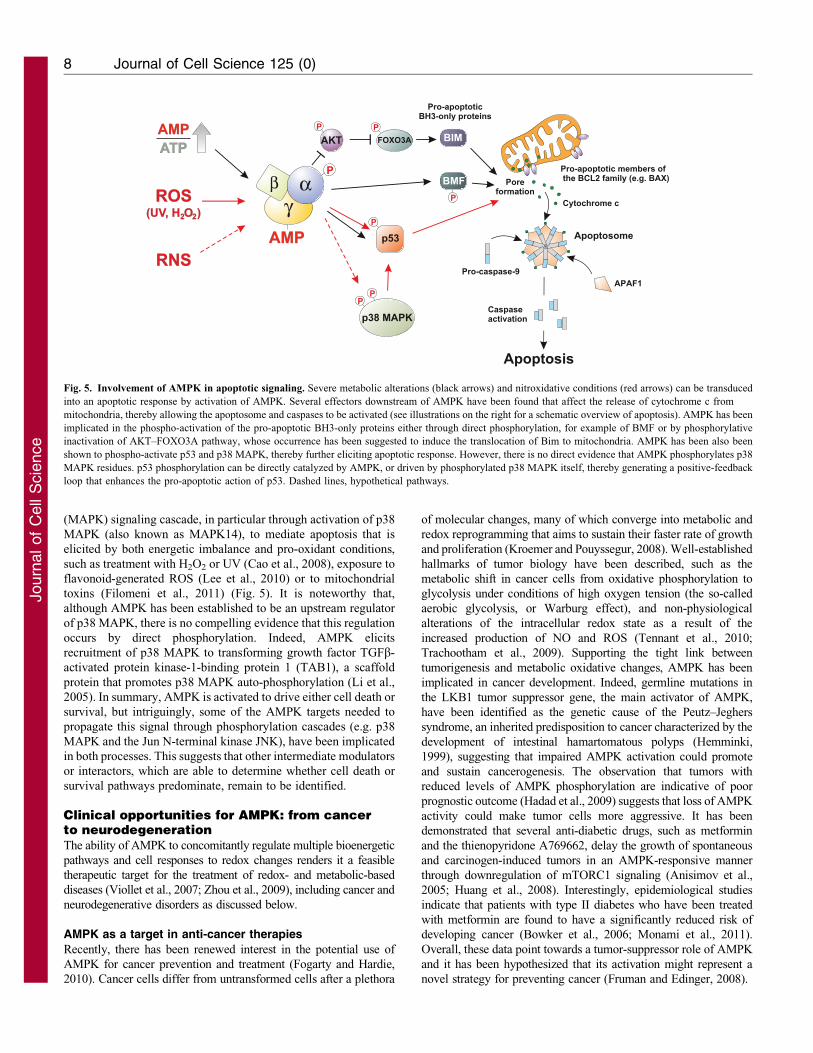
(MAPK) signaling cascade, in particular through activation of p38
MAPK (also known as MAPK14), to mediate apoptosis that is
elicited by both energetic imbalance and pro-oxidant conditions,
such as treatment with H2O2 or UV (Cao et al., 2008), exposure to
flavonoid-generated ROS (Lee et al., 2010) or to mitochondrial
toxins (Filomeni et al., 2011) (Fig. 5). It is noteworthy that,
although AMPK has been established to be an upstream regulator
of p38 MAPK, there is no compelling evidence that this regulation
occurs by direct phosphorylation. Indeed, AMPK elicits
recruitment of p38 MAPK to transforming growth factor TGFb-
activated protein kinase-1-binding protein 1 (TAB1), a scaffold
protein that promotes p38 MAPK auto-phosphorylation (Li et al.,
2005). In summary, AMPK is activated to drive either cell death or
survival, but intriguingly, some of the AMPK targets needed to
propagate this signal through phosphorylation cascades (e.g. p38
MAPK and the Jun N-terminal kinase JNK), have been implicated
in both processes. This suggests that other intermediate modulators
or interactors, which are able to determine whether cell death or
survival pathways predominate, remain to be identified.
Clinical opportunities for AMPK: from cancerto neurodegenerationThe ability of AMPK to concomitantly regulate multiple bioenergetic
pathways and cell responses to redox changes renders it a feasible
therapeutic target for the treatment of redox- and metabolic-based
diseases (Viollet et al., 2007; Zhou et al., 2009), including cancer and
neurodegenerative disorders as discussed below.
AMPK as a target in anti-cancer therapies
Recently, there has been renewed interest in the potential use of
AMPK for cancer prevention and treatment (Fogarty and Hardie,
2010). Cancer cells differ from untransformed cells after a plethora
of molecular changes, many of which converge into metabolic and
redox reprogramming that aims to sustain their faster rate of growth
and proliferation (Kroemer and Pouyssegur, 2008). Well-established
hallmarks of tumor biology have been described, such as the
metabolic shift in cancer cells from oxidative phosphorylation to
glycolysis under conditions of high oxygen tension (the so-called
aerobic glycolysis, or Warburg effect), and non-physiological
alterations of the intracellular redox state as a result of the
increased production of NO and ROS (Tennant et al., 2010;
Trachootham et al., 2009). Supporting the tight link between
tumorigenesis and metabolic oxidative changes, AMPK has been
implicated in cancer development. Indeed, germline mutations in
the LKB1 tumor suppressor gene, the main activator of AMPK,
have been identified as the genetic cause of the Peutz–Jeghers
syndrome, an inherited predisposition to cancer characterized by the
development of intestinal hamartomatous polyps (Hemminki,
1999), suggesting that impaired AMPK activation could promote
and sustain cancerogenesis. The observation that tumors with
reduced levels of AMPK phosphorylation are indicative of poor
prognostic outcome (Hadad et al., 2009) suggests that loss of AMPK
activity could make tumor cells more aggressive. It has been
demonstrated that several anti-diabetic drugs, such as metformin
and the thienopyridone A769662, delay the growth of spontaneous
and carcinogen-induced tumors in an AMPK-responsive manner
through downregulation of mTORC1 signaling (Anisimov et al.,
2005; Huang et al., 2008). Interestingly, epidemiological studies
indicate that patients with type II diabetes who have been treated
with metformin are found to have a significantly reduced risk of
developing cancer (Bowker et al., 2006; Monami et al., 2011).
Overall, these data point towards a tumor-suppressor role of AMPK
and it has been hypothesized that its activation might represent a
novel strategy for preventing cancer (Fruman and Edinger, 2008).
AMP
P
γαβ
Pro-apoptotic members ofthe BCL2 family (e.g. BAX)
APAF1
Cytochrome c
Apoptosome
Pro-caspase-9
Apoptosis
FOXO3AP
BIM
Pro-apoptoticBH3-only proteins
BMFP
p53P
p38 MAPKP
P
O )2(UV, H2
RNS
ROS
AMPATP
Poreformation
Caspaseactivation
AKTP
Fig. 5. Involvement of AMPK in apoptotic signaling. Severe metabolic alterations (black arrows) and nitroxidative conditions (red arrows) can be transduced
into an apoptotic response by activation of AMPK. Several effectors downstream of AMPK have been found that affect the release of cytochrome c from
mitochondria, thereby allowing the apoptosome and caspases to be activated (see illustrations on the right for a schematic overview of apoptosis). AMPK has been
implicated in the phospho-activation of the pro-apoptotic BH3-only proteins either through direct phosphorylation, for example of BMF or by phosphorylative
inactivation of AKT–FOXO3A pathway, whose occurrence has been suggested to induce the translocation of Bim to mitochondria. AMPK has been also been
shown to phospho-activate p53 and p38 MAPK, thereby further eliciting apoptotic response. However, there is no direct evidence that AMPK phosphorylates p38
MAPK residues. p53 phosphorylation can be directly catalyzed by AMPK, or driven by phosphorylated p38 MAPK itself, thereby generating a positive-feedback
loop that enhances the pro-apoptotic action of p53. Dashed lines, hypothetical pathways.
Journal of Cell Science 125 (0)8
Journ
alof
Cell
Scie
nce

In addition to studies that reveal the ability of AMPKactivators to act as anticancer agents, recent data suggests that
AMPK activation also represents a novel and feasible therapeuticstrategy for cancer treatment. For example, chemotherapeuticshave been developed that selectively affect the growth of tumorcells and their viability by promoting AMPK activation.
Particularly, the methylating drug temozolomide and the cell-permeable C6 ceramide have been shown to induce apoptosis inprimary cultured human glioblastoma cells and to sensitize
multiple cancer cell lines to doxorubicin by eliciting AMPKactivation (Ji et al., 2010; Zhang et al., 2010). Furthermore, theobservations that the phosphatidylinositol ether lipid analogues
(PIAs) (Memmott et al., 2008), the isatin-Schiff base copper(II)complexes (Filomeni et al., 2009; Filomeni et al., 2011) and somenutraceuticals (e.g. curcumin and selenium) (Hwang et al., 2006;Pan et al., 2008), are able to cause death in various cancer cell
lines by triggering AMPK-dependent apoptosis, further highlightthe potential role of AMPK as a promising target for drugsreversing, suppressing or preventing carcinogenic progression.
Nevertheless, it is worthwhile noting that, as observed for mostanticancer drugs, AMPK activators might also cause detrimentalside effects. Indeed, AMPK activation by anticancer compounds
has been shown to promote tumor cell survival instead ofcell death owing to activation of AMPK-mediated autophagicpathways (see below). Several lines of evidence show that
autophagy activation enables long-term survival, keeping cancercells alive when limited angiogenesis leads to nutrientdeprivation and hypoxia. AMPK-mediated autophagy appearsto enhance the resistance of cancer cells to chemotherapeutic
agents (e.g. cisplatin), and to naturally occurring molecules (e.g.polyphenols), which have been considered to be promisinganticancer drugs owing to their ability to induce metabolic
oxidative stress (Harhaji-Trajkovic et al., 2009; Filomeni et al.,2010b). In light of these results, it is reasonable to considerthat although targeting AMPK remains a feasible approach to
selectively kill tumor cells, the possible activation of AMPK-mediated pathways mediating tumor resistance, such asautophagy, should be taken into account.
Dual role of AMPK in neurodegenerative disease
Although AMPK-mediated autophagy might represent theAchilles’ heel of some chemotherapeutic approaches for the
treatment of proliferative diseases as discussed above (Bhutiaet al., 2010; Yang et al., 2010; Rosenfeldt, and Ryan, 2011), itcould be beneficial in non-renewing tissues, such as neurons
(Marino et al., 2011). Several observations demonstrate thatAMPK-mediated autophagy has a cytoprotective effect in somemodels of neurodegenerative disease, such as Huntington’s andAlzheimer’s diseases, because it contributes to the removal of
aggregate-prone mutant proteins and to sustaining neuronalbioenergetics, which is negatively affected underneurodegenerative-associated pro-oxidant conditions (Spencer
et al., 2009; Wong and Cuervo, 2010; Underwood et al., 2010;Vingtdeux et al., 2011; Vingtdeux et al., 2010). In support of thisassumption, it has been recently suggested that NO-mediated
inhibition of autophagy, which is associated with AMPKdephosphorylation, gives rise to a significant increase inhuntingtin-containing aggregates in neurons that express the
mutant form of the protein (Sarkar et al., 2011). However,although short-term activation of AMPK could mediate anadaptive neuronal quality control through induction of the
autophagic machinery, its persistent activation is not beneficialfor neurons (Ronnett et al., 2009). In fact, several in vitro studies
indicate that activation of AMPK over longer periods isharmful. Indeed, the pharmacological activator of AMPK, 5-aminoimidazole-4-carboxamide-1-b-D-ribofuranoside (AICAR),
was found to be pro-apoptotic in several human and mouseneuronal cell lines (Garcia-Gil et al., 2003). Moreover, it hasbeen shown that metformin significantly increases the in vivo
generation of b-amyloid peptide, which has a crucial role in thepathogenesis of Alzheimer’s disease (Chen, Y. et al., 2009).Likewise, in vivo studies suggest that AMPK activation that
occurs after cerebral ischemia is detrimental to neuronal survival.For example, genetic and pharmacological inhibition of AMPKunder these conditions reduces stroke damage (McCullough et al.,2005; Li et al., 2007), reinforcing the hypothesis that an excessive
and sustained activation of neuronal AMPK when oxygen andglucose substrates are lacking might induce a ‘metabolic-failure-like’ state leading to neuronal demise.
Conclusions and PerspectivesIn addition to the signaling network upstream of AMPK
activation, which senses and responds to metabolic alterationsin a concerted manner, a growing number of observations haverecently indicated that AMPK can be activated even upon
nitroxidative unbalance. Compelling evidence for whether thisactivation is direct, exclusively through cysteine oxidationwithout perturbing adenylate energy charge, or indirect,through the impairment of mitochondrial ATP-generating
machinery, is still limited. However, these findings, and thenovel concepts arising from them, reinforce the concept of thetight link between cellular (bio)energetics and redox status. They
also open new perspectives with regard to the wide impactthat AMPK can have on cellular processes that rely uponnitroxidative unbalance, and ultimately, on the role of AMPK in
human pathophysiology. Indeed, AMPK can activate proteintargets that are not restricted to metabolism, but are alsoimplicated in driving signaling pathways leading to diverse
cellular outcomes, such as apoptosis, proliferation andautophagy. Along this line, the possible use of drugs thatmodulate AMPK activity represents a clinical opportunity fortreatment of diseases that are tightly related to metabolic
dysfunction, and pathological states, such as cancer andneurodegeneration.
FundingThis work was partially supported by grants from Ministero dellaSalute, Progetto Giovani Ricercatori 2008 [grant number GR-2008-1138121]; Italian Association for Cancer Research (AIRC) [grantnumber IG 10636]; and the Ministero dell’Universita e della Ricerca(MIUR).
ReferencesAlexander, A., Cai, S. L., Kim, J., Nanez, A., Sahin, M., MacLean, K. H., Inoki, K.,
Guan, K. L., Shen, J., Person, M. D. et al. (2010). ATM signals to TSC2 in thecytoplasm to regulate mTORC1 in response to ROS. Proc. Natl. Acad. Sci. USA 107,4153-4158.
Almeida, A., Cidad, P., Delgado-Esteban, M., Fernandez, E., Garcıa-Nogales, P.
and Bolanos, J. P. (2005). Inhibition of mitochondrial respiration by nitric oxide: itsrole in glucose metabolism and neuroprotection. J. Neurosci. Res. 79, 166-171.
An, Z., Wang, H., Song, P., Zhang, M., Geng, X. and Zou, M. H. (2007). Nicotine-induced activation of AMP-activated protein kinase inhibits fatty acid synthase in3T3L1 adipocytes: a role for oxidant stress. J. Biol. Chem. 282, 26793-26801.
Anisimov, V. N., Berstein, L. M., Egormin, P. A., Piskunova, T. S., Popovich, I. G.,
Zabezhinski, M. A., Kovalenko, I. G., Poroshina, T. E., Semenchenko, A. V.,Provinciali, M. et al. (2005). Effect of metformin on life span and on the
AMPK in redox-metabolic signaling routes 9
Journ
alof
Cell
Scie
nce
development of spontaneous mammary tumors in HER-2/neu transgenic mice. Exp.
Gerontol. 40, 685-693.
Ataullakhanov, F. I. and Vitvitsky, V. M. (2002). What determines the intracellular
ATP concentration. Biosci. Rep. 22, 501-511.
Atkinson, D. E. (1968). The energy charge of the adenylate pool as a regulatory
parameter. Interaction with feedback modifiers. Biochemistry 7, 4030-4034.
Atkinson, D. E. (1970). Adenine nucleotides as universal stoichiometric metaboliccoupling agents. Adv. Enzyme Regul. 9, 207-219.
Barnes, K., Ingram, J. C., Porras, O. H., Barros, L. F., Hudson, E. R., Fryer, L. G.,
Foufelle, F., Carling, D., Hardie, D. G. and Baldwin, S. A. (2002). Activation of
GLUT1 by metabolic and osmotic stress: potential involvement of AMP-activatedprotein kinase (AMPK). J. Cell Sci. 115, 2433-2442.
Bess, E., Fisslthaler, B., Fromel, T. and Fleming, I. (2011). Nitric oxide-inducedactivation of the AMP-activated protein kinase a2 subunit attenuates IkB kinase
activity and inflammatory responses in endothelial cells. PLoS ONE 6, e20848.
Bhutia, S. K., Kegelman, T. P., Das, S. K., Azab, B., Su, Z. Z., Lee, S. G., Sarkar, D.
and Fisher, P. B. (2010). Astrocyte elevated gene-1 induces protective autophagy.Proc. Natl. Acad. Sci. USA 107, 22243-22248.
Biswas, S. C., Shi, Y., Sproul, A. and Greene, L. A. (2007). Pro-apoptotic Biminduction in response to nerve growth factor deprivation requires simultaneous
activation of three different death signaling pathways. J. Biol. Chem. 282, 29368-29374.
Bowker, S. L., Majumdar, S. R., Veugelers, P. and Johnson, J. A. (2006). Increasedcancer-related mortality for patients with type 2 diabetes who use sulfonylureas or
insulin. Diabetes Care 29, 254-258.
Brunet, A., Bonni, A., Zigmond, M. J., Lin, M. Z., Juo, P., Hu, L. S., Anderson,
M. J., Arden, K. C., Blenis, J. and Greenberg, M. E. (1999). Akt promotes cell
survival by phosphorylating and inhibiting a Forkhead transcription factor. Cell 96,857-868.
Calvert, J. W., Gundewar, S., Jha, S., Greer, J. J., Bestermann, W. H., Tian, R. and
Lefer, D. J. (2008). Acute metformin therapy confers cardioprotection against
myocardial infarction via AMPK-eNOS-mediated signaling. Diabetes 57, 696-705.
Cao, C., Lu, S., Kivlin, R., Wallin, B., Card, E., Bagdasarian, A., Tamakloe, T.,
Chu, W. M., Guan, K. L. and Wan, Y. (2008). AMP-activated protein kinasecontributes to UV- and H2O2-induced apoptosis in human skin keratinocytes. J. Biol.
Chem. 283, 28897-28908.
Cecconi, F. and Levine, B. (2008). The role of autophagy in mammalian development:
cell makeover rather than cell death. Dev. Cell 15, 344-357.
Chen, L., Jiao, Z. H., Zheng, L. S., Zhang, Y. Y., Xie, S. T., Wang, Z. X. and Wu,
J. W. (2009). Structural insight into the autoinhibition mechanism of AMP-activatedprotein kinase. Nature 459, 1146-1149.
Chen, Y., Zhou, K., Wang, R., Liu, Y., Kwak, Y. D., Ma, T., Thompson, R. C.,
Zhao, Y., Smith, L., Gasparini, L. et al. (2009). Antidiabetic drug metformin(GlucophageR) increases biogenesis of Alzheimer’s amyloid peptides via up-
regulating BACE1 transcription. Proc. Natl. Acad. Sci. USA 106, 3907-3912.
Chen, Z. P., Mitchelhill, K. I., Michell, B. J., Stapleton, D., Rodriguez-Crespo, I.,
Witters, L. A., Power, D. A., Ortiz de Montellano, P. R. and Kemp, B. E. (1999).AMP-activated protein kinase phosphorylation of endothelial NO synthase. FEBS
Lett. 443, 285-289.
Choi, S. L., Kim, S. J., Lee, K. T., Kim, J., Mu, J., Birnbaum, M. J., Soo Kim, S. and
Ha, J. (2001). The regulation of AMP-activated protein kinase by H(2)O(2).Biochem. Biophys. Res. Commun. 287, 92-97.
Concannon, C. G., Tuffy, L. P., Weisova, P., Bonner, H. P., Davila, D., Bonner, C.,
Devocelle, M. C., Strasser, A., Ward, M. W. and Prehn, J. H. (2010). AMP kinase-
mediated activation of the BH3-only protein Bim couples energy depletion to stress-induced apoptosis. J. Cell Biol. 189, 83-94.
Crute, B. E., Seefeld, K., Gamble, J., Kemp, B. E. and Witters, L. A. (1998).Functional domains of the alpha1 catalytic subunit of the AMP-activated protein
kinase. J. Biol. Chem. 273, 35347-35354.
Egan, D. F., Shackelford, D. B., Mihaylova, M. M., Gelino, S., Kohnz, R. A.,
Mair, W., Vasquez, D. S., Joshi, A., Gwinn, D. M., Taylor, R. et al. (2011).Phosphorylation of ULK1 (hATG1) by AMP-activated protein kinase connects
energy sensing to mitophagy. Science 331, 456-461.
Emerling, B. M., Weinberg, F., Snyder, C., Burgess, Z., Mutlu, G. M., Viollet, B.,
Budinger, G. R. and Chandel, N. S. (2009). Hypoxic activation of AMPK is
dependent on mitochondrial ROS but independent of an increase in AMP/ATP ratio.Free Radic. Biol. Med. 46, 1386-1391.
Filomeni, G., Piccirillo, S., Graziani, I., Cardaci, S., Da Costa Ferreira, A. M.,
Rotilio, G. and Ciriolo, M. R. (2009). The isatin-Schiff base copper(II) complex
Cu(isaepy)2 acts as delocalized lipophilic cation, yields widespread mitochondrialoxidative damage and induces AMP-activated protein kinase-dependent apoptosis.
Carcinogenesis 30, 1115-1124.
Filomeni, G., Desideri, E., Cardaci, S., Rotilio, G. and Ciriolo, M. R. (2010a). Under
the ROS…thiol network is the principal suspect for autophagy commitment.Autophagy 6, 999-1005.
Filomeni, G., Desideri, E., Cardaci, S., Graziani, I., Piccirillo, S., Rotilio, G. and
Ciriolo, M. R. (2010b). Carcinoma cells activate AMP-activated protein kinase-
dependent autophagy as survival response to kaempferol-mediated energeticimpairment. Autophagy 6, 202-216.
Filomeni, G., Cardaci, S., Da Costa Ferreira, A. M., Rotilio, G. and Ciriolo, M. R.
(2011). Metabolic oxidative stress elicited by the copper(II) complex [Cu(isaepy)2]
triggers apoptosis in SH-SY5Y cells through the induction of the AMP-activated
protein kinase/p38MAPK/p53 signalling axis: evidence for a combined use with 3-bromopyruvate in neuroblastoma treatment. Biochem. J. 437, 443-453.
Fogarty, S. and Hardie, D. G. (2010). Development of protein kinase activators:AMPK as a target in metabolic disorders and cancer. Biochim. Biophys. Acta 1804,581-591.
Foster, M. W., Hess, D. T. and Stamler, J. S. (2009). Protein S-nitrosylation in healthand disease: a current perspective. Trends Mol. Med. 15, 391-404.
Fruman, D. A. and Edinger, A. L. (2008). Cancer therapy: staying current with AMPK.Biochem. J. 412, e3-e5.
Garcia-Gil, M., Pesi, R., Perna, S., Allegrini, S., Giannecchini, M., Camici, M. andTozzi, M. G. (2003). 59-aminoimidazole-4-carboxamide riboside induces apoptosis inhuman neuroblastoma cells. Neuroscience 117, 811-820.
Ghillebert, R., Swinnen, E., Wen, J., Vandesteene, L., Ramon, M., Norga, K.,Rolland, F. and Winderickx, J. (2011). The AMPK/SNF1/SnRK1 fuel gauge andenergy regulator: structure, function and regulation. FEBS J. 278, 3978-3990.
Gwinn, D. M., Shackelford, D. B., Egan, D. F., Mihaylova, M. M., Mery, A.,Vasquez, D. S., Turk, B. E. and Shaw, R. J. (2008). AMPK phosphorylation ofraptor mediates a metabolic checkpoint. Mol. Cell 30, 214-226.
Hadad, S. M., Baker, L., Quinlan, P. R., Robertson, K. E., Bray, S. E., Thomson, G.,
Kellock, D., Jordan, L. B., Purdie, C. A., Hardie, D. G. et al. (2009). Histologicalevaluation of AMPK signalling in primary breast cancer. BMC Cancer 9, 307.
Hardie, D. G. (2007). AMP-activated/SNF1 protein kinases: conserved guardians ofcellular energy. Nat. Rev. Mol. Cell Biol. 8, 774-785.
Hardie, D. G. (2011). AMP-activated protein kinase: an energy sensor that regulates allaspects of cell function. Genes Dev. 25, 1895-1908.
Hardie, D. G., Carling, D. and Gamblin, S. J. (2011). AMP-activated protein kinase:also regulated by ADP? Trends Biochem. Sci. 36, 470-477.
Harhaji-Trajkovic, L., Vilimanovich, U., Kravic-Stevovic, T., Bumbasirevic, V. and
Trajkovic, V. (2009). AMPK-mediated autophagy inhibits apoptosis in cisplatin-treated tumour cells. J. Cell. Mol. Med. 13, 3644-3654.
Hawley, S. A., Pan, D. A., Mustard, K. J., Ross, L., Bain, J., Edelman, A. M.,
Frenguelli, B. G. and Hardie, D. G. (2005). Calmodulin-dependent protein kinasekinase-beta is an alternative upstream kinase for AMP-activated protein kinase. Cell
Metab. 2, 9-19.
Hawley, S. A., Ross, F. A., Chevtzoff, C., Green, K. A., Evans, A., Fogarty, S.,
Towler, M. C., Brown, L. J., Ogunbayo, O. A., Evans, A. M. et al. (2010). Use ofcells expressing gamma subunit variants to identify diverse mechanisms of AMPKactivation. Cell Metab. 11, 554-565.
Hemminki, A. (1999). The molecular basis and clinical aspects of Peutz-Jegherssyndrome. Cell. Mol. Life Sci. 55, 735-750.
Herrero-Martın, G., Høyer-Hansen, M., Garcıa-Garcıa, C., Fumarola, C., Farkas, T.,
Lopez-Rivas, A. and Jaattela, M. (2009). TAK1 activates AMPK-dependentcytoprotective autophagy in TRAIL-treated epithelial cells. EMBO J. 28, 677-685.
Huang, X., Wullschleger, S., Shpiro, N., McGuire, V. A., Sakamoto, K., Woods,
Y. L., McBurnie, W., Fleming, S. and Alessi, D. R. (2008). Important role of theLKB1-AMPK pathway in suppressing tumorigenesis in PTEN-deficient mice.Biochem. J. 412, 211-221.
Hwang, J. T., Kim, Y. M., Surh, Y. J., Baik, H. W., Lee, S. K., Ha, J. and Park, O. J.
(2006). Selenium regulates cyclooxygenase-2 and extracellular signal-regulatedkinase signaling pathways by activating AMP-activated protein kinase in coloncancer cells. Cancer Res. 66, 10057-10063.
Inoki, K., Ouyang, H., Zhu, T., Lindvall, C., Wang, Y., Zhang, X., Yang, Q.,Bennett, C., Harada, Y., Stankunas, K. et al. (2006). TSC2 integrates Wnt andenergy signals via a coordinated phosphorylation by AMPK and GSK3 to regulatecell growth. Cell 126, 955-968.
Jager, S., Handschin, C., St-Pierre, J. and Spiegelman, B. M. (2007). AMP-activatedprotein kinase (AMPK) action in skeletal muscle via direct phosphorylation of PGC-1alpha. Proc. Natl. Acad. Sci. USA 104, 12017-12022.
Ji, C., Yang, B., Yang, Y. L., He, S. H., Miao, D. S., He, L. and Bi, Z. G. (2010).Exogenous cell-permeable C6 ceramide sensitizes multiple cancer cell lines toDoxorubicin-induced apoptosis by promoting AMPK activation and mTORC1inhibition. Oncogene 29, 6557-6568.
Kahn, B. B., Alquier, T., Carling, D. and Hardie, D. G. (2005). AMP-activatedprotein kinase: ancient energy gauge provides clues to modern understanding ofmetabolism. Cell Metab. 1, 15-25.
Kawaguchi, T., Osatomi, K., Yamashita, H., Kabashima, T. and Uyeda, K. (2002).Mechanism for fatty acid ‘‘sparing’’ effect on glucose-induced transcription:regulation of carbohydrate-responsive element-binding protein by AMP-activatedprotein kinase. J. Biol. Chem. 277, 3829-3835.
Kilbride, S. M., Farrelly, A. M., Bonner, C., Ward, M. W., Nyhan, K. C.,Concannon, C. G., Wollheim, C. B., Byrne, M. M. and Prehn, J. H. (2010). AMP-activated protein kinase mediates apoptosis in response to bioenergetic stress throughactivation of the pro-apoptotic Bcl-2 homology domain-3-only protein BMF. J. Biol.
Chem. 285, 36199-36206.
Kim, J., Kundu, M., Viollet, B. and Guan, K. L. (2011). AMPK and mTOR regulateautophagy through direct phosphorylation of Ulk1. Nat. Cell Biol. 13, 132-141.
Kroemer, G. and Pouyssegur, J. (2008). Tumor cell metabolism: cancer’s Achilles’heel. Cancer Cell 13, 472-482.
Kroemer, G., Marino, G. and Levine, B. (2010). Autophagy and the integrated stressresponse. Mol. Cell 40, 280-293.
Kurth-Kraczek, E. J., Hirshman, M. F., Goodyear, L. J. and Winder, W. W. (1999).59 AMP-activated protein kinase activation causes GLUT4 translocation in skeletalmuscle. Diabetes 48, 1667-1671.
Journal of Cell Science 125 (0)10
Journ
alof
Cell
Scie
nce
Lee, Y. K., Hwang, J. T., Kwon, D. Y., Surh, Y. J. and Park, O. J. (2010). Inductionof apoptosis by quercetin is mediated through AMPKalpha1/ASK1/p38 pathway.Cancer Lett. 292, 228-236.
Levine, Y. C., Li, G. K. and Michel, T. (2007). Agonist-modulated regulation of AMP-activated protein kinase (AMPK) in endothelial cells. Evidence for an AMPK R Rac1R Akt R endothelial nitric-oxide synthase pathway. J. Biol. Chem. 282, 20351-20364.
Li, J., Miller, E. J., Ninomiya-Tsuji, J., Russell, R. R., 3rd and Young, L. H. (2005).AMP-activated protein kinase activates p38 mitogen-activated protein kinase byincreasing recruitment of p38 MAPK to TAB1 in the ischemic heart. Circ. Res. 97,872-879.
Li, J., Zeng, Z., Viollet, B., Ronnett, G. V. and McCullough, L. D. (2007).Neuroprotective effects of adenosine monophosphate-activated protein kinaseinhibition and gene deletion in stroke. Stroke 38, 2992-2999.
Liu, L., Hausladen, A., Zeng, M., Que, L., Heitman, J. and Stamler, J. S. (2001). Ametabolic enzyme for S-nitrosothiol conserved from bacteria to humans. Nature 410,490-494.
Long, Y. C. and Zierath, J. R. (2006). AMP-activated protein kinase signaling inmetabolic regulation. J. Clin. Invest. 116, 1776-1783.
Marino, G., Madeo, F. and Kroemer, G. (2011). Autophagy for tissue homeostasis andneuroprotection. Curr. Opin. Cell Biol. 23, 198-206.
McBride, A., Ghilagaber, S., Nikolaev, A. and Hardie, D. G. (2009). The glycogen-binding domain on the AMPK beta subunit allows the kinase to act as a glycogensensor. Cell Metab. 9, 23-34
McCullough, L. D., Zeng, Z., Li, H., Landree, L. E., McFadden, J. and Ronnett,G. V. (2005). Pharmacological inhibition of AMP-activated protein kinase providesneuroprotection in stroke. J. Biol. Chem. 280, 20493-20502.
Memmott, R. M., Gills, J. J., Hollingshead, M., Powers, M. C., Chen, Z., Kemp, B.,
Kozikowski, A. and Dennis, P. A. (2008). Phosphatidylinositol ether lipid analoguesinduce AMP-activated protein kinase-dependent death in LKB1-mutant non small celllung cancer cells. Cancer Res. 68, 580-588.
Merrill, G. F., Kurth, E. J., Hardie, D. G. and Winder, W. W. (1997). AICA ribosideincreases AMP-activated protein kinase, fatty acid oxidation, and glucose uptake inrat muscle. Am. J. Physiol. 273, E1107-E1112.
Mihaylova, M. M. and Shaw, R. J. (2011). The AMPK signalling pathway coordinatescell growth, autophagy and metabolism. Nat. Cell Biol. 13, 1016-1023.
Mitchelhill, K. I., Michell, B. J., House, C. M., Stapleton, D., Dyck, J., Gamble, J.,Ullrich, C., Witters, L. A. and Kemp, B. E. (1997). Posttranslational modificationsof the 59-AMP-activated protein kinase beta1 subunit. J. Biol. Chem. 272, 24475-24479.
Monami, M., Colombi, C., Balzi, D., Dicembrini, I., Giannini, S., Melani, C., Vitale, V.,
Romano, D., Barchielli, A., Marchionni, N. et al. (2011). Metformin and canceroccurrence in insulin-treated type 2 diabetic patients. Diabetes Care 34, 129-131.
Moncada, S. and Bolanos, J. P. (2006). Nitric oxide, cell bioenergetics andneurodegeneration. J. Neurochem. 97, 1676-1689.
Oakhill, J. S., Steel, R., Chen, Z. P., Scott, J. W., Ling, N., Tam, S. and Kemp, B. E.
(2011). AMPK is a direct adenylate charge-regulated protein kinase. Science 332,1433-1435.
Okoshi, R., Ozaki, T., Yamamoto, H., Ando, K., Koida, N., Ono, S., Koda, T.,Kamijo, T., Nakagawara, A. and Kizaki, H. (2008). Activation of AMP-activatedprotein kinase induces p53-dependent apoptotic cell death in response to energeticstress. J. Biol. Chem. 283, 3979-3987.
Pan, W., Yang, H., Cao, C., Song, X., Wallin, B., Kivlin, R., Lu, S., Hu, G., Di, W.
and Wan, Y. (2008). AMPK mediates curcumin-induced cell death in CaOV3ovarian cancer cells. Oncol. Rep. 20, 1553-1559.
Piantadosi, C. A. (2011). Regulation of mitochondrial processes by protein S-nitrosylation. Biochim. Biophys. Acta. (in press)
Poels, J., Spasic, M. R., Callaerts, P. and Norga, K. K. (2009). Expanding roles forAMP-activated protein kinase in neuronal survival and autophagy. Bioessays 31, 944-952.
Polekhina, G., Gupta, A., Michell, B. J., van Denderen, B., Murthy, S., Feil, S. C.,Jennings, I. G., Campbell, D. J., Witters, L. A., Parker, M. W. et al. (2003).AMPK beta subunit targets metabolic stress sensing to glycogen. Curr. Biol. 13, 867-871.
Reihill, J. A., Ewart, M. A., Hardie, D. G. and Salt, I. P. (2007). AMP-activatedprotein kinase mediates VEGF-stimulated endothelial NO production. Biochem.
Biophys. Res. Commun. 354, 1084-1088.Ronnett, G. V., Ramamurthy, S., Kleman, A. M., Landree, L. E. and Aja, S. (2009).
AMPK in the brain: its roles in energy balance and neuroprotection. J. Neurochem.
109 Suppl. 1, 17-23.Rosenfeldt, M. T. and Ryan, K. M. (2011). The multiple roles of autophagy in cancer.
Carcinogenesis 32, 955-963.Sarkar, S., Korolchuk, V. I., Renna, M., Imarisio, S., Fleming, A., Williams, A.,
Garcia-Arencibia, M., Rose, C., Luo, S., Underwood, B. R. et al. (2011). Complexinhibitory effects of nitric oxide on autophagy. Mol. Cell 43, 19-32.
Scherz-Shouval, R., Shvets, E., Fass, E., Shorer, H., Gil, L. and Elazar, Z. (2007).Reactive oxygen species are essential for autophagy and specifically regulate theactivity of Atg4. EMBO J. 26, 1749-1760.
Scott, J. W., Hawley, S. A., Green, K. A., Anis, M., Stewart, G., Scullion, G. A.,
Norman, D. G. and Hardie, D. G. (2004). CBS domains form energy-sensingmodules whose binding of adenosine ligands is disrupted by disease mutations. J.
Clin. Invest. 113, 274-284.
Spencer, B., Potkar, R., Trejo, M., Rockenstein, E., Patrick, C., Gindi, R., Adame, A.,
Wyss-Coray, T. and Masliah, E. (2009). Beclin 1 gene transfer activates autophagyand ameliorates the neurodegenerative pathology in alpha-synuclein models of
Parkinson’s and Lewy body diseases. J. Neurosci. 29, 13578-13588.
Steinberg, G. R. and Kemp, B. E. (2009). AMPK in health and disease. Physiol. Rev.
89, 1025-1078.
Tennant, D. A., Duran, R. V. and Gottlieb, E. (2010). Targeting metabolic
transformation for cancer therapy. Nat. Rev. Cancer 10, 267-277.
Trachootham, D., Alexandre, J. and Huang, P. (2009). Targeting cancer cells by
ROS-mediated mechanisms: a radical therapeutic approach? Nat. Rev. Drug Discov.
8, 579-591.
Tzatsos, A. and Tsichlis, P. N. (2007). Energy depletion inhibits phosphatidylinositol 3-kinase/Akt signaling and induces apoptosis via AMP-activated protein kinase-
dependent phosphorylation of IRS-1 at Ser-794. J. Biol. Chem. 282, 18069-18082.
Underwood, B. R., Imarisio, S., Fleming, A., Rose, C., Krishna, G., Heard, P.,
Quick, M., Korolchuk, V. I., Renna, M., Sarkar, S. et al. (2010). Antioxidants caninhibit basal autophagy and enhance neurodegeneration in models of polyglutamine
disease. Hum. Mol. Genet. 19, 3413-3429.
Vingtdeux, V., Chandakkar, P., Zhao, H., d’Abramo, C., Davies, P. and
Marambaud, P. (2011). Novel synthetic small-molecule activators of AMPK asenhancers of autophagy and amyloid-b peptide degradation. FASEB J. 25, 219-231.
Vingtdeux, V., Giliberto, L., Zhao, H., Chandakkar, P., Wu, Q., Simon, J. E., Janle, E.
M., Lobo, J., Ferruzzi, M. G., Davies, P. et al. (2010). AMP-activated protein kinasesignaling activation by resveratrol modulates amyloid-beta peptide metabolism. J. Biol.
Chem. 285, 9100-9113.
Viollet, B., Mounier, R., Leclerc, J., Yazigi, A., Foretz, M. and Andreelli, F. (2007).
Targeting AMP-activated protein kinase as a novel therapeutic approach for thetreatment of metabolic disorders. Diabetes Metab. 33, 395-402.
Weisova, P., Davila, D., Tuffy, L. P., Ward, M. W., Concannon, C. G. and Prehn,
J. H. (2011). Role of 59-adenosine monophosphate-activated protein kinase in cell
survival and death responses in neurons. Antioxid. Redox Signal. 14, 1863-1876.
Winder, W. W. and Hardie, D. G. (1996). Inactivation of acetyl-CoA carboxylase and
activation of AMP-activated protein kinase in muscle during exercise. Am. J. Physiol.
270, E299-E304.
Winder, W. W. and Hardie, D. G. (1999). AMP-activated protein kinase, a metabolicmaster switch: possible roles in type 2 diabetes. Am. J. Physiol. 277, E1-E10.
Wong, E. and Cuervo, A. M. (2010). Autophagy gone awry in neurodegenerativediseases. Nat. Neurosci. 13, 805-811.
Xiao, B., Heath, R., Saiu, P., Leiper, F. C., Leone, P., Jing, C., Walker, P. A., Haire, L.,
Eccleston, J. F., Davis, C. T. et al. (2007). Structural basis for AMP binding to
mammalian AMP-activated protein kinase. Nature 449, 496-500.
Xiao, B., Sanders, M. J., Underwood, E., Heath, R., Mayer, F. V., Carmena, D.,
Jing, C., Walker, P. A., Eccleston, J. F., Haire, L. F. et al. (2011). Structure ofmammalian AMPK and its regulation by ADP. Nature 472, 230-233.
Xie, Z., Dong, Y., Zhang, M., Cui, M. Z., Cohen, R. A., Riek, U., Neumann, D.,
Schlattner, U. and Zou, M. H. (2006). Activation of protein kinase C zeta byperoxynitrite regulates LKB1-dependent AMP-activated protein kinase in culturedendothelial cells. J. Biol. Chem. 281, 6366-6375.
Yang, P. M., Liu, Y. L., Lin, Y. C., Shun, C. T., Wu, M. S. and Chen, C. C. (2010).
Inhibition of autophagy enhances anticancer effects of atorvastatin in digestivemalignancies. Cancer Res. 70, 7699-7709.
Yu, L. F., Qiu, B. Y., Nan, F. J. and Li, J. (2010). AMPK activators as noveltherapeutics for type 2 diabetes. Curr. Top. Med. Chem. 10, 397-410.
Zhang, J., Xie, Z., Dong, Y., Wang, S., Liu, C. and Zou, M. H. (2008). Identificationof nitric oxide as an endogenous activator of the AMP-activated protein kinase in
vascular endothelial cells. J. Biol. Chem. 283, 27452-27461.
Zhang, W. B., Wang, Z., Shu, F., Jin, Y. H., Liu, H. Y., Wang, Q. J. and Yang, Y.
(2010). Activation of AMP-activated protein kinase by temozolomide contributes toapoptosis in glioblastoma cells via p53 activation and mTORC1 inhibition. J. Biol.
Chem. 285, 40461-40471.
Zheng, D., MacLean, P. S., Pohnert, S. C., Knight, J. B., Olson, A. L., Winder,
W. W. and Dohm, G. L. (2001). Regulation of muscle GLUT-4 transcription byAMP-activated protein kinase. J. Appl. Physiol. 91, 1073-1083.
Zhou, G., Myers, R., Li, Y., Chen, Y., Shen, X., Fenyk-Melody, J., Wu, M., Ventre, J.,
Doebber, T., Fujii, N. et al. (2001). Role of AMP-activated protein kinase in
mechanism of metformin action. J. Clin. Invest. 108, 1167-1174.
Zhou, G., Sebhat, I. K. and Zhang, B. B. (2009). AMPK activators--potential
therapeutics for metabolic and other diseases. Acta Physiol. (Oxf.) 196, 175-190.
Zmijewski, J. W., Banerjee, S., Bae, H., Friggeri, A., Lazarowski, E. R. and
Abraham, E. (2010). Exposure to hydrogen peroxide induces oxidation andactivation of AMP-activated protein kinase. J. Biol. Chem. 285, 33154-33164.
Zong, H., Ren, J. M., Young, L. H., Pypaert, M., Mu, J., Birnbaum, M. J. and
Shulman, G. I. (2002). AMP kinase is required for mitochondrial biogenesis inskeletal muscle in response to chronic energy deprivation. Proc. Natl. Acad. Sci. USA
99, 15983-15987.
Zou, M. H., Kirkpatrick, S. S., Davis, B. J., Nelson, J. S., Wiles, W. G. 4th,
Schlattner, U., Neumann, D., Brownlee, M., Freeman, M. B. and Goldman, M. H.
(2004). Activation of the AMP-activated protein kinase by the anti-diabetic drugmetformin in vivo. Role of mitochondrial reactive nitrogen species. J. Biol. Chem.
279, 43940-43951.
AMPK in redox-metabolic signaling routes 11
Journ
alof
Cell
Scie
nce