Vaccination against tumor cells expressing breast cancer ... · Proc. Nati. Acad. Sci. USA Vol. 87,...
5
Proc. Nati. Acad. Sci. USA Vol. 87, pp. 9498-9502, December 1990 Medical Sciences Vaccination against tumor cells expressing breast cancer epithelial tumor antigen (vaccine/recombinant vaccinia virus) MARA HAREUVENI*, CLAUDIE GAUTIER*, MARIE-PAULE KIENYt, DANIEL WRESCHNERt, PIERRE CHAMBONW, AND RICHARD LATHE*§ *Laboratoire de Gdn~tique Moldculaire des Eucaryotes-Unitd Associde 184 de Institut National de la Santd et de la Recherche Mddicale, Institut de Chimie Biologique, 11, rue Humann, 67085 Strasbourg Cddex, France; tTransg~ne SA, 11, rue de Molsheim, 67000 Strasbourg, France; and tDepartment of Microbiology, University of Tel Aviv, Tel Aviv, Israel Contributed by Pierre Chambon, September 6, 1990 ABSTRACT Ninety-one percent of breast tumors aber- rantly express an epithelial tumor antigen (ETA) identified by monoclonal antibody H23. Vaccinia virus recombinants ex- pressing tumor antigens have considerable promise in the active immunotherapy of cancer, and we have evaluated the potential of vaccinia recombinants expressing the secreted (S) and cell-associated (transmembrane, T) forms of H23 ETA to elicit immunity to tumor cells expressing ETA. Tumorigenic ras-transformed Fischer rat fibroblast lines FR-S and FR-T, expressing the S or T form of H23 ETA, respectively, were constructed for use in challenge experiments. Expression of H23 ETA in these lines was confirmed by Western blotting and immunofluorescence. When challenged by subcutaneous seed- ing of tumor cells, 97% (FR-S) and 91% (FR-T) of syngeneic Fischer rats rapidly developed tumors that failed to regress. Vaccination with recombinant vaccinia virus expressing ETA-T prior to challenge prevented tumor development in 82% of animals seeded with FR-T cells but in only 61% of animals seeded with FR-S. The vaccinia recombinant express- ing the S form was a less effective immunogen, and vaccination protected only 29-30% of animals from developing tumors upon challenge with either FR-S or -T cells. The increased immunogenicity of the recombinant expressing ETA-T was reflected in elevated levels of ETA-reactive antibody in vacci- nated animals, confirming that secreted antigens expressed from vaccinia virus are less effective immunogens than their membrane-associated counterparts. Tumor-associated antigens have considerable promise as targets for active or passive immunotherapy. Attempts have been made to elicit tumor immunity through inoculation of purified antigens or tumor lysates (1-7) or anti-idiotypic antibodies directed against tumor-associated antigens (8-10). Such approaches have met with only limited success, and we have used instead live recombinant vaccinia viruses express- ing tumor antigens to immunize against tumor cells. Inocu- lation of rodents with live recombinant vaccinia expressing tumor antigens of polyoma virus (11), bovine papillomavirus (12), and human papillomavirus (G. Meneguzzi, C. Cerni, M.-P.K., and R.L., unpublished work) was found to elicit rejection of cognate transformed cells. Although several lines of evidence suggest that an infective agent may contribute to breast cancer, the existence of such an agent has not been confirmed (13). We have therefore focused upon an endog- enous protein antigen that is aberrantly expressed in breast cancer. Monoclonal antibody (mAb) H23 was raised against particulate antigens released by T-47D breast tumor cells (14). mAb H23 detects an antigen [H23 epithelial tumor antigen (ETA)] present in 91% of breast tumors examined (14). H23 ETA can be detected in the body fluids of breast cancer patients, where its level correlates with disease status and poor prognosis (15). cDNA (16, 17) and genomic (18) cloning has revealed that a large central segment of the gene encoding H23 ETA consists of a multiple tandem repeat of a 60-nucleotide domain encoding a 20-amino acid sequence motif (17, 18). The number of repeat units differs substantially between individuals and between alleles (16), and copy- number variation has been observed between parent and child (M.H., unpublished data). The presence of similar variable-sized mucin-like glycoproteins in breast cancer and other adenocarcinomas has been reported by other groups (19-27). Sequence data indicate that H23 ETA is similar, if not identical, to the polymorphic episialins or epithelial mucins described by other groups (refs. 28-30; see refs. 31-33 for reviews). Comparison of the cDNA and genomic sequences encoding H23 ETA revealed the existence of two alternative mRNA species presumed to be generated by alternative splicing of a single precursor transcript (17). Translation of the two species is expected to result in two polypeptides that share a common secretion-signal sequence, amino terminus, and repeat array but that differ in their carboxyl-terminal regions. One of the two proteins, the transmembrane (T) form, contains a carboxyl-terminal hy- drophobic region (17) that is thought to be responsible for membrane association. The other species, the secreted (S) form, lacks this zone and appears in the culture medium (ref. 17 and unpublished data). Although RNA species corre- sponding to both forms are detected in breast cancer cells, the possibility that S-form mRNA is a partially spliced precursor of T mRNA has not been rigorously excluded (M.H. and R.L., unpublished data). We previously constructed vaccinia virus recombinants separately expressing the S and T forms of H23 ETA (VV- ETA-S and VV-ETA-T) and confirmed that the S form was predominantly secreted into the culture medium from VV- ETA-S-infected cells while the polypeptide encoded by VV- ETA-T remained cell-associated (M.H., D.W., M.-P.K., P.C., and R.L., unpublished data). We have now investigated the potential of the recombinants to elicit immunity to syngeneic tumor cells expressing H23 ETA and we report that rats inoculated with VV-ETA-T survive challenge with tumor cells expressing H23 ETA. MATERIALS AND METHODS Tumor Cells Expressing H23 ETA. Full-length cDNAs encoding the S and T forms of H23 ETA were constructed by Abbreviations: ETA, epithelial tumor antigen; mAb, monoclonal antibody; S. soluble; T, transmembrane. §Present address: AFRC Center for Genome Research, University of Edinburgh, King's Buildings, West Mains Road, Edinburgh EH9 3JQ, U.K. 9498 The publication costs of this article were defrayed in part by page charge payment. This article must therefore be hereby marked "advertisement" in accordance with 18 U.S.C. §1734 solely to indicate this fact. Downloaded by guest on May 2, 2020
Transcript of Vaccination against tumor cells expressing breast cancer ... · Proc. Nati. Acad. Sci. USA Vol. 87,...

Proc. Nati. Acad. Sci. USAVol. 87, pp. 9498-9502, December 1990Medical Sciences
Vaccination against tumor cells expressing breast cancer epithelialtumor antigen
(vaccine/recombinant vaccinia virus)
MARA HAREUVENI*, CLAUDIE GAUTIER*, MARIE-PAULE KIENYt, DANIEL WRESCHNERt, PIERRE CHAMBONW,AND RICHARD LATHE*§*Laboratoire de Gdn~tique Moldculaire des Eucaryotes-Unitd Associde 184 de Institut National de la Santd et de la Recherche Mddicale, Institut de ChimieBiologique, 11, rue Humann, 67085 Strasbourg Cddex, France; tTransg~ne SA, 11, rue de Molsheim, 67000 Strasbourg, France; and tDepartment ofMicrobiology, University of Tel Aviv, Tel Aviv, Israel
Contributed by Pierre Chambon, September 6, 1990
ABSTRACT Ninety-one percent of breast tumors aber-rantly express an epithelial tumor antigen (ETA) identified bymonoclonal antibody H23. Vaccinia virus recombinants ex-pressing tumor antigens have considerable promise in theactive immunotherapy of cancer, and we have evaluated thepotential of vaccinia recombinants expressing the secreted (S)and cell-associated (transmembrane, T) forms of H23 ETA toelicit immunity to tumor cells expressing ETA. Tumorigenicras-transformed Fischer rat fibroblast lines FR-S and FR-T,expressing the S or T form of H23 ETA, respectively, wereconstructed for use in challenge experiments. Expression ofH23 ETA in these lines was confirmed by Western blotting andimmunofluorescence. When challenged by subcutaneous seed-ing of tumor cells, 97% (FR-S) and 91% (FR-T) of syngeneicFischer rats rapidly developed tumors that failed to regress.Vaccination with recombinant vaccinia virus expressingETA-T prior to challenge prevented tumor development in82% of animals seeded with FR-T cells but in only 61% ofanimals seeded with FR-S. The vaccinia recombinant express-ing the S form was a less effective immunogen, and vaccinationprotected only 29-30% of animals from developing tumorsupon challenge with either FR-S or -T cells. The increasedimmunogenicity of the recombinant expressing ETA-T wasreflected in elevated levels of ETA-reactive antibody in vacci-nated animals, confirming that secreted antigens expressedfrom vaccinia virus are less effective immunogens than theirmembrane-associated counterparts.
Tumor-associated antigens have considerable promise astargets for active or passive immunotherapy. Attempts havebeen made to elicit tumor immunity through inoculation ofpurified antigens or tumor lysates (1-7) or anti-idiotypicantibodies directed against tumor-associated antigens (8-10).Such approaches have met with only limited success, and wehave used instead live recombinant vaccinia viruses express-ing tumor antigens to immunize against tumor cells. Inocu-lation of rodents with live recombinant vaccinia expressingtumor antigens of polyoma virus (11), bovine papillomavirus(12), and human papillomavirus (G. Meneguzzi, C. Cerni,M.-P.K., and R.L., unpublished work) was found to elicitrejection of cognate transformed cells. Although several linesof evidence suggest that an infective agent may contribute tobreast cancer, the existence of such an agent has not beenconfirmed (13). We have therefore focused upon an endog-enous protein antigen that is aberrantly expressed in breastcancer. Monoclonal antibody (mAb) H23 was raised againstparticulate antigens released by T-47D breast tumor cells(14). mAb H23 detects an antigen [H23 epithelial tumorantigen (ETA)] present in 91% of breast tumors examined
(14). H23 ETA can be detected in the body fluids of breastcancer patients, where its level correlates with disease statusand poor prognosis (15). cDNA (16, 17) and genomic (18)cloning has revealed that a large central segment of the geneencoding H23 ETA consists of a multiple tandem repeat of a60-nucleotide domain encoding a 20-amino acid sequencemotif (17, 18). The number of repeat units differs substantiallybetween individuals and between alleles (16), and copy-number variation has been observed between parent andchild (M.H., unpublished data). The presence of similarvariable-sized mucin-like glycoproteins in breast cancer andother adenocarcinomas has been reported by other groups(19-27). Sequence data indicate that H23 ETA is similar, ifnot identical, to the polymorphic episialins or epithelialmucins described by other groups (refs. 28-30; see refs.31-33 for reviews). Comparison of the cDNA and genomicsequences encoding H23 ETA revealed the existence of twoalternative mRNA species presumed to be generated byalternative splicing of a single precursor transcript (17).Translation of the two species is expected to result in twopolypeptides that share a common secretion-signal sequence,amino terminus, and repeat array but that differ in theircarboxyl-terminal regions. One of the two proteins, thetransmembrane (T) form, contains a carboxyl-terminal hy-drophobic region (17) that is thought to be responsible formembrane association. The other species, the secreted (S)form, lacks this zone and appears in the culture medium (ref.17 and unpublished data). Although RNA species corre-sponding to both forms are detected in breast cancer cells, thepossibility that S-form mRNA is a partially spliced precursorof T mRNA has not been rigorously excluded (M.H. andR.L., unpublished data).We previously constructed vaccinia virus recombinants
separately expressing the S and T forms of H23 ETA (VV-ETA-S and VV-ETA-T) and confirmed that the S form waspredominantly secreted into the culture medium from VV-ETA-S-infected cells while the polypeptide encoded by VV-ETA-T remained cell-associated (M.H., D.W., M.-P.K.,P.C., and R.L., unpublished data). We have now investigatedthe potential of the recombinants to elicit immunity tosyngeneic tumor cells expressing H23 ETA and we reportthat rats inoculated with VV-ETA-T survive challenge withtumor cells expressing H23 ETA.
MATERIALS AND METHODSTumor Cells Expressing H23 ETA. Full-length cDNAs
encoding the S and T forms of H23 ETA were constructed by
Abbreviations: ETA, epithelial tumor antigen; mAb, monoclonalantibody; S. soluble; T, transmembrane.§Present address: AFRC Center for Genome Research, University ofEdinburgh, King's Buildings, West Mains Road, Edinburgh EH93JQ, U.K.
9498
The publication costs of this article were defrayed in part by page chargepayment. This article must therefore be hereby marked "advertisement"in accordance with 18 U.S.C. §1734 solely to indicate this fact.
Dow
nloa
ded
by g
uest
on
May
2, 2
020
Proc. Natl. Acad. Sci. USA 87 (1990) 9499
reassembly of partial DNA clones (refs. 16 and 17; unpub-lished data) and inserted between BamHI and Sal I sites (Sform) or BamHI and EcoRV sites (T form) of pHMG (34), aplasmid comprising the promoter region, the untranslatedfirst exon, and the first intron of the mouse housekeepinggene encoding 3-hydroxy-3-methylglutaryl-CoA reductase.The polylinker is followed by a 123-base-pair fragment con-taining a polyadenylylation signal from simian virus 40 (34).Fischer rat FRrasl transformed fibroblasts (35) were cotrans-fected with pAG60 [a plasmid determining G418 resistance(36)] and either pHMG-ETA-S or pHMG-ETA-T expressionplasmid or the expression plasmid vector pHMG, by amodification (37) of the calcium phosphate precipitationmethod (38). G418-resistant foci (Geniticin, 500 ,ug/ml) weresubcultured and tested for reaction with mAb H23. Single-cell lines were established by limit dilution from positiveclones and flow cytofluorimetry was used to confirm that allcells expressed the antigen (data not shown). FR-0, FR-S,and FR-T cells (expressing no antigen, ETA-S, or ETA-T,respectively) were propagated in Dulbecco's modified Ea-gle's medium (DMEM, GIBCO) supplemented with 10% fetalbovine serum, penicillin G (100 units/ml), and streptomycin(100 ,ug/ml).
Vaccinia Recombinants. VV-ETA-S and VV-ETA-T(M.H., D.W., M.-P.K., P.C., and R.L., unpublished work)are vaccinia recombinants separately expressing the S and Tforms of H23 ETA under the control of the vaccinia 7.5Kpromoter. VV-0 is a nonrecombinant (thymidine kinase-deficient) control vaccinia virus. Viruses were propagated onmonolayer cultures of BHK21 cells at 37°C in DMEM sup-plemented with 1% fetal bovine serum and were purified andtitered on the same cells according to published protocols (11,39).
Expression Analysis. Transfected cells were grown asmonolayers on chamber slides (Lab-Tek), fixed in coldacetone, and incubated (10 min, 25°C) with mAb H23 at 10,g/ml in phosphate-buffered saline (PBS: 0.137 M NaCl/3mM KCI/9 mM Na2HPO4/1.5 mM KH2PO4, pH 7.4) with 5mM MgCl2, 5 mM CaC12, and 1% fetal bovine serum. Afterwashing, immune complexes were visualized by incubationwith fluorescein-conjugated rabbit anti-mouse IgG (Miles;1:50 in PBS with 1% fetal bovine serum) and photographedunder UV illumination. For Western blot analysis, 40-,ugprotein samples ofcell extracts (sonication) were analyzed bySDS/PAGE in 3-15% acrylamide linear gradient gels. Fol-lowing electrophoresis, the separated proteins were electro-transferred to nitrocellulose, and the blot was blocked in PBScontaining 5% skimmed milk, incubated with mAb H23 (10,ug/ml) in PBS plus 5% skimmed milk, washed, and incubatedwith 125I-labeled protein A (Amersham; 10 ,tCi/ml in PBSplus 5% skimmed milk; 1 ,uCi = 37 kBq) prior to exposure tox-ray film.Antibody Titers. Duplicate groups of three male and three
female animals were immunized with recombinant vacciniaviruses as described below for challenge experiments. Ani-mals were given booster injections at 10 days and blood wascollected by cardiac puncture ofanesthetized animals. Serumwas collected, clarified, and pooled in groups of three.Microwell dishes (Nunc) were preadsorbed with tumor cells(0.75 x 10⁵ per well) by drying overnight in alkaline medium(50 mM NaHCO3, pH 9.6). Wells were washed, dried, andsaturated with PBS plus 1% bovine serum albumin. Serumdilutions (1:50, 1:250, 1:1250) in PBS plus 0.01% Tween 20were added (60 min, 25°C). After washing, immobilizedantibody was detected by incubation with peroxidase-labeledanti-rat immunoglobulin and a commercial developmentagent as described (39). Color development at 490 nm wasdetermined using an automated microtiter plate reader (Mo-lecular Devices).
Vaccination and Challenge. Four- to five-week-old femaleor male IOPS Fischer rats (Iffa Credo, Saint Germain surl'Arbresle, France) were immunized with 10 gl of purifiedvirus containing 2 x 10⁷ plaque-forming units of VV-ETA-S,VV-ETA-T, or VV-0. Immunization was performed by tailscarification (intradermal). Vaccination was repeated at 10days, and challenge at 14 days was by subcutaneous seedingof tumor cells propagated in vitro and resuspended in PBS.Challenge doses (in 100 ,ul) were as follows: FR-0, 2 x 10⁴;FR-S, 4 x 10⁴; FR-T, 1.5 x 10⁵. All vaccination and challengeprocedures were performed under vapor anesthesia (5%halothane in 50% NO/50% 02). Tumor development wasmonitored by palpation.
RESULTSTumor Cells Expressing H23 ETA. To create an animal
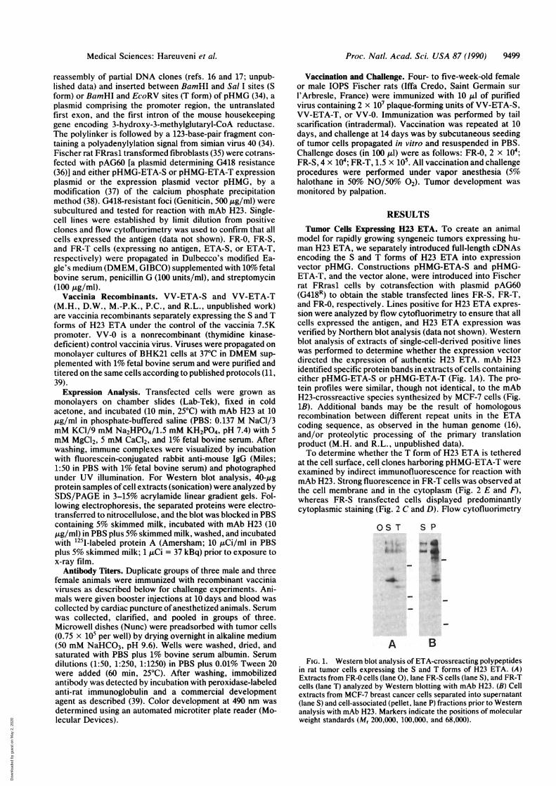
model for rapidly growing syngeneic tumors expressing hu-man H23 ETA, we separately introduced full-length cDNAsencoding the S and T forms of H23 ETA into expressionvector pHMG. Constructions pHMG-ETA-S and pHMG-ETA-T, and the vector alone, were introduced into Fischerrat FRrasl cells by cotransfection with plasmid pAG60(G418R) to obtain the stable transfected lines FR-S, FR-T,and FR-0, respectively. Lines positive for H23 ETA expres-sion were analyzed by flow cytofluorimetry to ensure that allcells expressed the antigen, and H23 ETA expression wasverified by Northern blot analysis (data not shown). Westernblot analysis of extracts of single-cell-derived positive lineswas performed to determine whether the expression vectordirected the expression of authentic H23 ETA. mAb H23identified specific protein bands in extracts of cells containingeither pHMG-ETA-S or pHMG-ETA-T (Fig. 1A). The pro-tein profiles were similar, though not identical, to the mAbH23-crossreactive species synthesized by MCF-7 cells (Fig.1B). Additional bands may be the result of homologousrecombination between different repeat units in the ETAcoding sequence, as observed in the human genome (16),and/or proteolytic processing of the primary translationproduct (M.H. and R.L., unpublished data).To determine whether the T form of H23 ETA is tethered
at the cell surface, cell clones harboring pHMG-ETA-T wereexamined by indirect immunofluorescence for reaction withmAb H23. Strong fluorescence in FR-T cells was observed atthe cell membrane and in the cytoplasm (Fig. 2 E and F),whereas FR-S transfected cells displayed predominantlycytoplasmic staining (Fig. 2 C and D). Flow cytofluorimetry
OS T S P
A BFIG. 1. Western blot analysis of ETA-crossreacting polypeptides
in rat tumor cells expressing the S and T forms of H23 ETA. (A)Extracts from FR-O cells (lane 0), lane FR-S cells (lane S), and FR-Tcells (lane T) analyzed by Western blotting with mAb H23. (B) Cellextracts from MCF-7 breast cancer cells separated into supernatant(lane S) and cell-associated (pellet, lane P) fractions prior to Westernanalysis with mAb H23. Markers indicate the positions of molecularweight standards (Mr 200,000, 100,000, and 68,000).
Medical Sciences: Hareuveni et al.
Dow
nloa
ded
by g
uest
on
May
2, 2
020
9500 Medical Sciences: Hareuveni et al.
1.0-a)
E: 1.5.a)cnc
0)0.5-
Vaccinia recombinantST ST ST ST
F
FIG. 2. Immunofluorescence analysis of rat tumor cells express-ing H23 ETA. Immobilized control FR-0 (A and B), FR-S (C and D),and FR-T (E and F) cells were counterstained with Evans blue (redfluorescence) and treated with mAb H23 and a second, fluorescein-labeled antibody (yellow fluorescence). Total fluorescence under UVillumination was photographed on color film (A, C, and E, positivephotography) and subsequently printed onto red-insensitive film toreveal specific H23 ETA yellow fluorescence (B, D, and F, negativephotography, laterally inverted). (x50.)
of the cell clones confirmed that the T form is present on thecell membrane, although some S form was also detected atthe cell surface (data not shown). Minor morphologicaldifferences were often observed between cells expressingETA and cells expressing no antigen (compare Fig. 2 C andE with A), though the significance of this alteration is notunderstood.Immune Reactivity in Vaccinated Rats. We previously
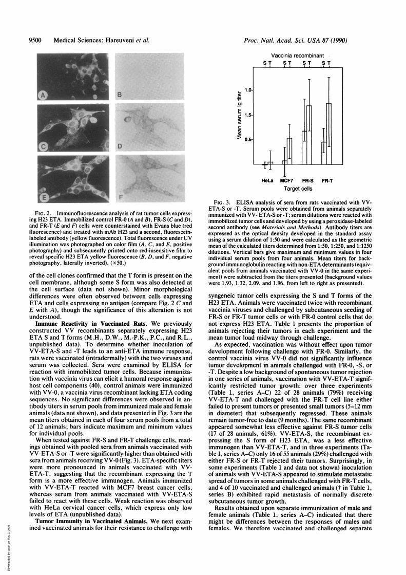
constructed VV recombinants separately expressing H23ETA S and T forms (M.H., D.W., M.-P.K., P.C., and R.L.,unpublished data). To determine whether inoculation ofVV-ETA-S and -T leads to an anti-ETA immune response,rats were vaccinated (intradermally) with the two viruses andserum was collected. Sera were examined by ELISA forreaction with immobilized tumor cells. Because immuniza-tion with vaccinia virus can elicit a humoral response againsthost cell components (40), control animals were immunizedwith VV-0, a vaccinia virus recombinant lacking ETA codingsequences. No significant differences were observed in an-tibody titers in serum pools from immunized male and femaleanimals (data not shown), and data presented in Fig. 3 are themean titers obtained in each of four serum pools from a totalof 12 animals; bars indicate maximum and minimum valuesfor individual pools.When tested against FR-S and FR-T challenge cells, read-
ings obtained with pooled sera from animals vaccinated withVV-ETA-S or -T were significantly higher than obtained withsera from animals receiving VV-0 (Fig. 3). ETA-specific titerswere more pronounced in animals vaccinated with VV-ETA-T, suggesting that the recombinant expressing the Tform is a more effective immunogen. Animals immunizedwith VV-ETA-T reacted with MCF7 breast cancer cells,whereas serum from animals vaccinated with VV-ETA-Sfailed to react with these cells. Weak reaction was observedwith HeLa cervical cancer cells, which express only lowlevels of ETA (unpublished data).Tumor Immunity in Vaccinated Animals. We next exam-
ined vaccinated animals for their resistance to challenge with
FIG. 3. ELISA analysis of sera from rats vaccinated with VV-ETA-S or -T. Serum pools were obtained from animals separatelyimmunized with VV- ETA-S or-T; serum dilutions were reacted withimmobilized tumor cells and developed by using a peroxidase-labeledsecond antibody (see Materials and Methods). Antibody titers areexpressed as the optical density developed in the standard assayusing a serum dilution of 1:50 and were calculated as the geometricmean of the calculated titers determined from 1:50, 1:250, and 1:1250dilutions. Vertical bars give maximum and minimum values in fourindividual serum pools from four animals. Mean titers for back-ground immunoglobulin reacting with non-ETA determinants (equiv-alent pools from animals vaccinated with VV-0 in the same experi-ment) were subtracted from the titers presented (background valueswere 1.93, 1.32, 2.09, and 1.96, from left to right as presented).
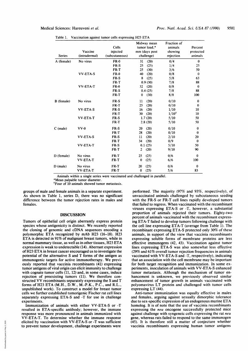
syngeneic tumor cells expressing the S and T forms of theH23 ETA. Animals were vaccinated twice with recombinantvaccinia viruses and challenged by subcutaneous seeding ofFR-S or FR-T tumor cells or with FR-O control cells that donot express H23 ETA. Table 1 presents the proportion ofanimals rejecting their tumors in each experiment and themean tumor load midway through challenge.As expected, vaccination was without effect upon tumor
development following challenge with FR-O. Similarly, thecontrol vaccinia virus VV-0 did not significantly influencetumor development in animals challenged with FR-O, -S, or-T. Despite a low background of spontaneous tumor rejectionin one series of animals, vaccination with VV-ETA-T signif-icantly restricted tumor growth: over three experiments(Table 1, series A-C) 22 of 28 animals (79%) receivingVV-ETA-T and challenged with the FR-T cell line eitherfailed to present tumors or presented small tumors (5-12 mmin diameter) that subsequently regressed. These animalsremain tumor-free to date (9 months). The same recombinantappeared somewhat less effective against FR-S tumor cells(17 of 28 animals, 61%). VV-ETA-S, the recombinant ex-pressing the S form of H23 ETA, was a less effectiveimmunogen than VV-ETA-T, and in three experiments (Ta-ble 1, series A-C) only 16 of 55 animals (29%) challenged witheither FR-S or FR-T rejected their tumors. Surprisingly, insome experiments (Table 1 and data not shown) inoculationof animals with VV-ETA-S appeared to stimulate metastaticspread oftumors in some animals challenged with FR-T cells,and 4 of 10 vaccinated and challenged animals (t in Table 1,series B) exhibited rapid metastasis of normally discretesubcutaneous tumor growth.
Results obtained upon separate immunization of male andfemale animals (Table 1, series A-C) indicated that theremight be differences between the responses of males andfemales. We therefore vaccinated and challenged separate
B
D
I
HeLa MCF7 FR-STarget cells
FR-T
Proc. Natl. Acad. Sci. USA 87 (1990)
.i:..:.z-
.1 :: .ii,.. 'O.
.4,
Dow
nloa
ded
by g
uest
on
May
2, 2
020
Proc. Nati. Acad. Sci. USA 87 (1990) 9501
Table 1. Vaccination against tumor cells expressing H23 ETA
Midway mean Fraction ofCells tumor load,* animals Percent
Vaccine injected mm (days post showing protectedSeries (intradermal) (subcutaneous) challenge) rejection animals
A (female) No virus FR-0 31 (20) 0/4 0FR-S 25 (25) 1/4 25FR-T 25 (30) 3/6 50
VV-ETA-S FR-0 40 (20) 0/8 0FR-S 8 (25) 5/8 63FR-T 0.9 (30) 7/8 88
VV-ETA-T FR-0 32 (20) 0/8 0FR-S 0.4 (25) 7/8 88FR-T 0 (30) 8/8 100
B (female) No virus FR-S 11 (20) 0/10 0FR-T 25 (20) 0/10 0
VV-ETA-S FR-S 16 (20) 1/10 10FR-T 30 (20) 1/1ot 10
VV-ETA-T FR-S 1.7 (20) 5/10 50FR-T 2.8 (20) 5/10 50
C (male) VV-0 FR-S 20 (20) 0/10 0FR-T 28 (20) 0/10 0
VV-ETA-S FR-S 11 (20) 2/10 20FR-T 34 (20) 0/9 0
VV-ETA-T FR-S 0.1 (25) 5/10 50FR-T 2 (20) 9/10 90
D (female) No virus FR-T 25 (25) 0/6 0VV-ETA-T FR-T 0 (25) 6/6 100
D (male) No virus FR-T 20 (25) 0/6 0VV-ETA-T FR-T 0 (25) 5/6 83
Animals within a single series were vaccinated and challenged in parallel.*Mean palpable tumor diameter.tFour of 10 animals showed tumor metastasis.
groups of male and female animals in a separate experiment.As shown in Table 1, series D, there was no significantdifference between the tumor rejection rates in males andfemales.
DISCUSSIONTumors of epithelial cell origin aberrantly express proteinspecies whose antigenicity is distinct. We recently reportedthe cloning of genomic and cDNA sequences encoding apolymorphic ETA recognized by mAb H23 (16-18). H23ETA is detected in 91% of malignant breast tumors, while innormal mammary tissue, as well as in other tissues, H23 ETAexpression is weak to undetectable (14). Aberrant expressionofH23 ETA in breast cancer encouraged us to investigate thepotential of the alternative S and T forms of the antigen asimmunogenic targets for active immunotherapy. We previ-ously reported that vaccinia recombinants (41) expressingtumor antigens of viral origin can elicit immunity to challengewith cognate tumor cells (11, 12) and, in some cases, inducerejection of preexisting tumors (11). We therefore con-structed VV recombinants separately expressing the S and Tforms of H23 ETA (M.H., D.W., M.-P.K., P.C., and R.L.,unpublished work). To construct a model for breast tumorcells we further established tumorigenic Fischer rat cell linesseparately expressing ETA-S and -T for use in challengeexperiments.Immunization of animals with either VV-ETA-S or -T
resulted in ETA-specific antibody; however, the immuneresponse was more pronounced in animals immunized withVV-ETA-T. To determine whether the immune responseelicited by vaccination with VV-ETA-S or -T was sufficientto prevent tumor development, challenge experiments were
performed. The majority (97% and 93%, respectively), ofunvaccinated animals challenged by subcutaneous seedingwith the FR-S or FR-T cell lines rapidly developed tumorsthat failed to regress. When vaccinated with the recombinantviruses expressing ETA-S or -T, however, a substantialproportion of animals rejected their tumors. Eighty-twopercent of animals vaccinated with the recombinant express-ing ETA-T failed to develop tumors following challenge withthe cell line expressing ETA-T (average from Table 1). Therecombinant expressing ETA-S protected only 30% of theseanimals, in support of the view that vaccinia recombinantsexpressing soluble forms of membrane proteins are lesseffective immunogens (42, 43). Vaccination against tumorlines expressing ETA-S was also somewhat less effective(29% and 61% overall tumor rejection frequencies in animalsvaccinated with VV-ETA-S and -T, respectively), indicatingthat an association with the cell membrane may be importantfor both target recognition and immunization. In some ex-periments, inoculation of animals with VV-ETA-S enhancedtumor metastasis. Although the mechanism of tumor en-hancement is unknown, we previously observed similarenhancement of tumor growth in animals vaccinated withpolyomavirus LT protein and challenged with tumor cellsexpressing LT (44).Anti-tumor immunization was equally effective in males
and females, arguing against sexually dimorphic tolerancedue to sex-specific expression ofan endogenous murine ETAhomolog. It is of note that the use of vaccinia recombinantsexpressing the neu oncogene successfully protected miceagainst challenge with syngeneic cells expressing the rat neugene, whereas rats failed to respond to the same immunogen(45). It is therefore still a matter of conjecture whethervaccinia recombinants expressing human tumor antigens
Medical Sciences: Hareuveni et al.
Dow
nloa
ded
by g
uest
on
May
2, 2
020
9502 Medical Sciences: Hareuveni et al.
such as p97 (46), the neu/c-erbB2 gene product (45), or ETA(this work) can elicit a therapeutic anti-tumor response inhuman. However, the efficient anti-tumor response reportedhere in rats will encourage further experiments to explore thisquestion.
We thank K. Dott and T. Kotkes for technical assistance and 1.Tsarfaty and 1. Keydar for helpful discussions. This work wassupported by the Association pour la Recherche contre le Cancer,the Institut National de la Sante et de la Recherche Mddicale(CNAMTS grant), the Centre National de la Recherche Scientifique,and the Ministere de la Recherche et de l'Enseignement Superieure.M.H. is the recipient of European Molecular Biology Organizationlong-term fellowship. D.W. was supported by the Israel CancerResearch Fund.
1. Sharon, R. & Naor, D. (1984) Cancer Immunol. Immunother.18, 203-208.
2. Hoover, H. C., Surdyke, M. G., Dangel, R. B., Peters, L. C.& Hanna, M. G. (1985) Cancer 15, 1236-1243.
3. Wallack, M. K., McNally, K., Michaelides, M., Bash, J.,Bartolucci, A., Siegler, H., Balch, C. & Wanebo, H. (1986) Am.Surg. 52, 148-151.
4. Dalianis, T. (1990) Adv. Cancer Res. 55, 57-85.5. Talarico, D., Ittmann, M., Balsari, A., Delli-Bovi, P., Basch,
R. S. & Basilico, C. (1990) Proc. Nail. Acad. Sci. USA 87,4222-4225.
6. Wallack, M. K., Steplewski, Z., Koprowski, H., Rosato, E.,George, J., Hulihan, B. & Johnson, J. (1977) Cancer 39,560-564.
7. Wallack, M. C., McNally, K. R., Leftheriotis, E., Siegler, H.,Balch, C., Wanebo, H. Bartolucci, A. A. & Bash, J. A. (1986)Cancer 57, 649-655.
8. Lee, V. K., Harriott, T. G., Kuchroo, V. K., Halliday, W. J.,Hellstrom, l. & Hellstrom, K. E. (1985) Proc. Nati. Acad. Sci.USA 82, 6286-6290.
9. Herlyn, D., Ross, A. H. & Koprowski, H. (1986) Science 232,100-102.
10. Nepom, G. T., Nelson, K. A., Holbeck, S. L., Hellstrom, l. &Hellstrom, K. E. (1984) Proc. Natl. Acad. Sci. USA 82, 2864-2867.
11. Lathe, R., Kieny, M. P., Gerlinger, P., Clertant, P., Guizani,I., Cuzin, F. & Chambon, P. (1987) Nature (London) 326,878-880.
12. Meneguzzi, G., Kieny, M. P., Lecocq, J. P., Chambon, P.,Cuzin, F. & Lathe, R. (1990) Vaccine 8, 199-206.
13. Hareuveni, M. & Lathe, R. (1990) Int. J. Cancer, in press.14. Keydar, I., Chou, C. S., Hareuveni, M., Tsarfaty, I., Sahar,
E., SeIzer, G., Chaitchik, S. & Hizi, A. (1989) Proc. Natl.Acad. Sci. USA 86, 1362-1366.
15. Tsarfaty, I., Chaitchik, S., Hareuveni, M., Horev, J., Hizi, A.,Wreschner, D. H. & Keydar, 1. (1988) in Breast Cancer Im-munodiagnosis and Immunotherapy, ed. Ceriani, R. L. (Ple-num, New York), pp. 161-169.
16. Hareuveni, M., Tsarfaty, I., Zaretsky, J., Kotkes, P., Horev,J., Zrihan, S., Weiss, M., Green, S., Lathe, R., Keydar, 1. &Wreschner, D. H. (1990) Eur. J. Biochem. 189, 475-486.
17. Wreschner, D., Hareuveni, M., Tsarfaty, 1, Smorodinsky, N.,Horev, J., Zaretsky, J., Kotkes, P., Weiss, M., Lathe, R.,Dion, A. S. & Keydar, 1. (1990) Eur. J. Biochem. 189,463-473.
18. Tsarfaty, I., Hareuveni, M., Horev, J., Zaretsky, J., Weiss, M.,Jeltsch, J. M., Gamier, J. M., Lathe, R., Keydar, I. & Wresch-ner, D. H. (1990) Gene, in press.
19. Bramwell, M. E., Bhavanandan, V. P., Wiseman, G. & Harris,H. (1983) Br. J. Cancer 48, 177-183.
20. Burchell, J. M., Durbin, H. & Taylor-Papadimitriou, J. (1983)J. Immunol. 131, 508-513.
21. Magnani, J. L., Stepewski, Z., Koprowski, H. & Ginsburg, V.(1983) Cancer Res. 43, 5489-5492.
22. Ceriani, R. L., Peterson, J. A. & Blank, E. W. (1984) CancerRes. 44, 3033-3039.
23. Hilkens, J., Buijis, F., Hilgers, J., Hagemann, P., Calafat, J.,Sonnenberg, A. & Van der Valk, M. (1984) Int. J. Cancer 34,197-206.
24. Lan, M. S., Finn, 0. J., Fernsten, P. D. & Metzgar, R. S.(1985) Cancer Res. 45, 305-310.
25. Abe, M. & Kufe, D. (1986) J. Cell. Physiol. 126, 126-136.26. Johnson, V. G., Schlom, J., Paterson, A. J., Bennett, J.,
Magnani, J. L. & Colcher, D. (1986) Cancer Res. 46, 850-857.27. Price, M. R., Edwards, S., Robins, R. A., Hilgers, J., Hilkens,
J. & Baldwin, R. (1986) Eur. J. Cancer Clin. Oncol. 22,115-117.
28. Siddiqui, J., Abe, M., Hayes, D., Shani, E., Yunis, E. & Kufe,D. (1988) Proc. Natl. Acad. Sci. USA 85, 2320-2323.
29. Gendler, S. J., Burchell, J. M., Duhig, T., Lamport, D., White,R., Parker, M. & Taylor-Papadimitriou, J. (1987) Proc. Natl.Acad. Sci. USA 84, 6060-6064.
30. Gendler, S. J., Taylor-Papadimitriou, J., Duhig, T., Rothbard,J. & Burchell, J. (1988) J. Biol. Chem. 263, 12820-12823.
31. Taylor-Papadimitriou, J. & Gendler, S. J. (1988) Cancer Rev.11/12, 11-24.
32. Hilgers, J., Zotter, S. & Kenemans, P. (1988) Cancer Rev.11/12, 3-10.
33. McKenzie, l. F. C. & Xing, P.-X. (1990) Cancer Cells 2,75-78.34. Gautier, C., Mehtali, M. & Lathe, R. (1989) Nucleic Acids Res.
17, 8389.35. Matriceau, L. M., Glaichenhaus, N., Gesnel, M. C. & Breath-
nach, R. (1985) EMBO J. 4, 1435-1440.36. Colbere-Garapin, F., Horodniceanu, F., Kourilsky, P. & Ga-
rapin, A. C. (1981) J. Mol. Biol. 150, 1-14.37. Wigler, M., Pellicer, A., Silverstein, S. & Axel, R. (1978) Cell
14, 725-731.38. Graham, F. L. & van der Eb, A. J. (1973) Virology 52,456-457.39. Kieny, M. P., Rautman, G., Schmitt, D., Dott, K., Wain-
Hobson, S., Laurent, A., Montagnier, L. & Lecocq, J. P.(1986) BiolTechnology 4, 790-795.
40. Lathe, R., Kieny, M. P., Dott, K., Gautier, C., Clertant, P.,Cuzin, F., Breitburd, F., Orth, G. & Meneguzzi, G. (1989) inVaccines for Sexually Transmitted Diseases, eds. Meheus, A.& Spier, R. E. (Butterworths, London), pp. 166-174.
41. Moss, B. & Flexner, C. (1987) Annu. Rev. Immunol. 5, 305-324.
42. Wiktor, T. J., Kieny, M. P. & Lathe, R. (1988) Appl. Virol.Res. 1, 69-90.
43. Kieny, M. P., Lathe, R., Dott, K., Schmitt, D., Girard, M.,Montagnier, L. & Lecocq, J. P. (1988) Prot. Engineer. 2,219-255.
44. Kieny, M. P., Gautier, C., Tomasetto, C., Kuhn, I., Hareu-veni, M., Clertant, P. & Lathe, R. (1990) Int. J. Cancer 45,185-189.
45. Bernards, R., Destree, A., McKenzie, S., Gordon, E., Wein-berg, R. A. & Panicali, D. (1987) Proc. Natl. Acad. Sci USA 84,6854-6858.
46. Estin, C. D., Stevenson, U. S., Plowman, G. D., Hu, S.-L.,Sridhar, P., Hellstrom, I., Brown, J. P. & Hellstrom, K. E.(1988) Proc. Natl. Acad. Sci. USA 85, 1052-1056.
Proc. Nati. Acad. Sci. USA 87 (1990)
Dow
nloa
ded
by g
uest
on
May
2, 2
020