Treatment of intracranial hypertension and aspects on...
8
Treatment of intracranial hypertension and aspects on lumbar dural puncture in severe bacterial meningitis. Grände, Per-Olof; Myhre, Erling; Nordström, Carl-Henrik; Schliamser, Silvia Published in: Acta Anaesthesiologica Scandinavica DOI: 10.1034/j.1399-6576.2002.460307.x 2002 Link to publication Citation for published version (APA): Grände, P-O., Myhre, E., Nordström, C-H., & Schliamser, S. (2002). Treatment of intracranial hypertension and aspects on lumbar dural puncture in severe bacterial meningitis. Acta Anaesthesiologica Scandinavica, 46(3), 264-270. https://doi.org/10.1034/j.1399-6576.2002.460307.x General rights Unless other specific re-use rights are stated the following general rights apply: Copyright and moral rights for the publications made accessible in the public portal are retained by the authors and/or other copyright owners and it is a condition of accessing publications that users recognise and abide by the legal requirements associated with these rights. • Users may download and print one copy of any publication from the public portal for the purpose of private study or research. • You may not further distribute the material or use it for any profit-making activity or commercial gain • You may freely distribute the URL identifying the publication in the public portal Read more about Creative commons licenses: https://creativecommons.org/licenses/ Take down policy If you believe that this document breaches copyright please contact us providing details, and we will remove access to the work immediately and investigate your claim.
Transcript of Treatment of intracranial hypertension and aspects on...
LUND UNIVERSITY
PO Box 117221 00 Lund+46 46-222 00 00
Treatment of intracranial hypertension and aspects on lumbar dural puncture in severebacterial meningitis.
Grände, Per-Olof; Myhre, Erling; Nordström, Carl-Henrik; Schliamser, Silvia
Published in:Acta Anaesthesiologica Scandinavica
DOI:10.1034/j.1399-6576.2002.460307.x
2002
Link to publication
Citation for published version (APA):Grände, P-O., Myhre, E., Nordström, C-H., & Schliamser, S. (2002). Treatment of intracranial hypertension andaspects on lumbar dural puncture in severe bacterial meningitis. Acta Anaesthesiologica Scandinavica, 46(3),264-270. https://doi.org/10.1034/j.1399-6576.2002.460307.x
General rightsUnless other specific re-use rights are stated the following general rights apply:Copyright and moral rights for the publications made accessible in the public portal are retained by the authorsand/or other copyright owners and it is a condition of accessing publications that users recognise and abide by thelegal requirements associated with these rights. • Users may download and print one copy of any publication from the public portal for the purpose of private studyor research. • You may not further distribute the material or use it for any profit-making activity or commercial gain • You may freely distribute the URL identifying the publication in the public portal
Read more about Creative commons licenses: https://creativecommons.org/licenses/Take down policyIf you believe that this document breaches copyright please contact us providing details, and we will removeaccess to the work immediately and investigate your claim.
Acta Anaesthesiol Scand 2002; 46: 264–270 Copyright C Acta Anaesthesiol Scand 2002Printed in Denmark. All rights reserved
ACTA ANAESTHESIOLOGICA SCANDINAVICA
0001-5172
Treatment of intracranial hypertension and aspects onlumbar dural puncture in severe bacterial meningitis
P-O. GRÄNDE1,4, E. B. MYHRE2, C-H. NORDSTRÖM3 and S. SCHLIAMSER2
Departments of 1Anaesthesia and Intensive Care, 2Infectious Diseases and 3Neurosurgery, University Hospital of Lund, and 4Department of Physiology,Lund University, Sweden
Background: Brain stem herniation due to raised intracranialpressure (ICP) is a common cause of mortality in severe bacterialmeningitis, but continuous measurements of ICP and the effectsof ICP-reducing therapy in these patients have, to our knowl-edge, not been described.Methods: During a four-year period, an ICP-monitoring devicewas implanted in patients admitted to our hospital with severebacterial meningitis and suspected intracranial hypertension.ICP above 20mmHg was treated using the Lund Concept, whichincludes antihypertensive therapy (b1-antagonist,a2-agonist),normalization of the plasma colloid osmotic pressure and theblood volume, and antistress therapy.Results: ICP above 20mmHg was found in all 12 patientsstudied. It was effectively reduced in all but two patients, whodied. Both patients had a low cerebral perfusion pressure (,10mmHg), dilated pupils at start of therapy and were beyond re-covery. Radiological signs of brain swelling were present in onlyfive patients. Seven patients recovered fully, while mild audio-logical impairment was observed in two and minor neurologicalsequelae in one patient. Eight patients showed signs suggestingimminent brain stem herniation before start of ICP-reducingtreatment, seven of whom had been subjected to diagnostic lum-
DESPITE effective antibiotic treatment and im-proved general intensive care, bacterial menin-
gitis continues to be associated with a high mortalityrate (1–5). In a recent US survey of unselected adultpatients with bacterial meningitis, the hospital mor-tality rate was 27% and the rate of neurological deficitpersisted at discharge was 9% (2). The mortality rateand the frequency of neurological sequelae weremuch higher in patients with more severe illness.
Causes of death in bacterial meningitis include cer-ebral vasculitis with infarction, herniation of braintissue, sepsis with circulatory failure, and coagulationdisturbances (6). Compression of the brain stem withcomplete cessation of cerebral circulation, however, isthe most important cause of death and sequelae inthese patients, and is due to increased intracranialpressure (ICP) (6–9). Brain edema is the main factorleading to increased ICP during bacterial meningitis,
264
bar dural puncture shortly before development of the brain stemsymptoms. These symptoms gradually regressed after initiationof therapy, and in one patient reversal of brain stem herniationwas documented by MRI.Conclusions: Severe bacterial meningitis can be associated withincreased ICP, which can be reduced using the Lund Concept.The high survival rate, the low frequency of sequelae and thereversal of signs of imminent brain stem herniation in thesehigh-risk patients indicated beneficial effects of the intervention.The study confirms earlier observations that lumbar dural punc-ture is potentially hazardous in patients with intracranial hyper-tension, because it may trigger brain stem herniation. A normalCT brain scan does not rule out intracranial hypertension.
Received 1 February, accepted for publication 23 August 2001
Key words: bacterial meningitis; brain stem herniation; CT-scan;intracranial pressure; lumbar dural puncture.
c Acta Anaesthesiologica Scandinavica 46 (2002)
but increased intracranial blood volume and disturb-ances in cerebrospinal fluid circulation can also be ofimportance (3).
Intracranial hypertension, especially when com-bined with signs of imminent brain stem compression,indicates a more severe disease and much higher mor-tality than in patients with uncomplicated bacterialmeningitis (3, 7, 10). Improvements in outcome cannot be expected to come from new antimicrobialagents, but from novel treatment modalities based ona more complete understanding of the pathogenesis,including mechanisms responsible for the develop-ment of brain edema.
The standard diagnostic procedure in bacterial men-ingitis is examination of cerebrospinal fluid obtainedby lumbar dural puncture. The benefit contra the riskof performing lumbar dural puncture in severe casesof bacterial meningitis with suspicion of intracranial
Meningitis and raised ICP
hypertension has been debated, because it may inducebrain stem herniation and thus precipitate a life-threatening situation (11, 12).
To the best of our knowledge, there are no pub-lished human data on serial measurement of ICP dur-ing severe bacterial meningitis, nor on the effect ofICP-reducing therapy on ICP and on outcome. A CT-scan of the brain is often used to assess the existenceof intracranial hypertension. Reliable measurement ofICP requires, however, placement of an intracranialmonitoring device. Standard management of severebacterial meningitis rarely includes invasive measure-ment of ICP and a therapeutic programme for insti-tution of ICP-reducing treatment.
The management of patients with severe bacterialmeningitis admitted to our hospital was revisedsome years ago, following a number of cases of fatalcerebral herniation after admission in otherwisehealthy individuals. An ICP-monitoring device isimplanted when clinical signs indicate intracranialhypertension, and the patient is treated with a strictICP–reducing protocol similar to that used in ourhospital for post traumatic brain edema (13, 14).This protocol, the Lund Concept for brain edematherapy, has been shown effective in reducing araised ICP and in improving outcome of braintrauma (15, 16). In the present paper we describeserial invasive ICP measurements in the patients ad-mitted to our hospital with severe life-threateningbacterial meningitis, the relationship between ICPand therapeutic intervention, as well as the func-tional outcome. The risk of lumbar dural puncturein patients with severe bacterial meningitis andraised ICP is discussed on the basis of the patientsincluded in this report.
Patient material and methodsThe present report comprises all patients below theage of 65 treated at the University Hospital of Lundbetween January 1994 and December 1997, admittedwith a diagnosis of bacterial meningitis and in whoma raised ICP exceeding 20 mmHg was documented.An intracranial pressure monitoring device was in-serted in patients with a clinical picture consideredindicative of increased ICP. This picture was a combi-nation of the following signs: progressive loss of con-sciousness, agitation, focal neurological signs, ocularmotility abnormalities (uni- or bilateral pupillary di-lation, impairment or loss of the reaction to light), cra-nial nerve palsy (N. occulomotorius, N. abducens), in-crease in blood pressure and bradycardia. Of thesesigns, loss of consciousness, agitation, increase inblood pressure and pupil dilation were considered
265
highly indicative of a life-threatening increase in ICP(17). The decision to insert the ICP measuring devicewas taken individually, based on assessment of eachpatient. Diagnostic lumbar puncture had invariablybeen performed in the emergency room before evalu-ation for ICP monitoring and transfer to the ICU.
The ICP monitoring device was implanted by stan-dard surgical procedure, and ICP was measured con-tinuously via an extracranial pressure transducer(Bentley, Inc., CA) connected to the intraventricularfluid space. Full intensive care was provided withventilatory support and sedation. Initial antibiotictreatment was adjusted according to the sensitivity ofisolated organisms.
Patients with an ICP exceeding 20 mmHg weretreated with ICP-reducing therapy. The breakpoint of20 mmHg used was arbitrarily based on earlierstudies, where the mortality rate following headtrauma was significantly higher with an ICP exceed-ing 20 mmHg (18). We used the Lund Concept forbrain edema treatment to reduce the ICP. This conceptincludes (i) antihypertensive treatment through b1-blockade (metoprolol 0.05–0.08 mg/kg ¿ 6–8 iv, alter-natively 50–100 mg ¿ 2 per enteral sond) and a2-agonist stimulation (clonidine 0.3–0.8 mg/kg ¿ 4–6 iv),(ii) infusion of thiopental to reduce brain metabolismand sympathetic stress (bolus doses of 2–4 mg/kg fol-lowed by continuous infusion of 0.5–3 mg/kg/h) (iii)volume substitution with plasma/albumin infusionand blood transfusion to a serum-albumin level of 35–40 g/l and Hb above 125 g/l to maintain normovolae-mia and a normal plasma colloid osmotic pressure,and (iv) volume-controlled mechanical ventilation(PaCO2 level of 4.6–5.0 kPa). It has been shown thatintracranial infections are associated with depressedcerebral autoregulation (19, 20). In this state a reduc-tion in arterial pressure will lower hydrostatic capil-lary pressure and thereby induce transcapillary ab-sorption. If these measures, as applied in our study,did not reduce ICP to a level below 25–30 mmHgwithin 24 h, dihydroergotamin was added intra-venously for a maximum of 2 days (3mg/kg ¿ 4–6).The antihypertensive therapy used was tailored to theICP value, if necessary, allowing a fall in the cerebralperfusion pressure (arterial pressure-ICP) to a lowestvalue of 50 mmHg for adults and 40 mmHg for youngchildren. Vasopressor support with the intention to in-crease the cerebral perfusion pressure was not used.Thiopental and clonidine in combination with fen-tanyl (3–5 mg/kg/h) were given primarily as anal-gesia, but provided also adequate antistress and seda-tive effects. After 2–3 days, thiopental was replaced bymidazolam to prevent barbiturate-induced pulmon-
P-O. Grände et al.
ary side-effects (21). The same zero baseline level wasused for measurements of arterial pressure and ICP.
Acute episodes of incipient brain stem symptomsbefore the patient was admitted to the ICU were man-aged in a few cases, using either short-term hyperven-tilation, a bolus infusion of thiopental (3–4 mg/kg), ortemporary drainage of cerebrospinal fluid or a combi-nation of these interventions. Osmotherapy was notused, due to the risk of an adverse rebound increasein ICP (22).
Results
During the four-year study period, 53 patients wereadmitted to our hospital with the primary diagnosisof bacterial meningitis. Fifteen patients had a clinicalpicture compatible with intracranial hypertension.Three patients were excluded due to moribund clin-ical condition on arrival at the hospital (one patient)or cardiovascular failure combined with coagulationdisturbances (two patients). An ICP measuring devicewas inserted into 12 patients without complications interms of intracranial haemorrhage or infection. Demo-graphic, clinical and outcome data of these 12 patientsare summarised in Table 1. Seven patients were 16years or younger; the oldest was 59 years. None of thepatients had underlying chronic diseases. Streptococ-cus pneumoniae was the most common aetiologydocumented by culture in six patients. Spinal fluidcultures were negative in four patients who had re-ceived antibiotics prior to specimen collection. Corti-costeroid therapy was given to six of the 12 patients(Table 1), a therapy recommended at the time of thestudy if it could be given before initiation of the anti-
Table1
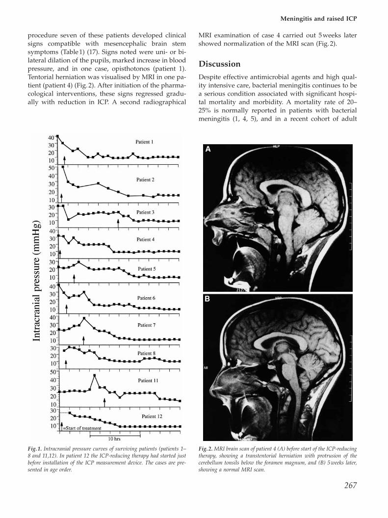
Demographic, clinical and outcome data.
Case Age Level of Etiologic Lumbar Brain stem Functionalno. (years) consciousness agent dural symptoms outcome
before sedation puncture(RLS)
1 2 4 S. pneumoniae Yes Yes Mild hearing impairment2 5 3–4 Unknown* Yes Yes Full recovery3 12 7 S. pneumoniae Yes Yes Full recovery4 14 7 S. pneumoniae Yes Yes Permanent mild ataxia and urinary bladder dysfunction5 15 4 Unknown* Yes No Full recovery6 16 3–4 Unknown* Yes No Full recovery7 16 3–4 S. pneumoniae Yes No Full recovery8 21 5 Unknown* Yes Yes Full recovery9 24 7 S. pneumoniae No Yes Died
10 35 7 S. pneumoniae Yes Yes Died11 47 3–4 N. meningitidis Yes No Full recovery12 59 4 S. pneumoniae Yes Yes Mild hearing impairment
*Cerebrospinal fluid culture negative.
266
biotic therapy (23, 24). Four patients were given di-hydroergotamine (patients 1, 4, 5 and 10).
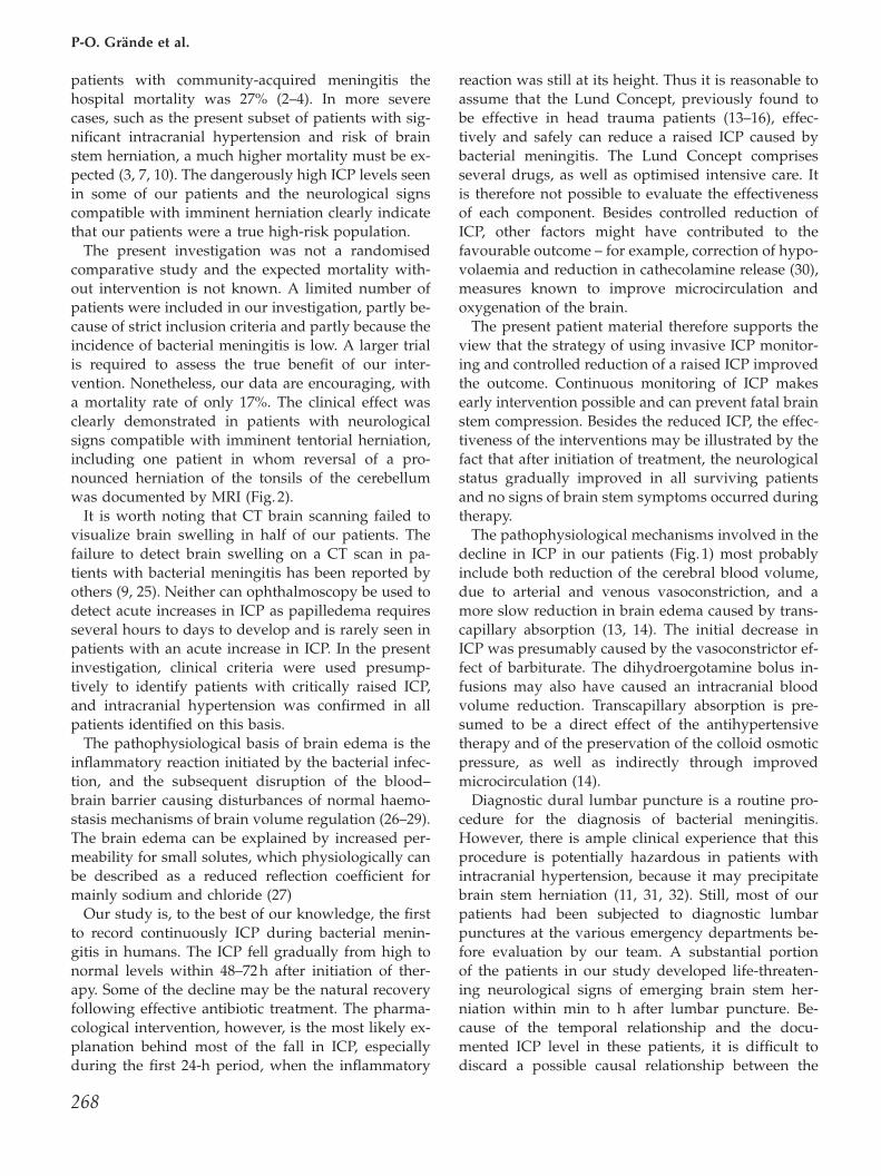
Intracranial hypertension with an initial ICP exceed-ing 20 mmHg was found in all 12 patients. ICP valuesbefore and after initiation of the ICP-reducing therapyare shown in Fig. 1. No further increase in ICP wasnoted after 2–3 h of treatment. In cases 5, 7 and 8 aclear trend of increasing ICP was broken shortly afterthe start of therapy. In general, ICP fell gradually butat various rates following commencement of the ICP-reducing treatment.
Ten patients survived, seven of whom recoveredcompletely. Two patients were left with mild hearingimpairment (patients 1 and 12) and one with perma-nent but mild ataxia combined with moderate urinarybladder dysfunction (patient 4). The follow-up timewas at least 2 years after hospital discharge. Two pa-tients died. Both showed insufficient cerebral per-fusion pressure (,10 mmHg) from the initiation oftherapies due to a significantly increased ICP, circum-stances suggesting virtually no cerebral blood flow(patients 9 and 10). They had bilaterally dilated pupilsnonresponsive to light stimulation. ICP continued tobe close to the mean arterial pressure, their neurologi-cal status did not improve, and they were both be-yond recovery.
Before implantation of the monitoring device, com-puterised tomography (CT scan) of the brain was per-formed in 10 patients and MRI was performed in oneof these patients (patient 4, Fig. 2). Radiological signsindicating brain swelling were seen on CT scan inonly five patients.
Diagnostic lumbar dural puncture was carried outin 11 of the 12 patients. Within 30 min to 2 h after the
Meningitis and raised ICP
procedure seven of these patients developed clinicalsigns compatible with mesencephalic brain stemsymptoms (Table 1) (17). Signs noted were uni- or bi-lateral dilation of the pupils, marked increase in bloodpressure, and in one case, opisthotonos (patient 1).Tentorial herniation was visualised by MRI in one pa-tient (patient 4) (Fig. 2). After initiation of the pharma-cological interventions, these signs regressed gradu-ally with reduction in ICP. A second radiographical
Fig.1. Intracranial pressure curves of surviving patients (patients 1–8 and 11,12). In patient 12 the ICP-reducing therapy had started justbefore installation of the ICP measurement device. The cases are pre-sented in age order.
267
MRI examination of case 4 carried out 5 weeks latershowed normalization of the MRI scan (Fig. 2).
Discussion
Despite effective antimicrobial agents and high qual-ity intensive care, bacterial meningitis continues to bea serious condition associated with significant hospi-tal mortality and morbidity. A mortality rate of 20–25% is normally reported in patients with bacterialmeningitis (1, 4, 5), and in a recent cohort of adult
Fig.2. MRI brain scan of patient 4 (A) before start of the ICP-reducingtherapy, showing a transtentorial herniation with protrusion of thecerebellum tonsils below the foramen magnum, and (B) 5weeks later,showing a normal MRI scan.
P-O. Grände et al.
patients with community-acquired meningitis thehospital mortality was 27% (2–4). In more severecases, such as the present subset of patients with sig-nificant intracranial hypertension and risk of brainstem herniation, a much higher mortality must be ex-pected (3, 7, 10). The dangerously high ICP levels seenin some of our patients and the neurological signscompatible with imminent herniation clearly indicatethat our patients were a true high-risk population.
The present investigation was not a randomisedcomparative study and the expected mortality with-out intervention is not known. A limited number ofpatients were included in our investigation, partly be-cause of strict inclusion criteria and partly because theincidence of bacterial meningitis is low. A larger trialis required to assess the true benefit of our inter-vention. Nonetheless, our data are encouraging, witha mortality rate of only 17%. The clinical effect wasclearly demonstrated in patients with neurologicalsigns compatible with imminent tentorial herniation,including one patient in whom reversal of a pro-nounced herniation of the tonsils of the cerebellumwas documented by MRI (Fig. 2).
It is worth noting that CT brain scanning failed tovisualize brain swelling in half of our patients. Thefailure to detect brain swelling on a CT scan in pa-tients with bacterial meningitis has been reported byothers (9, 25). Neither can ophthalmoscopy be used todetect acute increases in ICP as papilledema requiresseveral hours to days to develop and is rarely seen inpatients with an acute increase in ICP. In the presentinvestigation, clinical criteria were used presump-tively to identify patients with critically raised ICP,and intracranial hypertension was confirmed in allpatients identified on this basis.
The pathophysiological basis of brain edema is theinflammatory reaction initiated by the bacterial infec-tion, and the subsequent disruption of the blood–brain barrier causing disturbances of normal haemo-stasis mechanisms of brain volume regulation (26–29).The brain edema can be explained by increased per-meability for small solutes, which physiologically canbe described as a reduced reflection coefficient formainly sodium and chloride (27)
Our study is, to the best of our knowledge, the firstto record continuously ICP during bacterial menin-gitis in humans. The ICP fell gradually from high tonormal levels within 48–72 h after initiation of ther-apy. Some of the decline may be the natural recoveryfollowing effective antibiotic treatment. The pharma-cological intervention, however, is the most likely ex-planation behind most of the fall in ICP, especiallyduring the first 24-h period, when the inflammatory
268
reaction was still at its height. Thus it is reasonable toassume that the Lund Concept, previously found tobe effective in head trauma patients (13–16), effec-tively and safely can reduce a raised ICP caused bybacterial meningitis. The Lund Concept comprisesseveral drugs, as well as optimised intensive care. Itis therefore not possible to evaluate the effectivenessof each component. Besides controlled reduction ofICP, other factors might have contributed to thefavourable outcome – for example, correction of hypo-volaemia and reduction in cathecolamine release (30),measures known to improve microcirculation andoxygenation of the brain.
The present patient material therefore supports theview that the strategy of using invasive ICP monitor-ing and controlled reduction of a raised ICP improvedthe outcome. Continuous monitoring of ICP makesearly intervention possible and can prevent fatal brainstem compression. Besides the reduced ICP, the effec-tiveness of the interventions may be illustrated by thefact that after initiation of treatment, the neurologicalstatus gradually improved in all surviving patientsand no signs of brain stem symptoms occurred duringtherapy.
The pathophysiological mechanisms involved in thedecline in ICP in our patients (Fig. 1) most probablyinclude both reduction of the cerebral blood volume,due to arterial and venous vasoconstriction, and amore slow reduction in brain edema caused by trans-capillary absorption (13, 14). The initial decrease inICP was presumably caused by the vasoconstrictor ef-fect of barbiturate. The dihydroergotamine bolus in-fusions may also have caused an intracranial bloodvolume reduction. Transcapillary absorption is pre-sumed to be a direct effect of the antihypertensivetherapy and of the preservation of the colloid osmoticpressure, as well as indirectly through improvedmicrocirculation (14).
Diagnostic dural lumbar puncture is a routine pro-cedure for the diagnosis of bacterial meningitis.However, there is ample clinical experience that thisprocedure is potentially hazardous in patients withintracranial hypertension, because it may precipitatebrain stem herniation (11, 31, 32). Still, most of ourpatients had been subjected to diagnostic lumbarpunctures at the various emergency departments be-fore evaluation by our team. A substantial portionof the patients in our study developed life-threaten-ing neurological signs of emerging brain stem her-niation within min to h after lumbar puncture. Be-cause of the temporal relationship and the docu-mented ICP level in these patients, it is difficult todiscard a possible causal relationship between the

Meningitis and raised ICP
dural puncture and the brain stem symptoms as re-ported by many other investigators (11, 31, 32).Thus, there are strong indications suggesting thatlumbar puncture in patients with intracranial hyper-tension is associated with a significant risk of life-threatening complications and should be avoided.The problem facing the physician is to determinewhether a dangerous intracranial hypertension ispresent or not. Other diagnostic methods such asCT brain scan or opthalmoscopy are of little assist-ance, because findings do not correlate well with theICP level and may fail to identify patients at risk.Our study shows that patients with bacterial menin-gitis presenting clinical signs of intracranial hyper-tension invariably had dangerously high ICP levelsand were at risk of herniation, especially when sub-jected to lumbar puncture. A safe approach in suchcircumstances is to abstain from lumbar dural punc-ture and, if possible, insert an intraventricular ICP-monitoring device as used in our patients. Spinalfluid for culture and for clinical chemistry can safelybe obtained through the intraventricular catheter.
The mechanisms behind brain stem herniation, aswell as spinal headache, following lumbar duralpuncture is not understood, one fact which may ex-plain why lumbar dural puncture is still used ratheruncritically. It is classically explained as an effect ofloss of cerebrospinal fluid volume by leakagethrough the needle perforation of the dural sac (33).This, however, can not be the only explanation, be-cause the continuous production of cerebrospinalfluid should easily compensate for such an effect.We recently formulated the alternative explanation(34), that opening of the dural sac also results in re-duction in the intradural and interstitial brain press-ure, causing transvascular filtration across the dis-rupted blood–brain barrier and dilation of collapsedintracranial veins. Reduction in cerebrospinalcounter-pressure around the brain stem also allowsbrain tissue to move sagitally, with loss of volumebuffering capacity in this region (Fig. 2A).
We conclude that a significantly raised ICP level is alife-threatening condition during bacterial meningitis,and may precipitate brain stem compression, espe-cially after lumbar dural puncture. This situation canbe brought under control by pharmacological treat-ment of the brain edema using the Lund Concept,most probably reducing the imminent risk of her-niation. Continuous ICP measurement and aggressivemanagement of a raised ICP is warranted in severecases with suspected intracranial hypertension, andshould be considered in institutions capable of pro-viding this level of care.
269
AcknowledgmentsThe study was supported by the Medical Research Council(.11585), the Wallenberg Foundation, Region Skåne, and theMedical Faculty of Lund, Sweden.
References1. Quagliarello V, Scheld WM. Adjunctive therapy in bacterial
meningitis––what make sense? Infect Agents Dis 1992: 1: 167–171.
2. Aronin SI, Peduzzi P, Quagliarello VJ. Community-acquiredbacterial meningitis: Risk stratification for adverse clinicaloutcome and effect of antibiotic timing. Ann Intern Med 1998:129: 862–869.
3. Horwitz SJ, Boxerbaum B, O’Bell J. Cerebral herniation inbacterial meningitis in childhood. Ann Neurol 1980: 7: 524–528.
4. Quagliarello V, Scheld WM. Bacterial meningitis: patho-genesis, pathophysiology, and progress. N Eng J Med 1992:327: 864–872.
5. Bartfield AA. Bacterial meningitis. Prim Care Update Ob Gyns2000: 7: 49–54.
6. Spence M. Severe intracranial infections. Int Anesthesiol Clin1979: 17: 285–305.
7. Minns RA, Engelman HM, Stirling H. Cerebrospinal fluidpressure in pyogenic meningitis 1989. Arch Dis Child 1989:64: 814–820.
8. Schildkamp RI, Looder MC, Bijlmer HA, Dankert J, SchaltenRJ et al. Clinical manifestations and course of meningococcaldisease in 562 patients. Scand J Infect Dis 1996: 28: 47–51.
9. Rennick G, Shann F, de Campo J. Cerebral herniation duringbacterial meningitis in children. BMJ 1993: 306: 953–955.
10. Rebaud P, Berhier JC, Hartemann E, Floret D et al. Intra-cranial pressure in childhood central nervous system infec-tions. Intensive Care Med 1988: 14: 522–525.
11. Addy DP. When not to do a lumbar dural puncture. ArchDis Child 1987: 62: 873–875.
12. Harper JR. Timing of lumbar puncture in severe childhoodmeningitis. BM J 1985: 291: 651–652.
13. Asgeirsson B, Grände P-O, Nordström CH. A new therapyof post-trauma brain oedema based on haemodynamic prin-ciples for brain Volume regulation. Intensive Care Med 1994:20: 260–267.
14. Grände PO, Asgeirsson B, Nordström CH. Physiologic prin-ciples for Volume regulation of a tissue enclosed in a rigidshell with application to the injured brain. J Trauma 1997: 42:23–31.
15. Eker C, Asgeirsson B, Grände PO, Schale NW, NordstromCH et al. Improved outcome after head injury with a newtherapy based on principles for brain Volume regulation andpreserved microcirculation. Crit Care Med 1998: 26: 1881–1886.
16. Naredi S, Eden E, Zall S, Stephensen H, Rydenhag B et al. Astandardized neurosurgical neurointensive therapy directedtoward vasogenic edema after severe traumatic brain injury:clinical results. Intensive Care Med 1998: 24: 446–451.
17. Plum F, Posner JB. Diagnosis of stupor and coma, 2nd edn.Contemporary Neurology Series. Philadelphia: F. A. DavisCompany, 1972.
18. Jennet B, Teasdale G, Galbraith S, Pickard J, Grant H, Braak-man R et al. Severe head injuries in three countries. J NeurolNeurosurg Psychiatry 1977: 40: 291–298.
19. Tureen JH, Dworkin RJ, Kenny SL, Sachdeva M, Sande MAet al. Loss of cerebrovascular autoregulation in experimentalmeningitis in rabbits. J Clin Invest 1990: 85: 577–581.
P-O. Grände et al.
20. Möller K, Larsen FS, Qvist J, Wandall JH, Knudsen GM,Gjorup IE et al. Dependency of cerebral blood flow on meanarterial pressure in patients with acute bacterial meningitis.Crit Care Med 2000: 28: 1027–1031.
21. SchalenW, Messeter K, Nordström CH. Complications andside effects during thiopentone therapy in patients with se-vere head injuries. Acta Anesthesiol Scand 1992: 36: 369–377.
22. Kofke WA. Mannitol: potential for rebound intracranial hy-pertension? J Neurosurg Anaesthesiol 1993: 5: 1–3.
23. Pfister HW, Scheld WM. Brain injury in bacterial meningitis:therapeutic implications. Curr Opin Neurol 1997: 10: 254–259.
24. Liebel MH, Freij BJ, Syrogiannopoulos GA, Chane DF, HoytMJ, Stewart SM et al. Dexamethasone therapy for bacterialmeningitis: results of two double blind, placebo controlledtrials. N Engl J Med 1998: 319: 964–971.
25. Baker ND. The place of computed tomography and lumbarpuncture in suspected bacterial meningitis. Arch Dis Child1992: 67: 1417–1419.
26. Holmin S, Mathiesen T, Shetye J, Biberfeldt P et al. Intracer-ebral inflammatory response to experimental brain contu-sion. Acta Neurochir (Wien) 1995: 132: 110–119.
27. Fenstermacher JD. Volume regulation of the central nervoussystem. In: Staub NC, Taylor AE, eds. Edema. New York:Raven Press, 1984: 383–404.
28. Møller K, Stolze Larsen F, Bie P, Skinhøj P. The syndromeof inappropriate secretion of antidiuretic hormone and fluid
270
restriction in meningitis – how strong is the evidence? ScandInfect Dis 2001: 33: 13–26.
29. Spellerberg B, Tuomanen EI. The pathophysiology of pneu-mococcal meningitis. Ann Med 1994: 26: 411–418.
30. Payen D, Quintin I, Plaisence P, Chiron B, Lhoste F et al.Head injury: clonidine decreases plasma catecholamines.Crit Care Med 1990: 18: 392–395.
31. Duffy GP. Lumbar puncture in the presence of raised intra-cranial pressure. BMJ 1969: 1: 407–449.
32. Dodge PR, Swartz MN. Bacterial meningitis. A review ofselected aspects. N Engl J Med 1965: 272: 898–1010.
33. Gielen M. Post dural headache (PDPH): a review. Reg Anesth1989: 14: 101–106.
34. Kongstad L, Grände PO. Local vascular response during or-gan elevation. A model for cerebral effects of upright posi-tion and dural puncture. Acta Anaesthesiol Scand 1999: 43:438–446.
AddressP-O. GrändeDepartment of Anaesthesia and Intensive CareUniversity Hospital of LundSE-221 85 LundSwedene-mail: Per-Olof.Grande/mphy.lu.se