publication-theses.unistra.fr · Institut de Génétique et de Biologie Moléculaire et Cellulaire...
157
Institut de Génétique et de Biologie Moléculaire et Cellulaire UMR CNRS 7104/INSERM U596/ULP/Collège de France Thèse présentée à l'URF des Sciences de la Vie et de la Santé de l'Université Louis PASTEUR - Strasbourg I. Discipline Biologie Moléculaire pour obtenir le titre de Docteur de l'Université Louis PASTEUR de Strasbourg par Eduardo Castañeda Saucedo Role of the Retinoid X Receptor α (RXRα) and its Heterodimeric Partners during Skin Carcinogenesis Directeur: Daniel Metzger Co-directeur: Pierre Chambon Date de soutenance 1 Juillet 2004 Jury: Prof. Pierre Chambon Dr. Daniel Metzger Prof. Johan Auwerx Prof. Peter Angel Dr. Lluis Fajas
Transcript of publication-theses.unistra.fr · Institut de Génétique et de Biologie Moléculaire et Cellulaire...
Institut de Génétique et de Biologie Moléculaire et Cellulaire
UMR CNRS 7104/INSERM U596/ULP/Collège de France
Thèse
présentée àl'URF des Sciences de la Vie et de la Santé
de l'Université Louis PASTEUR - Strasbourg I.
Discipline Biologie Moléculaire
pour obtenir le titre de
Docteur de l'Université Louis PASTEUR de Strasbourg
par
Eduardo Castañeda Saucedo
Role of the Retinoid X Receptor α (RXRα) and its Heterodimeric
Partners during Skin Carcinogenesis
Directeur: Daniel Metzger
Co-directeur: Pierre Chambon
Date de soutenance 1 Juillet 2004
Jury: Prof. Pierre Chambon
Dr. Daniel Metzger
Prof. Johan Auwerx
Prof. Peter Angel
Dr. Lluis Fajas
Acknowledgments
To Prof. Pierre Chambon and Dr. Daniel Metzger for giving me theopportunity to prepare my PhD. thesis in their laboratory, for their
scientif ic and personal support, for their crit ical comments and for allwhat I have learned from them.
I would l ike to thank Prof. Johan Auwerx, Prof. Peter Angel and Dr.Lluis Fajas for accepting to be part of my Ph.D. jury.
To Dr. Patricio Gariglio, for all his support before and during my Ph.D.,for his crit ical comments, and most important for his fr iendship.
To Arup Indra, my colleague, collaborator and fr iend, with whom I haveworked during my Ph.D., for his advice, crit ical discussions and for all
the good time we spent together.
To Mei Li, for her scientif ic support, suggestions and crit ical comments,and in particular for her nice fr iendship.
Special thanks to Monique Duval, for all her incondit ional support withanimal work.
To all the current and former members of the laboratory, Jean-Marc,Takeshi, Meng, Ming, Phil ipp, Reiko, Yasunari, Nathalie, Celine,
Isabelle, Romain, Pierre, Michael, Michal, Ali, Bernadette, Christelle,Jean-François, Monica, Martha, Wojtek, Emilie, Valèrie, (and all thosewhom I may forget) for their crit ical comments, their support, and their
fr iendship.
To all people from the IGBMC common facil i t ies (histology service,oligonucleotide synthesis service, animal facil i ty, photographic service,informatics service). Special thanks to Kristina, Nadia and Bernard, for
their technical support. To all other people who make our work possibleat the IGBMC.
Special thanks to Collete Kutschis, for all her support and friendship.
To all my friends and colleagues, current and former members of theIGBMC, thanks to whom my time at the IGBMC has been so pleasent.
Special thanks to the "Ligue Contre le Cancer, comité du Bas-Rhin"for the f inancial support during my thesis.

Finally, thanks to Lourdes, my wife and my friend, for beingthere all the time, in the god and the not so good times, for her
motivation, her support and her love. Thank you .
AbreviationsAF-1AF-2AKAP-1BCCBMCEDBDDMBAECMERFCGM-CSFHATHDACHMTIHCIL-1IRSISCISHKGFK13K14LBDLXRMG'sNSAIDNRORSPMDPPARRARARRT-PCRrtTARXRSCCSCFSp-1SpCCTATamTetTPAUVVD3VDR
activation function 1activation function 2actinic keratosisactivator protein 1basal cell carcinomabasement membranecornified envelopeDNA binding domain7-12-dimethylben(a)anthraceneextra cellular matrixestrogen receptorfocal carcinomagranulocyte macrophage colony stimulator factorhistone acetyl transferasehistone deacetylase.histone methyl transferaseimmunohistochemistryinterleukin 1inner root sheathin situ carcinomain situ hybridisationkeratinocyte growth factorkeratin 13keratin 14ligand binding domainliver X receptormelanocytic growthsnon steroidal anti-inflammatory drugsnuclear receptorouter root sheathpost mitotic differentiatingperoxisome proliferator activated receptorretinoic acidretinoic acid receptorreverse transription coupled polymerase chain reactionreverse tetracycline-controlled transactivatorretinoic X receptorsquamous cell carcinomastem cell factorspecificity protein-1spindle cell carcinomatransit amplifyingtamoxifentetracyclin12-O-tetradecanoyl-phorbol-13-acetateultravioletvitamin D3vitamin D receptor

Table list.
Table 1. Common Neoplasm of the Skin .................................................................................. 11
Table 2. Mouse models of skin cancer ..................................................................................... 14
Table 3. Synthesis of the classical lipophilic hormones........................................................... 23
Table 4. Human and Mouse nuclear receptors ........................................................................ 26
Table 5. Expression of NRs in the skin..................................................................................... 32
Table 6. Summary of skin carcinogenesis in Tg.AC/RXRαep/- mice ......................................... 61
Table 7. Summary of RXRα and its heterodimeric partners functions during skin carcinogenesis. .... 70
Figure list.
Figure 1. Structure of human skin.............................................................................................. 2Figure 2. Keratinocyte proliferation unit................................................................................... 7Figure 3. The chemical skin carcinogenesis model.. ............................................................... 15Figure 4. The Tg.AC model...................................................................................................... 17Figure 5. The Cre-loxP system of homologous recombination................................................ 19Figure 6. Strategy to generate a tamoxifen-induceble Cre recombinase. ............................... 20Figure 7. Model of the mechanism of Cre-ERT2-mediated recombination in keratinocytes.... 21Figure 8. Structure of the major lipophylic hormones...................................................................... 24Figure 9. Modular structure of the NRs.. ................................................................................. 25Figure 10. Nuclear receptors response elements..................................................................... 27Figure 11. Mechanism of NR transcription regulation............................................................ 29Figure 12. Phenotype of RXRαep-/- mice................................................................................... 35Figure 13. Model for integration of nuclear receptors action in the skin. ............................. 39Figure 14. Role of PPARα during skin carcinogenesis ........................................................... 46Figure 15. Selective ablation of RXRα in epidermal keratinocytes of adult Tg.AC ................ 53Figure 16. Selective ablation of RXRα in epidermal keratinocytes of Tg.AC mice during skinmorphogenesis.......................................................................................................................... 56Figure 17. Formation of skin papillomas in RXRαep-/-(c) mice in response to a single DMBAapplication................................................................................................................................ 59Figure 18. Formation of melanocytic growths (MGs) in RXRαep-/-(c) mice in response to asingle DMBA application ......................................................................................................... 60Figure 19. Phenotypic changes in the skin of Tg.AC/RXRαep-/-(c) mice ............................................. 63Figure 20. Skin carcinogenesis in PPARγep-/- mice. ........................................................................ 74

I
RésuméLa peau des Mammifères comporte, sur sa face externe, l'épiderme, tissu épithélial
pavimenteux (squameux) stratifié et kératinisé (corné), composé de kératinocytes (espèce
majoritaire), de cellules pigmentaires (mélanocytes), de cellules immunitaires (cellules de
Langerhans) et de mécanorécepteurs (cellules de Merkel) (Kanitakis, 2002). L'épiderme est
ancré, via une lame basale, à du tissu conjonctif dense fibroélastique, le derme (Watt, 2002).
Ce tissu largement vascularisé, composé de fibroblastes et de cellules immunitaires
(mastocytes et histiocytes), repose sur l'hypoderme, association de tissu conjonctif lâche et de
tissu adipeux (Kanitakis, 2002). Des invaginations épidermiques dans les tissus sous-jacents
constituent des crêtes épidermiques ou forment les annexes épidermiques telles que les poils,
les glandes sébacées (holocrines), ou les glandes sudoriférantes (apocrines) (Fuchs, 1998;
Larsen, 1993; Mackenzie, 1975). La peau joue des fonctions très diverses, elle a des fonctions
métaboliques, sensorielles, thermorégulatrices, immunitaires, et elle représente une barrière
contre la déshydratation et les agressions externes (radiations UV, friction, parasites . . . )
Les rétinoïdes, métabolites actifs de la vitamine A, ont des effets pléïotropes sur l'homéostasie
et le développement des Vertébrés. La peau est un des tissus cibles des retinoïdes. En effet,
une carence ou un excès systémique en vitamine A entraîne une augmentation du nombre de
couches cornéocytaires de l'épiderme (hyperkératose) (Look et al., 1995; Tsambaos et al.,
1980; Tsambaos et al., 1985). Au niveau moléculaire, les rétinoïdes se lient à deux classes de
facteurs de transcription dont l'activité est contrôlée par des ligands spécifiques, les récepteurs
de l'acide rétinoique (RARα, β et γ) et les récepteurs X des retinoïdes (RXRα, β et γ)
(Chambon, 1994; Chambon, 1996). Les RXRs sont des partenaires de dimérisation pour
d'autres récepteurs nucléaires tels que le RARs, le récepteur de la vitamine D (VDR), les
récepteurs activés par les proliférateurs des péroxisomes (PPARs) et les "Liver X receptor"
(LXRs). RXRα est le RXR le plus fortement exprimé dans l'épiderme, où il joue un rôle clef
dans l'homéostasie de la peau (Li 2000,2001).
De nombreuses études montrent que, les RARs, le VDR, les PPARs et les LXRs, sont
également impliqués dans l'homéostasie de la peau. La vitamine D et des ligands spécifiques
pour les PPARs et les LXRs induisent la différentiation des kératinocytes de l'épiderme in
vivo et améliorent la récupération de la barrière de perméabilité après une disruption
mécanique (Fisher and Voorhees, 1996; Gibbs et al., 1996; Hanley et al., 1998; Hanley et al.,
1999; Hanley et al., 2000b; Komuves et al., 2002; Milde et al., 1991; Sakai and Demay, 2000;
Sorensen et al., 1997). De plus, les souris portant des mutations nulles du gène PPARα (souris

II
PPARα-/-) ou PPARβ (souris PPARβ-/-), présentent des retards de la cicatrisation des
blessures cutanées (Michalik et al., 2001), et les souris portant une mutation nulle du gène
VDR (souris VDR-/-) développent une alopécie sévère (Yoshizawa et al., 1997).
Des altérations des récepteurs RARs, RXRs et VDR sont associées à la transformation
cellulaire et la formation de tumeurs (Boudjelal et al., 2002; Darwiche et al., 1995; Issing and
Wustrow, 1996; Kumar et al., 1994; Reichrath et al., 1995). En effet, une diminution de
l'expression des RARs et des RXRs dans l'épiderme a été observée au cours de la
cancérogenèse cutanée chez l'homme et la souris (Xu et al., 2001). Il a été également montré
que l'expression de VDR est diminuée dans l'épitheliomes basocellulaires chez l'homme
(Reichrath et al., 1999a), et que la susceptibilité au développement de mélanomes chez
l'homme est associé à des polymorphismes du gène VDR (Hutchinson et al., 2000a).
L’application éctopique des rétinoïdes ou de la vitamine D, ainsi que de cartains ligands de
PPARα, peuvent diminuer la formation de papillomes, induites par un protocole de
cancérogenèse chimique, chez la souris (Kensler et al., 2000; Thuillier et al., 2000; Yu et al.,
1995Ê). D’autre part, il a été montré que des ligands de PPARγ inhibent la croissance de
cellules de mélanome en culture (Mossner et al., 2002). Ces données suggèrent donc une
action concertée des récepteurs VDR et PPARs avec RXRα dans la genèse des cancers
cutanés.
Une stratégie de mutagenèse somatique conditionnelle, qui repose sur l'activité de la
recombinase Cre, a été développée au laboratoire. La recombinase Cre est une recombinase de
la superfamille des intégrases, qui reconnaît des sites spécifiques appelés loxP (Austin et al.,
1981). Les sites loxP sont des séquences de 34 paires de bases comprenant une région de 8
paires de bases, flanqué de deux régions symétriques organisées en palindromes; la région
centrale donne une orientation au site de recombinaison. La position et l'orientation des sites
loxP déterminent la nature des produits de recombinaison. Si ces sites sont positionnés sur la
même molécule d'ADN et s'ils possèdent la même orientation, le segment d'ADN défini par
ces sites (floxé) peut être excisé. Pour obtenir l'invalidation d'un gène chez la souris, dans un
tissu donné et à un moment choisi par l'expérimentateur il faut donc disposer, d'une part,
d'animaux dont le gène est "floxé" et d'autre part exercer un contrôle spatio-temporel sur
l'activité de la recombinase Cre dans ces animaux. Songeant à contrôler l'activité de la
recombinase Cre chez la souris, des protéines de fusion de la recombinase Cre avec des
domaines de liaison au ligand (LBD) de récepteurs nucléaires insensibles à leurs ligands
naturels ont alors été réalisées au laboratoire (Metzger et al., 1995). Dans un premier temps,

III
une protéine de fusion de la recombinase Cre avec le LBD du récepteur ER humain portant
une mutation ponctuelle, qui excise une séquence génomique floxée en présence de
œstrogènes synthétiques (tamoxifèné) et ayant une activité nulle en présence d'œstradiol (E2),
(Cre-ERT) a été crée au laboratoire (Feil et al., 1996).
Une lignée transgénique pour la protéine Cre-ERT, dont la synthèse est contrôlée par le
promoteur réduit de la kératine K5 bovine (lignée K5-Cre-ERT), ce que limite l'expression de
la protéine de fusion aux épithélia de la peau et de la langue, montrent que l'excision d'un
fragment génomique dans tous les kératinocytes de l'épiderme est induite par injection du
ligand synthétique (tamoxifène). Une deuxième protéine de fusion, portant deux mutations
ponctuelles dans le LBD du récepteur ER humain (Cre-ERT2) a été généré au laboratoire.
Chez les animaux transgéniques pour la protéine de fusion Cre-ERT2, placée sous le contrôle
du promoteur réduit de la kératine K5 bovine (Indra et al., 1999), l'activité recombinase est
induite par le tamoxifèné de la même façon que chez les animaux K5-Cre-ERT, mais pour des
quantités dix fois moindres de ligand synthétique. Une excision d'un fragment génomique
dans tous les kératinocytes épidermiques peut aussi être induite chez des animaux K14-Cre-
ERT2 (Li et al., 2000), l'expression de la recombinase chimérique étant dirigée par un
promoteur dont l'activité, dans l'épiderme, est identique à celle du promoteur de la kératine 5.
Pour ces lignées, l'activité recombinase peut être contrôlée par un ligand synthétique, chez
l'adulte comme au cours du développement. Toutefois, la lignée de souris trangéniques pour
la recombinase Cre constitutive, placée sous le contrôle du promoteur de la kératine K14
(lignée K14-Cre), est un outil plus pratique si l'on souhaite invalider un gène pendant la mise
en place de l'épiderme.
Afin d'élucider le rôle éventuel de RXRα et des certains de ses partenaires de dimérisation
dans la genèse des cancers cutanés, nous avons utilisé des lignées des souris portant des
mutations des récepteurs nucléaires, soit sélectivement dans l'épiderme ou dans la lignée
germinale, que nous avons soumis à un protocole de cancérogenèse chimique. Dans un
premier temps, nous avons utilisé une lignée de souris transgénique bi-génique, établie au
laboratoire, qui exprime la recombinase chimérique Cre-ERT2 sous le contrôle du promoteur
de la kératine K14, dans la couche basale de l'épiderme, et dont l’activité est induite par le
tamoxifène, et qui porte des allèles RXRα conditionnels (Li et al., 2001b; Li et al., 2000), ce
qui permet d'invalider sélectivement RXRα dans les kératinocytes de l'épiderme à un moment
choisi. Nous avons généré des souris RXRαep-/-, chez lesquelles RXRα a été invalidé
sélectivement dans les kératinocytes de l’épiderme à l’âge adulte, et nous avons induit des

IV
tumeurs cutanées chez ces souris en utilisant un protocole de cancérogenèse chimique en deux
étapes (Yuspa et al., 1996). Dans un premier temps, l’initiation tumorale est induite par
l’application locale d’un cancérigène chimique, le 7-12-dimethylben(a)anthracene (DMBA),
lequel induit notamment une mutation de l’oncogène ras (Finch et al., 1996; Quintanilla et al.,
1986a). Dans un deuxième temps, l'application locale deux fois par semaine de 12-O-
tetradecanoyl-phorbol-13-acetate (TPA) stimule la croissance des cellules initiées et induit la
formation de tumeurs bénignes d’origine épithéliale (papillomes) qui peuvent se transformer
en carcinomes en poursuivant le traitement au TPA pendant 30-35 semaines (Yuspa, 1998;
Yuspa et al., 1990).
Nous avons observé que les souris RXRαep-/- traitées au DMBA et au TPA, développent deux
à trois fois plus de tumeurs que les souris contrôles, que la taille des tumeurs est augmentée,
et que ces tumeurs progresent fréquemment en carcinomes, alors que celles des souris
contrôles ne présentent que rarement une transformation cancéreuse. Nous avons également
montré que les souris RXRαep-/- ainsi traitées développent des lésions pigmentées bénignes
(nævus), lesquelles se transforment à forte fréquence en mélanomes, alors que les souris
contrôles traitées dans les mêmes conditions ne présentent que très peu de nævus. Ces
résultats montrent un rôle clef de RXRα dans la formation des tumeurs cutanées (carcinomes
et mélanomes) induites par un protocole de cancérogenèse chimique. Ces donnés indiquent
également que les kératinocytes mutés stimulent la formation de mélanomes, probablement
par des mécanismes paracrines.
Nous avons également traité des souris PPARγep-/-, chez lesquelles PPARγ est invalidé
sélectivement dans les kératinocytes de l’épiderme à l'âge adulte, et des souris knock-out
PPARα-/- avec du DMBA et du TPA. Nous avons montré que les souris PPARα-/- et
PPARγep-/- développent, comme les souris RXRαep-/-, deux à trois fois plus des tumeurs que
les souris contrôles. Contrairement aux souris RXRαep-/-, les souris PPARα-/- et PPARγep-/-
développent un nombre similaire de nævi que les souris contrôles et ne développent pas des
mélanomes. Nous avons également montré que les souris VDR-/- et les souris contrôles
traitées avec de DMBA/TPA développent un nombre similaire de papillomes, alors que le
nombre des nævi est augmenté chez les souris VDR-/-. Cependant, les nævi des souris VDR-/-,
contrairement à ceux des souris RXRαep-/-, ne se transforment pas en mélanomes. Ces
résultats suggèrent que les hétérodimèrs RXRα/PPARα et RXRα/PPARγ des kératinocytes,
mais ne pas les hétérodimères RXRα/VDR, jouent un rôle de suppresseur de tumeurs

V
d'origine épithéliale induites par application de DMBA/TPA. Par contre, les hétérodimères
RXRα/VDR seront impliqués dans le control de la proliferation des mélanocytes par les
kératinocytes, et peut-être dans la formation des mélanomes.
Pour mieux comprendre les mécanismes par lesquelles RXRα contrôle la cancérogenèse
cutanée, nous avons analysé les altérations de l’expression de gènes impliqués dans le
contrôle de la prolifération et la différenciation cellulaire, de l’apoptose, et de la migration et
de la communication cellulaire. Nous avons observé une diminution de l’expression du gène
Pten dans la peau des souris RXRαep-/- et des souris PPARγep-/-. Nous avons également
observé une augmentation de l'expression des intégrines α6 et β4 dans les couches
suprabasales de l'épiderme chez les souris RXRαep-/- et PPARγep-/-. Par contre, les niveaux
d'expression de Pten et des intégrines α6 et β4 ne sont pas altérés dans la peau des souris
VDR-/-. Il a été montré que la diminution de Pten est liée au développement de cancers chez
l'homme (Teng et al., 1997), et que l'invalidation sélective de Pten dans les kératinocytes de
l'épiderme entraîne le développement d'épithéliomes (Suzuki et al., 2003). Il a été également
montré que la surexpression des integrines α6β4 dans les couches suprabasales de l'épiderme
chez la souris, augmente la susceptibilité des souris au développement d'épithéliomes (Owens
et al., 2003). Ces donnés suggèrent donc que les hétérodimères RXRα/PPARγ dans les
kératinocytes, sont impliqués dans le contrôle de l'expression de Pten et des intégrines α6 et
β4, et que la diminution de Pten et la surexpression des intégrines dans les couches
suprabasales de l'épiderme, résultant de l'invalidation de ces hétérodimères, seraient à l'origine
de l'augmentation de la susceptibilité au développement d'épithéliomes chez les souris
RXRαep-/- et les souris PPARγep-/-.
D'autre part, nous avons montré que les niveaux d'expression de la e-cadhérine, une protéine
qui participe à l'adhésion cellulaire et qui est impliquée dans la communication kératinocytes-
mélanocytes (Jamal and Schneider, 2002), sont diminués chez les souris RXRαep-/- et les
souris VDR-/-, mais pas chez les souris PPARα-/- ou les souris PPARγep-/-. Ces données
suggèrent que les hétérodimères RXRα/VDR sont peut-être impliqués dans le contrôle de
l'expression de la e-cadherine, et dans la communication kératinocyte-mélanocyte.
Les oncogènes n-ras, h-ras et k-ras sont les gènes les plus souvent mutés dans les cancers
humains (Bos, 1989). Chez la souris, l’application ectopique de DMBA induit, entre autre,
des mutations de l’oncogène h-ras dans les kératinocytes de l’épiderme et de l’oncogène n-ras
dans les mélanocytes (Balmain et al., 1988). De plus, il a été montré que les souris chez
VI
lesquelles l’oncogène ras est invalidé sont partiellement résistantes à l’induction chimique des
tumeurs cutanées (Ise et al., 2000). Ces données suggèrent donc un rôle clef de l’oncogène ras
dans le développement des cancers cutanés.
Afin d’approfondir l’étude du rôle de RXRα dans la cancérogenèse cutanée, et en particulier
les possibles interactions entre les voies de signalisation ras et RXRα, nous avons utilisé un
modèle de souris génétiquement initiées, portant l’oncogène activé h-ras (souris transgéniques
Tg.AC) chez lesquelles des tumeurs epitheliales peuvent être induites par blessure cutanée,
exposition aux rayons UV ou application locale de TPA (Cannon et al., 1997; Leder et al.,
1990; Trempus et al., 1998). Nous avons croisé les souris Tg.AC avec des souris K14-
Cre/RXRαL2/L2, chez lesquelles RXRα est invalidé dans les kératinocytes de l’épiderme au
cours du développement embryonnaire (RXRαep-/-(c)), et avec des souris K14-Cre-
ERT2/RXRαL2/L2, chez lesquelles RXRα peut être invalidé à l’âge adulte par injection de
tamoxifène (RXRαep-/-). Nos résultats montrent que l’invalidation de RXRα au cours du
développement chez les souris Tg.AC (Tg.AC/RXRαep-/-(c)) est suffisante pour induire la
formation de papillomes. Par contre, lorsque RXRα est invalidé chez les souris Tg.AC
adultes (Tg.AC/RXRαep-/-) ces souris ne développent pas des tumeurs spontanées, mais
développent deux fois plus de tumeurs que les souris contrôles en réponse au traitement au
TPA. Des analyses biochimiques et histologiques ont montré que la plupart des tumeurs
spontanées chez les souris Tg.AC/RXRαep-/-(c), présentent des caractéristiques pré-
cancéreuses, et que certaines des ces tumeurs se transforment en carcinomes. Enfin, ni les
souris Tg.AC/RXRαep-/-(c), ni les souris Tg.AC/RXRαep-/- ne développent des mélanomes.
Ainsi, des mutations induites par l’application de DMBA, notamment des mutations de
l’oncogène ras dans les mélanocytes, sont probablement nécessaires pour la formation des
mélanomes chez les souris RXRαep-/-.
L’approche génétique que nous avons utilisé à donc permis de montrer le rôle de suppresseur
de tumeurs de RXRα dans la peau, fonction que serait importante principalement pendant
l’étape de promotion tumorale et médiée probablement par des hétérodimères RXRα/PPARα
ou RXRα/PPARγ pour les tumeurs d’origine épithéliale. De plus, RXRα dans les
kératinocytes, probablement sous forme d’hétérodimère avec VDR et d’autres récepteurs
nucléaires, contrôle également la formation de nævus et leur transformation en mélanomes.
Enfin, nos résultats suggèrent que RXRα joue son rôle anti-tumoral en contrôlant l’expression

VII
de gènes impliqués dans la régulation de la prolifération des kératinocytes et la
communication cellulaire, tels que Pten, la e-cadhérine et les intégrines α6 et β4.
VIII
Summary
Nuclear Receptors (NRs) are ligand-dependent transcription factors that play important roles
during embryonic development and homoeostasis. Several NRs, including the retinoic acid
receptors (RARα, β and γ), the retinoic X receptors (RXRα, β and γ), the vitamin D receptor
(VDR), the peroxisome proliferator activated receptors (PPARα, β and γ) and the liver X
receptors (LXRα and β), play key roles in the morphogenesis and homeostasis of the skin.
RXRα is the predominant retinoid receptor in the epidermis and a obligatory heterodimeric
partner for other non-steroidal NRs. Aberrant expression and function of several of these NRs
have been found during skin carcinogenesis, both in mouse and human. Natural or synthetic
ligands for RXRs, RARs, VDR and PPARs exhibit protective effects against tumor formation
in human and mouse. To investigate the possible role of keratinocytic RXRα in skin
caricnogenesis, we have subjected adult mice, in which RXRα was selectively ablated in
keratinocytes (RXRαep-/- mice), to the chemical two-step tumorigenesis protocol. Upon
DMBA/TPA treatment RXRαep-/- mice exhibited an increased tumor number, growth rate and
a much higher frequency to malignant carcinoma, than control mice. DMBA/TPA-treated
RXRαep-/- mice also develop melanocytic growths, which degenerate into melanoma.
Furthermore, mice in which PPARγ was selectively ablated in keratinocytes (PPARγep-/-), and
mice bearing a PPARα-null mutation are also more susceptible to DMBA/TPA-induced
epithelial tumors, than control mice. However, PPARγep-/- and PPARα-null mice did not
develop melanocytic growth or melanoma upon DMBA/TPA treatment. We also showed that
the susceptibility to epithelial tumors induced by DMBA/TPA is not affected in mice bearing
a null mutation of the VDR gene. However, VDR null mice developed a large number of
MGs in response to DMBA/TPA treatment, but in contrast to the MGs from RXRαep-/- mice,
these MGs did not progress to malignant melanoma. Thus, keratinocytic RXRα/PPARγ and
RXRα/PPARα heterodimers may function as suppressor of epithelial tumors, whereas
RXRα/VDR heterodimers may play a role in the formation of melanocytic growth. Further
studies are required to identify the heterodimerc partner(s) of RXRα involved in the
suppression of malignant melanoma.
We also used Tg.AC mice as an alternative model to study the role of RXRα during skin
carcinogenesis. Tg.AC transgenic mice carry an activated v-Ha-ras oncogene under the

IX
control of a partial ζ-globin promoter and are highly sensitive to tumor formation in response
to TPA treatment. Surprisingly, Tg.AC/RXRαep-/-(c) mice, carrying the Ha-ras oncogene and
in which the selective ablation of the RXRα alleles occurred in epidermal keratinocytes
during fetal epidermal morphogenesis, developed skin papillomas in the absence of any
promotion agent, some of which progressed to carcinoma. Moreover, in response to a single
topical application of DMBA, RXRαep-/-(c) mice, develop epithelial tumors. Thus, ablation of
RXRα in keratinocytes during epidermal morphogenesis, is sufficient to promote the
formation of Ha-ras initiated skin tumors.
All together our results demonstrate that keratinocytic RXRα is a tumor suppressor of
epithelial carcinogenesis (most probably within a heterodimer with PPARα and PPARγ) and
malignant melanoma, (most probably whitin a heterodimer with VDR and other NRs), via
paracrine mechanisms.


Index
1. Introduction..................................................... 1
1.1 Structure and Function of the Skin .............................................................................. 2
1.1.1 Epidermis ................................................................................................................. 3
1.1.2 Dermis. ...................................................................................................................... 4
1.1.3 Hypodermis. .............................................................................................................. 5
1.1.4 Hair Follicle.............................................................................................................. 5
1.1.5 Epidermal Stem Cells............................................................................................... 5
1.1.6 Skin homeostasis ...................................................................................................... 8
1.2 Skin cancer: an alteration of skin homeostasis.......................................................... 10
1.2.1 Melanoma................................................................................................................ 10
1.2.2 Non-melanoma skin cancer..................................................................................... 11
1.3 Mouse models of skin carcinogenesis ......................................................................... 13
1.3.1 The chemical skin carcinogenesis model ............................................................... 13
1.3.2 The TG.AC model.................................................................................................. 16
1.4 Tissue-specific temporally-controlled somatic mutagenesis in the mouse .............. 18
1.5 The Nuclear Receptor Superfamily ............................................................................ 22
1.5.1 Discovery and Characterization of the NRs............................................................ 23
1.5.2 Mechanism of Nuclear Receptors Action. .............................................................. 27
1.5.3 Coactivators and corepressors. ................................................................................ 28
1.6 Nuclear Receptors in Skin Homeostasis and Skin Carcinogenesis.......................... 31
1.6.1 The Retinoic Acid Receptors .................................................................................. 31
1.6.2 The Vitamin D Receptor ........................................................................................ 35
1.6.3 The Peroxisome Proliferator Activated Receptors.................................................. 36
1.6.4 The Liver X Receptors ............................................................................................ 38
1.6.5 Nuclear Receptors and Skin Carcinogenesis........................................................... 40
2. Results ........................................................... 43
2.1 Malignant transformation of DMBA/TPA-induced papillomas and melanocytic
growths in the skin of mice selectively lacking RXRalpha in epidermal keratinocytes
.............................................................................................................................................. 43
2.2 Role of PPARα in skin carcinogenesis. ...................................................................... 44
2.2.1 Introduction. ............................................................................................................ 44
2.2.2 Material and methods. ............................................................................................. 45
2.2.3 Results ..................................................................................................................... 45
2.2.4 Perspectives............................................................................................................. 47
2.3 Interaction between RXRα and the Ha-ras oncogene during mouse skin
carcinogenesis ..................................................................................................................... 48
2.3.1 Introduction. ............................................................................................................ 48
2.3.2 Materials and methods. ........................................................................................... 49
2.3.3 Results. .................................................................................................................... 52
2.3.4 Discussion. .............................................................................................................. 64
2.3.5 Perspectives............................................................................................................. 65
3. General Discussion......................................... 66
3.1 Keratinocytic RXRα and its heterodimerc partners as suppressors of epithelial tumors. .. 66
3.2 Keratinocytic RXRα and its heterodimerc partners as suppressors of malignant
melanoma. ........................................................................................................................... 69
3.3 General overview.......................................................................................................... 70
4. Conclusion and Perspectives. ........................ 72
5. Anexes ........................................................... 74
5.1 Anexe I........................................................................................................................... 74
Skin carcinogenesis in PPARγep-/-(c) mice. ....................................................................... 74
6. References ..................................................... 75

Introduction
1
1. Introduction
One of the main challenges of modern biological and medical research is to develop suitablemodels to study complex human diseases such as diabetes, obesity and cancer. The study ofsuch multifactorial and multigenic diseases requires appropriate models which recapitulatethe natural history, pathobiology and biochemistry of the human disease, includingbiological, genetic, etiologic and therapeutic related aspects such as a histological features,mutations, chromosomal changes, and gene expression profiles (Arch, 2002; Boelsterli,2003; Hann and Balmain, 2001; Kulkarni and Zisman, 2003; Liu et al., 2003b).
The mouse has proved immensely valuable as a model system to better understand variousaspects of human cancer. Indeed, studies of mouse models helped to define the nature ofcancer as a genetic disease, and demonstrated the causal role of genetic events found intumors (Hann and Balmain, 2001). Mouse models allow the isolation of all tumor stages, aswell as normal tissue, which are then amenable to pathological, genetic and biochemicalanalyses and, hence have been instrumental in investigating cancer-related genes and theirrole in carcinogenesis (Herzig and Christofori, 2002). Moreover, mouse models are valuabletools for the identification of early diagnostic markers and for testing new therapeuticagents. The most important criteria that should be taken in account to validate the “ideal”murine tumor model have been recently reviewed (Hann and Balmain, 2001).
The development of powerful molecular biology and genetic engineering techniques,allowing manipulating the mouse genome, contributed to the development of better mousemodels of human cancer. Genetic alterations observed in human cancers can be reproducedin the mouse by the introduction of transgenes or by introducing null mutations in the germline (Hann and Balmain, 2001; Herzig and Christofori, 2002; Wu and Pandolfi, 2001).Moreover, the recently developed strategy to introduce somatic mutations in the mousegenome by spatio-temporally controlled targeted somatic mutagenesis, gives an additionaladvantage for introducing genetic alterations in a given tissue and in specific stages ofcancer development.
Taking advantage of established mouse models of skin cancer, and of genetically modifiedmice allowing the introduction of somatic mutations in the mouse epidermis in a tissue-specific and temporally- controlled manner, we initiated an ambitious project to investigatethe role of some members of the hormone nuclear receptor superfamily in the developmentof skin cancer.
Introduction
2
1.1 Structure and Function of the Skin
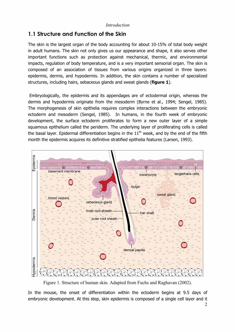
The skin is the largest organ of the body accounting for about 10-15% of total body weightin adult humans. The skin not only gives us our appearance and shape, it also serves otherimportant functions such as protection against mechanical, thermic, and environmentalimpacts, regulation of body temperature, and is a very important sensorial organ. The skin iscomposed of an association of tissues from various origins organized in three layers:epidermis, dermis, and hypodermis. In addition, the skin contains a number of specializedstructures, including hairs, sebaceous glands and sweat glands (figure 1).
Embryologically, the epidermis and its appendages are of ectodermal origin, whereas thedermis and hypodermis originate from the mesoderm (Byrne et al., 1994; Sengel, 1985).The morphogenesis of skin epithelia requires complex interactions between the embryonicectoderm and mesoderm (Sengel, 1985). In humans, in the fourth week of embryonicdevelopment, the surface ectoderm proliferates to form a new outer layer of a simplesquamous epithelium called the periderm. The underlying layer of proliferating cells is calledthe basal layer. Epidermal differentiation begins in the 11th week, and by the end of the fifthmonth the epidermis acquires its definitive stratified epithelia features (Larsen, 1993).
Figure 1. Structure of human skin. Adapted from Fuchs and Raghavan (2002).
In the mouse, the onset of differentiation within the ectoderm begins at 9.5 days ofembryonic development. At this step, skin epidermis is composed of a single cell layer and it

Introduction
3
starts to differentiate into a stratified epithelia around 10.5-11 days of development (Byrneet al., 1994; Kopan and Fuchs, 1989; McGowan and Coulombe, 1998). Between days 11 and17, the epidermis becomes fully differentiated, and acquires adult-like barrier properties(Byrne et al., 1994; Hardman et al., 1998). Formation of the hair follicle begins duringembryonic development. In humans mature follicles are formed by the 6th month ofdevelopment, whereas in mice they develop postnatally (Larsen, 1993; Panteleyev et al.,2001; Panteleyev et al., 1997).
1.1.1 Epidermis
Epidermis is a stratified epithelium that renews itself continuously. It is made of various celltypes, the majority of which (90-95%) are keratinocytes. The remaining cells includeLangerhans cells, melanocytes and Merkel cells (Grousson and Peguet-Navarro, 1998; Haakeand Scott, 1991; Hirobe, 1995; Kurosumi et al., 1979; Mackenzie, 1975). Epidermalkeratinocytes are arranged in four layers representing different stages of keratinocytedifferentiation: the basal, spinous, granular and cornified layers. Keratinocytes in the basallayer are undifferentiated proliferating cells, that are tightly attached to a multiproteicstructure that serves as an adherent connection between the epidermis and the dermis; thebasement membrane (BM) (Brakebusch et al., 2000; Chan, 1997; Hertle et al., 1991;Sonnenberg et al., 1991). Adhesion of basal keratinocytes to the BM is mediated bytransmembrane heterodimeric receptors called integrins. α6β4 integrins are the mainheterodimers involved in the attachment of basal keratinocytes to the BM (DiPersio et al.,2000; Georges-Labouesse et al., 1996). In contrast to other integrins that are distributedover the basal, apical, and lateral surfaces of basal cells, α6β4 integrins are primarily locatedat the basal surface, and are structural components of hemidesmosomes (Hertle et al., 1991;Sonnenberg et al., 1991; Watt, 2002).
In response to not well defined signals, keratinocytes in the basal layer detach from the BM,and initiate a terminal differentiation program. Keratinocytes migrate to the spinous layerwere they undergo morphological and biochemical changes, including decrease in thenuclear to cytoplasmic ratio, formation of new cell-to-cell adherent junctions (desmosomes),and loss of α6 and β1 integrins expression. Moreover, major changes in keratinocytecytoskeleton occur at this stage. The cytoskeleton of basal keratinocytes is composed of twomain intermediate filaments, the type II keratin 5 (K5) and the type I keratin K14 (K14)(Byrne et al., 1994; Chu and Weiss, 2002; Fuchs, 1995). In the spinous layer expression ofK5 and K14 is lost, and expression of keratins 1 (K1) and 10 (K10) is activated (Kim et al.,1984; Kopan and Fuchs, 1989; Loening et al., 1982; Moll et al., 1984).
Following the terminal differentiation program, keratinocytes migrate upwards to thegranular layer were they switch again gene expression programs to initiate the formation ofthe protective permeability barrier. The formation of the permeability barrier requirescoordinated activation of a complex set of events including keratinization, formation of the

Introduction
4
cornified envelope, formation of intracellular lamellar bodies, and induction of cell death.Keratinization results from the accumulation and cross-linking of cytoplasmic keratins, whichresults in the formation of a compact keratin network. In parallel, the expression of severalproteins that are structural components of the cornified envelope (CE) is activated (Marshallet al., 2001), including involucrin, loricrin, filaggrin and the small proline-rich proteins(Steinert and Marekov, 1995). This proteins are sequentially incorporated, via action ofepidermal transglutaminases (Greenberg et al., 1991; Peterson and Wuepper, 1984; Steinertand Marekov, 1999; Wilhelm et al., 1996), to an extremely tough protein polymeric structurelocated just below the cytoplasmic membrane. CE proteins are cross-linked to membraneanchored proteins and to the keratins complex in the cytoplasm (Steinert and Marekov,1997). In addition, several lipid species, synthetized by keratinocytes in specializedstructures called lamellar bodies (LB) (Weaver et al., 2002), are delivered selectively to theintercellular spaces at the stratum granulosum - stratum corneum interface (Elias et al.,1998), were they accumulate forming the epidermal permeability barrier (Eckert et al.,1997). Finally, cell death is induced in keratinocytes at late stages of differentiation(Gandarillas et al., 1999; Maruoka et al., 1997). Thus, the tight structure formed byinteractions between proteic and lipidic components of the cornified layer serves as amechanical protection and permeability barrier for the organism.
In addition to keratinocytes, human epidermis contains melanocytes, Langerhans cells andMerkel cells. Melanocytes originate from the neural crest and migrate into the epidermisduring embryonic development (Haake and Scott, 1991; Holbrook et al., 1989). In humans,melanocytes are distributed regularly among basal keratinocytes and in the hair follicle,whereas in mouse they are restricted to the hair follicle (Haake and Scott, 1991; Silver et al.,1977). Melanocytes produce melanin, the main natural pigment of the skin, and they transferit to the keratinocytes. Melanin not only gives the color to the skin, but protects the deeperlayers of the skin from solar radiation (Bessou-Touya et al., 1998). Langerhans cells aremobile dendritic, antigen presenting cells present in all stratified epithelia. They represent 3-6% of all cells in the epidermis, where they are considered as immature, becoming matureafter contact with antigens. Langerhans cells arise from the bone marrow. They first appearin the epidermis during development, and continue to migrate into the dermis throughoutlife. Merkel cells are pressure-detecting mechanoreceptors. In humans they lay at the baseof the epidermis in the thick skin of palmar and plantar regions, and are associated withunderlying nervous endings in the dermis (Larsen, 1993).
1.1.2 Dermis.
Dermis is a supportive and elastic tissue, which consists of cells and fibrous molecules. Inaddition to its protective and supportive role, the dermis also contains blood vessels andnerves. It undergoes continuous turnover, regulated by mechanisms controlling thesynthesis and degradation of its protein components. Fibroblasts (FB), the main cellpopulation in the dermis are responsible for the synthesis of all the proteic components of

Introduction
5
the dermis (Booth et al., 1980; Prockop, 1982; Tajima and Pinnell, 1981). A large majorityof dermal fibers are made of interstitial collagens, which accounts for 98% of the total massof dried dermis that are responsible for the mechanical resistance of the skin.
1.1.3 Hypodermis.
It is a fatty tissue representing the deepest part of the skin, separating it from the underlyingbody constituents. The hypodermis is composed mainly of adipocytes arranged in lobulesseparated by connective tissue. It plays an important role in thermoregulation, insulation,provision of energy, and protection from mechanical injuries (Hayward and Keatinge, 1981;Nunneley et al., 1985).
1.1.4 Hair Follicle
The hair follicle is an appendage of the epidermis. It develops from the embryonic epidermisas an epithelial extension. This epithelial extension differentiates into three enclosedepithelial cylinders. The central most cylinder forms the hair shaft. The outermost cylinderforms the outer root sheath (ORS) that separates the whole structure from the dermis. Themiddle cylinder, the inner root sheath (IRS), guides the shaft in its passage outward.
The hair cycles through periods of growth (anagen), regression (catagen) and rest (telogen).During anagen, the follicle grows deep into the hypodermis of the skin and the hair grows inlength. During catagen, cells in the lower part of the follicle undergo a massive wave ofapoptosis (Weedon and Strutton, 1984), whereas the regressing follicle maintains itsattachment to the hair shaft through both telogen and the induction of the next anagenphase (Lavker et al., 1998). The hair follicle continues to cycle during almost all life, but hairsynchronization is lost after the second cycle in mice (Bernot et al., 2002). At the bottom ofthe follicle is located the hair bulb, a terminal bell-like extremity containing matrix cellsresponsible for hair growth. The hair bulb forms an invagination that envelops the follicularpapilla, in which papillary fibroblast important for the regulation of hair growth are located(Jahoda and Reynolds, 1996; Paus, 1998). Sebaceous glands are located in the upper part ofthe follicle and made of an outer layer of basal cells, and of several layers of mature, lipid-laden cells. They produce the oily sebum that lubricates the skin and the hair. Anotherimportant structure located in the upper part of the follicles is the bulge (Kanitakis, 2002).The bulge is a special compartment, which is part of the ORS, were the hair follicle stemcells are located (Akiyama et al., 2000; Taylor et al., 2000).
1.1.5 Epidermal Stem Cells
The epidermis is a continuously regenerating tissue. Throughout adult life there is arequirement for the production of new interfollicular keratinocytes to replace the cells that

Introduction
6
are continually being shed from the surface of the skin. In response to skin injury, such asskin wounds, production of new keratinocytes is also required to repopulate the epidermis.Furthermore, during each anagen stage of the hair cycle new keratinocytes are produced toform the ORS of the growing follicle. Thus, a population of pluripotencial stem cells mustexist in the epidermis to accomplish all this regenerative tasks. Several experimentalapproaches demonstrated that the bulge of the hair follicle contains a population of putativekeratinocyte stem cells (Cotsarelis et al., 1990; Morris and Potten, 1999). It has been shownthat the progeny of the bulge stem cells can migrate up or down to populate theinterfollicular epidermis and the hair follicle, respectively (Cotsarelis et al., 1999; Liu et al.,2003a; Taylor et al., 2000), suggesting that bulge keratinocytes represent the pluripotentialkeratinocyte stem cell population. However, keratinocytes with stem cell properties havebeen also identified in the interfollicular epidermis (Ghazizadeh and Taichman, 2001; Joneset al., 1995), which may be responsible for the maintenance of the interfollicular epidermiskeratinocyte population. Moreover, it has been shown that stem cells from the interfollicularepidermis can differentiate to form a normal hair (Ferraris et al., 1997; Lavker and Sun,2000). These observations suggest that some plasticity may exist in the epidermalkeratinocyte population. Thus, differences between the bulge and the interfollicularepidermis microenvironment may play a crucial role in the determination of keratinocyteidentity in vivo.
The basal layer of the epidermis contains a heterogeneous population of keratinocytes inwhich three cell types are distinguished: epidermal stem cells, transit amplifying (TA) cells,and post mitotic differentiating (PMD) cells (Kaur and Li, 2000; Lavker and Sun, 1982;Pavlovitch et al., 1989). Interfollicular epidermal stem cells were first identified by theirability to retain radioactive labels for long periods of time on pulse-chase experiments(Bickenbach and Holbrook, 1987; Potten et al., 1982), and it is generally accepted that theyrepresent 2-10% of all basal keratinocytes (Alonso and Fuchs, 2003; Potten and Morris,1988). They are slow dividing cells with unlimited self-renewal capacity characterized byhigh expression levels of integrins α6 and β1 (Morris and Potten, 1994; Watt, 1988). Whencultured in vitro, stem cells adhere rapidly to the substrate and form colonies with greatreproductive capacity, that can be used to form a structurally complete epidermis inorganotypic cultures (Barrandon and Green, 1987; Bickenbach and Chism, 1998).Asymmetric division of epidermal stem cells gives rise to a daughter stem cell, identical tothe dividing stem cell, and to a TA cell (Jones et al., 1995; Savill, 2003; Savill and Sherratt,2003; Seery and Watt, 2000). In contrast to stem cells, TA cells divide rapidly, they havelimited proliferation capacity, exhibit lower levels of integrins α6 and β1 expression, and inculture take a longer time to adhere to the substrate (Adams and Watt, 1990; Jones et al.,1995; Kaur and Li, 2000; Tennenbaum et al., 1996). After few rounds of proliferation, TAcells stop dividing and initiate the terminal differentiation program, becoming PMD cells(Adams and Watt, 1990; Jaken and Yuspa, 1988; Jensen et al., 1985; Skerrow, 1978).
p63, a homologue of the tumor suppressor p53, has been identified as a putative marker forkeratinocyte stem cell (Mills et al., 1999; Pellegrini et al., 2001). Interestingly, in mouse
Introduction
7
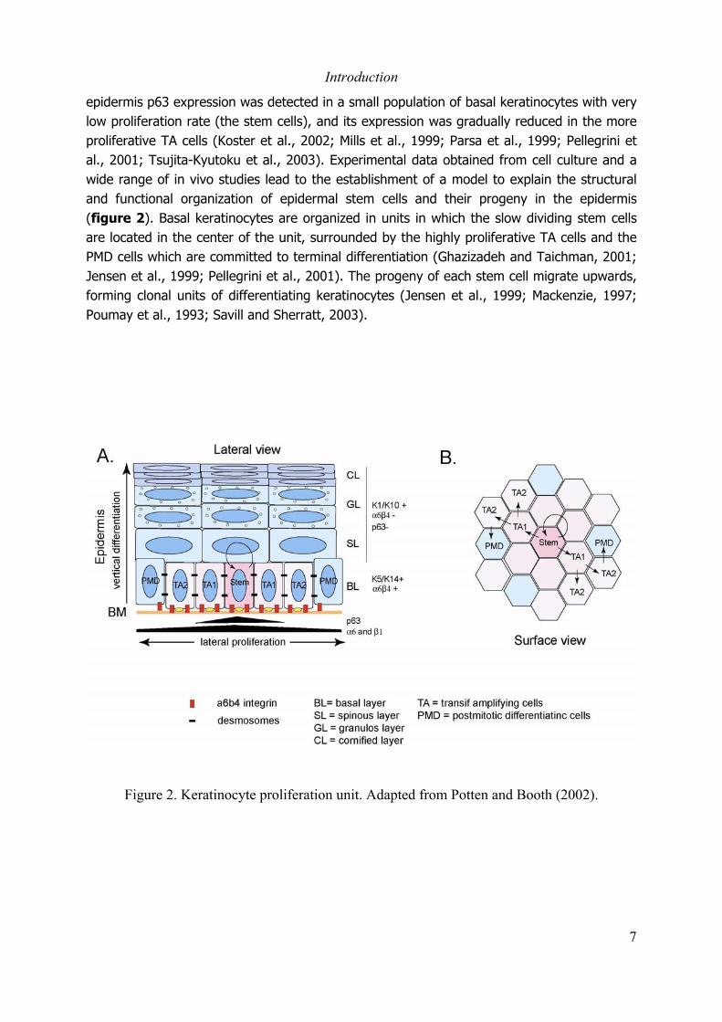
epidermis p63 expression was detected in a small population of basal keratinocytes with verylow proliferation rate (the stem cells), and its expression was gradually reduced in the moreproliferative TA cells (Koster et al., 2002; Mills et al., 1999; Parsa et al., 1999; Pellegrini etal., 2001; Tsujita-Kyutoku et al., 2003). Experimental data obtained from cell culture and awide range of in vivo studies lead to the establishment of a model to explain the structuraland functional organization of epidermal stem cells and their progeny in the epidermis(figure 2). Basal keratinocytes are organized in units in which the slow dividing stem cellsare located in the center of the unit, surrounded by the highly proliferative TA cells and thePMD cells which are committed to terminal differentiation (Ghazizadeh and Taichman, 2001;Jensen et al., 1999; Pellegrini et al., 2001). The progeny of each stem cell migrate upwards,forming clonal units of differentiating keratinocytes (Jensen et al., 1999; Mackenzie, 1997;Poumay et al., 1993; Savill and Sherratt, 2003).
Figure 2. Keratinocyte proliferation unit. Adapted from Potten and Booth (2002).

Introduction
8
1.1.6 Skin homeostasis
In healthy skin, basal keratinocytes proliferate slowly, and in response to not well definedsignals, some of the basal cells initiate the terminal differentiation program, migrateupwards, and are finally shed from the skin. However, in response to epidermal injury orunder certain pathological conditions, keratinocytes become activated. Activatedkeratinocytes are hyperproliferative, migratory, they change their cytoskeleton, augment thelevels of membrane receptors, and produce components of the extracellular matrix (ECM)(Freedberg et al., 2001). Activated keratinocytes produce signals to alert the surroundingcells, including other keratinocytes, FB, melanocytes, and lymphocytes. The affected cellstypes in turn produce their own paracrine and autocrine signals to potentiate the response.Once the damage has been repaired, de-activating signals are produced, promoting thereversal of the basal phenotype. Fine balance between keratinocyte proliferation anddifferentiation, and the capacity of keratinocytes to become activated in response to injurydepends on complex interactions between different cells types present in all threecompartments of the skin. Such interactions involve cell-cell contacts, production ofautocrine and paracrine factors, and ECM components.
Probably the most studied interaction between two cell types in the skin is that ofkeratinocytes and fibroblasts. It is well known that epidermal-mesenchymal interactions playa key role not only in skin morphogenesis, but also in the maintenance of skin homeostasis.In the epidermis, the BM physically separates keratinocytes and fibroblasts, thereforeinteractions between the two cell types depends on the production of secreted factors thatact in a paracrine manner. Several factors involved in the FB-keratinocyte communicationhave been identified. Keratinocyte growth factor (KGF) (Marchese et al., 1990; Rubin et al.,1989) and the granulocyte macrophage colony stimulation factor (GM-CSF) (Braunstein etal., 1994) for example, are fibroblast secreted factors that affect keratinocyte proliferation.On the other hand, keratinocytes produce and secrete interleukin 1 (IL-1) which, in additionto its autocrine effect on keratinocytes (Kupper et al., 1988), induces expression of KGF andGM-CSF in fibroblasts (Maas-Szabowski et al., 1999). Using organotropic cultures, Szabowskiand co-workers demonstrated that in the presence of fibroblast impaired in the production ofKGF and GM-CSF, keratinocyte proliferation and differentiation is strikingly affected(Szabowski et al., 2000). Thus, keratinocyte-fibroblast communication plays a major role inthe maintenance of skin homeostasis.
Another well known interaction between two different cell types in the skin is that ofkeratinocytes and melanocytes. Keratinocyte-melanocyte interactions have been largelydocumented in terms of skin pigmentation. Melanocytes contain highly specializedorganelles called melanosomes in which melanin is produced. Melanosomes undergo acomplex maturation process, and are transferred to keratinocytes through melanocytedendrites which are in direct contact with keratinocytes (Bessou-Touya et al., 1998; Cario-Andre et al., 1999; Seiberg et al., 2000). The relationship between keratinocytes andmelanocytes is not limited to the melanin transfer process. Studies using keratinocytes and

Introduction
9
melanocytes in culture, reconstituted epidermis, and animal models demonstrated thatepidermal keratinocytes are involved in the regulation of melanocyte growth anddifferentiation, migration and dendricity (Archambault et al., 1995; De Luca et al., 1988;Hirobe, 1994; Nakazawa et al., 1995; Scott and Haake, 1991; Tenchini et al., 1995; Valyi-Nagy and Herlyn, 1991; Valyi-Nagy et al., 1993; Valyi-Nagy et al., 1990). The mechanism bywhich keratinocytes control melanocytes function appears very complex. It involves cell-to-cell contacts (Hara et al., 1995; Hsu et al., 2000; Tang et al., 1994; Valyi-Nagy et al., 1993),keratinocyte-secreted factors (Donatien et al., 1993; Imokawa et al., 1992; Tenchini et al.,1995), and ECM components (Nakazawa et al., 1995).
In contrast to keratinocytes, FB and melanocytes, the role of adipocytes in the skin, and itsinteraction with other cell types, has been poorly investigated. For a long time, adipocyteswere considered as “passive cells” in the skin. However, recent experimental datademonstrate that adipocytes can affect the growth and differentiation of other cells types inthe skin, suggesting that they play an active role in the maintenance of skin homeostasis(Frank et al., 2000; Misago et al., 1998; Miyashita et al., 1992; Stallmeyer et al., 2001;Sugihara et al., 2001; Tokuda et al., 1999). Thus, maintenance of skin homeostasis requiresan orchestrated action of the different cells types on the three skin compartments. Locatedin the outer most layer of the skin, keratinocytes seem to play a central role in themaintenance of skin homeostasis.

Introduction
10
1.2 Skin cancer: an alteration of skin homeostasis
The skin is daily exposed to a variety of insults that disturb homeostasis. In normalconditions, skin homeostasis is reestablished by mechanisms involving the orchestratedaction of several cell types in the three compartments of the skin. Genetic alterationsaffecting the proliferation and/or differentiation of the cells in the skin can lead to permanentloss of homeostasis, resulting in pathological states such as psoriasis or cancer.
Exposure to ultraviolet (UV) radiations is considered as the main risk factor for thedevelopment of skin cancer in human (Armstrong and Kricker, 2001; de Gruijl, 1999;Schreiber et al., 1984). UV radiations causes photodamage in the DNA by inducing theformation of dimers between adjacent pyrimidine residues (Johnson, 1978; Regan et al.,1978; Snellman et al., 2003). The cell can respond to the UV-induced damage by repairingthe DNA to avoid harmful mutations (D'Ambrosio et al., 1981; Muramatsu et al., 1992;Runger et al., 1997), or if the damage is too great, by inducing apoptosis to removepotential cancer cells from the population (Iwasaki et al., 1996; Kulms and Schwarz, 2000).Failure of these pathways can results in UV-induced mutations that accumulate with time. Ifthese mutations affect genes involved in the control of cell proliferation, differentiation andapoptosis, the result can be the formation of tumors. Mutations in several genes involved inthe control of proliferation, differentiation and apoptosis have been identified in human skincancer including p53 (Einspahr et al., 1999; Ouhtit et al., 1998), patched (Aszterbaum et al.,1998; Bodak et al., 1999), b-raf (Kumar et al., 2003; Kumar et al., 2004), N-ras (Jiveskog etal., 1998; Reifenberger et al., 2004), and the INK4a-ARF locus (Kreimer-Erlacher et al.,2003).
Cancers of the skin can be originated from different cell types such as keratinocytes,melanocytes, fibroblast, and Merkel cells (Table 1) (Hsu et al., 2002; Smith and Skelton,2003). Depending on their origin, skin cancers are classified in two main groups; melanomaand non-melanoma skin cancer. Melanoma is a malignancy originated from melanocytes,whereas non-melanoma skin cancer includes cancers originated from cells other thanmelanocytes. The most frequent skin cancers are originated from melanocytes andkeratinocytes.
1.2.1 Melanoma
Melanoma is a malignancy originated from the melanocytes, and is considered the mostdangerous skin cancer. Long-term clinical and histopatological observations have led to thedefinition of five major subtypes of tumor progression (Clark, 1991).
Introduction
11
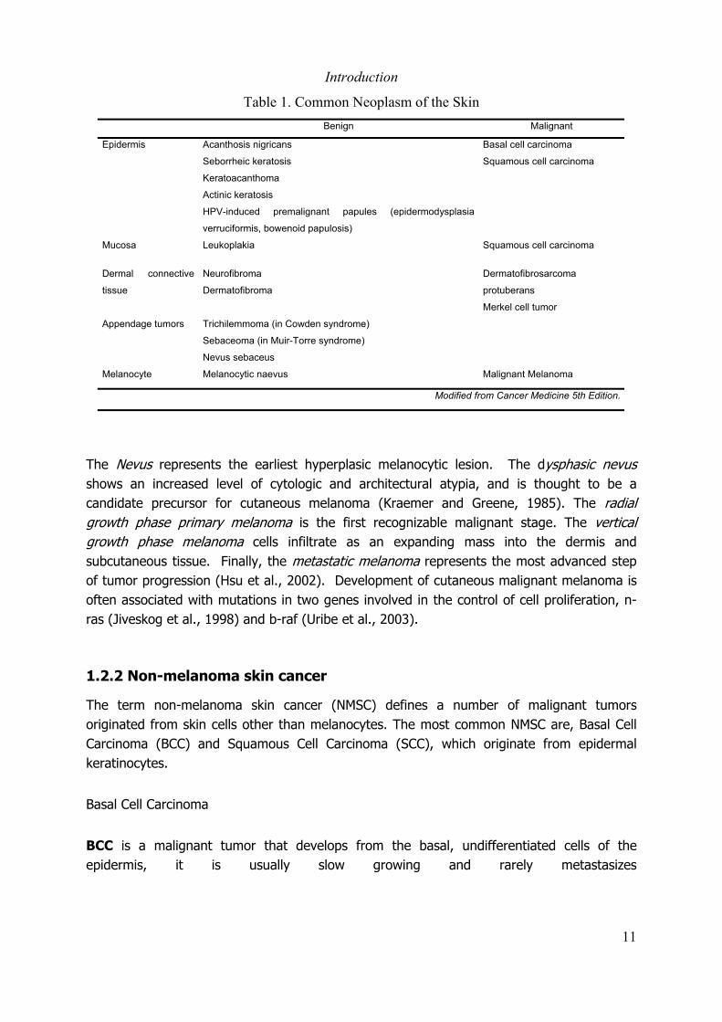
Table 1. Common Neoplasm of the SkinBenign Malignant
Epidermis Acanthosis nigricans
Seborrheic keratosis
Keratoacanthoma
Actinic keratosis
HPV-induced premalignant papules (epidermodysplasia
verruciformis, bowenoid papulosis)
Basal cell carcinoma
Squamous cell carcinoma
Mucosa Leukoplakia Squamous cell carcinoma
Dermal connective
tissue
Neurofibroma
Dermatofibroma
Dermatofibrosarcoma
protuberans
Merkel cell tumor
Appendage tumors Trichilemmoma (in Cowden syndrome)
Sebaceoma (in Muir-Torre syndrome)
Nevus sebaceus
Melanocyte Melanocytic naevus Malignant Melanoma
Modified from Cancer Medicine 5th Edition.
The Nevus represents the earliest hyperplasic melanocytic lesion. The dysphasic nevusshows an increased level of cytologic and architectural atypia, and is thought to be acandidate precursor for cutaneous melanoma (Kraemer and Greene, 1985). The radialgrowth phase primary melanoma is the first recognizable malignant stage. The verticalgrowth phase melanoma cells infiltrate as an expanding mass into the dermis andsubcutaneous tissue. Finally, the metastatic melanoma represents the most advanced stepof tumor progression (Hsu et al., 2002). Development of cutaneous malignant melanoma isoften associated with mutations in two genes involved in the control of cell proliferation, n-ras (Jiveskog et al., 1998) and b-raf (Uribe et al., 2003).
1.2.2 Non-melanoma skin cancer
The term non-melanoma skin cancer (NMSC) defines a number of malignant tumorsoriginated from skin cells other than melanocytes. The most common NMSC are, Basal CellCarcinoma (BCC) and Squamous Cell Carcinoma (SCC), which originate from epidermalkeratinocytes.
Basal Cell Carcinoma
BCC is a malignant tumor that develops from the basal, undifferentiated cells of theepidermis, it is usually slow growing and rarely metastasizes

Introduction
12
(Saldanha et al., 2003; Wong et al., 2003). The tumor appears as proliferating basaloid cellsforming cords and islands, invading into the dermis (Saldanha et al., 2003). The peripheralcells have characteristic palisading arrangement. A connection between the tumor islandand the epidermis can be demonstrated in most of the cases. The tumor may have focalareas of ulceration, with an accompanying chronic inflammatory infiltrate (Cancer Medicine5th edition). Mutations in genes such as p53 (Bolshakov et al., 2003; Ouhtit et al., 1998),patched (Bodak et al., 1999; de Gruijl et al., 2001; Lacour, 2002) and Fas (Boldrini et al.,2003) are frequently observed in human BCC.
Squamous Cell Carcinoma
Squamous Cell Carcinoma (SCC) is a malignant tumor arising from epidermal keratinocytes,which grows in a destructive way and metastasizes mainly via the lymphatic system. Actinickeratosis represents an early stage of SCC. It is characterized by atypical keratinocyteproliferation involving the suprabasal layers of the epidermis (Cockerell, 2000). However, thearchitectural organization of keratinocytes is not affected. In a more advanced stage, the socalled in situ SCC, the epidermal keratinocyte organization is lost, keratinocytes are arrangedhaphazardly with a “windblown” appearance. The border between epidermis and dermisremains sharp throughout the lesion. There is no tendency for keratinocytes to flatten outand mature, as they migrate from the basal layer upward, as is seen in normal epidermis.There is usually a moderate amount of chronic inflammatory infiltrate in the upper dermis.Bowen’s disease is an in situ SCC that occurs on non-exposed skin. In SCC the tumor cellsare seen to proliferate downward in cords and single cells into the dermis. The degree ofdifferentiation is determined by the extent of the tumor trying to recapitulate normalkeratinizing epithelium, with the Spindle-Cell Squamous Carcinoma (SpCC) representing theleast differentiated form (Cancer Medicine, 5th Edition). SCC present high frequency ofmutations in the p53 gene (Bolshakov et al., 2003; Einspahr et al., 1999) and in the INK4a-ARF locus (Kreimer-Erlacher et al., 2003). In addition, low frequency of H-Ras mutationshave also been detected in AK and SCC lesions (Spencer et al., 1995; van der Schroeff et al.,1990).

Introduction
13
1.3 Mouse models of skin carcinogenesis
A number of mouse models have been validated to study cancers originated from severalorgans including lung, mammary gland, nervous system, prostate and skin (MMHC NCIdatabase). Mouse models for both melanoma and non-melanoma skin cancer have beengenerated based on three main approaches: 1) induction of skin tumors by chemicaltreatment or exposure to UV-radiations; 2) generation of transgenic mice expressingoncogenic proteins under the control of tissue specific promoters; and 3) generation ofknockout mice with genetic alterations that are observed in human familial cancers. Someexamples of mouse models of melanoma and non-melanoma skin cancer are summarized intable 2. In this section I will describe the two models that we have used in our study, thetwo-step chemical carcinogenesis (DMBA/TPA) model, and the genetically pre-initiated(Tg.AC) mouse model.
1.3.1 The chemical skin carcinogenesis model
The two-step chemical skin carcinogenesis protocol has been used for over 50 years. In thismodel, skin tumors are induced by a single topical application of a chemical carcinogen intomouse skin, followed by repeated applications of a tumor promoter. The treatment gives riseto a series of phenotypical changes in the skin, including epidermal hyperplasia, formation ofbenign squamous tumors (papillomas) and the progression of some squamous papillomas tomalignant squamous carcinoma, with eventual metastasis of the cancer cells to distal organs(figure 3). The phenotypic evolution from normal keratinocytes through squamouspapilloma into squamous carcinoma is associated with genetic and epigenetic reproduciblechanges that characterize each stage of the process (Hennings et al., 1993).
Initiation
The term initiation defines a genetic event which consists on the introduction of a permanentmutation in the DNA of the cell, generally as a result of a DNA damage induced by genotoxicagents such as UV radiation or chemical substances. In the two-step chemical carcinogenesisprotocol, initiation is induced by topical application of the potent carcinogen 7,12-dimethylbenz(a)anthrancene (DMBA). Topical application of DMBA into mouse skin inducesactivation mutations in the H-ras, N-ras, and Ki-ras oncogenes, which results in theexpression of constitutively active Ras oncoproteins (Pazzaglia et al., 2001; Quintanilla et al.,1986a). Mutation and activation of Ha-ras oncogene are critical events for the formation ofskin tumors. Indeed, Ha-ras null mice develop significantly less tumors than wild-type micein response to a DMBA/TPA protocol (Ise et al., 2000), and the tumors induced in Ha-ras nullmice exhibit mutations in the K-ras gene, suggesting that activation of the K-ras gene canreplace the Ha-ras activation at the initiation step (Ise et al., 2000). The DMBA induced

Introduction
14
mutation in the Ha-ras oncogene is an irreversible event. However, the mutation is silent,and additional stimuli (promoter) are required to induce the formation of tumors.
Table 2. Mouse models of skin cancer
Cancer Type Model Phenotype Reference
Tyr-SV40E Predominantly ocular, but alsocutaneous melanomas with a high rateof metastasis
(Bradl et al., 1991)
Tpras Cutaneous melanomas develop aftertopical DMBA application
(Broome Powell etal., 1999)
K14-stem cellfactor
Hyperpigmentation with increasedmelanin production and increasedepidermal melanocytes
(Kunisada et al.,1998)
Ink4a∆2/3 Cutaneous melanomas develop afterDMBA treatment with occasionalmetastasis
(Krimpenfort et al.,2001)
Tyr-H-rasInk4a/Arf ∆2/3
Cutaneous and ocular melanomaswithout evidence of metastasis
(Chin et al., 1997)
Melanoma
Pten+/-Ink4a/Arf∆2/+
Cutaneous melanomas developspontaneously at a low rate
(You et al., 2002)
DMBA/TPA Squamous papilloma formation withprogression to invasive carcinoma
(Yuspa, 1998)
Ultravioletradiation
Squamous cell carcinomas andspindle cell squamous carcinoma
(Jiang et al., 1999)
K14-HPV16 earlyregion
Squamous hyperplasia with atypia withprogression to invasive carcinoma ofvarying degrees
(Arbeit et al.,1994)
K5-Gli Development of spontaneous BCC (Nilsson et al.,2000)
Non-melanomaskin cancer(NMSC)
Tg.AC Spontaneous squamous papillomas inold animals. Squamous papillomaswith progression to carcinoma afterTPA treatment or exposure to UV
(Leder et al.,1990)
Promotion
Promotion is accomplished by repeated applications of the tumor promoter 12-0-tetradecanoylphorbol-13-acetate (TPA). Experiments in vitro and in vivo demonstrated thatTPA stimulates differentiation of normal keratinocytes (Wei et al., 2003), induces basal cellhyperproliferation in the epidermis (Heyden et al., 1994), and a strong inflammatory reactionin the skin (Aldaz et al., 1985) of adult mice. In contrast, initiated cells have impaired ability

Introduction
15
to respond to differentiation signals (Kulesz-Martin et al., 1991), and in response to TPAtreatment, their proliferation to differentiation ratio may be increased, which results in theclonal expansion of the initiated population and the formation of squamous papillomas(Ghosh et al., 2004; Karen et al., 1999).
Malignant conversion
Conversion of a benign tumor to malignant carcinoma is characterized by an increasedautonomy from both the environment and the host. Papillomas induced by repeatedapplication of TPA may progress to malignancy in the absence of any additional exogenousexposure, although the rate of progression is slow and the frequency of malignantconversion is low (Hennings et al., 1993). In general 25-35 weeks of promotion are requiredto induce malignant conversion, and only few of the tumors may become malignant. Thetumor incidence, multiplicity and frequency of malignant conversion depend in great extenton the genetic background of the animals (Naito and DiGiovanni, 1989). It has been shownthat a subset of papillomas that have a greater risk for progression into malignancy (high-risk papillomas) can be identified at early stages during tumor progression (Glick et al.,1993). High-risk papillomas appear earlier and are characterized by an upregulation of α6β4integrins expression in the keratinocytes of the suprabasal layers (Tennenbaum et al., 1993),and reactivation of the keratin 13 (K13) expression (Rundhaug et al., 1997).
Figure 3. The chemical skin carcinogenesis model. (a) Initiation is induced by a single topicalapplication of the potent carcinogen 7,12-dimethylbenz(a)anthrancene (DMBA). (b)Promotion is achieved by repeated applications of 12-0-tetradecanoylphorbol-13-acetate(TPA). TPA treatment induces progressive changes in the skin, including epidermalhyperplasia, formation of benign papillomas, progression of the papillomas to carcinoma andthe eventual invasion of the malignant cells to the dermis and distal organs.

Introduction
16
Malignant conversion is characterized by invasion through the basement membrane andmigration into the underlying stroma. In situ carcinoma is an early stage of malignantprogression, and is characterized by the presence of disorganized basal-like keratinocytesthrough all the epidermal layers. In a more advanced stage (focal carcinoma), the basementmembrane separating the epidermis from the dermis is disrupted and the epidermal cellsinvade the underlying dermal compartment. Finally, in the most advanced stages ofmalignant conversion, the squamous cell carcinoma, epidermal keratinocytes are seen asnests or islands of cells invading the dermis. Malignant conversion involves importantchanges in cell-cell and cell-stoma interactions (Kerkela et al., 2000; Kerkela et al., 2001),and is often associated with mutations in p53 and upregulation of the AP-1 family oftranscription factors (Domann et al., 1994; Watts et al., 1995). Targeted expression of amutant p53 protein to the epidermis for example, results in accelerated chemically-inducedtumorigenesis and malignant transformation (Wang et al., 1998), whereas mice carrying anull mutation of the c-fos gene developed papillomas but not carcinomas in response toDMBA/TPA treatment (Saez et al., 1995), suggesting that p53 and c-fos play key roles duringthe progression stage.
1.3.2 The TG.AC model
In 1990, Leder and co-workers generated a transgenic mouse strain (Tg.AC) that carries theactivated v-Ha-ras oncogene under the control of the mouse embryonic α-like, zeta-globingene promoter (Leder et al., 1990). They found that Tg.AC mice spontaneously developedskin papillomas at areas of epidermal abrasion, and that in response to topical application ofTPA, Tg.AC mice developed multiple papillomas, some of which progressed to squamous cellcarcinomas.
Further studies on Tg.AC mice demonstrated that development of skin papillomas can alsobe induced by application of other known tumor promoters (Spalding et al., 1993), by fullthickness wounding of the skin (Cannon et al., 1997), and by exposure to UV-radiations(Trempus et al., 1998). It has been shown that transcriptional activation of the H-rastransgene in Tg.AC mice is a key event for the development of skin tumors. The expressionof the Ha-ras transgene is undetectable in the skin of unchallenged Tg.AC mice, but isexpressed in epidermal keratinocytes after 10 weeks of topical application of TPA (Battaloraet al., 2001), and at higher levels in all induced skin tumors (Cannon et al., 1997; Hansenand Tennant, 1994a; Hansen et al., 1996; Leder et al., 1990). The Tg.AC mouse has beenextensively used as a model for evaluating the carcinogenic potential of chemicals andpharmaceuticals (Sistare et al., 2002; Tennant et al., 1998), and for the study of genesinvolved in skin carcinogenesis. It has been shown for example, that double transgenic mice,bearing the Keratin 6-ornithine decarboxylase and v-Ha-ras transgenes, developedspontaneous skin tumors (Smith et al., 1998). Introduction of a null mutation of the kinasesuppressor of ras (KSR) gene into Tg.AC mice results in a complete abrogation of papillomaformation (Lozano et al., 2003). Thus, genetically pre-initiated Tg.AC mice represent an
Introduction
17
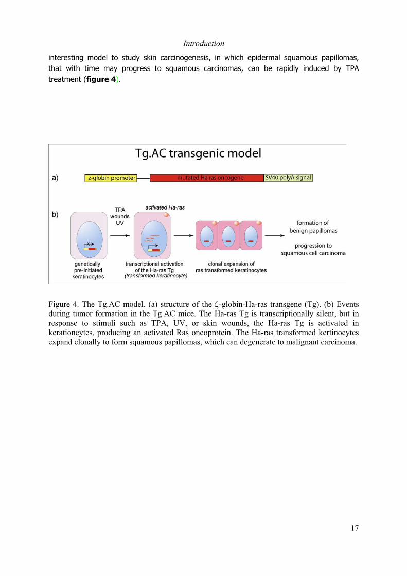
interesting model to study skin carcinogenesis, in which epidermal squamous papillomas,that with time may progress to squamous carcinomas, can be rapidly induced by TPAtreatment (figure 4).
Figure 4. The Tg.AC model. (a) structure of the ζ-globin-Ha-ras transgene (Tg). (b) Eventsduring tumor formation in the Tg.AC mice. The Ha-ras Tg is transcriptionally silent, but inresponse to stimuli such as TPA, UV, or skin wounds, the Ha-ras Tg is activated inkerationcytes, producing an activated Ras oncoprotein. The Ha-ras transformed kertinocytesexpand clonally to form squamous papillomas, which can degenerate to malignant carcinoma.

Introduction
18
1.4 Tissue-specific temporally-controlled somatic mutagenesis in
the mouse
Functional inactivation of genes by introduction of null mutations in the mouse germ line hasyield remarkable advances in understanding the roles played by specific gene products invivo. However, this strategy has some inherent limitations. For example, invalidation ofgenes coding for proteins that play essential functions during development can result in earlylethality. The effect of a germ line mutation may also be compensated during development.Furthermore, invalidation of genes that play important functions in different cell types mayresult in complex phenotypes. Thus, a strategy to introduce somatic mutations in a giventissue and in a temporally controlled manner is required to overcome this limitations. Thediscovery in the early 1980’s, by Austin and co-workers, of the Cre-loxP site-specifichomologous recombination system from the bacteriophage P1 (Austin et al., 1981), providedthe basis for the development of such technology.
The cre-loxP system consists on a site-specific recombinase, the recombinase Cre, whichrecognizes specific DNA sequences called “lox P” sites and induces homologousrecombination between two loxP sites. The loxP site is a 34 bp sequence composed of two13 bp palindromic sequences separated by 8 bp. The 8 bp between the two palindromesconfers an orientation to the loxP site. When two loxP sites are placed in head-to-headorientation, the Cre recombinase will induce an inversion of the sequence located betweenthe two loxP sites (floxed sequence), whereas recombination between two loxP sites placedin head-to-tail orientation results in the excision of the floxed sequence (Austin et al., 1981)(figure 5). The purification and characterization of the Cre recombinase by Abremski andHoess in 1984 (Abremski and Hoess, 1984), and the demonstration, a few years later, bySauer and Henderson that the Cre recombinase can efficiently induce the excision of a floxedDNA fragment in mammalian cells in culture (Sauer and Henderson, 1989), provided theexperimental basis for the development of a system to induce site-specific mutations in themammalian genome.
The first reports showing that Cre-mediated excision of DNA fragments can be induced inhigher organisms were published in the early 1990’s (Odell et al., 1990; Orban et al., 1992).In these reports the Cre recombinase was introduced as a transgene into Tobacco plants(Odell et al., 1990) or into transgenic mice (Orban et al., 1992) carrying a floxed DNAreporter gene. Excision of the floxed DNA fragment was efficiently induced in both transgenictobacco plants and transgenic mice. Later, using tissue-specific promoters to drive theexpression of the cre recombinase, several laboratories demonstrated the efficacy of the crerecombinase system to introduce mutations in a given gene in a tissue-specific manner(Kulkarni et al., 1999; Ray et al., 1999; Tarutani et al., 1997).

Introduction
19
Figure 5. The Cre-loxP system of homologous recombination.
On the other hand, two strategies have been used to achieve the temporal control of theCre-mediated recombination. One strategy was based in the control of the Cre expression atthe transcriptional level. For this purpose, double transgenic mice carrying the crerecombinase under the control of a tetracycline (Tet)- dependent promoter and a reversetetracycline-controlled transactivator (rtTA) transgene, must be generated. The rtTA proteinis constitutively expressed, but it remains inactive. Exogenous administration of tetracyclinewill activate the rtTA which can now bind to the Tet-dependent promoter to activateexpression of the cre recombinase (St-Onge et al., 1996; Utomo et al., 1999). The otherstrategy is based on the principle that the activity of some proteins can be modulated by thepresence of a specific ligand. Metzger and co-workers generated a fusion protein betweenthe Cre recombinase and the ligand binding domain (LBD) of the human Estrogen Receptorα (hERα), resulting in a Cre recombinase which activity is controlled by estrogens (Cre-ER).They established a cell line in which the Cre-ER is constitutively expressed and theydemonstrated that estradiol or the estrogen agonist/antagonist 4-hydroxytamoxifenefficiently induced the recombinase activity, whereas no activity was detected in the absenceof ligand or in the presence of a synthetic antiestrogen (Metzger et al., 1995). However,introduction of the Cre-ER into transgenic mice may result in the constitutive activation ofthe recombinase by endogenous estrogens. To avoid the activation by endogenousestrogens, a new fusion protein was generated in which the Cre recombinase was fused to amutated LBD of the hERα, resulting in a chimeric recombinase, Cre-ERT, which is activatedby tamoxifen, but not by natural estrogens (figure 6) (Feil et al., 1996). Using this Cre-ERT
chimeric recombinase, Feil and co-workers generated transgenic mice in which the Cre-ERT isexpressed under the control of the cytomegalovirus promoter, and they demonstrated thatexcision of a floxed DNA sequence was efficiently induced in several tissues of Cre-ERT
transgenic mice, after tamoxifen administration (Feil et al., 1996).

Introduction
20
Figure 6. Strategy to generate a tamoxifen-induceble Cre recombinase. Adapted from Metzger(2001)
However, the doses of tamoxifen required to induce the Cre-ERT activity in these transgenicmice were high, which may result in side effects due to the estrogen agonist/antagonistaction of tamoxifen. The problem was circumvented by the introduction of a secondmutation in the LBD of the Cre-ERT recombinase, resulting in a recombinase (Cre-ERT2)which was approximately 10-fold more efficiently induced by tamoxifen than the Cre-ERT
(Feil et al., 1997). Indra and co-workers generated transgenic mice in which the expressionof the Cre-ERT2 recombinases is driven by the bovine keratin 5 (K5) promoter to the basallayer of the epidermis, and demonstrated that excision of a floxed DNA can be efficientlyinduced in the basal layer of the epidermis of adult K5-Cre-ERT2 mice by administration of10-fold lower doses of tamoxifen than those required to activate the Cre-ERT (Indra et al.,1999). Similar results were obtained in transgenic mice in which the Cre-ERT2 recombinase isexpressed under the control of the keratin 14 (K14) promoter (Indra et al., 2000; Indra etal., 1999; Vasioukhin et al., 1999). The mechanism of tamoxifen-induced Cre-ERT2-mediatedmutagenesis in keratinocytes is represented in figure 7. On the other hand, the introductionof Cre mediated mutations selectively in epidermal keratinocytes during mouse embryonicdevelopment was achieved in transgenic mice in which a constitutively active Crerecombinase is expressed under the control of the K14 promoter (K14-Cre) (Indra et al.,2000). Thus, K5-Cre-ERT2 or K14-Cre-ERT2 and K14-Cre transgenic mice represent powerfultools to introduce somatic mutations in adult epidermis and during embryonic development,respectively.
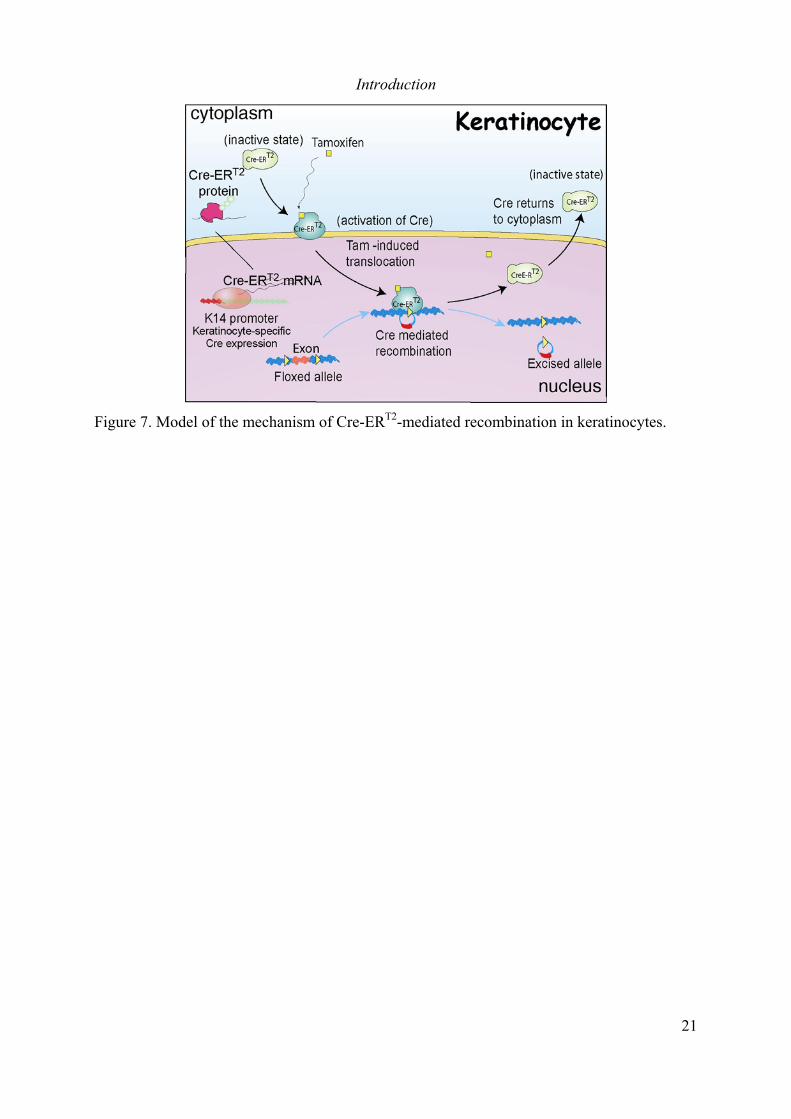
Introduction
21
Figure 7. Model of the mechanism of Cre-ERT2-mediated recombination in keratinocytes.
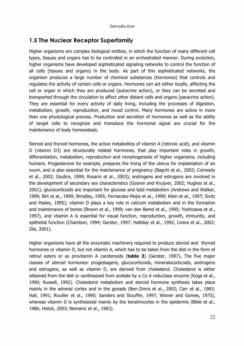
Introduction
22
1.5 The Nuclear Receptor Superfamily
Higher organisms are complex biological entities, in which the function of many different celltypes, tissues and organs has to be controlled in an orchestrated manner. During evolution,higher organisms have developed sophisticated signaling networks to control the function ofall cells (tissues and organs) in the body. As part of this sophisticated networks, theorganism produces a large number of chemical substances (hormones) that controls andregulates the activity of certain cells or organs. Hormones can act either locally, affecting thecell or organ in which they are produced (autocrine action), or they can be secreted andtransported through the circulation to affect other distant cells and organs (paracrine action).They are essential for every activity of daily living, including the processes of digestion,metabolism, growth, reproduction, and mood control. Many hormones are active in morethan one physiological process. Production and secretion of hormones as well as the abilityof target cells to recognize and transduce the hormonal signal are crucial for themaintenance of body homeostasis.
Steroid and thyroid hormones, the active metabolites of vitamin A (retinoic acid), and vitaminD (vitamin D3) are structurally related hormones, that play important roles in growth,differentiation, metabolism, reproduction and morphogenesis of higher organisms, includinghumans. Progesterone for example, prepares the lining of the uterus for implantation of anovum, and is also essential for the maintenance of pregnancy (Bagchi et al., 2003; Conneelyet al., 2002; Giudice, 1999; Rosario et al., 2003); androgens and estrogens are involved inthe development of secondary sex characteristics (Gooren and Kruijver, 2002; Hughes et al.,2001); glucocorticoids are important for glucose and lipid metabolism (Andrews and Walker,1999; Birt et al., 1999; Brindley, 1995; Fernandez-Mejia et al., 1999; Klein et al., 1997; Sivitzand Pasley, 1995); vitamin D plays a key role in calcium metabolism and in the formationand maintenance of bones (Brown et al., 1999; van den Bemd et al., 1995; Yoshizawa et al.,1997), and vitamin A is essential for visual function, reproduction, growth, immunity, andepithelial function (Chambon, 1994; Gerster, 1997; Halliday et al., 1992; Livera et al., 2002;Zile, 2001).
Higher organisms have all the enzymatic machinery required to produce steroid and thyroidhormones or vitamin D, but not vitamin A, which has to be taken from the diet in the form ofretinyl esters or as provitamin A carotenoids (table 3) (Gerster, 1997). The five majorclasses of steroid hormones: progestagens, glucocorticoids, mineralocorticoids, androgensand estrogens, as well as vitamin D, are derived from cholesterol. Cholesterol is eitherobtained from the diet or synthesized from acetate by a Co A reductase enzyme (Koga et al.,1990; Russell, 1992). Cholesterol metabolism and steroid hormone synthesis takes placemainly in the adrenal cortex and in the gonads (Ben-Zimra et al., 2002; Carr et al., 1983;Hall, 1991; Rouiller et al., 1990; Sanders and Stouffer, 1997; Wisner and Gomes, 1975),whereas vitamin D is synthesized mainly by the keratinocytes in the epidermis (Bikle et al.,1986; Holick, 2003; Nemanic et al., 1983).
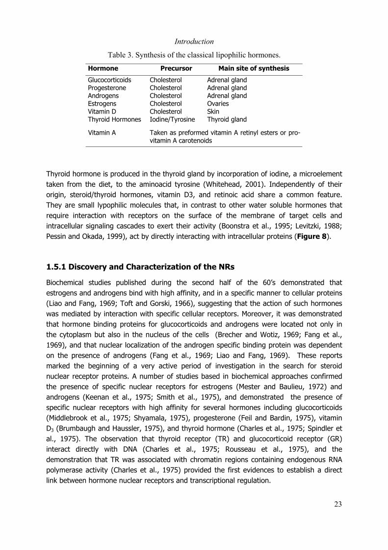
Introduction
23
Table 3. Synthesis of the classical lipophilic hormones.
Hormone Precursor Main site of synthesis
GlucocorticoidsProgesteroneAndrogensEstrogensVitamin DThyroid Hormones
CholesterolCholesterolCholesterolCholesterolCholesterolIodine/Tyrosine
Adrenal glandAdrenal glandAdrenal glandOvariesSkinThyroid gland
Vitamin A Taken as preformed vitamin A retinyl esters or pro-vitamin A carotenoids
Thyroid hormone is produced in the thyroid gland by incorporation of iodine, a microelementtaken from the diet, to the aminoacid tyrosine (Whitehead, 2001). Independently of theirorigin, steroid/thyroid hormones, vitamin D3, and retinoic acid share a common feature.They are small lypophilic molecules that, in contrast to other water soluble hormones thatrequire interaction with receptors on the surface of the membrane of target cells andintracellular signaling cascades to exert their activity (Boonstra et al., 1995; Levitzki, 1988;Pessin and Okada, 1999), act by directly interacting with intracellular proteins (Figure 8).
1.5.1 Discovery and Characterization of the NRs
Biochemical studies published during the second half of the 60’s demonstrated thatestrogens and androgens bind with high affinity, and in a specific manner to cellular proteins(Liao and Fang, 1969; Toft and Gorski, 1966), suggesting that the action of such hormoneswas mediated by interaction with specific cellular receptors. Moreover, it was demonstratedthat hormone binding proteins for glucocorticoids and androgens were located not only inthe cytoplasm but also in the nucleus of the cells (Brecher and Wotiz, 1969; Fang et al.,1969), and that nuclear localization of the androgen specific binding protein was dependenton the presence of androgens (Fang et al., 1969; Liao and Fang, 1969). These reportsmarked the beginning of a very active period of investigation in the search for steroidnuclear receptor proteins. A number of studies based in biochemical approaches confirmedthe presence of specific nuclear receptors for estrogens (Mester and Baulieu, 1972) andandrogens (Keenan et al., 1975; Smith et al., 1975), and demonstrated the presence ofspecific nuclear receptors with high affinity for several hormones including glucocorticoids(Middlebrook et al., 1975; Shyamala, 1975), progesterone (Feil and Bardin, 1975), vitaminD3 (Brumbaugh and Haussler, 1975), and thyroid hormone (Charles et al., 1975; Spindler etal., 1975). The observation that thyroid receptor (TR) and glucocorticoid receptor (GR)interact directly with DNA (Charles et al., 1975; Rousseau et al., 1975), and thedemonstration that TR was associated with chromatin regions containing endogenous RNApolymerase activity (Charles et al., 1975) provided the first evidences to establish a directlink between hormone nuclear receptors and transcriptional regulation.

Introduction
24
Figure 8. Structure of the major lipophylic hormones. From Molecular Biology of the Cell. 3rd
Edition.
The development of genetics and molecular biology in the 80’s, contributed to theidentification and characterization of the genes for several nuclear receptors. Thecharacterization of the GR (Govindan et al., 1985; Miesfeld et al., 1984) and the EstrogenReceptor α (ERα) genes (Green et al., 1986) in the early 1980’s marked the beginning of acascade of discoveries leading to the cloning and characterization of several NRs involved inhormonal signaling (Willson and Moore, 2002). Using a variety of genetic, biochemical, andmolecular biology techniques, a number of NR genes were cloned and characterized,including the chicken progesterone receptor gene (Gronemeyer et al., 1987), the genes forseveral human and mouse retinoic acid receptors (Brand et al., 1988; Giguere et al., 1987;Krust et al., 1989b; Petkovich et al., 1987; Zelent et al., 1989b), the human androgenreceptor (AR) gene (Lubahn et al., 1988; Trapman et al., 1988) and the TR gene (Chang etal., 1989).
In 1978, Wrange and Gustafson had already demonstrated, by biochemical approaches, thatthe GR has two functional domains: a DNA binding domain (DBD) and a ligand bindingdomain (LDB) (Wrange and Gustafsson, 1978). Comparison of the newly emerging nuclearreceptors give rise to the exciting observation that significant regional homology existedbetween the steroid hormone receptors as well as the cellular receptors for thyroid hormoneand retinoic acid (Krust et al., 1986; Owen and Zelent, 2000). NRs exhibit a modularstructure consisting of an NH2 terminal region (A/B), a conserved DNA-binding domain(DBD) or region C, a linker region D, and a conserved region E that contains the ligandbinding domain (LBD) (Freedman and Luisi, 1993; Kumar and Thompson, 1999; Laudet etal., 1992) (figure 9).
Introduction
25
The A/B domain is the most variable region both in size and sequence, and it contains atranscriptional activation domain (AF-1) that shows promoter and cell-specific activity, and isa target for phosphorylation mediated by various signaling pathways which can modulate thereceptor activity (Ali et al., 1993; Berry et al., 1990; Metzger et al., 1992; Rochette-Egly,2003; Tora et al., 1989). The DBD, the most conserved domain of NRs, confers the ability torecognize specific DNA target sequences and activate gene transcription. The DBD containstwo zinc fingers, one of which is involved in the recognition of core DNA motifs and the otheris involved in dimerization (Hard et al., 1990; Katahira et al., 1992; Lee et al., 1993; Maderet al., 1993; Zechel et al., 1994). The D domain, that is not well conserved among NRs,serves as a hinge between the DBD and the LBD, allowing the rotation of the DBD, and inmany cases harbors nuclear localization signals (Guiochon-Mantel et al., 1989; Lee andMahdavi, 1993; Picard et al., 1990; Picard et al., 1988; Picard and Yamamoto, 1987;Shyamala, 1975). The LBD is a multifunctional domain that, in addition to the binding ofligand, mediates homo- and heterodimerization (Fawell et al., 1990; Forman et al., 1989;O'Donnell and Koenig, 1990; Spanjaard et al., 1991). The LBD contains an activationfunction (AF-2) motif that participates in the recruitment of NRs associated proteins and isresponsible for the ligand-dependent transcriptional activation (Durand et al., 1994; LeDouarin et al., 1996; vom Baur et al., 1996). The availability of the sequences of NRs genesand the crystal structure of the NRs proteins together with the development of powerfulcomputer software and sequence databases gave rise to the new concept of “reverseendocrinology” in which the characterization of the receptor precedes the study of itsphysiological function, leading to the discovery of new hormonal responses (Aranda andPascual, 2001; Banner et al., 1993; Herz et al., 1997; Hirose et al., 1994; Moras andGronemeyer, 1998). The subfamilies of NRs from human and mouse are shown in table 4(NUREBASE).
Figure 9. Modular structure of the NRs. Adapted from Aranda and Pascual 2001.

Introduction
26
Table 4. Human and Mouse nuclear receptors

Introduction
27
1.5.2 Mechanism of Nuclear Receptors Action.
NRs interact with DNA through recognition of specific sequences in the promoter region oftarget genes, called response elements, which are composed of a core hexameric motif. Twoconsensus motifs have been identified: the sequence AGAACA that is preferentiallyrecognized by steroid class III receptors (see table 4), whereas AGG/TTCA served as arecognition motif for the receptors from the other classes (Beato et al., 1995). Althoughsome monomeric receptors can bind to a single hexameric motif, most receptors bind ashomo- or heterodimers to elements composed by two core hexameric motifs (Glass, 1994).For dimeric response elements, the half sites can be configured as palindromes, invertedrepeats, or direct repeats. Steroid hormone receptors typically bind to palindromic elementsas homodimers whereas non-steroidal receptors can bind as heterodimers to elements indifferent configurations. In that case, the arrangement and the spacing between motifs willdetermine the selectivity and specificity (Glass, 1994). Direct repeats with a spacing of 3, 4and 5 base pairs were observed to specify preferentially transcriptional responses to vitaminD, thyroid hormone and retinoic acid, respectively, whereas palindromic and invertedpalindromic elements appear to be regulated by several classes of receptors (figure 10)(Mangelsdorf et al., 1991; Naar et al., 1991; Schrader et al., 1994).
Figure 10. Nuclear receptors response elements. Adapted from Glass 1994.
Introduction
28
Moreover, it has been shown that dimerisation between receptors enhances their affinity forthey response elements (Jiang et al., 1997; Perlmann et al., 1996). Interestingly, the threeRetinoic X Receptors (RXRα, RXRβ, and RXRγ), that bind specifically to 9-cis retinoic acid,can form heterodimers with a number of non-steroidal nuclear receptors such as the RetinoicAcid Receptors (RARs), vitamin D3 receptor (VDR), TR, and the peroxisome proliferatoractivated receptors (PPARs), enhancing their DNA-binding to specific response elements andtheir transcriptional activity (Bardot et al., 1993; Issemann et al., 1993; Kliewer et al., 1992;Leid et al., 1992; Mangelsdorf and Evans, 1995; Schulman et al., 1998; Yu et al., 1991;Zhang et al., 1992). Thus, as common heterodimeric partners for non-steroidal nuclearreceptors, RXRs are central regulators of several hormonal signaling pathways.
1.5.3 Coactivators and corepressors.
Another important aspect of the mechanism of NR action is how this DNA-bound nuclearreceptors interact and modulate the activity of the transcriptional machinery. Using bothgenetic and biochemical approaches, several NR interacting proteins were identified (Beatoet al., 1995; Butt and Walfish, 1996; Chakravarti et al., 1996; Sande and Privalsky, 1996).Identification and characterization of these NR interacting proteins lead to the observationthat NRs act in conjunction with two classes of cofactors to regulate transcription in thecontext of chromatin: corepressors and coactivators (Kraus and Wong, 2002; Lemon andFreedman, 1999). The identification and characterization of accessory proteins that interactwith unliganded NRs (corepressors) (Chen and Evans, 1995; Horlein et al., 1995),demonstrated that NR mediated transcriptional repression occurs by two main mechanisms.NR/corepressors complexes can repress transcription either by directly interacting with thetranscriptional machinery (Fondell et al., 1996; Fondell et al., 1993; Muscat et al., 1998;Wong and Privalsky, 1998) or indirectly by modifying chromatin structure (Hu and Lazar,2000; Huang et al., 2000; Ito et al., 2000; Jenster, 1998; Nagy et al., 1997; Townson et al.,2004). Chromatin modifying factors with histone deacetylase (HDAC) activity are recruited tothe NR/corepressor complex through direct interaction with the corepressor (Hu and Lazar,2000; Huang et al., 2000; Ito et al., 2000; Jenster, 1998; Nagy et al., 1997; Townson et al.,2004) or mediated by other intermediate factors such as mSin3A (Hu and Lazar, 2000;Huang et al., 2000; Ito et al., 2000; Jenster, 1998; Nagy et al., 1997; Townson et al., 2004). The recruitment of HDAC factors to the NR complex results in deacetylation andcondensation of the chromatin, which renders the promoter inaccessible for thetranscriptional machinery. Upon binding of the ligand, conformational changes in the NRcomplex lead to exclusion of the corepressors from the complex and the subsequentrecruitment of coactivators (Driscoll et al., 1996; Egner et al., 2001; Leng et al., 1993; Lucket al., 2000; Muller et al., 1984; Yen et al., 1993) (Figure 11).
Three classes of coactivators have been described: histone acetyl transferases (HATs)(Blanco et al., 1998; Chen et al., 1997; Kraus et al., 1999; Li et al., 1999), histone methyl

Introduction
29
transferases (HMTs) (Strahl et al., 2001; Xu et al., 2004), and histone kinases (Banks et al.,2001; Bhattacharjee et al., 2001). In general, coactivators play one or more of the followingroles in nuclear receptor transcriptional activity: (1) they function as bridging factors torecruit cofactors to the DNA-bound NRs (Yao et al., 1996), (2) they acetylate nucleosomalhistones and various transcription factors at the promoters of target genes (Blanco et al.,1998; Chen et al., 1997), (3) they function as bridging factors between NRs and the basaltranscriptional machinery (TRAP/DRIP) (Lee and Lee Kraus, 2001; Muscat et al., 1998;Takeshita et al., 1996; Treuter et al., 1999).
Recruitment of coactivators or of components of the transcription machinery by the NRcomplexes is also modulated by phosphorylation events which, in cooperation with theligand, modulate the transcriptional activity of the NRs (Rochette-Egly, 2003). NRs can bephosphorylated by kinases that are associated with general transcription factors (Chen et al.,2000; Rochette-Egly et al., 1997), or that are activated in response to a variety ofextracellular signals (Adam-Stitah et al., 1999; Hoeck et al., 1989; Hsieh et al., 1991; Jurutkaet al., 1996; Kato et al., 1992; Lee et al., 2000; Pekarsky et al., 2001; Rochette-Egly et al.,1997).
In addition, NRs can also modulate transcription by interacting with other generaltranscription factors. It has been shown that several NRs exhibit a potent antagonistic effecton the AP-1 family of transcription factors (Saatcioglu et al., 1997; Salbert et al., 1993).However, in some cases NRs have positive effects on AP-1 transcriptional activity (Hanley etal., 2000b; Huang et al., 2003; Tseng et al., 2003). The precise mechanism of NRs anti AP-1activity is not clear, however two main mechanisms have been suggested. The first oneinvolves the inhibition of AP-1 binding to its DNA response elements either by physicalinteraction between NRs and AP-1 complexes (Aslam et al., 1999; Delerive et al., 1999;Fanjul et al., 1994; Kuroki et al., 1995; Salbert et al., 1993), or by blocking the JNK-dependent AP-1 activation (Bruna et al., 2003; Caelles et al., 1997; Gonzalez et al., 2000)(Caelles et al., 1997; Zhou et al., 1999).
Figure 11. Mechanism of NR transcription regulation. Adapted from Chen (2000).

Introduction
30
The second mechanism involves the competition for common cofactors required for both NRsand AP1 transcriptional activation such as CBP (Benkoussa et al., 2002; DiSepio et al., 1999;Horvai et al., 1997). NRs have been also shown to interact with the general transcriptionfactor Sp1. NR/Sp1 interaction can have negative or positive effect on Sp1 transcriptionalactivity. For example ER, VDR, and PPARs can synergistically activate Sp1 mediatedtranscription (Husmann et al., 2000; Krey et al., 1995; Krishnan et al., 1994; Liu andFreedman, 1994; Porter et al., 1996), whereas TR inhibits Sp1-dependent transcriptionalactivation (Xu et al., 1993). Thus, NRs are ligand-activated transcription factors involved ina sophisticated and extremely complex hormonal signaling network, not only as hormonesignal transducers, but also as integrators of multiple signaling pathways.
Introduction
31
1.6 Nuclear Receptors in Skin Homeostasis and Skin
Carcinogenesis.
Skin structure and homeostasis is affected by a number of hormonal signals such as sexhormones and vitamins. Differences between skin from man and woman for example aredetermined by the levels of estrogens and androgens (Rosenfield and Deplewski, 1995;Thornton, 2002; Tur, 1997). Vitamin A or vitamin D deficiency results in alterations of skinstructure and function (Fisher and Voorhees, 1996; Pavlovitch et al., 1984). For severalyears, major efforts have been done to understand the mechanisms by which hormonesaffect skin homeostasis. Different experimental approaches such as in situ hybridization(ISH), reverse-transcription coupled polymerase chain reaction (RT-PCR), andimmunohistochemistry (IHC), demonstrated the presence of various members of the NRsuperfamily in the skin (table 5). In this section, I will discuss some relevant in vitro and invivo studies demonstrating that NRs play important roles in the maintenance of skinhomeostasis. Because RXRs are heterodimeric partners for several non-steroidal nuclearreceptors, and RXRα is the predominant RXR in the skin, I will focus on known functions ofRXRα and its heterodimeric partners in skin homeostasis.
1.6.1 The Retinoic Acid Receptors
Retinoids and Skin Homeostasis
The observation that animals maintained in a vitamin A deficient diet exhibit epithelialalterations provided the first experimental evidence for the role of vitamin A in themaintenance of epithelial homeostasis (Bo, 1956; Bo, 1957; Hayes et al., 1970; Reiter,1965). Clinical and experimental data have shown that vitamin A deficiency affects epithelialhomeostasis of several organs including the eye (Pfister and Renner, 1978; Tseng et al.,1984), the cervix (Gijbels et al., 1992), the trachea (Lancillotti et al., 1992). Moreover,administration of exogenous vitamin A to experimental animals also results in alterations ofepidermal homeostasis. Exogenous vitamin A stimulates proliferation of both adult andembryonic epidermis (Tsambaos et al., 1980; Tsambaos et al., 1985), induces expression ofkeratins commonly expressed in hyperproliferative epidermis such as K6, K16 and K17(Eichner et al., 1992; Tsambaos et al., 1980; Tsambaos et al., 1985), and inhibitskeratinocyte terminal differentiation and the formation of the cornified layer (Brown et al.,1985; Eichner, 1986; Eichner et al., 1992; Gibbs et al., 1996).
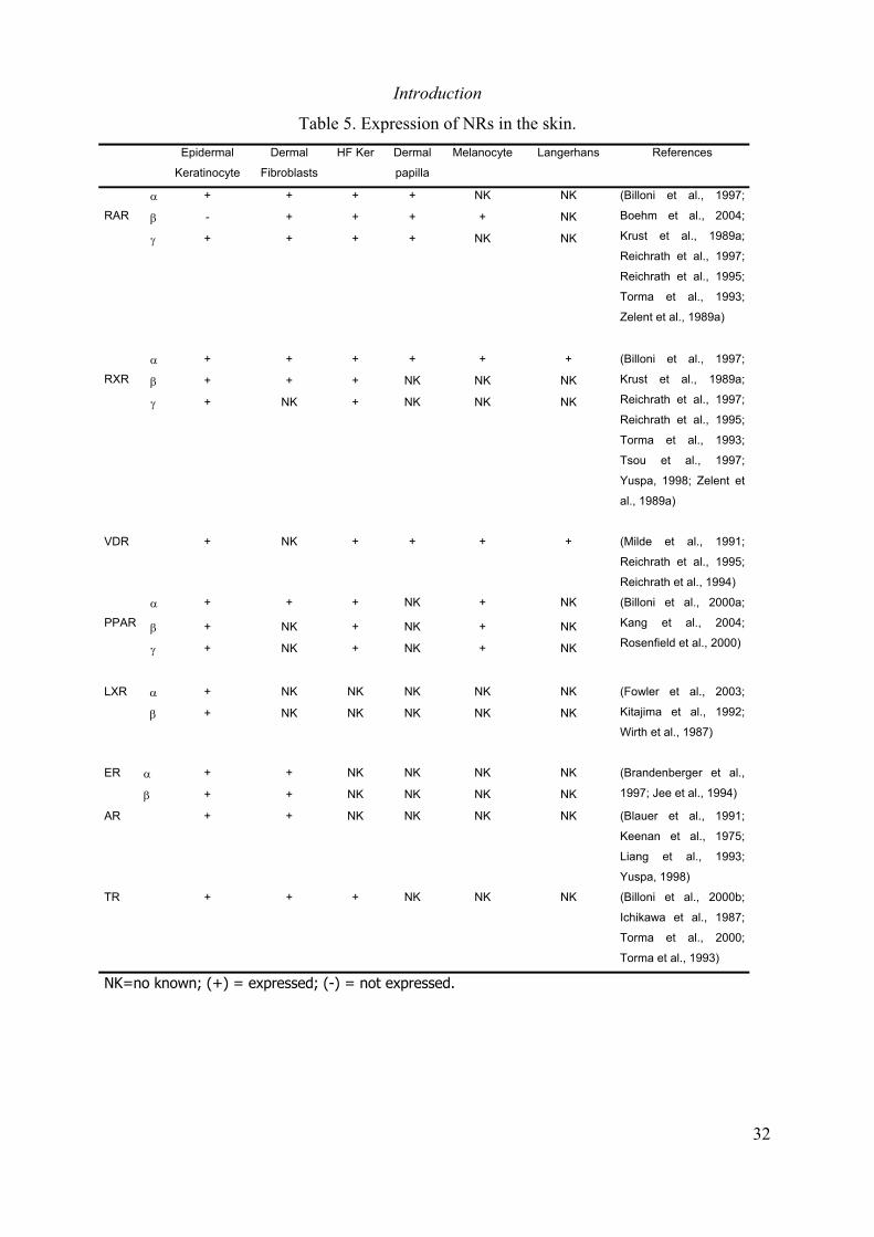
Introduction
32
Table 5. Expression of NRs in the skin.Epidermal
Keratinocyte
Dermal
Fibroblasts
HF Ker Dermal
papilla
Melanocyte Langerhans References
α + + + + NK NK
β - + + + + NK
γ +
RAR
+ + + NK NK
(Billoni et al., 1997;
Boehm et al., 2004;
Krust et al., 1989a;
Reichrath et al., 1997;
Reichrath et al., 1995;
Torma et al., 1993;
Zelent et al., 1989a)
α + + + + + +
β + + + NK NK NK
γ +
RXR
NK + NK NK NK
(Billoni et al., 1997;
Krust et al., 1989a;
Reichrath et al., 1997;
Reichrath et al., 1995;
Torma et al., 1993;
Tsou et al., 1997;
Yuspa, 1998; Zelent et
al., 1989a)
VDR + NK + + + + (Milde et al., 1991;
Reichrath et al., 1995;
Reichrath et al., 1994)
α + + + NK + NK
β + NK + NK + NKPPAR
γ + NK + NK + NK
(Billoni et al., 2000a;
Kang et al., 2004;
Rosenfield et al., 2000)
α + NK NK NK NK NKLXR
β + NK NK NK NK NK
(Fowler et al., 2003;
Kitajima et al., 1992;
Wirth et al., 1987)
α + + NK NK NK NKER
β + + NK NK NK NK
(Brandenberger et al.,
1997; Jee et al., 1994)
AR + + NK NK NK NK (Blauer et al., 1991;
Keenan et al., 1975;
Liang et al., 1993;
Yuspa, 1998)
TR + + + NK NK NK (Billoni et al., 2000b;
Ichikawa et al., 1987;
Torma et al., 2000;
Torma et al., 1993)
NK=no known; (+) = expressed; (-) = not expressed.
Introduction
33
In agreement with these results, RA stimulates proliferation and inhibits terminaldifferentiation of keratinocytes in culture (Gibbs et al., 1996; Varani et al., 1989), indicatingthat RA affects epidermal homeostasis, at least in part, by directly modulating keratinocyteproliferation and differentiation.
The Retinoic Acid Receptors in the Skin
Expression of retinoic acid receptors in the skin was first demonstrated by northern blot andRT-PCR analysis. These experiments demonstrated the presence of RARα, RARγ and RXRαmRNAs in both normal skin and kertinocytes in culture (Elder et al., 1992; Torma et al.,1993). It was also shown that the most abundant transcripts in normal skin are those forRXRα and RARγ (Torma et al., 1993). In agreement with data obtained from RNA analysis,western blot and immunohistochemical (IHC) analysis demonstrated that RARα, RARγ andRXRα proteins are present in the skin, and that RXRα and RARγ are the predominantisotypes (Fisher et al., 1994). In addition, it was shown that RXRβ and RXRγ are alsoexpressed in normal skin (Reichrath et al., 1997; Sherman and Partridge, 1997). Differentexperimental approaches demonstrated that protein levels of RXRs are 5 times higher thanthose of RARs (Fisher et al., 1994), and that in several cell types of normal skin, includingkeratinocyte, melanocyte, FB and dendritic cells, RXRα exhibit the highest levels ofexpression (Reichrath et al., 1995). RXRα is also the main isotypes in the hair follicle (Billoniet al., 1997).
In the early 90’s, a functional role of retinoic acid receptors in keratinocytes wasdemonstrated by transfection experiments, in which expression of different retinoic acidreceptor isotypes resulted in inhibition of the expression of several keratins (Blumenberg etal., 1992; Eichner et al., 1992; Tomic et al., 1990). Moreover, it was shown that introductionof a truncated RARγ protein, lacking the A/B and the DBD domains, into keratinocytes inculture inhibits their ability to undergo terminal differentiation (Aneskievich and Fuchs,1992). Cotransfection of human keratinocytes with expression vectors for dominant negativemutant retinoic acid and retinoic X receptors reduced the retinol-induced activation of aretinoic acid receptor-dependent reporter gene (Kurlandsky et al., 1994).
Targeted disruption of the RARα (Lufkin et al., 1993), RARβ (Mendelsohn et al., 1994) orRARγ (Lohnes et al., 1993) genes in the mouse resulted in no apparent defects in skindevelopment or in the maintenance of adult skin homeostasis. However, using RARα andRARγ null keratinocytes in culture, Goyette and co-workers showed that both RARα and RARγmediate RA-induced changes in keratinocyte gene expression (Goyette et al., 2000).Moreover, they demonstrated that RARγ is the principal receptor contributing to all- trans-retinoic acid (RA)-mediated growth arrest in this system. Furthermore, transgenic mice inwhich a dominant negative form of RARα is expressed in the basal layer of the epidermisexhibited abnormalities in skin development (Saitou et al., 1995) and defects in theformation of cornified layer, due to altered lipid processing, resulting in abnormal water loss
Introduction
34
(Attar et al., 1997; Imakado et al., 1995). Similarly, RXRβ null (Kastner et al., 1996) or RXRγnull mice (Krezel et al., 1996) have no defects in skin development or in the maintenance ofadult skin homeostasis. Moreover, RXRα+/-/RXRβ−null/RXRγ-null mice are viable and do notexhibit evident skin defects, indicating that one copy of the RXRα alleles is sufficient to exertmost of the RXRs functions during development and for the maintenance of skin homeostasisin adult mice (Krezel et al., 1996). In contrast, mice carrying a null mutation of the RXRαgene die in utero at 14.5 dpc (Kastner et al., 1994). Therefore, to investigate the role ofRXRα in adult mouse skin, Feng and co-workers generated transgenic mice in which adominant negative form of RXRα (dnRXRα) is expressed in the suprabasal keratinocytes ofthe epidermis (Feng et al., 1997). They showed that suprabasal expression of the dnRXRαresults in loss of RAR-induced basal keratinocyte proliferation but does not affect normalkeratinocytes proliferation and differentiation.
Using a spatio- temporally-controlled targeted somatic mutagenesis system, Chapellier andco-workers demonstrated that selective ablation of RARα, RARγ or RXRα in keratinocytes ofthe suprabasal (SB) layers of mouse epidermis does not affect epidermal homeostasis(Chapellier et al., 2002). However, they found that mice lacking RARγ in SB keratinocytesand mice lacking RXRα in all epidermal layers, exhibited impaired response to RA-inducedbasal keratinocyte proliferation, suggesting that RXRα/RARγ heterodimers in SBkeratinocytes mediate RA effect on basal keratinocyte proliferation via paracrine signals(Chapellier et al., 2002). This results, together with those obtained by Goyette and co-workers, using mutant keratinocytes in culture (Goyette et al., 2000), and by Feng and co-workers, using dnRXRα transgenic mice, indicate that RXRα/RARγ heterodimers in SBkeratinocytes are the mediators of RA effects in epidermal keratinocytes, but play a minorrole in normal skin homeostasis.
RXRα is the main retinoic acid receptor isotype expressed in the basal keratinocytes of theepidermis and, as a heterodimeric partner for other nuclear receptors, it may play a key rolenot only in response of RA, but also in other signaling pathways affecting keratinocyteproliferation and differentiation. To study the role of RXRα in basal keratinocytes, thelaboratory of Professor Pierre Chambon and Dr. Daniel Metzger generated transgenic mouselines in which a constitutively active or a tamoxifen-inducible Cre recombinase are expressedselectively in the basal keratinocyte of the epidermis (Indra et al., 2000; Indra et al., 1999).Using these basal keratinocyte specific Cre transgenic lines they showed that selectiveablation of RXRα alleles in epidermal keratinocytes of adult mice (RXRαep-/- mice) results inprogressive hair loss, hair follicle degeneration, epidermal hyperplasia, and skin inflammation(figure 12). They also demonstrated that RXRα plays a key role in anagen initiation duringhair cycle and in the control of epidermal keratinocyte proliferation and terminaldifferentiation (Li et al., 2001b; Li et al., 2000). All together, these data demonstrate thatkeratinocytic-RXRα plays a key role in the maintenance of skin homeostasis.
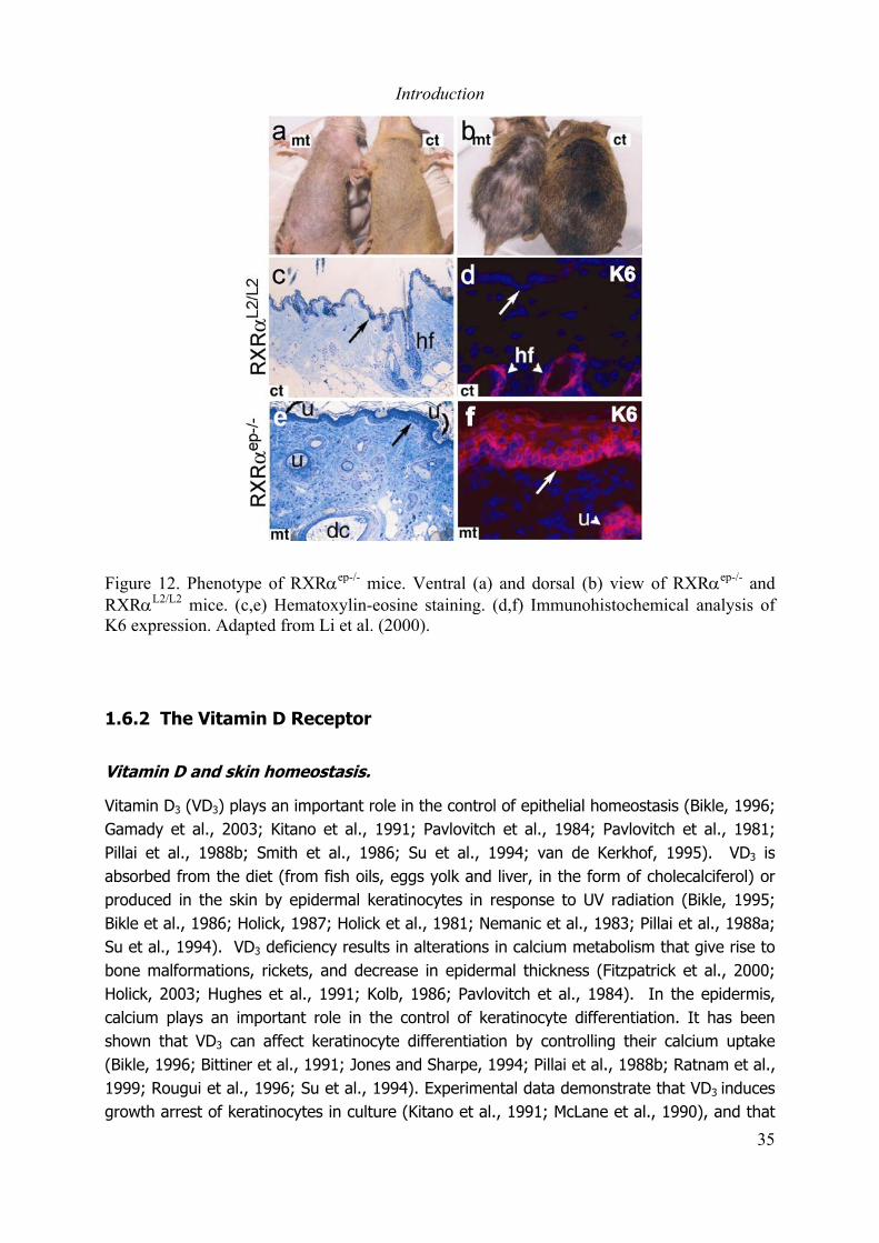
Introduction
35
Figure 12. Phenotype of RXRαep-/- mice. Ventral (a) and dorsal (b) view of RXRαep-/- andRXRαL2/L2 mice. (c,e) Hematoxylin-eosine staining. (d,f) Immunohistochemical analysis ofK6 expression. Adapted from Li et al. (2000).
1.6.2 The Vitamin D Receptor
Vitamin D and skin homeostasis.
Vitamin D3 (VD3) plays an important role in the control of epithelial homeostasis (Bikle, 1996;Gamady et al., 2003; Kitano et al., 1991; Pavlovitch et al., 1984; Pavlovitch et al., 1981;Pillai et al., 1988b; Smith et al., 1986; Su et al., 1994; van de Kerkhof, 1995). VD3 isabsorbed from the diet (from fish oils, eggs yolk and liver, in the form of cholecalciferol) orproduced in the skin by epidermal keratinocytes in response to UV radiation (Bikle, 1995;Bikle et al., 1986; Holick, 1987; Holick et al., 1981; Nemanic et al., 1983; Pillai et al., 1988a;Su et al., 1994). VD3 deficiency results in alterations in calcium metabolism that give rise tobone malformations, rickets, and decrease in epidermal thickness (Fitzpatrick et al., 2000;Holick, 2003; Hughes et al., 1991; Kolb, 1986; Pavlovitch et al., 1984). In the epidermis,calcium plays an important role in the control of keratinocyte differentiation. It has beenshown that VD3 can affect keratinocyte differentiation by controlling their calcium uptake(Bikle, 1996; Bittiner et al., 1991; Jones and Sharpe, 1994; Pillai et al., 1988b; Ratnam et al.,1999; Rougui et al., 1996; Su et al., 1994). Experimental data demonstrate that VD3 inducesgrowth arrest of keratinocytes in culture (Kitano et al., 1991; McLane et al., 1990), and that
Introduction
36
topical application of VD3 on psoriatic lesions reduces the number of proliferative cells (Kitanoet al., 1991). Thus, VD3 has pro-differentiation and anti-proliferative effects in epidermalkeratinocytes, which is most likely related to the effect of VD3 on keratinocyte calciummetabolism (Bikle, 1995; Su et al., 1994). Thus, epidermal keratinocytes are not only asource of VD3, but also a target of its action.
The Vitamin D Receptor in the Skin
VDR is expressed in all layers of the epidermis and in the hair follicle (Milde et al., 1991;Reichrath et al., 1996; Reichrath et al., 1994). The observation that VDR expression in thehair follicle varies during different stages of the hair cycle (Reichrath et al., 1994), suggestedthat VDR may play an important role in hair cycling. To study the role of VDR in vivo,Yoshizawa and co-workers generated a mice in which a null mutation of the VDR gene wasintroduced (Yoshizawa et al., 1997). VDR null mice exhibited a progressive alopecia anddegeneration of the hair follicles (Li et al., 2000; Yoshizawa et al., 1997), which results froma defect in the initiation of the hair cycle (entrance to anagen) (Sakai and Demay, 2000).The hair cycle defect leading to the development of alopecia in VDR null mice can beprevented by restoration of VDR expression in epidermal keratinocytes (Chen et al., 2001),but not by the normalization of mineral ion homeostasis (Amling et al., 1999), demonstratingthat VDR is required in keratinocytes for normal hair cycle. Interestingly, using hair-reconstitution assays in nude mice, Sakai and co-workers demonstrated that hair cycledefects in VDR null mice are the result of abnormal epidermal-mesenchymal interactions dueto an impairment in the capacity of keratinocytes to respond to dermal signals (Sakai et al.,2001). The hair cycle defects in VDR null mice are similar to those observed in RXRαep-/-
mice, suggesting that RXRα/VDR heterodimers in keratinocytes play a major role in thecontrol of hair cycle (Li et al., 2001b; Li et al., 2000).
In contrast to its role in hair cycling, VDR seem to play a minor role in the control ofkeratinocyte proliferation and differentiation. In 2000, Sakai and Demay reported that theepidermis of VDR null mice was indistinguishable from that of wild-type mice, and that VDRnull keratinocytes in culture proliferate and differentiate normally (Sakai and Demay, 2000).However, in a more detailed study Xie and co-workers showed that VDR null mice exhibitdefects in the expression of keratinocyte differentiation markers such as fillagrin, involucrin,and loricrin, and the presence of abnormal granules in the granular layer (Xie et al., 2002).These results suggest that in addition to its role in the control of hair cycle, VDR is alsoinvolved in the regulation of keratinocyte differentiation.
1.6.3 The Peroxisome Proliferator Activated Receptors
Fatty acids are structural components of the cornified layer of the epidermis (Grubauer et al.,1989). It has been shown that synthesis of cholesterol and fatty acid by keratinocyte isrequired for the formation and maintenance of the epidermal permeability barrier (Feingold

Introduction
37
et al., 1990; Mao-Qiang et al., 1993; Yang et al., 1995; Zettersten et al., 1997). For a longtime, fatty acids were considered as “simple” structural components of the cornified layer.However experimental data demonstrated that several lipid species can regulate keratinocytegrowth and differentiation. Ceramides for example, induce differentiation and inhibitproliferation of keratinocytes in culture (Geilen et al., 1997; Jung et al., 1998; Pillai et al.,1996), whereas topical applications of glucosylceramides stimulate keratinocyte proliferationin the epidermis of hairless mice (Marsh et al., 1995). These data suggested that beside theirfunction as structural components of the epidermis, fatty acids may also play a role asmodulators of keratinocytes function.
The Peroxisome Proliferator Activated Receptors (PPARs) were initially described as nuclearreceptors that can be activated by synthetic compounds called peroxisome proliferators(Dreyer et al., 1993; Dreyer et al., 1992; Green, 1992; Issemann and Green, 1990). ThreePPAR isotypes, PPARα, PPARβ and PPARγ, have been identified (Dreyer et al., 1992; Fajas etal., 1998; Fajas et al., 1999). Interestingly, PPARs can be activated by a variety of fatty acids(Dreyer et al., 1993; Keller et al., 1993; Schmidt et al., 1992). Very little was known aboutthe role of fatty acids on the regulation of keratinocyte proliferation and differentiationbefore the demonstration that PPARs are expressed in the epidermis. The three PPARisotypes are expressed in mouse epidermis during fetal development and their expressiondecreases progressively from the interfollicular epithelium after birth (Michalik et al., 2001).In adult skin, PPARα and PPARβ are the prominent isotypes, and it has been shown thatexpression of PPARα and PPARβ is increased in adult epidermis after various stimuli inducingkeratinocyte proliferation and differentiation (Braissant et al., 1996; Braissant and Wahli,1998; Michalik et al., 2001).
Experiments using skin explants of rat embryos demonstrated that PPARα ligands stimulatedifferentiation of fetal epidermal permeability barrier (Hanley et al., 1997). In adult mice,topical application of PPARα ligands stimulate differentiation of epidermal keratinocytes inwild-type animals, but not in PPARα null mice (Hanley et al., 2000a; Komuves et al., 2000).In line with this observations, PPARα activators induce an increase in involucrin mRNA levelsin cultured human keratinocytes (Komuves et al., 2000). These data suggest that PPARα isinvolved in the control or keratinocyte differentiation. However, PPARα null mice did notexhibit skin defect, indicating that PPARα plays a minor role in healthy skin. In agreementwith the observation that PPARα expression is reactivated in the skin in response to stress,PPARα null mice exhibited retarded wound healing (Michalik et al., 2001), indicating thatPPARα is important for re-epithealization of skin wounds.
A role for PPARβ in the control of keratinocyte differentiation has been shown byexperiments using keratinocytes in culture in which activators of PPARβ stimulatedifferentiation (Matsuura et al., 1999; Schmuth et al., 2004; Tan et al., 2001; Westergaardet al., 2001). Comparison of PPARβ heterozygous mice with wild-type control animalsrevealed a significant increase in the keratinocyte proliferation rate but no evident

Introduction
38
phenotypic changes in the epidermis of adult PPARβ mutant mice (Michalik et al., 2001).However, PPARβ null mice exhibit an increased hyperplastic response to TPA treatment, anda retardation in the healing of skin wounds (Michalik et al., 2001; Peters et al., 2000).Interestingly, PPARβ null keratinocytes in culture exhibit impaired adhesion and migrationcapacities (Michalik et al., 2001), and impaired response to inflammatory-cytokines induceddifferentiation (Di-Poi et al., 2002; Michalik et al., 2001; Tan et al., 2001). Thus, PPARβ playskey roles in the response of the skin to stress.
Studies in cell culture systems demonstrated that PPARγ ligands inhibit the growth (Ellis etal., 2000) and enhance calcium-induced differentiation of cultured keratinocytes (Evans etal., 1993). In rat skin explants, PPARγ ligands exhibit no effect on the development of skinbarrier function or epidermal morphology (Hanley et al., 1997). In adult mice, topicalapplication of PPARγ activators to mouse skin stimulates epidermal proliferation,differentiation, and apoptosis, which results in no net changes in epidermal morphology(Mao-Qiang et al., in press ). However, PPARγ activators inhibit TPA-induced cutaneousinflammation, and improve permeability barrier recovery after mechanical barrier disruption(induced by sequential applications of cellophane tape) (Mao-Qiang et al., in press ).
Thus, the three PPAR isotypes seem to play a role in the maintenance of skin homeostasis,in particular in response to injury. Because the absence of a single receptor has onlyminimal effects in skin homeostasis, there might be functional compensations between thethree receptor isotypes.
1.6.4 The Liver X Receptors
Liver X Receptors (LXRs) were first identified as orphan nuclear receptors that interact withthe RXRs (Miyata et al., 1996; Willy et al., 1995). LXRs are activated by oxidative derivativesof cholesterol called oxysterols. Recently, it has been demonstrated that both LXR isoforms,LXRα and LXRβ, are expressed in keratinocytes in culture (Hanley et al., 2000b) and in fetalskin (Komuves et al., 2002), but only LXRβ has been detected in adult mouse epidermis(Komuves et al., 2002). Epidermal keratinocytes can synthesize oxysterols de novo(Thiboutot et al., 2003), suggesting that activation of LXRs in keratinocytes may play a rolein epidermal maturation and homeostasis. The role of LXRs in epidermal homeostasis isfurther supported by the observations that oxysterols stimulate keratinocyte differentiationboth in culture and adult mice skin (Hanley et al., 2000b; Hanley et al., 2001; Komuves etal., 2002), and that oxysterols and LXR synthetic ligands inhibit both TPA-induced (irritantdermatitis) and oxazolone-induced (allergic dermatitis) inflammation by a mechanisminvolving both LXRα and LXRβ (Fowler et al., 2003). LXRα null mice do not exhibit anyepidermal alterations, but LXRβ null mice and LXRα/LXRβ null mice exhibit a slight decreasein the expression of epidermal differentiation markers, and thinning of the epidermis, but notsignificant functional cutaneous abnormalities (Fowler et al., 2003; Komuves et al., 2002).These data suggest that LXRs may play important roles in the skin in response to stress.

Introduction
39
Thus, several non-steroidal NRs including RXRs, RARs, VDR, PPARs and LXRs are expressedin the skin and have overlapping and independent roles in skin homeostasis. RXRα, thepredominant RXR receptor in the skin and a heterodimeric partner for other non-steroidalNRs, seem to play a central role as an integrator (regulator) of several signaling pathways.Based on the data available to date, a model can be proposed in which specific heterodimersparticipate selectively in a given process. RXRα/RARs (mainly RXRα/RARγ) are involved inthe control of keratinocyte proliferation and differentiation, in particular in response toexcess of RA. RXRα/VDR heterodimers play an important role in the control of hair cyclingand probably in the regulation of keratinocyte differentiation. RXRα/PPARs seem to play animportant function in response to stress, for example during wound healing and afterexposure to UV-radiations. Finally, RXRα/LXRs may participate in the control of keratinocytedifferentiation and skin inflammation (figure 13).
Figure 13. Model for integration of nuclear receptors action in the skin.
Introduction
40
1.6.5 Nuclear Receptors and Skin Carcinogenesis
The Retinoic Acid Receptors
In the early 70’s, a series of studies by Bollag and co-workers showed that vitamin A has aprotective effect against skin tumor formation in mice (Bollag, 1971; Bollag, 1972; Bollag,1974; Bollag, 1975). During the last three decades a number of experimental and clinicaldata demonstrated that retinoids can efficiently inhibit skin tumor formation, tumor growth,and malignant progression. Using mouse models in which skin tumors are induced by topicalapplication of chemical carcinogens (Athar et al., 1991; De Luca et al., 1993; Verma et al.,1980; Weeks et al., 1979) or by exposure to UV radiations (Athar et al., 1991), severalgroups have shown that natural or synthetic retinoids exhibit a protective effect against skintumor formation. Retinoic acid treated mice develop less and smaller tumors than nontreated mice (De Luca et al., 1993; De Luca et al., 1996; Gensler et al., 1987; Verma et al.,1979; Weeks et al., 1979), and the rate of malignant conversion is reduced in retinoid-treated mice (Chen et al., 1994a; Chen et al., 1994b; Chen et al., 1995; De Luca et al.,1993; De Luca et al., 1996).
Early clinical trials showed that retinoids exhibit a protective effect in the treatment ofmelanoma (Levine and Meyskens, 1980) and non-melanoma skin cancers (Lippman et al.,1988; Lippman et al., 1987a; Lippman et al., 1987b). Larger clinical trials demonstrated thatadministration of retinol decreases the probability of developing secondary SCC of the skin(Moon et al., 1997), and has a preventive effect in patients with a high risk to develop skincancer (DiGiovanna, 2001) such as xeroderma pigmentosum patients (Anolik et al., 1998;Giannotti et al., 2003; Saade et al., 1999), organ transplantation recipients (George et al.,2002; McNamara et al., 2002; Smit et al., 2004), and patients with Kaposi sarcoma(Aboulafia et al., 2003; Bernstein et al., 2002; Miles et al., 2002). Moreover, it wasdemonstrated that retinoids induce regression of some dysplastic nevus, the precursors ofmelanoma, to benign nevi in humans, suggesting that retinoids may also have benefic effecton the treatment of melanoma (Meyskens et al., 1986).
The precise mechanism by which retinoids exert their anti tumor activity in the skin is notclear. However it has been demonstrated that retinoids inhibit growth of HSCC cell lines(Braakhuis et al., 1997; Cooper et al., 1997; Klaassen et al., 2001; Lotan, 1993), and induceapoptosis of HSCC (Braakhuis et al., 1997; Cooper et al., 1997; Giandomenico et al., 1998;Hail and Lotan, 2001; Islam et al., 2000; Liaudet-Coopman et al., 1997; Lotan, 1996) andmelanoma cells lines (Danielsson et al., 1999). Interestingly, is has been shown thatretinoids have differential effects on normal and malignant keratinocytes. Whereas in theformer it induces growth arrest, in the later it induces apoptosis (Hail and Lotan, 2001).
RXRα and RARγ are the main retinoic acid receptors in the skin. In situ hybridization studieson samples of human normal skin, and AK and SCC lesions, showed a progressive decrease
Introduction
41
in the transcript levels of RARα and RARγ, and all three RXRs, which is associated with theprogression of squamous skin carcinogenesis (Xu et al., 2001). In agreement with theseobservations, it has been shown that RARα and RARγ mRNA levels are gradually decreasedin epidermal keratinocytes during mouse skin carcinogenesis (Darwiche et al., 1995).Interestingly, overexpression of an oncogenic Ha-ras in keratinocytes resulted in a decreaseof RARα and RARγ protein levels and transactivation activity (Darwiche et al., 1996).Moreover, blocking RARγ expression in HNSCC cells by antisense RNA resulted in decreasedRA-induced growth arrest (Oridate et al., 1996), and overexpression of RARα or RARγ intoneoplastic mouse epidermal cells, with low endogenous levels of these receptors, resulted inan increased RA-induced growth arrest and apoptosis (Hatoum et al., 2001). Recently, is hasbeen shown RARγ null, but not RARα null keratinocytes, carrying an activated v-Ha-rasoncogene, formed squamous cell carcinoma when grafted into nude mice (Chen et al.,2004). Furthermore, the expression of RXRα and RARβ in melanoma cells is lower than innormal melanocytes or nevus cells (Boehm et al., 2004), suggesting that expression of RXRαand RARβ is lost during melanoma formation. All together, the above data strongly suggestthat RA receptors play an important role in the development of skin cancers and in the anti-tumor effects of retinoids.
The Vitamin D receptor
Vitamin D has anti-proliferative and pro-differentiating properties on epidermal cells, and isan interesting candidate for the treatment of skin cancer. Studies of chemically-induced skincarcinogenesis in mice demonstrated that VD3 partially blocks tumor formation whentopically applied on the back skin of experimental mice (Chida et al., 1985; Pence et al.,1991; Wood et al., 1983). Vitamin D3 also has antiproliferative (Enepekides et al., 1999) andpro-apoptotic effects on squamous cell carcinoma cells in culture (Bernardi et al., 2002;McGuire et al., 2001; Satake et al., 2003). Furthermore, VD3 can induce growth arrest (Evanset al., 1996) and inhibit invasiveness of malignant melanoma cells in vitro and in vivo models(Yudoh et al., 1999). However, clinical use of VD3 is not yet validated because of its stronghypercalcemic effects. Synthetic analogs of VD3 with anti-proliferative pro-differentiatingeffect but with low calcemic activities have been developed (Brown et al., 1990; Hansen etal., 2000; Mathiasen et al., 1993; Posner et al., 2001; Posner et al., 2002; Posner et al.,1998). Several of this compounds exhibit enhanced anti-proliferative and pro-differentiatingeffects on cancer cells in culture (Akutsu et al., 2001; Hansen and Maenpaa, 1997;Koshizuka et al., 1999; Prudencio et al., 2001). They can induce growth arrest (Posner et al.,2001) and apoptosis (Danielsson et al., 1998; Danielsson et al., 1997) in melanoma cell linesin culture, and it has been demonstrated that they partially inhibit chemical carcinogenesis invivo (El Abdaimi et al., 1999; Kensler et al., 2000). Interestingly, it has been shown thatvitamin D in combination with retinoids exhibit synergistic effects in the induction ofapoptosis on melanoma cells (Danielsson et al., 1999), the suppression of SCC growth(Enepekides et al., 1999), and the inhibition of tumor-induced angiogenesis in nude mice(Majewski et al., 1995; Majewski et al., 1993).

Introduction
42
The vitamin D receptor is expressed in several cell types in the skin, where it plays a key rolein the control of hair cycle and keratinocyte differentiation. A link between the susceptibilityto develop skin cancer and the function of the VDR has been established from theobservations that VDR expression is higher in BCC lesions compared to normal skin(Reichrath et al., 1999b) and that polymorphisms of the VDR gene, which would be expectedto result in impaired function, are associated with susceptibility and prognosis in malignantmelanoma (Hutchinson et al., 2000b). Recently, Zinser and co-workers demonstrated thatpre-treatment of VDR null mice with medroxyprogesterone acetate (PMA), a syntheticprogesterone analog that enhances epithelial proliferation, followed by oral administration ofthe carcinogen DMBA results in spontaneous development of skin tumors in 85% of treatedanimals (Zinser et al., 2002).
The Peroxisome Proliferator Activated Receptors
The three PPAR isotypes play important roles in skin homeostasis. They seem to beparticularly important under conditions of stress such as wound healing and after exposureto UV radiations. However, their role during skin carcinogenesis has been poorlyinvestigated. It has been shown that PPARα ligands possess receptor mediated, anti-inflammatory activities in both irritant and allergic contact dermatitis animal models (Sheu etal., 2002), and that they can reverse expression of inflammatory cytokines induced by UVBin keratinocytes in culture (Kippenberger et al., 2001). Interestingly, activators of PPARαpartially inhibit tumor formation in a two step chemical carcinogenesis protocol (Thuillier etal., 2000). Moreover, PPARγ mediates the non-steroidal anti-inflammatory drugs (NSAID)antineoplastic effect on human oral squamous carcinoma cells (Nikitakis et al., 2002), andPPARγ specific ligands can block melanoma cell growth in culture (Mossner et al., 2002).Recently, Nicol and co-workers demonstrated that, in response to a chemical carcinogenesisprotocol, PPARγ+/- mice exhibit an increased number of tumors per mouse when comparedto wild-type mice (Nicol et al., 2004). Similarly, Kim and co-workers showed that PPARβ nullmice subjected to a chemical skin carcinogenesis protocol exhibited an enhanced onset oftumor formation, tumor size and tumor multiplicity when compared to wild-type mice (Kimet al., 2004). Thus, RXRα and its heterodimeric partners may play important roles in thedevelopment of skin cancer, but their precise role is not clear.
Manuscript in preparation
43
2. Results
2.1 Malignant transformation of DMBA/TPA-induced papillomas and
melanocytic growths in the skin of mice selectively lacking RXRα in
epidermal keratinocytes (Manuscript in preparation)
Indra A.K.1, Castaneda, E.1, Antal, M.C.2, Jiang, M.1, Messaddeq, N.1, Gariglio, P.3,
Kato, S.4, Wahli, W.5, Desvergne, B.5, Metzger, D.1,2 and Chambon, P.1, 2*.
1Institut de Génétique et de Biologie Moléculaire et Cellulaire (CNRS, INSERM, ULP,
Collège de France); 2Institut Clinique de la Souris, B.P.10142, 67404, ILLKIRCH,
Strasbourg, France; 3Depto. Genetica y Biol. Mol. Cinvestav-IPN, Mexico; 4Institute
of Molecular & Cellular Biosciences, University of Tokyo, Yayoi, Bunkyo-ku, Tokyo
113, Japan;5Unité CIG Science, Centre Intégratif de Génomique (CIG) UNIL,
Lausanne
*Corresponding author
IGBMC, BP 163 67404 Illkirch Cedex, France
(Tel: 33 +3 88 65 32/13/15/ ; Fax : 33 +3 88 65 32 03
e-mail: [email protected])
Short title : Role of RXRα in skin tumorigenesis
Key words: conditional targeted somatic mutagenesis, RXRα, epidermis, skin cancer,
chemical carcinogenesis, DMBA/TPA, carcinoma, melanoma, mouse.


Manuscript in preparation
1
Summary:
Ligands for several nuclear receptors, including those of Retinoid X Receptors(RXRs), Retinoic Acid Receptors (RARs), Peroxisome Proliferator-activatedReceptors (PPARs) and Vitamin D3 Receptor (VDR) have a suppressive effecton epidermal tumorigenesis. RXRs, the obligatory heterodimeric partners for
RARs, PPARs, VDR and Liver X receptors (LXRs), and in particular RXRα, themajor RXR isotype expressed in the epidermis, could be critical in the control
of skin tumorigenesis. We show that adult mice, in which RXRα was selectivelyablated in keratinocytes, subjected to the two-step skin tumorigenesisprotocol, develop an increased number of epidermal tumors, which progress ata much higher frequency to malignant carcinoma, thus establishing a tumor
modifier role for this receptor. Moreover, as under similar conditions, RXRα
mutant mice also develop a higher number of melanocytic growths, some of
which degenerate into melanoma, RXRα-ablated epidermal keratinocytes mightcontrol in a paracrine manner melanocyte proliferation and their malignant
progression. Finally, our data indicate that PPARγ, PPARα, LXRβ and VDRdifferentially act as skin tumor modifiers.
Introduction :
The incidence of skin cancers is growing at an alarming rate. Melanoma incidence
and mortality rate increases are among the highest of all cancers. While mortality due
to non-melanoma skin cancers is lower, squamous cell carcinomas (SCC) are
invasive and more than 10% metastasize (Kwa et al., 1992; Stratton et al., 2000 ).
Retinoids (principally all-trans retinoic acid, the active vitamin A derivative) are
involved in the control of growth and differentiation of normal, premalignant and
malignant cell types (Altucci and Gronemeyer, 2001). Their effects are mediated
through their binding to heterodimers formed between the members of two families of
nuclear receptors (NRs), the retinoic acid (RA) (RARα, β and γ) and retinoid X

Manuscript in preparation
2
(RXRα, β and γ) receptors (Chambon, 1996; Mangelsdorf et al., 1995 and Refs
therein). Vitamin A deficiency in humans and animal models is associated with
epithelial squamous metaplasia prone to malignant conversion (Hong and Itri, 1994;
Moon et al., 1994), and aberrant expression and function of RARs have been found
in carcinomas, as well as in premalignant lesions (Sun and Lotan, 2002). Natural and
synthetic retinoids are used for the treatment of skin disorders including psoriasis and
acnea (Livrea, 2000), as well as chemopreventive agents for epidermal cancers
(Altucci and Gronemeyer, 2001; Sun and Lotan, 2002). Altered expression of RARs
and RXRs found during carcinogenesis may reduce the ability of epidermal cell to
respond to RA and contribute to carcinoma development, both in mouse (Darwiche et
al., 1995; Darwiche et al., 1996) and human (Sun and Lotan, 2002). In mice, topical
application of RA inhibits papilloma formation and dietary RA has been effective in
preventing tumor formation and malignant conversion of chemically-induced mouse
skin papillomas (De Luca et al., 1996,; Tennenbaum et al., 1998 and refs therein).
The two-step chemical tumorigenesis mouse model has been extensively used to
study initiation, promotion and progression (malignant conversion) of skin tumors, as
the evolution of human squamous cell carcinoma (SCC) has many similarities with
chemically-induced squamous cell tumors in the mouse (Yuspa, 1998, and refs there
in). The initiation stage is achieved through a single topical appplication of 7,12-
dimethyl-benz[a]anthracene (DMBA) which is known to result in an activating
transversion mutation (A182->T) in the second base of codon 61 of the c-Ha-ras
gene (Yuspa, 1998, and refs therein). The second stage, promotion, is accomplished
by repeated topical treatment with a tumor promoter, such as the phorbol ester 12-O-
tetradecanoylphorbol-13 acetate (TPA), which causes a sustained hyperplasia and
inflammation, and then a selective clonal expansion into benign papillomas (Yuspa,
1998, and refs therein). The third stage, progression, is the malignant conversion of
benign papillomas to invasive SCC. Cutaneous papillomas and SCCs induced by
DMBA/TPA treatment, as well as human SCC, have been shown to contain the Ha-
ras gene codon 61 (A182 -> T) mutation (Nelson et al., 1992, and refs therein) and
targeted deletion of the Ha-ras gene drastically decreases the incidence of
DMBA/TPA-induced epidermal papillomas (Ise et al., 2000). Interestingly, benign
melanocytic dermal tumors (nevi) are induced upon a single topical DMBA

Manuscript in preparation
3
application. However, such nevi in which N-ras but not Ha-ras mutations at codon 61
have been found, do not progress to malignant melanomas (Husain et al., 1991).
RXRα, RXRβ, RARα and RARγ are expressed in human and mouse epidermis, and
RXRα and RARγ are the predominant receptors (Chapellier et al., 2002; Fisher and
Voorhees, 1996). The RXRα null mutation is lethal in utero between gestation days
13.5 and 16.5 (Kastner et al., 1994; Sucov et al., 1994), which precluded the genetic
study of its physiological function in skin homeostasis. This study became possible
by conditional inactivation of RXRα using the Cre/loxP technology and transgenic
mice selectively expressing the tamoxifen-inducible Cre-ERT2 recombinase in the
epidermal basal layer (Li et al., 2000; Metzger et al., 2003). RXRα ablation in
epidermal keratinocytes of adult mice resulted in alopecia, epidermal interfollicular
hyperplasia with keratinocyte hyperproliferation and aberrant terminal differentiation,
accompanied by a skin inflammatory reaction (Li et al., 2001b; Li et al., 2000).
Interestingly, RARs appear to be dispensible for homeostatic renewal of epidermal
keratinocytes in the adult mouse, while the proliferative effect of topically-applied RA
is transduced in suprabasal keratinocytes by RXRα/RARγ heterodimers, resulting in
the release of a paracrine signal, which in turn stimulates the proliferation of basal
keratinocytes (Chapellier et al., 2002). Importantly, RXRs are also known to
heterodimerize with other members of the nuclear receptor superfamily (e.g. PPARs,
VDR and LXRs, see (Laudet, 2002)) that are expressed in the epidermis (Fowler et
al., 2003; Li et al., 2000; Mao-Qiang et al., 2004; Schmuth et al., 2004, and refs.
therein; Sheu et al., 2002). Ligands of PPARs and VDR have been shown to inhibit
the growth of squamous cell carcinoma cell lines in vitro and multistage skin
tumorigenesis in vivo (Kensler et al., 2000; Kopelovich et al., 2002; Nikitakis et al.,
2002) and those of LXRs display anti-inflammatory activity against TPA induced
cutaneous inflammation (Fowler et al., 2003). Thus, RXR(s) and in particular RXRα,
could to be critical in transducing cellular signals controlling skin carcinogenesis.
To investigate the possible role of keratinocytic RXRα in papilloma formation and
their malignant conversion, as well as in the formation of melanocytic growths and
their progression to melanoma, we have subjected adult mice, in which RXRα was

Manuscript in preparation
4
selectively ablated in keratinocytes, to the chemical two-step tumorigenesis protocol.
Upon DMBA/TPA treatment these mutant mice exhibited an increased number of
epidermal tumors which were also bigger in size and progressed with a much higher
frequency to malignant carcinoma, thus establishing a role of RXRα in tumor
suppression. We also show that these DMBA/TPA-treated mutants develop
melanocytic growths, which degenerate into melanoma and metastasize to distal
lymph nodes, and appear to be induced in a paracrine manner by signals emanating
from the RXRα-ablated epidermal keratinocytes. Furthermore, using mice bearing
either a VDR-null mutation or a selective ablation of PPARγ in their epidermal
keratinocytes, we also investigated the role of these NRs as possible partners of
RXRα in tumor suppression.
Manuscript in preparation
5
Results :
Selective ablation of RXRα in keratinocytes increases the formation andgrowth of epidermal tumors in DMBA/TPA treated mice
RXRαep-/- mice (for epidermal keratinocyte-selective RXRα-null genotype)
selectively lacking RXRα in keratinocytes of interfollicular epidermis and hair follicle
outer root sheath, as well as control (CT) RXRαep-/+ and RXRαL2/L2 mice carrying
either one RXRα L- null allele in their epidermal keratinocytes or two floxed RXRα L2
alleles in their germ line, were generated by tamoxifen (Tam) treatment of K14-Cre-
ERT2/RXRαL2/L2, K14-Cre-ERT2/RXRαL2/+ and RXRαL2/L2 littermates,
respectively (see Experimental Procedures). As reported, RXRαep-/- mice exhibited
an epidermal keratinocyte hyperproliferation and abnormal differentiation (with
keratin 6 expression), a skin inflammation, and developed an extensive alopecia
within 10-15 weeks after RXRα ablation, whereas the skin and fur of CT mice were
similar to those of WT mice (data not shown, see (Li et al., 2000; Metzger et al.,
2003)).
To induce the formation of papilloma-like tumors, CT and RXRαep-/- female mice
were topically treated, two weeks after Tam administration, with a single dose of the
tumor initiator DMBA, and then twice a week with the tumor promoter TPA for 25-30
weeks [RXRαep-/-(a) mice, see Fig. 1A, and Experimental Procedures]. Seven to
eight weeks after the start of TPA treatment, epidermal tumors developed in all CT
and RXRαep-/-(a) mice, and their number and size increased with time (Fig.1B, and
data not shown). However, RXRαep-/-(a) mice developed approximately twice as
many tumors as CT mice [after 30 weeks, an average of 12 and 28 tumors were
present in CT and RXRαep-/-(a) mice, respectively (Fig. 1B & 1C)]. Moreover, CT
tumors were never longer than 12 mm, whereas ~10 % and 3 % of the RXRαep-/-(a)
tumors were 12 to 16 mm and 30 to 40 mm long, respectively (Fig.1D, and data not
shown). The number of tumors was 2-fold lower in males. However, RXRαep-/-(a)
males also exhibited twice as many tumors as CT littermates, and their size was also
Manuscript in preparation
6
increased (data not shown). All further analyses were performed on female mice. In
all cases, DMBA initiation was required to generate these tumors, as none appeared
in CT or RXRαep-/- mice treated with TPA alone (data not shown).
To investigate whether RXRα was involved in tumor initiation and/or promotion, mice
were first treated with DMBA, and then for 7 weeks with TPA before Tam-induced
RXRα selective ablation (RXRαep-/-(b) mice, Fig.1A). Tumors appeared 6 to 7
weeks after the start of TPA treatment in both CT and RXRαep-/-(b) mice, and by 30
weeks the latter had developed twice as many tumors as CT mice (Fig. 1E and F,
and data not shown). Thus, the lack of RXRα in epidermal keratinocytes does not
affect tumor initiation by DMBA, while it enhances tumor promotion. Accordingly, the
H-ras codon 61 "activating" mutation that was previously identified in DMBA/TPA-
induced epidermal tumors (see Introduction) was similarly present in both CT and
RXRαep-/- tumors (data not shown).
Tumors generated in RXRαep-/- mice have a “high risk” of malignant
conversion
Changes in keratin gene expression occurring during DMBA/TPA tumorigenesis have
been used to classify the induced papillomas into “high” and “low” risk for malignant
progression. Loss of expression of keratins K1 and K10 that are expressed in the
differentiating suprabasal layers of normal epidermis and in “low risk” papillomas, and
the concomitant appearance of K13 normally not expressed in skin, are used as
indicators of early stages of malignant conversion (Nischt et al., 1988, and reference
therein; Rundhaug et al., 1997). Immunohistochemistry of tumors (8-16 mm in length)
taken 22 weeks after the start of DMBA/TPA treatment were performed using
antibodies against keratins K5, K1, K10 and K13 (Nischt et al., 1988, and reference
therein; Rundhaug et al., 1997) (Fig. 2A). K5, "normally" selectively expressed in the
basal cell layer, was prominent in that layer and persisted throughout most of the
upper (suprabasal) layers of highly stratified tumors from CT and RXRαep-/-(a) mice,
but higher K5 levels were usually found in RXRαep-/-(a) tumors (Fig. 2A, a and b).
K1 and K10, strongly expressed in suprabasal cell layers of more than 70 % of CT
Manuscript in preparation
7
tumors (Fig. 2c and e), were expressed in less than 25 % of RXRαep-/-(a) tumors
(Fig. 2d and f). Tumors strongly expressing K13 were 3-4 times more frequent in
RXRαep-/-(a) than in CT mice, and more than 70 % of the tumor area was stained
(instead of 15% in CT mice; Fig. 2A, g and h, and data not shown).
Up-regulation of α6 and β4 integrins, normally expressed in basal cells and mainly
found in the basement membrane, as well as their presence into several cell layers
above the basement membrane, are associated with a high risk of malignant
conversion (Tennenbaum et al., 1993). α6 and β4 integrin expression was much
stronger in all RXRαep-/-(a) tumors and in most cases extended into suprabasal
layers (Fig 2A, i and j, and data not shown), while it was restricted to the basal layer
in >90 % of CT tumors. Finally, in accordance with an increased vascularity of
RXRαep-/-(a) tumors (data not shown), 70 % of RXRαep-/-(a) tumors exhibited
increased immunostaining of CD31 which labels endothelial intercellular junctions
(Fig. 2A, k and l). Taken together, the above data indicate that, upon RXRα ablation
in epidermal keratinocytes, DMBA/TPA-induced tumors exhibit a higher progression
to malignancy.
DMBA/TPA-induced epidermal tumors are known to degenerate at a low rate into
squamous cell carcinoma (SCC), but not into basal cell carcinoma (BCC) (Yuspa,
1998, and refs therein) (see Table 1). Nine tumors from each of 6 CT and 6
RXRαep-/-(a) mice were examined. As previously reported (Yuspa, 1998), most of
CT tumors analysed after 25 weeks of DMBA/TPA treatment were benign papillomas
characterised by skin folds integrated by a core of connective tissue and lined by an
acanthotic, hyperkeratotic, stratified squamous epithelium (Fig.2Ba, and data not
shown). 20 % of the papillomas exhibited a basal cell hyperplasia (Fig. 2Bb), 3%
displayed in situ carcinoma (data not shown), but foci of early SCC were only
observed 30 weeks after tumor initiation in ~14 % of the tumors (Fig. 2Bc, Table I,
and data not shown). In contrast, 25 weeks after tumor initiation, ~30% of RXRαep-/-
(a) papillomas exhibited basal cell hyperplasia (Fig. 2Cb), 10 % displayed in situ
carcinoma (not shown), and foci of SCC were observed in 40 % of the tumors (Fig.
2Cc). Five weeks later, 46% of these tumors had progressed to differentiated and

Manuscript in preparation
8
undifferentiated SCC with extensive local invasion of cancer cells into the dermis and
underlying muscles (Fig. 2Cd-g, Table I, and data not shown). Well differentiated
SCC [Fig. 2Cd, note the keratin pearls (white arrow) with keratinisation in the center],
moderately differentiated SCC [Fig. 2Ce; the cells are markedly irregular in shape
and size, and nuclei are hyperchromatic (blue arrowhead) with prominent nucleoli;
note also the internal keratinisation (asterisk)], as well as poorly differentiated SCC
[Fig. 2Cf; note the presence of pleiomorphic cancer cells with abnormal nuclei and
prominent nucleoli], were observed in 30 to 45 % of the tumors (Table I). Highly
aggressive anaplastic SCC (~3 %; Fig. 2Cg), and basal cell carcinoma (~6 %;
Fig.2C, h and i), previously not reported in the two step chemical tumorigenesis
model, were also found in RXRαep-/-(a), but not in CT mice (Table I).
Melanoma formation in skin of RXRαep-/- mice upon DMBA/TPA treatment A single DMBA application to skin of WT mice induces the formation of nevi, which
are benign melanocytic growths (MGs) that become visible after 10-12 weeks, and
are characterized by subepidermal accumulation of melanocytes (Epstein, 1992 and
refs. therein; Husain et al., 1991 ). Interestingly, a single DMBA treatment of
RXRαep-/- mice resulted in a ~7-fold increase in the number of MGs which were
generally larger than those of CT mice (Fig. 1, G and H, and data not shown).
Multiple TPA applications after DMBA initiation further increased the number of MGs
in CT and mutant mice (Fig. 1H; compare also Fig. 1C and G, and data not shown).
After 30 weeks of DMBA/TPA treatment, ~10 nevi were observed per CT mouse
(generally with a diameter below 2 mm), whereas >50 MGs were found per RXRαep-
/-(a) mouse (~75% with a diameter over 2 mm) (Fig. 1H). When RXRα was ablated 8
weeks after DMBA treatment (Fig. 1A), the number of MGs was only ~3 fold higher in
RXRαep-/-(b) than in CT mice, and only 40% of them had a diameter larger than 2
mm (Fig. 1H). These results indicate that both MG initiation and promotion might be
increased upon ablation of RXRα.
Histological analysis of MGs (>2mm) generated by DMBA or DMBA/TPA treatment
showed an accumulation of melanocytes in the dermis of CT mice, whereas a larger
number of densely melanin-laden melanocytes was seen in both the dermis and

Manuscript in preparation
9
hypodermis of RXRαep-/- skin sections, accompanied by a vertical invasion of the
underlying musculature in 5–10 % of the cases (compare Fig. 3A, a and b, and data
not shown). However, only few melanocytes (1-2 %) were found in the epidermis of
both CT and RXRαep-/- mice, and in most cases the basement membrane appeared
intact (Fig 3A, a and b; and data not shown). As previously reported (Ghadially,
1997), electron microscopy revealed that melanocytes from CT nevi contained mainly
stage III and IV melanosomes [Fig. 3A, c and e, and data not shown)]. On the other
hand, when compared with CT melanocytes, most of the melanocytes present in
MGs from RXRαep-/- mice showed a ~10-fold increase in melanosomes containing
melanin granules, and their nuclei were more elongated (Fig. 3, d and f). Moreover, in
~40% of the RXRαep-/- melanocytes, 40-50% of the melanosomes had the
appearance of vesicles exhibiting a variable electron opacity with a distinct
substructure, a characteristic of human melanoma cells (Ghadially, 1997) [Fig 3A d,
and f, arrows, and data not shown]. In addition, the membrane of a number of
melanosomes was disrupted and smaller melanin granules were scattered in the
cellular matrix (Fig. 3Af, and data not shown). In rare cases, oval shaped vesicles
contained a longitudinally-oriented component made of rods, which is also
characteristic of human melanoma cells (Fig. 3Ag, (Ghadially, 1997)). Taken
together, these results indicate the formation of melanomas in the skin of RXRαep-/-
mice.
N-Ras mutations in codon 61 have been detected in either mouse or human nevi and
melanomas (Husain et al., 1991; Kumar et al., 2004 and refs. therein). After 30
weeks of DMBA/TPA treatment, we PCR-amplified and cloned the first N-Ras exon
from 6 and 8 individual MGs (>2 mm in size) collected from CT and RXRαep-/-(a)
mice, respectively. Ten independent transformants from each MGs were sequenced.
Glycine to Valine (GGT GTT) and Glycine to Alanine (GGT GCT) transversion
mutations at codon 12 were found in 2/8 and 1/8 RXRαep-/-(a) MGs, respectively
(data not shown). None of the 6 control MGs showed mutation at codon 12. In
addition, 1/8 RXRαep-/-(a) and 1/6 control MGs had a Glycine to Valine point
mutation of N-ras codon 13 (data not shown). In contrast to human melanoma
(Demunter et al., 2001), no mutation of N-Ras codon 18 was detected in any of the

Manuscript in preparation
10
mutant or control MGs. No mutation of N-Ras codons 12 and 13 could be detected in
4 control and 4 mutant epidermal tumors. Fifteen MGs and 20 epidermal tumors (4-8
mm in size) from RXRαep-/-(a) and CT mice (collected 30 weeks after DMBA/ TPA
application) were also analyzed for possible mutation of N-Ras codon 61. None of
them had a codon 61 A182/T mutation (analysed by PCR and restriction digestion;
data not shown), while an A to C substitution was found in the second base of codon
61 [CAA-> CCA, a change from Glutamine (Gln) to Proline (Pro)] in one RXRαep-/-
(a) MG, but not in CT MGs. In contrast to human melanoma and nevi reports (Laud et
al., 2003; Pollock et al., 2003 and refs therein), no B-raf mutation could be detected
in 15 MGs from either CT or RXRαep-/-(a) mice (data not shown).
To demonstrate that RXRα was not ablated in MGs, PCR genotyping was performed
on DNA isolated from large nevi of CT and RXRαep-/- mice (Experimental
Procedures). RXRα L2 alleles, but no RXRα L- alleles, were found in RXRαep-/-
melanocytic tumors induced by either DMBA or DMBA/TPA, whereas as expected
only L- alleles were found in their surrounding epidermis (Fig. 3B). These results
demonstrate that the formation of nevi and melanomas in DMBA-treated RXRαep-/-
mice is associated with RXRα ablation in keratinocytes, but not in melanocytes.
Melanoma from RXRαep-/- mice metastasize to distal lymph nodesMelanoma cells are known to express specific chemokine receptors and to
metastasize preferentially in lymph nodes, lung and liver (Murakami et al., 2002;
Payne and Cornelius, 2002). Autopsies performed 30 weeks after DMBA treatment
revealed that subiliac lymph nodes were enlarged and pigmented in 2 out of 4
RXRαep-/- mice, but not in CT mice (0/4) (Fig. 4A, a and b, and data not shown).
Histological analysis showed few pigmented cells in lymphosinuses and some
lymphatic vessels (10-20%) of mutant mice (Fig. 4Ad, and data not shown). In
contrast, no pigmented cells were seen in lymphosinuses from CT mice, and very few
(5–6 fold less than in mutant mice) were present in lymphatic vessels (Fig. 4Ac, and
data not shown).
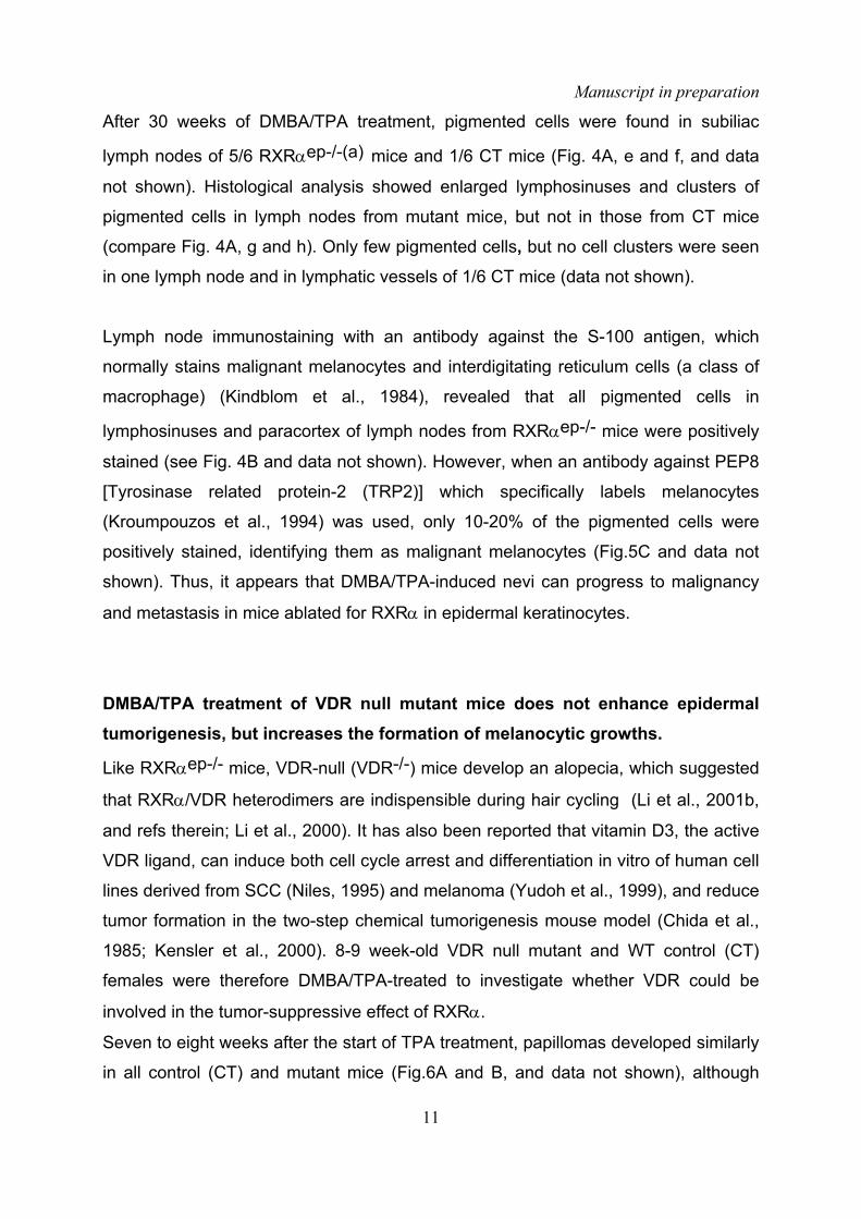
Manuscript in preparation
11
After 30 weeks of DMBA/TPA treatment, pigmented cells were found in subiliac
lymph nodes of 5/6 RXRαep-/-(a) mice and 1/6 CT mice (Fig. 4A, e and f, and data
not shown). Histological analysis showed enlarged lymphosinuses and clusters of
pigmented cells in lymph nodes from mutant mice, but not in those from CT mice
(compare Fig. 4A, g and h). Only few pigmented cells, but no cell clusters were seen
in one lymph node and in lymphatic vessels of 1/6 CT mice (data not shown).
Lymph node immunostaining with an antibody against the S-100 antigen, which
normally stains malignant melanocytes and interdigitating reticulum cells (a class of
macrophage) (Kindblom et al., 1984), revealed that all pigmented cells in
lymphosinuses and paracortex of lymph nodes from RXRαep-/- mice were positively
stained (see Fig. 4B and data not shown). However, when an antibody against PEP8
[Tyrosinase related protein-2 (TRP2)] which specifically labels melanocytes
(Kroumpouzos et al., 1994) was used, only 10-20% of the pigmented cells were
positively stained, identifying them as malignant melanocytes (Fig.5C and data not
shown). Thus, it appears that DMBA/TPA-induced nevi can progress to malignancy
and metastasis in mice ablated for RXRα in epidermal keratinocytes.
DMBA/TPA treatment of VDR null mutant mice does not enhance epidermaltumorigenesis, but increases the formation of melanocytic growths.
Like RXRαep-/- mice, VDR-null (VDR-/-) mice develop an alopecia, which suggested
that RXRα/VDR heterodimers are indispensible during hair cycling (Li et al., 2001b,
and refs therein; Li et al., 2000). It has also been reported that vitamin D3, the active
VDR ligand, can induce both cell cycle arrest and differentiation in vitro of human cell
lines derived from SCC (Niles, 1995) and melanoma (Yudoh et al., 1999), and reduce
tumor formation in the two-step chemical tumorigenesis mouse model (Chida et al.,
1985; Kensler et al., 2000). 8-9 week-old VDR null mutant and WT control (CT)
females were therefore DMBA/TPA-treated to investigate whether VDR could be
involved in the tumor-suppressive effect of RXRα.
Seven to eight weeks after the start of TPA treatment, papillomas developed similarly
in all control (CT) and mutant mice (Fig.6A and B, and data not shown), although

Manuscript in preparation
12
they were in general smaller in VDR-/- than in CT mice (see Fig. 5A-C). Histological
analysis of 40 tumors, 30 weeks after DMBA initiation revealed that, unlike RXRαep-
/- mutants, 95% of the papillomas were benign, while the others only exhibited foci of
early SCC (Table 1, and data not shown).
Melanocytic growths also appeared on DMBA and DMBA/TPA-treated skin of VDR-/-
mice, and their number was strikingly high to reach ~70 MGs after 30 weeks of TPA
application (instead of ~10 MGs in CT mice, Fig 5D). In both cases, histological
analysis of 30 MGs from CT and VDR-/- mice showed the presence of dermal
melanin-laden melanocytes (Fig 5E, and data not shown), and electron microscopy
revealed that they contained mostly stage III and stage IV melanosomes which are
characteristics of benign nevi (Fig 5F). However, the number of melanosomes was
approximately 2-fold increased in ~10% of the melanocytes present in VDR-/- MGs
(Fig 5F, and data not shown).
Selective ablation of PPARγ in keratinocytes enhances DMBA/TPA-induced
epidermal tumorigenesis.
To investigate whether the tumor suppressive effect of RXRα could be due to events
controlled by RXRα/PPARγ heterodimers, PPARγep-/- (for epidermal keratinocyte-
selective PPARγ-null genotype) mice, as well as their control (CT) littermates
(PPARγL2/L2) which carry two PPARγ L2 floxed alleles in their germ line (Imai et al.,
2004), were obtained by tamoxifen administration to K14-CreERT2/ PPARγL2/L2 and
PPARγL2/L2 littermates, respectively. Unlike RXRαep-/- and VDR-/- mice, PPARγep-
/- mice did not exhibit an alopecia. Furthermore, in contrast to RXRαep-/- mice,
PPARγep-/- mice showed only a modest increase in keratinocyte proliferation 6-8
weeks after Tam-induced ablation (≈15 % when compared to CT mice, as
determined from IHC staining of the Ki67 proliferation marker; data not shown), and
did not exhibit an abnormal interfollicular epidermis differentiation nor a skin
inflammatory reaction (Mao-Qiang et al., 2004, and our unpublished data).
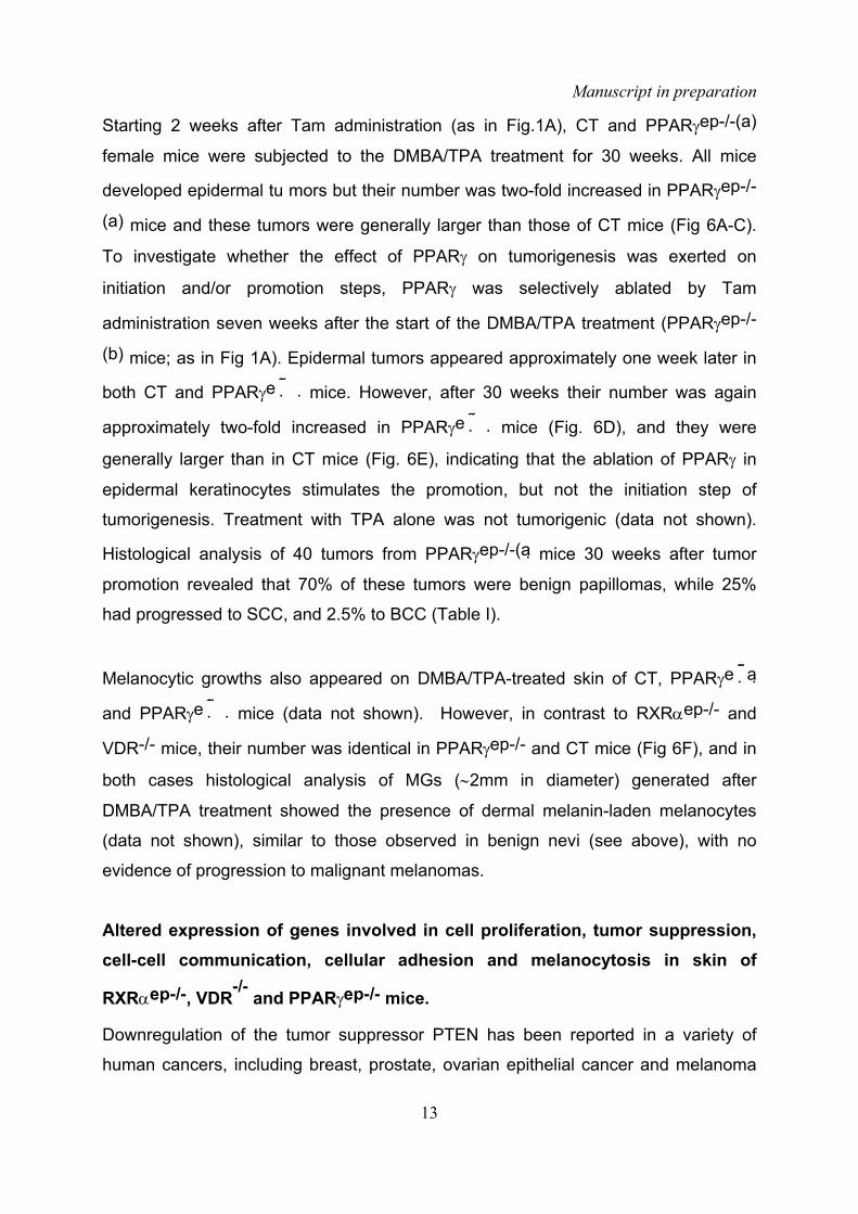
Manuscript in preparation
13
Starting 2 weeks after Tam administration (as in Fig.1A), CT and PPARγep-/-(a)
female mice were subjected to the DMBA/TPA treatment for 30 weeks. All mice
developed epidermal tu mors but their number was two-fold increased in PPARγep-/-
(a) mice and these tumors were generally larger than those of CT mice (Fig 6A-C).
To investigate whether the effect of PPARγ on tumorigenesis was exerted on
initiation and/or promotion steps, PPARγ was selectively ablated by Tam
administration seven weeks after the start of the DMBA/TPA treatment (PPARγep-/-
(b) mice; as in Fig 1A). Epidermal tumors appeared approximately one week later in
both CT and PPARγe� � mice. However, after 30 weeks their number was again
approximately two-fold increased in PPARγe� � mice (Fig. 6D), and they were
generally larger than in CT mice (Fig. 6E), indicating that the ablation of PPARγ in
epidermal keratinocytes stimulates the promotion, but not the initiation step of
tumorigenesis. Treatment with TPA alone was not tumorigenic (data not shown).
Histological analysis of 40 tumors from PPARγep-/-(a mice 30 weeks after tumor
promotion revealed that 70% of these tumors were benign papillomas, while 25%
had progressed to SCC, and 2.5% to BCC (Table I).
Melanocytic growths also appeared on DMBA/TPA-treated skin of CT, PPARγe� a
and PPARγe� � mice (data not shown). However, in contrast to RXRαep-/- and
VDR-/- mice, their number was identical in PPARγep-/- and CT mice (Fig 6F), and in
both cases histological analysis of MGs (∼2mm in diameter) generated after
DMBA/TPA treatment showed the presence of dermal melanin-laden melanocytes
(data not shown), similar to those observed in benign nevi (see above), with no
evidence of progression to malignant melanomas.
Altered expression of genes involved in cell proliferation, tumor suppression,cell-cell communication, cellular adhesion and melanocytosis in skin of
RXRαep-/-, VDR-/- and PPARγep-/- mice.
Downregulation of the tumor suppressor PTEN has been reported in a variety of
human cancers, including breast, prostate, ovarian epithelial cancer and melanoma
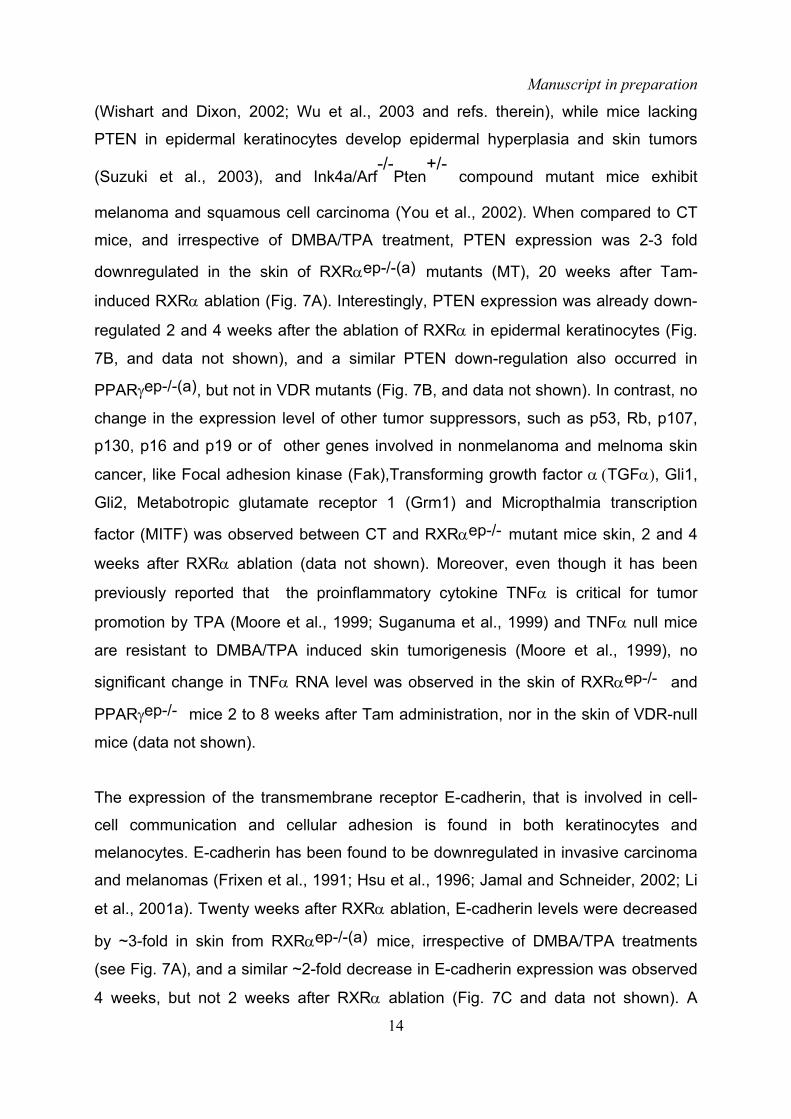
Manuscript in preparation
14
(Wishart and Dixon, 2002; Wu et al., 2003 and refs. therein), while mice lacking
PTEN in epidermal keratinocytes develop epidermal hyperplasia and skin tumors
(Suzuki et al., 2003), and Ink4a/Arf-/-
Pten+/-
compound mutant mice exhibit
melanoma and squamous cell carcinoma (You et al., 2002). When compared to CT
mice, and irrespective of DMBA/TPA treatment, PTEN expression was 2-3 fold
downregulated in the skin of RXRαep-/-(a) mutants (MT), 20 weeks after Tam-
induced RXRα ablation (Fig. 7A). Interestingly, PTEN expression was already down-
regulated 2 and 4 weeks after the ablation of RXRα in epidermal keratinocytes (Fig.
7B, and data not shown), and a similar PTEN down-regulation also occurred in
PPARγep-/-(a), but not in VDR mutants (Fig. 7B, and data not shown). In contrast, no
change in the expression level of other tumor suppressors, such as p53, Rb, p107,
p130, p16 and p19 or of other genes involved in nonmelanoma and melnoma skin
cancer, like Focal adhesion kinase (Fak),Transforming growth factor α (TGFα), Gli1,
Gli2, Metabotropic glutamate receptor 1 (Grm1) and Micropthalmia transcription
factor (MITF) was observed between CT and RXRαep-/- mutant mice skin, 2 and 4
weeks after RXRα ablation (data not shown). Moreover, even though it has been
previously reported that the proinflammatory cytokine TNFα is critical for tumor
promotion by TPA (Moore et al., 1999; Suganuma et al., 1999) and TNFα null mice
are resistant to DMBA/TPA induced skin tumorigenesis (Moore et al., 1999), no
significant change in TNFα RNA level was observed in the skin of RXRαep-/- and
PPARγep-/- mice 2 to 8 weeks after Tam administration, nor in the skin of VDR-null
mice (data not shown).
The expression of the transmembrane receptor E-cadherin, that is involved in cell-
cell communication and cellular adhesion is found in both keratinocytes and
melanocytes. E-cadherin has been found to be downregulated in invasive carcinoma
and melanomas (Frixen et al., 1991; Hsu et al., 1996; Jamal and Schneider, 2002; Li
et al., 2001a). Twenty weeks after RXRα ablation, E-cadherin levels were decreased
by ~3-fold in skin from RXRαep-/-(a) mice, irrespective of DMBA/TPA treatments
(see Fig. 7A), and a similar ~2-fold decrease in E-cadherin expression was observed
4 weeks, but not 2 weeks after RXRα ablation (Fig. 7C and data not shown). A

Manuscript in preparation
15
similar decrease was also observed in the skin of VDR null mutants (Fig. 7C),
whereas expression of E-cadherin was unaltered in PPARγep-/- mutants (see Fig.
7C).
It has been shown that targeted expression of α6β4 integrins in the suprabasal cell
layers of transgenic mouse epidermis dramatically increased the frequency of
papillomas and carcinomas induced by chemical tumorigenesis (Owens et al., 2003).
We have seen above that α6 and β4 integrin expression was much stronger in all
RXRαep-/- tumors, and in most cases extended into suprabasal layers (Fig.2A). Most
interestingly, 5-8 weeks after Tam injection, α6β4 integrins, which are normally
expressed by basal layer cells and located in the basement membrane, were found
to be strongly expressed in the upper hyperplastic suprabasal cell layers of RXRαep-
/- epidermis as evidenced by IHC (data not shown). Similarly, α6β4 integrins were
also found to be abnormally expressed in the epidermal suprabasal cell layers of
~25% of PPARγ ep-/- mice, but not in the epidermis of VDR null mice (data not
shown).
Overexpression of the hepatocyte growth factor (HGF) in basal keratinocytes of
transgenic mice leads to epidermal and dermal melanocytosis (Kunisada et al.,
2000). However, no significant change in the overall expression level of HGF and its
receptor c-Met was observed in the skin of RXRαep-/-, VDR-/- or PPARγep-/- mutant
mice 4 weeks after DMBA/TPA treatment (data not shown).
The ligand for the c-kit receptor, the stem cell factor SCF that is produced by
keratinocytes, plays an important role in melanocyte proliferation, survival and
migration (Kunisada et al., 2001; Yoshida et al., 2001). SCF can be produced in two
isoforms by alternative splicing yielding either a mRNA encoding only a membrane-
bound isoform (SCF220) or a mRNA encoding a longer membrane-bound isoform
(SCF248) from which a soluble secreted protein is released by proteolysis (SCF164).
Overexpression of SCF in epidermal keratinocytes leads to epidermal and dermal
melanocytosis (Kunisada et al., 1998a; Kunisada et al., 1998b). We therefore
investigated whether the increase in melanoma-like melanocytic tumors in

Manuscript in preparation
16
DMBA/TPA-treated RXRαep-/-
mutants could be related to altered expression of
SCF. Increased expression (~3 fold) of the SCF220 membrane-bound form was
observed in the skin of RXRαep-/-mutants (MT), irrespective of DMBA/TPA treatment
(Fig. 7A). Note, however, that this treatment also increased the basal level of
SCF220 in CT mice. In contrast, there was little, if any change in the expression level
of the SCF248 membrane-bound form in both CT and RXRαep-/- mice. An increase
in the expression level of total SCF was also observed in RXRαep-/- mice 4 weeks
after RXRα ablation (Fig.7D), whereas there was no significant change in its
expression in VDR-/-
and PPARγep-/- mutant mice (Fig.7D). We also looked at the
expression pattern of the SCF receptor c-kit, as loss of its expression is often
associated in human melanoma cells with progression to a metastatic phenotype
(Bar-Eli, 2001; Huang et al., 1998). 20-22 weeks after DMBA/TPA application, the
level of c-kit was reduced by 2-fold in skin of RXRαep-/- mice, but not in VDR-/- and
PPARγep-/- mutants, when compared to CT mice (Fig. 7A).
The POU domain transcription factor Brn-2 mRNA is overexpressed in human
melanoma compared to normal melanocytes, and is strongly upregulated by MAP
kinase signalling downstream of receptor tyrosine kinases (RTKs) and BRAF
(Goodall et al., 2004). Interestingly, Brn-2 mRNA level was significantly increased in
2/6 MGs from RXRαep-/- mice, but not in MGs from CT mice (data not shown).

Manuscript in preparation
17
Discussion
Keratinocytic RXRα and PPARγ act as tumor modifiers of both promotion andmalignant progression of epidermal tumors induced by topical DMBA/TPAtreatment.Using the well characterized DMBA/TPA-induced two-stage chemical skin
carcinogenesis system and keratinocyte-selective gene disruption in adult mice, we
have investigated here the role possibly played by RXRα and some of its
heterodimerization-partners in epidermal tumorigenesis. Our results show clearly that
RXRα acts as a tumor modifier of both the TPA-induced promotion of benign
papilloma tumors and their subsequent progression to malignancy. We also show
that the DMBA-induced initiation step which results in H-ras activation is not affected
by RXRα ablation.
Tumor promotion by TPA is known to induce the expression of the cytokine TNFα in
epidermal keratinocytes. This induction, which results in keratinocyte
hyperproliferation and skin inflammation, is critical for tumor promotion by TPA
(Moore et al., 1999; Suganuma et al., 1999). As a skin inflammation is also
generated upon RXRα ablation in epidermal keratinocytes (Li et al., 2001b ; Li et al.,
2000), and given the known associations between chronic inflammation and cancer
(Coussens and Werb, 2002), the question arises whether the promoting effect of
RXRα ablation on papilloma formation could reflect a further increase in TNFα
production. We could not detect such an increase , thus making it unlikely that TNFα
could be responsible for the more efficient tumor promotion resulting from RXRα
ablation in keratinocytes. Note, however, that irrespective of RXRα ablation in
keratinocytes, the TPA-induced promotion of epidermal tumors was efficiently
suppressed by inhibitors of AP-1 activity, such as the glucocorticoid fluocinolone
acetonide (FA) (Huang et al., 1997, our unpublished data).

Manuscript in preparation
18
In quest of partners with which RXRα may heterodimerize in skin keratinocytes in
order to modify the TPA-induced promotion and malignant progression of epidermal
tumors initiated by DMBA treatment, we also subjected mice ablated for NRs known
to be expressed in the skin to topical DMBA/TPA-induced skin tumorigenesis. The
three PPAR isotypes were selected for this study, because PPARα (Sheu et al.,
2002), PPARβ(δ) (Schmuth et al., 2004) and PPARγ (Mao-Qiang et al., 2004)
activators are known to inhibit TPA-induced cutaneous inflammation. The selective
ablation of PPARβ in basal keratinocytes had no significant effect on DMBA/TPA-
induced skin tumorigenesis (our unpublished results). However, as carcinogenicity
induced by DMBA/TPA was enhanced in PPARβ null mice (Kim et al., 2004), PPARβ
might exert its tumor modifier activity in non-keratinocytes, and /or might control
molecular mechanism in keratinocytes of the forming epidermis that prevented
tumorigenesis. In contrast, in PPARγep-/- mice ablated for PPARγ in epidermal
keratinocytes, the rate of formation of epidermal tumors, as well as their malignant
progression to carcinomas, were similar to those observed in RXRαep-/- mice even
though, in contrast to RXRα, the selective ablation of PPARγ in keratinocyte does not
result in skin inflammation (Mao-Qiang et al., 2004, and our unpublished results).
Thus, PPARγ is likely to be a major heterodimerization partner for RXRα in the
process which leads to increased tumor promotion and malignant progression in
DMBA/TPA-treated mice. Interestingly, germ line null mutations of PPARα or LXRβ,
the activations of which is also known to inhibit TPA-induced cutaneous inflammation
(Fowler et al., 2003), similarly resulted in 1.5 - 2-fold increased rates of formation of
DMBA/TPA induced tumors (our unpublished results). Selective ablation of PPARα
and LXRβ in keratinocytes is therefore required to determine whether, besides
RXRα/PPARγ heterodimers, RXRα/PPARα and RXRα/LXRβ heterodimers also act
as cell-autonomous epidermal tumor modifiers. On the other hand, germline null
mutations of either VDR, RARα, RARγ or RARβ had no significant effect on
DMBA/TPA-induced epidermal tumor formation (this study, and our unpublished
data).
In a search for genes whose altered expression upon RXRα ablation in skin
keratinocytes could possibly account for the DMBA/TPA-induced increased in

Manuscript in preparation
19
epidermal tumor formation and progression to malignancy, we looked at the
expression of a number of genes whose misexpression is known to affect
DMBA/TPA-induced epidermal tumorigenesis. Although the expression of most of
these genes was not significantly different in DMBA/TPA-treated skin of control and
RXRαep-/- mutant mice, changes in the expression of two genes was particularly
relevant. The PTEN gene, whose expression was 2-3-fold decreased (Fig. 7), is a
tumor suppressor gene that modulates several cellular functions, including cell
migration, survival and proliferation, through the inhibition of PI3-kinase-mediated
signalling cascades (Sulis and Parsons, 2003). Importantly, selective disruption of
the PTEN gene in epidermal keratinocytes has shown that PTEN functions as a
tumor suppressor for both spontaneous and induced skin tumors in mice (Suzuki et
al., 2003). Moreover, it has been reported that PTEN expression is upregulated by
PPARγ ligands, and PPAR response elements have been localized in the genomic
sequence upstream of the PTEN coding sequence (Patel et al., 2001). Most
interestingly, we found that ablation of PPARγ in skin keratinocytes also significantly
reduces PTEN expression in the skin (Fig. 7). Thus, the similarly increased tumor
formation and progression to malignancy observed for DMBA/TPA-treated mice upon
selective ablation of RXRα or PPARγ in keratinocytes, might be caused at least in
part by a downregulation of the expression of the tumor suppressor PTEN, due to the
lack of RXRα/PPARγ heterodimers.
The suprabasal expression of α6β4 integrin, that we observed in DMBA/TPA induced
epidermal tumors of RXRαep-/- mice (see Results section) has been correlated with
a high risk of malignant conversion to SCC (Tennenbaum et al., 1993). Interestingly,
it has been recently shown that targeted expression of α6β4 in keratinocytes of the
epidermal suprabasal layers of transgenic mice dramatically increases the rate of
DMBA/TPA-induced promotion and malignant progression of epidermal tumors, and
it was suggested that this ectopic α6β4 integrin expression promotes tumorigenesis
by preventing TGFβ from suppressing clonal expansion of initiated basal
keratinocytes (Owens et al., 2003). As approximately 30% of PPARγ ep-/- mice also
exhibit suprabasal expression of α6β4 (our unpublished results) and both PPARγ
(Westergaard et al., 2001), and RXRα (Chapellier et al., 2002), are present in
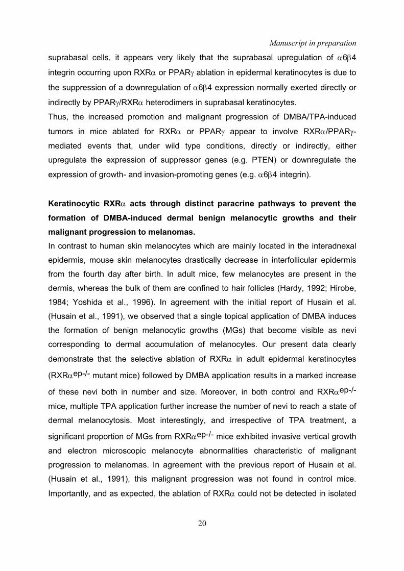
Manuscript in preparation
20
suprabasal cells, it appears very likely that the suprabasal upregulation of α6β4
integrin occurring upon RXRα or PPARγ ablation in epidermal keratinocytes is due to
the suppression of a downregulation of α6β4 expression normally exerted directly or
indirectly by PPARγ/RXRα heterodimers in suprabasal keratinocytes.
Thus, the increased promotion and malignant progression of DMBA/TPA-induced
tumors in mice ablated for RXRα or PPARγ appear to involve RXRα/PPARγ-
mediated events that, under wild type conditions, directly or indirectly, either
upregulate the expression of suppressor genes (e.g. PTEN) or downregulate the
expression of growth- and invasion-promoting genes (e.g. α6β4 integrin).
Keratinocytic RXRα acts through distinct paracrine pathways to prevent theformation of DMBA-induced dermal benign melanocytic growths and theirmalignant progression to melanomas.In contrast to human skin melanocytes which are mainly located in the interadnexal
epidermis, mouse skin melanocytes drastically decrease in interfollicular epidermis
from the fourth day after birth. In adult mice, few melanocytes are present in the
dermis, whereas the bulk of them are confined to hair follicles (Hardy, 1992; Hirobe,
1984; Yoshida et al., 1996). In agreement with the initial report of Husain et al.
(Husain et al., 1991), we observed that a single topical application of DMBA induces
the formation of benign melanocytic growths (MGs) that become visible as nevi
corresponding to dermal accumulation of melanocytes. Our present data clearly
demonstrate that the selective ablation of RXRα in adult epidermal keratinocytes
(RXRαep-/- mutant mice) followed by DMBA application results in a marked increase
of these nevi both in number and size. Moreover, in both control and RXRαep-/-
mice, multiple TPA application further increase the number of nevi to reach a state of
dermal melanocytosis. Most interestingly, and irrespective of TPA treatment, a
significant proportion of MGs from RXRαep-/- mice exhibited invasive vertical growth
and electron microscopic melanocyte abnormalities characteristic of malignant
progression to melanomas. In agreement with the previous report of Husain et al.
(Husain et al., 1991), this malignant progression was not found in control mice.
Importantly, and as expected, the ablation of RXRα could not be detected in isolated

Manuscript in preparation
21
MGs, while it was efficiently ablated in MG-free epidermis and in the epidermis
surrounding MGs.
We therefore conclude that the genetically DMBA-initiated proliferation of
melanocytes and their possible malignant transformation must be controlled by
paracrine/juxtacrine signals which emanate from keratinocytes, and are regulated by
RXRα. Morevorer, these signals must be different from those which control the
formation of epidermal squamous tumors, as PPARγep-/- mutants develop epidermal
tumors upon DMBA/TPA treatment as efficiently as RXRαep-/- mutants, whereas
they do not exhibit any increase in MGs formation, when compared to control mice.
On the other hand, VDR-null mutants which do not show any enhancement of
epidermal tumorigenesis upon DMBA/TPA treatment, exhibit a marked increase in
benign MGs, which, however, do not progress to malignancy. Thus, the events
which, upon DMBA/TPA treatment, underlie the suppressive effect of RXRα on
epidermal tumorigenesis might be mediated by RXRα/PPARγ heterodimers (see
above), while those which underlie the suppressive effect of RXRα on the formation
of benign MGs could be mediated by RXRα/VDR heterodimers. On the other hand,
the events underlying the malignant conversion of benign MGs to melanomas might
be mediated by heterodimers between RXRα and NR partner(s) different from those
mutated in the present study. That different mechanisms are at work in these
tumorigenesis processes, is further supported by the absolute requirement of the
tumor promoter TPA to generate DMBA-initiated epidermal tumors in RXR�
mutants, whereas this promoter is dispensible to generate benign melanocytic
growths and melanomas. Along the same lines, it is remarkable that the topical
application of the glucocorticoid FA, which antagonizes the activity of AP-1, fully
prevents the occurrence of epidermal tumors in DMBA/TPA-treated RXRαep-/- mice,
while it does not affect the generation of MGs and their malignant progression (our
unpublished data).
The non-cell autonomous molecular events that, upon topical DMBA initiation,
underlie in the epidermis and dermis the keratinocyte-originated RXRα-mediated
control of dermal MG generation and malignant transformation, are unknown. In vitro
keratinocyte-melanocyte cocultures, as well as overexpression studies in transgenic
mice, have suggested that the proliferation and differentiation of mammalian

Manuscript in preparation
22
melanocytes are regulated by factors produced by keratinocytes. More specifically
endothelin ET-1, as well as the stem cell factor (SCF) and the hepatocyte growth
factor (HGF) could be such factors (Hirobe et al., 2002). In particular, analysis of
transgenic mice expressing SCF in epidermal keratinocytes led to the conclusion that
SCF acts as a key factor to support the temporally and spatially coordinated survival,
proliferation, differentiation and migration of melanocyte lineages (Kunisada et al.,
1998b). Moreover, the soluble form of SCF (Refs in Results section) may be required
for the migration of c-kit-expressing melanoblasts/melanocytes, while membrane-
bound SCF is required for their survival, proliferation and differentiation (Wehrle-
Haller and Weston, 1995). In a preliminary analysis, we found that SCF expression
was significantly increased in the skin of RXR� mutants, which suggests that the
ablation of RXRα in epidermal keratinocytes may directly or indirectly lead to an
overproduction of SCF, which in DMBA-initiated melanocytes/melanoblasts could
lead to their proliferation and enhance the formation of MGs. More sophisticated
studies are obviously required to characterize the epidermal and dermal factors
whose expression is altered by ablation of RXRα in epidermal keratinocytes, in order
to elucidate how the development of DMBA-initiated melanocytic tumors is
environmentally regulated by RXRα-controlled events in epidermal keratinocytes.

Manuscript in preparation
23
Acknowledgements
We thank B. Lane, E. Georges, S. Yuspa and V. Hearing for kindly providing antibodies, N.
Ghyselinck for RAR-null mice, S. Kato for VDR-null mice, Deltagen for LXRβ null mice and
F.J. Gonzalez for PPARα - null mice. We also thank M. Mark, A. Van Es and R. Matyas for
helpful discussion, R. Lorentz, M. Duval, B. Chofflet, S. Bronner and C. Gérard for excellent
technical help, M. LeMeur, E. Metzger and the animal facility staff for animal care and for
useful suggestions, the Histology service staff and IGBMC common facility for assistance, the
secretarial staff for typing and the illustration staff for preparing the figures. This work was
supported from funds from Centre National de la Recherche Scientifique, the institut National
de la Santé et de la Recherche Médicale, the Collège de France, the Hôpital Universitaire de
Strasbourg, de Association pour la Recherche Médicale, the Human Frontier Science
Program, the Ministère de l’Éducation Nationale de la Recherche et de la Technologie, the
Swiss National Science Foundation, the Etat de Vaud, and the European Community and by a
UICC Translational Cancer Research Fellowship funded by Novartis (Switzerland).
(Prior to publication, a copy of the manuscript is to be provided to UICC to be
forwarded to Novartis for information)

Manuscript in preparation
24
Experimental Procedures:
Mice and Carcinogenic treatment
To selectively ablate RXRα in epidermal keratinocytes of adult mice, mice carrying
LoxP-site-containing (floxed) RXRα L2 alleles were bred with hemizygous K14-Cre-
ERT2(tg/0) transgenic mice, to produce K14-Cre-ERT2(tg/0)/RXRαL2/L2 mice (Li et
al., 2000; Metzger et al., 2003). 8-12 week old female mice, were injected with 0.1
mg of tamoxifen (100 µl sunflower oil) for 5 days (Metzger et al., 2003) to produce
control (CT) (from K14-Cre-ER T2 0/0 and K14-Cre-ER T2 tg/0 RXRα L2/+ mice) and
RXRαep-/- mice (from K14-Cre-ER T2 tg/0 RXRα L2/L2 mice). 2 weeks after the first
tamoxifen injection, RXRα ablation in the epidermis was verified by Southern
hybridisation of DNA isolated from tail biopsies after separation of the epidermis and
the dermis (Li et al., 2000). CT and PPARγep-/- mice were generated in a similar way
by breeding K14-Cre-ERT2(tg/0) transgenic mice with PPAR��� mice (Imai et
al., 2004).
Groups of control (CT) and mutant (RXRαep-/-, PPARγep-/- and VDR-/-) animals
were housed in a temperature and humidity controlled room with 12h cycles of light
and dark. 14 days after last Tam injection, mice in each group were shaved to
synchronize the hair cycle and treated the day after with a single dose of 50 µg
DMBA in 100 µl acetone. One week after DMBA application, 5 µg TPA (in 200 µl
acetone) was applied topically twice a week for 30 weeks. The number and size of
the papillomas per mouse were weekly recorded.
Determination of the number and size of the tumorsThe length of the papillomas and the diameter of the melanocytic growths (nevi) were
measured with a Vernier calliper on isofluorane anesthesised mice.
Manuscript in preparation
25
Histological and electron microscopic analysisTumors were excised 25 or 30 weeks after DMBA/TPA application, fixed in Bouin’s
fixative and embedded in paraffin. All skin biopsies were matched for age, sex and
body site and all papillomas were matched for age and sex. 5 µm paraffin sections of
tumor and skin, 2 µm semithin sections of skin samples and 70 nm ultrathin sections
for EM were made as described (Li et al., 2001b). Subiliac lymph nodes were fixed in
4% paraformaldehyde, photo-graphed, embedded in paraffin and 5 µm-thick sections
were stained with hematoxylin and eosin.

Manuscript in preparation
26
Immunohistochemistry
Tumors (8–16 mm) were excised 22 weeks after DMBA/TPA application and
immediately embedded in OCT and frozen on dry ice. 10 µm-thick frozen sections
from CT and RXRαep-/- tumors were reacted with primary antibodies [mouse
monoclonal anti -K10, rabbit polyclonal anti-K5 (gifts from Prof. Brigitte Lane, Cell
Structure research group, University of Dundee]), rabbit polyclonal anti-K1 (Babco),
rabbit polyclonal anti-K13 (gift from Prof. S. Yuspa, NIH), rabbit polyclonal α6-integrin
(gift from E. Georges), and biotin-conjugated rat monoclonal anti-CD31(Pharmingen)
and processed as described (Li et al., 2001b). To detect melanocytes 5 µm paraffin
sections of subiliac lymph nodes from CT and mutant animals were reacted with
primary antibodies [rabbit polyclonal α-PEP8 (TRP2) [a gift from Vincent Hearing,
NIH] or rabbit polyclonal S-100 (Dako)], and processed as described (Pavan and
Tilghman, 1994; Sotillo et al., 2001).
Genotyping of the RXRα allelesDorsal skin biopsies with and without black spots (nevi / melanocytic growths) were
taken from control (CT) and mutant (RXRαep-/-) animals. Samples were incubated
overnight in 4mg/ml Dispase (neutral protease, Boehringer Mannheim) at 4°C. The
following day the epidermis was peeled off from the dermis under a stereo-zoom
binocular (Leica) and the melanocytic growths were micro-dissected out from the
remaining dermal tissue. The hair follicles were carefully plucked from the nevi under
the binocular, rinsed several times with 1XPBS to remove the adhering cells and
transferred into separate eppendorf tubes along with the corresponding epidermis.
Genomic DNA from the epidermis and dermis was isolated as described (Li et al.,
2000). (Li et al., 2001b)A semi-quantitative PCR with different dilution of the isolated
DNA was performed for 26 cycles in Perkin Elmer DNA light cycler to amplify the L2
and L- RXRα allele as described (Li et al., 2000).
Detection of mutations in the H-ras gene.

Manuscript in preparation
27
Mutation of the codon 61 of the H-ras gene (Acc No. AF081118) was detected by
PCR amplification of a 280 bp amplicon followed by restriction digest as described
(Nelson et al., 1992). DNA was isolated from papillomas (4-8 mm; 25 CT and 25 MT),
lesion free skin (10 CT and 10 MT) and nevi (15 CT and 15 MT) biopsied from 6 CT
and 6 RXRαep-/- animals 25 weeks after DMBA/TPA treatment. List of the primers
are available on request.The 280 bp amplicons of exon II (encompassing codon 61)
from some papillomas (5 CTand 5 MT) were also cloned and sequenced to detect
mutation in the codon 61using primers as described (Ise et al., 2000). Mutations in
the codon 12 and 13 of the H-Ras gene exon I were analysed from papillomas (10
CT and 10 MT), lesion free skin biopsies (4 CT and 4 MT) and nevi (4 CT and 4 MT)
of 6 CT and RXRαep-/- animals, respectively, by PCR amplification of exon I using
specific primers, followed by cloning and sequencing on both strands as described
(Ise et al., 2000).
Detection of mutations in the N-ras gene.A 120 bp amplicon of the second exon of the N-Ras gene encompassing codon 61
was amplified by PCR using specific primers. DNA was isolated from papillomas (20
CT and 20 MT) and nevi (>2mm size; 15 CT and 15 MT) microdissected under
binocular from the remaining adhering cells, from 6 CT and 6 RXRαep-/- animals 25
weeks after DMBA/TPA treatment. As mutation at the codon 61 in N-Ras produces
either a NlaIII (CAA->CAT; A to T transversion in the third base) or a BfaI (CAA-
>CTA; A to T transversion in the second base) restriction site, the PCR amplified
products were restriction digested by NlaIII and BfaI respectively, and resolved by
agarose gel electrophoresis. The gel was blotted into a nylon membrane and
hybridized with a 32P labelled internal oligonucleotide. Primers are available on
request. Amplified products were also cloned and 10 transformants for each sample
were sequenced on both strands as mentioned by Ise et al.(Ise et al., 2000).
Detection of the codon 12 and codon 13 mutation in the N-ras gene was done by
PCR amplification of exon I from papillomas (4 CT and 4 MT) and nevi (6 CT and 8
MT) of 6 CT and RXRαep-/- animals, respectively, using primers as described by Ise
et al. (Ise et al., 2000), followed by cloning and sequencing on both strands.
Manuscript in preparation
28
Quantitative RT-PCRRNA was extracted from approximately 100 mg of dorsal body skin or tumor with
TrizolR Reagent (GIBCO BRL, Life technologies), dissolved in RNase-free water at a
concentration of 1 µg/µl after quantification by spectrophotometric analyses. cDNA
synthesis was performed with Superscript II reverse transcription kit (GIBCO BRL,
Life technologies) using 5 µg of total RNA, according to the manufacturer’s
instructions. 1 µl of cDNA was used in a 30 µl semiquantitative PCR reaction with 1X
PCR buffer, 100 ng of each primer, an optimal concentration of MgCl2 (1.5 mM), 0.2
mM dNTP and 2 U of AmpliTaq (Perkin Elmer, Foster City, Calif.). Real time PCR
was performed with a LightCycler (Roche Diagnostics) and the SYBR green protocol.
For that purpose 5 µl of each cDNA was diluted to 20 µl and 2 µl of the diluted cDNA
was used for each amplification. Amplification specificity was verified by melting
curve analysis and the data quantified with the LightCycler software. The primers
were [S=5’-GTGCTCTCTTCAACATTAGGT-3’; As=5’-TCCAGTATAAG
GCTCCAAAAG-3’] for SCF, [S=5’-CGGATCACAAAGATT TGCGAT-3’; As=5’-ATA
GGACCAGACATCACTTTC-3’] for c-Kit, [S=5’-AA GGTTTTCTAC AGCA TCACC-3’;
As=5’-CACTGTGATCACTATCTCCAT-3’] for E-cadherin, [S=5’-
AGATGACAATCATGTTGCAGC-3’ ; As=5’-CATCATCTTGTGAAACAG C AG -3’] for
PTEN and [S=5’-GTAATGATCAGTCAACGGG GGAC-3’; As=5’-
CCAGCAAGCTTGCAACCTTAACCA-3’] for HPRT. The amplified products in
exponential phase were electrophoresed on 2% agarose gel, transferred onto nylon
membrane and hybridized with the following radiolabelled nucleotides for SCF
(248+164) and SCF(220), [5’-AAGGCCCCTGAAGACTCGGGCCTACAAT GG-3’], c-
KIT(I)[5’-CTGCCCGTGAAGTGGATGGCACCAGAGAGC-3’], E-cad- herin, (I)[5’-
CAGCCTCTGGATAGAGAAGCCATTGCCAAG-3’], PTEN(I), [5’-GT
AATGATTTGTGCATATTTATTGCATCGG-3’] and HPRT(I), [5’-GCTTTCCCTG
GTTAAGCAGTACAGCCCC-3’].
Statistical analysesWhere relevant, data were compared by Unpaired Student’s t-test with corrections for
unequal variance using Statview 5 programme (Abacus Concepts, CA). Values are
reported as mean ± SEM and the significance level was set at p≤ 0.05.

Manuscript in preparation
29
Figure legends and Tables:
Figure 1. DMBA/TPA-induced epidermal tumors and melanocytic growths in
RXRαep-/- mice.
A. Timing of tamoxifen (Tam)-induced RXRα ablation in epidermal keratinocytes, and
DMBA/TPA induced tumorigenesis. Tam-treatment (0.1 mg for 5 consecutive days)
was started either 18 days before ("a") or 8 weeks after ("b") topical DMBA
application. TPA was topically applied twice a week (blue arrows) for up to 30 weeks.
B. Formation of epidermal tumors in DMBA/TPA treated control (CT) and RXRαep-/-
(a) mice, as determined macroscopically and plotted against the number of weeks of
TPA treatment. Values are expressed as mean +/- SEM (n=7 mice).
C. Dorsal view of CT (K14-Cre-ERT2(0/0)/RXRα
L2/L2) (left) and RXRαep-/-(a) (right)
mice after 25 weeks of DMBA/TPA treatment.
D. Epidermal tumor length distribution in CT and RXRαep-/-(a) mice, after 30 weeks
of DMBA/TPA treatment. Values are expressed as mean +/- SEM (n=7 mice).
E. Formation of epidermal tumors in DMBA/TPA-treated CT and RXRαep-/-(b) mice
in which RXRα was ablated in keratinocytes during TPA promotion. Values are
expressed as mean +/- SEM (n = 7 mice).
F. Epidermal tumor length distribution in CT and RXRαep-/-(b) mice, after 30 weeks
of DMBA/TPA treatment. Values are expressed as mean +/- SEM (n = 7 mice).
G. Dorsal view of CT (K14-Cre-ERT2(0/0)
/RXRαL2/L2
) (left) and RXRαep-/- (right)
mice 25 weeks after DMBA treatment. Arrows point to melanocytic growths.
H. Size distribution of melanocytic growths in CT, RXRαep-/-, RXRαep-/-(a) and
RXRαep-/-(b) mice. The number of MGs, 1 – 2 and 2 – 4 mm in diameter, was
determined on 7 CT, 7 RXRαep-/-, 7 RXRαep-/-(a), and 7 RXRαep-/-(b) mice, 30
weeks after DMBA and DMBA/TPA treatments, as indicated. Values are expressed
as mean +/- SEM.
Manuscript in preparation
30
Figure 2. Immuno-histochemical and histological analyses of skin tumors
induced by DMBA/TPA treatment in RXRαep-/- mice.A. Representative sections of tumor biopsies from control (CT) (a, c, e, g, i, k) and
RXRαep-/-(a) (b, d, f, h, j, l) mice, taken after 22 weeks of DMBA/TPA treatment,
and stained with antibodies against K5 (a, b), K1 (c, d), K10 (e, f), K13 (g, h), α6
integrin (i, j) and CD31 (k, l) are presented. Red corresponds to antibody staining and
blue to DNA DAPI staining. Scale bar in Aa: 25 µm.
B. Representative hematoxylin- and eosin-stained 5 µm-thick paraffin sections from
CT biopsies taken after 25 (a - b) and 30 (c) weeks of DMBA/TPA treatment. (a),
suprabasal cell hyperplasia (acanthosis) within a benign papilloma; (b), basal cell
hyperplasia within a benign papilloma; (c), focus of early squamous cell carcinoma.
C. Representative hematoxylin- and eosin-stained 5 µm-thick paraffin sections from
RXRαep-/- tumor biopsies taken after 25 (a-c) and 30 (d-i) weeks of DMBA/TPA
treatment. (a), suprabasal cell hyperplasia within a benign papilloma ; (b), basal cell
hyperplasia within a benign papilloma ; (c), focus of early squamous cell carcinoma;
(d), (e) and (f), well, moderately and poorly differentiated SCC, respectively; (g)
anaplastic SCC with spindle-shaped cells and absence (or minimal) keratinisation; (h)
and (i), basal cell carcinoma with densely packed basophilic cells with
hyperchromatic nuclei resembling those of epidermal basal cells.
Hyperplastic basal (blue brackets) and suprabasal (black brackets) cell layers in “b”
(B and C) and (a,b) [B and C] are indicated. Black arrows in (B)a and (C)a point to
basal cell layers; white arrow in C(d) points to a keratin pearl; black and blue
arrowheads in C(c) and (C)e, point to a mitosis and abnormal nuclei, respectively;
asterisk in C(e) indicates internal cell keratinisation. Scale bar (in Ba) = 33 µm for
Ba,b and Ca,b; 22 µm for Bc and Cc-i.
Figure 3.(A) Histological and ultrastructural analyses of melanocytic growths
(MGs) from CT (control) and RXRαep-/-(a) mice. (a) and (b) : Hematoxylin and
eosin-stained paraffin sections (5 µm thick) of (a) CT (K14-Cre-ER T2(0/0)
/RXRαL2/L2) and (b) RXRαep-/-(a) MGs in skin biopsies taken 25 weeks after
DMBA/TPA treatment. The arrow in (b) points to melanocytic invasion of the
hypodermis. E, epidermis ; D, dermis ; H, hypodermis. Scale bar=33 µm. (c) to (g) :

Manuscript in preparation
31
electron microscopic analyses of CT (c and e) and RXRαep-/-(a) (d, f and g) MGs
isolated 25 weeks after DMBA/TPA treatment. Note in the inset (g), the oval-shaped
subcellular structure containing a longitudinally oriented component made of rods.
Arrows in (c),(d) and (f) point to control "normal" dermal melanocytes (in c), and
abnormally shaped melanosomes typical of malignant melanoma (in d and f),
respectively. Arrowheads in “c” and “e” point towards a melanin-laden melanophage
and melanosomes of stage III and IV, respectively. N, nucleus. Scale bars : 2.5 µm
for c and d ; 1 µm for e and f and 0.25 µm for the g inset.
(B) RXRα is not ablated in melanocytic growths from RXRαep-/- mice. The
presence of RXRα L2 and L- alleles was analysed by PCR in DNA from epidermis
(E), dermis (D) isolated from MG free skin biopsies and melanocytic growths (MG)
from CT (lanes 1 and 2) (K14-Cre-ERT2(0/0)/RXRαL2/L2) and RXRαep-/- (lanes 3-
10) mice, 25 weeks after DMBA or DMBA/TPA treatments, as indicated. The position
of the RXRα L2 and L- alleles is indicated.
Figure 4. Metastasis of melanocytes in lymph nodes of DMBA and DMBA/TPA-
treated RXRαep-/- mice.
A. Subiliac lymph nodes from CT (a,e) and RXRαep-/- (b,f) mice isolated 30 weeks
after DMBA (a,b) and DMBA/TPA (e,f) treatment, respectively. Hematoxylin- and
eosin-stained 5 µm paraffin sections of subiliac lymph nodes from CT (c,g) and
RXRαep-/- (d,h) mice after DMBA (c,d) and DMBA/TPA (g,h) treatment, respectively.
Scale bar (in "c") = 33 µm. The arrow in “d” and arrowheads in “h” point to
pigmented cells.
B. Representative sections of subiliac lymph nodes from CT (a) and RXRαep-/- (b)
taken 30 weeks after DMBA application and stained with antibodies to S-100. Arrows
point to S-100 positive cells. Scale bar (in "a"), 33 µm.
C. Representative sections of subiliac lymph nodes from CT (a) and RXRαep-/- (b)
taken 30 weeks after DMBA application and stained with antibodies to α-PEP8. The
arrow indicates α-PEP8 positive malignant melanocytes. Scale bar (in "a"), 33 µm.
Manuscript in preparation
32
Figure 5. DMBA/TPA-induced epidermal tumors and melanocytic growths in
VDR-/-
mice.
A. Dorsal view of CT (control, VDR+/+)(left) and VDR-/- (right) mice 25 weeks after the
start of DMBA/TPA treatment. Arrows point to papilloma-like epidermal tumors.
B. Formation of epidermal tumors in DMBA/TPA treated CT and VDR-/- mice, as
determined macroscopically and plotted against the number of weeks after the start
of TPA treatment. Values are expressed as mean +/- SEM (n=7 mice).
C. Length distribution of epidermal tumors in CT and VDR-/- mice, as measured
30 weeks after the start of the DMBA/TPA treatment. Values are expressed as mean
+/- SEM (n=7 mice).
D. Size distribution of melanocytic growths in CT and VDR-/- mice. The number
of MGs (1-2 and 2-4 mm in diameter) was determined on 7 CT and 7 VDR -/- mice,
30 weeks after the start of TPA treatment. Values are expressed as mean +/- SEM
E. Hematoxylin- and eosin-stained paraffin sections (5 µm thick) of (a) CT and (b)
VDR-/- MGs in skin biopsies taken after 25 weeks of DMBA/TPA treatment. Scale
bar=33 µm.
F. Electron microscopic analysis of (a) CT and (b) VDR-/- MGs taken after 25
weeks of DMBA/TPA treatment. N, nucleus. Scale bar: 2.5 µm.
Figure 6. DMBA/TPA-induced epidermal tumors and melanocytic growths in
PPARγ ep-/-(a) and PPARγ ep-/-(b) mice.A. Formation of papilloma like epidermal tumors in DMBA/TPA-treated control (CT)
and PPARγ ep-/-(a) mice, as determined macroscopically and plotted against the
number of weeks after the start of the TPA treatment. Values are expressed as mean
+/- SEM (n=7 mice).
B. Dorsal view of CT [K14-Cre-ERT2(0/0)/PPARγL2/L2] (left) and PPARγ ep-/-(a)
(right) mice 25 weeks after the start of DMBA/TPA treatment.
C. Length distribution of epidermal tumors in CT and PPARγ ep-/-(a) mice as
measured after 25 weeks of DMBA/TPA treatment. Values are expressed as mean

Manuscript in preparation
33
+/- SEM (n=7 mice).
D. Formation of papilloma-like tumors in DMBA/TPA-treated PPARγ ep-/-(b) mice in
which PPARγ was ablated in keratinocytes during TPA promotion. Values are
expressed as mean +/- SEM (n = 7 mice).
E. Length distribution of epidermal tumors in CT and PPARγ ep-/-(b) mice, after 25
weeks of TPA treatment. Values are expressed as mean +/- SEM (n = 7 mice).
F. Size distribution (diameter) of MGs in CT, PPARγ ep-/-(a) and PPARγ ep-/-(b)
mice. The number of MGs (1 – 2 and 2 – 4 mm in diameter) was determined on 7 CT,
7 PPARγ ep-/-(a) and 7 PPARγ ep-/-(b) mice, after 25 weeks of TPA treatment.
Values are expressed as mean +/- SEM.
Figure 7. (A) RT-PCR analyses on skin biopsies from CT and RXRαep-/-(a) (MT)mice. Semi-quantitative RT-PCR was performed for SCF(248+164), SCF(220), c-kit,
E-cadherin, PTEN and HPRT on dorsal skin biopsies from control (CT) and RXRαep-
/-(a) mutant mice collected after 20-22 weeks of DMBA/TPA treatment as indicated.
HPRT was used as an internal control. SCF(248+164) and SCF(220) represent the
longer and the shorter membrane bound form of SCF after deletion of exon 6,
respectively.
(B-D) Quantitative real-time RT-PCR determination of relative RNA levels of PTEN
(B), E-cadherin (C) and SCF (D) in dorsal skin biopsies from CT, VDR-/-
mice, and
RXRαep-/- or PPARγep-/- mice 4 weeks after Tamoxifen-treatment. In all three cases
no DMBA/TPA treatment was applied. *P< 0.05, **P<0.01 and #, p≥ 0.05.
Table I. Incidence of malignant progression in papillomas generated by a 30-
week DMBA/TPA treatment of mice selectively lacking either RXRα or PPARγ in
epidermal keratinocytes, or VDR-/-
mice.
Epidermal tumors obtained 30 weeks after DMBA/TPA application from 6 control, 6
RXRαep-/-(a), 6 PPARγep-/-(a) and 6 VDR-/- mice were analysed and histologically
classified, as indicated.

Manuscript in preparation
34
References :
Altucci, L., and Gronemeyer, H. (2001). The promise of retinoids to fight against cancer. Nat Rev Cancer1, 181-193.Bar-Eli, M. (2001). Gene regulation in melanoma progression by the AP-2 transcription factor. PigmentCell Res 14, 78-85.Chambon, P. (1996). A decade of molecular biology of retinoic acid receptors. Faseb J 10, 940-954.Chapellier, B., Mark, M., Messaddeq, N., Calleja, C., Warot, X., Brocard, J., Gerard, C., Li, M., Metzger,D., Ghyselinck, N. B., and Chambon, P. (2002). Physiological and retinoid-induced proliferations ofepidermis basal keratinocytes are differently controlled. Embo J 21, 3402-3413.Chida, K., Hashiba, H., Fukushima, M., Suda, T., and Kuroki, T. (1985). Inhibition of tumor promotion inmouse skin by 1 alpha,25-dihydroxyvitamin D3. Cancer Res 45, 5426-5430.Coussens, L. M., and Werb, Z. (2002). Inflammation and cancer. Nature 420, 860-867.Darwiche, N., Celli, G., Tennenbaum, T., Glick, A. B., Yuspa, S. H., and De Luca, L. M. (1995). Mouseskin tumor progression results in differential expression of retinoic acid and retinoid X receptors. CancerRes 55, 2774-2782.Darwiche, N., Scita, G., Jones, C., Rutberg, S., Greenwald, E., Tennenbaum, T., Collins, S. J., De Luca, L.M., and Yuspa, S. H. (1996). Loss of retinoic acid receptors in mouse skin and skin tumors is associatedwith activation of the ras(Ha) oncogene and high risk for premalignant progression. Cancer Res 56, 4942-4949.De Luca, L. M., Tarone, R., Huynh, M., Jones, C. S., and Chen, L. C. (1996). Dietary retinoic acid inhibitsmouse skin carcinogenesis irrespective of age at initiation. Nutr Cancer 25, 249-257.Demunter, A., Ahmadian, M. R., Libbrecht, L., Stas, M., Baens, M., Scheffzek, K., Degreef, H., De Wolf-Peeters, C., and van Den Oord, J. J. (2001). A novel N-ras mutation in malignant melanoma is associatedwith excellent prognosis. Cancer Res 61, 4916-4922.Epstein, J. H. (1992). Experimental models for primary melanoma. Photodermatol PhotoimmunolPhotomed 9, 91-98.Fisher, G. J., and Voorhees, J. J. (1996). Molecular mechanisms of retinoid actions in skin. Faseb J 10,1002-1013.Fowler, A. J., Sheu, M. Y., Schmuth, M., Kao, J., Fluhr, J. W., Rhein, L., Collins, J. L., Willson, T. M.,Mangelsdorf, D. J., Elias, P. M., and Feingold, K. R. (2003). Liver X receptor activators display anti-inflammatory activity in irritant and allergic contact dermatitis models: liver-X-receptor-specificinhibition of inflammation and primary cytokine production. J Invest Dermatol 120, 246-255.Frixen, U. H., Behrens, J., Sachs, M., Eberle, G., Voss, B., Warda, A., Lochner, D., and Birchmeier, W.(1991). E-cadherin-mediated cell-cell adhesion prevents invasiveness of human carcinoma cells. J Cell Biol113, 173-185.Ghadially, F. N. (1997). Ultrastructural Pathology of the Cell and Matrix, Vol 2, Fourth edition edn,Butterworth-Heinemann).Goodall, J., Martinozzi, S., Dexter, T. J., Champeval, D., Carreira, S., Larue, L., and Goding, C. R. (2004).Brn-2 expression controls melanoma proliferation and is directly regulated by beta-catenin. Mol Cell Biol24, 2915-2922.Hardy, M. H. (1992). The secret life of the hair follicle. Trends Genet 8, 55-61.Hirobe, T. (1984). Histochemical survey of the distribution of the epidermal melanoblasts and melanocytesin the mouse during fetal and postnatal periods. Anat Rec 208, 589-594.Hirobe, T., Furuya, R., Akiu, S., Ifuku, O., and Fukuda, M. (2002). Keratinocytes control the proliferationand differentiation of cultured epidermal melanocytes from ultraviolet radiation B-induced pigmentedspots in the dorsal skin of hairless mice. Pigment Cell Res 15, 391-399.Hong, W. K., and Itri, L. M. (1994). Retinoids and human cancer. In The Retinoids, M. B. Sporn, A. B.Roberts, and D. S. Goodman, eds. (New York, Raven Press), pp. 597-658.Hsu, M. Y., Wheelock, M. J., Johnson, K. R., and Herlyn, M. (1996). Shifts in cadherin profiles betweenhuman normal melanocytes and melanomas. J Investig Dermatol Symp Proc 1, 188-194.Huang, C., Ma, W. Y., Dawson, M. I., Rincon, M., Flavell, R. A., and Dong, Z. (1997). Blocking activatorprotein-1 activity, but not activating retinoic acid response element, is required for the antitumorpromotion effect of retinoic acid. Proc Natl Acad Sci U S A 94, 5826-5830.Huang, S., Jean, D., Luca, M., Tainsky, M. A., and Bar-Eli, M. (1998). Loss of AP-2 results indownregulation of c-KIT and enhancement of melanoma tumorigenicity and metastasis. Embo J 17, 4358-4369.

Manuscript in preparation
35
Husain, Z., Pathak, M. A., Flotte, T., and Wick, M. M. (1991). Role of ultraviolet radiation in theinduction of melanocytic tumors in hairless mice following 7,12-dimethylbenz(a)anthracene applicationand ultraviolet irradiation. Cancer Res 51, 4964-4970.Imai, T., Takakuwa, R., Marchand, S., Dentz, E., Bornert, J. M., Messaddeq, N., Wendling, O., Mark, M.,Desvergne, B., Wahli, W., et al. (2004). Peroxisome proliferator-activated receptor gamma is required inmature white and brown adipocytes for their survival in the mouse. Proc Natl Acad Sci U S A 101, 4543-4547.Ise, K., Nakamura, K., Nakao, K., Shimizu, S., Harada, H., Ichise, T., Miyoshi, J., Gondo, Y., Ishikawa, T.,Aiba, A., and Katsuki, M. (2000). Targeted deletion of the H-ras gene decreases tumor formation in mouseskin carcinogenesis. Oncogene 19, 2951-2956.Jamal, S., and Schneider, R. J. (2002). UV-induction of keratinocyte endothelin-1 downregulates E-cadherin in melanocytes and melanoma cells. J Clin Invest 110, 443-452.Kastner, P., Grondona, J. M., Mark, M., Gansmuller, A., LeMeur, M., Decimo, D., Vonesch, J. L., Dolle,P., and Chambon, P. (1994). Genetic analysis of RXR alpha developmental function: convergence of RXRand RAR signaling pathways in heart and eye morphogenesis. Cell 78, 987-1003.Kensler, T. W., Dolan, P. M., Gange, S. J., Lee, J. K., Wang, Q., and Posner, G. H. (2000). Conceptuallynew deltanoids (vitamin D analogs) inhibit multistage skin tumorigenesis. Carcinogenesis 21, 1341-1345.Kim, D. J., Akiyama, T. E., Harman, F. S., Burns, A. M., Shan, W., Ward, J. M., Kennett, M. J., Gonzalez,F. J., and Peters, J. M. (2004). Peroxisome proliferator-activated receptor beta (delta)-dependentregulation of ubiquitin C expression contributes to attenuation of skin carcinogenesis. J Biol Chem 279,23719-23727.Kindblom, L. G., Lodding, P., Rosengren, L., Baudier, J., and Haglid, K. (1984). S-100 protein inmelanocytic tumors. An immunohistochemical investigation of benign and malignant melanocytic tumorsand metastases of malignant melanoma and a characterization of the antigen in comparison to humanbrain. Acta Pathol Microbiol Immunol Scand [A] 92, 219-230.Kopelovich, L., Fay, J. R., Glazer, R. I., and Crowell, J. A. (2002). Peroxisome proliferator-activatedreceptor modulators as potential chemopreventive agents. Mol Cancer Ther 1, 357-363.Kroumpouzos, G., Urabe, K., Kobayashi, T., Sakai, C., and Hearing, V. J. (1994). Functional analysis ofthe slaty gene product (TRP2) as dopachrome tautomerase and the effect of a point mutation on itscatalytic function. Biochem Biophys Res Commun 202, 1060-1068.Kumar, R., Angelini, S., Snellman, E., and Hemminki, K. (2004). BRAF mutations are common somaticevents in melanocytic nevi. J Invest Dermatol 122, 342-348.Kunisada, T., Lu, S. Z., Yoshida, H., Nishikawa, S., Mizoguchi, M., Hayashi, S., Tyrrell, L., Williams, D.A., Wang, X., and Longley, B. J. (1998a). Murine cutaneous mastocytosis and epidermal melanocytosisinduced by keratinocyte expression of transgenic stem cell factor. J Exp Med 187, 1565-1573.Kunisada, T., Yamazaki, H., and Hayashi, S. I. (2001). Review: ligands for receptor tyrosine kinasesexpressed in the skin as environmental factors for melanocyte development. J Investig Dermatol SympProc 6, 6-9.Kunisada, T., Yamazaki, H., Hirobe, T., Kamei, S., Omoteno, M., Tagaya, H., Hemmi, H., Koshimizu, U.,Nakamura, T., and Hayashi, S. I. (2000). Keratinocyte expression of transgenic hepatocyte growth factoraffects melanocyte development, leading to dermal melanocytosis. Mech Dev 94, 67-78.Kunisada, T., Yoshida, H., Yamazaki, H., Miyamoto, A., Hemmi, H., Nishimura, E., Shultz, L. D.,Nishikawa, S., and Hayashi, S. (1998b). Transgene expression of steel factor in the basal layer of epidermispromotes survival, proliferation, differentiation and migration of melanocyte precursors. Development125, 2915-2923.Kwa, R. E., Campana, K., and Moy, R. L. (1992). Biology of cutaneous squamous cell carcinoma. J AmAcad Dermatol 26, 1-26.Laud, K., Kannengiesser, C., Avril, M. F., Chompret, A., Stoppa-Lyonnet, D., Desjardins, L., Eychene, A.,Demenais, F., Lenoir, G. M., and Bressac-de Paillerets, B. (2003). BRAF as a melanoma susceptibilitycandidate gene? Cancer Res 63, 3061-3065.Laudet, V. a. G., H (2002). The Nuclear Receptor FactsBook, First edn (London, UK, Academic Press).Li, G., Schaider, H., Satyamoorthy, K., Hanakawa, Y., Hashimoto, K., and Herlyn, M. (2001a).Downregulation of E-cadherin and Desmoglein 1 by autocrine hepatocyte growth factor during melanomadevelopment. Oncogene 20, 8125-8135.Li, M., Chiba, H., Warot, X., Messaddeq, N., Gerard, C., Chambon, P., and Metzger, D. (2001b). RXR-alpha ablation in skin keratinocytes results in alopecia and epidermal alterations. Development 128, 675-688.

Manuscript in preparation
36
Li, M., Indra, A. K., Warot, X., Brocard, J., Messaddeq, N., Kato, S., Metzger, D., and Chambon, P.(2000). Skin abnormalities generated by temporally controlled RXRalpha mutations in mouse epidermis.Nature 407, 633-636.Livrea, M. A. (2000). Vitamin A and Retinoids: An update of Biological Aspects and Clinical Applications(Basel, Switzerland, Birkhauser Verlag).Mangelsdorf, D. J., Thummel, C., Beato, M., Herrlich, P., Schutz, G., Umesono, K., Blumberg, B.,Kastner, P., Mark, M., Chambon, P., and et al. (1995). The nuclear receptor superfamily: the seconddecade. Cell 83, 835-839.Mao-Qiang, M., Fowler, A. J., Schmuth, M., Lau, P., Chang, S., Brown, B. E., Moser, A. H., Michalik, L.,Desvergne, B., Wahli, W., et al. (2004). Peroxisome proliferator-activated receptor (PPAR)-gammaactivation stimulates keratinocyte differentiation. J Invest Dermatol (In Press).Metzger, D., Indra, A. K., Li, M., Chapellier, B., Calleja, C., Ghyselinck, N. B., and Chambon, P. (2003).Targeted conditional somatic mutagenesis in the mouse: temporally-controlled knock out of retinoidreceptors in epidermal keratinocytes. Methods Enzymol 364, 379-408.Moon, R. C., Mehta, R. G., and Rao, K. J. V. N. (1994). Retinoids and cancer in experimental animals. InThe Retinoids, M. B. Sporn, A. B. Roberts, and D. S. Goodman, eds. (New York, Raven Press), pp. 573-595.Moore, R. J., Owens, D. M., Stamp, G., Arnott, C., Burke, F., East, N., Holdsworth, H., Turner, L.,Rollins, B., Pasparakis, M., et al. (1999). Mice deficient in tumor necrosis factor-alpha are resistant to skincarcinogenesis. Nat Med 5, 828-831.Murakami, T., Maki, W., Cardones, A. R., Fang, H., Tun Kyi, A., Nestle, F. O., and Hwang, S. T. (2002).Expression of CXC chemokine receptor-4 enhances the pulmonary metastatic potential of murine B16melanoma cells. Cancer Res 62, 7328-7334.Nelson, M. A., Futscher, B. W., Kinsella, T., Wymer, J., and Bowden, G. T. (1992). Detection of mutantHa-ras genes in chemically initiated mouse skin epidermis before the development of benign tumors. ProcNatl Acad Sci U S A 89, 6398-6402.Nikitakis, N. G., Hebert, C., Lopes, M. A., Reynolds, M. A., and Sauk, J. J. (2002). PPARgamma-mediatedantineoplastic effect of NSAID sulindac on human oral squamous carcinoma cells. Int J Cancer 98, 817-823.Niles, R. M. (1995). Use of vitamins A and D in chemoprevention and therapy of cancer: control of nuclearreceptor expression and function. Vitamins, cancer and receptors. Adv Exp Med Biol 375, 1-15.Nischt, R., Roop, D. R., Mehrel, T., Yuspa, S. H., Rentrop, M., Winter, H., and Schweizer, J. (1988).Aberrant expression during two-stage mouse skin carcinogenesis of a type I 47-kDa keratin, K13,normally associated with terminal differentiation of internal stratified epithelia. Mol Carcinog 1, 96-108.Owens, D. M., Romero, M. R., Gardner, C., and Watt, F. M. (2003). Suprabasal alpha6beta4 integrinexpression in epidermis results in enhanced tumourigenesis and disruption of TGFbeta signalling. J CellSci 116, 3783-3791.Patel, L., Pass, I., Coxon, P., Downes, C. P., Smith, S. A., and Macphee, C. H. (2001). Tumor suppressorand anti-inflammatory actions of PPARgamma agonists are mediated via upregulation of PTEN. CurrBiol 11, 764-768.Pavan, W. J., and Tilghman, S. M. (1994). Piebald lethal (sl) acts early to disrupt the development ofneural crest-derived melanocytes. Proc Natl Acad Sci U S A 91, 7159-7163.Payne, A. S., and Cornelius, L. A. (2002). The role of chemokines in melanoma tumor growth andmetastasis. J Invest Dermatol 118, 915-922.Pollock, P. M., Harper, U. L., Hansen, K. S., Yudt, L. M., Stark, M., Robbins, C. M., Moses, T. Y.,Hostetter, G., Wagner, U., Kakareka, J., et al. (2003). High frequency of BRAF mutations in nevi. NatGenet 33, 19-20.Rundhaug, J. E., Gimenez-Conti, I., Stern, M. C., Budunova, I. V., Kiguchi, K., Bol, D. K., Coghlan, L. G.,Conti, C. J., DiGiovanni, J., Fischer, S. M., et al. (1997). Changes in protein expression during multistagemouse skin carcinogenesis. Mol Carcinog 20, 125-136.Schmuth, M., Haqq, C. M., Cairns, W. J., Holder, J. C., Dorsam, S., Chang, S., Lau, P., Fowler, A. J.,Chuang, G., Moser, A. H., et al. (2004). Peroxisome proliferator-activated receptor (PPAR)-beta/deltastimulates differentiation and lipid accumulation in keratinocytes. J Invest Dermatol 122, 971-983.Sheu, M. Y., Fowler, A. J., Kao, J., Schmuth, M., Schoonjans, K., Auwerx, J., Fluhr, J. W., Man, M. Q.,Elias, P. M., and Feingold, K. R. (2002). Topical peroxisome proliferator activated receptor-alphaactivators reduce inflammation in irritant and allergic contact dermatitis models. J Invest Dermatol 118,94-101.Sotillo, R., Garcia, J. F., Ortega, S., Martin, J., Dubus, P., Barbacid, M., and Malumbres, M. (2001).Invasive melanoma in Cdk4-targeted mice. Proc Natl Acad Sci U S A 98, 13312-13317.
Manuscript in preparation
37
Stratton, S. P., Dorr, R. T., and Alberts, D. S. (2000). The state-of-the-art in chemoprevention of skincancer. Eur J Cancer 36, 1292-1297.Sucov, H. M., Dyson, E., Gumeringer, C. L., Price, J., Chien, K. R., and Evans, R. M. (1994). RXR alphamutant mice establish a genetic basis for vitamin A signaling in heart morphogenesis. Genes Dev 8, 1007-1018.Suganuma, M., Okabe, S., Marino, M. W., Sakai, A., Sueoka, E., and Fujiki, H. (1999). Essential role oftumor necrosis factor alpha (TNF-alpha) in tumor promotion as revealed by TNF-alpha-deficient mice.Cancer Res 59, 4516-4518.Sulis, M. L., and Parsons, R. (2003). PTEN: from pathology to biology. Trends Cell Biol 13, 478-483.Sun, S. Y., and Lotan, R. (2002). Retinoids and their receptors in cancer development andchemoprevention. Crit Rev Oncol Hematol 41, 41-55.Suzuki, A., Itami, S., Ohishi, M., Hamada, K., Inoue, T., Komazawa, N., Senoo, H., Sasaki, T., Takeda, J.,Manabe, M., et al. (2003). Keratinocyte-specific Pten deficiency results in epidermal hyperplasia,accelerated hair follicle morphogenesis and tumor formation. Cancer Res 63, 674-681.Tennenbaum, T., Lowry, D., Darwiche, N., Morgan, D. L., Gartsbein, M., Hansen, L., De Luca, L. M.,Hennings, H., and Yuspa, S. H. (1998). Topical retinoic acid reduces skin papilloma formation butresistant papillomas are at high risk for malignant conversion. Cancer Res 58, 1435-1443.Tennenbaum, T., Weiner, A. K., Belanger, A. J., Glick, A. B., Hennings, H., and Yuspa, S. H. (1993). Thesuprabasal expression of alpha 6 beta 4 integrin is associated with a high risk for malignant progression inmouse skin carcinogenesis. Cancer Res 53, 4803-4810.Wehrle-Haller, B., and Weston, J. A. (1995). Soluble and cell-bound forms of steel factor activity playdistinct roles in melanocyte precursor dispersal and survival on the lateral neural crest migrationpathway. Development 121, 731-742.Westergaard, M., Henningsen, J., Svendsen, M. L., Johansen, C., Jensen, U. B., Schroder, H. D.,Kratchmarova, I., Berge, R. K., Iversen, L., Bolund, L., et al. (2001). Modulation of keratinocyte geneexpression and differentiation by PPAR-selective ligands and tetradecylthioacetic acid. J Invest Dermatol116, 702-712.Wishart, M. J., and Dixon, J. E. (2002). PTEN and myotubularin phosphatases: from 3-phosphoinositidedephosphorylation to disease. Phosphatase and tensin homolog deleted on chromosome ten. Trends CellBiol 12, 579-585.Wu, H., Goel, V., and Haluska, F. G. (2003). PTEN signaling pathways in melanoma. Oncogene 22, 3113-3122.Yoshida, H., Kunisada, T., Grimm, T., Nishimura, E. K., Nishioka, E., and Nishikawa, S. I. (2001).Review: melanocyte migration and survival controlled by SCF/c-kit expression. J Investig Dermatol SympProc 6, 1-5.Yoshida, H., Kunisada, T., Kusakabe, M., Nishikawa, S., and Nishikawa, S. I. (1996). Distinct stages ofmelanocyte differentiation revealed by anlaysis of nonuniform pigmentation patterns. Development 122,1207-1214.You, M. J., Castrillon, D. H., Bastian, B. C., O'Hagan, R. C., Bosenberg, M. W., Parsons, R., Chin, L., andDePinho, R. A. (2002). Genetic analysis of Pten and Ink4a/Arf interactions in the suppression oftumorigenesis in mice. Proc Natl Acad Sci U S A 99, 1455-1460.Yudoh, K., Matsuno, H., and Kimura, T. (1999). 1alpha,25-dihydroxyvitamin D3 inhibits in vitroinvasiveness through the extracellular matrix and in vivo pulmonary metastasis of B16 mouse melanoma.J Lab Clin Med 133, 120-128.Yuspa, S. H. (1998). The pathogenesis of squamous cell cancer: lessons learned from studies of skincarcinogenesis. J Dermatol Sci 17, 1-7.

Manuscript in preparation
38

Manuscript in preparation
39

Manuscript in preparation
40

Manuscript in preparation
41
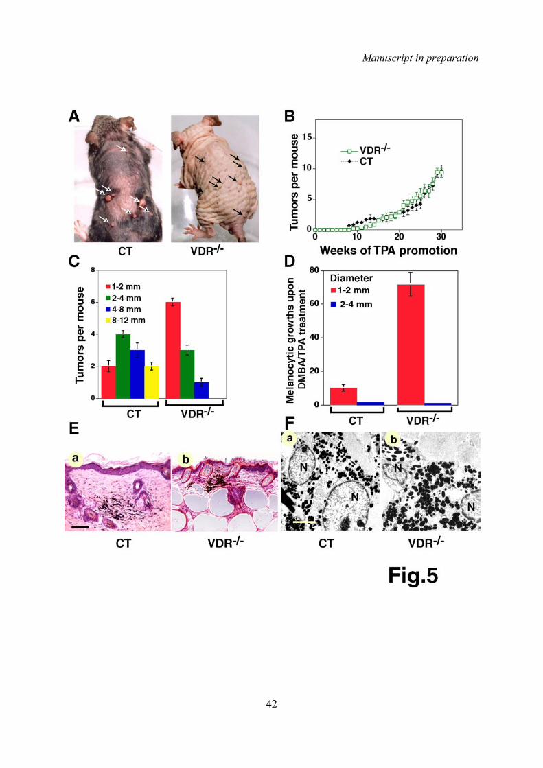
Manuscript in preparation
42

Manuscript in preparation
43
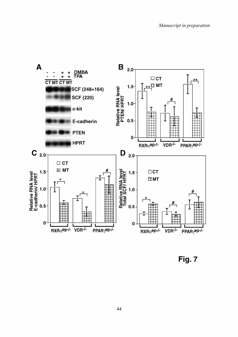
Manuscript in preparation
44

Manuscript in preparation
45

Results
44
2.2 Role of PPARα in skin carcinogenesis.
2.2.1 Introduction.
Peroxisome proliferator-activated receptors (PPARs) are ligand-activated transcription factors
that belong to the nuclear receptor superfamily (Keller et al., 1993; Wahli et al., 1999),.
Three PPAR isotypes, PPARα, PPARδ/β and PPARγ have been identified (Fajas et al., 1998;
Fajas et al., 1999; Issemann et al., 1993). PPARs are activated by the peroxisome
proliferators, fibrates, fatty acids and eicosanoids (Forman et al., 1997; Kliewer et al., 1997).
They modulate the transcription of a variety of target genes involved in lipid metabolism
(Wahli et al., 1999), and play a key role in the control of proliferation and apoptosis in
various cell types (Asou et al., 1999; Cattley, 2003; Hanley et al., 1998).
All PPAR isotypes are expressed in mouse epidermis during fetal development. Their
expression decreases progressively from the interfollicular epithelium after birth (Michalik et
al., 2001). In adult epidermis PPARα and PPARβ are the prominent isotypes, and it has been
shown that expression of PPARα and PPARβ is increased in adult epidermis after various
stimuli (Braissant et al., 1996; Braissant and Wahli, 1998; Michalik et al., 2001).
Several experimental approaches demonstrated that PPARs play key roles in skin
development and in the maintenance of skin homeostasis. PPARβ activators stimulate
differentiation of keratinocytes in culture (Tan et al., 2001) and in vivo (Schmuth et al.,
2004). Moreover, it has been shown that PPARβ plays key roles in skin wound healing,
keratinocyte response to inflammation and keratinocyte apoptosis (Di-Poi et al., 2002;
Michalik et al., 2001; Tan et al., 2001). Recently, Kim and co-workers demonstrated that
PPARβ null mice exhibited an increased susceptibility to DMBA/TPA-induced skin
carcinogenesis (Kim et al., 2004). PPARγ activators also stimulate differentiation of epidermal
keratinocytes in vivo, and improve permeability barrier recovery after mechanical disruption
(Mao-Qiang, et al. in press). Moreover, it was shown that PPARγ mediate the non-steroidal
anti-inflammatory drugs (NSAID) antineoplastic effect on human oral squamous carcinoma
cells (Nikitakis et al., 2002), and that PPARγ specific ligands can block melanoma cell growth
in culture (Mossner et al., 2002). As we show in section 2.1, PPARγep-/- mice, in which PPARγ
was selectively ablated in epidermal keratinocytes, exhibited an increased susceptibility to

Results
45
chemically- induced skin carcinogenesis, demonstrating a key role of PPARγ during
chemically-induced skin carcinogenesis.
PPARα ligands stimulate the differentiation of fetal epidermal permeability barrier (Hanley et
al., 1997), and possess receptor mediated, anti-inflammatory activities in both irritant and
allergic contact dermatitis animal models (Sheu et al., 2002). Moreover, PPARα activators
can reverse the expression of inflammatory cytokines induced by UVB in keratinocytes in
culture (Kippenberger et al., 2001). Interestingly, it has been shown that PPARα activators
partially inhibit tumor formation in a two-step chemical carcinogenesis protocol (Thuillier et
al., 2000), suggesting that PPARα, may also play an important role during skin
carcinogenesis. To investigate the possible role of PPARα in skin carcinogenesis, we
subjected PPARα null mice to a chemically-induced skin carcinogenesis protocol.
2.2.2 Material and methods.
Mice and carcinogenic treatment. PPARα null mice were previously described (Lee and
Gonzalez, 1996). PPARα null mutants and control wild-type (CT) animals were housed in a
temperature and humidity controlled room with 12h cycles of light and dark. 10-12 week-old
PPARα (n=10) and WT (n=10) mice were shaved to synchronize the hair cycle and treated
the day after with a single dose of 50 µg DMBA in 100 µl acetone. One week after the DMBA
application, 5 µg TPA (in 200 µl acetone) was applied topically twice a week. The number of
papillomas per mouse was weekly recorded.
2.2.3 Results
PPARα null mice exhibit an increased susceptibility to chemically-induced skin
tumors.
To determine whether PPARα is involved in skin carcinogenesis, we subjected PPARα null
mice to a two-step chemical carcinogenesis protocol. PPARα null and CT mice were treated
with a single dose of DMBA, followed by twice a week applications of TPA. Both DMBA/TPA-
treated PPARα null and CT mice developed papilloma-like tumors after 10 and 12 weeks of
TPA promotion, respectively (figure 14A). After 15 weeks of TPA promotion, all PPARα null
mice developed papillomas, whereas only 80% of CT mice exhibited papillomas. Moreover,
PPARα null mice developed two to three times more tumors than CT mice (figure 14B).
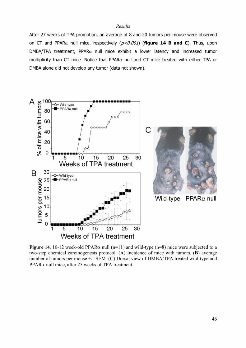
Results
46
After 27 weeks of TPA promotion, an average of 8 and 20 tumors per mouse were observed
on CT and PPARα null mice, respectively (p<0.001) (figure 14 B and C). Thus, upon
DMBA/TPA treatment, PPARα null mice exhibit a lower latency and increased tumor
multiplicity than CT mice. Notice that PPARα null and CT mice treated with either TPA or
DMBA alone did not develop any tumor (data not shown).
Figure 14. 10-12 week-old PPARα null (n=11) and wild-type (n=8) mice were subjected to atwo-step chemical carcinogenesis protocol. (A) Incidence of mice with tumors. (B) averagenumber of tumors per mouse +/- SEM. (C) Dorsal view of DMBA/TPA treated wild-type andPPARα null mice, after 25 weeks of TPA treatment.
Results
47
2.2.4 Perspectives.
To investigate whether PPARα null mice exhibit an enhanced response to TPA treatment, we
will compare the morphological and biochemcal alterations induced by topical application of
TPA to back skin of PPARα and WT mice, including epidermal thickness, keratinocyte
hyperproliferation and suprabasal expression of integrins α6β4. We have taken skin biopsies
from unchallenged PPARα null and WT mice, or PPARα null and WT mice treated either with
acetone or with two topical applications of TPA that we will use for histological and IHC
analysis.
In our previous study, both RXRαep-/- and PPARγep-/- mice exhibit increased suceptibility to
skin tumor formation and high frequency malignant conversion, in response to DMBA/TPA
treatment. To determine if PPARα plays a role in the malignant conversion of DMBA/TPA-
induced skin tumors, DMBA/TPA treated PPARα and CT mice will be sacrificed after 30 weeks
of TPA promotion, tumor biosies will be taken and analyzed by histology.
Moreover, we found that the increased susceptibility to papilloma and carcinoma formation
in RXRαep-/- and PPARγep-/- mice was correlated with a downregulation of the tumor
suppressor Pten, in the skin of these animals. Moreover, RXRαep-/- mice also exhibited
increased expression of SCF. To determine if the expression of Pten and SCF is also
deregulated in the skin of PPARα mice, we have taken skin biopsies from PPARα null and CT
mice and extracted the total RNA. We will measure the transcript levels of Pten and SCF in
these samples by real-time quantitative RT-PCR. Because PPARα is also expressed in other
cells types than keratinocytes, the generation of keratinocyte-specific PPARα mutant mice
will help to better understand its role during skin carcinogenesis.

Results
48
2.3 Interaction between RXRα and the Ha-ras oncogene during
mouse skin carcinogenesis
2.3.1 Introduction.
Retinoids, the active derivatives of vitamin A, are involved in the control of growth and
differentiation of normal, pre-malignant and malignant cell types (Altucci and Gronemeyer,
2001). Vitamin A deficiency in humans and animal models is associated with epithelial
squamous metaplasia (Hong and Itri, 1994; Moon et al., 1994). On the other hand, natural
and synthetic retinoids can efficiently block chemically-induced skin tumor formation
(Tennenbaum et al., 1998) and prevent malignant conversion in mice (Chen et al., 1994a;
Chen et al., 1995; De Luca et al., 1996), and are used as chemo preventive agents for
epithelial cancers in humans (Altucci and Gronemeyer, 2001; Lippman and Lotan, 2000;
Niles, 2000). Retinoids action is mediated by members of two families of nuclear receptors
(NRs), the retinoic acid receptors (RARα, β and γ) and the retinoic X receptors (RXRα, β and
γ) (Chambon, 1996; Mangelsdorf and Evans, 1995). Aberrant expression and function of
RARs and RXRs that may contribute to carcinoma development, have been found during skin
carcinogenesis, both in mouse (Darwiche et al., 1995; Darwiche et al., 1996) and human (Xu
et al., 2001). Previous studies from our laboratory demonstrated that selective ablation of
RXRα in epidermal keratinocytes during skin morphogenesis (RXRαep-/-(c) mice) (Li et al.,
2001b), or induced in keratinocytes of adult mice (RXRαep-/- mice) (Li et al., 2000), results in
extensive alopecia, epidermal hyperplasia, hair follicle degeneration, and skin inflammation
(Li et al., 2001b; Li et al., 2000), demonstrating a key role for RXRα in skin homeostasis.
Moreover, as shown in section 2.1, RXRαep-/- mice are more susceptible to chemically-
induced skin tumors than wild-type mice. Indeed, RXRαep-/- mice treated with DMBA/TPA
exhibit an increased epithelial tumor number, growth rate and incidence of malignant
conversion when compared to control mice. Furthermore, RXRαep-/- mice developed
malignant melanoma in response to a single topical application of DMBA. Thus, keratinocytic
RXRα has a suppressive effect on DMBA/TPA-induced skin tumorigenesis. Moreover, when
RXRα was ablated during the promotion stage of the two-step carcinogenesis protocol,
several weeks after the DMBA initiation, resulted in similar increase in tumor formation and
malignant conversion, indicating that keratinocytic RXRα plays a critical role during the
promotion stage of chemical carcinogenesis.

Results
49
As discussed in section 1.3.1, topical application of DMBA into mouse skin induces activation
mutations in the H-ras oncogene in keratinocytes, (Pazzaglia et al., 2001; Quintanilla et al.,
1986a). Mutation and activation of Ha-ras oncogene are critical events for the formation of
skin tumors. Indeed, Ha-ras null mice develop significantly less tumors than wild-type mice
in response to a DMBA/TPA protocol (Ise et al., 2000), and the tumors induced in Ha-ras null
mice exhibit mutations in the K-ras gene, suggesting that activation of the K-ras gene can
replace the Ha-ras activation at the initiation step (Ise et al., 2000). To further investigate
the role of RXRα in the development of skin cancer, an in particular in the Ha-ras mediated
carcinogenesis, we used the Tg.AC mouse model (described in section 1.3.2) as an
alternative model to the two-stage chemical skin carcinogenesis protocol. Tg.AC transgenic
mice carry an activated v-Ha-ras oncogene under the control of a partial ζ-globin promoter
(Leder et al., 1990), and are considered as “genetically pre-initiated mice”. Tg.AC mice
develop skin tumors in response to specific carcinogens and tumor promoters in a very rapid
and highly selective fashion (Battalora et al., 2001; Leder et al., 1990; Leder et al., 2002;
Tennant et al., 2001). The Tg.AC mouse has been used to identify carcinogens (Tennant et
al., 1995; Tennant et al., 1996) and for the study of genes involved in skin carcinogenesis
(Humble et al., 1998; Lan et al., 2000; Leder et al., 2002). To study the role of RXRα during
the promotion stage of Ha-ras-induced skin tumors, we generated Tg.AC/RXRαep-/-(c) and
Tg.AC/RXRαep-/- mice, carrying the Ha-ras oncogene and in which the selective ablation of
the RXRα alleles either occurred in epidermal keratinocytes during fetal epidermal
morphogenesis or was induced in adult mice, repectively.
2.3.2 Materials and methods.
Mice. K14-Cretg/o/RXRαL2/L2 (RXRαep-/-(c)) and K14-Cre-ERT2(tg/o)/RXRαL2/L2 (RXRαep-/-) mice, in
which RXRα alleles are selectively ablated in epidermal keratinocytes during embryonic
development or in adult mice, respectively, were described (Li et al., 2001b; Li et al., 2000).
Tg.AC transgenic mice, carrying an activated Ha-ras oncogene under the control of a partial
ζ-globin promoter (Leder et al., 1990), were purchased from Taconic. To selectively ablate
RXRα alleles in epidermal keratinocytes of Tg.AC mice during skin morphogenesis, K14-
Cretg/o/RXRαL2/L2 mice, carrying both RXRα floxed alleles and hemizygous for the K14-Cre
transgene, were crossed with Tg.AC mice hemizygous for the ζ-globin Ha-ras transgene. The
resulting Tg.AC/K14-Cretg/o/RXRαL2/+ and Tg.AC/RXRαL2/+ mice were intercrossed to generate
Tg.AC/K14-Cretg/o/RXRαL2/L2 (Tg.AC/RXRαep-/-(c)) and Tg.AC/RXRαL2/L2 control mice. To
Results
50
selectively ablate RXRα alleles in epidermal keratinocytes of adult Tg.AC mice, K14-Cre-
ERT2(tg/o)/RXRαL2/L2 mice were bred with hemizygous Tg.AC mice to generate Tg.AC/K14-Cre-
ERT2(tg/o)/RXRαL2/+ and Tg.AC/RXRαL2/+ mice. Breeding of Tg.AC/K14-Cre-ERT2(tg/o)/RXRαL2/+
with Tg.AC/RXRαL2/+ mice resulted in Tg.AC/K14-Cre-ERT2/RXRαL2/L2, carrying the Ha-ras and
the K14-Cre-ERT2 transgenes and both RXRα alleles floxed, and Tg.AC/RXRαL2/L2 mice. 6-7
week-old Tg.AC/K14-Cre-ERT2(tg/o)/RXRαL2/L2 females were injected intraperitonealy with 0.1
mg of tamoxifen for 5 consecutive days (Indra et al., 1999) to generate Tg.AC/RXRαep-/-
mice. Tg.AC/RXRαL2/L2 littermates were injected with the same dose of tamoxifen and used
as controls (CT) in all experiments. Mice were genotyped for the presence of K14-Cre and
K14-Cre-ERT2 transgenes and for the presense of wt, L2 or L- RXRα alleles by PCR as
described (Li et al., 2000). The presence of the ζ-globin Ha-ras transgene was detected by
PCR using primers directed to the SV40 polyadenylation sequence from the transgene (Fwd:
5’-AATTCTGAAGGAAAGTCC-3’, reverse: 5’-TGGACAAACTACCTACAG–3’). The Tg.AC
responder genotype was confirmed by Southern blotting (Kantz et al., 1999; Thompson et
al., 2001). DNA was digested with BamHI and probed with a radiolabeled 660 bp DNA
segment specific for the ζ-globin promoter of the transgene. The ζ-globin probe was
obtained by PCR amplification from genomic DNA of Tg.AC transgenic mice (ζ-globin
primers: fwd 5’-GACAGGGATGGCTCAGACAA–3’; reverse: 5’-CTGCTCGACAAGCTTTGATCT–3’).
TPA treatment. Mice were housed in a temperature and humidity controlled room with 12h
cycles of light and dark. Back skin of 8-9 week-old Tg.AC/RXRαep-/ and Tg.AC/RXRαL2/L2 mice
was shaved using electric clippers. One week after shaving, 5 µg of TPA (in 200 µl of
acetone) were applied topically twice a week to the back skin of the animals. TPA promotion
was continued for 13 weeks.
DMBA treatment. Back skin of 6-8 week-old RXRαep-/-(c) and RXRαL2/L2 mice was shaved
and one day after shaving, a single dose of 50 µg of DMBA in 100 µl acetone was topically
applied.
Histology and immunohistochemistry. Mice were anesthetized, skin and tumor biopsies
were surgically removed, fixed in 4% paraformaldehide over night at 4°C and embedded in
paraffin. 7 µm paraffin sections were stained with Hematoxylin and eosine for histological
analysis. For IHC analysis, paraffin sections were deparaffinized, permeabilized with PBS
Triton 0.1% for 2 min, blocked with 5% normal goat serum for 1h, incubated with a rabbit

Results
51
polyclonal anti-keratin 13 antibody (gift from Prof. S. Yuspa, NIH) o/n at 4°C and with a goat
anti-rabbit fluorescent secondary antibody for 1h and mounted with DAPI.
Real-Time RT-PCR. After surgical removal, skin biopsies were immediately frozen in liquid
nitrogen. RNA was extracted from skin biopsies with TrizolR Reagent (Gibco BRL, Life
technologies) following the manufacturer’s instructions. 3-5 µg of RNA were used for reverse
transcription reaction using Superscript II reverse transcriptase (Gibco BRL, Life
technologies) and 0.2 mM oligo dT in a final volume of 20 µl, following the manufacturer’s
instructions. cDNA preparations were diluted 1:10 or 1:20 in ultra pure (milliQ) water, and 2
µl of diluted cDNA were used for quantitative real-time PCR, using the Quantitect PCR
amplification kit (Qiagen) in a final reaction volume of 10 µl, in a LightCycler instrument
(Roche Diagnostics). To generate calibration curves, sequential dilutions of 3 µg of cDNA
from CT mice skin were prepared. cDNA was diluted 10, 100, 1000 and 10000 times. A
calibration curve with the 4 dilutions and the undiluted cDNA was generated for each
transcript. The relative levels of expression for each gene were normalized to HPRT. The
results represent the fold difference in the amount of transcript compared to the CT levels.
Data shown represent the average of 3 independent quantifications. The primers used for
RT-PCR were HPRT (s=5’-GTAATGATCAGTCAACGGGGGAC-3’; as=5’-
CCAGCAAGCTTGCAACCTTAACCA-3’), odc (s=5’-GACGATAAGGATGCGTTCTATGTT-3’;
as=5’-CTGGCAGCAT ACTTGATTTGAGA-3’), c-jun (s=5’-GAGCGGATCAAGGCAGAGAGGAAG;
as=5’-TGTGCCACCTGTTCCCTGAGC-3’), cox-2 (s=5‘-TCAAAACCGTGGGGAATGTAT-3’; as=5’-
AGGATGTAGTGCACTGTGTTT-3’), c-myc (s=5’-AAAACGACAAGAGGCG GACAC-3’; as=5’-
GCTTGTGCTCGTCTGCTTGAA-3’), IL1-RI (s=5’-CATCAAGGCAGTGGGAACTT-3’; as=5’-
ACTTTTGCAACACT TGGCCT-3’), IL1-RII (s=5’-GCATCATTGGGGTCAAGACT-3’; as=5’-
GATCCTCCC TTGTGACTGGA-3’) and icIL-1ra (s=5’-CTGGGTACTTACAAGGACC-3’; as=5’-CGT
GACTATAAGGGGCT-3’ ).

Results
52
2.3.3 Results.
Selective ablation of RXRα in epidermal keratinocytes of “genetically pre-
initiated” adult mice increases the susceptibility to TPA-induced skin
tumorigenesis.
To investigate whether selective ablation of RXRα in epidermal keratinocytes of Tg.AC mice
has an effect on the formation of TPA-induced tumors, we generated Tg.AC/RXRαep-/- mice,
bearing the Ha-ras oncogene and in which RXRα was selectively ablated in epidermal
keratinocytes at adult stage through tamoxifen (Tam) administration to 6-7 week-old
Tg.AC/K14-Cre-ERT2/RXRαL2/L2 mice. As previously seen in RXRαep-/- mice (Li et al., 2000),
Tg.AC/RXRαep-/- mice developed progressive alopecia, hair follicle degeneration and
epidermal hyperplasia starting 7-8 weeks after Tam administration (data not shown). To
induce skin tumors on Tg.AC/RXRαL2/L2 (CT) and Tg.AC/RXRαep-/- (MT) mice, TPA was
topically applied twice a week to back skin of 9-10 week-old CT and MT mice and tumor
appearance, number and size were recorded weekly. After 4 weeks of TPA treatment,
papilloma-like tumors appeared in 12% of CT and MT mice, and after 8 weeks of promotion
all mice developed tumors (fig 15A). However, MT mice developed two to three times more
tumors than their CT littermates (fig 15B), to the extend that after 13 weeks of TPA
treatment, CT and MT mice exhibited an average of 20 and 50 papilloma-like tumors,
respectively. Because of the large number of tumors developed in CT and MT mice in
response to TPA applications, the treatment was stopped after 13 weeks. Moreover, when
TPA promotion was started at the age of 23 weeks, CT mice exhibited a 2-3 fold increase in
tumor number and growth rate, when compared to CT mice in which the TPA treatment was
started at the age of 10 weeks (data not shown), in agreement with previous reports
showing that tumor incidence and multiplicity in Tg.AC mice in response to TPA-promotion
are higher in older animals (Battalora et al., 2001). However, under these conditions, MT
mice still developed two to three times more tumors than CT mice (data not shown).
Furthermore, irrespective of the age at which the TPA treatment was started, 62% (10 out
of 16) of MT mice developed more than 50 papillomas, whereas only 7% (1 out of 15) of CT
mice developed more than 50 tumors (data not shown).

Results
53
Figure 15. Selective ablation of RXRα in epidermal keratinocytes of adult Tg.AC mice increase thesusceptibility to TPA-induced skin tumorigenesis. 9-10 week-old Tg.AC/RXRαep-/- and Tg.AC/RXRαL2/L2 micewere treated twice a week with topical applications of TPA. (A) Percentage of mice with tumors. (B) Averagenumber of tumor per mouse +/- SEM. (C) Tumor size (length) distribution after 13 weeks of TPA treatment. Thetotal number of tumors per category is shown. (D) IHC analysis of K13 expression. High-risk papillomas fromMT (a) and CT (b) mice, and low-risk papilloma from CT mice (c). Scale bar 100 µm for a and b, 25 µm in c.(E) Hematoxylin-eosine staining of TPA-induced tumors from Tg.AC/RXRαL2/L2 and Tg.AC/RXRαep-/- mice,after 13 weeks of TPA treatment. (a) squamous papilloma from CT mice; (b) squamous papilloma withhyperplasia and (c) focal carcinoma from MT mice. Arrowhead in (b) points to an area of atypical hyperplasia.Arrow in (c) points to epidermal cells invading into the dermis. Scale bar 50 µm for a and b, 25 µm in c.
Results
54
The length of more than 99% of the tumors from both CT and MT mice in which TPA was
started at the age of 9-10 weeks, measured one week after the end of TPA treatment, varied
between 0.5 and 6 mm. Aproximately 1% of the tumors had a size between 6 and 8 mm in
both cases. No significant difference in tumor size distribution was observed between CT and
MT mice (Fig 15C, and data not shown). However, when the tumor size was measured 10
weeks after the end of TPA treatment, 3 MT mice exhibited tumors larger than 15 mm in
length, whereas in CT mice the tumors were never larger than 8 mm (data not shown).
Thus, tumors of MT mice grew at a much higher rate than those of CT mice after the TPA
treatment had been stopped.
To investigate whether ablation of RXRα in epidermal keratinocytes of Tg.AC mice has an
effect on the malignant conversion of TPA-induced skin tumors, tumor biopsies from CT and
MT mice, in which TPA was started at the age of 9-10 weeks, were removed 2 weeks after
the end of TPA treatment and analyzed by IHC. Keratin 13 (K13) was shown to be expressed
in papillomas with a “high risk” for malignant conversion, but not in low risk papillomas or
normal skin (Glick et al., 1993; Tennenbaum et al., 1996). 60% of the tumors from MT mice
exhibited large areas of K13 expression (figure 15Da and data not shown), whereas
only few tumors (less than 1%) from CT mice exhibited such large areas of K13 staining
(figure 15Db and data not shown). In more than 90% of tumors from CT mice K13
expression was either not detected, or in only a few cells (figure 15Dc). Histological
analysis revealed that most of the tumors from CT and MT mice were benign squamous
papillomas (figure 15E a and b), with few tumors exhibiting atypical basal cell hyperplasia
(figure 15Eb). 8% of the tumors from MT mice were identified as focal carcinoma (FC)
(figure 15Ec), whereas no such tumors were observed in CT mice. No evidence of
squamous cell carcinoma (SCC) or basal cell carcinoma (BCC) was found in the tumors from
either CT or MT mice (Table 6). Note that topical application of TPA to CT or MT mice did
not results in formation of melanocytic growths (MGs). This is in contrast to what occurs
when RXRαep-/- mice are subjected to a DMBA/TPA two-step chemical tumorigenesis protocol.
Indeed, initiation with a single application of DMBA, followed by twice a week applications of
TPA resulted in the formation of epithelial tumors and MGs in RXRαep-/- mice (see section
2.1).
Thus, ablation of RXRα in genetically pre-initiated adul mice results in an increased incidence
of TPA-induced epidermal tumors, which exhibit features of high-risk papillomas.

Results
55
Selective ablation of RXRα in keratinocytes of Tg.AC mice during skin
morphogenesis results in spontaneous papilloma formation and a high rate of
progression to malignancy.
To investigate the effect of RXRα ablation during skin morphogenesis in the tumor
susceptibility of Tg.AC mice, we generated Tg.AC/RXRαep-/-(c) mice, carrying the Ha-ras
oncogene and in which the RXRα alleles are selectively ablated in epidermal keratinocytes
early during skin formation, from Tg.AC/K14-Cre/RXRαL2/L2 mice. As reported for RXRαep-/-(c)
mice (Li et al., 2001b), Tg.AC/RXRαep-/-(c) mice exhibited a progressive alopecia, which
started in ventral skin, near the hind legs, and with time spreads over the whole body. Hair
loss begined at 4-5 weeks of age, and at the age of 16 weeks, all Tg.AC/RXRαep-/-(c) mice
exhibited a severe hair loss (60% loss in dorsal and 80% in ventral skin). Surprisingly,
papilloma-like tumors appeared spontaneously on the ventral skin of 12-13 week-old
Tg.AC/RXRαep-/-(c) mice (figure 16A d). By 16 weeks of age, all Tg.AC/RXRαep-/-(c) mice had
developed skin tumors (figure 16B), both on the ventral and dorsal skin (figure 16A e,f).
In contrast, no tumors appeared in Tg.AC/RXRαL2/L2 (figure 16A a and b) or RXRαep-/- mice
(Figure 16A c and table 6). The number of tumors in Tg.AC/RXRαep-/-(c) mice increased
with age, and an average of 16 tumors per mouse was observed in 26-28 week-old mice
(figure 16C). Some of the tumors from Tg.AC/RXRαep-/-(c) mice exhibited a high growth rate,
reaching more than 15 mm in length in 16-25 week-old mice (data not shown).
To investigate whether spontaneous skin tumors from Tg.AC/RXRαep-/-(c) mice progress to
malignancy, tumor biopsies were taken from 16-25 week-old mice. The IHC analysis of K13
expression revealed that all tumors from Tg.AC/RXRαep-/-(c) mice exhibited large areas with
high levels of K13 expression (figure 16D a and b), indicating that these tumors are at a
high risk of conversion into malignancy. Histological analysis showed that most of the
tumors from Tg.AC/RXRαep-/-(c) mice were benign squamous papillomas (figure 16Ea).
However, 11.5%, 7.7% and 3.8% of 20-25 week-old Tg.AC/RXRαep-/-(c) mice developed ISC
(figure 16Eb), FC (figure 16Ec) and SCC (data not shown), respectively (table 6).

Results
56
Figure 16. Selective ablation of RXRα in epidermal keratinocytes of Tg.AC mice during skin morphogenesisresults in formation of spontaneous skin tumors in adult mice. (A) ventral (a,c,d) an dorsal (b,e,f) view of 38week-old Tg.AC/RXRαL2/L2 (a,b), 20 week-old RXRαep-/-(c) (c) and 16 week-old (d) and 20 week-old (e,f)Tg.AC/RXRαep-/-(c) females; (B) Percentage of mice with spontaneous tumors. (C) The average number oftumors per mouse +/- SEM. (D) IHC analysis of K13 expression in tumors from Tg.AC/RXRαep-/-(c) mice. Scalebar 50 µm. (E) Hematoxylin-eosine staining of tumors from Tg.AC/RXRαep-/-(c) mice. (a) benign papilloma; (b)in situ carcinoma; (c) focal carcinoma. Arrows point to the basement membrane. Arrowheads in (c) point to cellsinvading into the dermis through the basement membrane. Scale bar 20 µm for a and b; 10 µm in c.
Results
57
These data demonstrate that selective ablation of RXRα in epidermal keratinocytes of
genetically pre-initiated mice during skin morphogenesis is sufficient to induce the formation
of high-risk papillomas at adult stage, that with time progress to malignancy in the absence
of any tumor promoter.
It has been shown that the formation of skin tumors in Tg.AC mice is correlated with the
transcriptional activation of the Ha-ras transgene in epidermal keratinocytes (Hansen et al.,
1995; Hansen and Tennant, 1994a). While Ha-ras transgene expression is not detected in
the skin of unchallenged Tg.AC mice, expression of the transgene is induced after topical
application of TPA, and is detected in all tumors induced in Tg.AC mice (Battalora et al.,
2001; Cannon et al., 1997; Hansen and Tennant, 1994a). Moreover, the induction of the Ha-
ras transgene expression and the sensitivity to tumor promotion in Tg.AC mice increase with
age (Battalora et al., 2001). To investigate whether RXRα ablation in keratinocytes of Tg.AC
mice during skin formation results in the transcriptional activation of the Ha-ras transgene,
we measured the levels of the Ha-ras transgene transcript in the skin of Tg.AC/RXRαep-/-(c)
mice 2, 4, 8 and 12 weeks after birth, by quantitative RT-PCR. No significant increase in the
expression levels of the Ha-ras transgene was observed in the skin of Tg.AC/RXRαep-/-(c)
mice, when compared to Tg.AC/RXRαL2/L2 mice (data not shown). This indicated that ablation
of RXRα in keratinocytes of Tg.AC mice during epidermal morphogenesis does not alter the
expression of the Ha-ras transgene post-natally.
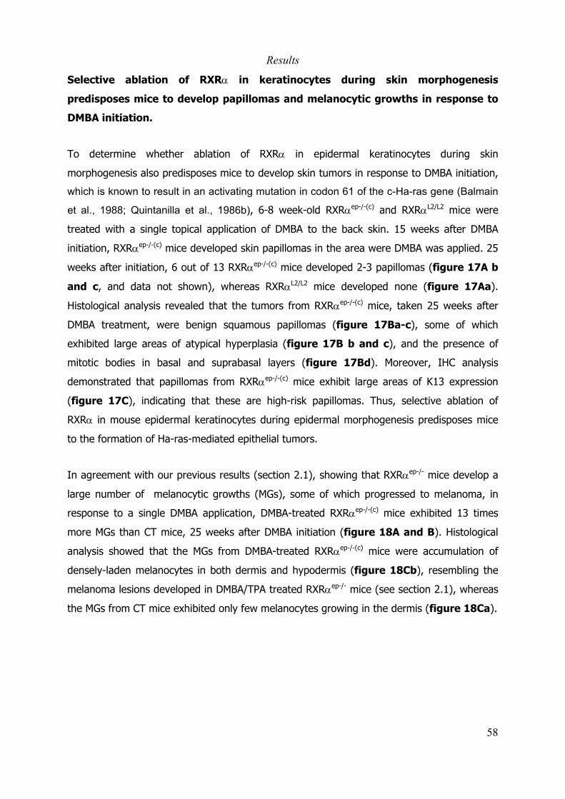
Results
58
Selective ablation of RXRα in keratinocytes during skin morphogenesis
predisposes mice to develop papillomas and melanocytic growths in response to
DMBA initiation.
To determine whether ablation of RXRα in epidermal keratinocytes during skin
morphogenesis also predisposes mice to develop skin tumors in response to DMBA initiation,
which is known to result in an activating mutation in codon 61 of the c-Ha-ras gene (Balmain
et al., 1988; Quintanilla et al., 1986b), 6-8 week-old RXRαep-/-(c) and RXRαL2/L2 mice were
treated with a single topical application of DMBA to the back skin. 15 weeks after DMBA
initiation, RXRαep-/-(c) mice developed skin papillomas in the area were DMBA was applied. 25
weeks after initiation, 6 out of 13 RXRαep-/-(c) mice developed 2-3 papillomas (figure 17A b
and c, and data not shown), whereas RXRαL2/L2 mice developed none (figure 17Aa).
Histological analysis revealed that the tumors from RXRαep-/-(c) mice, taken 25 weeks after
DMBA treatment, were benign squamous papillomas (figure 17Ba-c), some of which
exhibited large areas of atypical hyperplasia (figure 17B b and c), and the presence of
mitotic bodies in basal and suprabasal layers (figure 17Bd). Moreover, IHC analysis
demonstrated that papillomas from RXRαep-/-(c) mice exhibit large areas of K13 expression
(figure 17C), indicating that these are high-risk papillomas. Thus, selective ablation of
RXRα in mouse epidermal keratinocytes during epidermal morphogenesis predisposes mice
to the formation of Ha-ras-mediated epithelial tumors.
In agreement with our previous results (section 2.1), showing that RXRαep-/- mice develop a
large number of melanocytic growths (MGs), some of which progressed to melanoma, in
response to a single DMBA application, DMBA-treated RXRαep-/-(c) mice exhibited 13 times
more MGs than CT mice, 25 weeks after DMBA initiation (figure 18A and B). Histological
analysis showed that the MGs from DMBA-treated RXRαep-/-(c) mice were accumulation of
densely-laden melanocytes in both dermis and hypodermis (figure 18Cb), resembling the
melanoma lesions developed in DMBA/TPA treated RXRαep-/- mice (see section 2.1), whereas
the MGs from CT mice exhibited only few melanocytes growing in the dermis (figure 18Ca).

Results
59
Figure 17. Formation of skin papillomas in RXRαep-/-(c) mice in response to a single DMBAapplication. (A) Dorsal view of RXRαL2/L2 (a) and RXRαep-/-(c) (b,c) mice, 25 weeks after theapplication of DMBA. White arrowheads point to papillomas. (B) Hematoxylin-eosine staining oftumors from DMBA-treated RXRαep-/-(c) mice, 25 weeks after DMBA initiation. (a) benign papilloma,(b,c) benign papillomas with atypical hyperplasia (arrowheads). (d) higher magnification of apapilloma. Arrows point to basement membrane. Arrowheads in (d) point to mitotic figures. The insertis a higher magnification of a mitotic figure. Scale bar 50 µm in a; 25 µm in b and c; 10 µm in d. (C)IHC detection of keratin 13 expression in papillomas from RXRαep-/-(c) mice. Scale bar 50 µm.
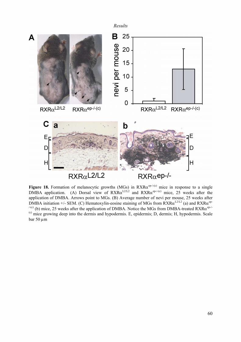
Results
60
Figure 18. Formation of melanocytic growths (MGs) in RXRαep-/-(c) mice in response to a singleDMBA application. (A) Dorsal view of RXRαL2/L2 and RXRαep-/-(c) mice, 25 weeks after theapplication of DMBA. Arrows point to MGs. (B) Average number of nevi per mouse, 25 weeks afterDMBA initiation +/- SEM. (C) Hematoxylin-eosine staining of MGs from RXRαL2/L2 (a) and RXRαep-
/-(c) (b) mice, 25 weeks after the application of DMBA. Notice the MGs from DMBA-treated RXRαep-/-
(c) mice growing deep into the dermis and hypodermis. E, epidermis; D, dermis; H, hypodermis. Scalebar 50 µm

Results
61Table 6. Summary of skin carcinogenesis in Tg.AC/RXRαep/- mice
Results
62
Skin alterations in Tg.AC mice ablated for RXRα in epidermal keratinocytes during
epidermal morphogenesis.
To determine whether skin alterations observed in Tg.AC/RXRαep-/-(c) mice are related to
those induced by topical application of TPA, skin biopsies from unchallenged Tg.AC/RXRαL2/L2
(CT) and Tg.AC/RXRαep-/- mice, CT and Tg.AC/RXRαep-/- mice treated with two topical
applications of TPA, and unchallenged Tg.AC/RXRαep-/-(c) mice were removed and prepared
for histological analysis. Topical application of TPA to skin of CT mice resulted in a 3-4 fold
increase in epidermal thickness, when compared to the skin of untreated CT mice.
Untreated 20-22 week-old Tg.AC/RXRαep-/- mice exhibited already a 2-fold increase in
epidermal thickness when compared to untreated CT mice, and in response to TPA
treatment, the epidermal thickness was further by 7-8 fold, when compared to untreated CT
mice. Interestingly, the epidermis of untreated Tg.AC/RXRαep-/-(c) mice at the age of 8 weeks,
was 3-4 fold thicker than that of Tg.AC/RXRαL2/L2 mice (figure 19A and B). Preliminary
results indicate that Tg.AC/RXRαep-/-(c) mice exhibit epidermal keratinocyte hyperproliferation,
similar to the keratinocyte hyperproliferation induced by topical application of TPA to the
back skin of Tg.AC/RXRαL2/L2 mice (not shown).
It has been shown that topical application of TPA to back skin of wild-type mice, results in
significant increase in the transcript levels of genes such as ornithine decarboxylase (odc), c-
jun , cyclooxygenase-2 (cox-2), c-myc, the IL-1 receptor type I (IL1-RI), and the intracellular
IL-1 receptor antagonist (icIL-1ra) (Chun et al., 2003; Kakar and Roy, 1994; Limtrakul Pn et
al., 2001). To examine whether ablation of RXRα in epidermal keratinocytes during
epidermal morphogenesis results in gene expression changes similar to those induced by
TPA application, we compared the transcript levels of these genes in the skin of
Tg.AC/RXRαep-/-(c) mice and TPA-treated Tg.AC/RXRαL2/L2 mice. As expected, topical
application of TPA to back skin of Tg.AC/RXRαL2/L2 mice resulted in significant increase in the
mRNA levels of odc, c-jun, cox-2, c-myc, icIL-1ra and IL1-RI, when compared to untreated
Tg.AC/RXRαL2/L2 mice (figure 19C). In contrast, no significant increase in the transcript
levels of these genes was observed in the skin of Tg.AC/RXRαep-/-(c) mice (figure 19C).
Thus, changes on gene expression induced by selective ablation of RXRα in keratinocytes
during skin development, leading to the formation of skin papillomas, appear to be different
from those induced by topical application of TPA to adult skin.

Results
63
Figure 19. Phenotypic changes in the skin of Tg.AC/RXRαep-/-(c) mice. (A) Hematoxylin-eosine stainedskin sections from 10 week-old Tg.AC/RXRαL2/L2 females non treated (a) or treated with two topicalapplications of TPA (b), and untreated 20 week-old Tg.AC/RXRαep-/- (c) and 8 week-oldTg.AC/RXRαep-/-(c) (d) females. Scale bar 20 µm (B) Quantification of epidermal thickness fromsamples described in (A). Results are the average epidermal thickness in µm +/- SEM. *** p < 0.001.(C) Quantitative RT-PCR analysis of mRNA levels in skin samples from 10 week-oldTg.AC/RXRαL2/L2 females non treated or treated with two topical applications of TPA, and untreated 8week-old Tg.AC-RXRαep-/-(c) females. (A). Data shown are the relative mRNA levels +/- SEM,normalized against HPRT, and represents the average of three independent quantifications on six miceper group. * p =0.1, *** p < 0.001.
Results
64
2.3.4 Discussion.
We have previously shown that ablation of RXRα in epidermal keratinocytes of adult mice
results in an increased susceptibility to DMBA/TPA-induced skin tumorigenesis (see section
2.1). Here we show that keratinocytic RXRα exerts a tumor suppressor effect during the
promotion stage of Ha-ras-mediated skin papillomas. Indeed, selective ablation of RXRα in
epidermal keratinocytes of genetically pre-initiated (Tg.AC) adult mice, carrying an activated
Ha-ras oncogene, results in increased skin tumor formation in response to TPA promotion,
and a higher incidence of high-risk papillomas.
Interestingly, selective ablation of RXRα in epidermal keratinocytes of Tg.AC mice during skin
morphogenesis (Tg.AC/RXRαep-/-(c) mice), is sufficient on its own, to induce the formation of
high-risk papillomas, which with time progress to malignancy. Furthermore, ablation of RXRα
in keratinocytes during epidermal morphogenesis (RXRαep-/-(c) mice), is sufficient for the
promotion of skin tumors in response to a single applicaiton of DMBA, which also exhibit
features of high-risk papillomas. Interestingly, in contrast to the 100% incidence of skin
tumors observed in 16 week-old Tg.AC/RXRαep-/-(c) mice, only 60% of RXRαep-/-(c) mice
developed skin tumors 25 weeks after DMBA initiation. Moreover, Tg.AC/RXRαep-/-(c) mice
developed an average of 16 tumors, whereas RXRαep-/-(c) mice developed only 2-3 tumors.
These differences might be explained by the fact that in Tg.AC/RXRαep-/-(c) mice all the
epidermal cells carry the activated Ha-ras oncogene, whereas in RXRαep-/- mice, DMBA
application may induce the activation mutations of the Ha-ras oncogene only in few
epidermal cells.
Thus, the cellular alterations resulting from the genetic ablation of RXRα in keratinocytes
during epidermal morphogenesis are probably sufficient to promote the clonal expansion of
Ha-ras initiated keratinocytes. In contrast, ablation of RXRα in keratinocytes of adult mice
most likely potentiates the tumor promotion effect of TPA, but is not sufficient on its own to
promote the clonal expansion of initiated keratinocytes. It has been suggested that
epidermal keratinocyte stem cells are the target of carcinogens and the precursors of skin
tumors in response to tumor promoters (Morris et al., 2000). Ablation of RXRα early during
epidermal morphogenesis may affect keratinocyte commitment, resulting in the generation
of keratinocytes with enhanced proliferative potential. In the presence of Ha-ras mutations,
RXRα null keratinocytes will clonaly expand and form tumors. Further studies are required to
Results
65
understand how ablation of RXRα in epidermal keratinocytes, early during development,
results in the promotion of Ha-ras mediated skin tumors.
2.3.5 Perspectives.
In section 2.1, we show that epithelial tumors induced by DMBA/TPA-treatment in RXRαep-/-
mice, progressed with high frequency to SCC after 30 weeks of TPA promotion. In this study,
application of TPA to Tg.AC/RXRαep-/- mice resulted in the formation of a large number of
papillomas after few weeks of TPA treatment. The promotion was interrupted after 13
weeks, and the mice were sacrificed and tumor biopsies collected 2 to 10 weeks after the
end of the treatment. Under this experimental conditions, TPA-induced tumors in
Tg.AC/RXRαep-/- mice did not progress to SCC, most likely because of the shortness of the
TPA treatment (only 13 weeks compared to 30 weeks in the DMBA/TPA protocol). To further
investigate if RXRα ablation in the epidermis of adult Tg.AC mice has an effect on the
incidence of progression to SCC, lower doses of TPA will be used for the promotion of skin
tumors in Tg.AC/RXRαep-/- mice, which may allow us to continue the TPA treatment for
longer than 13 weeks and assess the effect of RXRα ablation on the frequency of malignant
conversion. On the other hand, we found that the tumors from 20-25 week-old
Tg.AC/RXRαep-/-(c) mice progressed to SCC, but the frequency of progression was low.
However, it is likely that in older animals the frequency of conversion will increase. To
address this question, we have generated new groups of Tg.AC/RXRαep-/-(c) mice, from which
tumor biopsies will be taken and analyzed by histology, at 30, 35 and 40 weeks of age.
Similarly, we have generated new groups of RXRαep-/-(c) mice that have been treated with
DMBA, and from which tumor biopsies will be taken 30 and 35 weeks after DMBA initiation.
Finally, a detailed comparison of the cellular and molecular changes induced upon selective
ablation of RXRα in epidermal keratinocytes during embryonic development, or at the adult
stage is required to understand the mechanisms underlying the formation of spontaneous
papillomas and carcinomas in Tg.AC/RXRαep-/-(c) but not in Tg.AC/RXRαep-/- mice. To address
this question, we are collecting skin samples from Tg.AC/RXRαep-/-(c) and Tg.AC/RXRαep-/-
mice at different time points after birth and after tamoxifen administration, respectively, that
we will analyze by histology, immunohistochemistry, and real-time quantitative RT-PCR.

General Discussion
66
3. General Discussion
3.1 Keratinocytic RXRα and its heterodimeric partners as suppressors of
epithelial tumors.
RXRα is the main retinoic acid receptor in the epidermis (Reichrath et al., 1997), where itplays critical roles in the maintenance of skin homeostasis (Li et al., 2001b; Li et al., 2000).To investigate the possible role of RXRα during skin carcinogenesis, we took advantage of amouse model previously generated in the laboratory, in which RXRα alleles are selectivelyablated in epidermal keratinocytes of adult mice (RXRαep-/- mice). We demonstrate thatRXRαep-/- mice exhibit a higher susceptibility to develop papillomas and carcinomas thancontrol mice, in response to a DMBA/TPA treatment, indicating that keratinocytic RXRα has atumor suppressor effect on the formation of DMBA-TPA-induced skin tumors. Because RXRαis a heterodimeric partner for several non-steroidal NRs, including the VDR, PPARs and LXRs,the increased susceptibility to tumor formation and malignant conversion in RXRαep-/- mice,may result from alterations of more than one signaling pathway in keratinocytes. To identifythe heterodimeric partner(s) that may function, together with RXRα in keratinocytes, tosuppress the formation of chemically-induced epithelial tumors, we used mouse lines inwhich null mutations of various NRs have been introduced, either in the germ line orselectively in epidermal keratinocyte at the adult stage.
We found that, in response to DMBA/TPA treatment, PPARγep-/- mice exhibit highersusceptibility to the formation of papillomas and carcinomas than control mice. This dataindicate that in keratinocytes, RXRα/PPARγ heterodimers act as suppressors of chemically-induced epithelial tumorigenesis. PPARα null mice also exhibited increased incidence ofpapilloma formation compared to control mice, in response to DMBA/TPA treatment. BecausePPARα is also expressed in dermal fibroblasts (Billoni et al., 2000a), the possibility that thelack of PPARα in fibroblasts may contribute to the increased tumor incidence observed in thePPARα null mice, after DMBA/TPA treatment, can not be excluded. However, theobservations that PPARα expression is reactivated in mouse epidermal keratinocytes duringwound healing or after TPA treatment (Michalik et al., 2001), and that PPARα activatorsinduce apoptosis and decrease proliferation of keratinocytes in vivo (Komuves et al., 2000),supports the idea that keratinocytic RXRα/PPARα heterodimers may also play key roles assuppressors of epithelial tumor formation. We also show that VDR null mice exhibited similartumor incidence and multiplicity than WT mice upon DMBA/TPA treatment, indicating thatVDR does not play crucial roles during epithelial carcinogenesis.

General Discussion
67
It has been shown that initiated keratinocytes exhibit abnormal response to severaldifferentiation signals (Parkinson, 1985). In contrast to normal keratinocytes, in which TPAinduces differentiation, initiated cells respond to TPA treatment by undergoing proliferation,which results in the clonal expansion of the initiated cells and the subsequent formation ofsquamous papillomas (Karen et al., 1999). Accelerated keratinocyte differentiation in Cox-1null and Cox-2 null mice for example, resulted in a decreased incidence of skin tumorsinduced by DMBA/TPA treatment (Tiano et al., 2002). In contrast, resistance to Ca++induced differentiation in keratinocytes of transgenic mice overexpressing Cyclin D1 in theepidermis, resulted in increased tumor formation in response to DMBA/TPA (Yamamoto etal., 2002). We have previously shown that RXRαep-/- mice exhibited abnormal keratinocyteterminal differentiation, as indicated by the significant decrease in the expression of filaggrinin the suprabasal layers of the epidermis (Li et al., 2001b; Li et al., 2000). PPARα and PPARγplay important roles in the control of keratinocyte differentiation in vivo (Komuves et al.,2000). Thus, disruption of RXRα/PPARα or RXRα/PPARγ heterodimers in keratinocytes mayresult in impairment of keratinocyte differentiation, contributing to the increased tumorformation in response to DMBA/TPA treatment.
Stimulation of keratinocyte proliferation by TPA, during the promotion stage of the chemicalcarcinogenesis protocol, plays a key role in the formation of skin tumors. A single topicalapplication of TPA to mice skin results in a strong increase in keratinocyte proliferation in thebasal layer of the epidermis (Karen et al., 1999). Interestingly, it has been shown thatselective ablation of the tumor suppressor BRCA-1 in mice epidermis resulted in increasedkeratinocyte proliferation and increased susceptibility to DMBA/TPA induced tumors (Bertonet al., 2003). Similarly, overexpression of DP-1, a subunit of the E2F transcription factorfamily, in the basal keratinocytes of mice epidermis, results in increased keratinocyteproliferation and enhanced tumor formation in response to DMBA/TPA treatment (Wang etal., 2001). Ablation of RXRα or PPARγ in epidermal keratinocytes results in keratinocytehyperproliferation, which may also contribute to the increased susceptibility to tumorformation in response to DMBA/TPA treatment.
Interestingly, both RXRαep-/- and PPARγep-/- mice exhibited increase suprabasal expression ofα6β4 integrins in the epidermis and a decrease of Pten expression in the skin. Recently, ithas been demonstrated that targeted expression of α6β4 integrins in the suprabasal layersof transgenic mouse epidermis dramatically increased the frequency of papillomas,carcinomas and metastases induced by chemical carcinogenesis (Owens et al., 2003).Moreover, the selective ablation of Pten alleles in epidermal keratinocytes results indevelopment of epidermal hyperplasia and skin tumors (Suzuki et al., 2003). Thus, it is likelythat the disruption of the RXRα/PPARγ heterodimers in keratinocytes results in deregulationof α6β4 integrins and Pten expression, leading to an increased susceptibility to epithelialtumor formation in response to DMBA/TPA treatment. Recently, a functional PPAR responseelement has been identified in the Pten promoter (Patel et al., 2001), strongly supporting theidea that RXRα/PPARγ heterodimers may directly regulate Pten expression in keratinocytes.
General Discussion
68
When RXRα ablation was induced in mice epidermis 9 weeks after DMBA initiation, duringthe TPA promotion stage, mutant mice also exhibited higher tumor multiplicity, tumor growthrate and incidence of conversion into malignancy than control mice, indicating that RXRαplay minor roles during the initiation stage. Furthermore, the selective ablation of RXRαalleles in epidermal keratinocytes of “genetically pre-initiated” (Tg.AC) adult mice (Leder etal., 1990), also resulted in an increased susceptibility to TPA-induced skin tumors. This dataindicate that RXRα plays minor roles in the initiation stage, and suggest that the increasedsusceptibility to tumor formation in RXRαep-/- mice is due to alterations in the response toTPA promotion.
A striking observation was that selective ablation of RXRα alleles in epidermal keratinocytesof Tg.AC mice (Leder et al., 1990), during epidermal morphogenesis (Tg.AC/RXRαep-/-(c)
mice), resulted in the formation of spontaneous skin papillomas and carcinomas, in theabsence of any tumor promotion agent. Similarly, a single topical application of DMBA to theback skin of RXRαep-/-(c) mice, in which RXRα alleles were selectively ablated in epidermalkeratinocytes during epidermal morphogenesis, results in the spontaneous formation ofepithelial tumors. This results demonstrate that skin alterations induced by ablation of RXRαin keratinocytes during epidermal morphogenesis, are sufficient to promote the clonalexpansion of a population of initiated keratinocytes, carrying Ha-ras mutations, and for theformation and malignant progression of epithelial tumors. One possible explanation to thespontaneous formation of epithelial tumors in Tg.AC/RXRαep-/-(c) mice and in RXRαep-/-(c) micein response to a single DMBA topical application is that ablation of RXRα early duringepidermal morphogenesis may affect keratinocyte commitment, resulting in the generationof epidermal cells with enhanced proliferative potential, which in the presence of Ha-rasmutations will clonally expand and form tumors. Recently, it has been shown that micecarrying a null mutation of the phospholipase C delta1 (PLCδ1) develop progressive alopecia,epidermal hyperplasia and dermal cysts (Nakamura et al., 2003), resembling the phenotypeof RXRαep-/- mice. Interestingly, PLCδ1 null mice developed spontaneous skin tumors and theauthors suggest that PLCδ1 is required for skin stem cell lineage commitment. Anotherpossibility is that upon disruption of hair follicles in RXRαep-/-(c) mice, the keratinocyte stemcells from the bulge will migrate upwards to the interfollicular epidermis, where, exposed toa new microenvironmental stimuli, their proliferation rate will be increased, contributing tothe development of epithelial tumors. Several lines of evidence indicate that keratinocytesfrom the hair follicle contribute in a great extent to the development of epithelial tumors inresponse to DMBA/TPA treatment (Binder et al., 1997). Similarly, Hansen and co-workersshowed that TPA-induced papillomas in Tg.AC mice are mainly from follicular origin (Hansenand Tennant, 1994b). Further studies are required to elucidate the possible role of RXRα inthe commitment of keratinocytes during epidermal morphogenesis and in adult skin.Interestingly, PPARγep-/-(c) mice, in which PPARγ alleles are selectively ablated in keratinocytesduring epidermal morphogenesis (anexe I), or PPARα null or VDR null mice did notdeveloped epithelial tumors in response to a single topical application of DMBA, suggesting

General Discussion
69
that disruption of several RXRα/NR heterodimers may be required to promote the formationof epithelial tumors in response to single topical application of DMBA.
3.2 Keratinocytic RXRα and its heterodimeric partners as suppressors
of malignant melanoma.
In addition to epithelial tumors, RXRαep-/- mice developed a large number of melanocyticgrowths, which progress to malignant melanoma, in response to DMBA/TPA treatment. Incontrast to RXRαep-/- mice, PPARα null or PPARγep-/- mice did not develop melanocyticgrowths or melanoma, upon DMBA/TPA treatment, suggesting that these receptors are notinvolved in the formation of melanoma. Interestingly, DMBA/TPA-treated VDR null micedeveloped a large number of MGs, but in contrast to the MGs induced in RXRαep-/- byDMBA/TPA treatment, the MGs from VDR null mice did not degenerate into melanoma. Inmice, melanocytes are located mainly in the hair follicle. The observation that RXRαep-/- andVDR null mice (Li et al., 2000), but not in PPARα null or PPARγep-/- mice, developed aprogressive alopecia and hair follicle degeneration, suggests that degeneration of the hairfollicle is an important event for the formation of MGs. Upon disruption of the hair folliclemelanocytes might be release to the dermal compartment, where their growth anddifferentiation properties may change, becoming more susceptible to malignanttransformation. At the molecular level, a significant decrease in the expression levels of e-cadherin, an adhesion molecule that plays key roles in keratinocyte-melanocytecommunication (Jamal and Schneider, 2002), was observed in the skin of both RXRαep-/- andVDR null mice. It has been shown that down regulation of e-cadherin is associated withmelanoma progression (Li et al., 2001a), and that overexpression of e-cadherin in melanomacells restores the keratinocyte-mediated growth control (Hsu et al., 2000). Thus,degeneration of the hair follicle and loss of keratinocyte-melanocyte adhesion resulting fromthe disruption of RXRα/VDR heterodimers in keratinocytes may generate a permissiveenvironment for the growth of DMBA-initiated melanocytes. However, the observation thatMGs from RXRαep-/-, but not those of VDR mice progress to malignant melanoma, indicatethat additional alterations observed in RXRαep-/- but not in VDR null mice (ie down regulationof Pten), are required for the degeneration of these MG’s to melanoma. It has been shownthat down regulation of Pten is associated with melanoma development in human (Wu et al.,2003), and that in mice, invalidation of one Pten allele in a Inka/ARF∆2/3 background resultsin the development of malignant melanoma (You et al., 2002). We found that the levels ofPten mRNA were down regulated in the skin of RXRαep-/- mice, but not in that of VDR nullmice. These data suggest that degeneration of the hair follicle and decreased e-cadherinexpression in the skin may be sufficient for the formation of benign melanocytic growths,whereas Pten down regulation and/or other alterations might be required for their malignanttransformation.

General Discussion
70
3.3 General overview.
RXRs are heterodimeric partners for several hormone nuclear receptors including RARs, VDR,PPARs and LXRs. In the epidermis, RXRα is the major RXR isotype. As described in section2.1, selective ablation of RXRα in epidermal keratinocytes results in increased susceptibilityto chemical skin carcinogenesis (DMBA/TPA). RXRαep-/- mice exhibited an increased incidenceof epithelial tumor formation and malignant conversion than control mice. In addition,RXRαep-/- mice also developed melanoma in response to a single DMBA application. This isassociated to changes in gene expression including decrease of Pten and e-cadherin, andincrease in SCF and SB expression of α6β4 integrins. These alterations have been associatedto cancer development in humans and mouse models. The ablation of any singleheterodimeric partner for RXRα recapitulates all the alterations observed in RXRαep-/- mice,indicating that those alterations are the result of the disruption of more than one singlesignaling pathway. Interestingly, PPARα null and PPARγep-/- mice, but not VDR null mice,exhibited similar increased papilloma incidence, than RXRαep-/- mice, and PPARγep-/- mice alsoexhibited decrease in Pten expression and SB expression of α6β4 integrins. In contrast, VDRnull, but not PPARα null or PPARγep-/- mice exhibited hair follicle degeneration, decrease of e-cadherin expression and increased MGs (table 7). These data indicate that the twocarcinogenic events, development of papillomas and melanoma, observed in RXRαep-/-
mutants can be functionally separated. RXRα/PPARα and RXRα/PPARγ heterodimers seemto play major roles as suppressors of epithelial tumors, whereas keratinocytic RXRα/VDRheterodimers seem to play an important role in the control of melanocytes growth.Interestingly, the combined alterations observed in VDR null mice (hair follicle degeneration,down regulation of e-cadherin and formation of MG's) and in PPARγep-/- mice (downregulation of Pten, suprabasal expression of α6β4 integrins and increased papillomaformation) recapitulate almost all the alterations observed in RXRαep-/- mice.
Table 7. Summary of RXRα and its heterodimeric partners functions during skin carcinogenesis.
General Discussion
71
Thus, ablation of both PPARγ (or PPARα) and VDR in keratinocytes may completelyrecapitulate the phenotype observed in RXRαep-/- mice (table 7). However, the contributionof other heterodimeric partners of RXRα can not be discarded. The generation of micelacking PPARα or VDR selectively in epidermal keratinocytes, may help to elucidate theprecise role of this receptors during skin carcinogenesis.
We have also shown that ablation of RXRα in keratinocytes during epidermal morphogenesis,but not at adult stage, is sufficient for the promotion of epithelial tumors in response to asingle DMBA application. Interestingly, PPARγep-/-(c) mice, in which PPARγ was selectivelyablated in epidermal keratinocytes during epidermal morphogenesis, or PPARα null and VDRnull mice, in which null mutations of the PPARα and VDR genes have been introduced in thegerm line, respectively, did not develop epithelial tumors in response to a single topicalapplication of DMBA (table 7). We can hypothesize that the simultaneous ablation of morethan one RXRα heterodimeric partner, PPARα and PPARγ, PPARα and VDR or PPARγ andVDR for example, may also results in spontaneous development of skin tumors in responseto a single topical application of DMBA.
Conclusions and Perspectives
72
4. Conclusion and Perspectives.
Our data demonstrate that keratinocytic RXRα plays a key role during the promotion stage ofthe two-step chemical skin carcinogenesis, most likely by preventing the clonal expansion ofthe initiated cells. Keratinocytic RXRα/PPARγ heterodimers, and probablyRXRα/PPARα heterodimers, function as suppressors of DMBA/TPA-induced epithelial tumors.Moreover, keratinocytic RXRα plays key roles in the control of melanocyte growth andmelanoma formation in response to a single topical application of DMBA. Degeneration of thehair follicle and down regulation of Pten and e-cadherin resulting from the ablation ofkeratinocytic RXRα predispose for melanoma formation. Our results suggest thatkeratinocytic RXRα/VDR heterodimers control melanocyte growth, and disruption of theseheterodimers in keratinocytes results in the formation of MG's in response to a single DMBAapplication. However, the disruption of other RXRα/NR heterodimers may be required for thedegeneration of these MG's into melanoma. Thus, keratinocytic RXRα acts as a tumorsuppressor of chemically-induced melanoma and non-melanoma skin cancer in mice.
To determine whether PPARα and PPARγ may have overlapping and independent functionsduring chemically-induced skin carcinogenesis, we are establishing double PPARγep-/-/PPARαnull mice that will be subjected to a DMBA/TPA two-step carcinogenesis protocol. Moreover,generation of keratinocyte-specific PPARα mutant mice may help to better understand therole of keratinocytic PPARα during skin caricnogenesis. Similarly, because VDR is alsoexpressed in other cell types, including melanocytes, the generation of a keratinocyte-specific VDR knockout mice will help to clarify the function of keratinocytic RXRα/VDRheterodimers in the development of carcinoma and melanoma.
We have also shown that ablation of RXRα in keratinocytes during epidermal morphogenesisis sufficient to promote their clonal expansion in presence of an activated Ha-ras oncogeneand the formation of epithelial tumors. Our data suggest that RXRα may play critical roles inthe commitment of epidermal keratinocytes during development and that ablation of RXRαduring epidermal morphogenesis may results in the generation of keratinocytes withincreased proliferative potential.
So far, our studies using the chemically-induced carcinogenesis and Tg.AC mice are based onthe mutation and activation of the Ha-ras oncogene. It will be interesting to extend thisstudies to other models in which tumor formation is independent of the Ha-ras oncogene, forexample, using UV-induced carcinogenesis or Human Papillomavirus (HPV)-inducedcarcinogenesis. On the other hand, it will be interesting to investigate the possible role ofRXRα and its heterodimeric partners in the development of other epithelial cancers such ascervical, colon and prostate cancer. We have already established a collaboration with the

Conclusions and Perspectives
73
laboratories of Dr. Patricio Gariglio and Dr. Luis Covarrubias in Mexico to study the role ofRXRα in HPV-induced cervical cancer.
Our genetic approach has lead to the identification of NRs heterodimers that play specificfunctions during skin carcinogenesis, and we hope that it will provide the basis for thedevelopment of new anti-cancer strategies based in combinatory treatments, in whichinhibition or induction of a specific cellular process could be achieved by targeting a givenNRs heterodimer.
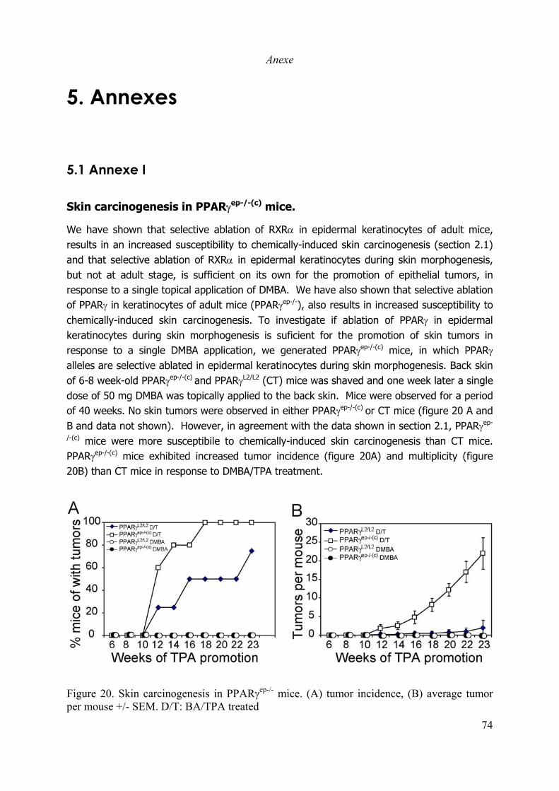
Anexe
74
5. Annexes
5.1 Annexe I
Skin carcinogenesis in PPARγep-/-(c) mice.
We have shown that selective ablation of RXRα in epidermal keratinocytes of adult mice,results in an increased susceptibility to chemically-induced skin carcinogenesis (section 2.1)and that selective ablation of RXRα in epidermal keratinocytes during skin morphogenesis,but not at adult stage, is sufficient on its own for the promotion of epithelial tumors, inresponse to a single topical application of DMBA. We have also shown that selective ablationof PPARγ in keratinocytes of adult mice (PPARγep-/-), also results in increased susceptibility tochemically-induced skin carcinogenesis. To investigate if ablation of PPARγ in epidermalkeratinocytes during skin morphogenesis is suficient for the promotion of skin tumors inresponse to a single DMBA application, we generated PPARγep-/-(c) mice, in which PPARγalleles are selective ablated in epidermal keratinocytes during skin morphogenesis. Back skinof 6-8 week-old PPARγep-/-(c) and PPARγL2/L2 (CT) mice was shaved and one week later a singledose of 50 mg DMBA was topically applied to the back skin. Mice were observed for a periodof 40 weeks. No skin tumors were observed in either PPARγep-/-(c) or CT mice (figure 20 A andB and data not shown). However, in agreement with the data shown in section 2.1, PPARγep-
/-(c) mice were more susceptibile to chemically-induced skin carcinogenesis than CT mice.PPARγep-/-(c) mice exhibited increased tumor incidence (figure 20A) and multiplicity (figure20B) than CT mice in response to DMBA/TPA treatment.
Figure 20. Skin carcinogenesis in PPARγep-/- mice. (A) tumor incidence, (B) average tumorper mouse +/- SEM. D/T: BA/TPA treated

Anexe
6. References
Aboulafia, D.M., Norris, D., Henry, D., Grossman, R.J., Thommes, J., Bundow, D., Yocum, R.C. and Stevens, V. (2003) 9-cis-retinoic acidcapsules in the treatment of AIDS-related Kaposi sarcoma: results of a phase 2 multicenter clinical trial. Arch Dermatol, 139,178-186.
Abremski, K. and Hoess, R. (1984) Bacteriophage P1 site-specific recombination. Purification and properties of the Cre recombinase protein.J Biol Chem, 259, 1509-1514. 1502: Austin S et al. A novel role for site-specifi...[PMID: 7026049]Related Articles, Links.
Adams, J.C. and Watt, F.M. (1990) Changes in keratinocyte adhesion during terminal differentiation: reduction in fibronectin bindingprecedes alpha 5 beta 1 integrin loss from the cell surface. Cell, 63, 425-435.
Adam-Stitah, S., Penna, L., Chambon, P. and Rochette-Egly, C. (1999) Hyperphosphorylation of the retinoid X receptor alpha by activated c-Jun NH2-terminal kinases. J Biol Chem, 274, 18932-18941.
Akiyama, M., Smith, L.T. and Shimizu, H. (2000) Changing patterns of localization of putative stem cells in developing human hair follicles.J Invest Dermatol, 114, 321-327.
Akutsu, N., Lin, R., Bastien, Y., Bestawros, A., Enepekides, D.J., Black, M.J. and White, J.H. (2001) Regulation of gene Expression by1alpha,25-dihydroxyvitamin D3 and Its analog EB1089 under growth-inhibitory conditions in squamous carcinoma Cells. MolEndocrinol, 15, 1127-1139. 1122: Prudencio J et al. Action of low calcemic 1121alpha...[PMID: 11353784]Related Articles,Links.
Aldaz, C.M., Conti, C.J., Gimenez, I.B., Slaga, T.J. and Klein-Szanto, A.J. (1985) Cutaneous changes during prolonged application of 12-O-tetradecanoylphorbol-13-acetate on mouse skin and residual effects after cessation of treatment. Cancer Res, 45, 2753-2759.
Ali, S., Metzger, D., Bornert, J.M. and Chambon, P. (1993) Modulation of transcriptional activation by ligand-dependent phosphorylation ofthe human oestrogen receptor A/B region. Embo J, 12, 1153-1160.
Alonso, L. and Fuchs, E. (2003) Stem cells of the skin epithelium. Proc Natl Acad Sci U S A, 100 Suppl 1, 11830-11835.Altucci, L. and Gronemeyer, H. (2001) The promise of retinoids to fight against cancer. Nat Rev Cancer, 1, 181-193.Amling, M., Priemel, M., Holzmann, T., Chapin, K., Rueger, J.M., Baron, R. and Demay, M.B. (1999) Rescue of the skeletal phenotype of
vitamin D receptor-ablated mice in the setting of normal mineral ion homeostasis: formal histomorphometric and biomechanicalanalyses. Endocrinology, 140, 4982-4987.
Andrews, R.C. and Walker, B.R. (1999) Glucocorticoids and insulin resistance: old hormones, new targets. Clin Sci (Lond), 96, 513-523.Aneskievich, B.J. and Fuchs, E. (1992) Terminal differentiation in keratinocytes involves positive as well as negative regulation by retinoic
acid receptors and retinoid X receptors at retinoid response elements. Mol Cell Biol, 12, 4862-4871.Anolik, J.H., Di Giovanna, J.J. and Gaspari, A.A. (1998) Effect of isotretinoin therapy on natural killer cell activity in patients with
xeroderma pigmentosum. Br J Dermatol, 138, 236-241.Aranda, A. and Pascual, A. (2001) Nuclear hormone receptors and gene expression. Physiol Rev, 81, 1269-1304.Arbeit, J.M., Munger, K., Howley, P.M. and Hanahan, D. (1994) Progressive squamous epithelial neoplasia in K14-human papillomavirus
type 16 transgenic mice. J Virol, 68, 4358-4368.Arch, J.R. (2002) Lessons in obesity from transgenic animals. J Endocrinol Invest, 25, 867-875.Archambault, M., Yaar, M. and Gilchrest, B.A. (1995) Keratinocytes and fibroblasts in a human skin equivalent model enhance melanocyte
survival and melanin synthesis after ultraviolet irradiation. J Invest Dermatol, 104, 859-867.Armstrong, B.K. and Kricker, A. (2001) The epidemiology of UV induced skin cancer. J Photochem Photobiol B, 63, 8-18.Aslam, F., McCabe, L., Frenkel, B., van Wijnen, A.J., Stein, G.S., Lian, J.B. and Stein, J.L. (1999) AP-1 and vitamin D receptor (VDR)
signaling pathways converge at the rat osteocalcin VDR element: requirement for the internal activating protein-1 site for vitaminD-mediated trans-activation. Endocrinology, 140, 63-70.
Asou, H., Verbeek, W., Williamson, E., Elstner, E., Kubota, T., Kamada, N. and Koeffler, H.P. (1999) Growth inhibition of myeloidleukemia cells by troglitazone, a ligand for peroxisome proliferator activated receptor gamma, and retinoids. Int J Oncol, 15,1027-1031.
Aszterbaum, M., Rothman, A., Johnson, R.L., Fisher, M., Xie, J., Bonifas, J.M., Zhang, X., Scott, M.P. and Epstein, E.H., Jr. (1998)Identification of mutations in the human PATCHED gene in sporadic basal cell carcinomas and in patients with the basal cellnevus syndrome. J Invest Dermatol, 110, 885-888.
Athar, M., Agarwal, R., Wang, Z.Y., Lloyd, J.R., Bickers, D.R. and Mukhtar, H. (1991) All-trans retinoic acid protects against conversion ofchemically induced and ultraviolet B radiation-induced skin papillomas to carcinomas. Carcinogenesis, 12, 2325-2329.
Attar, P.S., Wertz, P.W., McArthur, M., Imakado, S., Bickenbach, J.R. and Roop, D.R. (1997) Inhibition of retinoid signaling in transgenicmice alters lipid processing and disrupts epidermal barrier function. Mol Endocrinol, 11, 792-800.
Austin, S., Ziese, M. and Sternberg, N. (1981) A novel role for site-specific recombination in maintenance of bacterial replicons. Cell, 25,729-736. 723: Orban PC et al. Tissue- and site-specific DNA...[PMID: 1495975]Related Articles, Links.
Bagchi, I.C., Cheon, Y.P., Li, Q. and Bagchi, M.K. (2003) Progesterone receptor-regulated gene networks in implantation. Front Biosci, 8,s852-861.
Balmain, A., Brown, K., Akhurst, R.J. and Fee, F.M. (1988) Molecular analysis of chemical carcinogenesis in the skin. Br J Cancer Suppl, 9,72-75.
Banks, G.C., Deterding, L.J., Tomer, K.B. and Archer, T.K. (2001) Hormone-mediated dephosphorylation of specific histone H1 isoforms. JBiol Chem, 276, 36467-36473.
Banner, C.D., Gottlicher, M., Widmark, E., Sjovall, J., Rafter, J.J. and Gustafsson, J.A. (1993) A systematic analytical chemistry/cell assayapproach to isolate activators of orphan nuclear receptors from biological extracts: characterization of peroxisome proliferator-activated receptor activators in plasma. J Lipid Res, 34, 1583-1591.
Bardot, O., Aldridge, T.C., Latruffe, N. and Green, S. (1993) PPAR-RXR heterodimer activates a peroxisome proliferator response elementupstream of the bifunctional enzyme gene. Biochem Biophys Res Commun, 192, 37-45.
Barrandon, Y. and Green, H. (1987) Three clonal types of keratinocyte with different capacities for multiplication. Proc Natl Acad Sci U S A,84, 2302-2306.
AnexeBattalora, M.S., Spalding, J.W., Szczesniak, C.J., Cape, J.E., Morris, R.J., Trempus, C.S., Bortner, C.D., Lee, B.M. and Tennant, R.W.
(2001) Age-dependent skin tumorigenesis and transgene expression in the Tg.AC (v-Ha-ras) transgenic mouse. Carcinogenesis,22, 651-659.
Beato, M., Herrlich, P. and Schutz, G. (1995) Steroid hormone receptors: many actors in search of a plot. Cell, 83, 851-857.Benkoussa, M., Brand, C., Delmotte, M.H., Formstecher, P. and Lefebvre, P. (2002) Retinoic acid receptors inhibit AP1 activation by
regulating extracellular signal-regulated kinase and CBP recruitment to an AP1-responsive promoter. Mol Cell Biol, 22, 4522-4534.
Ben-Zimra, M., Koler, M., Melamed-Book, N., Arensburg, J., Payne, A.H. and Orly, J. (2002) Uterine and placental expression ofsteroidogenic genes during rodent pregnancy. Mol Cell Endocrinol, 187, 223-231.
Bernardi, R.J., Johnson, C.S., Modzelewski, R.A. and Trump, D.L. (2002) Antiproliferative effects of 1alpha,25-dihydroxyvitamin D(3) andvitamin D analogs on tumor-derived endothelial cells. Endocrinology, 143, 2508-2514.
Bernot, K.M., Coulombe, P.A. and McGowan, K.M. (2002) Keratin 16 expression defines a subset of epithelial cells during skinmorphogenesis and the hair cycle. J Invest Dermatol, 119, 1137-1149.
Bernstein, Z.P., Chanan-Khan, A., Miller, K.C., Northfelt, D.W., Lopez-Berestein, G. and Gill, P.S. (2002) A multicenter phase II study ofthe intravenous administration of liposomal tretinoin in patients with acquired immunodeficiency syndrome-associated Kaposi'ssarcoma. Cancer, 95, 2555-2561.
Berry, M., Metzger, D. and Chambon, P. (1990) Role of the two activating domains of the oestrogen receptor in the cell-type and promoter-context dependent agonistic activity of the anti-oestrogen 4-hydroxytamoxifen. Embo J, 9, 2811-2818.
Berton, T.R., Matsumoto, T., Page, A., Conti, C.J., Deng, C.X., Jorcano, J.L. and Johnson, D.G. (2003) Tumor formation in mice withconditional inactivation of Brca1 in epithelial tissues. Oncogene, 22, 5415-5426.
Bessou-Touya, S., Picardo, M., Maresca, V., Surleve-Bazeille, J.E., Pain, C. and Taieb, A. (1998) Chimeric human epidermal reconstructs tostudy the role of melanocytes and keratinocytes in pigmentation and photoprotection. J Invest Dermatol, 111, 1103-1108.
Bhattacharjee, R.N., Banks, G.C., Trotter, K.W., Lee, H.L. and Archer, T.K. (2001) Histone H1 phosphorylation by Cdk2 selectivelymodulates mouse mammary tumor virus transcription through chromatin remodeling. Mol Cell Biol, 21, 5417-5425.
Bickenbach, J.R. and Chism, E. (1998) Selection and extended growth of murine epidermal stem cells in culture. Exp Cell Res, 244, 184-195.Bickenbach, J.R. and Holbrook, K.A. (1987) Label-retaining cells in human embryonic and fetal epidermis. J Invest Dermatol, 88, 42-46.Bikle, D.D. (1995) 1,25(OH)2D3-regulated human keratinocyte proliferation and differentiation: basic studies and their clinical application.
J Nutr, 125, 1709S-1714S.Bikle, D.D. (1996) 1,25(OH)2D3-modulated calcium induced keratinocyte differentiation. J Investig Dermatol Symp Proc, 1, 22-27.Bikle, D.D., Nemanic, M.K., Gee, E. and Elias, P. (1986) 1,25-Dihydroxyvitamin D3 production by human keratinocytes. Kinetics and
regulation. J Clin Invest, 78, 557-566.Billoni, N., Buan, B., Gautier, B., Collin, C., Gaillard, O., Mahe, Y.F. and Bernard, B.A. (2000a) Expression of peroxisome proliferator
activated receptors (PPARs) in human hair follicles and PPAR alpha involvement in hair growth. Acta Derm Venereol, 80, 329-334.
Billoni, N., Buan, B., Gautier, B., Gaillard, O., Mahe, Y.F. and Bernard, B.A. (2000b) Thyroid hormone receptor beta1 is expressed in thehuman hair follicle. Br J Dermatol, 142, 645-652.
Billoni, N., Gautier, B., Mahe, Y.F. and Bernard, B.A. (1997) Expression of retinoid nuclear receptor superfamily members in human hairfollicles and its implication in hair growth. Acta Derm Venereol, 77, 350-355.
Binder, R.L., Gallagher, P.M., Johnson, G.R., Stockman, S.L., Smith, B.J., Sundberg, J.P. and Conti, C.J. (1997) Evidence that initiatedkeratinocytes clonally expand into multiple existing hair follicles during papilloma histogenesis in SENCAR mouse skin. MolCarcinog, 20, 151-158.
Birt, D.F., Yaktine, A. and Duysen, E. (1999) Glucocorticoid mediation of dietary energy restriction inhibition of mouse skin carcinogenesis.J Nutr, 129, 571S-574S.
Bittiner, B., Bleehen, S.S. and MacNeil, S. (1991) 1 alpha,25(OH)2 vitamin D3 increases intracellular calcium in human keratinocytes. Br JDermatol, 124, 230-235. 237: Kitano Y et al. Suppression of proliferation ...[PMID: 1907555]Related Articles, Links.
Blanco, J.C., Minucci, S., Lu, J., Yang, X.J., Walker, K.K., Chen, H., Evans, R.M., Nakatani, Y. and Ozato, K. (1998) The histone acetylasePCAF is a nuclear receptor coactivator. Genes Dev, 12, 1638-1651.
Blauer, M., Vaalasti, A., Pauli, S.L., Ylikomi, T., Joensuu, T. and Tuohimaa, P. (1991) Location of androgen receptor in human skin. J InvestDermatol, 97, 264-268.
Blumenberg, M., Connolly, D.M. and Freedberg, I.M. (1992) Regulation of keratin gene expression: the role of the nuclear receptors forretinoic acid, thyroid hormone, and vitamin D3. J Invest Dermatol, 98, 42S-49S.
Bo, W.J. (1956) The relationship between vitamin A deficiency and estrogen in producing uterine metaplasia in the rat. Anat Rec, 124, 619-627.
Bo, W.J. (1957) Relation of vitamin A deficiency and estrogen to induction of keratinizing metaplasia in the uterus of the rat. Am J ClinNutr, 5, 666-673.
Bodak, N., Queille, S., Avril, M.F., Bouadjar, B., Drougard, C., Sarasin, A. and Daya-Grosjean, L. (1999) High levels of patched genemutations in basal-cell carcinomas from patients with xeroderma pigmentosum. Proc Natl Acad Sci U S A, 96, 5117-5122.
Boehm, N., Samama, B., Cribier, B. and Rochette-Egly, C. (2004) Retinoic-acid receptor beta expression in melanocytes. Eur J Dermatol,14, 19-23.
Boelsterli, U.A. (2003) Animal models of human disease in drug safety assessment. J Toxicol Sci, 28, 109-121.Boldrini, L., Loggini, B., Gisfredi, S., Zucconi, Y., Baldinotti, F., Fogli, A., Simi, P., Cervadoro, G., Barachini, P., Basolo, F., Pingitore, R.
and Fontanini, G. (2003) Mutations of Fas (APO-1/CD95) and p53 genes in nonmelanoma skin cancer. J Cutan Med Surg, 7, 112-118.
Bollag, W. (1971) Therapy of chemically induced skin tumors of mice with vitamin A palmitate and vitamin A acid. Experientia, 27, 90-92.Bollag, W. (1972) Prophylaxis of chemically induced benign and malignant epithelial tumors by vitamin A acid (retinoic acid). Eur J
Cancer, 8, 689-693.Bollag, W. (1974) Therapeutic effects of an aromatic retinoic acid analog on chemically induced skin papillomas and carcinomas of mice.
Eur J Cancer, 10, 731-737.Bollag, W. (1975) Therapy of epithelial tumors with an aromatic retinoic acid analog. Chemotherapy, 21, 236-247.Bolshakov, S., Walker, C.M., Strom, S.S., Selvan, M.S., Clayman, G.L., El-Naggar, A., Lippman, S.M., Kripke, M.L. and Ananthaswamy,
H.N. (2003) p53 mutations in human aggressive and nonaggressive basal and squamous cell carcinomas. Clin Cancer Res, 9, 228-234.
Boonstra, J., Rijken, P., Humbel, B., Cremers, F., Verkleij, A. and van Bergen en Henegouwen, P. (1995) The epidermal growth factor. CellBiol Int, 19, 413-430.

AnexeBooth, B.A., Polak, K.L. and Uitto, J. (1980) Collagen biosynthesis by human skin fibroblasts. I. Optimization of the culture conditions for
synthesis of type I and type III procollagens. Biochim Biophys Acta, 607, 145-160.Bos, J.L. (1989) ras oncogenes in human cancer: a review. Cancer Res, 49, 4682-4689.Boudjelal, M., Voorhees, J.J. and Fisher, G.J. (2002) Retinoid signaling is attenuated by proteasome-mediated degradation of retinoid
receptors in human keratinocyte HaCaT cells. Exp Cell Res, 274, 130-137.Braakhuis, B.J., Klaassen, I., van der Leede, B.M., Cloos, J., Brakenhoff, R.H., Copper, M.P., Teerlink, T., Hendriks, H.F., van der Saag,
P.T. and Snow, G.B. (1997) Retinoid metabolism and all-trans retinoic acid-induced growth inhibition in head and necksquamous cell carcinoma cell lines. Br J Cancer, 76, 189-197.
Bradl, M., Klein-Szanto, A., Porter, S. and Mintz, B. (1991) Malignant melanoma in transgenic mice. Proc Natl Acad Sci U S A, 88, 164-168.
Braissant, O., Foufelle, F., Scotto, C., Dauca, M. and Wahli, W. (1996) Differential expression of peroxisome proliferator-activated receptors(PPARs): tissue distribution of PPAR-alpha, -beta, and -gamma in the adult rat. Endocrinology, 137, 354-366.
Braissant, O. and Wahli, W. (1998) Differential expression of peroxisome proliferator-activated receptor-alpha, -beta, and -gamma during ratembryonic development. Endocrinology, 139, 2748-2754. 2742: Braissant O et al. Differential expression of pe...[PMID:8536636]Related Articles, Links.
Brakebusch, C., Grose, R., Quondamatteo, F., Ramirez, A., Jorcano, J.L., Pirro, A., Svensson, M., Herken, R., Sasaki, T., Timpl, R., Werner,S. and Fassler, R. (2000) Skin and hair follicle integrity is crucially dependent on beta 1 integrin expression on keratinocytes.Embo J, 19, 3990-4003.
Brand, N., Petkovich, M., Krust, A., Chambon, P., de The, H., Marchio, A., Tiollais, P. and Dejean, A. (1988) Identification of a secondhuman retinoic acid receptor. Nature, 332, 850-853.
Brandenberger, A.W., Tee, M.K., Lee, J.Y., Chao, V. and Jaffe, R.B. (1997) Tissue distribution of estrogen receptors alpha (ER-alpha) andbeta (ER-beta) mRNA in the midgestational human fetus. J Clin Endocrinol Metab, 82, 3509-3512.
Braunstein, S., Kaplan, G., Gottlieb, A.B., Schwartz, M., Walsh, G., Abalos, R.M., Fajardo, T.T., Guido, L.S. and Krueger, J.G. (1994) GM-CSF activates regenerative epidermal growth and stimulates keratinocyte proliferation in human skin in vivo. J Invest Dermatol,103, 601-604.
Brecher, P.I. and Wotiz, H.H. (1969) Dissociation of estradiol from a uterine nuclear receptor. Endocrinology, 84, 718-726.Brindley, D.N. (1995) Role of glucocorticoids and fatty acids in the impairment of lipid metabolism observed in the metabolic syndrome. Int
J Obes Relat Metab Disord, 19 Suppl 1, S69-75.Broome Powell, M., Gause, P.R., Hyman, P., Gregus, J., Lluria-Prevatt, M., Nagle, R. and Bowden, G.T. (1999) Induction of melanoma in
TPras transgenic mice. Carcinogenesis, 20, 1747-1753.Brown, A.J., Dusso, A. and Slatopolsky, E. (1999) Vitamin D. Am J Physiol, 277, F157-175.Brown, A.J., Finch, J.L., Lopez-Hilker, S., Dusso, A., Ritter, C., Pernalete, N. and Slatopolsky, E. (1990) New active analogues of vitamin D
with low calcemic activity. Kidney Int Suppl, 29, S22-27.Brown, R., Gray, R.H. and Bernstein, I.A. (1985) Retinoids alter the direction of differentiation in primary cultures of cutaneous
keratinocytes. Differentiation, 28, 268-278.Brumbaugh, P.F. and Haussler, M.R. (1975) Specific binding of 1alpha,25-dihydroxycholecalciferol to nuclear components of chick
intestine. J Biol Chem, 250, 1588-1594.Bruna, A., Nicolas, M., Munoz, A., Kyriakis, J.M. and Caelles, C. (2003) Glucocorticoid receptor-JNK interaction mediates inhibition of the
JNK pathway by glucocorticoids. Embo J, 22, 6035-6044.Butt, T.R. and Walfish, P.G. (1996) Human nuclear receptor heterodimers: opportunities for detecting targets of transcriptional regulation
using yeast. Gene Expr, 5, 255-268.Byrne, C., Tainsky, M. and Fuchs, E. (1994) Programming gene expression in developing epidermis. Development, 120, 2369-2383.Caelles, C., Gonzalez-Sancho, J.M. and Munoz, A. (1997) Nuclear hormone receptor antagonism with AP-1 by inhibition of the JNK
pathway. Genes Dev, 11, 3351-3364.Cannon, R.E., Spalding, J.W., Trempus, C.S., Szczesniak, C.J., Virgil, K.M., Humble, M.C. and Tennant, R.W. (1997) Kinetics of wound-
induced v-Ha-ras transgene expression and papilloma development in transgenic Tg.AC mice. Mol Carcinog, 20, 108-114.Cario-Andre, M., Bessou, S., Gontier, E., Maresca, V., Picardo, M. and Taieb, A. (1999) The reconstructed epidermis with melanocytes: a
new tool to study pigmentation and photoprotection. Cell Mol Biol (Noisy-le-grand), 45, 931-942.Carr, B.R., Parker, C.R., Jr., Ohashi, M., MacDonald, P.C. and Simpson, E.R. (1983) Regulation of human fetal testicular secretion of
testosterone: low-density lipoprotein-cholesterol and cholesterol synthesized de novo as steroid precursor. Am J Obstet Gynecol,146, 241-247.
Cattley, R.C. (2003) Regulation of cell proliferation and cell death by peroxisome proliferators. Microsc Res Tech, 61, 179-184. 173: RobertsRA et al. PPAR alpha and the regulation...[PMID: 12505304]Related Articles, Links.
Chakravarti, D., LaMorte, V.J., Nelson, M.C., Nakajima, T., Schulman, I.G., Juguilon, H., Montminy, M. and Evans, R.M. (1996) Role ofCBP/P300 in nuclear receptor signalling. Nature, 383, 99-103.
Chambon, P. (1994) The retinoid signaling pathway: molecular and genetic analyses. Semin Cell Biol, 5, 115-125.Chambon, P. (1996) A decade of molecular biology of retinoic acid receptors. Faseb J, 10, 940-954.Chan, L.S. (1997) Human skin basement membrane in health and in autoimmune diseases. Front Biosci, 2, d343-352.Chang, C., Kokontis, J., Liao, S.S. and Chang, Y. (1989) Isolation and characterization of human TR3 receptor: a member of steroid receptor
superfamily. J Steroid Biochem, 34, 391-395.Chapellier, B., Mark, M., Messaddeq, N., Calleja, C., Warot, X., Brocard, J., Gerard, C., Li, M., Metzger, D., Ghyselinck, N.B. and
Chambon, P. (2002) Physiological and retinoid-induced proliferations of epidermis basal keratinocytes are differently controlled.Embo J, 21, 3402-3413.
Charles, M.A., Ryffel, G.U., Obinata, M., McCarthy, B.J. and Baxter, J.D. (1975) Nuclear receptors for thyroid hormone: evidence fornonrandom distribution within chromatin. Proc Natl Acad Sci U S A, 72, 1787-1791.
Chen, C.F., Goyette, P. and Lohnes, D. (2004) RARgamma acts as a tumor suppressor in mouse keratinocytes. Oncogene.Chen, C.H., Sakai, Y. and Demay, M.B. (2001) Targeting expression of the human vitamin D receptor to the keratinocytes of vitamin D
receptor null mice prevents alopecia. Endocrinology, 142, 5386-5389. 5314: Bikle DD et al. Vitamin D regulatedkeratinoc...[PMID: 12520529]Related Articles, Links.
Chen, D., Riedl, T., Washbrook, E., Pace, P.E., Coombes, R.C., Egly, J.M. and Ali, S. (2000) Activation of estrogen receptor alpha by S118phosphorylation involves a ligand-dependent interaction with TFIIH and participation of CDK7. Mol Cell, 6, 127-137.
Chen, H., Lin, R.J., Schiltz, R.L., Chakravarti, D., Nash, A., Nagy, L., Privalsky, M.L., Nakatani, Y. and Evans, R.M. (1997) Nuclearreceptor coactivator ACTR is a novel histone acetyltransferase and forms a multimeric activation complex with P/CAF andCBP/p300. Cell, 90, 569-580.
AnexeChen, J.D. and Evans, R.M. (1995) A transcriptional co-repressor that interacts with nuclear hormone receptors. Nature, 377, 454-457.Chen, L.C., Kirchhoff, S. and De Luca, L.M. (1994a) Effect of excess dietary retinoic acid on skin papilloma and carcinoma formation
induced by a complete carcinogenesis protocol in female Sencar mice. Cancer Lett, 78, 63-67.Chen, L.C., Sly, L. and De Luca, L.M. (1994b) High dietary retinoic acid prevents malignant conversion of skin papillomas induced by a
two-stage carcinogenesis protocol in female SENCAR mice. Carcinogenesis, 15, 2383-2386.Chen, L.C., Tarone, R., Huynh, M. and De Luca, L.M. (1995) High dietary retinoic acid inhibits tumor promotion and malignant conversion
in a two-stage skin carcinogenesis protocol using 7,12-dimethylbenz[a]anthracene as the initiator and mezerein as the tumorpromoter in female SENCAR mice. Cancer Lett, 95, 113-118.
Chida, K., Hashiba, H., Fukushima, M., Suda, T. and Kuroki, T. (1985) Inhibition of tumor promotion in mouse skin by 1 alpha,25-dihydroxyvitamin D3. Cancer Res, 45, 5426-5430.
Chin, L., Pomerantz, J., Polsky, D., Jacobson, M., Cohen, C., Cordon-Cardo, C., Horner, J.W., 2nd and DePinho, R.A. (1997) Cooperativeeffects of INK4a and ras in melanoma susceptibility in vivo. Genes Dev, 11, 2822-2834.
Chu, P.G. and Weiss, L.M. (2002) Keratin expression in human tissues and neoplasms. Histopathology, 40, 403-439.Chun, K.S., Keum, Y.S., Han, S.S., Song, Y.S., Kim, S.H. and Surh, Y.J. (2003) Curcumin inhibits phorbol ester-induced expression of
cyclooxygenase-2 in mouse skin through suppression of extracellular signal-regulated kinase activity and NF-kappaB activation.Carcinogenesis, 24, 1515-1524.
Clark, W.H., Jr. (1991) Human cutaneous malignant melanoma as a model for cancer. Cancer Metastasis Rev, 10, 83-88.Cockerell, C.J. (2000) Histopathology of incipient intraepidermal squamous cell carcinoma ("actinic keratosis"). J Am Acad Dermatol, 42,
11-17.Conneely, O.M., Mulac-Jericevic, B., DeMayo, F., Lydon, J.P. and O'Malley, B.W. (2002) Reproductive functions of progesterone receptors.
Recent Prog Horm Res, 57, 339-355.Cooper, M.P., Klaassen, I., Brakenhoff, R.H., Cloos, J., Snow, G.B. and Braakhuis, B.J. (1997) All-trans retinoic acid induced gene
expression and growth inhibition in head and neck cancer cell lines. Oral Oncol, 33, 270-274.Cotsarelis, G., Kaur, P., Dhouailly, D., Hengge, U. and Bickenbach, J. (1999) Epithelial stem cells in the skin: definition, markers,
localization and functions. Exp Dermatol, 8, 80-88.Cotsarelis, G., Sun, T.T. and Lavker, R.M. (1990) Label-retaining cells reside in the bulge area of pilosebaceous unit: implications for
follicular stem cells, hair cycle, and skin carcinogenesis. Cell, 61, 1329-1337.D'Ambrosio, S.M., Slazinski, L., Whetstone, J.W. and Lowney, E. (1981) Excision repair of UV-induced pyrimidine dimers in human skin in
vivo. J Invest Dermatol, 77, 311-313.Danielsson, C., Fehsel, K., Polly, P. and Carlberg, C. (1998) Differential apoptotic response of human melanoma cells to 1 alpha,25-
dihydroxyvitamin D3 and its analogues. Cell Death Differ, 5, 946-952.Danielsson, C., Mathiasen, I.S., James, S.Y., Nayeri, S., Bretting, C., Hansen, C.M., Colston, K.W. and Carlberg, C. (1997) Sensitive
induction of apoptosis in breast cancer cells by a novel 1,25- dihydroxyvitamin D3 analogue shows relation to promoterselectivity. J Cell Biochem, 66, 552-562.
Danielsson, C., Torma, H., Vahlquist, A. and Carlberg, C. (1999) Positive and negative interaction of 1,25-dihydroxyvitamin D3 and theretinoid CD437 in the induction of human melanoma cell apoptosis. Int J Cancer, 81, 467-470.
Darwiche, N., Celli, G., Tennenbaum, T., Glick, A.B., Yuspa, S.H. and De Luca, L.M. (1995) Mouse skin tumor progression results indifferential expression of retinoic acid and retinoid X receptors. Cancer Res, 55, 2774-2782.
Darwiche, N., Scita, G., Jones, C., Rutberg, S., Greenwald, E., Tennenbaum, T., Collins, S.J., De Luca, L.M. and Yuspa, S.H. (1996) Loss ofretinoic acid receptors in mouse skin and skin tumors is associated with activation of the ras(Ha) oncogene and high risk forpremalignant progression. Cancer Res, 56, 4942-4949.
de Gruijl, F.R. (1999) Skin cancer and solar UV radiation. Eur J Cancer, 35, 2003-2009.de Gruijl, F.R., van Kranen, H.J. and Mullenders, L.H. (2001) UV-induced DNA damage, repair, mutations and oncogenic pathways in skin
cancer. J Photochem Photobiol B, 63, 19-27.De Luca, L.M., Sly, L., Jones, C.S. and Chen, L.C. (1993) Effects of dietary retinoic acid on skin papilloma and carcinoma formation in
female SENCAR mice. Carcinogenesis, 14, 539-542.De Luca, L.M., Tarone, R., Huynh, M., Jones, C.S. and Chen, L.C. (1996) Dietary retinoic acid inhibits mouse skin carcinogenesis
irrespective of age at initiation. Nutr Cancer, 25, 249-257.De Luca, M., D'Anna, F., Bondanza, S., Franzi, A.T. and Cancedda, R. (1988) Human epithelial cells induce human melanocyte growth in
vitro but only skin keratinocytes regulate its proper differentiation in the absence of dermis. J Cell Biol, 107, 1919-1926.Delerive, P., De Bosscher, K., Besnard, S., Vanden Berghe, W., Peters, J.M., Gonzalez, F.J., Fruchart, J.C., Tedgui, A., Haegeman, G. and
Staels, B. (1999) Peroxisome proliferator-activated receptor alpha negatively regulates the vascular inflammatory gene responseby negative cross-talk with transcription factors NF-kappaB and AP-1. J Biol Chem, 274, 32048-32054.
DiGiovanna, J.J. (2001) Retinoid chemoprevention in patients at high risk for skin cancer. Med Pediatr Oncol, 36, 564-567.DiPersio, C.M., van der Neut, R., Georges-Labouesse, E., Kreidberg, J.A., Sonnenberg, A. and Hynes, R.O. (2000) alpha3beta1 and
alpha6beta4 integrin receptors for laminin-5 are not essential for epidermal morphogenesis and homeostasis during skindevelopment. J Cell Sci, 113 ( Pt 17), 3051-3062.
Di-Poi, N., Tan, N.S., Michalik, L., Wahli, W. and Desvergne, B. (2002) Antiapoptotic role of PPARbeta in keratinocytes via transcriptionalcontrol of the Akt1 signaling pathway. Mol Cell, 10, 721-733. 710: Mossner R et al. Agonists of peroxisome prolif...[PMID:12230498]Related Articles, Links.
DiSepio, D., Sutter, M., Johnson, A.T., Chandraratna, R.A. and Nagpal, S. (1999) Identification of the AP1-antagonism domain of retinoicacid receptors. Mol Cell Biol Res Commun, 1, 7-13.
Domann, F.E., Jr., Levy, J.P., Finch, J.S. and Bowden, G.T. (1994) Constitutive AP-1 DNA binding and transactivating ability of malignantbut not benign mouse epidermal cells. Mol Carcinog, 9, 61-66.
Donatien, P., Surleve-Bazeille, J.E., Thody, A.J. and Taieb, A. (1993) Growth and differentiation of normal human melanocytes in a TPA-free, cholera toxin-free, low-serum medium and influence of keratinocytes. Arch Dermatol Res, 285, 385-392.
Dreyer, C., Keller, H., Mahfoudi, A., Laudet, V., Krey, G. and Wahli, W. (1993) Positive regulation of the peroxisomal beta-oxidationpathway by fatty acids through activation of peroxisome proliferator-activated receptors (PPAR). Biol Cell, 77, 67-76.
Dreyer, C., Krey, G., Keller, H., Givel, F., Helftenbein, G. and Wahli, W. (1992) Control of the peroxisomal beta-oxidation pathway by anovel family of nuclear hormone receptors. Cell, 68, 879-887. 872: Issemann I et al. The peroxisome proliferator-a...[PMID:8240670]Related Articles, Links.
Driscoll, J.E., Seachord, C.L., Lupisella, J.A., Darveau, R.P. and Reczek, P.R. (1996) Ligand-induced conformational changes in the humanretinoic acid receptor detected using monoclonal antibodies. J Biol Chem, 271, 22969-22975.
AnexeDurand, B., Saunders, M., Gaudon, C., Roy, B., Losson, R. and Chambon, P. (1994) Activation function 2 (AF-2) of retinoic acid receptor
and 9-cis retinoic acid receptor: presence of a conserved autonomous constitutive activating domain and influence of the nature ofthe response element on AF-2 activity. Embo J, 13, 5370-5382.
Eckert, R.L., Crish, J.F. and Robinson, N.A. (1997) The epidermal keratinocyte as a model for the study of gene regulation and celldifferentiation. Physiol Rev, 77, 397-424.
Egner, U., Heinrich, N., Ruff, M., Gangloff, M., Mueller-Fahrnow, A. and Wurtz, J.M. (2001) Different ligands-different receptorconformations: modeling of the hER alpha LBD in complex with agonists and antagonists. Med Res Rev, 21, 523-539.
Eichner, R. (1986) Epidermal effects of retinoids: in vitro studies. J Am Acad Dermatol, 15, 789-797. 783: Magnaldo T et al. Expression ofloricrin is neg...[PMID: 1378029]Related Articles, Links.
Eichner, R., Kahn, M., Capetola, R.J., Gendimenico, G.J. and Mezick, J.A. (1992) Effects of topical retinoids on cytoskeletal proteins:implications for retinoid effects on epidermal differentiation. J Invest Dermatol, 98, 154-161.
Einspahr, J.G., Alberts, D.S., Warneke, J.A., Bozzo, P., Basye, J., Grogan, T.M., Nelson, M.A. and Bowden, G.T. (1999) Relationship of p53mutations to epidermal cell proliferation and apoptosis in human UV-induced skin carcinogenesis. Neoplasia, 1, 468-475.
El Abdaimi, K., Papavasiliou, V., Rabbani, S.A., Rhim, J.S., Goltzman, D. and Kremer, R. (1999) Reversal of hypercalcemia with thevitamin D analogue EB1089 in a human model of squamous cancer. Cancer Res, 59, 3325-3328.
Elder, J.T., Astrom, A., Pettersson, U., Tavakkol, A., Krust, A., Kastner, P., Chambon, P. and Voorhees, J.J. (1992) Retinoic acid receptorsand binding proteins in human skin. J Invest Dermatol, 98, 36S-41S.
Elias, P.M., Cullander, C., Mauro, T., Rassner, U., Komuves, L., Brown, B.E. and Menon, G.K. (1998) The secretory granular cell: theoutermost granular cell as a specialized secretory cell. J Investig Dermatol Symp Proc, 3, 87-100.
Ellis, C.N., Varani, J., Fisher, G.J., Zeigler, M.E., Pershadsingh, H.A., Benson, S.C., Chi, Y. and Kurtz, T.W. (2000) Troglitazone improvespsoriasis and normalizes models of proliferative skin disease: ligands for peroxisome proliferator-activated receptor-gammainhibit keratinocyte proliferation. Arch Dermatol, 136, 609-616.
Enepekides, D.J., Black, M.J. and White, J.H. (1999) The independent and combined effects of RAR-, RXR-, and VDR-selective ligands onthe growth of squamous cell carcinoma in vitro. J Otolaryngol, 28, 83-89.
Evans, C.B., Pillai, S. and Goldyne, M.E. (1993) Endogenous prostaglandin E2 modulates calcium-induced differentiation in human skinkeratinocytes. Prostaglandins Leukot Essent Fatty Acids, 49, 777-781.
Evans, S.R., Houghton, A.M., Schumaker, L., Brenner, R.V., Buras, R.R., Davoodi, F., Nauta, R.J. and Shabahang, M. (1996) Vitamin Dreceptor and growth inhibition by 1,25-dihydroxyvitamin D3 in human malignant melanoma cell lines. J Surg Res, 61, 127-133.
Fajas, L., Fruchart, J.C. and Auwerx, J. (1998) PPARgamma3 mRNA: a distinct PPARgamma mRNA subtype transcribed from anindependent promoter. FEBS Lett, 438, 55-60.
Fajas, L., Schoonjans, K., Gelman, L., Kim, J.B., Najib, J., Martin, G., Fruchart, J.C., Briggs, M., Spiegelman, B.M. and Auwerx, J. (1999)Regulation of peroxisome proliferator-activated receptor gamma expression by adipocyte differentiation and determination factor1/sterol regulatory element binding protein 1: implications for adipocyte differentiation and metabolism. Mol Cell Biol, 19, 5495-5503. 5494: Wahli W et al. Fatty acids, eicosanoids, and...[PMID: 10086196]Related Articles, Links.
Fang, S., Anderson, K.M. and Liao, S. (1969) Receptor proteins for androgens. On the role of specific proteins in selective retention of 17-beta-hydroxy-5-alpha-androstan-3-one by rat ventral prostate in vivo and in vitro. J Biol Chem, 244, 6584-6595.
Fanjul, A., Dawson, M.I., Hobbs, P.D., Jong, L., Cameron, J.F., Harlev, E., Graupner, G., Lu, X.P. and Pfahl, M. (1994) A new class ofretinoids with selective inhibition of AP-1 inhibits proliferation. Nature, 372, 107-111.
Fawell, S.E., Lees, J.A., White, R. and Parker, M.G. (1990) Characterization and colocalization of steroid binding and dimerization activitiesin the mouse estrogen receptor. Cell, 60, 953-962.
Feil, P.D. and Bardin, C.W. (1975) Cytoplasmic and nuclear progesterone receptors in the guinea pig uterus. Endocrinology, 97, 1398-1407.Feil, R., Brocard, J., Mascrez, B., LeMeur, M., Metzger, D. and Chambon, P. (1996) Ligand-activated site-specific recombination in mice.
Proc Natl Acad Sci U S A, 93, 10887-10890.Feil, R., Wagner, J., Metzger, D. and Chambon, P. (1997) Regulation of Cre recombinase activity by mutated estrogen receptor ligand-
binding domains. Biochem Biophys Res Commun, 237, 752-757.Feingold, K.R., Man, M.Q., Menon, G.K., Cho, S.S., Brown, B.E. and Elias, P.M. (1990) Cholesterol synthesis is required for cutaneous
barrier function in mice. J Clin Invest, 86, 1738-1745.Feng, X., Peng, Z.H., Di, W., Li, X.Y., Rochette-Egly, C., Chambon, P., Voorhees, J.J. and Xiao, J.H. (1997) Suprabasal expression of a
dominant-negative RXR alpha mutant in transgenic mouse epidermis impairs regulation of gene transcription and basalkeratinocyte proliferation by RAR-selective retinoids. Genes Dev, 11, 59-71.
Fernandez-Mejia, C., Medina-Martinez, O., Martinez-Perez, L. and Goodman, P.A. (1999) The human insulin gene contains multipletranscriptional elements that respond to glucocorticoids. Pancreas, 18, 336-341.
Ferraris, C., Bernard, B.A. and Dhouailly, D. (1997) Adult epidermal keratinocytes are endowed with pilosebaceous forming abilities. Int JDev Biol, 41, 491-498.
Finch, J.S., Albino, H.E. and Bowden, G.T. (1996) Quantitation of early clonal expansion of two mutant 61st codon c-Ha- ras alleles inDMBA/TPA treated mouse skin by nested PCR/RFLP. Carcinogenesis, 17, 2551-2557.
Fisher, G.J., Talwar, H.S., Xiao, J.H., Datta, S.C., Reddy, A.P., Gaub, M.P., Rochette-Egly, C., Chambon, P. and Voorhees, J.J. (1994)Immunological identification and functional quantitation of retinoic acid and retinoid X receptor proteins in human skin. J BiolChem, 269, 20629-20635.
Fisher, G.J. and Voorhees, J.J. (1996) Molecular mechanisms of retinoid actions in skin. Faseb J, 10, 1002-1013.Fitzpatrick, S., Sheard, N.F., Clark, N.G. and Ritter, M.L. (2000) Vitamin D-deficient rickets: a multifactorial disease. Nutr Rev, 58, 218-
222.Fondell, J.D., Brunel, F., Hisatake, K. and Roeder, R.G. (1996) Unliganded thyroid hormone receptor alpha can target TATA-binding protein
for transcriptional repression. Mol Cell Biol, 16, 281-287.Fondell, J.D., Roy, A.L. and Roeder, R.G. (1993) Unliganded thyroid hormone receptor inhibits formation of a functional preinitiation
complex: implications for active repression. Genes Dev, 7, 1400-1410.Forman, B.M., Chen, J. and Evans, R.M. (1997) Hypolipidemic drugs, polyunsaturated fatty acids, and eicosanoids are ligands for
peroxisome proliferator-activated receptors alpha and delta. Proc Natl Acad Sci U S A, 94, 4312-4317.Forman, B.M., Yang, C.R., Au, M., Casanova, J., Ghysdael, J. and Samuels, H.H. (1989) A domain containing leucine-zipper-like motifs
mediate novel in vivo interactions between the thyroid hormone and retinoic acid receptors. Mol Endocrinol, 3, 1610-1626.Fowler, A.J., Sheu, M.Y., Schmuth, M., Kao, J., Fluhr, J.W., Rhein, L., Collins, J.L., Willson, T.M., Mangelsdorf, D.J., Elias, P.M. and
Feingold, K.R. (2003) Liver X receptor activators display anti-inflammatory activity in irritant and allergic contact dermatitismodels: liver-X-receptor-specific inhibition of inflammation and primary cytokine production. J Invest Dermatol, 120, 246-255.242: Komuves LG et al. Oxysterol stimulation of epid...[PMID: 11851872]Related Articles, Links.

AnexeFrank, S., Stallmeyer, B., Kampfer, H., Kolb, N. and Pfeilschifter, J. (2000) Leptin enhances wound re-epithelialization and constitutes a
direct function of leptin in skin repair. J Clin Invest, 106, 501-509.Freedberg, I.M., Tomic-Canic, M., Komine, M. and Blumenberg, M. (2001) Keratins and the keratinocyte activation cycle. J Invest
Dermatol, 116, 633-640.Freedman, L.P. and Luisi, B.F. (1993) On the mechanism of DNA binding by nuclear hormone receptors: a structural and functional
perspective. J Cell Biochem, 51, 140-150.Fuchs, E. (1995) Keratins and the skin. Annu Rev Cell Dev Biol, 11, 123-153.Fuchs, E. (1998) Beauty is skin deep: the fascinating biology of the epidermis and its appendages. Harvey Lect, 94, 47-77.Gamady, A., Koren, R., Ron, D., Liberman, U.A. and Ravid, A. (2003) Vitamin D enhances mitogenesis mediated by keratinocyte growth
factor receptor in keratinocytes. J Cell Biochem, 89, 440-449.Gandarillas, A., Goldsmith, L.A., Gschmeissner, S., Leigh, I.M. and Watt, F.M. (1999) Evidence that apoptosis and terminal differentiation
of epidermal keratinocytes are distinct processes. Exp Dermatol, 8, 71-79.Geilen, C.C., Wieder, T. and Orfanos, C.E. (1997) Ceramide signalling: regulatory role in cell proliferation, differentiation and apoptosis in
human epidermis. Arch Dermatol Res, 289, 559-566.Gensler, H.L., Watson, R.R., Moriguchi, S. and Bowden, G.T. (1987) Effects of dietary retinyl palmitate or 13-cis-retinoic acid on the
promotion of tumors in mouse skin. Cancer Res, 47, 967-970.George, R., Weightman, W., Russ, G.R., Bannister, K.M. and Mathew, T.H. (2002) Acitretin for chemoprevention of non-melanoma skin
cancers in renal transplant recipients. Australas J Dermatol, 43, 269-273.Georges-Labouesse, E., Messaddeq, N., Yehia, G., Cadalbert, L., Dierich, A. and Le Meur, M. (1996) Absence of integrin alpha 6 leads to
epidermolysis bullosa and neonatal death in mice. Nat Genet, 13, 370-373.Gerster, H. (1997) Vitamin A--functions, dietary requirements and safety in humans. Int J Vitam Nutr Res, 67, 71-90.Ghazizadeh, S. and Taichman, L.B. (2001) Multiple classes of stem cells in cutaneous epithelium: a lineage analysis of adult mouse skin.
Embo J, 20, 1215-1222.Ghosh, A.K., Bhattacharyya, S., Lakos, G., Chen, S.J., Mori, Y. and Varga, J. (2004) Disruption of transforming growth factor beta signaling
and profibrotic responses in normal skin fibroblasts by peroxisome proliferator-activated receptor gamma. Arthritis Rheum, 50,1305-1318.
Giandomenico, V., Vaccari, G., Fiorucci, G., Percario, Z., Vannuchi, S., Matarrese, P., Malorni, W., Romeo, G. and Affabris, G.R. (1998)Apoptosis and growth inhibition of squamous carcinoma cells treated with interferon-alpha, IFN-beta and retinoic acid areassociated with induction of the cyclin-dependent kinase inhibitor p21. Eur Cytokine Netw, 9, 619-631.
Giannotti, B., Vanzi, L., Difonzo, E.M. and Pimpinelli, N. (2003) The treatment of basal cell carcinomas in a patient with xerodermapigmentosum with a combination of imiquimod 5% cream and oral acitretin. Clin Exp Dermatol, 28 Suppl 1, 33-35.
Gibbs, S., Backendorf, C. and Ponec, M. (1996) Regulation of keratinocyte proliferation and differentiation by all-trans-retinoic acid, 9-cis-retinoic acid and 1,25-dihydroxy vitamin D3. Arch Dermatol Res, 288, 729-738.
Giguere, V., Ong, E.S., Segui, P. and Evans, R.M. (1987) Identification of a receptor for the morphogen retinoic acid. Nature, 330, 624-629.Gijbels, M.J., van der Ham, F., van Bennekum, A.M., Hendriks, H.F. and Roholl, P.J. (1992) Alterations in cytokeratin expression precede
histological changes in epithelia of vitamin A-deficient rats. Cell Tissue Res, 268, 197-203.Giudice, L.C. (1999) Genes associated with embryonic attachment and implantation and the role of progesterone. J Reprod Med, 44, 165-
171.Glass, C.K. (1994) Differential recognition of target genes by nuclear receptor monomers, dimers, and heterodimers. Endocr Rev, 15, 391-
407.Glick, A.B., Kulkarni, A.B., Tennenbaum, T., Hennings, H., Flanders, K.C., O'Reilly, M., Sporn, M.B., Karlsson, S. and Yuspa, S.H. (1993)
Loss of expression of transforming growth factor beta in skin and skin tumors is associated with hyperproliferation and a high riskfor malignant conversion. Proc Natl Acad Sci U S A, 90, 6076-6080.
Gonzalez, M.V., Jimenez, B., Berciano, M.T., Gonzalez-Sancho, J.M., Caelles, C., Lafarga, M. and Munoz, A. (2000) Glucocorticoidsantagonize AP-1 by inhibiting the Activation/phosphorylation of JNK without affecting its subcellular distribution. J Cell Biol,150, 1199-1208.
Gooren, L.J. and Kruijver, F.P. (2002) Androgens and male behavior. Mol Cell Endocrinol, 198, 31-40.Govindan, M.V., Devic, M., Green, S., Gronemeyer, H. and Chambon, P. (1985) Cloning of the human glucocorticoid receptor cDNA.
Nucleic Acids Res, 13, 8293-8304.Goyette, P., Feng Chen, C., Wang, W., Seguin, F. and Lohnes, D. (2000) Characterization of retinoic acid receptor-deficient keratinocytes. J
Biol Chem, 275, 16497-16505.Green, S. (1992) Peroxisome proliferators: a model for receptor mediated carcinogenesis. Cancer Surv, 14, 221-232.Green, S., Walter, P., Greene, G., Krust, A., Goffin, C., Jensen, E., Scrace, G., Waterfield, M. and Chambon, P. (1986) Cloning of the human
oestrogen receptor cDNA. J Steroid Biochem, 24, 77-83. 10: Zelent A et al. Cloning of murine alpha and b...[PMID:2544807]Related Articles, Links.
Greenberg, C.S., Birckbichler, P.J. and Rice, R.H. (1991) Transglutaminases: multifunctional cross-linking enzymes that stabilize tissues.Faseb J, 5, 3071-3077.
Gronemeyer, H., Turcotte, B., Quirin-Stricker, C., Bocquel, M.T., Meyer, M.E., Krozowski, Z., Jeltsch, J.M., Lerouge, T., Garnier, J.M. andChambon, P. (1987) The chicken progesterone receptor: sequence, expression and functional analysis. Embo J, 6, 3985-3994.
Grousson, J. and Peguet-Navarro, J. (1998) Functional relevance of CD40 expression in human epidermis. Cell Biol Toxicol, 14, 345-350.Grubauer, G., Feingold, K.R., Harris, R.M. and Elias, P.M. (1989) Lipid content and lipid type as determinants of the epidermal permeability
barrier. J Lipid Res, 30, 89-96.Guiochon-Mantel, A., Loosfelt, H., Lescop, P., Sar, S., Atger, M., Perrot-Applanat, M. and Milgrom, E. (1989) Mechanisms of nuclear
localization of the progesterone receptor: evidence for interaction between monomers. Cell, 57, 1147-1154.Haake, A.R. and Scott, G.A. (1991) Physiologic distribution and differentiation of melanocytes in human fetal and neonatal skin equivalents.
J Invest Dermatol, 96, 71-77.Hail, N., Jr. and Lotan, R. (2001) Synthetic retinoid CD437 promotes rapid apoptosis in malignant human epidermal keratinocytes and G1
arrest in their normal counterparts. J Cell Physiol, 186, 24-34.Hall, P.F. (1991) Cytochrome P-450 C21scc: one enzyme with two actions: hydroxylase and lyase. J Steroid Biochem Mol Biol, 40, 527-532.Halliday, G.M., Ho, K.K. and Barnetson, R.S. (1992) Regulation of the skin immune system by retinoids during carcinogenesis. J Invest
Dermatol, 99, 83S-86S. 13: De Luca LM et al. Dietary retinoids are essenti...[PMID: 1451104]Related Articles, Links.Hanley, K., Jiang, Y., Crumrine, D., Bass, N.M., Appel, R., Elias, P.M., Williams, M.L. and Feingold, K.R. (1997) Activators of the nuclear
hormone receptors PPARalpha and FXR accelerate the development of the fetal epidermal permeability barrier. J Clin Invest,100, 705-712.
AnexeHanley, K., Jiang, Y., He, S.S., Friedman, M., Elias, P.M., Bikle, D.D., Williams, M.L. and Feingold, K.R. (1998) Keratinocyte
differentiation is stimulated by activators of the nuclear hormone receptor PPARalpha. J Invest Dermatol, 110, 368-375.Hanley, K., Komuves, L.G., Bass, N.M., He, S.S., Jiang, Y., Crumrine, D., Appel, R., Friedman, M., Bettencourt, J., Min, K., Elias, P.M.,
Williams, M.L. and Feingold, K.R. (1999) Fetal epidermal differentiation and barrier development In vivo is accelerated bynuclear hormone receptor activators. J Invest Dermatol, 113, 788-795.
Hanley, K., Komuves, L.G., Ng, D.C., Schoonjans, K., He, S.S., Lau, P., Bikle, D.D., Williams, M.L., Elias, P.M., Auwerx, J. and Feingold,K.R. (2000a) Farnesol stimulates differentiation in epidermal keratinocytes via PPARalpha. J Biol Chem, 275, 11484-11491.
Hanley, K., Ng, D.C., He, S.S., Lau, P., Min, K., Elias, P.M., Bikle, D.D., Mangelsdorf, D.J., Williams, M.L. and Feingold, K.R. (2000b)Oxysterols induce differentiation in human keratinocytes and increase Ap-1-dependent involucrin transcription. J InvestDermatol, 114, 545-553.
Hanley, K., Wood, L., Ng, D.C., He, S.S., Lau, P., Moser, A., Elias, P.M., Bikle, D.D., Williams, M.L. and Feingold, K.R. (2001)Cholesterol sulfate stimulates involucrin transcription in keratinocytes by increasing Fra-1, Fra-2, and Jun D. J Lipid Res, 42, 390-398. 392: Hanley K et al. Farnesol stimulates different...[PMID: 10753967]Related Articles, Links.
Hann, B. and Balmain, A. (2001) Building 'validated' mouse models of human cancer. Curr Opin Cell Biol, 13, 778-784.Hansen, C.M., Hamberg, K.J., Binderup, E. and Binderup, L. (2000) Seocalcitol (EB 1089): a vitamin D analogue of anti-cancer potential.
Background, design, synthesis, pre-clinical and clinical evaluation. Curr Pharm Des, 6, 803-828.Hansen, C.M. and Maenpaa, P.H. (1997) EB 1089, a novel vitamin D analog with strong antiproliferative and differentiation-inducing effects
on target cells. Biochem Pharmacol, 54, 1173-1179.Hansen, L.A., Spalding, J.W., French, J.E. and Tennant, R.W. (1995) A transgenic mouse model (TG.AC) for skin carcinogenesis: inducible
transgene expression as a second critical event. Prog Clin Biol Res, 391, 223-235.Hansen, L.A. and Tennant, R. (1994a) Focal transgene expression associated with papilloma development in v- Ha-ras-transgenic TG.AC
mice. Mol Carcinog, 9, 143-154.Hansen, L.A. and Tennant, R.W. (1994b) Follicular origin of epidermal papillomas in v-Ha-ras transgenic TG.AC mouse skin. Proc Natl
Acad Sci U S A, 91, 7822-7826.Hansen, L.A., Trempus, C.S., Mahler, J.F. and Tennant, R.W. (1996) Association of tumor development with increased cellular proliferation
and transgene overexpression, but not c-Ha-ras mutations, in v-Ha-ras transgenic Tg.AC mice. Carcinogenesis, 17, 1825-1833.Hara, M., Yaar, M. and Gilchrest, B.A. (1995) Endothelin-1 of keratinocyte origin is a mediator of melanocyte dendricity. J Invest Dermatol,
105, 744-748.Hard, T., Kellenbach, E., Boelens, R., Maler, B.A., Dahlman, K., Freedman, L.P., Carlstedt-Duke, J., Yamamoto, K.R., Gustafsson, J.A. and
Kaptein, R. (1990) Solution structure of the glucocorticoid receptor DNA-binding domain. Science, 249, 157-160.Hardman, M.J., Sisi, P., Banbury, D.N. and Byrne, C. (1998) Patterned acquisition of skin barrier function during development.
Development, 125, 1541-1552.Hatoum, A., El-Sabban, M.E., Khoury, J., Yuspa, S.H. and Darwiche, N. (2001) Overexpression of retinoic acid receptors alpha and gamma
into neoplastic epidermal cells causes retinoic acid-induced growth arrest and apoptosis. Carcinogenesis, 22, 1955-1963.Hayes, K.C., McCombs, H.L. and Faherty, T.P. (1970) The fine structure of vitamin A deficiency. I. Parotid duct metaplasia. Lab Invest, 22,
81-89.Hayward, M.G. and Keatinge, W.R. (1981) Roles of subcutaneous fat and thermoregulatory reflexes in determining ability to stabilize body
temperature in water. J Physiol, 320, 229-251.Hennings, H., Glick, A.B., Greenhalgh, D.A., Morgan, D.L., Strickland, J.E., Tennenbaum, T. and Yuspa, S.H. (1993) Critical aspects of
initiation, promotion, and progression in multistage epidermal carcinogenesis. Proc Soc Exp Biol Med, 202, 1-8.Hertle, M.D., Adams, J.C. and Watt, F.M. (1991) Integrin expression during human epidermal development in vivo and in vitro.
Development, 112, 193-206.Herz, J.M., Thomsen, W.J. and Yarbrough, G.G. (1997) Molecular approaches to receptors as targets for drug discovery. J Recept Signal
Transduct Res, 17, 671-776.Herzig, M. and Christofori, G. (2002) Recent advances in cancer research: mouse models of tumorigenesis. Biochim Biophys Acta, 1602, 97-
113.Heyden, A., Lutzow-Holm, C., Clausen, O.P., Thrane, E.V., Brandtzaeg, P., Roop, D.R., Yuspa, S.H. and Huitfeldt, H.S. (1994) Application
of cantharidin or 12-O-tetradecanoylphorbol-13-acetate on mouse epidermis induces a cell population shift that causes alteredkeratin distribution. Differentiation, 57, 187-193.
Hirobe, T. (1994) Keratinocytes are involved in regulating the developmental changes in the proliferative activity of mouse epidermalmelanoblasts in serum-free culture. Dev Biol, 161, 59-69.
Hirobe, T. (1995) Structure and function of melanocytes: microscopic morphology and cell biology of mouse melanocytes in the epidermisand hair follicle. Histol Histopathol, 10, 223-237.
Hirose, T., Fujimoto, W., Tamaai, T., Kim, K.H., Matsuura, H. and Jetten, A.M. (1994) TAK1: molecular cloning and characterization of anew member of the nuclear receptor superfamily. Mol Endocrinol, 8, 1667-1680.
Hoeck, W., Rusconi, S. and Groner, B. (1989) Down-regulation and phosphorylation of glucocorticoid receptors in cultured cells.Investigations with a monospecific antiserum against a bacterially expressed receptor fragment. J Biol Chem, 264, 14396-14402.
Holbrook, K.A., Underwood, R.A., Vogel, A.M., Gown, A.M. and Kimball, H. (1989) The appearance, density and distribution ofmelanocytes in human embryonic and fetal skin revealed by the anti-melanoma monoclonal antibody, HMB-45. Anat Embryol(Berl), 180, 443-455.
Holick, M.F. (1987) Photosynthesis of vitamin D in the skin: effect of environmental and life-style variables. Fed Proc, 46, 1876-1882.Holick, M.F. (2003) Vitamin D: A millenium perspective. J Cell Biochem, 88, 296-307.Holick, M.F., MacLaughlin, J.A. and Doppelt, S.H. (1981) Regulation of cutaneous previtamin D3 photosynthesis in man: skin pigment is
not an essential regulator. Science, 211, 590-593.Hong, W.K. and Itri, L.M. (1994) Retinoids and human cancer. Raven Press, New York.Horlein, A.J., Naar, A.M., Heinzel, T., Torchia, J., Gloss, B., Kurokawa, R., Ryan, A., Kamei, Y., Soderstrom, M., Glass, C.K. and et al.
(1995) Ligand-independent repression by the thyroid hormone receptor mediated by a nuclear receptor co-repressor. Nature, 377,397-404.
Horvai, A.E., Xu, L., Korzus, E., Brard, G., Kalafus, D., Mullen, T.M., Rose, D.W., Rosenfeld, M.G. and Glass, C.K. (1997) Nuclearintegration of JAK/STAT and Ras/AP-1 signaling by CBP and p300. Proc Natl Acad Sci U S A, 94, 1074-1079.
Hsieh, J.C., Jurutka, P.W., Galligan, M.A., Terpening, C.M., Haussler, C.A., Samuels, D.S., Shimizu, Y., Shimizu, N. and Haussler, M.R.(1991) Human vitamin D receptor is selectively phosphorylated by protein kinase C on serine 51, a residue crucial to its trans-activation function. Proc Natl Acad Sci U S A, 88, 9315-9319.
AnexeHsu, M.Y., Meier, F. and Herlyn, M. (2002) Melanoma development and progression: a conspiracy between tumor and host. Differentiation,
70, 522-536.Hsu, M.Y., Meier, F.E., Nesbit, M., Hsu, J.Y., Van Belle, P., Elder, D.E. and Herlyn, M. (2000) E-cadherin expression in melanoma cells
restores keratinocyte-mediated growth control and down-regulates expression of invasion-related adhesion receptors. Am JPathol, 156, 1515-1525. 1513: Li G et al. Downregulation of E-cadherin ...[PMID: 11781826]Related Articles, Links.
Hu, X. and Lazar, M.A. (2000) Transcriptional repression by nuclear hormone receptors. Trends Endocrinol Metab, 11, 6-10.Huang, E.Y., Zhang, J., Miska, E.A., Guenther, M.G., Kouzarides, T. and Lazar, M.A. (2000) Nuclear receptor corepressors partner with
class II histone deacetylases in a Sin3-independent repression pathway. Genes Dev, 14, 45-54.Huang, Y., Boskovic, G. and Niles, R.M. (2003) Retinoic acid-induced AP-1 transcriptional activity regulates B16 mouse melanoma growth
inhibition and differentiation. J Cell Physiol, 194, 162-170.Hughes, I.A., Lim, H.N., Martin, H., Mongan, N.P., Dovey, L., Ahmed, S.F. and Hawkins, J.R. (2001) Developmental aspects of androgen
action. Mol Cell Endocrinol, 185, 33-41.Hughes, M.R., Malloy, P.J., O'Malley, B.W., Pike, J.W. and Feldman, D. (1991) Genetic defects of the 1,25-dihydroxyvitamin D3 receptor. J
Recept Res, 11, 699-716.Humble, M.C., Szczesniak, C.J., Luetteke, N.C., Spalding, J.W., Cannon, R.E., Hansen, L.A., Lee, D.C. and Tennant, R.W. (1998) TGF
alpha is dispensable for skin tumorigenesis in Tg.AC mice. Toxicol Pathol, 26, 562-569.Husmann, M., Dragneva, Y., Romahn, E. and Jehnichen, P. (2000) Nuclear receptors modulate the interaction of Sp1 and GC-rich DNA via
ternary complex formation. Biochem J, 352 Pt 3, 763-772.Hutchinson, P.E., Osborne, J.E., Lear, J.T., Smith, A.G., Bowers, P.W., Morris, P.N., Jones, P.W., York, C., Strange, R.C. and Fryer, A.A.
(2000a) Vitamin D receptor polymorphisms are associated with altered prognosis in patients with malignant melanoma. ClinCancer Res, 6, 498-504. 492: Reichrath J et al. Analysis of 491,425-dihydroxyvit...[PMID: 10433950]Related Articles, Links.
Hutchinson, P.E., Osborne, J.E., Lear, J.T., Smith, A.G., Bowers, P.W., Morris, P.N., Jones, P.W., York, C., Strange, R.C. and Fryer, A.A.(2000b) Vitamin D receptor polymorphisms are associated with altered prognosis in patients with malignant melanoma. ClinCancer Res, 6, 498-504.
Ichikawa, K., Hughes, I.A., Horwitz, A.L. and DeGroot, L.J. (1987) Characterization of nuclear thyroid hormone receptors of cultured skinfibroblasts from patients with resistance to thyroid hormone. Metabolism, 36, 392-399.
Imakado, S., Bickenbach, J.R., Bundman, D.S., Rothnagel, J.A., Attar, P.S., Wang, X.J., Walczak, V.R., Wisniewski, S., Pote, J., Gordon,J.S. and et al. (1995) Targeting expression of a dominant-negative retinoic acid receptor mutant in the epidermis of transgenicmice results in loss of barrier function. Genes Dev, 9, 317-329.
Imokawa, G., Yada, Y. and Miyagishi, M. (1992) Endothelins secreted from human keratinocytes are intrinsic mitogens for humanmelanocytes. J Biol Chem, 267, 24675-24680.
Indra, A.K., Li, M., Brocard, J., Warot, X., Bornert, J.M., Gerard, C., Messaddeq, N., Chambon, P. and Metzger, D. (2000) Targeted somaticmutagenesis in mouse epidermis. Horm Res, 54, 296-300.
Indra, A.K., Warot, X., Brocard, J., Bornert, J.M., Xiao, J.H., Chambon, P. and Metzger, D. (1999) Temporally-controlled site-specificmutagenesis in the basal layer of the epidermis: comparison of the recombinase activity of the tamoxifen- inducible Cre-ER(T)and Cre-ER(T2) recombinases. Nucleic Acids Res, 27, 4324-4327.
Ise, K., Nakamura, K., Nakao, K., Shimizu, S., Harada, H., Ichise, T., Miyoshi, J., Gondo, Y., Ishikawa, T., Aiba, A. and Katsuki, M. (2000)Targeted deletion of the H-ras gene decreases tumor formation in mouse skin carcinogenesis. Oncogene, 19, 2951-2956.
Islam, T.C., Skarin, T., Sumitran, S. and Toftgard, R. (2000) Retinoids induce apoptosis in cultured keratinocytes. Br J Dermatol, 143, 709-719.
Issemann, I. and Green, S. (1990) Activation of a member of the steroid hormone receptor superfamily by peroxisome proliferators. Nature,347, 645-650.
Issemann, I., Prince, R.A., Tugwood, J.D. and Green, S. (1993) The peroxisome proliferator-activated receptor:retinoid X receptorheterodimer is activated by fatty acids and fibrate hypolipidaemic drugs. J Mol Endocrinol, 11, 37-47. 33: Fajas L et al.Regulation of peroxisome prol...[PMID: 10409739]Related Articles, Links.
Issing, W.J. and Wustrow, T.P. (1996) Expression of retinoic acid receptors in squamous cell carcinomas and their possible implication forchemoprevention. Anticancer Res, 16, 2373-2377.
Ito, K., Barnes, P.J. and Adcock, I.M. (2000) Glucocorticoid receptor recruitment of histone deacetylase 2 inhibits interleukin-1beta-inducedhistone H4 acetylation on lysines 8 and 12. Mol Cell Biol, 20, 6891-6903.
Iwasaki, K., Izawa, M. and Mihara, M. (1996) UV-induced apoptosis in rat skin. J Dermatol Sci, 12, 31-35.Jahoda, C.A. and Reynolds, A.J. (1996) Dermal-epidermal interactions. Adult follicle-derived cell populations and hair growth. Dermatol
Clin, 14, 573-583.Jaken, S. and Yuspa, S.H. (1988) Early signals for keratinocyte differentiation: role of Ca2+-mediated inositol lipid metabolism in normal
and neoplastic epidermal cells. Carcinogenesis, 9, 1033-1038.Jamal, S. and Schneider, R.J. (2002) UV-induction of keratinocyte endothelin-1 downregulates E-cadherin in melanocytes and melanoma
cells. J Clin Invest, 110, 443-452. 442: Hsu MY et al. E-cadherin expression in mela...[PMID: 10793063]Related Articles, Links.Jee, S.H., Lee, S.Y., Chiu, H.C., Chang, C.C. and Chen, T.J. (1994) Effects of estrogen and estrogen receptor in normal human melanocytes.
Biochem Biophys Res Commun, 199, 1407-1412.Jensen, P.K., Norgard, J.O. and Bolund, L. (1985) Changes in basal cell subpopulations and tissue differentiation in human epidermal
cultures treated with epidermal growth factor and cholera toxin. Virchows Arch B Cell Pathol Incl Mol Pathol, 49, 325-340.Jensen, U.B., Lowell, S. and Watt, F.M. (1999) The spatial relationship between stem cells and their progeny in the basal layer of human
epidermis: a new view based on whole-mount labelling and lineage analysis. Development, 126, 2409-2418.Jenster, G. (1998) Coactivators and corepressors as mediators of nuclear receptor function: an update. Mol Cell Endocrinol, 143, 1-7.Jiang, G., Lee, U. and Sladek, F.M. (1997) Proposed mechanism for the stabilization of nuclear receptor DNA binding via protein
dimerization. Mol Cell Biol, 17, 6546-6554.Jiang, W., Ananthaswamy, H.N., Muller, H.K. and Kripke, M.L. (1999) p53 protects against skin cancer induction by UV-B radiation.
Oncogene, 18, 4247-4253.Jiveskog, S., Ragnarsson-Olding, B., Platz, A. and Ringborg, U. (1998) N-ras mutations are common in melanomas from sun-exposed skin
of humans but rare in mucosal membranes or unexposed skin. J Invest Dermatol, 111, 757-761.Johnson, B.E. (1978) Formation of thymine containing dimers in skin exposed to ultraviolet radiation. Bull Cancer, 65, 283-297.Jones, K.T. and Sharpe, G.R. (1994) Intracellular free calcium and growth changes in single human keratinocytes in response to vitamin D
and five 20-epi-analogues. Arch Dermatol Res, 286, 123-129.Jones, P.H., Harper, S. and Watt, F.M. (1995) Stem cell patterning and fate in human epidermis. Cell, 80, 83-93.

AnexeJung, E.M., Griner, R.D., Mann-Blakeney, R. and Bollag, W.B. (1998) A potential role for ceramide in the regulation of mouse epidermal
keratinocyte proliferation and differentiation. J Invest Dermatol, 110, 318-323.Jurutka, P.W., Hsieh, J.C., Nakajima, S., Haussler, C.A., Whitfield, G.K. and Haussler, M.R. (1996) Human vitamin D receptor
phosphorylation by casein kinase II at Ser-208 potentiates transcriptional activation. Proc Natl Acad Sci U S A, 93, 3519-3524.Kakar, S.S. and Roy, D. (1994) Curcumin inhibits TPA induced expression of c-fos, c-jun and c-myc proto-oncogenes messenger RNAs in
mouse skin. Cancer Lett, 87, 85-89.Kang, H.Y., Chung, E., Lee, M., Cho, Y. and Kang, W.H. (2004) Expression and function of peroxisome proliferator-activated receptors in
human melanocytes. Br J Dermatol, 150, 462-468.Kanitakis, J. (2002) Anatomy, histology and immunohistochemistry of normal human skin. Eur J Dermatol, 12, 390-399; quiz 400-391.Kantz, D.C., Lacks, G.D. and Cannon, R.E. (1999) Chemiluminescence-based method for genotyping Tg.AC responder mice. Biotechniques,
27, 278-280.Karen, J., Wang, Y., Javaherian, A., Vaccariello, M., Fusenig, N.E. and Garlick, J.A. (1999) 12-O-tetradecanoylphorbol-13-acetate induces
clonal expansion of potentially malignant keratinocytes in a tissue model of early neoplastic progression. Cancer Res, 59, 474-481.
Kastner, P., Grondona, J.M., Mark, M., Gansmuller, A., LeMeur, M., Decimo, D., Vonesch, J.L., Dolle, P. and Chambon, P. (1994) Geneticanalysis of RXR alpha developmental function: convergence of RXR and RAR signaling pathways in heart and eyemorphogenesis. Cell, 78, 987-1003.
Kastner, P., Mark, M., Leid, M., Gansmuller, A., Chin, W., Grondona, J.M., Decimo, D., Krezel, W., Dierich, A. and Chambon, P. (1996)Abnormal spermatogenesis in RXR beta mutant mice. Genes Dev, 10, 80-92.
Katahira, M., Knegtel, R., Schilthius, J., Boelens, R., Eib, D., van der Saag, P. and Kaptein, R. (1992) The structure of the human retinoicacid receptor-beta DNA-binding domain determined by NMR. Nucleic Acids Symp Ser, 65-66.
Kato, S., Tora, L., Yamauchi, J., Masushige, S., Bellard, M. and Chambon, P. (1992) A far upstream estrogen response element of theovalbumin gene contains several half-palindromic 5'-TGACC-3' motifs acting synergistically. Cell, 68, 731-742.
Kaur, P. and Li, A. (2000) Adhesive Properties of Human Basal Epidermal Cells: An Analysis of Keratinocyte Stem Cells, TransitAmplifying Cells, and Postmitotic Differentiating Cells. J Invest Dermatol, 114, 413-420.
Keenan, B.S., Meyer, W.J., Hadjian, A.J. and Migeon, C.J. (1975) Androgen receptor in human skin fibroblasts. Characterization of aspecific 17beta-hydroxy-5alpha-androstan-3-one-protein complex in cell sonicates and nuclei. Steroids, 25, 535-552.
Keller, H., Dreyer, C., Medin, J., Mahfoudi, A., Ozato, K. and Wahli, W. (1993) Fatty acids and retinoids control lipid metabolism throughactivation of peroxisome proliferator-activated receptor-retinoid X receptor heterodimers. Proc Natl Acad Sci U S A, 90, 2160-2164.
Kensler, T.W., Dolan, P.M., Gange, S.J., Lee, J.K., Wang, Q. and Posner, G.H. (2000) Conceptually new deltanoids (vitamin D analogs)inhibit multistage skin tumorigenesis. Carcinogenesis, 21, 1341-1345.
Kerkela, E., Ala-Aho, R., Jeskanen, L., Rechardt, O., Grenman, R., Shapiro, S.D., Kahari, V.M. and Saarialho-Kere, U. (2000) Expression ofhuman macrophage metalloelastase (MMP-12) by tumor cells in skin cancer. J Invest Dermatol, 114, 1113-1119.
Kerkela, E., Ala-aho, R., Lohi, J., Grenman, R., V, M.K. and Saarialho-Kere, U. (2001) Differential patterns of stromelysin-2 (MMP-10) andMT1-MMP (MMP-14) expression in epithelial skin cancers. Br J Cancer, 84, 659-669.
Kim, D.J., Akiyama, T.E., Harman, F.S., Burns, A.M., Shan, W., Ward, J.M., Kennett, M.J., Gonzalez, F.J. and Peters, J.M. (2004)PPARbeta {(d)}-dependent regulation of ubiquitin C expression contributes to attenuation of skin carcinogenesis. J Biol Chem.
Kim, K.H., Marchuk, D. and Fuchs, E. (1984) Expression of unusually large keratins during terminal differentiation: balance of type I andtype II keratins is not disrupted. J Cell Biol, 99, 1872-1877.
Kippenberger, S., Loitsch, S.M., Grundmann-Kollmann, M., Simon, S., Dang, T.A., Hardt-Weinelt, K., Kaufmann, R. and Bernd, A. (2001)Activators of peroxisome proliferator-activated receptors protect human skin from ultraviolet-B-light-induced inflammation. JInvest Dermatol, 117, 1430-1436. 1418: Sheu MY et al. Topical peroxisome proliferat...[PMID: 11851881]Related Articles,Links.
Kitajima, Y., Owaribe, K., Nishizawa, Y. and Yaoita, H. (1992) Control of the distribution of hemidesmosome components in culturedkeratinocytes: Ca2+ and phorbol esters. J Dermatol, 19, 770-773.
Kitano, Y., Ikeda, N. and Okano, M. (1991) Suppression of proliferation of human epidermal keratinocytes by 1,25-dihydroxyvitamin D3.Analysis of its effect on psoriatic lesion and of its mechanism using human keratinocytes in culture. Eur J Clin Invest, 21, 53-58.58: Hashimoto K et al. Growth-inhibitory effects of ...[PMID: 2390499]Related Articles, Links.
Klaassen, I., Brakenhoff, R.H., Smeets, S.J., Snow, G.B. and Braakhuis, B.J. (2001) Metabolism and growth inhibition of four retinoids inhead and neck squamous normal and malignant cells. Br J Cancer, 85, 630-635.
Klein, J.D., Price, S.R., Bailey, J.L., Jacobs, J.D. and Sands, J.M. (1997) Glucocorticoids mediate a decrease in AVP-regulated ureatransporter in diabetic rat inner medulla. Am J Physiol, 273, F949-953.
Kliewer, S.A., Sundseth, S.S., Jones, S.A., Brown, P.J., Wisely, G.B., Koble, C.S., Devchand, P., Wahli, W., Willson, T.M., Lenhard, J.M.and Lehmann, J.M. (1997) Fatty acids and eicosanoids regulate gene expression through direct interactions with peroxisomeproliferator-activated receptors alpha and gamma. Proc Natl Acad Sci U S A, 94, 4318-4323. 4312: Forman BM et al.Hypolipidemic drugs, polyunsa...[PMID: 9113986]Related Articles, Links.
Kliewer, S.A., Umesono, K., Mangelsdorf, D.J. and Evans, R.M. (1992) Retinoid X receptor interacts with nuclear receptors in retinoic acid,thyroid hormone and vitamin D3 signalling. Nature, 355, 446-449.
Koga, T., Shimada, Y., Kuroda, M., Tsujita, Y., Hasegawa, K. and Yamazaki, M. (1990) Tissue-selective inhibition of cholesterol synthesisin vivo by pravastatin sodium, a 3-hydroxy-3-methylglutaryl coenzyme A reductase inhibitor. Biochim Biophys Acta, 1045, 115-120.
Kolb, E. (1986) [Recent knowledge about the significance of 1,25-hydroxyvitamin D and the development of genetically-induced rickets]. ZGesamte Inn Med, 41, 641-646.
Komuves, L.G., Hanley, K., Lefebvre, A.M., Man, M.Q., Ng, D.C., Bikle, D.D., Williams, M.L., Elias, P.M., Auwerx, J. and Feingold, K.R.(2000) Stimulation of PPARalpha promotes epidermal keratinocyte differentiation in vivo. J Invest Dermatol, 115, 353-360.
Komuves, L.G., Schmuth, M., Fowler, A.J., Elias, P.M., Hanley, K., Man, M.Q., Moser, A.H., Lobaccaro, J.M., Williams, M.L.,Mangelsdorf, D.J. and Feingold, K.R. (2002) Oxysterol stimulation of epidermal differentiation is mediated by liver X receptor-beta in murine epidermis. J Invest Dermatol, 118, 25-34. 23: Hanley K et al. Oxysterols induce differentia...[PMID:10692116]Related Articles, Links.
Kopan, R. and Fuchs, E. (1989) A new look into an old problem: keratins as tools to investigate determination, morphogenesis, anddifferentiation in skin. Genes Dev, 3, 1-15.
AnexeKoshizuka, K., Koike, M., Asou, H., Cho, S.K., Stephen, T., Rude, R.K., Binderup, L., Uskokovic, M. and Koeffler, H.P. (1999) Combined
effect of vitamin D3 analogs and paclitaxel on the growth of MCF-7 breast cancer cells in vivo. Breast Cancer Res Treat, 53,113-120.
Koster, M.I., Huntzinger, K.A. and Roop, D.R. (2002) Epidermal differentiation: transgenic/knockout mouse models reveal genes involvedin stem cell fate decisions and commitment to differentiation. J Investig Dermatol Symp Proc, 7, 41-45.
Kraemer, K.H. and Greene, M.H. (1985) Dysplastic nevus syndrome. Familial and sporadic precursors of cutaneous melanoma. DermatolClin, 3, 225-237.
Kraus, W.L., Manning, E.T. and Kadonaga, J.T. (1999) Biochemical analysis of distinct activation functions in p300 that enhancetranscription initiation with chromatin templates. Mol Cell Biol, 19, 8123-8135.
Kraus, W.L. and Wong, J. (2002) Nuclear receptor-dependent transcription with chromatin. Is it all about enzymes? Eur J Biochem, 269,2275-2283.
Kreimer-Erlacher, H., Seidl, H., Back, B., Cerroni, L., Kerl, H. and Wolf, P. (2003) High frequency of ultraviolet mutations at the INK4a-ARF locus in squamous cell carcinomas from psoralen-plus-ultraviolet-A-treated psoriasis patients. J Invest Dermatol, 120, 676-682.
Krey, G., Mahfoudi, A. and Wahli, W. (1995) Functional interactions of peroxisome proliferator-activated receptor, retinoid-X receptor, andSp1 in the transcriptional regulation of the acyl-coenzyme-A oxidase promoter. Mol Endocrinol, 9, 219-231.
Krezel, W., Dupe, V., Mark, M., Dierich, A., Kastner, P. and Chambon, P. (1996) RXR gamma null mice are apparently normal andcompound RXR alpha +/-/RXR beta -/-/RXR gamma -/- mutant mice are viable. Proc Natl Acad Sci U S A, 93, 9010-9014.
Krimpenfort, P., Quon, K.C., Mooi, W.J., Loonstra, A. and Berns, A. (2001) Loss of p16Ink4a confers susceptibility to metastatic melanomain mice. Nature, 413, 83-86.
Krishnan, V., Wang, X. and Safe, S. (1994) Estrogen receptor-Sp1 complexes mediate estrogen-induced cathepsin D gene expression inMCF-7 human breast cancer cells. J Biol Chem, 269, 15912-15917.
Krust, A., Green, S., Argos, P., Kumar, V., Walter, P., Bornert, J.M. and Chambon, P. (1986) The chicken oestrogen receptor sequence:homology with v-erbA and the human oestrogen and glucocorticoid receptors. Embo J, 5, 891-897. 897: Green S et al. Humanoestrogen receptor cDNA...[PMID: 3754034]Related Articles, Links.
Krust, A., Kastner, P., Petkovich, M., Zelent, A. and Chambon, P. (1989a) A third human retinoic acid receptor, hRAR-gamma. Proc NatlAcad Sci U S A, 86, 5310-5314. 5315: Ruberte E et al. Retinoic acid receptors and c...[PMID: 1849812]Related Articles, Links.
Krust, A., Kastner, P., Petkovich, M., Zelent, A. and Chambon, P. (1989b) A third human retinoic acid receptor, hRAR-gamma. Proc NatlAcad Sci U S A, 86, 5310-5314.
Kulesz-Martin, M.F., Blumenson, L.E., Manly, K.F., Siracky, J. and East, C.J. (1991) Tumor progression of murine epidermal cells aftertreatment in vitro with 12-O-tetradecanoylphorbol-13-acetate or retinoic acid. Cancer Res, 51, 4701-4706.
Kulkarni, R.N., Bruning, J.C., Winnay, J.N., Postic, C., Magnuson, M.A. and Kahn, C.R. (1999) Tissue-specific knockout of the insulinreceptor in pancreatic beta cells creates an insulin secretory defect similar to that in type 2 diabetes. Cell, 96, 329-339.
Kulkarni, R.N. and Zisman, A. (2003) Lessons for human diabetes from experimental mouse models. Curr Diab Rep, 3, 168-175.Kulms, D. and Schwarz, T. (2000) Molecular mechanisms of UV-induced apoptosis. Photodermatol Photoimmunol Photomed, 16, 195-201.Kumar, R., Angelini, S. and Hemminki, K. (2003) Activating BRAF and N-Ras mutations in sporadic primary melanomas: an inverse
association with allelic loss on chromosome 9. Oncogene, 22, 9217-9224.Kumar, R., Angelini, S., Snellman, E. and Hemminki, K. (2004) BRAF mutations are common somatic events in melanocytic nevi. J Invest
Dermatol, 122, 342-348.Kumar, R., Shoemaker, A.R. and Verma, A.K. (1994) Retinoic acid nuclear receptors and tumor promotion: decreased expression of retinoic
acid nuclear receptors by the tumor promoter 12- O-tetradecanoylphorbol-13-acetate. Carcinogenesis, 15, 701-705.Kumar, R. and Thompson, E.B. (1999) The structure of the nuclear hormone receptors. Steroids, 64, 310-319.Kunisada, T., Yoshida, H., Yamazaki, H., Miyamoto, A., Hemmi, H., Nishimura, E., Shultz, L.D., Nishikawa, S. and Hayashi, S. (1998)
Transgene expression of steel factor in the basal layer of epidermis promotes survival, proliferation, differentiation and migrationof melanocyte precursors. Development, 125, 2915-2923.
Kupper, T.S., Lee, F., Birchall, N., Clark, S. and Dower, S. (1988) Interleukin 1 binds to specific receptors on human keratinocytes andinduces granulocyte macrophage colony-stimulating factor mRNA and protein. A potential autocrine role for interleukin 1 inepidermis. J Clin Invest, 82, 1787-1792.
Kurlandsky, S.B., Xiao, J.H., Duell, E.A., Voorhees, J.J. and Fisher, G.J. (1994) Biological activity of all-trans retinol requires metabolicconversion to all-trans retinoic acid and is mediated through activation of nuclear retinoid receptors in human keratinocytes. JBiol Chem, 269, 32821-32827.
Kuroki, Y., Shiozawa, S., Kano, J. and Chihara, K. (1995) Competition between c-fos and 1,25(OH)2 vitamin D3 in the transcriptionalcontrol of type I collagen synthesis in MC3T3-E1 osteoblastic cells. J Cell Physiol, 164, 459-464.
Kurosumi, K., Kurosumi, U. and Inoue, K. (1979) Morphological and morphometric studies with the electron microscope on the Merkel cellsand associated nerve terminals of normal and denervated skin. Arch Histol Jpn, 42, 243-261.
Lacour, J.P. (2002) Carcinogenesis of basal cell carcinomas: genetics and molecular mechanisms. Br J Dermatol, 146 Suppl 61, 17-19.Lan, L., Trempus, C. and Gilmour, S.K. (2000) Inhibition of ornithine decarboxylase (ODC) decreases tumor vascularization and reverses
spontaneous tumors in ODC/Ras transgenic mice. Cancer Res, 60, 5696-5703.Lancillotti, F., Darwiche, N., Celli, G. and De Luca, L.M. (1992) Retinoid status and the control of keratin expression and adhesion during
the histogenesis of squamous metaplasia of tracheal epithelium. Cancer Res, 52, 6144-6152.Larsen, W.J. (1993) Development of the Integumentary System. Human Embryology. CHURCHILL LIVINGSTONE INC, 419-433.Laudet, V., Hanni, C., Coll, J., Catzeflis, F. and Stehelin, D. (1992) Evolution of the nuclear receptor gene superfamily. Embo J, 11, 1003-
1013.Lavker, R.M., Risse, B., Brown, H., Ginsburg, D., Pearson, J., Baker, M.S. and Jensen, P.J. (1998) Localization of plasminogen activator
inhibitor type 2 (PAI-2) in hair and nail: implications for terminal differentiation. J Invest Dermatol, 110, 917-922.Lavker, R.M. and Sun, T.T. (1982) Heterogeneity in epidermal basal keratinocytes: morphological and functional correlations. Science, 215,
1239-1241.Lavker, R.M. and Sun, T.T. (2000) Epidermal stem cells: properties, markers, and location. Proc Natl Acad Sci U S A, 97, 13473-13475.Le Douarin, B., vom Baur, E., Zechel, C., Heery, D., Heine, M., Vivat, V., Gronemeyer, H., Losson, R. and Chambon, P. (1996) Ligand-
dependent interaction of nuclear receptors with potential transcriptional intermediary factors (mediators). Philos Trans R SocLond B Biol Sci, 351, 569-578.
Leder, A., Kuo, A., Cardiff, R.D., Sinn, E. and Leder, P. (1990) v-Ha-ras transgene abrogates the initiation step in mouse skin tumorigenesis:effects of phorbol esters and retinoic acid. Proc Natl Acad Sci U S A, 87, 9178-9182.
AnexeLeder, A., Lebel, M., Zhou, F., Fontaine, K., Bishop, A. and Leder, P. (2002) Genetic interaction between the unstable v-Ha-RAS transgene
(Tg.AC) and the murine Werner syndrome gene: transgene instability and tumorigenesis. Oncogene, 21, 6657-6668.Lee, H.Y., Suh, Y.A., Robinson, M.J., Clifford, J.L., Hong, W.K., Woodgett, J.R., Cobb, M.H., Mangelsdorf, D.J. and Kurie, J.M. (2000)
Stress pathway activation induces phosphorylation of retinoid X receptor. J Biol Chem, 275, 32193-32199.Lee, K.C. and Lee Kraus, W. (2001) Nuclear receptors, coactivators and chromatin: new approaches, new insights. Trends Endocrinol
Metab, 12, 191-197.Lee, M.S., Kliewer, S.A., Provencal, J., Wright, P.E. and Evans, R.M. (1993) Structure of the retinoid X receptor alpha DNA binding
domain: a helix required for homodimeric DNA binding. Science, 260, 1117-1121.Lee, S.S. and Gonzalez, F.J. (1996) Targeted disruption of the peroxisome proliferator-activated receptor alpha gene, PPAR alpha. Ann N Y
Acad Sci, 804, 524-529.Lee, Y. and Mahdavi, V. (1993) The D domain of the thyroid hormone receptor alpha 1 specifies positive and negative transcriptional
regulation functions. J Biol Chem, 268, 2021-2028.Leid, M., Kastner, P., Lyons, R., Nakshatri, H., Saunders, M., Zacharewski, T., Chen, J.Y., Staub, A., Garnier, J.M., Mader, S. and et al.
(1992) Purification, cloning, and RXR identity of the HeLa cell factor with which RAR or TR heterodimerizes to bind targetsequences efficiently. Cell, 68, 377-395.
Lemon, B.D. and Freedman, L.P. (1999) Nuclear receptor cofactors as chromatin remodelers. Curr Opin Genet Dev, 9, 499-504.Leng, X., Tsai, S.Y., O'Malley, B.W. and Tsai, M.J. (1993) Ligand-dependent conformational changes in thyroid hormone and retinoic acid
receptors are potentially enhanced by heterodimerization with retinoic X receptor. J Steroid Biochem Mol Biol, 46, 643-661.Levine, N. and Meyskens, F.L. (1980) Topical vitamin-A-acid therapy for cutaneous metastatic melanoma. Lancet, 2, 224-226.Levitzki, A. (1988) From epinephrine to cyclic AMP. Science, 241, 800-806.Li, G., Schaider, H., Satyamoorthy, K., Hanakawa, Y., Hashimoto, K. and Herlyn, M. (2001a) Downregulation of E-cadherin and
Desmoglein 1 by autocrine hepatocyte growth factor during melanoma development. Oncogene, 20, 8125-8135.Li, M., Chiba, H., Warot, X., Messaddeq, N., Gerard, C., Chambon, P. and Metzger, D. (2001b) RXR-alpha ablation in skin keratinocytes
results in alopecia and epidermal alterations. Development, 128, 675-688.Li, M., Indra, A.K., Warot, X., Brocard, J., Messaddeq, N., Kato, S., Metzger, D. and Chambon, P. (2000) Skin abnormalities generated by
temporally controlled RXRalpha mutations in mouse epidermis. Nature, 407, 633-636.Li, Q., Imhof, A., Collingwood, T.N., Urnov, F.D. and Wolffe, A.P. (1999) p300 stimulates transcription instigated by ligand-bound thyroid
hormone receptor at a step subsequent to chromatin disruption. Embo J, 18, 5634-5652.Liang, T., Hoyer, S., Yu, R., Soltani, K., Lorincz, A.L., Hiipakka, R.A. and Liao, S. (1993) Immunocytochemical localization of androgen
receptors in human skin using monoclonal antibodies against the androgen receptor. J Invest Dermatol, 100, 663-666.Liao, S. and Fang, S. (1969) Receptor-proteims for androgens and the mode of action of androgens on gene transcription in ventral prostate.
Vitam Horm, 27, 17-90.Liaudet-Coopman, E.D., Berchem, G.J. and Wellstein, A. (1997) In vivo inhibition of angiogenesis and induction of apoptosis by retinoic
acid in squamous cell carcinoma. Clin Cancer Res, 3, 179-184.Limtrakul Pn, P., Anuchapreeda, S., Lipigorngoson, S. and Dunn, F.W. (2001) Inhibition of carcinogen induced c-Ha-ras and c-fos proto-
oncogenes expression by dietary curcumin. BMC Cancer, 1, 1.Lippman, S.M., Kessler, J.F., Al-Sarraf, M., Alberts, D.S., Itri, L.M., Mattox, D., Von Hoff, D.D., Loescher, L. and Meyskens, F.L. (1988)
Treatment of advanced squamous cell carcinoma of the head and neck with isotretinoin: a phase II randomized trial. Invest NewDrugs, 6, 51-56. 52: Lippman SM et al. Retinoids as preventive and t...[PMID: 3552220]Related Articles, Links.
Lippman, S.M., Kessler, J.F. and Meyskens, F.L., Jr. (1987a) Retinoids as preventive and therapeutic anticancer agents (Part I). Cancer TreatRep, 71, 391-405.
Lippman, S.M., Kessler, J.F. and Meyskens, F.L., Jr. (1987b) Retinoids as preventive and therapeutic anticancer agents (Part II). CancerTreat Rep, 71, 493-515. 493: Lippman SM et al. Retinoids as preventive and t...[PMID: 3548957]Related Articles, Links.
Lippman, S.M. and Lotan, R. (2000) Advances in the development of retinoids as chemopreventive agents. J Nutr, 130, 479S-482S.Liu, M. and Freedman, L.P. (1994) Transcriptional synergism between the vitamin D3 receptor and other nonreceptor transcription factors.
Mol Endocrinol, 8, 1593-1604.Liu, Y., Lyle, S., Yang, Z. and Cotsarelis, G. (2003a) Keratin 15 promoter targets putative epithelial stem cells in the hair follicle bulge. J
Invest Dermatol, 121, 963-968.Liu, Y.J., Araujo, S., Recker, R.R. and Deng, H.W. (2003b) Molecular and genetic mechanisms of obesity: implications for future
management. Curr Mol Med, 3, 325-340.Livera, G., Rouiller-Fabre, V., Pairault, C., Levacher, C. and Habert, R. (2002) Regulation and perturbation of testicular functions by vitamin
A. Reproduction, 124, 173-180.Loening, T., Staquet, M.J., Schmitt, D. and Thivolet, J. (1982) Immunocytochemical and ultrastructural localization of keratin polypeptides
in normal epidermal and mucosal cells and tissues. J Invest Dermatol, 78, 44-47.Lohnes, D., Kastner, P., Dierich, A., Mark, M., LeMeur, M. and Chambon, P. (1993) Function of retinoic acid receptor gamma in the mouse.
Cell, 73, 643-658.Look, J., Landwehr, J., Bauer, F., Hoffmann, A.S., Bluethmann, H. and LeMotte, P. (1995) Marked resistance of RAR gamma-deficient mice
to the toxic effects of retinoic acid. Am J Physiol, 269, E91-98.Lotan, R. (1993) Squamous cell differentiation markers in normal, premalignant, and malignant epithelium: effects of retinoids. J Cell
Biochem Suppl, 17F, 167-174.Lotan, R. (1996) Retinoids and their receptors in modulation of differentiation, development, and prevention of head and neck cancers.
Anticancer Res, 16, 2415-2419.Lozano, J., Xing, R., Cai, Z., Jensen, H.L., Trempus, C., Mark, W., Cannon, R. and Kolesnick, R. (2003) Deficiency of kinase suppressor of
Ras1 prevents oncogenic ras signaling in mice. Cancer Res, 63, 4232-4238.Lubahn, D.B., Joseph, D.R., Sar, M., Tan, J., Higgs, H.N., Larson, R.E., French, F.S. and Wilson, E.M. (1988) The human androgen
receptor: complementary deoxyribonucleic acid cloning, sequence analysis and gene expression in prostate. Mol Endocrinol, 2,1265-1275.
Luck, L.A., Barse, J.L., Luck, A.M. and Peck, C.H. (2000) Conformational changes in the human estrogen receptor observed by (19)F NMR.Biochem Biophys Res Commun, 270, 988-991.
Lufkin, T., Lohnes, D., Mark, M., Dierich, A., Gorry, P., Gaub, M.P., LeMeur, M. and Chambon, P. (1993) High postnatal lethality and testisdegeneration in retinoic acid receptor alpha mutant mice. Proc Natl Acad Sci U S A, 90, 7225-7229.
Maas-Szabowski, N., Shimotoyodome, A. and Fusenig, N.E. (1999) Keratinocyte growth regulation in fibroblast cocultures via a doubleparacrine mechanism. J Cell Sci, 112 ( Pt 12), 1843-1853.
Mackenzie, I.C. (1975) Ordered structure of the epidermis. J Invest Dermatol, 65, 45-51.

AnexeMackenzie, I.C. (1997) Retroviral transduction of murine epidermal stem cells demonstrates clonal units of epidermal structure. J Invest
Dermatol, 109, 377-383.Mader, S., Chen, J.Y., Chen, Z., White, J., Chambon, P. and Gronemeyer, H. (1993) The patterns of binding of RAR, RXR and TR homo-
and heterodimers to direct repeats are dictated by the binding specificites of the DNA binding domains. Embo J, 12, 5029-5041.Majewski, S., Marczak, M., Szmurlo, A., Jablonska, S. and Bollag, W. (1995) Retinoids, interferon alpha, 1,25-dihydroxyvitamin D3 and
their combination inhibit angiogenesis induced by non-HPV-harboring tumor cell lines. RAR alpha mediates the antiangiogeniceffect of retinoids. Cancer Lett, 89, 117-124.
Majewski, S., Szmurlo, A., Marczak, M., Jablonska, S. and Bollag, W. (1993) Inhibition of tumor cell-induced angiogenesis by retinoids,1,25- dihydroxyvitamin D3 and their combination. Cancer Lett, 75, 35-39.
Mangelsdorf, D.J. and Evans, R.M. (1995) The RXR heterodimers and orphan receptors. Cell, 83, 841-850.Mangelsdorf, D.J., Umesono, K., Kliewer, S.A., Borgmeyer, U., Ong, E.S. and Evans, R.M. (1991) A direct repeat in the cellular retinol-
binding protein type II gene confers differential regulation by RXR and RAR. Cell, 66, 555-561.Mao-Qiang, M., Elias, P.M. and Feingold, K.R. (1993) Fatty acids are required for epidermal permeability barrier function. J Clin Invest, 92,
791-798.Marchese, C., Rubin, J., Ron, D., Faggioni, A., Torrisi, M.R., Messina, A., Frati, L. and Aaronson, S.A. (1990) Human keratinocyte growth
factor activity on proliferation and differentiation of human keratinocytes: differentiation response distinguishes KGF from EGFfamily. J Cell Physiol, 144, 326-332.
Marsh, N.L., Elias, P.M. and Holleran, W.M. (1995) Glucosylceramides stimulate murine epidermal hyperproliferation. J Clin Invest, 95,2903-2909.
Marshall, D., Hardman, M.J., Nield, K.M. and Byrne, C. (2001) Differentially expressed late constituents of the epidermal cornifiedenvelope. Proc Natl Acad Sci U S A, 98, 13031-13036.
Maruoka, Y., Harada, H., Mitsuyasu, T., Seta, Y., Kurokawa, H., Kajiyama, M. and Toyoshima, K. (1997) Keratinocytes become terminallydifferentiated in a process involving programmed cell death. Biochem Biophys Res Commun, 238, 886-890.
Mathiasen, I.S., Colston, K.W. and Binderup, L. (1993) EB 1089, a novel vitamin D analogue, has strong antiproliferative and differentiationinducing effects on cancer cells. J Steroid Biochem Mol Biol, 46, 365-371.
Matsuura, H., Adachi, H., Smart, R.C., Xu, X., Arata, J. and Jetten, A.M. (1999) Correlation between expression of peroxisome proliferator-activated receptor beta and squamous differentiation in epidermal and tracheobronchial epithelial cells. Mol Cell Endocrinol, 147,85-92.
McGowan, K.M. and Coulombe, P.A. (1998) Onset of keratin 17 expression coincides with the definition of major epithelial lineages duringskin development. J Cell Biol, 143, 469-486.
McGuire, T.F., Trump, D.L. and Johnson, C.S. (2001) Vitamin D(3)-induced apoptosis of murine squamous cell carcinoma cells. Selectiveinduction of caspase-dependent MEK cleavage and up-regulation of MEKK-1. J Biol Chem, 276, 26365-26373. 26364: Akutsu Net al. Amphiregulin is a vitamin D26363 ...[PMID: 11237771]Related Articles, Links.
McLane, J.A., Katz, M. and Abdelkader, N. (1990) Effect of 1,25-dihydroxyvitamin D3 on human keratinocytes grown under differentculture conditions. In Vitro Cell Dev Biol, 26, 379-387. 310: Matsunaga T et al. 371,324(R)-dihydroxyvitamin D373, ...[PMID:2162363]Related Articles, Links.
McNamara, I.R., Muir, J. and Galbraith, A.J. (2002) Acitretin for prophylaxis of cutaneous malignancies after cardiac transplantation. JHeart Lung Transplant, 21, 1201-1205.
Mendelsohn, C., Mark, M., Dolle, P., Dierich, A., Gaub, M.P., Krust, A., Lampron, C. and Chambon, P. (1994) Retinoic acid receptor beta 2(RAR beta 2) null mutant mice appear normal. Dev Biol, 166, 246-258.
Mester, J. and Baulieu, E.E. (1972) Nuclear estrogen receptor of chick liver. Biochim Biophys Acta, 261, 236-244.Metzger, D., Clifford, J., Chiba, H. and Chambon, P. (1995) Conditional site-specific recombination in mammalian cells using a ligand-
dependent chimeric Cre recombinase. Proc Natl Acad Sci U S A, 92, 6991-6995.Metzger, D., Losson, R., Bornert, J.M., Lemoine, Y. and Chambon, P. (1992) Promoter specificity of the two transcriptional activation
functions of the human oestrogen receptor in yeast. Nucleic Acids Res, 20, 2813-2817.Meyskens, F.L., Jr., Edwards, L. and Levine, N.S. (1986) Role of topical tretinoin in melanoma and dysplastic nevi. J Am Acad Dermatol,
15, 822-825.Michalik, L., Desvergne, B., Tan, N.S., Basu-Modak, S., Escher, P., Rieusset, J., Peters, J.M., Kaya, G., Gonzalez, F.J., Zakany, J., Metzger,
D., Chambon, P., Duboule, D. and Wahli, W. (2001) Impaired skin wound healing in peroxisome proliferator-activated receptor(PPAR)alpha and PPARbeta mutant mice. J Cell Biol, 154, 799-814.
Middlebrook, J.L., Wong, M.D., Ishii, D.N. and Aronow, L. (1975) Subcellular distribution of glucocorticoid receptors in mouse fibroblasts.Biochemistry, 14, 180-186.
Miesfeld, R., Okret, S., Wikstrom, A.C., Wrange, O., Gustafsson, J.A. and Yamamoto, K.R. (1984) Characterization of a steroid hormonereceptor gene and mRNA in wild-type and mutant cells. Nature, 312, 779-781.
Milde, P., Hauser, U., Simon, T., Mall, G., Ernst, V., Haussler, M.R., Frosch, P. and Rauterberg, E.W. (1991) Expression of 1,25-dihydroxyvitamin D3 receptors in normal and psoriatic skin. J Invest Dermatol, 97, 230-239.
Miles, S.A., Dezube, B.J., Lee, J.Y., Krown, S.E., Fletcher, M.A., Saville, M.W., Kaplan, L., Groopman, J., Scadden, D.T., Cooley, T., VonRoenn, J. and Friedman-Kien, A. (2002) Antitumor activity of oral 9-cis-retinoic acid in HIV-associated Kaposi's sarcoma. Aids,16, 421-429.
Mills, A.A., Zheng, B., Wang, X.J., Vogel, H., Roop, D.R. and Bradley, A. (1999) p63 is a p53 homologue required for limb and epidermalmorphogenesis. Nature, 398, 708-713.
Misago, N., Toda, S., Sugihara, H., Kohda, H. and Narisawa, Y. (1998) Proliferation and differentiation of organoid hair follicle cells co-cultured with fat cells in collagen gel matrix culture. Br J Dermatol, 139, 40-48.
Miyashita, M., Baba, S. and Suzuki, H. (1992) Localized proliferative effect of preadipocytes on cultured human epidermal keratinocytes.Acta Derm Venereol, 72, 413-417.
Miyata, K.S., McCaw, S.E., Patel, H.V., Rachubinski, R.A. and Capone, J.P. (1996) The orphan nuclear hormone receptor LXR alphainteracts with the peroxisome proliferator-activated receptor and inhibits peroxisome proliferator signaling. J Biol Chem, 271,9189-9192.
Moll, R., Moll, I. and Franke, W.W. (1984) Differences of expression of cytokeratin polypeptides in various epithelial skin tumors. ArchDermatol Res, 276, 349-363.
Moon, R.C., Mehta, R.G. and Rao, K.J.V.N. (1994) Retinoids and cancer in experimental animals. Raven Press, New York.Moon, T.E., Levine, N., Cartmel, B., Bangert, J.L., Rodney, S., Dong, Q., Peng, Y.M. and Alberts, D.S. (1997) Effect of retinol in preventing
squamous cell skin cancer in moderate-risk subjects: a randomized, double-blind, controlled trial. Southwest Skin Cancer

AnexePrevention Study Group. Cancer Epidemiol Biomarkers Prev, 6, 949-956. 916: Agarwal R et al. Protection against malignant...[PMID: 8876671]Related Articles, Links.
Moras, D. and Gronemeyer, H. (1998) The nuclear receptor ligand-binding domain: structure and function. Curr Opin Cell Biol, 10, 384-391.Morris, R.J. and Potten, C.S. (1994) Slowly cycling (label-retaining) epidermal cells behave like clonogenic stem cells in vitro. Cell Prolif,
27, 279-289.Morris, R.J. and Potten, C.S. (1999) Highly persistent label-retaining cells in the hair follicles of mice and their fate following induction of
anagen. J Invest Dermatol, 112, 470-475.Morris, R.J., Tryson, K.A. and Wu, K.Q. (2000) Evidence that the epidermal targets of carcinogen action are found in the interfollicular
epidermis of infundibulum as well as in the hair follicles. Cancer Res, 60, 226-229.Mossner, R., Schulz, U., Kruger, U., Middel, P., Schinner, S., Fuzesi, L., Neumann, C. and Reich, K. (2002) Agonists of peroxisome
proliferator-activated receptor gamma inhibit cell growth in malignant melanoma. J Invest Dermatol, 119, 576-582.Muller, R.E., Beebe, D.M., Bercel, E., Traish, A.M. and Wotiz, H.H. (1984) Estriol and estradiol interactions with the estrogen receptor in
vivo and in vitro. J Steroid Biochem, 20, 1039-1046.Muramatsu, T., Kobayashi, N., Tada, H., Yamaji, M., Shirai, T., Mori, T. and Ohnishi, T. (1992) Induction and repair of UVB-induced
cyclobutane pyrimidine dimers and (6-4) photoproducts in organ-cultured normal human skin. Arch Dermatol Res, 284, 232-237.Muscat, G.E., Burke, L.J. and Downes, M. (1998) The corepressor N-CoR and its variants RIP13a and RIP13Delta1 directly interact with the
basal transcription factors TFIIB, TAFII32 and TAFII70. Nucleic Acids Res, 26, 2899-2907.Naar, A.M., Boutin, J.M., Lipkin, S.M., Yu, V.C., Holloway, J.M., Glass, C.K. and Rosenfeld, M.G. (1991) The orientation and spacing of
core DNA-binding motifs dictate selective transcriptional responses to three nuclear receptors. Cell, 65, 1267-1279.Nagy, L., Kao, H.Y., Chakravarti, D., Lin, R.J., Hassig, C.A., Ayer, D.E., Schreiber, S.L. and Evans, R.M. (1997) Nuclear receptor
repression mediated by a complex containing SMRT, mSin3A, and histone deacetylase. Cell, 89, 373-380.Naito, M. and DiGiovanni, J. (1989) Genetic background and development of skin tumors. Carcinog Compr Surv, 11, 187-212.Nakamura, Y., Fukami, K., Yu, H., Takenaka, K., Kataoka, Y., Shirakata, Y., Nishikawa, S., Hashimoto, K., Yoshida, N. and Takenawa, T.
(2003) Phospholipase Cdelta1 is required for skin stem cell lineage commitment. Embo J, 22, 2981-2991.Nakazawa, K., Nakazawa, H., Collombel, C. and Damour, O. (1995) Keratinocyte extracellular matrix-mediated regulation of normal human
melanocyte functions. Pigment Cell Res, 8, 10-18.Nemanic, M.K., Whitney, J., Arnaud, S., Herbert, S. and Elias, P.M. (1983) Vitamin D3 production by cultured human keratinocytes and
fibroblasts. Biochem Biophys Res Commun, 115, 444-450.Nicol, C.J., Yoon, M., Ward, J.M., Yamashida, M., Fukamachi, K., Peters, J.M. and Gonzalez, F.J. (2004) PPAR{gamma} influences
susceptibility to DMBA-induced mammary, ovarian and skin carcinogenesis. Carcinogenesis.Nikitakis, N.G., Hamburger, A.W. and Sauk, J.J. (2002) The nonsteroidal anti-inflammatory drug sulindac causes down-regulation of signal
transducer and activator of transcription 3 in human oral squamous cell carcinoma cells. Cancer Res, 62, 1004-1007.Niles, R.M. (2000) Recent advances in the use of vitamin A (retinoids) in the prevention and treatment of cancer. Nutrition, 16, 1084-1089.Nilsson, M., Unden, A.B., Krause, D., Malmqwist, U., Raza, K., Zaphiropoulos, P.G. and Toftgard, R. (2000) Induction of basal cell
carcinomas and trichoepitheliomas in mice overexpressing GLI-1. Proc Natl Acad Sci U S A, 97, 3438-3443.Nunneley, S.A., Wissler, E.H. and Allan, J.R. (1985) Immersion cooling: effect of clothing and skinfold thickness. Aviat Space Environ Med,
56, 1177-1182.Odell, J., Caimi, P., Sauer, B. and Russell, S. (1990) Site-directed recombination in the genome of transgenic tobacco. Mol Gen Genet, 223,
369-378. 366: Sauer B et al. Targeted insertion of exogeno...[PMID: 2288914]Related Articles, Links.O'Donnell, A.L. and Koenig, R.J. (1990) Mutational analysis identifies a new functional domain of the thyroid hormone receptor. Mol
Endocrinol, 4, 715-720.Orban, P.C., Chui, D. and Marth, J.D. (1992) Tissue- and site-specific DNA recombination in transgenic mice. Proc Natl Acad Sci U S A, 89,
6861-6865. 6864: Russell SH et al. Directed excision of a transg...[PMID: 1495484]Related Articles, Links.Oridate, N., Esumi, N., Lotan, D., Hing, W.K., Rochette-Egly, C., Chambon, P. and Lotan, R. (1996) Implication of retinoic acid receptor
gamma in squamous differentiation and response to retinoic acid in head and neck SqCC/Y1 squamous carcinoma cells.Oncogene, 12, 2019-2028.
Ouhtit, A., Nakazawa, H., Armstrong, B.K., Kricker, A., Tan, E., Yamasaki, H. and English, D.R. (1998) UV-radiation-specific p53 mutationfrequency in normal skin as a predictor of risk of basal cell carcinoma. J Natl Cancer Inst, 90, 523-531.
Owen, G.I. and Zelent, A. (2000) Origins and evolutionary diversification of the nuclear receptor superfamily. Cell Mol Life Sci, 57, 809-827.
Owens, D.M., Romero, M.R., Gardner, C. and Watt, F.M. (2003) Suprabasal alpha6beta4 integrin expression in epidermis results inenhanced tumourigenesis and disruption of TGFbeta signalling. J Cell Sci, 116, 3783-3791.
Panteleyev, A.A., Jahoda, C.A. and Christiano, A.M. (2001) Hair follicle predetermination. J Cell Sci, 114, 3419-3431.Panteleyev, A.A., Paus, R., Wanner, R., Nurnberg, W., Eichmuller, S., Thiel, R., Zhang, J., Henz, B.M. and Rosenbach, T. (1997) Keratin 17
gene expression during the murine hair cycle. J Invest Dermatol, 108, 324-329.Parkinson, E.K. (1985) Defective responses of transformed keratinocytes to terminal differentiation stimuli. Their role in epidermal tumour
promotion by phorbol esters and by deep skin wounding. Br J Cancer, 52, 479-493.Parsa, R., Yang, A., McKeon, F. and Green, H. (1999) Association of p63 with proliferative potential in normal and neoplastic human
keratinocytes. J Invest Dermatol, 113, 1099-1105.Patel, L., Pass, I., Coxon, P., Downes, C.P., Smith, S.A. and Macphee, C.H. (2001) Tumor suppressor and anti-inflammatory actions of
PPARgamma agonists are mediated via upregulation of PTEN. Curr Biol, 11, 764-768.Paus, R. (1998) Principles of hair cycle control. J Dermatol, 25, 793-802.Pavlovitch, J.H., Galoppin, L., Rizk, M., Didierjean, L. and Balsan, S. (1984) Alterations in rat epidermis provoked by chronic vitamin D
deficiency. Am J Physiol, 247, E228-233.Pavlovitch, J.H., Rizk, M., Delecluse, C. and Balsan, S. (1981) [Effect of vitamin D on skin metabolism (author's transl)]. Ann Endocrinol
(Paris), 42, 477-481. 476: Bittiner B et al. 471 alpha,425(OH)472 vitamin D473 in...[PMID: 2018729]Related Articles, Links.Pavlovitch, J.H., Rizk-Rabin, M., Gervaise, M., Metezeau, P. and Grunwald, D. (1989) Cell subpopulations within proliferative and
differentiating compartments of epidermis. Am J Physiol, 256, C977-986.Pazzaglia, S., Mancuso, M., Primerano, B., Rebessi, S., Biozzi, G., Covelli, V. and Saran, A. (2001) Analysis of c-Ha-ras gene mutations in
skin tumors induced in carcinogenesis-susceptible and carcinogenesis-resistant mice by different two-stage protocols or tumorpromoter alone. Mol Carcinog, 30, 111-118.
Pekarsky, Y., Hallas, C., Palamarchuk, A., Koval, A., Bullrich, F., Hirata, Y., Bichi, R., Letofsky, J. and Croce, C.M. (2001) Aktphosphorylates and regulates the orphan nuclear receptor Nur77. Proc Natl Acad Sci U S A, 98, 3690-3694.
AnexePellegrini, G., Dellambra, E., Golisano, O., Martinelli, E., Fantozzi, I., Bondanza, S., Ponzin, D., McKeon, F. and De Luca, M. (2001) p63
identifies keratinocyte stem cells. Proc Natl Acad Sci U S A, 98, 3156-3161.Pence, B.C., Richard, B.C., Lawlis, R.S. and Kuratko, C.N. (1991) Effects of dietary calcium and vitamin D3 on tumor promotion in mouse
skin. Nutr Cancer, 16, 171-181.Perlmann, T., Umesono, K., Rangarajan, P.N., Forman, B.M. and Evans, R.M. (1996) Two distinct dimerization interfaces differentially
modulate target gene specificity of nuclear hormone receptors. Mol Endocrinol, 10, 958-966.Pessin, J.E. and Okada, S. (1999) Insulin and EGF receptors integrate the Ras and Rap signaling pathways. Endocr J, 46 Suppl, S11-16.Peters, J.M., Lee, S.S., Li, W., Ward, J.M., Gavrilova, O., Everett, C., Reitman, M.L., Hudson, L.D. and Gonzalez, F.J. (2000) Growth,
adipose, brain, and skin alterations resulting from targeted disruption of the mouse peroxisome proliferator-activated receptorbeta(delta). Mol Cell Biol, 20, 5119-5128.
Peterson, L.L. and Wuepper, K.D. (1984) Epidermal and hair follicle transglutaminases and crosslinking in skin. Mol Cell Biochem, 58, 99-111.
Petkovich, M., Brand, N.J., Krust, A. and Chambon, P. (1987) A human retinoic acid receptor which belongs to the family of nuclearreceptors. Nature, 330, 444-450.
Pfister, R.R. and Renner, M.E. (1978) The corneal and conjunctival surface in vitamin A deficiency: a scanning electron microscopy study.Invest Ophthalmol Vis Sci, 17, 874-883.
Picard, D., Kumar, V., Chambon, P. and Yamamoto, K.R. (1990) Signal transduction by steroid hormones: nuclear localization isdifferentially regulated in estrogen and glucocorticoid receptors. Cell Regul, 1, 291-299.
Picard, D., Salser, S.J. and Yamamoto, K.R. (1988) A movable and regulable inactivation function within the steroid binding domain of theglucocorticoid receptor. Cell, 54, 1073-1080.
Picard, D. and Yamamoto, K.R. (1987) Two signals mediate hormone-dependent nuclear localization of the glucocorticoid receptor. Embo J,6, 3333-3340.
Pillai, S., Bikle, D.D. and Elias, P.M. (1988a) 1,25-Dihydroxyvitamin D production and receptor binding in human keratinocytes varies withdifferentiation. J Biol Chem, 263, 5390-5395.
Pillai, S., Bikle, D.D. and Elias, P.M. (1988b) Vitamin D and epidermal differentiation: evidence for a role of endogenously producedvitamin D metabolites in keratinocyte differentiation. Skin Pharmacol, 1, 149-160. 144: Smith EL et al. Effect of 141 alpha,125-dihydrox...[PMID: 2423618]Related Articles, Links.
Pillai, S., Cho, S., Mahajan, M., Frew, L. and Rawlings, A.V. (1996) Synergy between vitamin D precursor 25-hydroxyvitamin D and shortchain ceramides on keratinocyte proliferation and differentiation. J Investig Dermatol Symp Proc, 1, 39-43.
Porter, W., Wang, F., Wang, W., Duan, R. and Safe, S. (1996) Role of estrogen receptor/Sp1 complexes in estrogen-induced heat shockprotein 27 gene expression. Mol Endocrinol, 10, 1371-1378.
Posner, G.H., Crawford, K.R., Peleg, S., Welsh, J.E., Romu, S., Gewirtz, D.A., Gupta, M.S., Dolan, P. and Kensler, T.W. (2001) A non-calcemic sulfone version of the vitamin D(3) analogue seocalcitol (EB 1089): chemical synthesis, biological evaluation andpotency enhancement of the anticancer drug adriamycin. Bioorg Med Chem, 9, 2365-2371.
Posner, G.H., Halford, B.A., Peleg, S., Dolan, P. and Kensler, T.W. (2002) Conceptually new low-calcemic oxime analogues of the hormone1 alpha,25-dihydroxyvitamin D(3): synthesis and biological testing. J Med Chem, 45, 1723-1730.
Posner, G.H., Lee, J.K., Wang, Q., Peleg, S., Burke, M., Brem, H., Dolan, P. and Kensler, T.W. (1998) Noncalcemic, antiproliferative,transcriptionally active, 24-fluorinated hybrid analogues of the hormone 1alpha, 25-dihydroxyvitamin D3. Synthesis andpreliminary biological evaluation. J Med Chem, 41, 3008-3014.
Potten, C.S. and Morris, R.J. (1988) Epithelial stem cells in vivo. J Cell Sci Suppl, 10, 45-62.Potten, C.S., Wichmann, H.E., Loeffler, M., Dobek, K. and Major, D. (1982) Evidence for discrete cell kinetic subpopulations in mouse
epidermis based on mathematical analysis. Cell Tissue Kinet, 15, 305-329.Poumay, Y., Boucher, F., Leclercq-Smekens, M., Degen, A. and Leloup, R. (1993) Basal cell adhesion to a culture substratum controls the
polarized spatial organization of human epidermal keratinocytes into proliferating basal and terminally differentiating suprabasalpopulations. Epithelial Cell Biol, 2, 7-16.
Prockop, D.J. (1982) How does a skin fibroblast make type I collagen fibers? J Invest Dermatol, 79 Suppl 1, 3s-6s.Prudencio, J., Akutsu, N., Benlimame, N., Wang, T., Bastien, Y., Lin, R., Black, M.J., Alaoui-Jamali, M.A. and White, J.H. (2001) Action of
low calcemic 1alpha,25-dihydroxyvitamin D3 analogue EB1089 in head and neck squamous cell carcinoma. J Natl Cancer Inst,93, 745-753. 743: McGuire TF et al. Vitamin D(743)-induced apoptosi...[PMID: 11331275]Related Articles, Links.
Quintanilla, M., Brown, K., Ramsden, M. and Balmain, A. (1986a) Carcinogen-specific mutation and amplification of Ha-ras during mouseskin carcinogenesis. Nature, 322, 78-80.
Quintanilla, M., Brown, K., Ramsden, M. and Balmain, A. (1986b) Carcinogen-specific mutation and amplification of Ha-ras during mouseskin carcinogenesis. Nature, 322, 78-80.
Ratnam, A.V., Bikle, D.D. and Cho, J.K. (1999) 1,25 dihydroxyvitamin D3 enhances the calcium response of keratinocytes. J Cell Physiol,178, 188-196.
Ray, M.K., Fagan, S.P., Moldovan, S., DeMayo, F.J. and Brunicardi, F.C. (1999) Beta cell-specific ablation of target gene using Cre-loxPsystem in transgenic mice. J Surg Res, 84, 199-203.
Regan, J.D., Carrier, W.L., Smith, D.P., Waters, R. and Lee, W.H. (1978) Pyrimidine dimer excision in human cells and skin cancer. NatlCancer Inst Monogr, 141-143.
Reichrath, J., Collins, E.D., Epple, S., Kerber, A., Norman, A.W. and Bahmer, F.A. (1996) Immunohistochemical detection of 1,25-dihydroxyvitamin D3 receptors (VDR) in human skin. A comparison of five antibodies. Pathol Res Pract, 192, 281-289.
Reichrath, J., Kamradt, J., Zhu, X.H., Kong, X.F., Tilgen, W. and Holick, M.F. (1999a) Analysis of 1,25-dihydroxyvitamin D(3) receptors(VDR) in basal cell carcinomas. Am J Pathol, 155, 583-589.
Reichrath, J., Kamradt, J., Zhu, X.H., Kong, X.F., Tilgen, W. and Holick, M.F. (1999b) Analysis of 1,25-dihydroxyvitamin D(3) receptors(VDR) in basal cell carcinomas. Am J Pathol, 155, 583-589.
Reichrath, J., Mittmann, M., Kamradt, J. and Muller, S.M. (1997) Expression of retinoid-X receptors (-alpha,-beta,-gamma) and retinoic acidreceptors (-alpha,-beta,-gamma) in normal human skin: an immunohistological evaluation. Histochem J, 29, 127-133.
Reichrath, J., Munssinger, T., Kerber, A., Rochette-Egly, C., Chambon, P., Bahmer, F.A. and Baum, H.P. (1995) In situ detection of retinoid-X receptor expression in normal and psoriatic human skin. Br J Dermatol, 133, 168-175.
Reichrath, J., Schilli, M., Kerber, A., Bahmer, F.A., Czarnetzki, B.M. and Paus, R. (1994) Hair follicle expression of 1,25-dihydroxyvitaminD3 receptors during the murine hair cycle. Br J Dermatol, 131, 477-482. 473: Milde P et al. Expression of 471,425-dihydroxyv...[PMID: 1649228]Related Articles, Links.
Reifenberger, J., Knobbe, C.B., Sterzinger, A.A., Blaschke, B., Schulte, K.W., Ruzicka, T. and Reifenberger, G. (2004) Frequent alterationsof Ras signaling pathway genes in sporadic malignant melanomas. Int J Cancer, 109, 377-384.
AnexeReiter, R.J. (1965) Uterine Keratinizing Metaplasia and Plasma Levels of Vitamin a in Rats Fed Vitamin a-Deficient Diets. Tex Rep Biol
Med, 23, 486-493.Rochette-Egly, C. (2003) Nuclear receptors: integration of multiple signalling pathways through phosphorylation. Cell Signal, 15, 355-366.Rochette-Egly, C., Adam, S., Rossignol, M., Egly, J.M. and Chambon, P. (1997) Stimulation of RAR alpha activation function AF-1 through
binding to the general transcription factor TFIIH and phosphorylation by CDK7. Cell, 90, 97-107.Rosario, G., Sachdeva, G., Okulicz, W.C., Ace, C.I., Katkam, R.R. and Puri, C.P. (2003) Role of progesterone in structural and biochemical
remodeling of endometrium. Front Biosci, 8, s924-935.Rosenfield, R.L. and Deplewski, D. (1995) Role of androgens in the developmental biology of the pilosebaceous unit. Am J Med, 98, 80S-
88S.Rosenfield, R.L., Deplewski, D. and Greene, M.E. (2000) Peroxisome proliferator-activated receptors and skin development. Horm Res, 54,
269-274. 217: Michalik L et al. Nuclear hormone receptors and...[PMID: 11595815]Related Articles, Links.Rougui, Z., Pavlovitch, J. and Rizk-Rabin, M. (1996) In vivo effects of vitamin D on the proliferation and differentiation of rat keratinocytes.
J Cell Physiol, 168, 385-394.Rouiller, V., Gangnerau, M.N., Vayssiere, J.L. and Picon, R. (1990) Cholesterol side-chain cleavage activity in rat fetal gonads: a limiting
step for ovarian steroidogenesis. Mol Cell Endocrinol, 72, 111-120.Rousseau, G.G., Higgins, S.J., Baxter, J.D., Gelfand, D. and Tomkins, G.M. (1975) Binding of glucocorticoid receptors to DNA. J Biol
Chem, 250, 6015-6021.Rubin, J.S., Osada, H., Finch, P.W., Taylor, W.G., Rudikoff, S. and Aaronson, S.A. (1989) Purification and characterization of a newly
identified growth factor specific for epithelial cells. Proc Natl Acad Sci U S A, 86, 802-806.Rundhaug, J.E., Gimenez-Conti, I., Stern, M.C., Budunova, I.V., Kiguchi, K., Bol, D.K., Coghlan, L.G., Conti, C.J., DiGiovanni, J., Fischer,
S.M., Winberg, L.D. and Slaga, T.J. (1997) Changes in protein expression during multistage mouse skin carcinogenesis. MolCarcinog, 20, 125-136.
Runger, T.M., Epe, B., Moller, K., Dekant, B. and Hellfritsch, D. (1997) Repair of directly and indirectly UV-induced DNA lesions and ofDNA double-strand breaks in cells from skin cancer-prone patients with the disorders dysplastic nevus syndrome or basal cellnevus syndrome. Recent Results Cancer Res, 143, 337-351.
Russell, D.W. (1992) Cholesterol biosynthesis and metabolism. Cardiovasc Drugs Ther, 6, 103-110.Saade, M., Debahy, N.E. and Houjeily, S. (1999) Clinical remission of xeroderma pigmentosum-associated squamous cell carcinoma with
isotretinoin and chemotherapy: case report. J Chemother, 11, 313-317.Saatcioglu, F., Lopez, G., West, B.L., Zandi, E., Feng, W., Lu, H., Esmaili, A., Apriletti, J.W., Kushner, P.J., Baxter, J.D. and Karin, M.
(1997) Mutations in the conserved C-terminal sequence in thyroid hormone receptor dissociate hormone-dependent activationfrom interference with AP-1 activity. Mol Cell Biol, 17, 4687-4695.
Saez, E., Rutberg, S.E., Mueller, E., Oppenheim, H., Smoluk, J., Yuspa, S.H. and Spiegelman, B.M. (1995) c-fos is required for malignantprogression of skin tumors. Cell, 82, 721-732.
Saitou, M., Sugai, S., Tanaka, T., Shimouchi, K., Fuchs, E., Narumiya, S. and Kakizuka, A. (1995) Inhibition of skin development bytargeted expression of a dominant-negative retinoic acid receptor. Nature, 374, 159-162.
Sakai, Y. and Demay, M.B. (2000) Evaluation of keratinocyte proliferation and differentiation in vitamin D receptor knockout mice.Endocrinology, 141, 2043-2049. 2049: Sakai Y et al. Metabolic and cellular analys...[PMID: 11306599]Related Articles, Links.
Sakai, Y., Kishimoto, J. and Demay, M.B. (2001) Metabolic and cellular analysis of alopecia in vitamin D receptor knockout mice. J ClinInvest, 107, 961-966. 910: Schuster I et al. Selective inhibition of vitam...[PMID: 11179750]Related Articles, Links.
Salbert, G., Fanjul, A., Piedrafita, F.J., Lu, X.P., Kim, S.J., Tran, P. and Pfahl, M. (1993) Retinoic acid receptors and retinoid X receptor-alpha down-regulate the transforming growth factor-beta 1 promoter by antagonizing AP-1 activity. Mol Endocrinol, 7, 1347-1356.
Saldanha, G., Fletcher, A. and Slater, D.N. (2003) Basal cell carcinoma: a dermatopathological and molecular biological update. Br JDermatol, 148, 195-202.
Sande, S. and Privalsky, M.L. (1996) Identification of TRACs (T3 receptor-associating cofactors), a family of cofactors that associate with,and modulate the activity of, nuclear hormone receptors. Mol Endocrinol, 10, 813-825.
Sanders, S.L. and Stouffer, R.L. (1997) Localization of steroidogenic enzymes in macaque luteal tissue during the menstrual cycle andsimulated early pregnancy: immunohistochemical evidence supporting the two-cell model for estrogen production in the primatecorpus luteum. Biol Reprod, 56, 1077-1087.
Satake, K., Takagi, E., Ishii, A., Kato, Y., Imagawa, Y., Kimura, Y. and Tsukuda, M. (2003) Anti-tumor effect of vitamin A and D on headand neck squamous cell carcinoma. Auris Nasus Larynx, 30, 403-412.
Sauer, B. and Henderson, N. (1989) Cre-stimulated recombination at loxP-containing DNA sequences placed into the mammalian genome.Nucleic Acids Res, 17, 147-161. 148: Schwenk F et al. A cre-transgenic mouse strain...[PMID: 8559668]Related Articles, Links.
Savill, N.J. (2003) Mathematical models of hierarchically structured cell populations under equilibrium with application to the epidermis.Cell Prolif, 36, 1-26.
Savill, N.J. and Sherratt, J.A. (2003) Control of epidermal stem cell clusters by Notch-mediated lateral induction. Dev Biol, 258, 141-153.Schmidt, A., Endo, N., Rutledge, S.J., Vogel, R., Shinar, D. and Rodan, G.A. (1992) Identification of a new member of the steroid hormone
receptor superfamily that is activated by a peroxisome proliferator and fatty acids. Mol Endocrinol, 6, 1634-1641.Schmuth, M., Haqq, C.M., Cairns, W.J., Holder, J.C., Dorsam, S., Chang, S., Lau, P., Fowler, A.J., Chuang, G., Moser, A.H., Brown, B.E.,
Mao-Qiang, M., Uchida, Y., Schoonjans, K., Auwerx, J., Chambon, P., Willson, T.M., Elias, P.M. and Feingold, K.R. (2004)Peroxisome Proliferator-Activated Receptor (PPAR)-beta/delta Stimulates Differentiation and Lipid Accumulation inKeratinocytes. J Invest Dermatol, 122, 971-983.
Schrader, M., Muller, K.M., Nayeri, S., Kahlen, J.P. and Carlberg, C. (1994) Vitamin D3-thyroid hormone receptor heterodimer polaritydirects ligand sensitivity of transactivation. Nature, 370, 382-386.
Schreiber, M.M., Moon, T.E. and Bozzo, P.D. (1984) Chronic solar ultraviolet damage associated with malignant melanoma of the skin. JAm Acad Dermatol, 10, 755-759.
Schulman, I.G., Shao, G. and Heyman, R.A. (1998) Transactivation by retinoid X receptor-peroxisome proliferator-activated receptorgamma (PPARgamma) heterodimers: intermolecular synergy requires only the PPARgamma hormone-dependent activationfunction. Mol Cell Biol, 18, 3483-3494.
Scott, G.A. and Haake, A.R. (1991) Keratinocytes regulate melanocyte number in human fetal and neonatal skin equivalents. J InvestDermatol, 97, 776-781.
Seery, J.P. and Watt, F.M. (2000) Asymmetric stem-cell divisions define the architecture of human oesophageal epithelium. Curr Biol, 10,1447-1450.

AnexeSeiberg, M., Paine, C., Sharlow, E., Andrade-Gordon, P., Costanzo, M., Eisinger, M. and Shapiro, S.S. (2000) The protease-activated
receptor 2 regulates pigmentation via keratinocyte-melanocyte interactions. Exp Cell Res, 254, 25-32.Sengel, P. (1985) Role of extracellular matrix in the development of skin and cutaneous appendages. Prog Clin Biol Res, 171, 123-135.Sherman, J.A. and Partridge, M. (1997) Expression of retinoic acid receptors in normal, dysplastic and malignant oral epithelia. Br J Oral
Maxillofac Surg, 35, 260-266.Sheu, M.Y., Fowler, A.J., Kao, J., Schmuth, M., Schoonjans, K., Auwerx, J., Fluhr, J.W., Man, M.Q., Elias, P.M. and Feingold, K.R. (2002)
Topical peroxisome proliferator activated receptor-alpha activators reduce inflammation in irritant and allergic contact dermatitismodels. J Invest Dermatol, 118, 94-101. 119: Tan NS et al. Critical roles of PPAR beta/d...[PMID: 11751632]Related Articles,Links.
Shyamala, G. (1975) Glucocorticoid receptors in mouse mammary tumors: Specific binding to nuclear components. Biochemistry, 14, 437-444.
Silver, A.F., Fleischmann, R.D. and Chase, H.B. (1977) The fine structure of the melanocytes of the adult mouse hair follicle during theiramelanotic phase (telogen and early anagen). Am J Anat, 150, 653-658.
Sistare, F.D., Thompson, K.L., Honchel, R. and DeGeorge, J. (2002) Evaluation of the Tg.AC transgenic mouse assay for testing the humancarcinogenic potential of pharmaceuticals--practical pointers, mechanistic clues, and new questions. Int J Toxicol, 21, 65-79.
Sivitz, W.I. and Pasley, A.M. (1995) Glucocorticoid effects on glucose transport and transporter gene expression in L6 muscle cells.Biochem Biophys Res Commun, 206, 51-56.
Skerrow, C.J. (1978) Intercellular adhesion and its role in epidermal differentiation. Invest Cell Pathol, 1, 23-37.Smit, J.V., de Sevaux, R.G., Blokx, W.A., van de Kerkhof, P.C., Hoitsma, A.J. and de Jong, E.M. (2004) Acitretin treatment in
(pre)malignant skin disorders of renal transplant recipients: Histologic and immunohistochemical effects. J Am Acad Dermatol,50, 189-196.
Smith, A.A., McLean, W.S., Hansson, V., Nayfeh, S.N. and French, F.S. (1975) Androgen receptor in nuclei of rat testis. Steroids, 25, 569-586.
Smith, E.L., Walworth, N.C. and Holick, M.F. (1986) Effect of 1 alpha,25-dihydroxyvitamin D3 on the morphologic and biochemicaldifferentiation of cultured human epidermal keratinocytes grown in serum-free conditions. J Invest Dermatol, 86, 709-714. 705:Pavlovitch JH et al. [Effect of vitamin D on skin ...[PMID: 6803660]Related Articles, Links.
Smith, K. and Skelton, H. (2003) Overview of Merkel cell carcinoma and recent advances in research. Int J Dermatol, 42, 749-751.Smith, M.K., Trempus, C.S. and Gilmour, S.K. (1998) Co-operation between follicular ornithine decarboxylase and v-Ha-ras induces
spontaneous papillomas and malignant conversion in transgenic skin. Carcinogenesis, 19, 1409-1415.Snellman, E., Strozyk, M., Segerback, D., Klimenko, T. and Hemminki, K. (2003) Effect of the spectral range of a UV lamp on the
production of cyclobutane pyrimidine dimers in human skin in situ. Photodermatol Photoimmunol Photomed, 19, 281-286.Sonnenberg, A., Calafat, J., Janssen, H., Daams, H., van der Raaij-Helmer, L.M., Falcioni, R., Kennel, S.J., Aplin, J.D., Baker, J., Loizidou,
M. and et al. (1991) Integrin alpha 6/beta 4 complex is located in hemidesmosomes, suggesting a major role in epidermal cell-basement membrane adhesion. J Cell Biol, 113, 907-917.
Sorensen, S., Solvsten, H., Politi, Y. and Kragballe, K. (1997) Effects of vitamin D3 on keratinocyte proliferation and differentiation in vitro:modulation by ligands for retinoic acid and retinoid X receptors. Skin Pharmacol, 10, 144-152.
Spalding, J.W., Momma, J., Elwell, M.R. and Tennant, R.W. (1993) Chemically induced skin carcinogenesis in a transgenic mouse line(TG.AC) carrying a v-Ha-ras gene. Carcinogenesis, 14, 1335-1341.
Spanjaard, R.A., Darling, D.S. and Chin, W.W. (1991) Ligand-binding and heterodimerization activities of a conserved region in the ligand-binding domain of the thyroid hormone receptor. Proc Natl Acad Sci U S A, 88, 8587-8591.
Spencer, J.M., Kahn, S.M., Jiang, W., DeLeo, V.A. and Weinstein, I.B. (1995) Activated ras genes occur in human actinic keratoses,premalignant precursors to squamous cell carcinomas. Arch Dermatol, 131, 796-800.
Spindler, B.J., MacLeod, K.M., Ring, J. and Baxter, J.D. (1975) Thyroid hormone receptors. Binding characteristics and lack of hormonaldependency for nuclear localization. J Biol Chem, 250, 4113-4119.
Stallmeyer, B., Kampfer, H., Podda, M., Kaufmann, R., Pfeilschifter, J. and Frank, S. (2001) A novel keratinocyte mitogen: regulation ofleptin and its functional receptor in skin repair. J Invest Dermatol, 117, 98-105.
Steinert, P.M. and Marekov, L.N. (1995) The proteins elafin, filaggrin, keratin intermediate filaments, loricrin, and small proline-richproteins 1 and 2 are isodipeptide cross-linked components of the human epidermal cornified cell envelope. J Biol Chem, 270,17702-17711.
Steinert, P.M. and Marekov, L.N. (1997) Direct evidence that involucrin is a major early isopeptide cross-linked component of thekeratinocyte cornified cell envelope. J Biol Chem, 272, 2021-2030.
Steinert, P.M. and Marekov, L.N. (1999) Initiation of assembly of the cell envelope barrier structure of stratified squamous epithelia. MolBiol Cell, 10, 4247-4261.
St-Onge, L., Furth, P.A. and Gruss, P. (1996) Temporal control of the Cre recombinase in transgenic mice by a tetracycline responsivepromoter. Nucleic Acids Res, 24, 3875-3877. 3814: Feil R et al. Ligand-activated site-specifi...[PMID: 8855277]Related Articles,Links.
Strahl, B.D., Briggs, S.D., Brame, C.J., Caldwell, J.A., Koh, S.S., Ma, H., Cook, R.G., Shabanowitz, J., Hunt, D.F., Stallcup, M.R. and Allis,C.D. (2001) Methylation of histone H4 at arginine 3 occurs in vivo and is mediated by the nuclear receptor coactivator PRMT1.Curr Biol, 11, 996-1000.
Su, M.J., Bikle, D.D., Mancianti, M.L. and Pillai, S. (1994) 1,25-Dihydroxyvitamin D3 potentiates the keratinocyte response to calcium. JBiol Chem, 269, 14723-14729. 14720: Bikle DD. 14721,14725(OH)14722D14723-regulated human k...[PMID: 7782932]RelatedArticles, Links.
Sugihara, H., Toda, S., Yonemitsu, N. and Watanabe, K. (2001) Effects of fat cells on keratinocytes and fibroblasts in a reconstructed ratskin model using collagen gel matrix culture. Br J Dermatol, 144, 244-253.
Suzuki, A., Itami, S., Ohishi, M., Hamada, K., Inoue, T., Komazawa, N., Senoo, H., Sasaki, T., Takeda, J., Manabe, M., Mak, T.W. andNakano, T. (2003) Keratinocyte-specific Pten deficiency results in epidermal hyperplasia, accelerated hair follicle morphogenesisand tumor formation. Cancer Res, 63, 674-681.
Szabowski, A., Maas-Szabowski, N., Andrecht, S., Kolbus, A., Schorpp-Kistner, M., Fusenig, N.E. and Angel, P. (2000) c-Jun and JunBantagonistically control cytokine-regulated mesenchymal-epidermal interaction in skin. Cell, 103, 745-755.
Tajima, S. and Pinnell, S.R. (1981) Collagen synthesis by human skin fibroblasts in culture: studies of fibroblasts explanted from papillaryand reticular dermis. J Invest Dermatol, 77, 410-412.
Takeshita, A., Yen, P.M., Misiti, S., Cardona, G.R., Liu, Y. and Chin, W.W. (1996) Molecular cloning and properties of a full-lengthputative thyroid hormone receptor coactivator. Endocrinology, 137, 3594-3597.

AnexeTan, N.S., Michalik, L., Noy, N., Yasmin, R., Pacot, C., Heim, M., Fluhmann, B., Desvergne, B. and Wahli, W. (2001) Critical roles of
PPAR beta/delta in keratinocyte response to inflammation. Genes Dev, 15, 3263-3277. 3220: Smith KJ et al. Peroxisomes indermatology. P...[PMID: 11685671]Related Articles, Links.
Tang, A., Eller, M.S., Hara, M., Yaar, M., Hirohashi, S. and Gilchrest, B.A. (1994) E-cadherin is the major mediator of human melanocyteadhesion to keratinocytes in vitro. J Cell Sci, 107, 983-992. 986: Hara M et al. Endothelin-981 of keratinocyte ...[PMID:7490466]Related Articles, Links.
Tarutani, M., Itami, S., Okabe, M., Ikawa, M., Tezuka, T., Yoshikawa, K., Kinoshita, T. and Takeda, J. (1997) Tissue-specific knockout ofthe mouse Pig-a gene reveals important roles for GPI-anchored proteins in skin development. Proc Natl Acad Sci U S A, 94,7400-7405.
Taylor, G., Lehrer, M.S., Jensen, P.J., Sun, T.T. and Lavker, R.M. (2000) Involvement of follicular stem cells in forming not only the folliclebut also the epidermis. Cell, 102, 451-461.
Tenchini, M.L., Morra, F., Soranzo, C. and Malcovati, M. (1995) Effects of keratinocyte-secreted soluble factors on spreading, number ofdendrites and cell-cell contacts of human epidermal melanocytes and dermal fibroblasts: a quantitative analysis. Epithelial CellBiol, 4, 143-155.
Teng, D.H., Hu, R., Lin, H., Davis, T., Iliev, D., Frye, C., Swedlund, B., Hansen, K.L., Vinson, V.L., Gumpper, K.L., Ellis, L., El-Naggar,A., Frazier, M., Jasser, S., Langford, L.A., Lee, J., Mills, G.B., Pershouse, M.A., Pollack, R.E., Tornos, C., Troncoso, P., Yung,W.K., Fujii, G., Berson, A., Steck, P.A. and et al. (1997) MMAC1/PTEN mutations in primary tumor specimens and tumor celllines. Cancer Res, 57, 5221-5225.
Tennant, R.W., French, J.E. and Spalding, J.W. (1995) Identifying chemical carcinogens and assessing potential risk in short- term bioassaysusing transgenic mouse models. Environ Health Perspect, 103, 942-950.
Tennant, R.W., Spalding, J. and French, J.E. (1996) Evaluation of transgenic mouse bioassays for identifying carcinogens andnoncarcinogens. Mutat Res, 365, 119-127.
Tennant, R.W., Stasiewicz, S., Eastin, W.C., Mennear, J.H. and Spalding, J.W. (2001) The Tg.AC (v-Ha-ras) transgenic mouse: nature of themodel. Toxicol Pathol, 29, 51-59.
Tennant, R.W., Tice, R.R. and Spalding, J.W. (1998) The transgenic Tg.AC mouse model for identification of chemical carcinogens. ToxicolLett, 102-103, 465-471.
Tennenbaum, T., Belanger, A.J., Quaranta, V. and Yuspa, S.H. (1996) Differential regulation of integrins and extracellular matrix binding inepidermal differentiation and squamous tumor progression. J Investig Dermatol Symp Proc, 1, 157-161.
Tennenbaum, T., Lowry, D., Darwiche, N., Morgan, D.L., Gartsbein, M., Hansen, L., De Luca, L.M., Hennings, H. and Yuspa, S.H. (1998)Topical retinoic acid reduces skin papilloma formation but resistant papillomas are at high risk for malignant conversion. CancerRes, 58, 1435-1443.
Tennenbaum, T., Weiner, A.K., Belanger, A.J., Glick, A.B., Hennings, H. and Yuspa, S.H. (1993) The suprabasal expression of alpha 6 beta4 integrin is associated with a high risk for malignant progression in mouse skin carcinogenesis. Cancer Res, 53, 4803-4810.
Thiboutot, D., Jabara, S., McAllister, J.M., Sivarajah, A., Gilliland, K., Cong, Z. and Clawson, G. (2003) Human skin is a steroidogenictissue: steroidogenic enzymes and cofactors are expressed in epidermis, normal sebocytes, and an immortalized sebocyte cell line(SEB-1). J Invest Dermatol, 120, 905-914.
Thompson, K.L., Rosenzweig, B.A., Honchel, R., Cannon, R.E., Blanchard, K.T., Stoll, R.E. and Sistare, F.D. (2001) Loss of criticalpalindromic transgene promoter sequence in chemically induced Tg.AC mouse skin papillomas expressing transgene-derivedmRNA. Mol Carcinog, 32, 176-186.
Thornton, M.J. (2002) The biological actions of estrogens on skin. Exp Dermatol, 11, 487-502.Thuillier, P., Anchiraico, G.J., Nickel, K.P., Maldve, R.E., Gimenez-Conti, I., Muga, S.J., Liu, K.L., Fischer, S.M. and Belury, M.A. (2000)
Activators of peroxisome proliferator-activated receptor-alpha partially inhibit mouse skin tumor promotion. Mol Carcinog, 29,134-142.
Tiano, H.F., Loftin, C.D., Akunda, J., Lee, C.A., Spalding, J., Sessoms, A., Dunson, D.B., Rogan, E.G., Morham, S.G., Smart, R.C. andLangenbach, R. (2002) Deficiency of either cyclooxygenase (COX)-1 or COX-2 alters epidermal differentiation and reducesmouse skin tumorigenesis. Cancer Res, 62, 3395-3401.
Toft, D. and Gorski, J. (1966) A receptor molecule for estrogens: isolation from the rat uterus and preliminary characterization. Proc NatlAcad Sci U S A, 55, 1574-1581.
Tokuda, Y., Toda, S., Masaki, Z. and Sugihara, H. (1999) Proliferation and differentiation of rat dorsal prostatic epithelial cells in collagengel matrix culture, focusing upon effects of adipocytes. Int J Urol, 6, 509-519.
Tomic, M., Jiang, C.K., Epstein, H.S., Freedberg, I.M., Samuels, H.H. and Blumenberg, M. (1990) Nuclear receptors for retinoic acid andthyroid hormone regulate transcription of keratin genes. Cell Regul, 1, 965-973.
Tora, L., White, J., Brou, C., Tasset, D., Webster, N., Scheer, E. and Chambon, P. (1989) The human estrogen receptor has two independentnonacidic transcriptional activation functions. Cell, 59, 477-487.
Torma, H., Karlsson, T., Michaelsson, G., Rollman, O. and Vahlquist, A. (2000) Decreased mRNA levels of retinoic acid receptor alpha,retinoid X receptor alpha and thyroid hormone receptor alpha in lesional psoriatic skin. Acta Derm Venereol, 80, 4-9.
Torma, H., Rollman, O. and Vahlquist, A. (1993) Detection of mRNA transcripts for retinoic acid, vitamin D3, and thyroid hormone (c-erb-A) nuclear receptors in human skin using reverse transcription and polymerase chain reaction. Acta Derm Venereol, 73, 102-107.
Townson, S.M., Kang, K., Lee, A.V. and Oesterreich, S. (2004) Structure-function analysis of the ER alpha corepressor SAFB1:Identification of a potent transcriptional repression domain. J Biol Chem.
Trapman, J., Klaassen, P., Kuiper, G.G., van der Korput, J.A., Faber, P.W., van Rooij, H.C., Geurts van Kessel, A., Voorhorst, M.M.,Mulder, E. and Brinkmann, A.O. (1988) Cloning, structure and expression of a cDNA encoding the human androgen receptor.Biochem Biophys Res Commun, 153, 241-248.
Trempus, C.S., Mahler, J.F., Ananthaswamy, H.N., Loughlin, S.M., French, J.E. and Tennant, R.W. (1998) Photocarcinogenesis andsusceptibility to UV radiation in the v-Ha-ras transgenic Tg.AC mouse. J Invest Dermatol, 111, 445-451.
Treuter, E., Johansson, L., Thomsen, J.S., Warnmark, A., Leers, J., Pelto-Huikko, M., Sjoberg, M., Wright, A.P., Spyrou, G. and Gustafsson,J.A. (1999) Competition between thyroid hormone receptor-associated protein (TRAP) 220 and transcriptional intermediaryfactor (TIF) 2 for binding to nuclear receptors. Implications for the recruitment of TRAP and p160 coactivator complexes. J BiolChem, 274, 6667-6677.
Tsambaos, D., Mahrle, G. and Orfanos, C.E. (1980) Epidermal changes induced by oral excess of aromatic retinoid in guinea pigs. ArchDermatol Res, 267, 141-152.
Tsambaos, D., Stadler, R., Hilt, K., Zimmermann, B. and Orfanos, C.E. (1985) Effects of arotinoid ethyl ester on epithelial differentiationand proliferation. Ciba Found Symp, 113, 97-116.

AnexeTseng, L., Tang, M., Wang, Z. and Mazella, J. (2003) Progesterone receptor (hPR) upregulates the fibronectin promoter activity in human
decidual fibroblasts. DNA Cell Biol, 22, 633-640.Tseng, S.C., Hatchell, D., Tierney, N., Huang, A.J. and Sun, T.T. (1984) Expression of specific keratin markers by rabbit corneal,
conjunctival, and esophageal epithelia during vitamin A deficiency. J Cell Biol, 99, 2279-2286.Tsou, H.C., Xie, X.X., Yao, Y.J., Ping, X.L. and Peacocke, M. (1997) Expression of retinoid X receptors in human dermal fibroblasts. Exp
Cell Res, 236, 493-500.Tsujita-Kyutoku, M., Kiuchi, K., Danbara, N., Yuri, T., Senzaki, H. and Tsubura, A. (2003) p63 expression in normal human epidermis and
epidermal appendages and their tumors. J Cutan Pathol, 30, 11-17.Tur, E. (1997) Physiology of the skin--differences between women and men. Clin Dermatol, 15, 5-16.Uribe, P., Wistuba, II and Gonzalez, S. (2003) BRAF mutation: a frequent event in benign, atypical, and malignant melanocytic lesions of
the skin. Am J Dermatopathol, 25, 365-370.Utomo, A.R., Nikitin, A.Y. and Lee, W.H. (1999) Temporal, spatial, and cell type-specific control of Cre-mediated DNA recombination in
transgenic mice. Nat Biotechnol, 17, 1091-1096. 1017: Tronche F et al. Disruption of the glucocortic...[PMID: 10471508]RelatedArticles, Links.
Valyi-Nagy, I.T. and Herlyn, M. (1991) Regulation of growth and phenotype of normal human melanocytes in culture. Cancer Treat Res, 54,85-101.
Valyi-Nagy, I.T., Hirka, G., Jensen, P.J., Shih, I.M., Juhasz, I. and Herlyn, M. (1993) Undifferentiated keratinocytes control growth,morphology, and antigen expression of normal melanocytes through cell-cell contact. Lab Invest, 69, 152-159.
Valyi-Nagy, I.T., Murphy, G.F., Mancianti, M.L., Whitaker, D. and Herlyn, M. (1990) Phenotypes and interactions of human melanocytesand keratinocytes in an epidermal reconstruction model. Lab Invest, 62, 314-324.
van de Kerkhof, P.C. (1995) Biological activity of vitamin D analogues in the skin, with special reference to antipsoriatic mechanisms. Br JDermatol, 132, 675-682. 618: Bollag WB et al. Biphasic effect of 671,625-dihyd...[PMID: 7706369]Related Articles, Links.
van den Bemd, G.J., Pols, H.A., Birkenhager, J.C., Kleinekoort, W.M. and van Leeuwen, J.P. (1995) Differential effects of 1,25-dihydroxyvitamin D3-analogs on osteoblast- like cells and on in vitro bone resorption. J Steroid Biochem Mol Biol, 55, 337-346.
van der Schroeff, J.G., Evers, L.M., Boot, A.J. and Bos, J.L. (1990) Ras oncogene mutations in basal cell carcinomas and squamous cellcarcinomas of human skin. J Invest Dermatol, 94, 423-425.
Varani, J., Nickoloff, B.J., Dixit, V.M., Mitra, R.S. and Voorhees, J.J. (1989) All-trans retinoic acid stimulates growth of adult humankeratinocytes cultured in growth factor-deficient medium, inhibits production of thrombospondin and fibronectin, and reducesadhesion. J Invest Dermatol, 93, 449-454.
Vasioukhin, V., Degenstein, L., Wise, B. and Fuchs, E. (1999) The magical touch: genome targeting in epidermal stem cells induced bytamoxifen application to mouse skin. Proc Natl Acad Sci U S A, 96, 8551-8556.
Verma, A.K., Shapas, B.G., Rice, H.M. and Boutwell, R.K. (1979) Correlation of the inhibition by retinoids of tumor promoter-inducedmouse epidermal ornithine decarboxylase activity and of skin tumor promotion. Cancer Res, 39, 419-425.
Verma, A.K., Slaga, T.J., Wertz, P.W., Mueller, G.C. and Boutwell, R.K. (1980) Inhibition of skin tumor promotion by retinoic acid and itsmetabolite 5,6-epoxyretinoic acid. Cancer Res, 40, 2367-2371.
vom Baur, E., Zechel, C., Heery, D., Heine, M.J., Garnier, J.M., Vivat, V., Le Douarin, B., Gronemeyer, H., Chambon, P. and Losson, R.(1996) Differential ligand-dependent interactions between the AF-2 activating domain of nuclear receptors and the putativetranscriptional intermediary factors mSUG1 and TIF1. Embo J, 15, 110-124.
Wahli, W., Devchand, P.R., A, I.J. and Desvergne, B. (1999) Fatty acids, eicosanoids, and hypolipidemic agents regulate gene expressionthrough direct binding to peroxisome proliferator-activated receptors. Adv Exp Med Biol, 447, Fajas L et al. PPARgamma3mRNA: a distinct P...[PMID: 9821958]Related Articles, Links.
Wang, D., Russell, J., Xu, H. and Johnson, D.G. (2001) Deregulated expression of DP1 induces epidermal proliferation and enhances skincarcinogenesis. Mol Carcinog, 31, 90-100.
Wang, X.J., Greenhalgh, D.A., Jiang, A., He, D., Zhong, L., Medina, D., Brinkley, B.R. and Roop, D.R. (1998) Expression of a p53 mutantin the epidermis of transgenic mice accelerates chemical carcinogenesis. Oncogene, 17, 35-45.
Watt, F.M. (1988) Epidermal stem cells in culture. J Cell Sci Suppl, 10, 85-94.Watt, F.M. (2002) Role of integrins in regulating epidermal adhesion, growth and differentiation. Embo J, 21, 3919-3926.Watts, R.G., Ben-Ari, E.T., Bernstein, L.R., Birrer, M.J., Winterstein, D., Wendel, E. and Colburn, N.H. (1995) c-jun and multistage
carcinogenesis: association of overexpression of introduced c-jun with progression toward a neoplastic endpoint in mouse JB6cells sensitive to tumor promoter-induced transformation. Mol Carcinog, 13, 27-36.
Weaver, T.E., Na, C.L. and Stahlman, M. (2002) Biogenesis of lamellar bodies, lysosome-related organelles involved in storage andsecretion of pulmonary surfactant. Semin Cell Dev Biol, 13, 263-270.
Weedon, D. and Strutton, G. (1984) The recognition of early stages of catagen. Am J Dermatopathol, 6, 553-555.Weeks, C.E., Slaga, T.J., Hennings, H., Gleason, G.L. and Bracken, W.M. (1979) Inhibition of phorbol ester-induced tumor promotion in
mice by vitamin A analog and anti-inflammatory steroid. J Natl Cancer Inst, 63, 401-406.Wei, S.J., Trempus, C.S., Cannon, R.E., Bortner, C.D. and Tennant, R.W. (2003) Identification of Dss1 as a 12-O-tetradecanoylphorbol-13-
acetate- responsive gene expressed in keratinocyte progenitor cells, with possible involvement in early skin tumorigenesis. J BiolChem, 278, 1758-1768.
Westergaard, M., Henningsen, J., Svendsen, M.L., Johansen, C., Jensen, U.B., Schroder, H.D., Kratchmarova, I., Berge, R.K., Iversen, L.,Bolund, L., Kragballe, K. and Kristiansen, K. (2001) Modulation of keratinocyte gene expression and differentiation by PPAR-selective ligands and tetradecylthioacetic acid. J Invest Dermatol, 116, 702-712. 720: Billoni N et al. Expression of peroxisomeprol...[PMID: 11200828]Related Articles, Links.
Whitehead, S.N.a.S. (2001) Endocrinology. An Integrated Approach.Wilhelm, B., Meinhardt, A. and Seitz, J. (1996) Transglutaminases: purification and activity assays. J Chromatogr B Biomed Appl, 684, 163-
177.Willson, T.M. and Moore, J.T. (2002) Genomics versus orphan nuclear receptors--a half-time report. Mol Endocrinol, 16, 1135-1144.Willy, P.J., Umesono, K., Ong, E.S., Evans, R.M., Heyman, R.A. and Mangelsdorf, D.J. (1995) LXR, a nuclear receptor that defines a
distinct retinoid response pathway. Genes Dev, 9, 1033-1045.Wirth, P.J., Yuspa, S.H., Thorgeirsson, S.S. and Hennings, H. (1987) Induction of common patterns of polypeptide synthesis and
phosphorylation by calcium and 12-O-tetradecanoylphorbol-13-acetate in mouse epidermal cell culture. Cancer Res, 47, 2831-2838.
Wisner, J.R., Jr. and Gomes, W.R. (1975) Comparative ability of rat seminiferous tubules, interstitium, and whole testes to utilizecholesterol-1,5-3H as a substrate for testosterone synthesis and the intratesticular distribution of cholesterol side-chain cleavageenzyme. Steroids, 26, 769-783.

AnexeWong, C.S., Strange, R.C. and Lear, J.T. (2003) Basal cell carcinoma. Bmj, 327, 794-798.Wong, C.W. and Privalsky, M.L. (1998) Transcriptional repression by the SMRT-mSin3 corepressor: multiple interactions, multiple
mechanisms, and a potential role for TFIIB. Mol Cell Biol, 18, 5500-5510.Wood, A.W., Chang, R.L., Huang, M.T., Uskokovic, M. and Conney, A.H. (1983) 1 alpha, 25-Dihydroxyvitamin D3 inhibits phorbol ester-
dependent chemical carcinogenesis in mouse skin. Biochem Biophys Res Commun, 116, 605-611.Wrange, O. and Gustafsson, J.A. (1978) Separation of the hormone- and DNA-binding sites of the hepatic glucocorticoid receptor by means
of proteolysis. J Biol Chem, 253, 856-865.Wu, H., Goel, V. and Haluska, F.G. (2003) PTEN signaling pathways in melanoma. Oncogene, 22, 3113-3122.Wu, X. and Pandolfi, P.P. (2001) Mouse models for multistep tumorigenesis. Trends Cell Biol, 11, S2-9.Xie, Z., Komuves, L., Yu, Q.C., Elalieh, H., Ng, D.C., Leary, C., Chang, S., Crumrine, D., Yoshizawa, T., Kato, S. and Bikle, D.D. (2002)
Lack of the vitamin D receptor is associated with reduced epidermal differentiation and hair follicle growth. J Invest Dermatol,118, 11-16.
Xu, J., Thompson, K.L., Shephard, L.B., Hudson, L.G. and Gill, G.N. (1993) T3 receptor suppression of Sp1-dependent transcription fromthe epidermal growth factor receptor promoter via overlapping DNA-binding sites. J Biol Chem, 268, 16065-16073.
Xu, W., Cho, H., Kadam, S., Banayo, E.M., Anderson, S., Yates, J.R., 3rd, Emerson, B.M. and Evans, R.M. (2004) A methylation-mediatorcomplex in hormone signaling. Genes Dev, 18, 144-156.
Xu, X.C., Wong, W.Y., Goldberg, L., Baer, S.C., Wolf, J.E., Ramsdell, W.M., Alberts, D.S., Lippman, S.M. and Lotan, R. (2001)Progressive decreases in nuclear retinoid receptors during skin squamous carcinogenesis. Cancer Res, 61, 4306-4310.
Yamamoto, H., Ochiya, T., Takeshita, F., Toriyama-Baba, H., Hirai, K., Sasaki, H., Sakamoto, H., Yoshida, T., Saito, I. and Terada, M.(2002) Enhanced skin carcinogenesis in cyclin D1-conditional transgenic mice: cyclin D1 alters keratinocyte response to calcium-induced terminal differentiation. Cancer Res, 62, 1641-1647.
Yang, L., Mao-Qiang, M., Taljebini, M., Elias, P.M. and Feingold, K.R. (1995) Topical stratum corneum lipids accelerate barrier repair aftertape stripping, solvent treatment and some but not all types of detergent treatment. Br J Dermatol, 133, 679-685.
Yao, T.P., Ku, G., Zhou, N., Scully, R. and Livingston, D.M. (1996) The nuclear hormone receptor coactivator SRC-1 is a specific target ofp300. Proc Natl Acad Sci U S A, 93, 10626-10631.
Yen, P.M., Sugawara, A., Forgione, M., Spanjaard, R.A., Macchia, E., Cheng, S.Y. and Chin, W.W. (1993) Region-specific anti-thyroidhormone receptor (TR) antibodies detect changes in TR structure due to ligand-binding and dimerization. Mol Cell Endocrinol,97, 93-99.
Yoshizawa, T., Handa, Y., Uematsu, Y., Takeda, S., Sekine, K., Yoshihara, Y., Kawakami, T., Arioka, K., Sato, H., Uchiyama, Y.,Masushige, S., Fukamizu, A., Matsumoto, T. and Kato, S. (1997) Mice lacking the vitamin D receptor exhibit impaired boneformation, uterine hypoplasia and growth retardation after weaning. Nat Genet, 16, 391-396.
You, M.J., Castrillon, D.H., Bastian, B.C., O'Hagan, R.C., Bosenberg, M.W., Parsons, R., Chin, L. and DePinho, R.A. (2002) Geneticanalysis of Pten and Ink4a/Arf interactions in the suppression of tumorigenesis in mice. Proc Natl Acad Sci U S A, 99, 1455-1460.
Yu, J., Papavasiliou, V., Rhim, J., Goltzman, D. and Kremer, R. (1995) Vitamin D analogs: new therapeutic agents for the treatment ofsquamous cancer and its associated hypercalcemia. Anticancer Drugs, 6, 101-108.
Yu, V.C., Delsert, C., Andersen, B., Holloway, J.M., Devary, O.V., Naar, A.M., Kim, S.Y., Boutin, J.M., Glass, C.K. and Rosenfeld, M.G.(1991) RXR beta: a coregulator that enhances binding of retinoic acid, thyroid hormone, and vitamin D receptors to their cognateresponse elements. Cell, 67, 1251-1266.
Yudoh, K., Matsuno, H. and Kimura, T. (1999) 1alpha,25-dihydroxyvitamin D3 inhibits in vitro invasiveness through the extracellularmatrix and in vivo pulmonary metastasis of B16 mouse melanoma. J Lab Clin Med, 133, 120-128.
Yuspa, S.H. (1998) The pathogenesis of squamous cell cancer: lessons learned from studies of skin carcinogenesis. J Dermatol Sci, 17, 1-7.Yuspa, S.H., Dlugosz, A.A., Denning, M.F. and Glick, A.B. (1996) Multistage carcinogenesis in the skin. J Investig Dermatol Symp Proc, 1,
147-150.Yuspa, S.H., Hennings, H., Roop, D., Strickland, J. and Greenhalgh, D.A. (1990) The malignant conversion step of mouse skin
carcinogenesis. Environ Health Perspect, 88, 193-195.Zechel, C., Shen, X.Q., Chambon, P. and Gronemeyer, H. (1994) Dimerization interfaces formed between the DNA binding domains
determine the cooperative binding of RXR/RAR and RXR/TR heterodimers to DR5 and DR4 elements. Embo J, 13, 1414-1424.Zelent, A., Krust, A., Petkovich, M., Kastner, P. and Chambon, P. (1989a) Cloning of murine alpha and beta retinoic acid receptors and a
novel receptor gamma predominantly expressed in skin. Nature, 339, 714-717. 711: Mattei MG et al. Mapping of the humanretinoic...[PMID: 2844649]Related Articles, Links.
Zelent, A., Krust, A., Petkovich, M., Kastner, P. and Chambon, P. (1989b) Cloning of murine alpha and beta retinoic acid receptors and anovel receptor gamma predominantly expressed in skin. Nature, 339, 714-717.
Zettersten, E.M., Ghadially, R., Feingold, K.R., Crumrine, D. and Elias, P.M. (1997) Optimal ratios of topical stratum corneum lipidsimprove barrier recovery in chronologically aged skin. J Am Acad Dermatol, 37, 403-408.
Zhang, X.K., Hoffmann, B., Tran, P.B., Graupner, G. and Pfahl, M. (1992) Retinoid X receptor is an auxiliary protein for thyroid hormoneand retinoic acid receptors. Nature, 355, 441-446.
Zhou, X.F., Shen, X.Q. and Shemshedini, L. (1999) Ligand-activated retinoic acid receptor inhibits AP-1 transactivation by disrupting c-Jun/c-Fos dimerization. Mol Endocrinol, 13, 276-285.
Zile, M.H. (2001) Function of vitamin A in vertebrate embryonic development. J Nutr, 131, 705-708.Zinser, G.M., Sundberg, J.P. and Welsh, J. (2002) Vitamin D(3) receptor ablation sensitizes skin to chemically induced tumorigenesis.
Carcinogenesis, 23, 2103-2109.