Pigmentation of the viscera and carcasses (chromatosis) in … · 2016-08-16 · Pigmentation of...
4
Ciência Rural, v.43, n.2, fev, 2013. Ciência Rural, Santa Maria, v.43, n.2, p.338-341, fev, 2013 ISSN 0103-8478 Luciano da Anunciação Pimentel I João Ricardo Barbosa Araújo II Antônio Flávio Medeiros Dantas I Odaci Fernandes de Oliveira III Rosane Maria Trindade Medeiros I Franklin Riet-Correa I* Pigmentation of the viscera and carcasses (chromatosis) in sheep in the Brazilian northeastern region Pigmentação das vísceras e carcaças (cromatose) em ovinos na região Nordeste do Brasil Received 07.02.12 Approved 08.28.12 Returned by the author 10.17.12 CR-2012-0364.R3 ABSTRACT We report exogenous pigmentation in sheep grazing in native pastures in northeastern Brazil. The sheep carcasses from a farm were condemned at the slaughterhouse due to pigmentation of the carcasses and viscera. In visits to the farm, bluish-purple pigmentation of the mucosa was observed in the sheep. In two necropsied sheep, a bluish-purple pigment was observed in the skin, subcutaneous tissue, fat, muscles, cartilage, bones, serous membranes of the forestomachs, kidneys, adrenal glands, and the mucosa of the uterus, urinary bladder, urethra, vagina, trachea, bronchi, and bronchioles. Some bone surfaces, the intima of large arteries, tendons, muscle insertions, and ligaments had a yellow-brown or light brown pigment. However, the pigment was not observed upon histologic examination of tissues, suggesting that the pigmentation is caused by a plant. Two plants, Rhamnidium molle and Pereskia bahiensis, were fed to experimental sheep and rabbits, but did not cause pigmentation. Key words: exogenous pigmentation, ruminants, carcass condemnation. RESUMO Descreve-se pigmentação exógena em ovinos, pastejando numa área de pastagem nativa da região nordeste do Brasil. Os ovinos de uma fazenda, destinados ao abate, tiveram as carcaças rejeitadas pelo frigorífico em virtude da pigmentação apresentada nos tecidos. Em visitas à fazenda, foi observada pigmentação azul-violeta nas mucosas de ovelhas. Em dois ovinos necropsiados, pigmento azul-violeta foi observado na pele, tecido subcutâneo, gordura, músculos, cartilagens, ossos, serosa dos pré-estômagos, rins, glândulas adrenais, mucosa do útero, bexiga urinária, uretra, vagina, traqueia, brônquios e bronquíolos. Algumas superfícies ósseas, íntima de grandes artérias, tendões, inserções musculares e ligamentos tinham pigmento castanho-amarelo ou castanho claro. No entanto, o pigmento não foi observado nos tecidos após processamento para o exame histológico, o que sugere que a pigmentação é causada por uma substância exógena, provavelmente presente em uma planta consumida. Duas plantas, Rhamnidium molle e Pereskia bahiensis , foram fornecidas experimentalmente a ovinos e coelhos, mas não causaram pigmentação. Palavras-chave: pigmentação exógena, ruminantes, condenação de carcaças. Tissue pigmentation in domestic animals can be caused by exogenous substances, such as carotenoids, or by endogenous substances, as occurs in certain genetic diseases (JONES et al., 1997; VARASCHIN et al., 1998). In Colombia, the consumption of the plants Bunchosia pseudonitida and Bunchosia armeniaca cause pigmentation of different tissues in cattle and sheep (GÁMEZ et al., 1983a, b). In Australia, diffuse black pigment identified as lipofuscin, and observed in the liver, portal lymph nodes and, to a lesser extent, in the lungs of 3% of asymptomatic sheep slaughtered in a slaughterhouse in Brisbane was - NOTE - I Hospital Veterinário, Universidade Federal de Campina Grande (UFCG), 58708-110, Patos, PB, Brasil. E-mail: [email protected]. *Autor para correspondência. II Instituto para o Desenvolvimento Social do Semiárido, Senhor do Bonfim, BA, Brasil. III Departamento de Botânica, Universidade Federal Rural do Semiárido (UFERSA), Mossoró, RN, Brasil.
Transcript of Pigmentation of the viscera and carcasses (chromatosis) in … · 2016-08-16 · Pigmentation of...

338 Pimentel et al.
Ciência Rural, v.43, n.2, fev, 2013.
Ciência Rural, Santa Maria, v.43, n.2, p.338-341, fev, 2013
ISSN 0103-8478
Luciano da Anunciação PimentelI João Ricardo Barbosa AraújoII Antônio Flávio Medeiros DantasI
Odaci Fernandes de OliveiraIII Rosane Maria Trindade MedeirosI Franklin Riet-CorreaI*
Pigmentation of the viscera and carcasses (chromatosis) in sheep in the Braziliannortheastern region
Pigmentação das vísceras e carcaças (cromatose) em ovinos na região Nordeste do Brasil
Received 07.02.12 Approved 08.28.12 Returned by the author 10.17.12CR-2012-0364.R3
ABSTRACT
We report exogenous pigmentation in sheepgrazing in native pastures in northeastern Brazil. The sheepcarcasses from a farm were condemned at the slaughterhousedue to pigmentation of the carcasses and viscera. In visits tothe farm, bluish-purple pigmentation of the mucosa wasobserved in the sheep. In two necropsied sheep, a bluish-purplepigment was observed in the skin, subcutaneous tissue, fat,muscles, cartilage, bones, serous membranes of theforestomachs, kidneys, adrenal glands, and the mucosa of theuterus, urinary bladder, urethra, vagina, trachea, bronchi, andbronchioles. Some bone surfaces, the intima of large arteries,tendons, muscle insertions, and ligaments had a yellow-brownor light brown pigment. However, the pigment was not observedupon histologic examination of tissues, suggesting that thepigmentation is caused by a plant. Two plants, Rhamnidiummolle and Pereskia bahiensis, were fed to experimental sheepand rabbits, but did not cause pigmentation.
Key words: exogenous pigmentation, ruminants, carcasscondemnation.
RESUMO
Descreve-se pigmentação exógena em ovinos,pastejando numa área de pastagem nativa da região nordestedo Brasil. Os ovinos de uma fazenda, destinados ao abate,tiveram as carcaças rejeitadas pelo frigorífico em virtude dapigmentação apresentada nos tecidos. Em visitas à fazenda,foi observada pigmentação azul-violeta nas mucosas deovelhas. Em dois ovinos necropsiados, pigmento azul-violetafoi observado na pele, tecido subcutâneo, gordura, músculos,
cartilagens, ossos, serosa dos pré-estômagos, rins, glândulasadrenais, mucosa do útero, bexiga urinária, uretra, vagina,traqueia, brônquios e bronquíolos. Algumas superfícies ósseas,íntima de grandes artérias, tendões, inserções musculares eligamentos tinham pigmento castanho-amarelo ou castanhoclaro. No entanto, o pigmento não foi observado nos tecidosapós processamento para o exame histológico, o que sugereque a pigmentação é causada por uma substância exógena,provavelmente presente em uma planta consumida. Duasplantas, Rhamnidium molle e Pereskia bahiensis, foramfornecidas experimentalmente a ovinos e coelhos, mas nãocausaram pigmentação.
Palavras-chave: pigmentação exógena, ruminantes, condenaçãode carcaças.
Tissue pigmentation in domestic animalscan be caused by exogenous substances, such ascarotenoids, or by endogenous substances, as occursin certain genetic diseases (JONES et al., 1997;VARASCHIN et al., 1998). In Colombia, the consumptionof the plants Bunchosia pseudonitida and Bunchosiaarmeniaca cause pigmentation of different tissues incattle and sheep (GÁMEZ et al., 1983a, b). In Australia,diffuse black pigment identified as lipofuscin, andobserved in the liver, portal lymph nodes and, to alesser extent, in the lungs of 3% of asymptomatic sheepslaughtered in a slaughterhouse in Brisbane was
- NOTE -
IHospital Veterinário, Universidade Federal de Campina Grande (UFCG), 58708-110, Patos, PB, Brasil. E -mail:[email protected]. *Autor para correspondência.
IIInstituto para o Desenvolvimento Social do Semiárido, Senhor do Bonfim, BA, Brasil.IIIDepartamento de Botânica, Universidade Federal Rural do Semiárido (UFERSA), Mossoró, RN, Brasil.
339Pigmentation of the viscera and carcasses (chromatosis) in sheep in the Brazilian northeastern region.
Ciência Rural, v.43, n.2, fev, 2013.
associated with the consumption of Acacia aneura(WINTER, 1961; JONES et al., 1997). Similarpigmentation was reported in sheep from the FalklandIslands (ROWLAND & WHITLEY, 1983). In 1990, theinspection service of the United States Department ofAgriculture (USDA) published a note describing theretention of eight cattle carcasses with purplepigmentation of the viscera and different tissues.However, no morphological changes were observed intissues upon histological examination, and it wasconcluded that the pigmentation was most likely causedby the ingestion of a plant (STEPHENSON, 1990).
The aim of this study is to report exogenouspigmentation (chromatosis) of the carcass and visceraof sheep in the state of Bahia, Northeastern Brazil. Inaddition, we report the plants that were found on thefarm where the condition occurred and the experimentaladministration of two plants suspected as beingresponsible for the pigmentation.
The pigmentation occurred on a farm in themunicipality of Campo Formoso, located at
S10°30’33.9'’; W0.40°23’05.0'’, with an elevation of831m. According to farmers interviewed in 2009, youngand adult sheep exhibited blue/purple (violet)pigmentation of the mucous membranes 60 to 90 daysafter being placed in a particular area of nativevegetation (Caatinga). At slaughter, pigmentation wasobserved in the viscera and carcass. This pigmentationhad been observed on this farm for 8 years. In 2008, 30sheep sent to a slaughterhouse were condemned dueto the pigmentation of the carcasses and viscera.Despite the commercial rejection of the carcasses, farmworkers who ate the meat said that there are no changesin its taste and texture. There was no other species(goats, cattle or horses) grazing in the area where thepigmentation occurred.
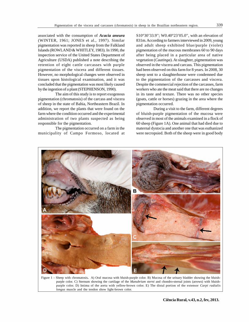
During a visit to the farm, different degreesof bluish-purple pigmentation of the mucosa wereobserved in most of the animals examined in a flock of60 sheep (Figure 1A). One animal that had died due tomaternal dystocia and another one that was euthanizedwere necropsied. Both of the sheep were in good body
Figure 1 - Sheep with chromatosis. A) Oral mucosa with bluish-purple color. B) Mucosa of the urinary bladder showing the bluish-purple color. C) Sternum showing the cartilage of the Manubrium sterni and chondro-sternal joints (arrows) with bluish-purple color. D) Intima of the aorta with yellow-brown color. E) The distal portion of the extensor Carpi radialislongus muscle and the tendon show light-brown color.
340 Pimentel et al.
Ciência Rural, v.43, n.2, fev, 2013.
condition, but their ocular, oral and vaginal mucousmembranes were bluish-purple in color. At necropsy,the bluish-purple color was observed to varyingintensities in the skin and subcutaneous tissue,muscles, surfaces of some articular cartilages and bones,particularly the inner skull and vertebrae. In theabdominal cavity, the pigment was observed in theserosa of the forestomachs, kidneys, adrenal glandsand the surface of the mucous membranes of the uterus,urinary bladder, urethra and vagina (Figures 1B and1C). In the thoracic cavity, the trachea, bronchi andbronchioles were also pigmented.
The bone surfaces of the carpal joints andother joints, fat, intima of the great arteries (Figure 1D),and some ligaments, particularly the nuchal ligament,showed yellow-brown pigmentation; in some of thejoint capsules, tendons, ligaments, and muscleinsertions, the pigmentation was light brown (Figure1E). Samples of the tissues affected were fixed in 10%buffered formalin, embedded in paraffin, and stainedby hematoxylin and eosin. No significant histologiclesions were observed, and the pigment did not remainin the tissues after processing.
The pastures where the cases occurred wereinspected for plants that could be the cause of thedisease. The native vegetation of the paddock,Caatinga, is typical of the Brazilian semiarid region: atype of forest characterized by small-sized deciduoustrees and shrubs. Caatinga has herbaceous flora thatis present only during the short rainy season(November-December to March-April) and that ismainly composed of species of Acanthaceae,Asteraceae, Convolvulaceae, Leguminosae andRubiaceae. Succulent plants of the Cactaceae,Euphorbiaceae, and Bromeliaceae families and vinesof the Bignoniaceae and Sapindaceae families are alsocommon (CARDOSO et al., 2011). The main plantspecies found in the paddock were as follows:Rhamnidium molle Reissek; Pereskia bahiensis
Gürke; Arrojadoa rhodantha (Gürke) Britton & Rose;Aspidosperma pyrifolium Mart.; Cnidoscolusbahianus (Ule) Pax & K.Hoffm; Cnidoscolusquercifolius Pohl; Croton spp.; Erythroxylum sp.;Jatropha mollissima (Pohl) Baill; Jatropha ribifolia(Pohl) Baill; Melochia tomentosa L.; Neocalyptrocalyxlongifolium (Mart.) Cornejo & Iltis; Poincianellapyramidalis (Tul.) L.P. Queiroz; Spondias tuberosaArruda; Tacinga palmadora (Britton & Rose) N.P.Taylor & Stuppy and Triplaris gardneriana Wedd.
The leaves of R. molle, a shrub with blackpigmentation in the leaves and a black fruit, and thefruits of P. bahiensis, which were mentioned by farmersas the cause of the disease, were administeredexperimentally to 3 sheep and 2 rabbits. The doses ofthe plants administered are given in table 1. The animalswere slaughtered at the end of the experiment; nopigmentation was observed in the carcasses andviscera.
The occurrence of the disease in a particulararea, with a high frequency in the affected flock,suggests that the pigmentation is due to plantconsumption. Furthermore, the observed pigmentationis very similar to the pigmentation caused byBunchosia spp. in Colombia (GÁMEZ et al., 1983a),which is characterized by the presence of bluish-purplepigment in asymptomatic animals that also disappearsafter tissue processing for histology. Thedisappearance of the pigment during tissue processingis probably due to its solubility in water or alcohol.
The negative results to reproduce thepigmentation with fruits of P. bahiensis and leaves ofR. molle suggest that these two plants are not thecause of the disease. Nevertheless, R. molle has acharacteristic black pigment in its leaves and fruits,which suggest that this plant is probably causing thepigmentation. In this case, the failure to reproduce thedisease can be due to a chemical modification of thepigment between the collection and the administration
Table 1 - Plant and doses administered to sheep and rabbits for the determination of the etiology of a chromatosis in sheep.
Plant Animal species Material and form of administration Daily dose Administration period
P. bahiensis Sheep Fresh refrigerated fruits, cut and mixed at 1% withconcentrated food.
20g kg-1 40 days
R. molle Sheep Dry ground leaves mixed at 40% with concentratedfood.
6g kg-1 33 days
R. molle Sheep Fresh leaves collected weekly and administered daily. 6g kg-1 120 daysR. molle Rabbit Fresh ground leaves mixed at 40% with concentrated
food.12g kg-1 30 days
R. molle Rabbit Similar than the other rabbit. 24g kg-1 60 days
341Pigmentation of the viscera and carcasses (chromatosis) in sheep in the Brazilian northeastern region.
Ciência Rural, v.43, n.2, fev, 2013.
of the plant. However, the possibility that some otherplant is responsible for the pigmentation is not ruledout.
ACKNOWLEDGMENTS
This research was financially supported by theNational Institute for Science and Technology for the Controlof Poisoning Plants, grant CNPq 573534/2008-0.
ETHICAL STATEMENT
We declare to whom correspond that we assumeany responsibility about any process realized during thedevelopment of the research entitled Pigmentation of theviscera and carcasses (chromatosis) in sheep. Likewise we areavailable to answer any questions that may be needed.
REFERENCES
CARDOSO, D.B.O.S. et al. Caatinga no contexto de umametacomunidade: evidências da biogeografia, padrõesfilogenéticos e abundância de espécies em Leguminosas. In:CARVALHO C.J.B. & ALMEIDA E.A.B. (Eds.). Biogeografiada América do Sul: padrões e processos. São Paulo: Roca,2011. p.241-260.
GÁMEZ, J.E.T. et al. Cromatosis de los bovinos o “vacamorada” em Colombia (1a parte). Carta Ganadera, v.20,n.6, p.25-30, 1983a.
GÁMEZ, J.E.T. et al. Cromatosis de los bovinos o “vacamorada” em Colombia (2a parte). Carta Ganadera, v.20,n.7, p.19-21 e p.65-68, 1983b.
JONES, T.C. et al. Mineral deposits and pigments. In: _____.et al. (Eds.). Veterinary pathology. Baltimore: LippincottWillians & Willians, 1997. p.57-80.
ROWLAND, A.C.; WHITLEY, R.S. A preliminary report onhepatic pigmentation in Falkland Islands sheep. VeterinaryResearch Communications, v.6, n.1, p.235-240, 1983.Available from: <http://www.springerlink.com/content/f2502u05kkh68288/>. Accessed: May, 8, 2011. doi: 10.1007/BF02214918.
STEPHENSON, S.L. The purple cow’s projected feast. 1990.USDA/FSIS veterinarian. Available from: <http://issuu.com/drshinola/docs/the_purple_cow>. Accesed: May, 8, 2011.
VARASCHIN, M.S. et al. Porfiria eritropeiética congênita embovino no Estado de Minas Gerais. Ciência Rural, v.28, n.4,p.695-698, 1998. Available from: <http://www.scielo.br/pdf/cr/v28n4/a26v28n4.pdf>. Accessed: May, 8, 2011. doi: doi.org/10.1590/S0103-84781998000400026.
WINTER, H. An environmental lipofuscin pigmentationof livers. Studies on the pigmentation affecting the sheepand other animals in certain districts of Australia .Australia: University of Queensland, 1961. 66p.


![Les matériaux élastomériques - EDP Sciencespropriétés et les forces qu’ils délivrent varient éga-lement [44]. Peu de données sont disponibles sur le proces-sus de pigmentation;](https://static.fdocuments.fr/doc/165x107/6107648a276fb763dd075ed9/les-matriaux-lastomriques-edp-sciences-proprits-et-les-forces-quails.jpg)


![Techniques de l’Inspection Post Mortemfiliere-bovine-tchad.com/classified/PAFiB_techniques_iPM[1].pdf · Les carcasses de bovins de plus de 6 mois doivent être fendues en 2. ...](https://static.fdocuments.fr/doc/165x107/5b9b894409d3f2d06f8d11f8/techniques-de-linspection-post-mortemfiliere-bovine-tchadcomclassifiedpafibtechniquesipm1pdf.jpg)