Paraneoplastic cutaneous manifestations: concepts and updates
14
An Bras Dermatol. 2013;88(1):9-22. 9 ▲ CONTINUED MEDICAL EDUCATION Paraneoplastic cutaneous manifestations: concepts and updates * Manifestações cutâneas paraneoplásicas: conceitos e atualizações Josenilson Antônio da Silva 1 Kleyton de Carvalho Mesquita 2 Ana Carolina de Souza Machado Igreja 3 Isabella Cristina Rodrigues Naves Lucas 1 Aline Ferreira Freitas 1 Sandra Maximiano de Oliveira 1 Izelda Maria Carvalho Costa 4 Iphis Tenfuss Campbell 5 Abstract: The skin often signals systemic changes. Some neoplastic diseases that affect internal organs may trigger several cutaneous manifestations. Although these dermatoses are relatively unusual, the recognition of some typical paraneoplastic dermatoses may lead to the early diagnosis of a neoplasm and determine a better prognosis. In this review article, we discuss the paraneoplastic cutaneous mani- festations strongly associated with neoplasms, which include acanthosis nigricans maligna, tripe palms, erythema gyratum repens, Bazex syndrome, acquired hypertrichosis lanuginosa, necrolytic migratory erythema, Leser-Trélat sign and paraneoplastic pemphigus. We also review the clinical manifestations of each condition and include updated knowledge on disease pathogenesis. Keywords: Neoplasms; Paraneoplastic syndromes; Skin manifestations Resumo: A pele é, muitas vezes, reflexo de manifestações sistêmicas. Doenças neoplásicas que afetam órgãos internos podem exibir manifestações cutâneas diversas. Apesar de relativamente incomuns, o reconhecimento de dermatoses paraneoplásicas pode levar ao diagnóstico precoce da neoplasia e, consequentemente, determinar melhor prognóstico. Nesta revisão serão discutidas as manifestações cutâneas paraneoplásicas com maior força de associação a neoplasias, que incluem acantose nigrican- te maligna, paquidermatoglifia adquirida, erythema gyratum repens, síndrome de Bazex, hipertricose lanuginosa adquirida, eritema necrolítico migratório, sinal de Leser-Trélat e pênfigo paraneoplásico. Para cada condição serão revisadas e atualizadas as manifestações clínicas, principais neoplasias asso- ciadas e etiopatogenia. Palavras-chave: Manifestações cutâneas; Neoplasias; Síndromes paraneoplásicas Received on 19.09.2011. Approved by the Advisory Board and accepted for publication on 16.10.2012. * Work conducted at University Hospital of Brasília - University of Brasília (HUB/UnB) – Brasília (DF), Brazil. Financial Support: None. Conflict of Interests: None. 1 MD. Catholic University of Brasília - UCB – Brasília (DF), Brazil. 2 MSc in Health Sciences (under course) – University of Brasília (UnB) - Dermatologist – Health State Secretaria of the Federal District of Brazil – Brasília (DF), Brazil. 3 MD - Resident in Dermatology, University Hospital of Brasília - University of Brasília (HUB-UnB) – Brasília (DF), Brazil. 4 PhD in Dermatology, Federal University of São Paulo (Unifesp) – Professor (PhD) of Dermatology, University of Brasília (UnB) – Brasília (DF), Brazil. 5 MSc in Dermatology, Federal University of São Paulo (Unifesp) - Professor of Dermatology, School of Medicine, University of Planalto Central (UNIPLAC) – Brasília (DF), Brazil. ©2013 by Anais Brasileiros de Dermatologia INTRODUCTION Paraneoplastic diseases may be defined as hor- monal, neurological or hematological disturbances and as clinical and biochemical imbalances associated with the presence of malignancies without direct asso- ciation with primary tumor invasion or metastasis. The skin may provide the doctor with signs that are suggestive of systemic diseases, thus contributing to the diagnosis of many diseases, including malignan- cies. 1,2 In 1868, Hebra was the first to suggest that skin pigmentation could indicate the presence of visceral cancer. 3 Since then, more than 50 dermatological con- ditions have been reported as potential markers of malignancy. 4 The skin may be directly or indirectly involved in malignancies. Direct involvement implies
Transcript of Paraneoplastic cutaneous manifestations: concepts and updates
An Bras Dermatol. 2013;88(1):9-22.
9
▲
CONTINUED MEDICAL EDUCATION
Paraneoplastic cutaneous manifestations: concepts and updates *
Manifestações cutâneas paraneoplásicas: conceitos e atualizações
Josenilson Antônio da Silva1 Kleyton de Carvalho Mesquita2
Ana Carolina de Souza Machado Igreja3 Isabella Cristina Rodrigues Naves Lucas1
Aline Ferreira Freitas1 Sandra Maximiano de Oliveira1
Izelda Maria Carvalho Costa4 Iphis Tenfuss Campbell5
Abstract: The skin often signals systemic changes. Some neoplastic diseases that affect internal organsmay trigger several cutaneous manifestations. Although these dermatoses are relatively unusual, therecognition of some typical paraneoplastic dermatoses may lead to the early diagnosis of a neoplasmand determine a better prognosis. In this review article, we discuss the paraneoplastic cutaneous mani-festations strongly associated with neoplasms, which include acanthosis nigricans maligna, tripe palms,erythema gyratum repens, Bazex syndrome, acquired hypertrichosis lanuginosa, necrolytic migratoryerythema, Leser-Trélat sign and paraneoplastic pemphigus. We also review the clinical manifestationsof each condition and include updated knowledge on disease pathogenesis.Keywords: Neoplasms; Paraneoplastic syndromes; Skin manifestations
Resumo: A pele é, muitas vezes, reflexo de manifestações sistêmicas. Doenças neoplásicas que afetamórgãos internos podem exibir manifestações cutâneas diversas. Apesar de relativamente incomuns, oreconhecimento de dermatoses paraneoplásicas pode levar ao diagnóstico precoce da neoplasia e,consequentemente, determinar melhor prognóstico. Nesta revisão serão discutidas as manifestaçõescutâneas paraneoplásicas com maior força de associação a neoplasias, que incluem acantose nigrican-te maligna, paquidermatoglifia adquirida, erythema gyratum repens, síndrome de Bazex, hipertricoselanuginosa adquirida, eritema necrolítico migratório, sinal de Leser-Trélat e pênfigo paraneoplásico.Para cada condição serão revisadas e atualizadas as manifestações clínicas, principais neoplasias asso-ciadas e etiopatogenia.Palavras-chave: Manifestações cutâneas; Neoplasias; Síndromes paraneoplásicas
Received on 19.09.2011.Approved by the Advisory Board and accepted for publication on 16.10.2012. * Work conducted at University Hospital of Brasília - University of Brasília (HUB/UnB) – Brasília (DF), Brazil.
Financial Support: None.Conflict of Interests: None.
1 MD. Catholic University of Brasília - UCB – Brasília (DF), Brazil.2 MSc in Health Sciences (under course) – University of Brasília (UnB) - Dermatologist – Health State Secretaria of the Federal District of Brazil – Brasília (DF),
Brazil.3 MD - Resident in Dermatology, University Hospital of Brasília - University of Brasília (HUB-UnB) – Brasília (DF), Brazil. 4 PhD in Dermatology, Federal University of São Paulo (Unifesp) – Professor (PhD) of Dermatology, University of Brasília (UnB) – Brasília (DF), Brazil.5 MSc in Dermatology, Federal University of São Paulo (Unifesp) - Professor of Dermatology, School of Medicine, University of Planalto Central (UNIPLAC) –
Brasília (DF), Brazil.
©2013 by Anais Brasileiros de Dermatologia
INTRODUCTIONParaneoplastic diseases may be defined as hor-
monal, neurological or hematological disturbancesand as clinical and biochemical imbalances associatedwith the presence of malignancies without direct asso-ciation with primary tumor invasion or metastasis.The skin may provide the doctor with signs that aresuggestive of systemic diseases, thus contributing to
the diagnosis of many diseases, including malignan-cies.1,2 In 1868, Hebra was the first to suggest that skinpigmentation could indicate the presence of visceralcancer.3 Since then, more than 50 dermatological con-ditions have been reported as potential markers ofmalignancy.4 The skin may be directly or indirectlyinvolved in malignancies. Direct involvement implies
An Bras Dermatol. 2013;88(1):9-22.
10 Silva JA, Mesquita KC, Igreja ACSM, Lucas ICRN, Freitas AF, Oliveira SM, Costa IMC, Campbell IT
the presence of tumor cells in the skin caused bydirect tumor extension or metastasis. Indirect involve-ment, in turn, is caused by a variety of factors (inflam-matory, proliferative or metabolic factors) related tothe neoplasia, such as polypeptides, hormones,cytokines, antibodies or growth factors that act asmediators, interfering with cell communication and,consequently, with its activity. In this case, there is nopresence of neoplastic cells in the skin, and thisinvolvement is considered a dermatological paraneo-plastic syndrome.5,6
Paraneoplastic dermatoses are a heterogeneousgroup of clinical manifestations that may have abenign appearance. They are the second most com-mon paraneoplastic syndrome, only behindendocrine syndromes. It is not always easy to deter-mine the correlation between a dermatologic findingand an internal neoplasm or even to define the fre-quency of this association in the general population.4,5
Curth, in his studies of acanthosis nigricans maligna,proposed some criteria to assess the causal relation-ship between dermatological change and potentialunderlying malignancy (Chart 1).2,4,6 Given the widescope of the subject, we will discuss the dermatosesthat are highly correlated with malignancy, whoserecognition implies a mandatory investigation of inter-nal malignancy. Since cutaneous paraneoplastic syn-dromes commonly precede or follow visceral cancer,their recognition may result in earlier diagnosis andbetter prognosis for the patient (Chart 2).7,8
ACANTHOSIS NIGRICANS MALIGNAAcanthosis nigricans can be classified as benign
or malignant. The benign form (80%) is relativelycommon and may be associated with obesity, insulin
resistance, diabetes mellitus and drug use, contrary tothe malignant form, which is rare.2,3,9-12
Acanthosis nigricans maligna (ANM) was the firstdermatosis truly associated with malignant processes,being perhaps the best known among all associations.6
It occurs equally in both sexes without racial predilec-tion or familial association.1,9 The disease has a suddenonset with extensive and severe lesions that developquickly. It affects adults with an average age of 40 years.In contrast, the benign form usually manifests earlierin life and develops slowly.3,9,10
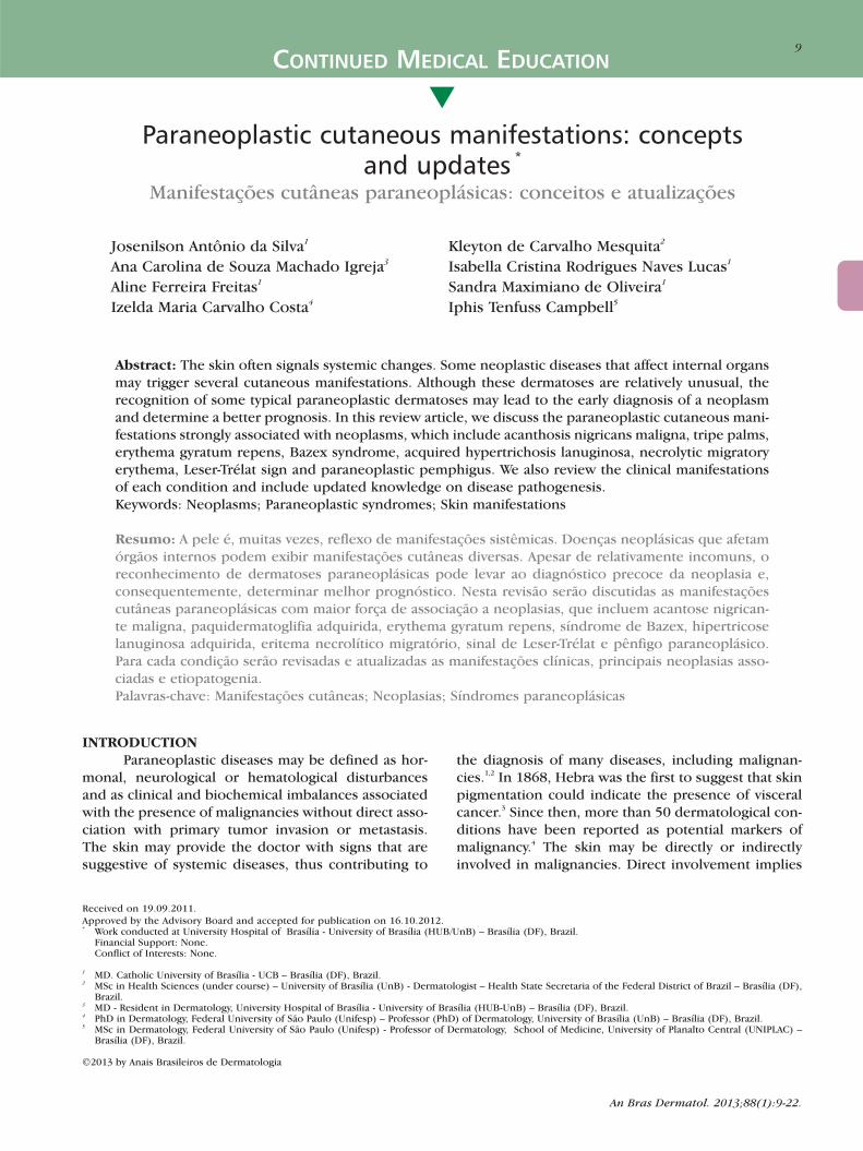
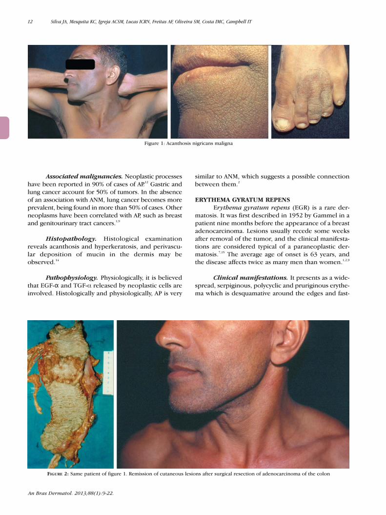
Clinical manifestations. It begins with sym-metrical hyperpigmentation in intertriginous areassuch as the axilla, cubital fossa, submammary, inguinaland posterior cervical regions, although any part ofthe body can be affected (Figure 1).3,8-10 Lesions thenbecome slightly infiltrated, with velvety hyperkeratot-ic plaques, commonly surrounded by acrochordons.3,9
There may be an association with generalized pruritusand involvement of mucosal surfaces, which present averrucous aspect in severe cases.6,9,11 Approximately25% of patients present concomitant involvement ofthe palmoplantar region in a pattern known as tripepalms (acquired pachydermatoglyphia). It may also beassociated with the sudden onset of multiple lesionsof seborrheic keratosis (Leser-Trélat sign).9 Both asso-ciations are described below.
Associated malignancies ANM can precede,occur simultaneously or occur after the diagnosis ofcancer.6,9 In a review study, this dermatological findingwas observed in 58% of patients before tumor diagno-sis.9 ANM is associated with 90% of all abdominal can-cers; 55-61% are of gastric origin, and adenocarcino-ma is found in 70-90% of cases (Figure 2).3,6,9,11,12 Otherless associated malignant conditions include uterine,liver, intestine, pancreas, thyroid, ovary, kidney,breast, lung, bladder and gallbladder cancers, mostlyconsisting of adenocarcinomas.9,11An association withlymphomas and mycosis fungoides has also beenreported.1
ANM tends to evolve simultaneously with theunderlying neoplasia and it aggravates as the condi-tion worsens.12 It improves with treatment or relapsesin the occurrence of metastasis, often serving as astandard measure of progression or recurrence ofmalignancy (Figure 2).9 Despite the presence of acan-thosis nigricans in benign and non-neoplastic condi-tions, such as drug use and insulin resistance, adetailed medical history should be taken frompatients diagnosed with this dermatosis. The diagno-sis of ANM should be strongly considered in adultsover 40 years of age, without endocrinologicalchanges or genetically determined diseases with fast-
CHART 1: Curth’s criteria for the diagnosis of cutaneous paraneoplastic syndromes
1) Both conditions began simultaneously (neoplasiaand paraneoplasia)
2) Development of a parallel course *
3) The skin lesion is not associated with a genetic syn-drome
4) There is a specific type of neoplasia that occurs withparaneoplasia
5) The dermatosis is rare in the general population
6) There is a high frequency of association betweenboth conditions
* Treatment of the neoplasia results in regression of the skin lesion;recurrence of the neoplasia implies recurrence of the skin lesion.
An Bras Dermatol. 2013;88(1):9-22.
Paraneoplastic cutaneous manifestations: concepts and updates 11
growing skin lesions. In these individuals, an exten-sive gastrointestinal evaluation is mandatory.6,11
Prognosis, however, is poor. Underlying tumors pres-ent in patients with ANM tend to have an aggressivebehavior, which leads to a mean survival of 2 yearsafter the diagnosis of ANM.3,11,13
Histopathology. Histologically, ANM showshyperkeratosis, papillomatosis and some degree ofacanthosis with thickening of the spinous layer of theepidermis.2,12 The dark color is more related to hyper-keratosis than to the presence of melanin; therefore,the term “acanthosis nigricans” is merely descriptive,as there is no proliferation of melanocytes.2,6
Pathophysiology. The exact pathophysiologicalmechanism of ANM is not well defined.3 It is believedthat cytokines produced by neoplastic cells are involved,such as transforming growth factor alpha (TGF-α),insulin growth factor-like (IGF-1), fibroblast growth fac-tor (FGF) and melanocyte-stimulating hormone (MSH-α). TGF-αwould be structurally similar to the epidermal
growth factor (EGF-α), interacting with this receptorpresent in the surface of epidermal cells.2,3,11,13 So far, nofactor has been conclusively identified.6
ACQUIRED PACHYDERMATOGLYPHIAAlso referred to as tripe palms or acanthosis
palmaris, acquired pachydermatoglyphia (AP) is aterm that was introduced in the medical literature in1970 by Clark. It is a skin condition that is usuallyassociated with Leser-Trélat sign and ANM, being con-sidered by some authors much more a variant of ANMthan actually a new disease. It predominantly affectsadults, with a predilection for males (63% ofcases).2,9,12
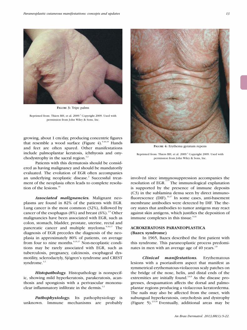
Clinical manifestations. It presents withyellowish, velvety, diffuse palmar hyperkeratosis,with accentuated dermatoglyphic patterns, leadingto a rough appearance that resembles the intestinalvillosities, which explains the term tripe palms(Figure 3).13,14
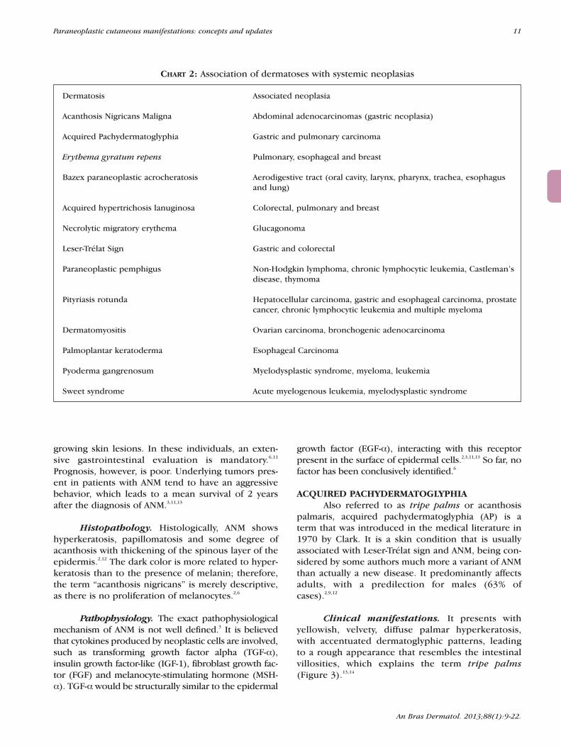
CHART 2: Association of dermatoses with systemic neoplasias
Dermatosis Associated neoplasia
Acanthosis Nigricans Maligna Abdominal adenocarcinomas (gastric neoplasia)
Acquired Pachydermatoglyphia Gastric and pulmonary carcinoma
Erythema gyratum repens Pulmonary, esophageal and breast
Bazex paraneoplastic acrocheratosis Aerodigestive tract (oral cavity, larynx, pharynx, trachea, esophagus and lung)
Acquired hypertrichosis lanuginosa Colorectal, pulmonary and breast
Necrolytic migratory erythema Glucagonoma
Leser-Trélat Sign Gastric and colorectal
Paraneoplastic pemphigus Non-Hodgkin lymphoma, chronic lymphocytic leukemia, Castleman’s disease, thymoma
Pityriasis rotunda Hepatocellular carcinoma, gastric and esophageal carcinoma, prostate cancer, chronic lymphocytic leukemia and multiple myeloma
Dermatomyositis Ovarian carcinoma, bronchogenic adenocarcinoma
Palmoplantar keratoderma Esophageal Carcinoma
Pyoderma gangrenosum Myelodysplastic syndrome, myeloma, leukemia
Sweet syndrome Acute myelogenous leukemia, myelodysplastic syndrome
An Bras Dermatol. 2013;88(1):9-22.
12 Silva JA, Mesquita KC, Igreja ACSM, Lucas ICRN, Freitas AF, Oliveira SM, Costa IMC, Campbell IT
Associated malignancies. Neoplastic processeshave been reported in 90% of cases of AP.12 Gastric andlung cancer account for 50% of tumors. In the absenceof an association with ANM, lung cancer becomes moreprevalent, being found in more than 50% of cases. Otherneoplasms have been correlated with AP, such as breastand genitourinary tract cancers.1,9
Histopathology. Histological examinationreveals acanthosis and hyperkeratosis, and perivascu-lar deposition of mucin in the dermis may beobserved.14
Pathophysiology. Physiologically, it is believedthat EGF-α and TGF-α released by neoplastic cells areinvolved. Histologically and physiologically, AP is very
similar to ANM, which suggests a possible connectionbetween them.2
ERYTHEMA GYRATUM REPENSErythema gyratum repens (EGR) is a rare der-
matosis. It was first described in 1952 by Gammel in apatient nine months before the appearance of a breastadenocarcinoma. Lesions usually recede some weeksafter removal of the tumor, and the clinical manifesta-tions are considered typical of a paraneoplastic der-matosis.7,15 The average age of onset is 63 years, andthe disease affects twice as many men than women.1,2,9
Clinical manifestations. It presents as a wide-spread, serpiginous, polycyclic and pruriginous erythe-ma which is desquamative around the edges and fast-
Figure 1: Acanthosis nigricans maligna
FIGURE 2: Same patient of figure 1. Remission of cutaneous lesions after surgical resection of adenocarcinoma of the colon
An Bras Dermatol. 2013;88(1):9-22.
Paraneoplastic cutaneous manifestations: concepts and updates 13
growing, about 1 cm/day, producing concentric figuresthat resemble a wood surface (Figure 4).9,10,15 Handsand feet are often spared. Other manifestationsinclude palmoplantar keratosis, ichthyosis and ony-chodystrophy in the sacral region.1,2
Patients with this dermatosis should be consid-ered as having malignancy and should be mandatorilyevaluated. The evolution of EGR often accompaniesan underlying neoplastic disease.2 Successful treat-ment of the neoplasia often leads to complete resolu-tion of the lesions.16
Associated malignancies. Malignant neo-plasms are found in 82% of the patients with EGR.Lung cancer is the most common (32%), followed bycancer of the esophagus (8%) and breast (6%).2,9 Othermalignancies have been associated with EGR, such ascolon, stomach, bladder, prostate, uterine, rectal andpancreatic cancer and multiple myeloma.9,10,17 Thediagnosis of EGR precedes the diagnosis of the neo-plasia in approximately 80% of patients, on averagefrom four to nine months.9,15,17 Non-neoplastic condi-tions may be rarely associated with EGR, such astuberculosis, pregnancy, calcinosis, esophageal dys-motility, sclerodactyly, Sjögren’s syndrome and CRESTsyndrome.1,2
Histopathology. Histopathology is nonspecif-ic, showing mild hyperkeratosis, parakeratosis, acan-thosis and spongiosis with a perivascular mononu-clear inflammatory infiltrate in the dermis.1.2
Pathophysiology. Its pathophysiology isunknown. Immune mechanisms are probably
involved since immunosuppression accompanies theresolution of EGR.
2,15
The immunological explanationis supported by the presence of immune deposits(C3) in the sublamina densa seen by direct immuno-fluorescence (DIF).16,17 In some cases, anti-basementmembrane antibodies were detected by DIF. The the-ory states that antibodies to tumor antigens may reactagainst skin antigens, which justifies the deposition ofimmune complexes in this tissue.9,16
ACROKERATOSIS PARANEOPLASTICA (Bazex syndrome)
In 1965, Bazex described the first patient withthis syndrome. This paraneoplastic process predomi-nates in men with an average age of 40 years.1,8
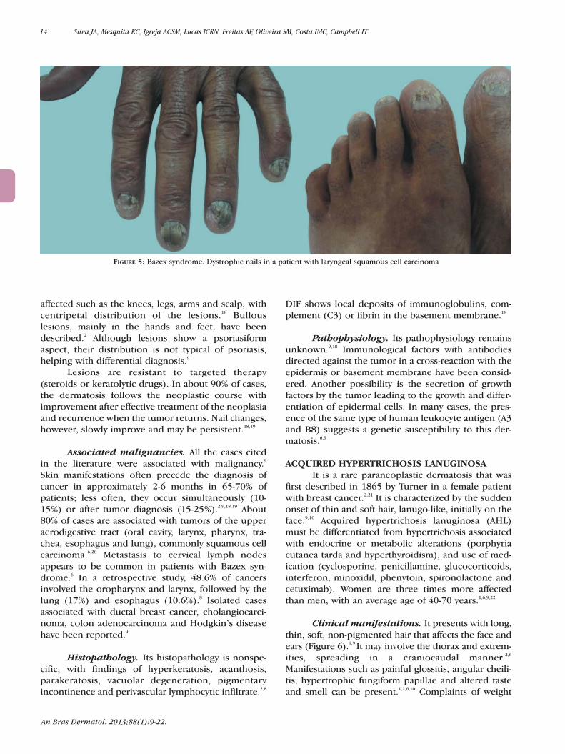
Clinical manifestations. Erythematouslesions with a psoriasiform aspect that manifest assymmetrical erythematous-violaceous scaly patches onthe bridge of the nose, helix, and distal ends of theextremities are initially found.2,6,9 As the disease pro-gresses, desquamation affects the dorsal and palmo-plantar regions producing a violaceous keratorderma.The nails may also be affected from the onset, withsubungual hyperkeratosis, onycholysis and dystrophy(Figure 5).2,9,10 Eventually, additional areas may be
FIGURE 3: Tripe palms
Reprinted from: Thiers BH, et al. 2009.6 Copyright 2009. Used with permission from John Wiley & Sons, Inc.
FIGURE 4: Erythema gyratum repens
Reprinted from: Thiers BH, et al. 2009.6 Copyright 2009. Used with permission from John Wiley & Sons, Inc.
An Bras Dermatol. 2013;88(1):9-22.
14 Silva JA, Mesquita KC, Igreja ACSM, Lucas ICRN, Freitas AF, Oliveira SM, Costa IMC, Campbell IT
affected such as the knees, legs, arms and scalp, withcentripetal distribution of the lesions.18 Bullouslesions, mainly in the hands and feet, have beendescribed.2 Although lesions show a psoriasiformaspect, their distribution is not typical of psoriasis,helping with differential diagnosis.9
Lesions are resistant to targeted therapy(steroids or keratolytic drugs). In about 90% of cases,the dermatosis follows the neoplastic course withimprovement after effective treatment of the neoplasiaand recurrence when the tumor returns. Nail changes,however, slowly improve and may be persistent.18,19
Associated malignancies. All the cases citedin the literature were associated with malignancy.9
Skin manifestations often precede the diagnosis ofcancer in approximately 2-6 months in 65-70% ofpatients; less often, they occur simultaneously (10-15%) or after tumor diagnosis (15-25%).2,9,18,19 About80% of cases are associated with tumors of the upperaerodigestive tract (oral cavity, larynx, pharynx, tra-chea, esophagus and lung), commonly squamous cellcarcinoma.6,20 Metastasis to cervical lymph nodesappears to be common in patients with Bazex syn-drome.6 In a retrospective study, 48.6% of cancersinvolved the oropharynx and larynx, followed by thelung (17%) and esophagus (10.6%).8 Isolated casesassociated with ductal breast cancer, cholangiocarci-noma, colon adenocarcinoma and Hodgkin’s diseasehave been reported.9
Histopathology. Its histopathology is nonspe-cific, with findings of hyperkeratosis, acanthosis,parakeratosis, vacuolar degeneration, pigmentaryincontinence and perivascular lymphocytic infiltrate.2,8
DIF shows local deposits of immunoglobulins, com-plement (C3) or fibrin in the basement membrane.18
Pathophysiology. Its pathophysiology remainsunknown.9,18 Immunological factors with antibodiesdirected against the tumor in a cross-reaction with theepidermis or basement membrane have been consid-ered. Another possibility is the secretion of growthfactors by the tumor leading to the growth and differ-entiation of epidermal cells. In many cases, the pres-ence of the same type of human leukocyte antigen (A3and B8) suggests a genetic susceptibility to this der-matosis.6,9
ACQUIRED HYPERTRICHOSIS LANUGINOSAIt is a rare paraneoplastic dermatosis that was
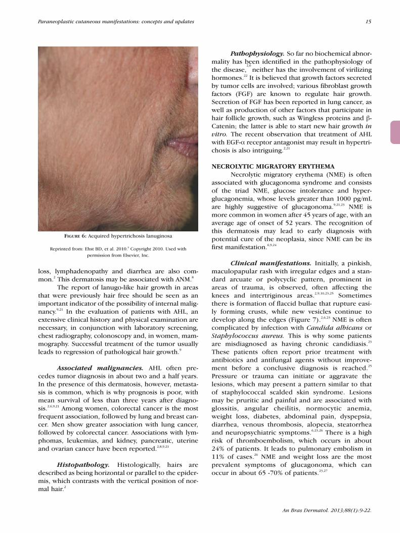
first described in 1865 by Turner in a female patientwith breast cancer.2,21 It is characterized by the suddenonset of thin and soft hair, lanugo-like, initially on theface.9,10 Acquired hypertrichosis lanuginosa (AHL)must be differentiated from hypertrichosis associatedwith endocrine or metabolic alterations (porphyriacutanea tarda and hyperthyroidism), and use of med-ication (cyclosporine, penicillamine, glucocorticoids,interferon, minoxidil, phenytoin, spironolactone andcetuximab). Women are three times more affectedthan men, with an average age of 40-70 years.1,6,9,22
Clinical manifestations. It presents with long,thin, soft, non-pigmented hair that affects the face andears (Figure 6).8,9 It may involve the thorax and extrem-ities, spreading in a craniocaudal manner.2,6
Manifestations such as painful glossitis, angular cheili-tis, hypertrophic fungiform papillae and altered tasteand smell can be present.1,2,6,10 Complaints of weight
FIGURE 5: Bazex syndrome. Dystrophic nails in a patient with laryngeal squamous cell carcinoma
An Bras Dermatol. 2013;88(1):9-22.
Paraneoplastic cutaneous manifestations: concepts and updates 15
loss, lymphadenopathy and diarrhea are also com-mon.2 This dermatosis may be associated with ANM.8
The report of lanugo-like hair growth in areasthat were previously hair free should be seen as animportant indicator of the possibility of internal malig-nancy.9,21 In the evaluation of patients with AHL, anextensive clinical history and physical examination arenecessary, in conjunction with laboratory screening,chest radiography, colonoscopy and, in women, mam-mography. Successful treatment of the tumor usuallyleads to regression of pathological hair growth.9
Associated malignancies. AHL often pre-cedes tumor diagnosis in about two and a half years.In the presence of this dermatosis, however, metasta-sis is common, which is why prognosis is poor, withmean survival of less than three years after diagno-sis.2,6,9,21 Among women, colorectal cancer is the mostfrequent association, followed by lung and breast can-cer. Men show greater association with lung cancer,followed by colorectal cancer. Associations with lym-phomas, leukemias, and kidney, pancreatic, uterineand ovarian cancer have been reported.2,8,9,21
Histopathology. Histologically, hairs aredescribed as being horizontal or parallel to the epider-mis, which contrasts with the vertical position of nor-mal hair.2
Pathophysiology. So far no biochemical abnor-mality has been identified in the pathophysiology ofthe disease,
2,6
neither has the involvement of virilizinghormones.22 It is believed that growth factors secretedby tumor cells are involved; various fibroblast growthfactors (FGF) are known to regulate hair growth.Secretion of FGF has been reported in lung cancer, aswell as production of other factors that participate inhair follicle growth, such as Wingless proteins and β-Catenin; the latter is able to start new hair growth invitro. The recent observation that treatment of AHLwith EGF-α receptor antagonist may result in hypertri-chosis is also intriguing.2,21
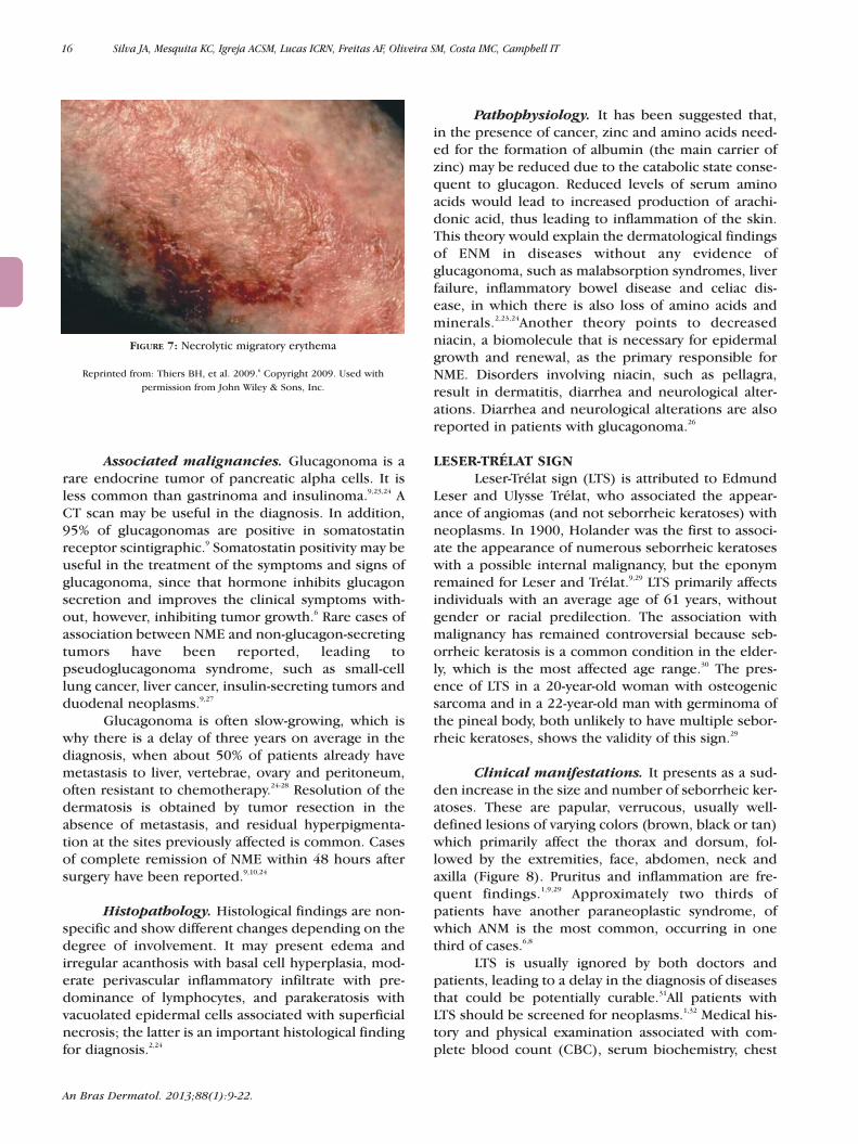
NECROLYTIC MIGRATORY ERYTHEMANecrolytic migratory erythema (NME) is often
associated with glucagonoma syndrome and consistsof the triad NME, glucose intolerance and hyper-glucagonemia, whose levels greater than 1000 pg/mLare highly suggestive of glucagonoma.9,21,23 NME ismore common in women after 45 years of age, with anaverage age of onset of 52 years. The recognition ofthis dermatosis may lead to early diagnosis withpotential cure of the neoplasia, since NME can be itsfirst manifestation.6,9,24
Clinical manifestations. Initially, a pinkish,maculopapular rash with irregular edges and a stan-dard arcuate or polycyclic pattern, prominent inareas of trauma, is observed, often affecting theknees and intertriginous areas.2,9,10,23,25 Sometimesthere is formation of flaccid bullae that rupture easi-ly forming crusts, while new vesicles continue todevelop along the edges (Figure 7).2,6,23 NME is oftencomplicated by infection with Candida albicans orStaphylococcus aureus. This is why some patientsare misdiagnosed as having chronic candidiasis.23
These patients often report prior treatment withantibiotics and antifungal agents without improve-ment before a conclusive diagnosis is reached.25
Pressure or trauma can initiate or aggravate thelesions, which may present a pattern similar to thatof staphylococcal scalded skin syndrome. Lesionsmay be pruritic and painful and are associated withglossitis, angular cheilitis, normocytic anemia,weight loss, diabetes, abdominal pain, dyspepsia,diarrhea, venous thrombosis, alopecia, steatorrheaand neuropsychiatric symptoms.6,23,26 There is a highrisk of thromboembolism, which occurs in about24% of patients. It leads to pulmonary embolism in11% of cases.26 NME and weight loss are the mostprevalent symptoms of glucagonoma, which canoccur in about 65 -70% of patients.23,27
FIGURE 6: Acquired hypertrichosis lanuginosa
Reprinted from: Ehst BD, et al. 2010.9 Copyright 2010. Used with permission from Elsevier, Inc.
16 Silva JA, Mesquita KC, Igreja ACSM, Lucas ICRN, Freitas AF, Oliveira SM, Costa IMC, Campbell IT
An Bras Dermatol. 2013;88(1):9-22.
Associated malignancies. Glucagonoma is arare endocrine tumor of pancreatic alpha cells. It isless common than gastrinoma and insulinoma.9,23,24 ACT scan may be useful in the diagnosis. In addition,95% of glucagonomas are positive in somatostatinreceptor scintigraphic.9 Somatostatin positivity may beuseful in the treatment of the symptoms and signs ofglucagonoma, since that hormone inhibits glucagonsecretion and improves the clinical symptoms with-out, however, inhibiting tumor growth.6 Rare cases ofassociation between NME and non-glucagon-secretingtumors have been reported, leading topseudoglucagonoma syndrome, such as small-celllung cancer, liver cancer, insulin-secreting tumors andduodenal neoplasms.9,27
Glucagonoma is often slow-growing, which iswhy there is a delay of three years on average in thediagnosis, when about 50% of patients already havemetastasis to liver, vertebrae, ovary and peritoneum,often resistant to chemotherapy.24-28 Resolution of thedermatosis is obtained by tumor resection in theabsence of metastasis, and residual hyperpigmenta-tion at the sites previously affected is common. Casesof complete remission of NME within 48 hours aftersurgery have been reported.9,10,24
Histopathology. Histological findings are non-specific and show different changes depending on thedegree of involvement. It may present edema andirregular acanthosis with basal cell hyperplasia, mod-erate perivascular inflammatory infiltrate with pre-dominance of lymphocytes, and parakeratosis withvacuolated epidermal cells associated with superficialnecrosis; the latter is an important histological findingfor diagnosis.2,24
Pathophysiology. It has been suggested that,in the presence of cancer, zinc and amino acids need-ed for the formation of albumin (the main carrier ofzinc) may be reduced due to the catabolic state conse-quent to glucagon. Reduced levels of serum aminoacids would lead to increased production of arachi-donic acid, thus leading to inflammation of the skin.This theory would explain the dermatological findingsof ENM in diseases without any evidence ofglucagonoma, such as malabsorption syndromes, liverfailure, inflammatory bowel disease and celiac dis-ease, in which there is also loss of amino acids andminerals.2,23,24Another theory points to decreasedniacin, a biomolecule that is necessary for epidermalgrowth and renewal, as the primary responsible forNME. Disorders involving niacin, such as pellagra,result in dermatitis, diarrhea and neurological alter-ations. Diarrhea and neurological alterations are alsoreported in patients with glucagonoma.26
LESER-TRÉLAT SIGNLeser-Trélat sign (LTS) is attributed to Edmund
Leser and Ulysse Trélat, who associated the appear-ance of angiomas (and not seborrheic keratoses) withneoplasms. In 1900, Holander was the first to associ-ate the appearance of numerous seborrheic keratoseswith a possible internal malignancy, but the eponymremained for Leser and Trélat.9,29 LTS primarily affectsindividuals with an average age of 61 years, withoutgender or racial predilection. The association withmalignancy has remained controversial because seb-orrheic keratosis is a common condition in the elder-ly, which is the most affected age range.30 The pres-ence of LTS in a 20-year-old woman with osteogenicsarcoma and in a 22-year-old man with germinoma ofthe pineal body, both unlikely to have multiple sebor-rheic keratoses, shows the validity of this sign.29
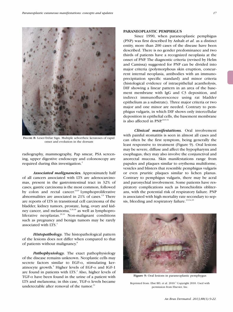
Clinical manifestations. It presents as a sud-den increase in the size and number of seborrheic ker-atoses. These are papular, verrucous, usually well-defined lesions of varying colors (brown, black or tan)which primarily affect the thorax and dorsum, fol-lowed by the extremities, face, abdomen, neck andaxilla (Figure 8). Pruritus and inflammation are fre-quent findings.1,9,29 Approximately two thirds ofpatients have another paraneoplastic syndrome, ofwhich ANM is the most common, occurring in onethird of cases.6,8
LTS is usually ignored by both doctors andpatients, leading to a delay in the diagnosis of diseasesthat could be potentially curable.31All patients withLTS should be screened for neoplasms.1,32 Medical his-tory and physical examination associated with com-plete blood count (CBC), serum biochemistry, chest
FIGURE 7: Necrolytic migratory erythema
Reprinted from: Thiers BH, et al. 2009.6 Copyright 2009. Used with permission from John Wiley & Sons, Inc.
Paraneoplastic cutaneous manifestations: concepts and updates 17
radiography, mammography, Pap smear, PSA screen-ing, upper digestive endoscopy and colonoscopy arerequired during this investigation.2
Associated malignancies. Approximately halfof all cancers associated with LTS are adenocarcino-mas, present in the gastrointestinal tract in 32% ofcases; gastric carcinoma is the most common, followedby colon and rectal cancer.6,8,9 Lymphoproliferativeabnormalities are associated in 21% of cases.1,9 Thereare reports of LTS in transitional cell carcinoma of thebladder, kidney tumors, prostate, lung, ovary and kid-ney cancer, and melanoma,9,10,30 as well as lymphopro-liferative neoplasias.29,33 Non-malignant conditionssuch as pregnancy and benign tumors may be rarelyassociated with LTS.1
Histopathology. The histopathological patternof the lesions does not differ when compared to thatof patients without malignancy.8
Pathophysiology. The exact pathophysiologyof the disease remains unknown. Neoplastic cells maysecrete factors similar to EGF-α, stimulating ker-atinocyte growth.9 Higher levels of EGF-α and IGF-1are found in patients with LTS.2 Also, higher levels ofTGF-α have been found in the urine of a patient withLTS and melanoma; in this case, TGF-α levels becameundetectable after removal of the tumor.31
PARANEOPLASTIC PEMPHIGUSSince 1990, when paraneoplastic pemphigus
(PNP) was first described by Anhalt et al. as a distinctentity, more than 200 cases of the disease have beendescribed. There is no gender predominance and twothirds of patients have a recognized neoplasia at theonset of PNP. The diagnostic criteria (revised by Helmand Camissa) suggested for PNP can be divided intomajor criteria (polymorphous skin eruption, concur-rent internal neoplasia, antibodies with an immuno-precipitation specific standard) and minor criteria(histological evidence of intraepithelial acantholysis,DIF showing a linear pattern in an area of the base-ment membrane with IgG and C3 deposition, andindirect immunofluorescence using rat bladderepithelium as a substrate). Three major criteria or twomajor and one minor are needed. Contrary to pem-phigus vulgaris, in which DIF shows only intercellulardeposition in epithelial cells, the basement membraneis also affected in PNP.2,8,9,34
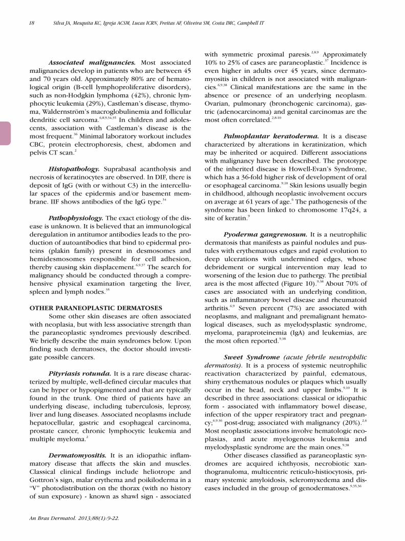
Clinical manifestations. Oral involvementwith painful stomatitis is seen in almost all cases andcan often be the first symptom, being generally theleast responsive to treatment (Figure 9). Oral lesionsmay be severe, diffuse and affect the hypopharynx andesophagus; they may also involve the conjunctival andanorectal mucosa. Skin manifestations range frompapules and plaques similar to erythema multiforme,vesicles and blisters that resemble pemphigus vulgarisor even pruritic plaques similar to lichen planus.Contrary to pemphigus vulgaris, there may be acraland paronychial involvement. Some patients have res-piratory complications such as bronchiolitis obliter-ans, with the potential risk of respiratory failure. PNPis associated with high mortality rate secondary to sep-sis, bleeding and respiratory failure.2,9,34,35
An Bras Dermatol. 2013;88(1):9-22.
Figure 9: Oral lesions in paraneoplastic pemphygus
Reprinted from: Ehst BD, et al. 2010.9 Copyright 2010. Used with permission from Elsevier, Inc.
FIGURE 8: Leser-Trélat Sign. Multiple seborrheic keratoses of rapidonset and evolution in the dorsum
18 Silva JA, Mesquita KC, Igreja ACSM, Lucas ICRN, Freitas AF, Oliveira SM, Costa IMC, Campbell IT
An Bras Dermatol. 2013;88(1):9-22.
Associated malignancies. Most associatedmalignancies develop in patients who are between 45and 70 years old. Approximately 80% are of hemato-logical origin (B-cell lymphoproliferative disorders),such as non-Hodgkin lymphoma (42%), chronic lym-phocytic leukemia (29%), Castleman’s disease, thymo-ma, Waldernström’s macroglobulinemia and folliculardendritic cell sarcoma.6,8,9,34,35 In children and adoles-cents, association with Castleman’s disease is themost frequent.36 Minimal laboratory workout includesCBC, protein electrophoresis, chest, abdomen andpelvis CT scan.2
Histopathology. Suprabasal acantholysis andnecrosis of keratinocytes are observed. In DIF, there isdeposit of IgG (with or without C3) in the intercellu-lar spaces of the epidermis and/or basement mem-brane. IIF shows antibodies of the IgG type.34
Pathophysiology. The exact etiology of the dis-ease is unknown. It is believed that an immunologicalderegulation in antitumor antibodies leads to the pro-duction of autoantibodies that bind to epidermal pro-teins (plakin family) present in desmosomes andhemidesmosomes responsible for cell adhesion,thereby causing skin displacement.6,9,37 The search formalignancy should be conducted through a compre-hensive physical examination targeting the liver,spleen and lymph nodes.35
OTHER PARANEOPLASTIC DERMATOSESSome other skin diseases are often associated
with neoplasia, but with less associative strength thanthe paraneoplastic syndromes previously described.We briefly describe the main syndromes below. Uponfinding such dermatoses, the doctor should investi-gate possible cancers.
Pityriasis rotunda. It is a rare disease charac-terized by multiple, well-defined circular macules thatcan be hyper or hypopigmented and that are typicallyfound in the trunk. One third of patients have anunderlying disease, including tuberculosis, leprosy,liver and lung diseases. Associated neoplasms includehepatocellular, gastric and esophageal carcinoma,prostate cancer, chronic lymphocytic leukemia andmultiple myeloma.2
Dermatomyositis. It is an idiopathic inflam-matory disease that affects the skin and muscles.Classical clinical findings include heliotrope andGottron’s sign, malar erythema and poikiloderma in a“V” photodistribution on the thorax (with no historyof sun exposure) - known as shawl sign - associated
with symmetric proximal paresis.2,8,9 Approximately10% to 25% of cases are paraneoplastic.37 Incidence iseven higher in adults over 45 years, since dermato-myositis in children is not associated with malignan-cies.6,9,38 Clinical manifestations are the same in theabsence or presence of an underlying neoplasm.Ovarian, pulmonary (bronchogenic carcinoma), gas-tric (adenocarcinoma) and genital carcinomas are themost often correlated.2,8-10
Palmoplantar keratoderma. It is a diseasecharacterized by alterations in keratinization, whichmay be inherited or acquired. Different associationswith malignancy have been described. The prototypeof the inherited disease is Howell-Evan’s Syndrome,which has a 36-fold higher risk of development of oralor esophageal carcinoma.9,10 Skin lesions usually beginin childhood, although neoplastic involvement occurson average at 61 years of age.6 The pathogenesis of thesyndrome has been linked to chromosome 17q24, asite of keratin.9
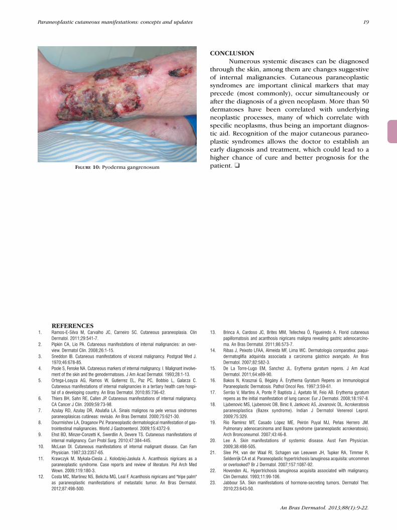
Pyoderma gangrenosum. It is a neutrophilicdermatosis that manifests as painful nodules and pus-tules with erythematous edges and rapid evolution todeep ulcerations with undermined edges, whosedebridement or surgical intervention may lead toworsening of the lesion due to pathergy. The pretibialarea is the most affected (Figure 10).9,38 About 70% ofcases are associated with an underlying condition,such as inflammatory bowel disease and rheumatoidarthritis.6,9 Seven percent (7%) are associated withneoplasms, and malignant and premalignant hemato-logical diseases, such as myelodysplastic syndrome,myeloma, paraproteinemia (IgA) and leukemias, arethe most often reported.9,38
Sweet Syndrome (acute febrile neutrophilicdermatosis). It is a process of systemic neutrophilicreactivation characterized by painful, edematous,shiny erythematous nodules or plaques which usuallyoccur in the head, neck and upper limbs.9,10 It isdescribed in three associations: classical or idiopathicform - associated with inflammatory bowel disease,infection of the upper respiratory tract and pregnan-cy;6,9.36 post-drug; associated with malignancy (20%).2,6
Most neoplastic associations involve hematologic neo-plasias, and acute myelogenous leukemia andmyelodysplastic syndrome are the main ones.9,38
Other diseases classified as paraneoplastic syn-dromes are acquired ichthyosis, necrobiotic xan-thogranuloma, multicentric reticulo-histiocytosis, pri-mary systemic amyloidosis, scleromyxedema and dis-eases included in the group of genodermatoses.9,35,36
Paraneoplastic cutaneous manifestations: concepts and updates 19
CONCLUSIONNumerous systemic diseases can be diagnosed
through the skin, among them are changes suggestiveof internal malignancies. Cutaneous paraneoplasticsyndromes are important clinical markers that mayprecede (most commonly), occur simultaneously orafter the diagnosis of a given neoplasm. More than 50dermatoses have been correlated with underlyingneoplastic processes, many of which correlate withspecific neoplasms, thus being an important diagnos-tic aid. Recognition of the major cutaneous paraneo-plastic syndromes allows the doctor to establish anearly diagnosis and treatment, which could lead to ahigher chance of cure and better prognosis for thepatient. ❑
An Bras Dermatol. 2013;88(1):9-22.
REFERENCESRamos-E-Silva M, Carvalho JC, Carneiro SC. Cutaneous paraneoplasia. Clin1.Dermatol. 2011;29:541-7.Pipkin CA, Lio PA. Cutaneous manifestations of internal malignancies: an over-2.view. Dermatol Clin. 2008;26:1-15.Sneddon IB. Cutaneous manifestations of visceral malignancy. Postgrad Med J.3.1970;46:678-85.Poole S, Fenske NA. Cutaneous markers of internal malignancy. I. Malignant involve-4.ment of the skin and the genodermatoses. J Am Acad Dermatol. 1993;28:1-13.Ortega-Loayza AG, Ramos W, Gutierrez EL, Paz PC, Bobbio L, Galarza C.5.Cutaneous manifestations of internal malignancies in a tertiary health care hospi-tal of a developing country. An Bras Dermatol. 2010;85:736-42.Thiers BH, Sahn RE, Callen JP. Cutaneous manifestations of internal malignancy.6.CA Cancer J Clin. 2009;59:73-98.Azulay RD, Azulay DR, Abulafia LA. Sinais malignos na pele versus síndromes7.paraneoplásicas cutâneas: revisão. An Bras Dermatol. 2000;75:621-30.Dourmishev LA, Draganov PV. Paraneoplastic dermatological manifestation of gas-8.trointestinal malignancies. World J Gastroenterol. 2009;15:4372-9.Ehst BD, Minzer-Conzetti K, Swerdlin A, Devere TS. Cutaneous manifestations of9.internal malignancy. Curr Probl Surg. 2010;47:384-445.McLean DI. Cutaneous manifestations of internal malignant disease. Can Fam10.Physician. 1987;33:2357-65.Krawczyk M, Mykala-Ciesla J, Kolodziej-Jaskula A. Acanthosis nigricans as a11.paraneoplastic syndrome. Case reports and review of literature. Pol Arch MedWewn. 2009;119:180-3.Costa MC, Martinez NS, Belicha MG, Leal F. Acanthosis nigricans and "tripe palm"12.as paraneoplastic manifestations of metastatic tumor. An Bras Dermatol.2012;87:498-500.
FIGURE 10: Pyoderma gangrenosum
Brinca A, Cardoso JC, Brites MM, Tellechea Ó, Figueiredo A. Florid cutaneous13.papillomatosis and acanthosis nigricans maligna revealing gastric adenocarcino-ma. An Bras Dermatol. 2011;86:573-7.Ribas J, Peixoto LFAA, Almeida MF, Lima WC. Dermatologia comparativa: paqui-14.dermatoglifia adquirida associada a carcinoma gástrico avançado. An BrasDermatol. 2007;82:582-3.De La Torre-Lugo EM, Sanchez JL. Erythema gyratum repens. J Am Acad15.Dermatol. 2011;64:e89-90.Bakos N, Krasznai G, Bégány Á. Erythema Gyratum Repens an Immunological16.Paraneoplastic Dermatosis. Pathol Oncol Res. 1997;3:59-61.Serrão V, Martins A, Ponte P, Baptista J, Apetato M, Feio AB. Erythema gyratum17.repens as the initial manifestation of lung cancer. Eur J Dermatol. 2008;18:197-8.Ljubenovic MS, Ljubenovic DB, Binic II, Jankovic AS, Jovanovic DL. Acrokeratosis18.paraneoplastica (Bazex syndrome). Indian J Dermatol Venereol Leprol.2009;75:329.Río Ramírez MT, Casado López ME, Peirón Puyal MJ, Peñas Herrero JM.19.Pulmonary adenocarcinoma and Bazex syndrome (paraneoplastic acrokeratosis).Arch Bronconeumol. 2007;43:46-8. Lee A. Skin manifestations of systemic disease. Aust Fam Physician.20.2009;38:498-505.Slee PH, van der Waal RI, Schagen van Leeuwen JH, Tupker RA, Timmer R,21.Seldenrijk CA et al. Paraneoplastic hypertrichosis lanuginosa acquisita: uncommonor overlooked? Br J Dermatol. 2007;157:1087-92.Hovenden AL. Hypertrichosis lanuginosa acquisita associated with malignancy.22.Clin Dermatol. 1993;11:99-106.Jabbour SA. Skin manifestations of hormone-secreting tumors. Dermatol Ther.23.2010;23:643-50.

20 Silva JA, Mesquita KC, Igreja ACSM, Lucas ICRN, Freitas AF, Oliveira SM, Costa IMC, Campbell IT
An Bras Dermatol. 2013;88(1):9-22.
MAILING ADDRESS:Kleyton de Carvalho MesquitaHospital Universitário de BrasíliaServiço de Dermatologia SGAN 605, Av. L2 Norte.70840-901 Brasilia – DF.E-mail: [email protected]
How to cite this article: Silva JA, Mesquita KC, Igreja ACSM, Lucas ICRN, Freitas AF, Oliveira SM, Costa IMC, CampbellIT. Paraneoplastic cutaneous manifestations: concepts and updates. An Bras Dermatol. 2013;88(1):9-22.
Mendoza-Guil F, Hernández-Jurado I, Burkhardt P, Linares J, Naranjo R. Necrolytic24.migratory erythema associated with glucagonoma. Actas Dermosifiliogr.2005;96:175-8. Teixeira RC, Nico MM, Ghideti AC. Necrolytic migratory erythema associated with25.glucagonoma: a report of 2 cases. Clinics (São Paulo). 2008;63:267-70.Qadan M, Visser B, Kim J, Pai R, Triadafilopoulos G. Abdominal Mass, Anemia,26.Diabetes Mellitus, and Necrolytic Migratory Erythema. Dig Dis Sci. 2012;57:1465-8.Dal Coleto CC, de Mello AP, Piquero-Casals J, Lima FR, Vilela MA, Festa-Neto C et27.al. Necrolytic migratory erythema associated with glucagonoma syndrome: a casereport. Rev Hosp Clin Fac Med São Paulo. 2001;56:183-8.Echenique-Elizondo M, Martínez de Lizarduy I. Glucagonoma and necrolytic migra-28.tory erythema. Rev Esp Enferm Dig. 2005;97(6):455-7.Bártholo RM, Bártholo TP, Florião RA. Leser-Trélat: Um sinal clínico revisitado.29.Pulmão RJ. 2009;18:3-56.Kluger N, Guillot B. Sign of Leser-Trelat with an adenocarcinoma of the prostate: a30.case report. Cases J. 2009;2:8868.
Constantinou C, Dancea H, Meade P. The sign of Leser-Trelat in colorectal adeno-31.carcinoma. Am Surg. 2010;76:340-1.Wieland CN, Kumar N. Sign of Leser-Trélat. Int J Dermatol. 2008;47:643-4.32.Yavasoglu I, Kadikoylu G, Bolaman Z. The Leser-Trelat sign is a associated with33.acute myeloid leukemia. Ann Hematol. 2011;90:363.Edgin WA, Pratt TC, Grimwood RE. Pemphigus Vulgaris and Paraneoplastic34.Pemphigus. Oral Maxillofac Surg Clin North Am. 2008;20:577-84.Boyce S, Harper J. Paraneoplastic dermatoses. Dermatol Clin. 2002;20:523-3.35.Shah A, Jack A, Liu H, Hopkins RS. Neoplastic/paraneoplastic dermatitis, fasciitis,36.and panniculitis. Rheum Dis Clin North Am. 2011;37:573-92.Pelosof LC, Gerber DE. Paraneoplastic Syndromes: an approach to diagnosis and37.treatment. Mayo Clin Proc. 2010;85:838-54.Chung VQ, Moschella SL, Zembowicz A, Liu V. Clinical and pathologic findings of38.paraneoplastic dermatoses. J Am Acad Dermatol. 2006;54:745-62.
Paraneoplastic cutaneous manifestations: concepts and updates 21
An Bras Dermatol. 2013;88(1):9-22.
1. More than 50 dermatologic conditions have beenreported as potential markers of malignancy. It is cor-rect to state the following about cutaneous paraneo-plastic manifestations:
a) They are easily recognized, especially when associatedwith histological examination, given the peculiar cell featu-res resulting from cell growth factors released by the tumorb) When they precede the onset of malignancy, their evolu-tion is not related to the patient's response to cancer therapy.c) Paraneoplastic manifestations comprise a heteroge-neous group of dermatoses that can occur directly(tumor invasion) or indirectly (distant interaction of theskin with substances released by the tumor).d) Due to their parallel course with the underlying neo-plasia, they serve as a standard for the evaluation of pro-gress or recurrence of malignancy
2. Curth’s criteria are used in the recognition of cuta-neous paraneoplastic syndromes. These criteria includeall of the following, except:
a) Parallel course of the neoplasm and cutaneous mani-festationb) Lack of association between cutaneous manifestationand genetic syndromec) Presence of tumor cells in skin lesionsd) High frequency of association between the neoplasmand cutaneous manifestation
3. Acanthosis nigricans maligna:a) It is closely associated with diabetic nephropathy.b) It is a condition commonly found in insulin-depen-dent diabetics with poor control of the disease.c) It has a sudden onset and is predominantly associatedwith abdominal carcinomas.d) The underlying neoplasms tend to have an indolentcourse, which leads to long-term survival of patients.
4. Which of the following exams should be first reques-ted of a patient with acanthosis nigricans malignant?
a) Thyroid ultrasound with dopplerb) Upper digestive tract endoscopyc) Mammographyd) Chest x-ray
5. Which of the following are NOT characteristics ofacquired dermatoglyphia?
a) It predominantly affects children with diseases of thelymphoid-hematopoietic tissue. b) It gives the palms a velvety yellowish aspect.c) It is often associated with Acanthosis NigricansMalignant.d) Lung and gastric carcinoma should be investigated
6. Widespread, serpiginous, polycyclic and pruriginouserythema which is desquamative around the edges andfast-growing, often associated with lung cancer. Thisdescription corresponds to:
a) Erythema gyratum repensb) Erythema migrans necrolyticc) Erythema annulare centrifugumd) Disseminated granuloma annulare
7. Male patient, 45 years old, smoker, presenting witherythematous scaly patches on the nose, helix, andextremities for six months. He was being treated withtopical corticosteroids and keratolytic drugs, withoutimprovement of his condition. He also presented withsubungual hyperkeratosis, onycholysis, and nail dys-trophy. He was diagnosed with squamous cell carcino-ma of the larynx. These dermatological symptoms pro-bably correspond to:
a) Seborrheic dermatitis and onychomycosisb) Psoriasis Vulgarisc) Bazex Syndromed) Palmoplantar keratoderma of Unna-Thost
8. Female patient, 42 years old, had been undergoinglaser hair removal treatment for 6 months with a non-dermatologist due to excessive hair growth on her face.She came to see the doctor due to treatment ineffecti-veness. On examination, thin, soft and non-pigmentedhair was observed on her face and ears.Complementary examination was normal. Based onthis description, which of the following is the most pru-dent course of action?
a) Regular monitoring with periodic examinations, sincethe condition can precede the onset of cancer.b) Referral to an endocrinologist for serial assessment ofhormonal profile. c) Maintenance of laser hair removal treatment, chan-ging the parameters of the device. d) Prescription of oral contraceptives with an antiandro-gen profile.
9. It is correct to state the following about acquiredhypertrichosis lanuginosa associated with malignancies:
a) Its pathogenesis is well established with overproduc-tion of virilizing hormones b) It is more common in men and usually spares the facec) In addition to a detailed clinical history, laboratoryscreening includes chest x-ray, colonoscopy and, inwomen, mammography.d) Metastasis is unusual and the patient usually has goodprognosis.
10. Which of the following findings should be primari-ly considered and investigated in a patient with erythe-ma migrans necrolytic?
a) Pancreatic nodule seen on computed tomography.b) Type IV Bosniak cysts on computed tomography.c) BI-RADS III findings on digital mammography.d) Chammas III thyroid nodule on ecodoppler.
11. Early diagnosis and treatment of glucagonomaincrease the potential for cure of cancer. Surgical resec-tion is the treatment of choice, since the tumor is oftenresistant to chemotherapy. Which of the followingoptions make up the triad of glucagonoma: glucoseintolerance, hyperglucagonemia and:
a) Erythema gyratum repensb) Erythema migrans necrolyticc) Halstead-Cullen Signd) Erythema chronicum migrans
▲
QUESTIONS
An Bras Dermatol. 2013;88(1):9-22.
22 Silva JA, Mesquita KC, Igreja ACSM, Lucas ICRN, Freitas AF, Oliveira SM, Costa IMC, Campbell IT
12. Patient, 70 years old, uncommunicative, accompa-nied by a caregiver in a nursing home. The caregivernoted the recent onset of numerous brownish papulesand plaques, with a verrucous and waxy surface in thedorsum. The patient also presented significant weightloss and frequent nausea. The presumptive diagnosis ofthe underlying disease is:
a) Squamous cell carcinoma of the larynx.b) Gastric Adenocarcinomac) Prostate Adenocarcinoma. d) Malignant Melanoma.
13. It is incorrect to state the following about the Leser-Trelat sign:
a) It primarily affects the chest and dorsumb) Pruritic and eczematous lesions are frequent findingsc) Association with paraneoplastic syndrome is com-mon, especially with ANMd) Histopathology of skin lesions is essential for diagnos-tic confirmation of the Leser-Trelat sign
14) 68-year-old patient presenting with night sweats,weight loss and Pel-Ebstein fever. Extremely painfuloral erosions develop. The probable diagnosis is:
a) Pyostomatitis vegetansb) Bullous pemphigoidc) Behcet's Diseased) Paraneoplastic pemphigus
15) Which of the following options characterizes para-neoplastic pemphigus:
a) Preferential involvement of the skin of the trunk whencompared to oral and conjunctival mucosaeb) Positive indirect immunofluorescence with rat blad-der substratec) Low mortality rated) The autoantibodies produced by the tumor exclusive-ly affect the skin and mucosae
16. Female patient, 62 years old, presenting withperiorbital erythema and edema, violaceous papules oninterphalangeal joints with periungual erythema andtelangiectasias. Which of the following is the most ade-quate initial management:
a) Oral corticosteroids and observation, since it is anidiopathic diseaseb) Alendronate and diltiazem, given its frequent associa-tion with calcinosis in this age groupc) Transvaginal ultrasound and chest X-ray, due to theknown association with lung and ovarian carcinoma.d) Interruption of drug use, as clinical manifestationsprobably correspond to a drug-induced skin rash.
17. Which of the following alternatives is the correctassociation between a paraneoplastic manifestationand a neoplasia:
a) Acanthosis nigricans malignant - LymphoproliferativeDisordersb) Tripe palms - Medullary Thyroid Carcinomac) Bazex paraneoplastic acrokeratosis - aerodigestive tractd) Erythema migrans necrolytic - Pancreatic adenocarcinoma
18) Acute febrile neutrophilic dermatosis, also knownas Sweet syndrome, has several causes. The following
should be primarily investigated, except:a) Infection of the upper respiratory tractb) Prior use of drugsc) Hematological Malignanciesd) Rheumatoid Arthritis
19. Patient with a deep ulceration with underminededges in the pretibial region resulting from local trau-ma. Biopsy of the border of the ulcer showed neutrop-hilic inflammation with abscesses and necrosis. It iscorrect to state the following:
a) Most cases are idiopathicb) Nikolsky’s sign is typical of this conditionc) Inflammatory bowel diseases and neoplasms shouldbe investigatedd) Pentavalent antimony, despite its adverse effects,remains the treatment of choice.
20. Male patient, 67 years old, diagnosed with broncho-genic carcinoma. T3N1M0 (stage IIIA), undergoing che-motherapy with cisplatin and etoposide. One weekafter the second treatment cycle, he developed pruritusand erythema on waxy brownish pre-existing papulesand plaques on the upper limbs and bosom, as well ashyperpigmentation in the dorsal surface of extremitiesand nails. Which of the following is the most likelyhypothesis for the clinical manifestations described:
a) It is Bazex paraneoplastic acrokeratosis, often occur-ring in association with carcinomas of the upper aerodi-gestive tract and indicating poor prognosis.b) The clinical manifestations correspond to the Leser-Trelat sign and indicate metastases screening.c) The clinical manifestations refer to skin reactions tothe antineoplastic drugs used and do not require inter-ruption of treatment.d) The clinical manifestations refer to photosensitivitydue to the use of alkylating agents and indicate substitu-tion of the chemotherapy regimen, due to the risk ofserious drug eruptions in case of reexposure.
PapersInformation for all members: The EMC-D
questionnaire is now available at the homepageof the Brazilian Annals of Dermatology:www.anaisdedermatologia.org.br. The deadlinefor completing the questionnaire is 30 daysfrom the date of online publication.
1. d2. b3. d4. b5. b
6. d7. d 8. c9. b10. b
11. c12. b13. d14. c15. b
16. a17. a18. b19. a20. d
Answer keyMycosis fungoides and Sézary syndrome:clinical, histopathological and immunohis-tochemical review andupdate2012;87(6):817-30.