Module Endocrinologie NODULE THYROÏDIEN...
94
NODULE THYROÏDIEN Stratification du risque de malignité TIRADS - Elastographie DIUE 2016 Module Endocrinologie Par cœur ! Centre de Pathologie et d’Imagerie Unité Thyroïde et Tumeurs Endocrines Hôpital La Pitié-Salpêtrière Université Pierre et Marie Curie - Paris VI Dr Gilles Russ Dr Jean Tramalloni Message important ! En jaune dans le texte
Transcript of Module Endocrinologie NODULE THYROÏDIEN...
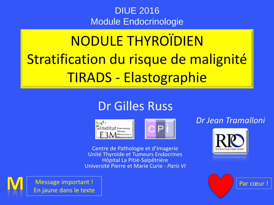
NODULE THYROÏDIEN Stratification du risque de malignité
TIRADS - Elastographie
DIUE 2016
Module Endocrinologie
Par cœur !
Centre de Pathologie et d’Imagerie Unité Thyroïde et Tumeurs Endocrines
Hôpital La Pitié-Salpêtrière Université Pierre et Marie Curie - Paris VI
Dr Gilles Russ Dr Jean Tramalloni
Message important ! En jaune dans le texte
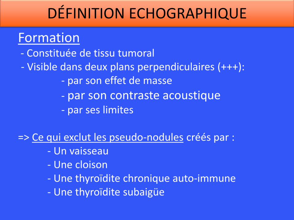
Formation - Constituée de tissu tumoral - Visible dans deux plans perpendiculaires (+++): - par son effet de masse
- par son contraste acoustique - par ses limites
=> Ce qui exclut les pseudo-nodules créés par :
- Un vaisseau - Une cloison - Une thyroïdite chronique auto-immune - Une thyroïdite subaigüe
DÉFINITION ECHOGRAPHIQUE
1- Histoire naturelle du nodule
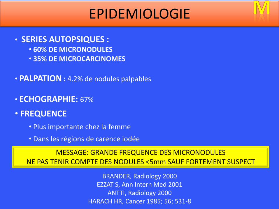
• SERIES AUTOPSIQUES : • 60% DE MICRONODULES • 35% DE MICROCARCINOMES
• PALPATION : 4.2% de nodules palpables
• ECHOGRAPHIE: 67%
• FREQUENCE
• Plus importante chez la femme
• Dans les régions de carence iodée
BRANDER, Radiology 2000 EZZAT S, Ann Intern Med 2001
ANTTI, Radiology 2000 HARACH HR, Cancer 1985; 56; 531-8
EPIDEMIOLOGIE
MESSAGE: GRANDE FREQUENCE DES MICRONODULES NE PAS TENIR COMPTE DES NODULES <5mm SAUF FORTEMENT SUSPECT
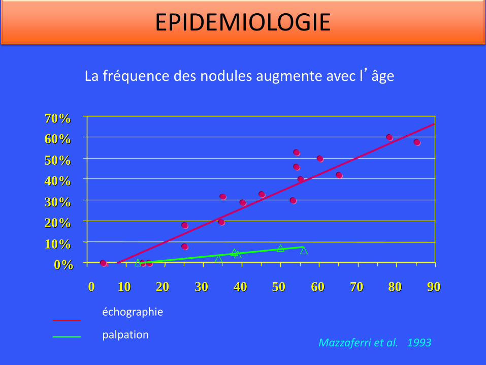
La fréquence des nodules augmente avec l’âge
0% 0%
10% 10%
20% 20%
30% 30%
40% 40%
50% 50%
60% 60%
70% 70%
0 0 10 10 20 20 30 30 40 40 50 50 60 60 70 70 80 80 90 90
Mazzaferri et al. 1993
échographie
palpation
EPIDEMIOLOGIE
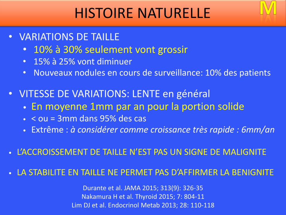
• VARIATIONS DE TAILLE • 10% à 30% seulement vont grossir • 15% à 25% vont diminuer • Nouveaux nodules en cours de surveillance: 10% des patients
• VITESSE DE VARIATIONS: LENTE en général • En moyenne 1mm par an pour la portion solide • < ou = 3mm dans 95% des cas • Extrême : à considérer comme croissance très rapide : 6mm/an
• L’ACCROISSEMENT DE TAILLE N’EST PAS UN SIGNE DE MALIGNITE
• LA STABILITE EN TAILLE NE PERMET PAS D’AFFIRMER LA BENIGNITE
Durante et al. JAMA 2015; 313(9): 326-35 Nakamura H et al. Thyroid 2015; 7: 804-11
Lim DJ et al. Endocrinol Metab 2013; 28: 110-118
HISTOIRE NATURELLE
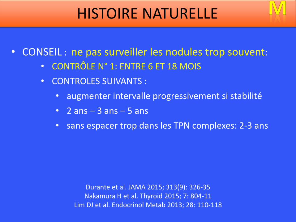
• CONSEIL : ne pas surveiller les nodules trop souvent:
• CONTRÔLE N° 1: ENTRE 6 ET 18 MOIS
• CONTROLES SUIVANTS :
• augmenter intervalle progressivement si stabilité
• 2 ans – 3 ans – 5 ans
• sans espacer trop dans les TPN complexes: 2-3 ans
HISTOIRE NATURELLE
Durante et al. JAMA 2015; 313(9): 326-35 Nakamura H et al. Thyroid 2015; 7: 804-11
Lim DJ et al. Endocrinol Metab 2013; 28: 110-118
2. Conduite de l’examen
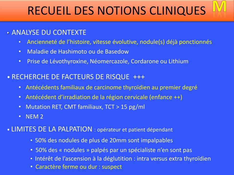
• ANALYSE DU CONTEXTE
• Ancienneté de l’histoire, vitesse évolutive, nodule(s) déjà ponctionnés
• Maladie de Hashimoto ou de Basedow
• Prise de Lévothyroxine, Néomercazole, Cordarone ou Lithium
• RECHERCHE DE FACTEURS DE RISQUE +++
• Antécédents familiaux de carcinome thyroïdien au premier degré
• Antécédent d’irradiation de la région cervicale (enfance ++)
• Mutation RET, CMT familiaux, TCT > 15 pg/ml
• NEM 2
• LIMITES DE LA PALPATION : opérateur et patient dépendant
• 50% des nodules de plus de 20mm sont impalpables
• 50% des « nodules » palpés par un spécialiste n’en sont pas
• Intérêt de l’ascension à la déglutition : intra versus extra thyroïdien
• Caractère ferme ou dur : suspect
RECUEIL DES NOTIONS CLINIQUES
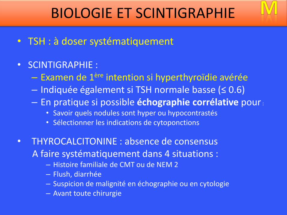
• TSH : à doser systématiquement
• SCINTIGRAPHIE : – Examen de 1ère intention si hyperthyroïdie avérée – Indiquée également si TSH normale basse (≤ 0.6) – En pratique si possible échographie corrélative pour :
• Savoir quels nodules sont hyper ou hypocontrastés • Sélectionner les indications de cytoponctions
• THYROCALCITONINE : absence de consensus A faire systématiquement dans 4 situations :
– Histoire familiale de CMT ou de NEM 2 – Flush, diarrhée – Suspicion de malignité en échographie ou en cytologie – Avant toute chirurgie
BIOLOGIE ET SCINTIGRAPHIE
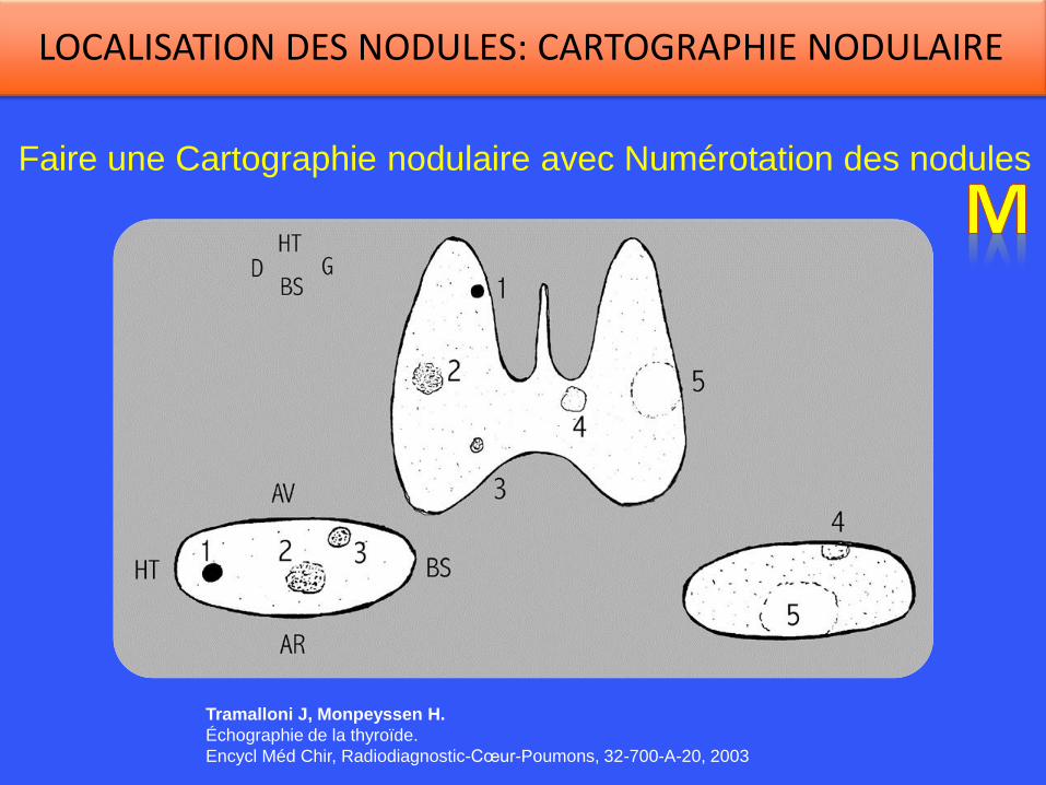
Faire une Cartographie nodulaire avec Numérotation des nodules
Tramalloni J, Monpeyssen H.
Échographie de la thyroïde.
Encycl Méd Chir, Radiodiagnostic-Cœur-Poumons, 32-700-A-20, 2003
LOCALISATION DES NODULES: CARTOGRAPHIE NODULAIRE
Chaque nodule est numéroté. Ce numéro est repris lors des examens ultérieurs.
Son numéro ne doit jamais être changé.
Un numéro ne représente qu’un seul nodule.
Si un nodule disparaît, son numéro n’est pas ré-attribué.
Chaque nouveau nodule reçoit un nouveau numéro.
SCHEMA: LES 6 REGLES D’OR
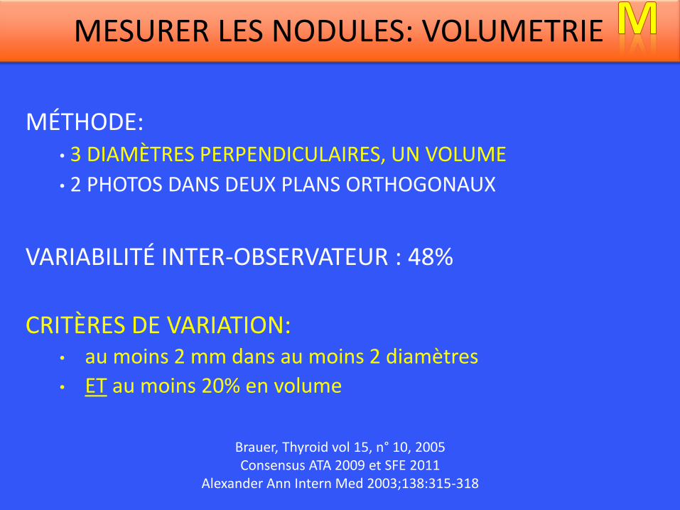
MÉTHODE: • 3 DIAMÈTRES PERPENDICULAIRES, UN VOLUME
• 2 PHOTOS DANS DEUX PLANS ORTHOGONAUX
VARIABILITÉ INTER-OBSERVATEUR : 48%
CRITÈRES DE VARIATION: • au moins 2 mm dans au moins 2 diamètres
• ET au moins 20% en volume
Brauer, Thyroid vol 15, n° 10, 2005 Consensus ATA 2009 et SFE 2011
Alexander Ann Intern Med 2003;138:315-318
MESURER LES NODULES: VOLUMETRIE
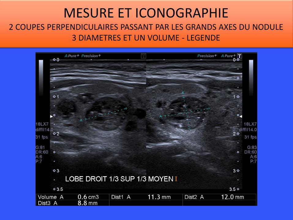
MESURE ET ICONOGRAPHIE 2 COUPES PERPENDICULAIRES PASSANT PAR LES GRANDS AXES DU NODULE
3 DIAMETRES ET UN VOLUME - LEGENDE
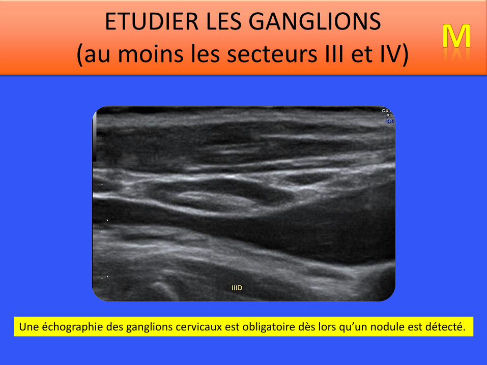
ETUDIER LES GANGLIONS (au moins les secteurs III et IV)
Une échographie des ganglions cervicaux est obligatoire dès lors qu’un nodule est détecté.
3. SÉMÉIOLOGIE ET STRATIFICATION DU RISQUE: un changement de paradigme
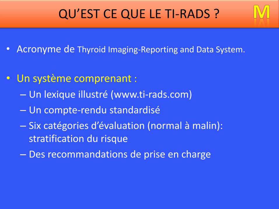
• Acronyme de Thyroid Imaging-Reporting and Data System.
• Un système comprenant :
– Un lexique illustré (www.ti-rads.com)
– Un compte-rendu standardisé
– Six catégories d’évaluation (normal à malin): stratification du risque
– Des recommandations de prise en charge
QU’EST CE QUE LE TI-RADS ?
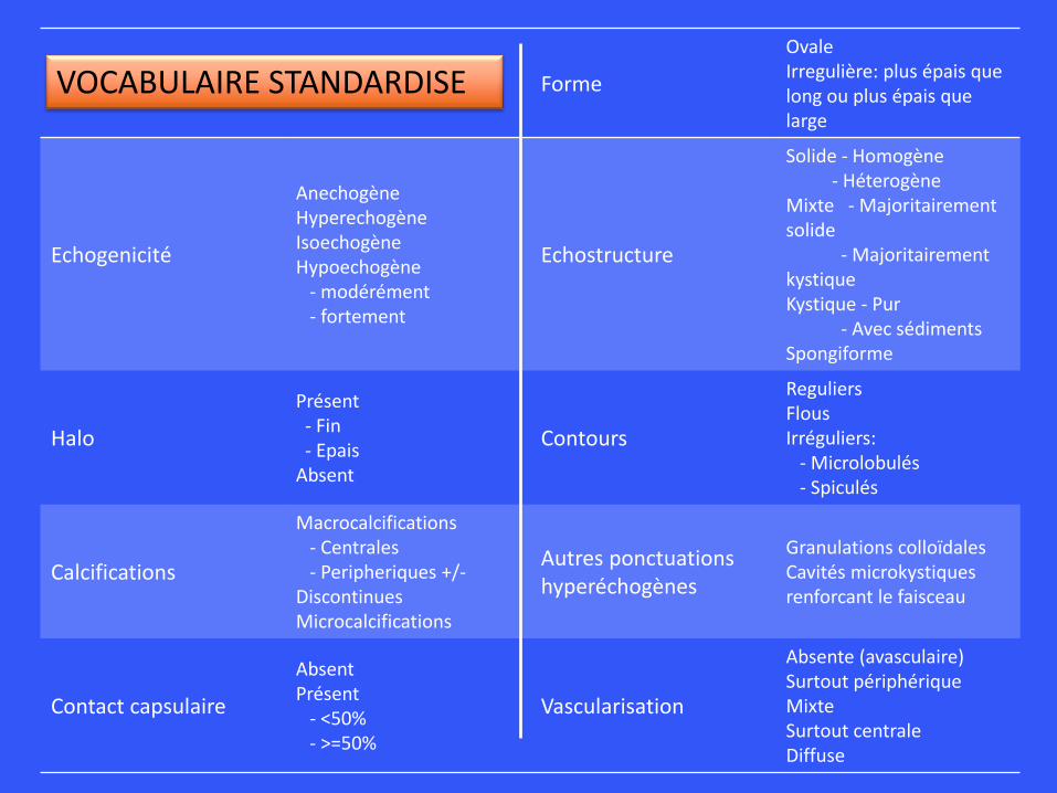
Forme
Ovale Irregulière: plus épais que long ou plus épais que large
Echogenicité
Anechogène Hyperechogène Isoechogène Hypoechogène - modérément - fortement
Echostructure
Solide - Homogène - Héterogène Mixte - Majoritairement solide - Majoritairement kystique Kystique - Pur - Avec sédiments Spongiforme
Halo
Présent - Fin - Epais Absent
Contours
Reguliers Flous Irréguliers: - Microlobulés - Spiculés
Calcifications
Macrocalcifications - Centrales - Peripheriques +/- Discontinues Microcalcifications
Autres ponctuations hyperéchogènes
Granulations colloïdales Cavités microkystiques renforcant le faisceau
Contact capsulaire
Absent Présent - <50% - >=50%
Vascularisation
Absente (avasculaire) Surtout périphérique Mixte Surtout centrale Diffuse
VOCABULAIRE STANDARDISE
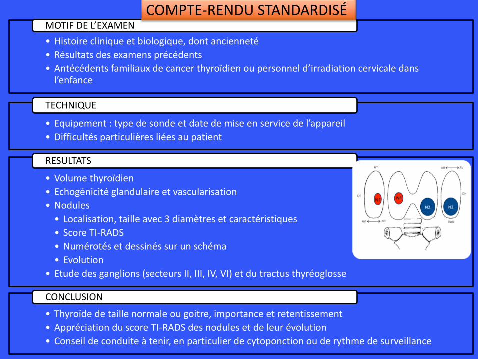
• Histoire clinique et biologique, dont ancienneté
• Résultats des examens précédents
• Antécédents familiaux de cancer thyroïdien ou personnel d’irradiation cervicale dans l’enfance
MOTIF DE L’EXAMEN
• Equipement : type de sonde et date de mise en service de l’appareil
• Difficultés particulières liées au patient
TECHNIQUE
• Volume thyroïdien
• Echogénicité glandulaire et vascularisation
• Nodules
• Localisation, taille avec 3 diamètres et caractéristiques
• Score TI-RADS
• Numérotés et dessinés sur un schéma
• Evolution
• Etude des ganglions (secteurs II, III, IV, VI) et du tractus thyréoglosse
RESULTATS
• Thyroïde de taille normale ou goitre, importance et retentissement
• Appréciation du score TI-RADS des nodules et de leur évolution
• Conseil de conduite à tenir, en particulier de cytoponction ou de rythme de surveillance
CONCLUSION
COMPTE-RENDU STANDARDISÉ
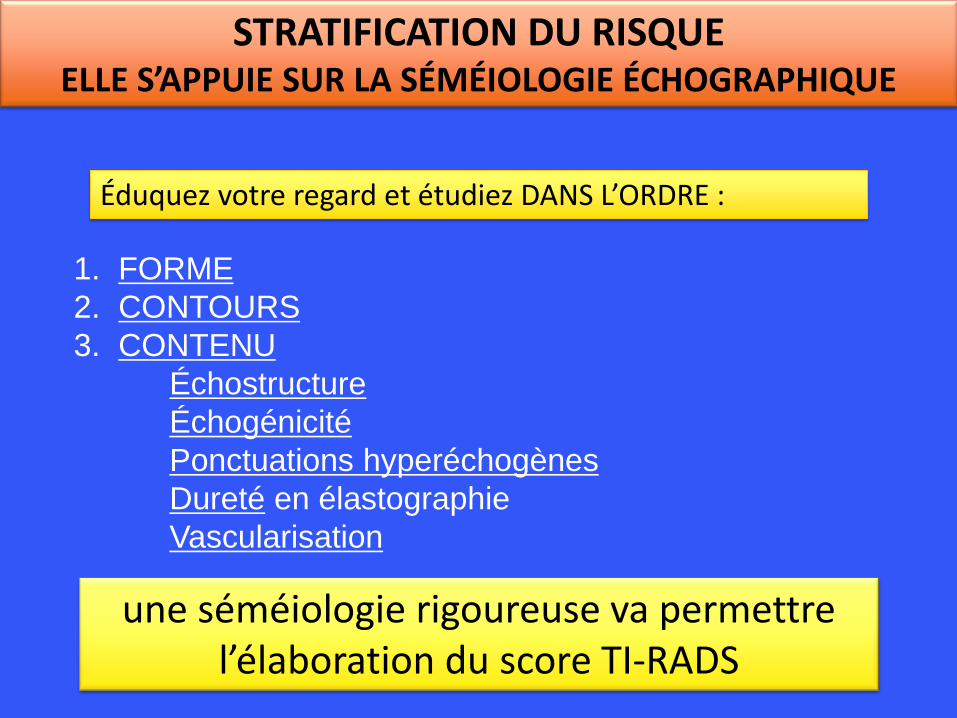
1. FORME
2. CONTOURS
3. CONTENU
Échostructure
Échogénicité
Ponctuations hyperéchogènes
Dureté en élastographie
Vascularisation
Éduquez votre regard et étudiez DANS L’ORDRE :
une séméiologie rigoureuse va permettre l’élaboration du score TI-RADS
STRATIFICATION DU RISQUE ELLE S’APPUIE SUR LA SÉMÉIOLOGIE ÉCHOGRAPHIQUE
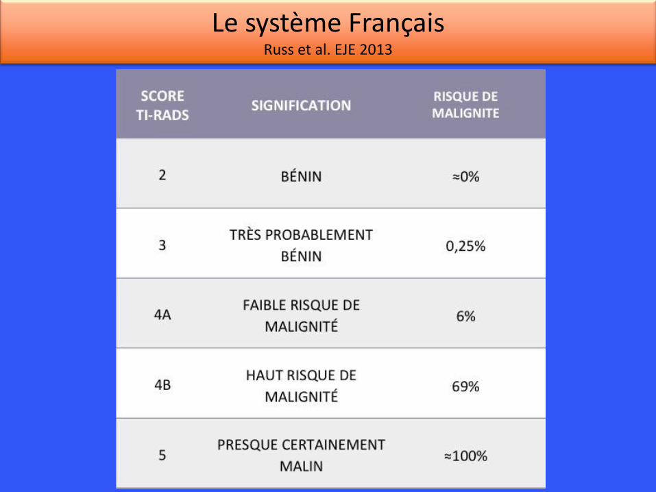
Le système Français Russ et al. EJE 2013
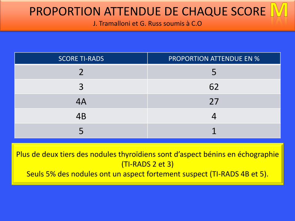
SCORE TI-RADS PROPORTION ATTENDUE EN %
2 5
3 62
4A 27
4B 4
5 1
PROPORTION ATTENDUE DE CHAQUE SCORE J. Tramalloni et G. Russ soumis à C.O
Plus de deux tiers des nodules thyroïdiens sont d’aspect bénins en échographie (TI-RADS 2 et 3)
Seuls 5% des nodules ont un aspect fortement suspect (TI-RADS 4B et 5).
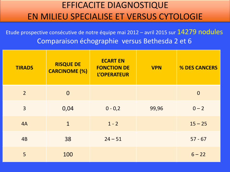
TIRADS RISQUE DE
CARCINOME (%)
ECART EN FONCTION DE L’OPERATEUR
VPN % DES CANCERS
2 0 0
3 0,04 0 - 0,2 99,96 0 – 2
4A 1 1 - 2 15 – 25
4B 38 24 – 51 57 - 67
5 100 6 – 22
Etude prospective consécutive de notre équipe mai 2012 – avril 2015 sur 14279 nodules Comparaison échographie versus Bethesda 2 et 6
EFFICACITE DIAGNOSTIQUE EN MILIEU SPECIALISE ET VERSUS CYTOLOGIE
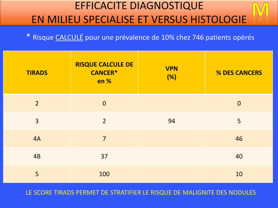
TIRADS RISQUE CALCULE DE
CANCER* en %
VPN (%)
% DES CANCERS
2 0 0
3 2 94 5
4A 7 46
4B 37 40
5 100 10
* Risque CALCULÉ pour une prévalence de 10% chez 746 patients opérés
EFFICACITE DIAGNOSTIQUE EN MILIEU SPECIALISE ET VERSUS HISTOLOGIE
LE SCORE TIRADS PERMET DE STRATIFIER LE RISQUE DE MALIGNITE DES NODULES
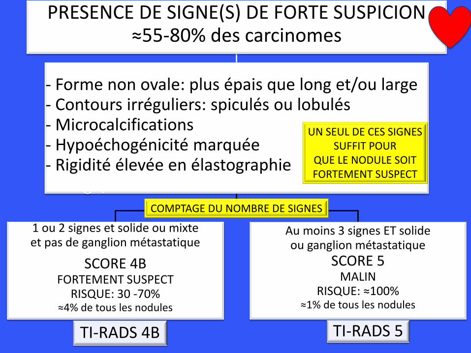
PRESENCE DE SIGNE(S) DE FORTE SUSPICION ≈55-80% des carcinomes
- Forme non ovale: plus épais que long et/ou large - Contours irréguliers: spiculés ou lobulés - Microcalcifications - Hypoéchogénicité marquée - Rigidité élevée en élastographie feature is enough, solid or mixed nodule
1 ou 2 signes et solide ou mixte et pas de ganglion métastatique
SCORE 4B FORTEMENT SUSPECT
RISQUE: 30 -70% ≈4% de tous les nodules
TI-RADS 4B
Au moins 3 signes ET solide ou ganglion métastatique
SCORE 5 MALIN
RISQUE: ≈100% ≈1% de tous les nodules
TI-RADS 5
COMPTAGE DU NOMBRE DE SIGNES
UN SEUL DE CES SIGNES SUFFIT POUR
QUE LE NODULE SOIT FORTEMENT SUSPECT
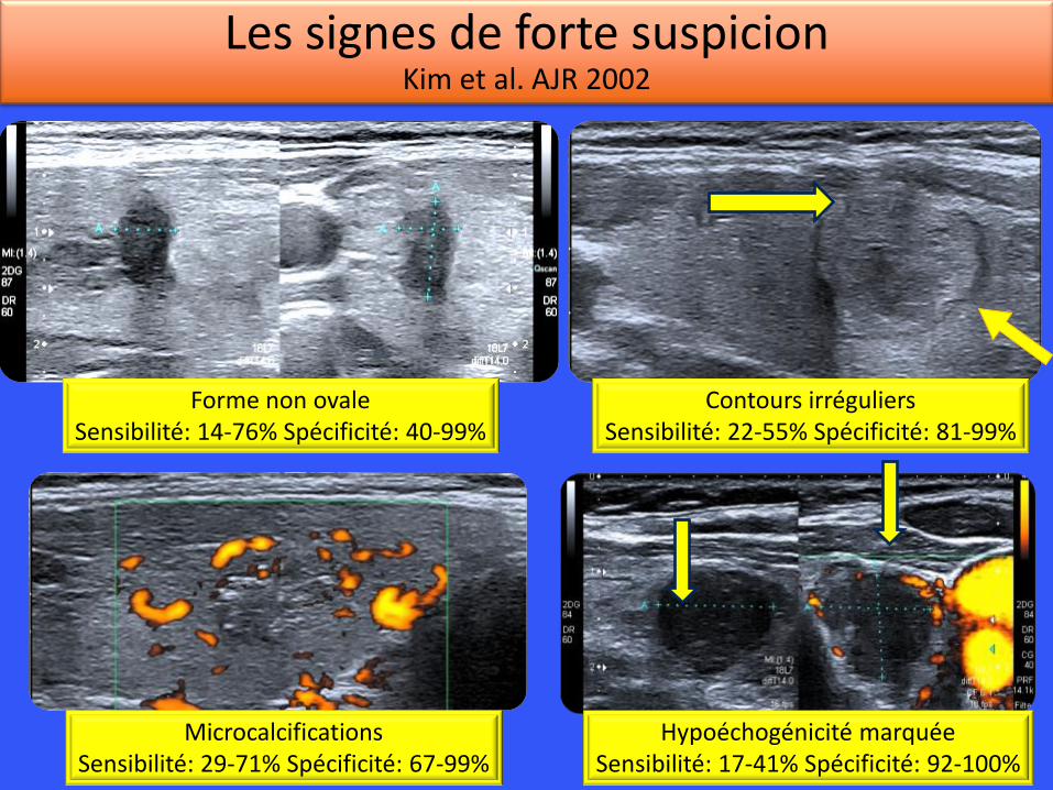
Les signes de forte suspicion Kim et al. AJR 2002
Forme non ovale Sensibilité: 14-76% Spécificité: 40-99%
Contours irréguliers Sensibilité: 22-55% Spécificité: 81-99%
Microcalcifications Sensibilité: 29-71% Spécificité: 67-99%
Hypoéchogénicité marquée Sensibilité: 17-41% Spécificité: 92-100%
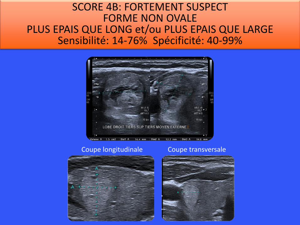
SCORE 4B: FORTEMENT SUSPECT FORME NON OVALE
PLUS EPAIS QUE LONG et/ou PLUS EPAIS QUE LARGE Sensibilité: 14-76% Spécificité: 40-99%
Coupe longitudinale Coupe transversale
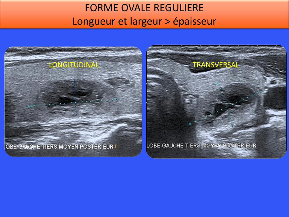
LONGITUDINAL TRANSVERSAL
FORME OVALE REGULIERE Longueur et largeur > épaisseur
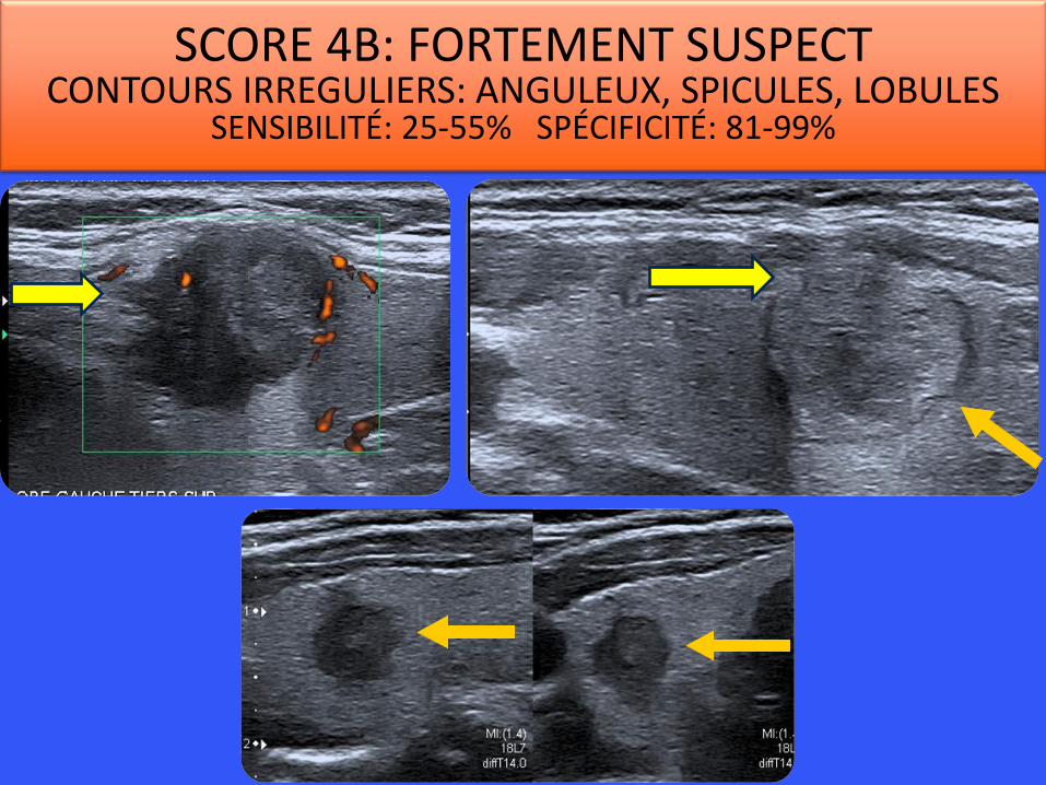
SCORE 4B: FORTEMENT SUSPECT CONTOURS IRREGULIERS: ANGULEUX, SPICULES, LOBULES
SENSIBILITÉ: 25-55% SPÉCIFICITÉ: 81-99%
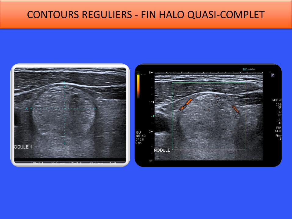
CONTOURS REGULIERS - FIN HALO QUASI-COMPLET
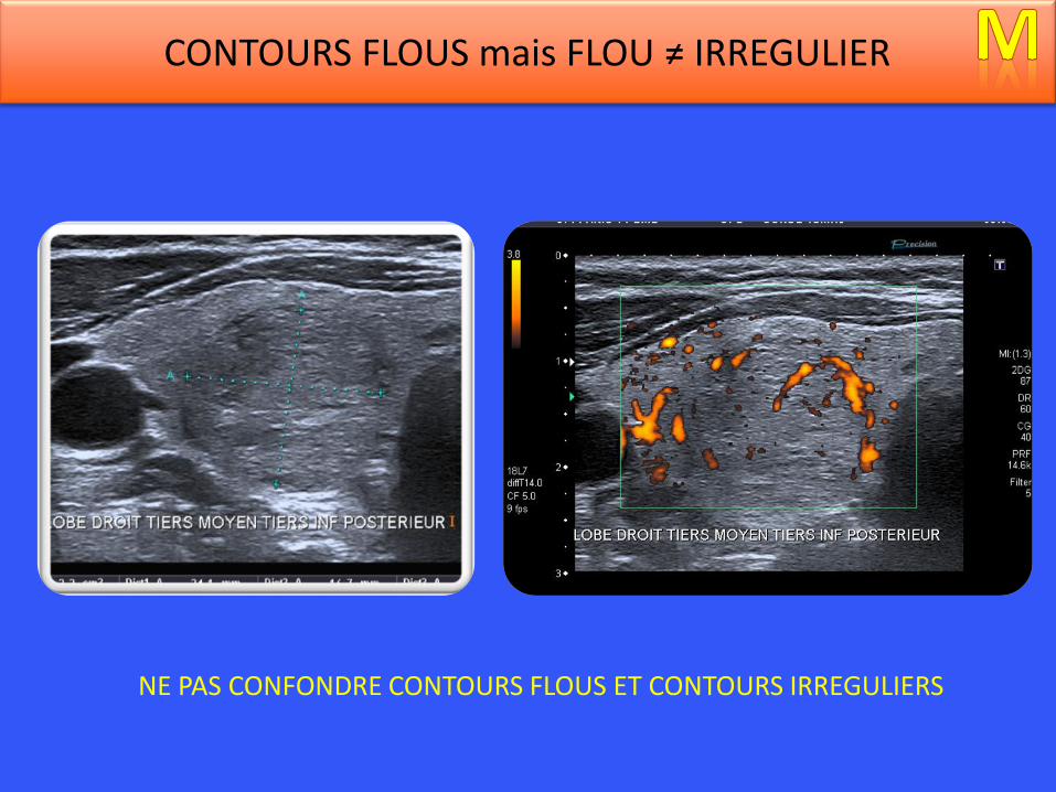
CONTOURS FLOUS mais FLOU ≠ IRREGULIER
NE PAS CONFONDRE CONTOURS FLOUS ET CONTOURS IRREGULIERS
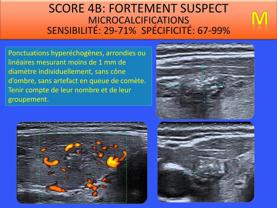
SCORE 4B: FORTEMENT SUSPECT MICROCALCIFICATIONS
SENSIBILITÉ: 29-71% SPÉCIFICITÉ: 67-99%
Ponctuations hyperéchogènes, arrondies ou linéaires mesurant moins de 1 mm de diamètre individuellement, sans cône d’ombre, sans artefact en queue de comète. Tenir compte de leur nombre et de leur groupement.
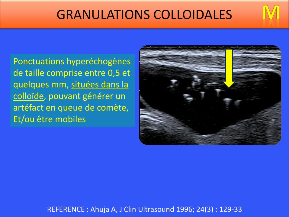
Ponctuations hyperéchogènes de taille comprise entre 0,5 et quelques mm, situées dans la colloïde, pouvant générer un artéfact en queue de comète, Et/ou être mobiles
REFERENCE : Ahuja A, J Clin Ultrasound 1996; 24(3) : 129-33
GRANULATIONS COLLOIDALES
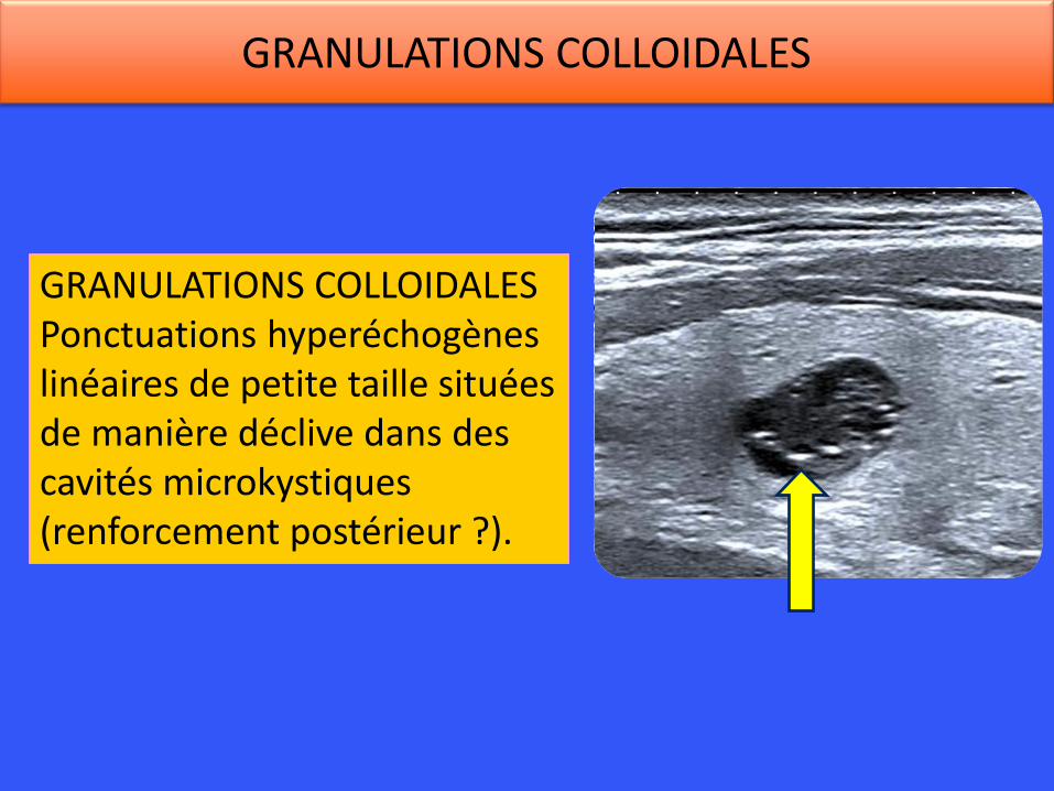
GRANULATIONS COLLOIDALES Ponctuations hyperéchogènes linéaires de petite taille situées de manière déclive dans des cavités microkystiques (renforcement postérieur ?).
GRANULATIONS COLLOIDALES
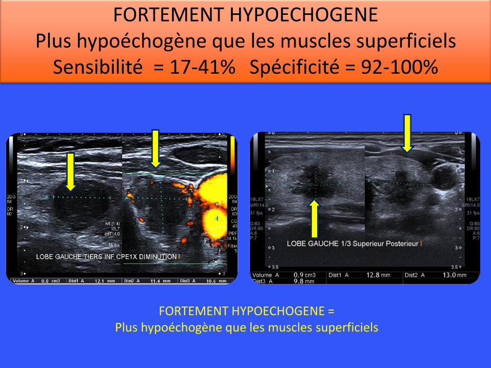
FORTEMENT HYPOECHOGENE Plus hypoéchogène que les muscles superficiels
Sensibilité = 17-41% Spécificité = 92-100%
FORTEMENT HYPOECHOGENE = Plus hypoéchogène que les muscles superficiels
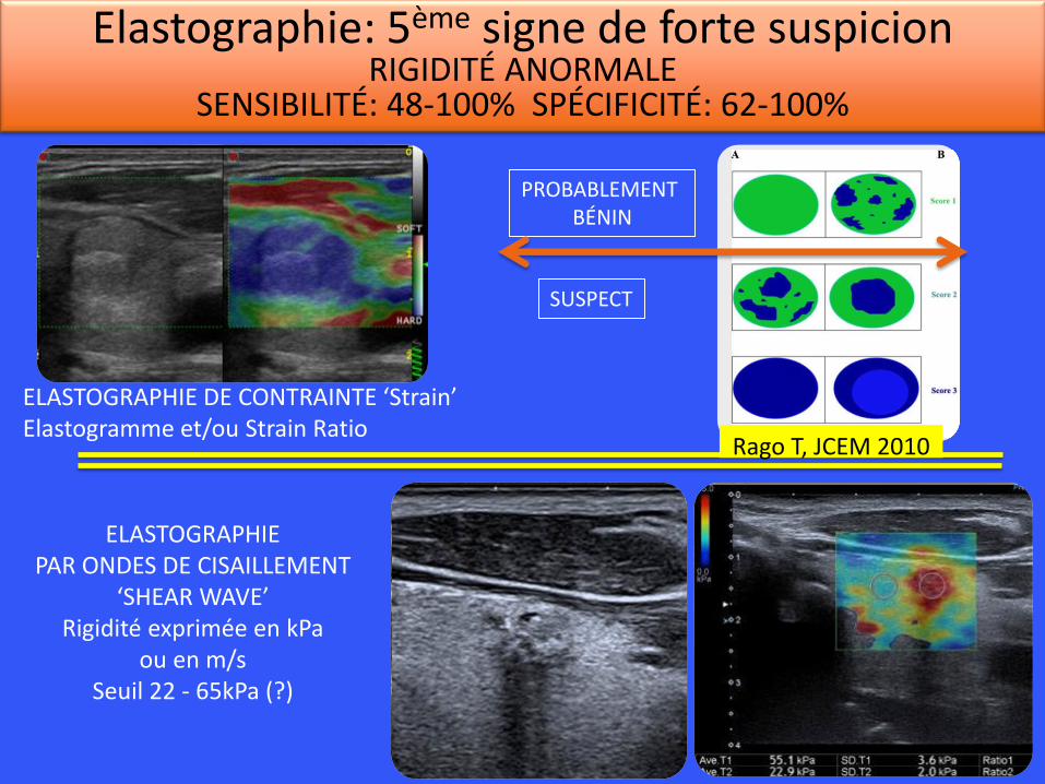
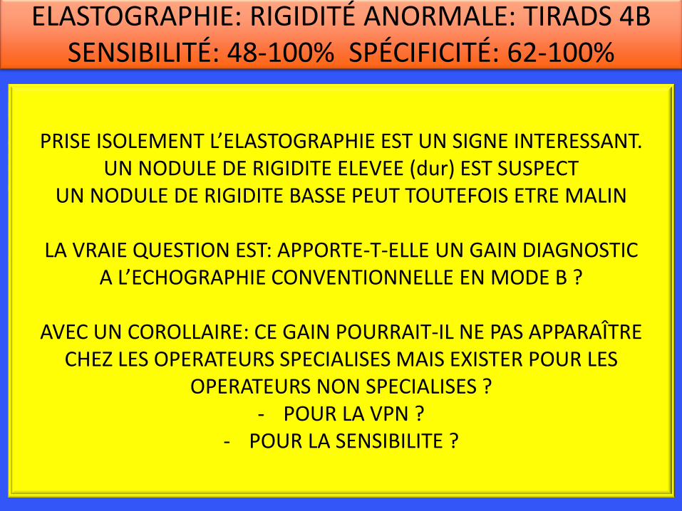
Elastographie: 5ème signe de forte suspicion RIGIDITÉ ANORMALE
SENSIBILITÉ: 48-100% SPÉCIFICITÉ: 62-100%
PROBABLEMENT BÉNIN
SUSPECT
ELASTOGRAPHIE DE CONTRAINTE ‘Strain’ Elastogramme et/ou Strain Ratio
ELASTOGRAPHIE PAR ONDES DE CISAILLEMENT
‘SHEAR WAVE’ Rigidité exprimée en kPa
ou en m/s Seuil 22 - 65kPa (?)
Rago T, JCEM 2010
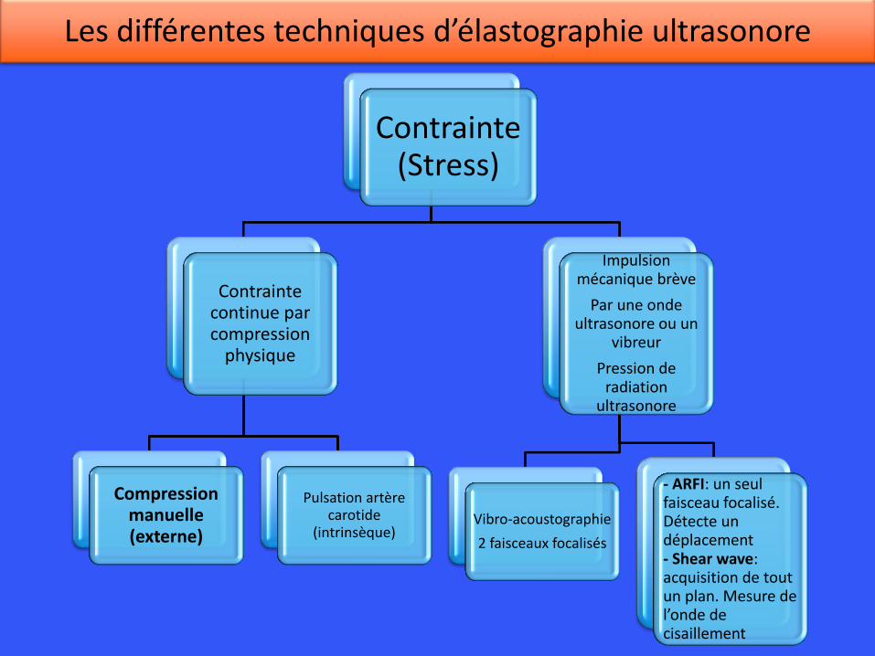
Contrainte(Stress)
Contrainte continue par compression
physique
Compression manuelle (externe)
Pulsation artère carotide
(intrinsèque)
Impulsion mécanique brève
Par une onde ultrasonore ou un
vibreur
Pression de radiation
ultrasonore
Vibro-acoustographie
2 faisceaux focalisés
- ARFI: un seul faisceau focalisé. Détecte un déplacement - Shear wave: acquisition de tout un plan. Mesure de l’onde de cisaillement
Les différentes techniques d’élastographie ultrasonore
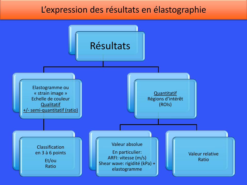
Résultats
Elastogramme ou « strain image »
Echelle de couleur Qualitatif
+/- semi-quantitatif (ratio)
Classification en 3 à 6 points
Et/ou Ratio
Quantitatif Régions d’intérêt
(ROIs)
Valeur absolue
En particulier: ARFI: vitesse (m/s)
Shear wave: rigidité (kPa) + elastogramme
Valeur relative Ratio
L’expression des résultats en élastographie
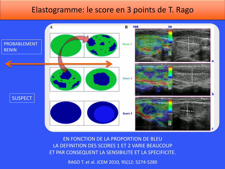
RAGO T. et al. JCEM 2010, 95(12: 5274-5280
PROBABLEMENT BENIN
SUSPECT
EN FONCTION DE LA PROPORTION DE BLEU LA DEFINITION DES SCORES 1 ET 2 VARIE BEAUCOUP
ET PAR CONSEQUENT LA SENSIBILITE ET LA SPECIFICITE.
Elastogramme: le score en 3 points de T. Rago
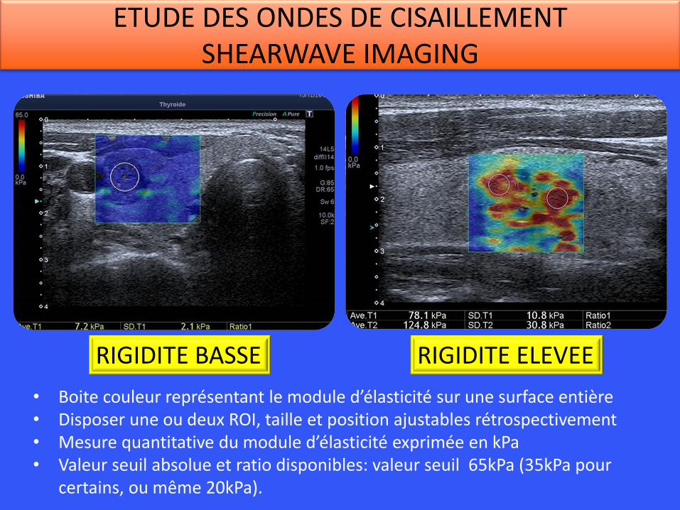
ETUDE DES ONDES DE CISAILLEMENT SHEARWAVE IMAGING
• Boite couleur représentant le module d’élasticité sur une surface entière • Disposer une ou deux ROI, taille et position ajustables rétrospectivement • Mesure quantitative du module d’élasticité exprimée en kPa • Valeur seuil absolue et ratio disponibles: valeur seuil 65kPa (35kPa pour
certains, ou même 20kPa).
RIGIDITE ELEVEE RIGIDITE BASSE
ELASTOGRAPHIE: RIGIDITÉ ANORMALE: TIRADS 4B SENSIBILITÉ: 48-100% SPÉCIFICITÉ: 62-100%
SCORE 3 RIGIDITE ELEVEE
SCORE 1 RIGIDITE BASSE
VPN 98%* ?? *Veyrieres et al. Eur J Radiol 2012; 81: 3965-72 * Zhang B et al. J Ultrasound Med 2013; 32: 2163-9 * Nell S et al. Eur J Radiol 2015; 84: 652:61
PRISE ISOLEMENT L’ELASTOGRAPHIE EST UN SIGNE INTERESSANT. UN NODULE DE RIGIDITE ELEVEE (dur) EST SUSPECT
UN NODULE DE RIGIDITE BASSE PEUT TOUTEFOIS ETRE MALIN
LA VRAIE QUESTION EST: APPORTE-T-ELLE UN GAIN DIAGNOSTIC A L’ECHOGRAPHIE CONVENTIONNELLE EN MODE B ?
AVEC UN COROLLAIRE: CE GAIN POURRAIT-IL NE PAS APPARAÎTRE
CHEZ LES OPERATEURS SPECIALISES MAIS EXISTER POUR LES OPERATEURS NON SPECIALISES ?
- POUR LA VPN ? - POUR LA SENSIBILITE ?
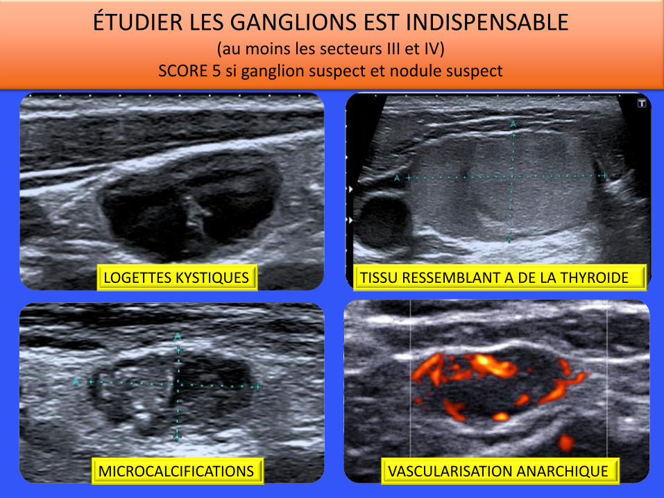
ÉTUDIER LES GANGLIONS EST INDISPENSABLE (au moins les secteurs III et IV)
SCORE 5 si ganglion suspect et nodule suspect
LOGETTES KYSTIQUES TISSU RESSEMBLANT A DE LA THYROIDE
MICROCALCIFICATIONS VASCULARISATION ANARCHIQUE
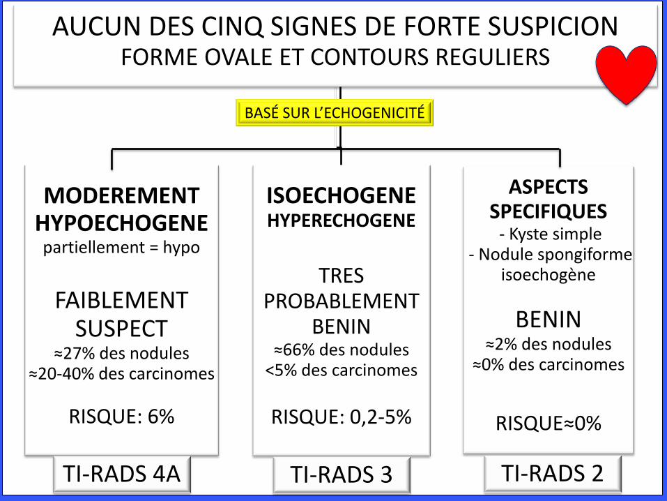
AUCUN DES CINQ SIGNES DE FORTE SUSPICION FORME OVALE ET CONTOURS REGULIERS
MODEREMENT HYPOECHOGENE partiellement = hypo
FAIBLEMENT
SUSPECT ≈27% des nodules
≈20-40% des carcinomes
RISQUE: 6%
TI-RADS 4A
ISOECHOGENE HYPERECHOGENE
TRES PROBABLEMENT
BENIN ≈66% des nodules
<5% des carcinomes
RISQUE: 0,2-5%
TI-RADS 3
ASPECTS SPECIFIQUES - Kyste simple
- Nodule spongiforme isoechogène
BENIN ≈2% des nodules
≈0% des carcinomes
RISQUE≈0%
TI-RADS 2
BASÉ SUR L’ECHOGENICITÉ
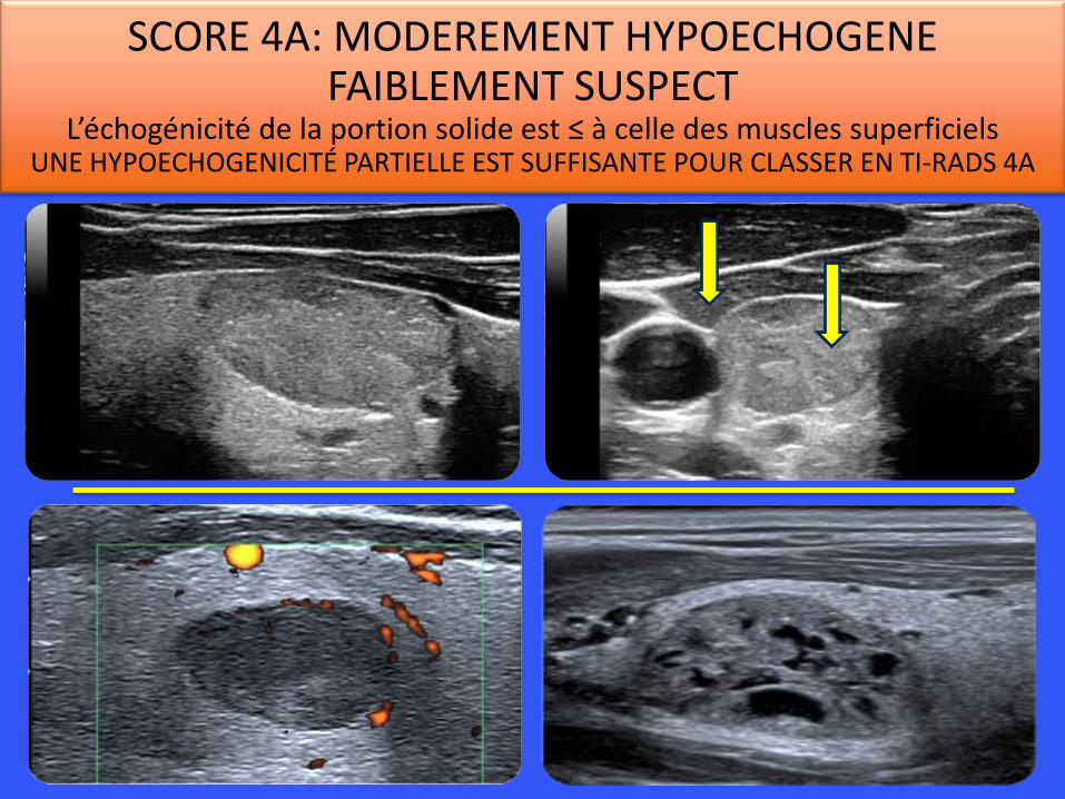
SCORE 4A: MODEREMENT HYPOECHOGENE FAIBLEMENT SUSPECT
L’échogénicité de la portion solide est ≤ à celle des muscles superficiels UNE HYPOECHOGENICITÉ PARTIELLE EST SUFFISANTE POUR CLASSER EN TI-RADS 4A

ECHOGENICITÉ: MODEREMENT HYPOECHOGÈNE L’échogénicité dépend du gain
GAIN EXCESSIF GAIN OPTIMISE
ISOECHOGENE OU MODEREMENT HYPOECHOGENE ?
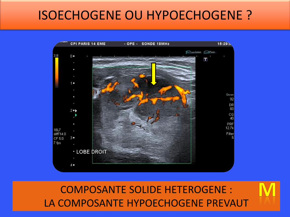
COMPOSANTE SOLIDE HETEROGENE : LA COMPOSANTE HYPOECHOGENE PREVAUT
ISOECHOGENE OU HYPOECHOGENE ?
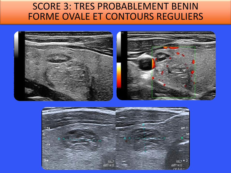
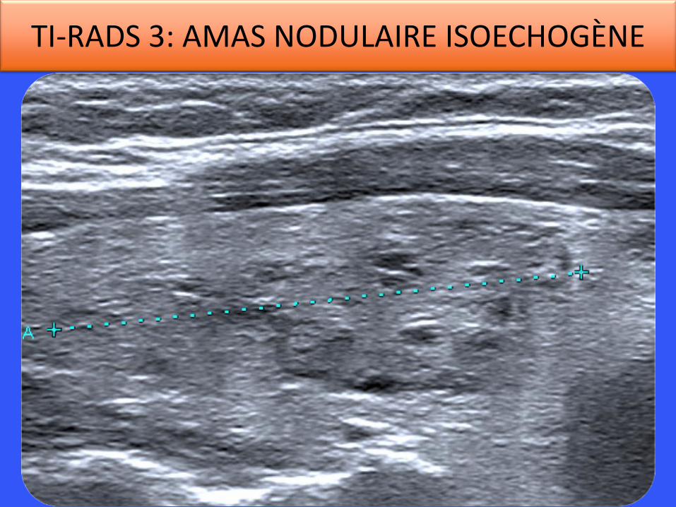
SCORE 3: TRES PROBABLEMENT BENIN FORME OVALE ET CONTOURS REGULIERS
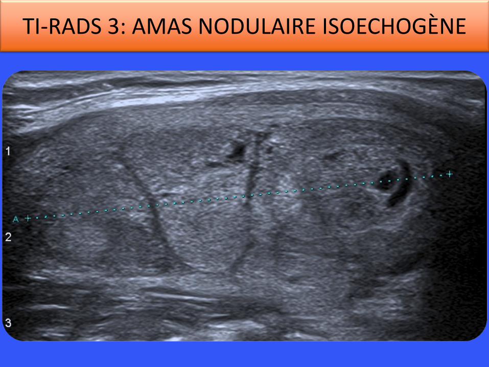
TI-RADS 3: AMAS NODULAIRE ISOECHOGÈNE
TI-RADS 3: AMAS NODULAIRE ISOECHOGÈNE
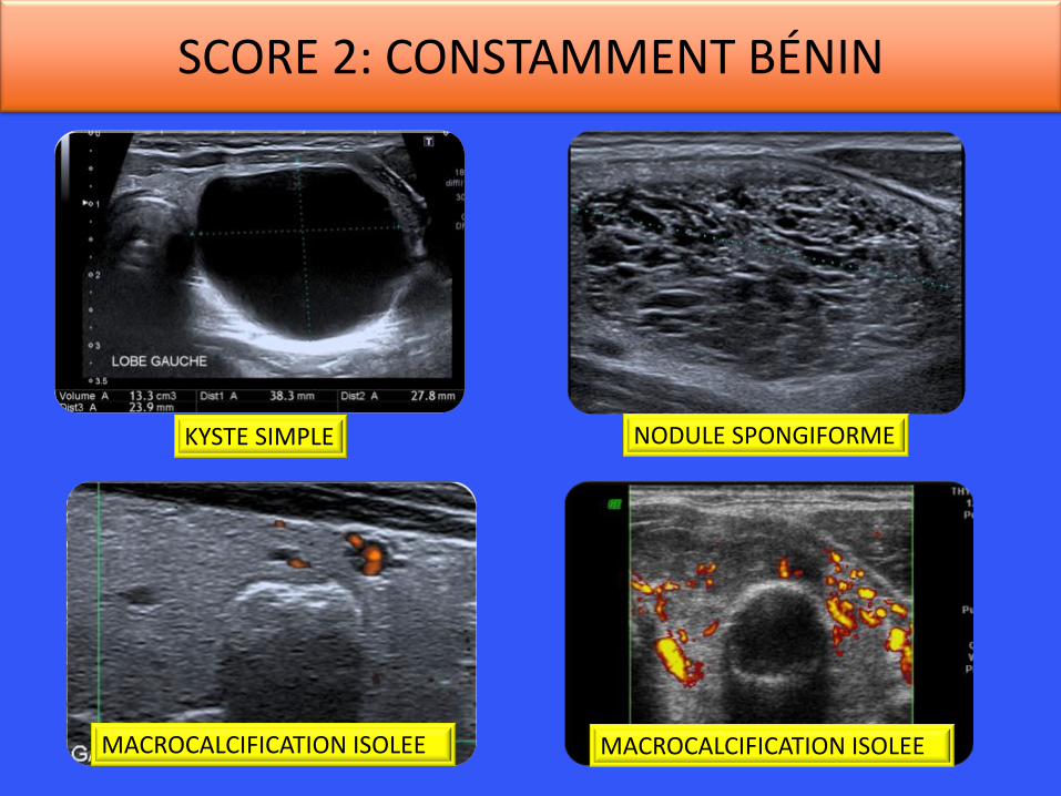
SCORE 2: CONSTAMMENT BÉNIN
KYSTE SIMPLE NODULE SPONGIFORME
MACROCALCIFICATION ISOLEE MACROCALCIFICATION ISOLEE
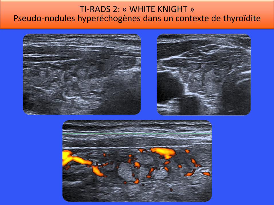
TI-RADS 2: « WHITE KNIGHT » Pseudo-nodules hyperéchogènes dans un contexte de thyroïdite
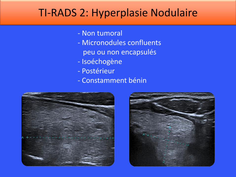
- Non tumoral - Micronodules confluents peu ou non encapsulés - Isoéchogène - Postérieur - Constamment bénin
TI-RADS 2: Hyperplasie Nodulaire
4. TIRADS: un phénomène mondial

LA STRATIFICATION DU RISQUE Les publications
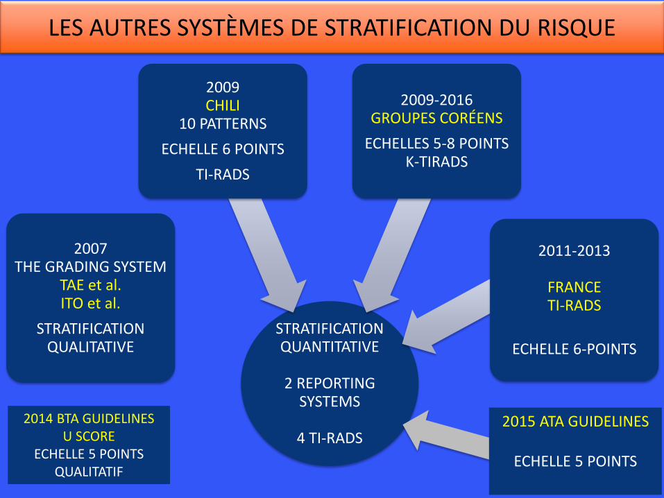
STRATIFICATION QUANTITATIVE
2 REPORTING
SYSTEMS
4 TI-RADS
2007 THE GRADING SYSTEM
TAE et al. ITO et al.
STRATIFICATION QUALITATIVE
2009 CHILI
10 PATTERNS
ECHELLE 6 POINTS
TI-RADS
2009-2016 GROUPES CORÉENS
ECHELLES 5-8 POINTS K-TIRADS
2011-2013
FRANCE TI-RADS
ECHELLE 6-POINTS
2014 BTA GUIDELINES U SCORE
ECHELLE 5 POINTS QUALITATIF
LES AUTRES SYSTÈMES DE STRATIFICATION DU RISQUE
2015 ATA GUIDELINES
ECHELLE 5 POINTS
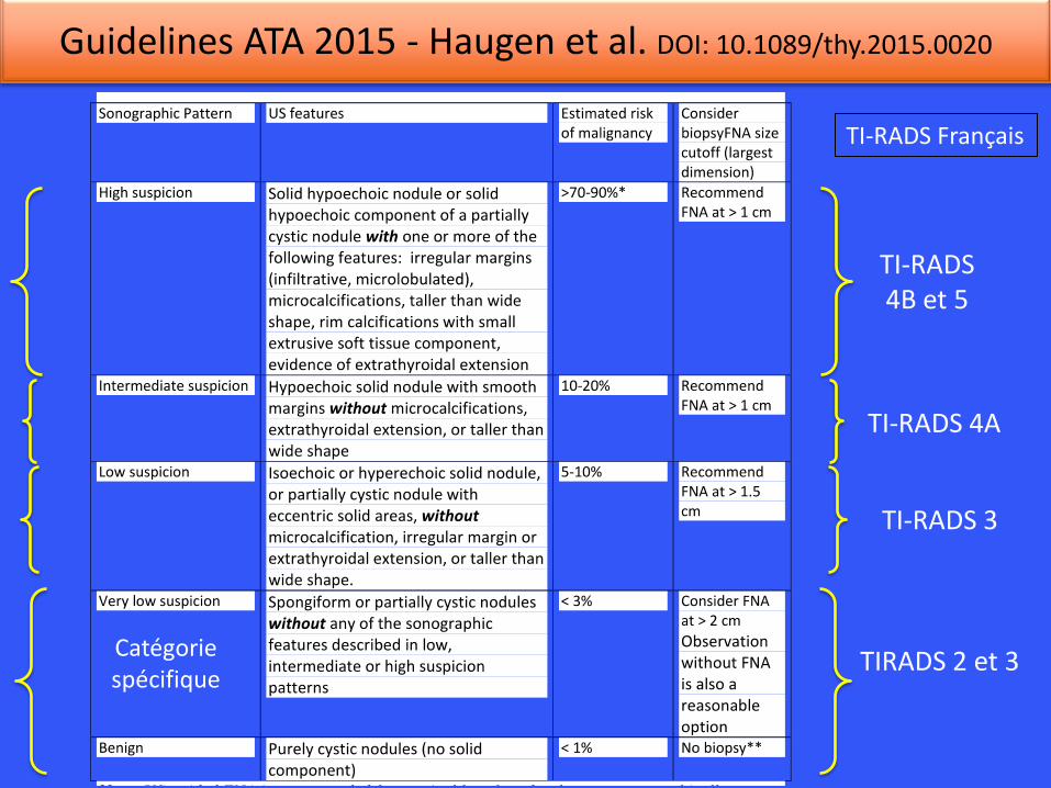
TIRADS 2 et 3
TI-RADS 3
TI-RADS 4A
TI-RADS 4B et 5
Guidelines ATA 2015 - Haugen et al. DOI: 10.1089/thy.2015.0020
TI-RADS Français
Catégorie spécifique
• Comptage du nombre de signes de suspicion • 3 sous-catégories de suspicion (4A, 4B and 4C)
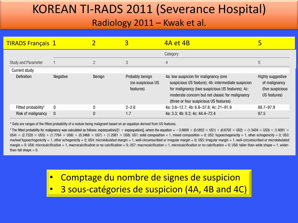
KOREAN TI-RADS 2011 (Severance Hospital) Radiology 2011 – Kwak et al.
TIRADS Français 1 2 3 4A et 4B 5
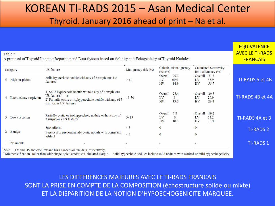
KOREAN TI-RADS 2015 – Asan Medical Center Thyroid. January 2016 ahead of print – Na et al.
TI-RADS 5 et 4B
TI-RADS 4B et 4A
TI-RADS 4A et 3
TI-RADS 2
EQUIVALENCE AVEC LE TI-RADS
FRANCAIS
TI-RADS 1
LES DIFFERENCES MAJEURES AVEC LE TI-RADS FRANCAIS SONT LA PRISE EN COMPTE DE LA COMPOSITION (échostructure solide ou mixte)
ET LA DISPARITION DE LA NOTION D’HYPOECHOGENICITE MARQUEE.
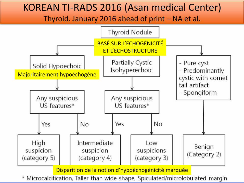
KOREAN TI-RADS 2016 (Asan medical Center) Thyroid. January 2016 ahead of print – NA et al.
BASÉ SUR L’ECHOGÉNICITÉ ET L’ECHOSTRUCTURE
Majoritairement hypoéchogène
Disparition de la notion d’hypoéchogénicité marquée
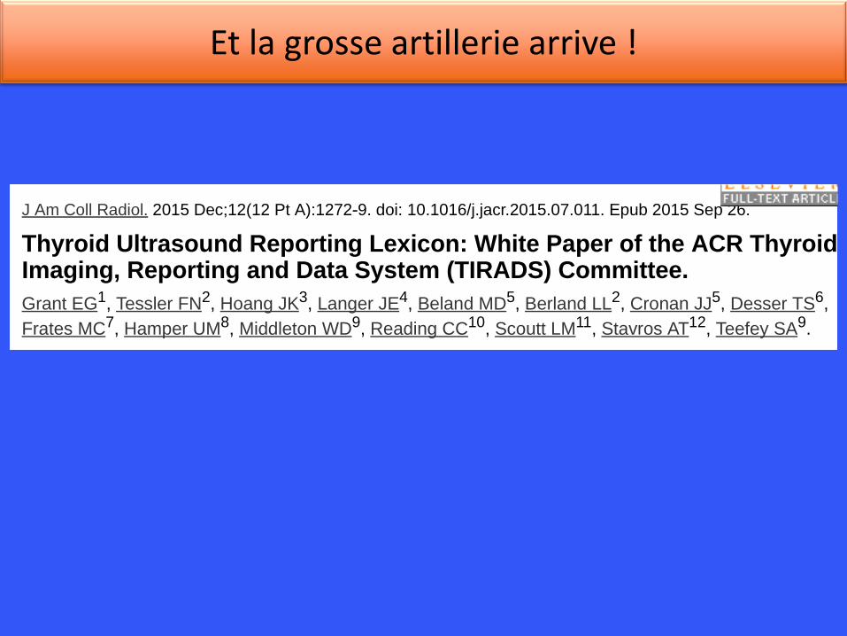
Et la grosse artillerie arrive ! 5/3/2016 Thyroid Ultrasound Reporting Lexicon: White Paper of the ACR Thyroid Imaging, Reporting and Data System (TIRADS) Committee. - PubMed - NCBI
http://www.ncbi.nlm.nih.gov/pubmed/26419308 1/2
KEYWORDS:
PubMed Commons home
How to join PubMed Commons
J Am Coll Radiol. 2015 Dec;12(12 Pt A):12729. doi: 10.1016/j.jacr.2015.07.011. Epub 2015 Sep 26.
Thyroid Ultrasound Reporting Lexicon: White Paper of the ACR ThyroidImaging, Reporting and Data System (TIRADS) Committee.
Grant EG , Tessler FN , Hoang JK , Langer JE , Beland MD , Berland LL , Cronan JJ , Desser TS ,
Frates MC , Hamper UM , Middleton WD , Reading CC , Scoutt LM , Stavros AT , Teefey SA .
Abstract
Ultrasound is the most commonly used imaging technique for the evaluation of thyroid nodules.
Sonographic findings are often not specific, and definitive diagnosis is usually made through fine
needle aspiration biopsy or even surgery. In reviewing the literature, terms used to describe
nodules are often poorly defined and inconsistently applied. Several authors have recently
described a standardized risk stratification system called the Thyroid Imaging, Reporting and
Data System (TIRADS), modeled on the BIRADS system for breast imaging. However, most of
these TIRADS classifications have come from individual institutions, and none has been widely
adopted in the United States. Under the auspices of the ACR, a committee was organized to
develop TIRADS. The eventual goal is to provide practitioners with evidencebased
recommendations for the management of thyroid nodules on the basis of a set of welldefined
sonographic features or terms that can be applied to every lesion. Terms were chosen on the
basis of demonstration of consistency with regard to performance in the diagnosis of thyroid
cancer or, conversely, classifying a nodule as benign and avoiding followup. The initial portion of
this project was aimed at standardizing the diagnostic approach to thyroid nodules with regard to
terminology through the development of a lexicon. This white paper describes the consensus
process and the resultant lexicon.
Copyright © 2015 American College of Radiology. Published by Elsevier Inc. All rights reserved.
Thyroid nodule; structured reporting; thyroid cancer; thyroid imaging; ultrasound
PMID: 26419308 [PubMed in process]
PubMed Commons
0 comments
Abstract
1 2 3 4 5 2 5 6
7 8 9 10 11 12 9
Author information
LinkOut more resources
Full text links
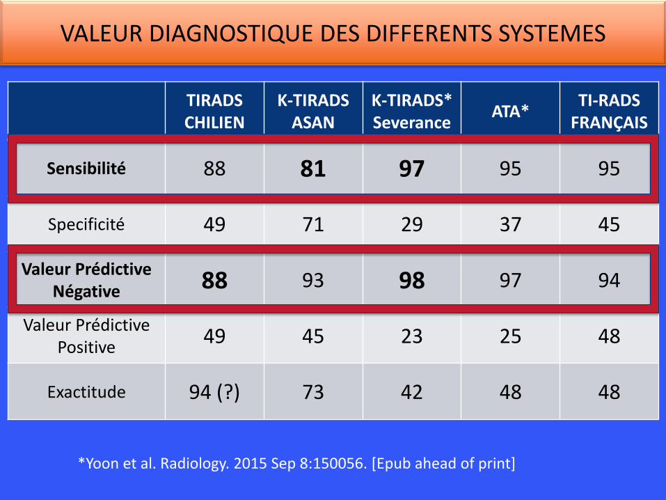
TIRADS CHILIEN
K-TIRADS ASAN
K-TIRADS* Severance
ATA* TI-RADS
FRANÇAIS
Sensibilité 88 81 97 95 95
Specificité 49 71 29 37 45
Valeur Prédictive Négative 88 93 98 97 94
Valeur Prédictive Positive
49 45 23 25 48
Exactitude 94 (?) 73 42 48 48
VALEUR DIAGNOSTIQUE DES DIFFERENTS SYSTEMES
*Yoon et al. Radiology. 2015 Sep 8:150056. [Epub ahead of print]
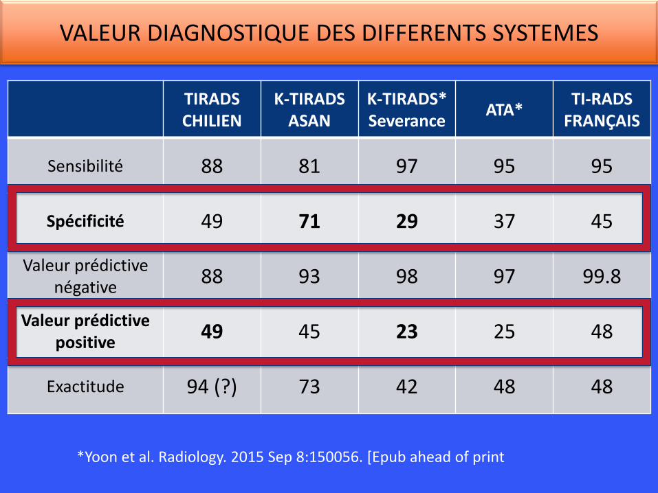
TIRADS CHILIEN
K-TIRADS ASAN
K-TIRADS* Severance
ATA* TI-RADS
FRANÇAIS
Sensibilité 88 81 97 95 95
Spécificité 49 71 29 37 45
Valeur prédictive négative
88 93 98 97 99.8
Valeur prédictive positive
49 45 23 25 48
Exactitude 94 (?) 73 42 48 48
VALEUR DIAGNOSTIQUE DES DIFFERENTS SYSTEMES
*Yoon et al. Radiology. 2015 Sep 8:150056. [Epub ahead of print
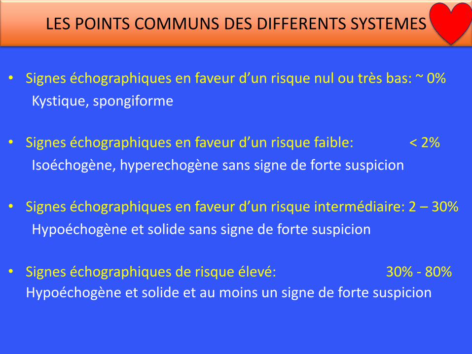
LES POINTS COMMUNS DES DIFFERENTS SYSTEMES
• Signes échographiques en faveur d’un risque nul ou très bas: ~ 0%
Kystique, spongiforme
• Signes échographiques en faveur d’un risque faible: < 2%
Isoéchogène, hyperechogène sans signe de forte suspicion
• Signes échographiques en faveur d’un risque intermédiaire: 2 – 30%
Hypoéchogène et solide sans signe de forte suspicion
• Signes échographiques de risque élevé: 30% - 80%
Hypoéchogène et solide et au moins un signe de forte suspicion
5. FAUX NEGATIFS DU TIRADS
FAUX NEGATIFS DU TI-RADS NODULES KYSTIQUES
• Les nodules majoritairement kystiques carcinomateux représentent 1-3% du total des carcinomes et la moitié des faux négatifs du TI-RADS.
• Il s’agit dans l’immense majorité des cas de carcinomes papillaires de variante classique avec une forte composante kystique.
• 2 signes de la partie solide sont à prendre en compte: – Hypoéchogénicité – La présence de microcalcifications
• La cytoponction doit cibler la composante solide.
Lee et al. Thyroid 2009, 19(4); 341-46
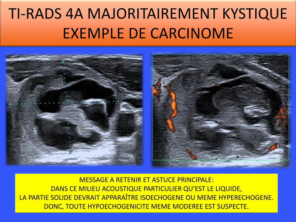
TI-RADS 4A MAJORITAIREMENT KYSTIQUE EXEMPLE DE CARCINOME
MESSAGE A RETENIR ET ASTUCE PRINCIPALE: DANS CE MILIEU ACOUSTIQUE PARTICULIER QU’EST LE LIQUIDE,
LA PARTIE SOLIDE DEVRAIT APPARAÎTRE ISOECHOGENE OU MEME HYPERECHOGENE. DONC, TOUTE HYPOECHOGENICITE MEME MODEREE EST SUSPECTE.
FAUX NEGATIFS DU TI-RADS NODULES SOLIDES ISOECHOGENES
• Les nodules solides isoéchogènes sans signes de forte suspicion représentent 1-3% des carcinomes et la moitié des faux négatifs du TI-RADS.
• Il s’agit dans l’immense majorité des cas de carcinomes papillaires de variante folliculaire et plus rarement de carcinomes folliculaires.
• Les carcinomes sont isoéchogènes dans 15% des cas, dans la plupart des cas avec des signes de forte suspicion.
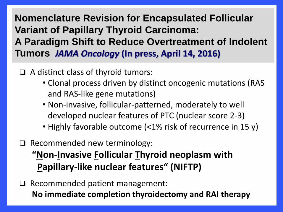
Nomenclature Revision for Encapsulated Follicular
Variant of Papillary Thyroid Carcinoma:
A Paradigm Shift to Reduce Overtreatment of Indolent
Tumors JAMA Oncology (In press, April 14, 2016)
A distinct class of thyroid tumors: • Clonal process driven by distinct oncogenic mutations (RAS
and RAS-like gene mutations) • Non-invasive, follicular-patterned, moderately to well
developed nuclear features of PTC (nuclear score 2-3) • Highly favorable outcome (<1% risk of recurrence in 15 y)
Recommended new terminology:
“Non-Invasive Follicular Thyroid neoplasm with Papillary-like nuclear features“ (NIFTP)
Recommended patient management: No immediate completion thyroidectomy and RAI therapy
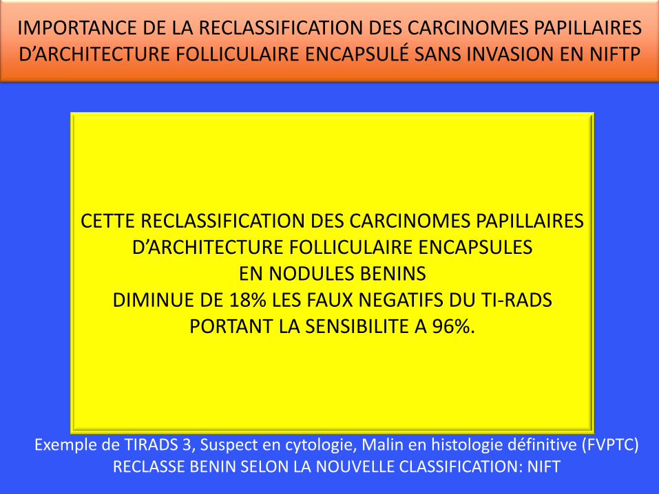
IMPORTANCE DE LA RECLASSIFICATION DES CARCINOMES PAPILLAIRES D’ARCHITECTURE FOLLICULAIRE ENCAPSULÉ SANS INVASION EN NIFTP
Un nouveau concept, le « NIFTP » (non invasive follicular thyroid neoplasm with papillary like nuclear features), remplace le carcinome papillaire d’architecture folliculaire encapsulé.
Ceux-ci sont reclassés comme bénins.
Exemple de TIRADS 3, Suspect en cytologie, Malin en histologie définitive (FVPTC) RECLASSE BENIN SELON LA NOUVELLE CLASSIFICATION: NIFT
CETTE RECLASSIFICATION DES CARCINOMES PAPILLAIRES D’ARCHITECTURE FOLLICULAIRE ENCAPSULES
EN NODULES BENINS DIMINUE DE 18% LES FAUX NEGATIFS DU TI-RADS
PORTANT LA SENSIBILITE A 96%.
• FAUX NEGATIFS (15 cas)
• ≤ 10mm : aucun cas • > 10mm et ≤ 20mm : 6 cas, aucun reclassé en NIFT : 4,9% • >20mm et ≤ 40mm: 5 cas dont 2 reclassés en NIFT : 5,9% à 3,5% • > 40mm: 4 cas dont 1 reclassé en NIFT: 16% à 12%
dont 2 Bethesda II dont 1 NIFT soit 1 cas donc 4% • TOUS CANCERS (296 cas)
• ≤ 10mm : 64 cas • > 10mm et ≤ 20mm : 122 cas • >20mm et ≤ 40mm: 85 cas • > 40mm: 25 cas
0
2
4
6
8
10
12
≤ 10mm ≤ 20mm ≤ 40mm > 40mm
Faux négatifs ( %)
Faux négatifs ( %)
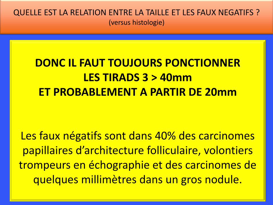
DONC IL FAUT TOUJOURS PONCTIONNER LES TIRADS 3 > 40mm
ET PROBABLEMENT A PARTIR DE 20mm
Les faux négatifs sont dans 40% des carcinomes papillaires d’architecture folliculaire, volontiers
trompeurs en échographie et des carcinomes de quelques millimètres dans un gros nodule.
QUELLE EST LA RELATION ENTRE LA TAILLE ET LES FAUX NEGATIFS ? (versus histologie)
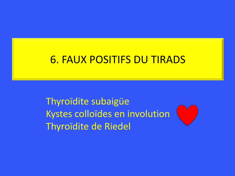
6. FAUX POSITIFS DU TIRADS
Thyroïdite subaigüe Kystes colloïdes en involution Thyroïdite de Riedel
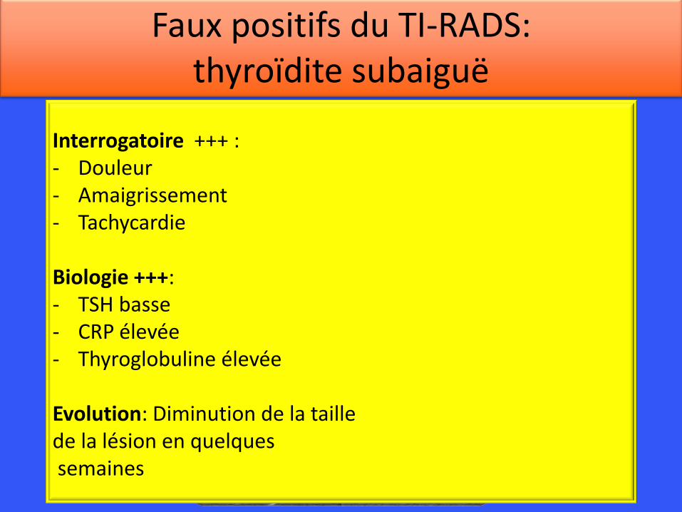
Faux positifs du TI-RADS: thyroïdite subaiguë
Interrogatoire +++ : - Douleur - Amaigrissement - Tachycardie Biologie +++: - TSH basse - CRP élevée - Thyroglobuline élevée
Evolution: Diminution de la taille de la lésion en quelques semaines
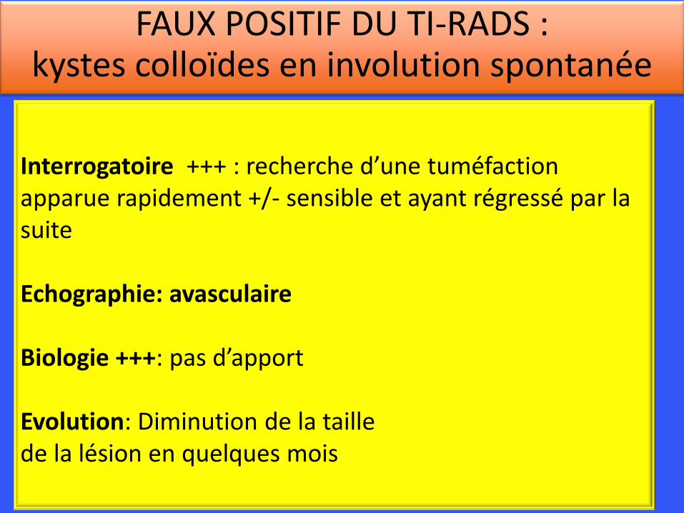
FAUX POSITIF DU TI-RADS : kystes colloïdes en involution spontanée
16/03/2016 16/03/2016
28/11/2014 31/05/2013
Interrogatoire +++ : recherche d’une tuméfaction apparue rapidement +/- sensible et ayant régressé par la suite Echographie: avasculaire Biologie +++: pas d’apport
Evolution: Diminution de la taille de la lésion en quelques mois
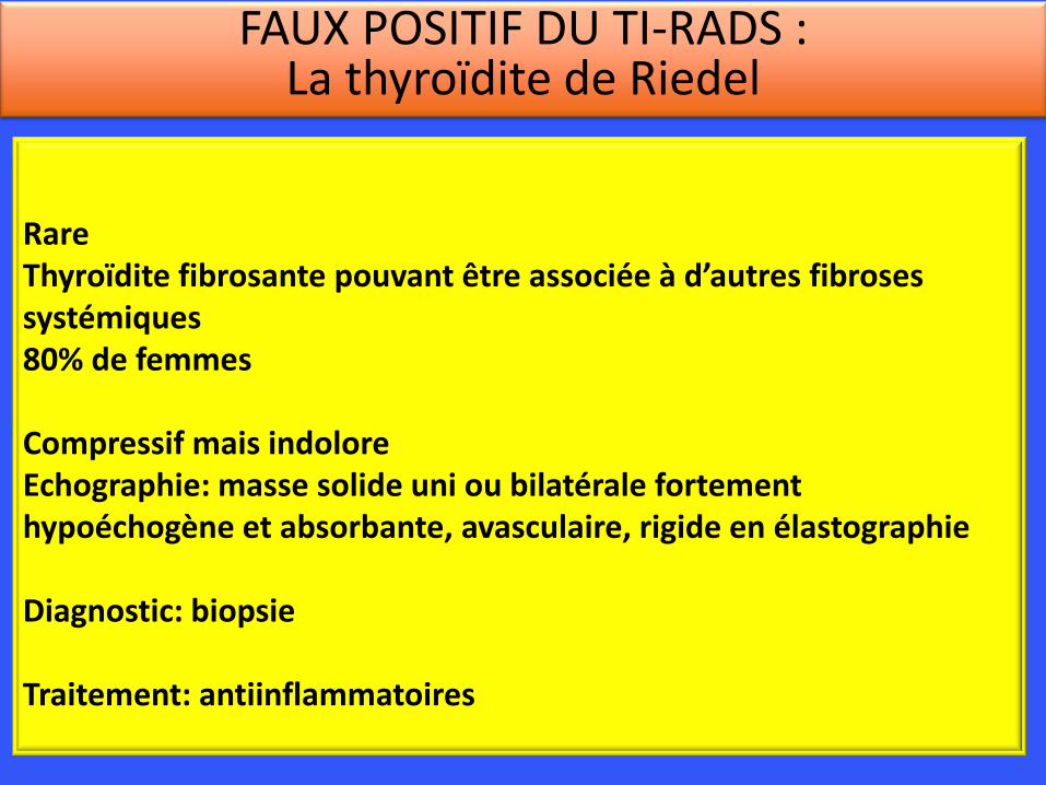
FAUX POSITIF DU TI-RADS : La thyroïdite de Riedel
Rare Thyroïdite fibrosante pouvant être associée à d’autres fibroses systémiques 80% de femmes Compressif mais indolore Echographie: masse solide uni ou bilatérale fortement hypoéchogène et absorbante, avasculaire, rigide en élastographie Diagnostic: biopsie Traitement: antiinflammatoires
7. LES SIGNES QUI NE SONT PAS DANS LE TI-RADS
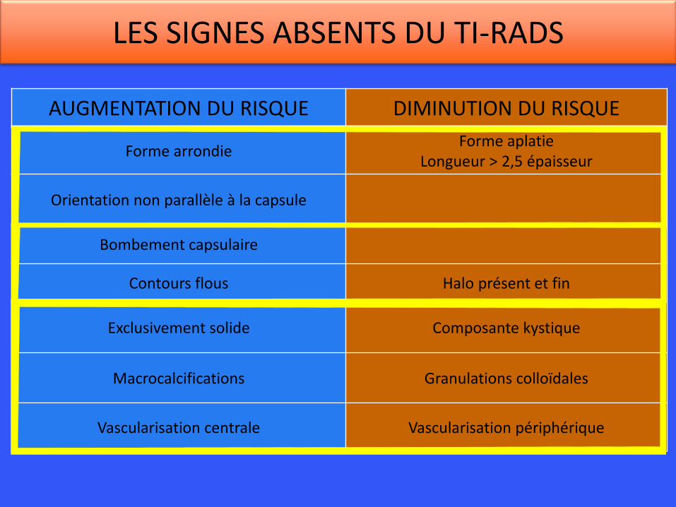
LES SIGNES ABSENTS DU TI-RADS
AUGMENTATION DU RISQUE DIMINUTION DU RISQUE
Forme arrondie Forme aplatie
Longueur > 2,5 épaisseur
Orientation non parallèle à la capsule
Bombement capsulaire
Contours flous Halo présent et fin
Exclusivement solide Composante kystique
Macrocalcifications Granulations colloïdales
Vascularisation centrale Vascularisation périphérique
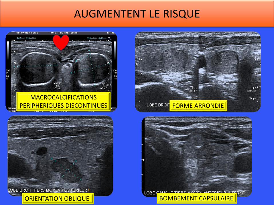
AUGMENTENT LE RISQUE
MACROCALCIFICATIONS PERIPHERIQUES DISCONTINUES FORME ARRONDIE
ORIENTATION OBLIQUE BOMBEMENT CAPSULAIRE
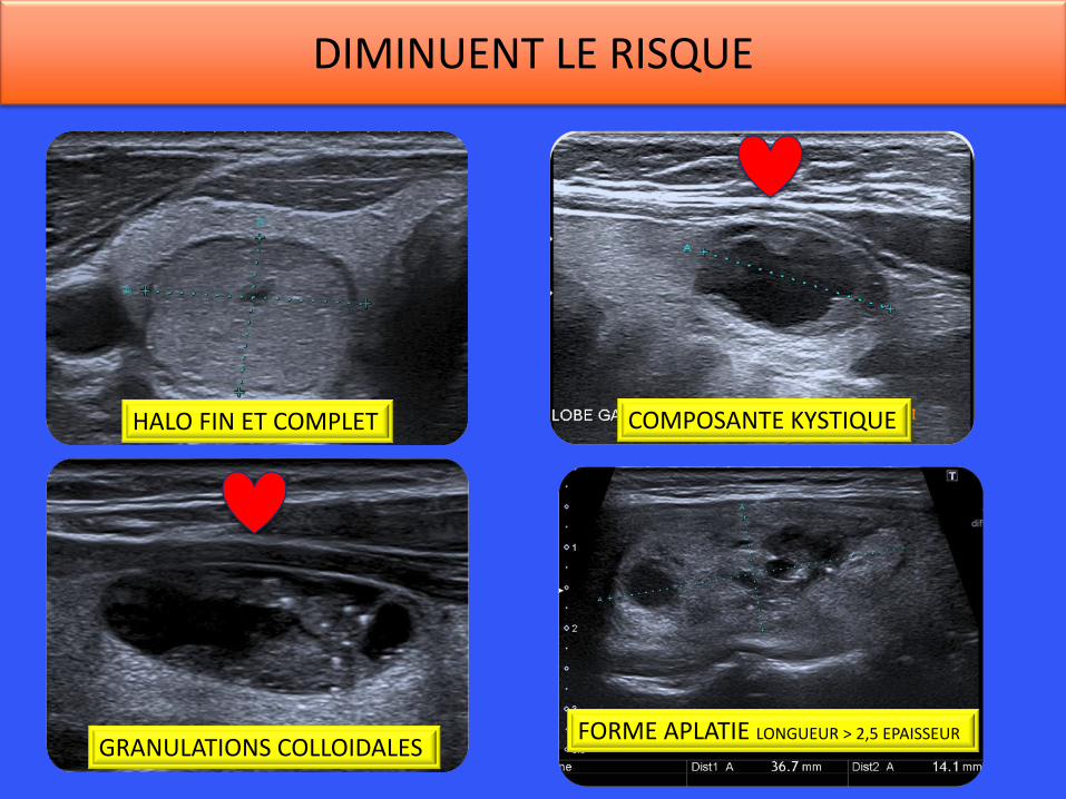
DIMINUENT LE RISQUE
HALO FIN ET COMPLET COMPOSANTE KYSTIQUE
GRANULATIONS COLLOIDALES FORME APLATIE LONGUEUR > 2,5 EPAISSEUR
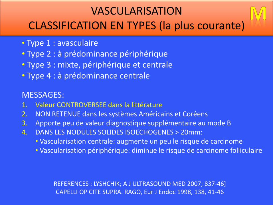
• Type 1 : avasculaire • Type 2 : à prédominance périphérique • Type 3 : mixte, périphérique et centrale • Type 4 : à prédominance centrale
MESSAGES: 1. Valeur CONTROVERSEE dans la littérature 2. NON RETENUE dans les systèmes Américains et Coréens 3. Apporte peu de valeur diagnostique supplémentaire au mode B 4. DANS LES NODULES SOLIDES ISOECHOGENES > 20mm:
• Vascularisation centrale: augmente un peu le risque de carcinome • Vascularisation périphérique: diminue le risque de carcinome folliculaire
REFERENCES : LYSHCHIK; A J ULTRASOUND MED 2007; 837-46] CAPELLI OP CITE SUPRA. RAGO, Eur J Endoc 1998, 138, 41-46
VASCULARISATION CLASSIFICATION EN TYPES (la plus courante)
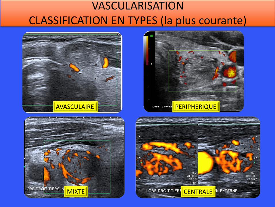
VASCULARISATION CLASSIFICATION EN TYPES (la plus courante)
PERIPHERIQUE AVASCULAIRE
MIXTE CENTRALE
8. LA THYROÏDE MULTINODULAIRE
Kunreuther E, ATA 2004
Papini E, JCEM 2002
Kim EK, AJR 2002
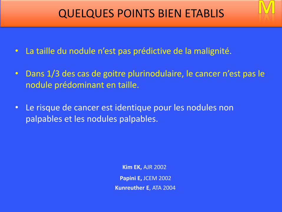
QUELQUES POINTS BIEN ETABLIS
• La taille du nodule n’est pas prédictive de la malignité.
• Dans 1/3 des cas de goitre plurinodulaire, le cancer n’est pas le nodule prédominant en taille.
• Le risque de cancer est identique pour les nodules non palpables et les nodules palpables.
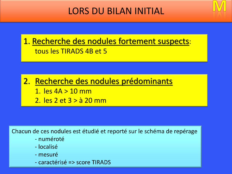
1. Recherche des nodules fortement suspects: tous les TIRADS 4B et 5
2. Recherche des nodules prédominants 1. les 4A > 10 mm 2. les 2 et 3 > à 20 mm
LORS DU BILAN INITIAL
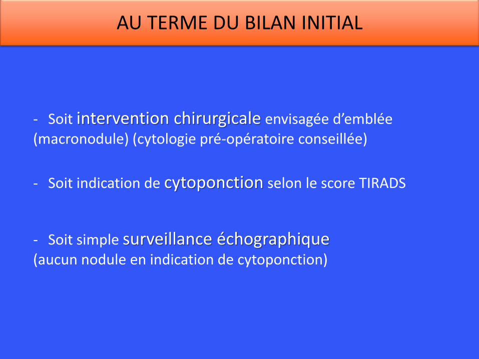
- Soit simple surveillance échographique (aucun nodule en indication de cytoponction)
- Soit intervention chirurgicale envisagée d’emblée (macronodule) (cytologie pré-opératoire conseillée)
- Soit indication de cytoponction selon le score TIRADS
AU TERME DU BILAN INITIAL
1. Tout résultat de cytoponction malin ou suspect
2. Les macro-nodules (> 30 ou 40 mm de grand diamètre) ? THERMO-ABLATION
3. Les signes de compression: dysphagie, dyspnée, dysphonie THERMO-ABLATION
4. ± Quand la surveillance devient trop contraignante
INDICATIONS CHIRURGICALES
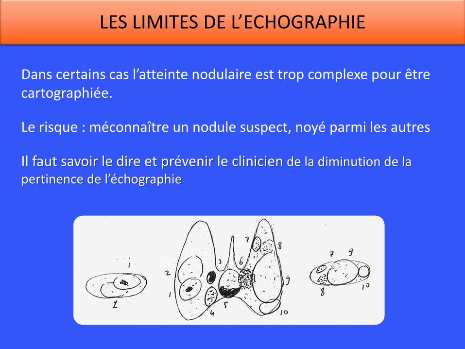
Dans certains cas l’atteinte nodulaire est trop complexe pour être cartographiée.
Le risque : méconnaître un nodule suspect, noyé parmi les autres
Il faut savoir le dire et prévenir le clinicien de la diminution de la pertinence de l’échographie
LES LIMITES DE L’ECHOGRAPHIE
CONCLUSION: LES POINTS IMPORTANTS
• Les nodules doivent être • Cartographiés • Mesurés dans trois diamètres
• Une échographie ganglionnaire doit être systématiquement associée
• Le système TI-RADS: • Permet de détecter 95% des carcinomes thyroïdiens • De caractériser un nodule comme bénin avec un risque
d’erreur < 2% • Rationalise (et diminue) les indications de cytoponctions
• Dans les thyroïdes multinodulaires, il faut rechercher d’abord les nodules suspects (TI-RADS 5, 4B, 4A).

BIBLIOGRAPHIE
REFERENCES BIBLIOGRAPHIQUES : LES INDISPENSABLES
• New sonographic criteria for recommending fine-needle aspiration biopsy of non palpable solid nodules of the thyroid Eun-Kyung Kim, AJR 178; March 2002 p 687-691
• US features of thyroid malignancy : pearls and pitfalls Jenny K Hoang, Radiographics 2007 ; 27 : 847-860
• Common and uncommon sonographic features of papillary thyroid carcinoma Bryan K Chan, J Ultrasound Med 22 : 1083-1090, 2003
• Sonography of thyroid nodules A « classic pattern » diagnostic approach Carl C Reading, Ultrasound Quarterly 2005; 21 : 157-165
• Ultrasonographic evaluation of thyroid nodules in 900 patients : comparison among ultrasonographic, cytological and histological findings. Yasuhiro Ito, Thyroid vol 17, number 12, 2007, p 1269-76
New Sonographic Criteria for recommending fine needle aspiration biopsy of non palpable solid nodules of the thyroid. Eun-Kyung Kim, AJR 2002; 178 : 687-691 Thyroid nodule shape and prediction of malignancy. Erik K Alexander, Thyroid, vol 14, number 11, 2004, 953-958 Thyroid nodule shape suggests malignancy. Carlo Cappelli, European Journal of Endocrinology, 2006, 155, 27-31 Diagnostic value of ultrasonography to distinguish between benign and malignant lesions in the management of thyroid nodules. Hyun Jung Tae, Thyroid, Vol 17, Number 5, 2007, p461-466 Benign and malignant thyroid nodules : US differentiation – Multicenter retrospective study. Won-Jin Moon, Radiology, vol 247, number 3, June 2008, 762-770
Les articles fondateurs sur les signes de bénignité et de malignité en échographie
-Carl Reading : Sonography of thyroid nodules : a « classic pattern » diagnostic approach. Ultrasound Quarterly: September 2005 - Volume 21 - Issue 3 - pp 157-165 - Yasuhiro Ito : Ultrasonographic evaluation of thyroid nodules in 900 patients : comparison among ultrasonographic, cytological and histological findings. Thyroid 2007, 17, 1269-1276 - John A Bonavita : Pattern recognition of benign nodules at ultrasound of the thyroid : which nodules can be left alone ? AJR 2009, 193, 207-213 - Vivek Virmani : Sonographic patterns of benign thyroid Nodules: verification at our institution. AJR:196, April 2011 p891-95
Les concepts de pattern et de grading
Valeur prédictive des signes échographiques
• Rago et al. EJE 1998 138: 41-46
• Papini et al. JCEM 2002 87(5): 1941-46
• Kim et al. AJR 2002 178: 687-91
• Cappelli et al. EJE 2006 155: 27-31
• Moon et al. Radiology 2008 247(3): 762
• Bonavita et al. AJR 2009 193: 207-13
• Russ et al. J Radiol 2011 92: 701-13
• Bojunga et al. Thyroid 2010 20(10): 1145-50
• Trimboli et al. JCEM 2012 97(12): 4524-30
• Bojunga et al. PLOS One 2012 7(8): 1-8
• Zhang et al. PLOS One 2012 7(11): 1-8
• Tae HJ et al., Thyroid 2007;17(5) : 461. Diagnostic value of ultrasonography to distinguish between benign and malignant lesions in the management of thyroid nodules.
• Ito Y et al., Thyroid 2007; 17, 1269-1276 Ultrasonographic evaluation of thyroid nodules in 900 patients : comparison among ultrasonographic, cytological and histological findings.
• Horvath et al.. JCEM, may 2009, 90(5), 1748-51 An ultrasonogram reporting system for thyroid nodules stratifying cancer risk for clinical management.
• Park et al.. Thyroid 2009, 19 (11), 257-64 A proposal for a thyroid imaging reporting and data system for ultrasound features of thyroid carcinoma.
• Kwak et al., Radiology 2011; 260; 892-99 Thyroid Imaging Reporting and Data System for US features of nodules. A step in establishing better stratification of cancer risk.
• Kwak JY et al., Korean J Radiol 2013; 14(1): 110-117 Image reporting and characterization system for ultrasound features of thyroid nodules: multicentric korean retrospective study
• Russ et al. J Radiol 2011;92;701-13 Le système TI-RADS en échographie thyroïdienne.
• Russ et al. Eur J Endocrinol 2013, 168 (5); 649-55 Prospective evaluation of thyroid imaging reporting and data system on 4550 nodules with and without elastography.
• Haugen et al. Thyroid 2016; 26(1): 1-133 2015 ATA Guidelines for adult patients with thyroid nodules and differentiated thyroid cancer
• Na et al. Thyroid. February 2016 ahead of print TIRADS risk stratification of thyroid nodules: categorization based on solidity and echogenicity
Stratification du risque
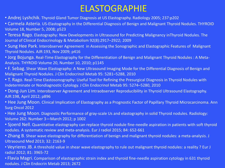
• Andrej Lyshchik. Thyroid Gland Tumor Diagnosis at US Elastography. Radiology 2005; 237:p202
• Carmela Asteria. US-Elastography in the Differential Diagnosis of Benign and Malignant Thyroid Nodules. THYROID Volume 18, Number 5, 2008; p523
• Teresa Rago. Elastography: New Developments in Ultrasound for Predicting Malignancy inThyroid Nodules. The Journal of Clinical Endocrinology & Metabolism 92(8):2917–2922; 2009
• Sung Hee Park. Interobserver Agreement in Assessing the Sonographic and Elastographic Features of Malignant Thyroid Nodules. AJR:193, Nov 2009; p416
• Jorg Bojunga. Real-Time Elastography for the Differentiation of Benign and Malignant Thyroid Nodules : A Meta-Analysis. THYROID Volume 20, Number 10, 2010; p1145
• F. Sebag. Shear Wave Elastography: A New Ultrasound Imaging Mode for the Differential Diagnosis of Benign and Malignant Thyroid Nodules. J Clin Endocrinol Metab 95: 5281–5288, 2010
• T. Rago. Real-Time Elastosonography: Useful Tool for Refining the Presurgical Diagnosis in Thyroid Nodules with Indeterminate or Nondiagnostic Cytology. J Clin Endocrinol Metab 95: 5274–5280, 2010
• Dong-Jun Lim. Interobserver Agreement and Intraobserver Reproducibility in Thyroid Ultrasound Elastography. AJR:198, April 2012; p896
• Hee Jung Moon. Clinical Implication of Elastography as a Prognostic Factor of Papillary Thyroid Microcarcinoma. Ann Surg Oncol 2012
• Hee Jung Moon. Diagnostic Performance of gray-scale Us and elastography in solid Thyroid nodules. Radiology: Volume 262: Number 3—March 2012; p 1002
• Sjoerd Nell. Quantitative elastography can replace thyroid nodule fine-needle aspiration in patients with soft thyroid nodules. A systematic review and meta-analysis. Eur J radiol 2015; 84: 652-661
• Zhang B. Shear wave elastography for differentiation of benign and malignant thyroid nodules: a meta-analysis. J Ultrasound Med 2013; 32: 2163-9
• Veyrieres JB. A threshold value in shear wave elastography to rule out malignant thyroid nodules: a reality ? Eur J radiol 2012; 81: 3965-72
• Flavia Magri. Comparison of elastographic strain index and thyroid fine-needle aspiration cytology in 631 thyroid nodules. J Clin Endocrin Metab 2013; 2672
ELASTOGRAPHIE