Microbiolog to Introduction y - Weebly
27
Introduction to Microbiolog y Anas Abu-Humaidan M.D. Ph.D. Lecture 2
Transcript of Microbiolog to Introduction y - Weebly

Introduction to
Microbiology
Anas Abu-HumaidanM.D. Ph.D.
Lecture 2

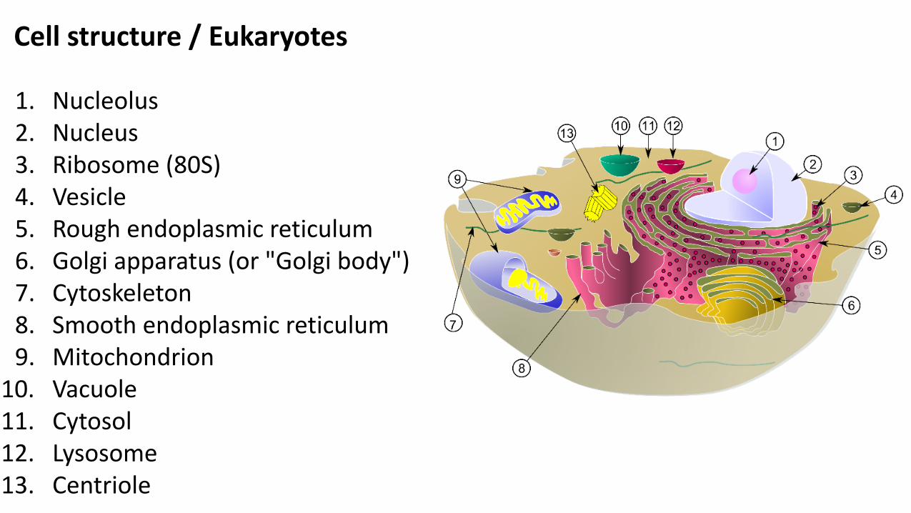
1. Nucleolus2. Nucleus3. Ribosome (80S)4. Vesicle5. Rough endoplasmic reticulum6. Golgi apparatus (or "Golgi body")7. Cytoskeleton8. Smooth endoplasmic reticulum9. Mitochondrion
10. Vacuole11. Cytosol12. Lysosome13. Centriole
Cell structure / Eukaryotes

Cell structure / Eukaryotes / Nucleus
• The nucleus contains the genome, andis surrounded by a porus membrane totraffic proteins.
• The nucleolus is important in ribosomebiogenesis.
• DNA wraps around histones formingnucleosomes, which coil to form fiberscalled chromatin which coils and loopsto form chromosomes.
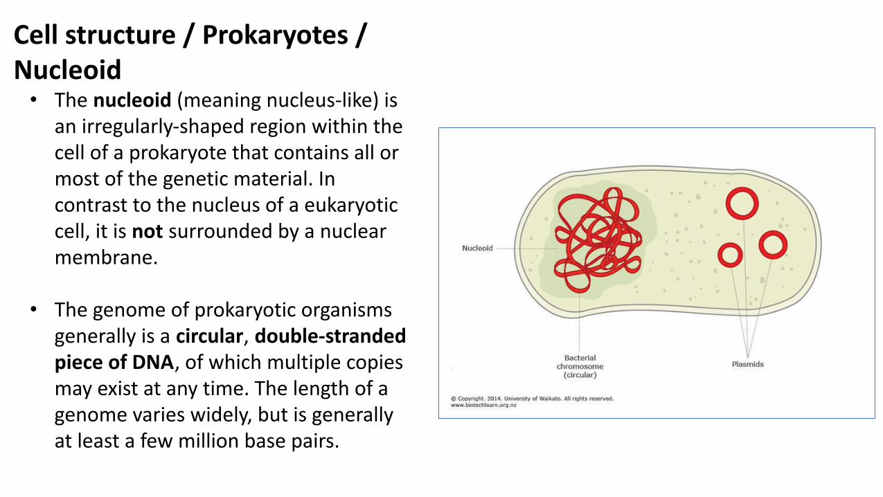
Cell structure / Prokaryotes /Nucleoid• The nucleoid (meaning nucleus-like) is
an irregularly-shaped region within thecell of a prokaryote that contains all ormost of the genetic material. Incontrast to the nucleus of a eukaryoticcell, it is not surrounded by a nuclearmembrane.
• The genome of prokaryotic organismsgenerally is a circular, double-strandedpiece of DNA, of which multiple copiesmay exist at any time. The length of agenome varies widely, but is generallyat least a few million base pairs.

Top Adenine thymidine(A-T) pairing; bottom: guanine-cytosine (G-C) pair

Prokaryotes vs Eukaryotes DNA
Prokaryotic DNA:
• Is found freely in the cytoplasm (withina region called the nucleoid)
• Is naked (i.e. not bound with proteinsand therefore doesn’t form chromatin)
• Genomes are compact (contain littlerepetitive DNA and no introns)
• Contains extra-chromosomal plasmids
• Is circular in shape
Eukaryotic DNA:
• Is contained within a nucleus
• Is bound to histone proteins
• Genomes contain large amounts ofnon-coding and repetitive DNA(including introns)
• Do not contain plasmids (butorganelles such as the mitochondriamay contain their own chromosomes)
• Are linear in shape

1. Nucleolus2. Nucleus3. Ribosome (80S)4. Vesicle5. Rough endoplasmic reticulum6. Golgi apparatus (or "Golgi body")7. Cytoskeleton8. Smooth endoplasmic reticulum9. Mitochondrion
10. Vacuole11. Cytosol12. Lysosome13. Centriole
Cell structure / Eukaryotes

1. Nucleolus2. Nucleus3. Ribosome (80S)4. Vesicle5. Rough endoplasmic reticulum6. Golgi apparatus (or "Golgi body")7. Cytoskeleton8. Smooth endoplasmic reticulum9. Mitochondrion
10. Vacuole11. Cytosol12. Lysosome13. Centriole
Cell structure / Eukaryotes

Prokaryotes vs Eukaryotes ribosomes
• Prokaryotes have smallerribosomes, this property isused to target bacterialprotein synthesis withantibiotics.

Transcription and translation
• Prokaryotes couple transcription withtranslation, because of no nuclear membrane.
1. Nucleolus2. Nucleus3. Ribosome (80S)4. Vesicle5. Rough endoplasmic reticulum6. Golgi apparatus (or "Golgi body")7. Cytoskeleton8. Smooth endoplasmic reticulum9. Mitochondrion
10. Vacuole11. Cytosol12. Lysosome13. Centriole
Cell structure / Eukaryotes

Cell structure / Eukaryotes / Vesicles
• A lipid bilayer, similar to the cellmembrane, that functions incompartmentalizing cellular processesand substances.
• Involved in metabolism, transport andstorage of molecules, as well as reactionchambers.
• Include: Vacuoles, lysosomes, transportvesicles and secretory vesicles.
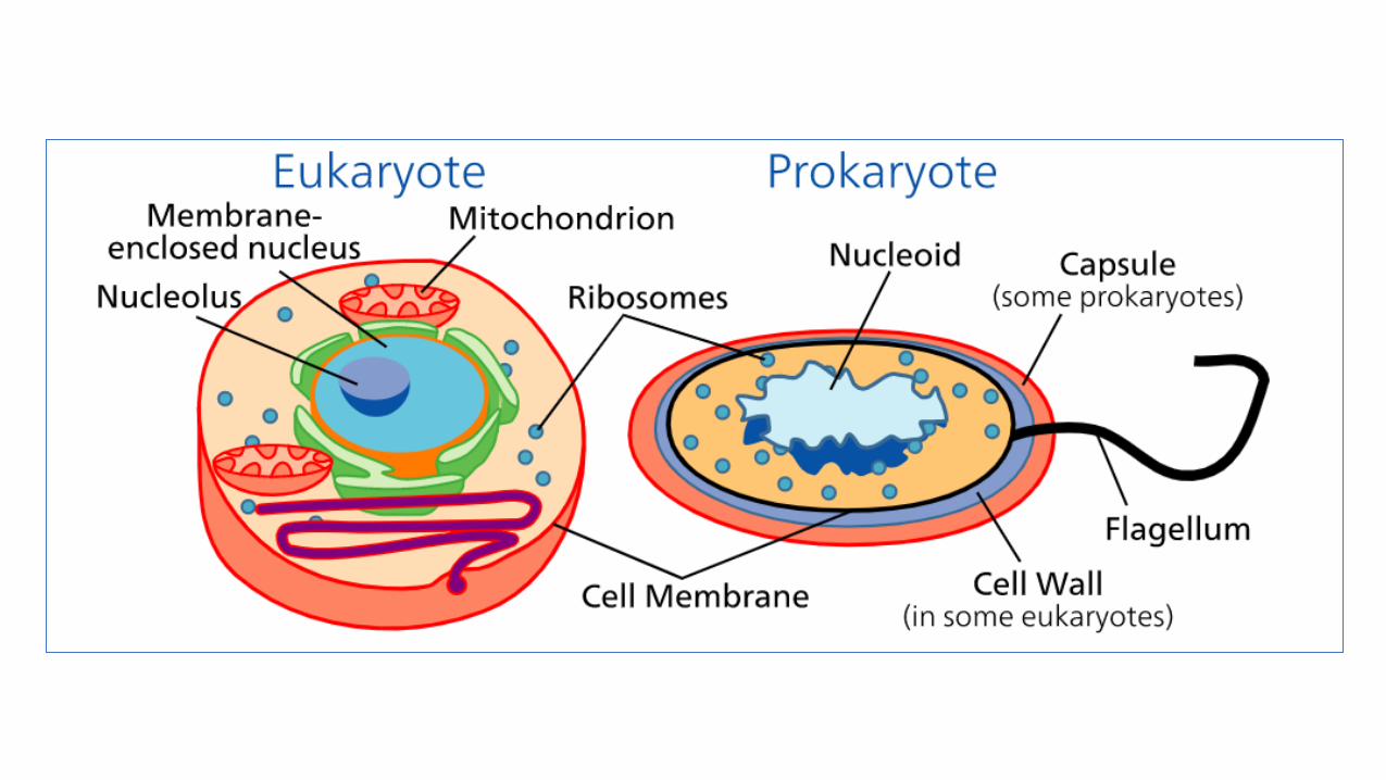
• Prokaryotes DO NOT contain organellessurrounded by a membrane.

1. Nucleolus2. Nucleus3. Ribosome4. Vesicle5. Rough endoplasmic reticulum6. Golgi apparatus (or "Golgi body")7. Cytoskeleton8. Smooth endoplasmic reticulum9. Mitochondrion
10. Vacuole11. Cytosol12. Lysosome13. Centriole
Cell structure / Eukaryotes

Cell structure / Eukaryotes and prokaryotes / Cytoskeleton
• The cytoskeleton is a scaffold thatholds the structure of the cell, andfacilitates transport in the cell, as wellas motility and migration.
• Microfilaments are polymers of theprotein actin.
• Cillia and flagella are made ofmicrotubules (protein cylinders madeof tubulin) enclosed in a membrane.
• Not only eukaryotes, but alsoprokaryotes possess a cytoskeleton,with actin and tubulin - like proteins.
Actin Tubulin

Cell structure / Prokaryotes
• Circular DNA is packaged in nucleoid.More than one nucleoid can be found,but all are similar (haploid).
• Cytoplasm contains smaller ribosomes(70S), and inclusion bodies thatfunction in the storage of energy or asa reservoir of structural building blocks(e.g. Glycogen, PHB, polyphosphate).
• Cell membrane have high proteindensity, and lacks sterols found ineukaryotes.

Cell structure / Eukaryotes/ Cell membrane functions
The plasma membrane (also calledthe cell membrane) is a phospholipidbilayer with embedded proteins thatencloses every living cell. Thismembrane blocks uncontrolledmovements of water-solublematerials into or out of the cell. Thevarious proteins embedded in thephospholipid bilayer penetrate intoand through the bilayer three-dimensionally.

Cell structure / Prokaryotes/ Cell membrane functions
1. Permeability and transport (activeand passive).
2. Electron transport and oxidativephosphorylation
3. Excretion of hydrolyticexoenzymes and pathogenicityproteins.
4. Biosynthetic functions
5. Chemotactic systems


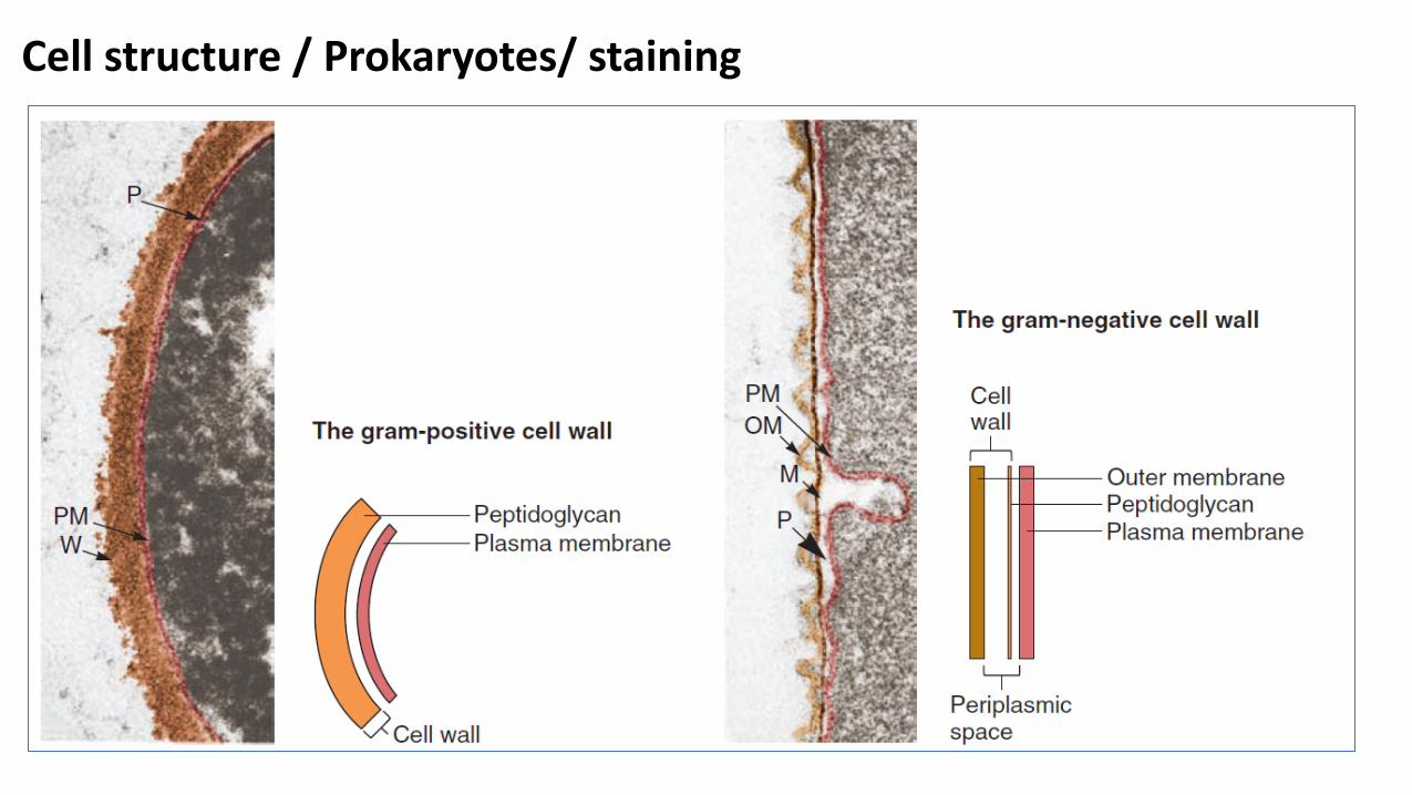
Cell structure / Prokaryotes/Gram (+) Cell wall
• High tensile strength owing toPeptidoglycans.
• Plays a role in cell division andits own synthesis and is anantigenic determinant.
• Teichoic acid is found inGram(+) bacteria andcontributes to tensile strengh,porousity and antiginicity.

Cell structure / Prokaryotes/Gram (-) Cell wall
• Smaller peptidoglycan layer, but an extra outermembrane, with Lipopolysaccharide (LPS) outerleaflet.
• LPS is an antigenic determinant.
• Antibiotics pass slowly through the outermembrane contributing to the high antibioticres. of Gram (-)s.
• Permiplasm contains transporters anddetoxifiers (e.g. β-lactamase, important inantibiotic resistance).

Scanning electron microscopy experiments performed on E. coli (K12 MG1655), S. haemolyticus (MTCC 3383), and S. haemolyticus protoplasts.
Computational antimicrobial peptide design and evaluation against multidrug-resistant clinical isolates of bacteria• December 2017• Journal of Biological Chemistry 293(10):jbc.M117.805499
Cell structure / Prokaryotes/ staining

Cell structure / Prokaryotes/ staining / Gram staining

Cell structure / Prokaryotes/ Cell wall
• Mycoplasmas lack a cell wall,leading to variability in shape, andresistance to antibiotics that attackthe cell wall.
• Some bacteria form an externallayer of polysaccharides called acapsule when rigid, or a slime layerwhen loose, which contributes toinvasiveness and resistance tophagocytosis.

Conclusion
• Differences exist between prokaryotic and human eukaryotic cell structure,mainly in the nucleus, lipid bilayer organelles, and cell wall.
• Bacteria can be divided on the basis of their cell wall differences into grampositive and gram negative bacteria.
Further reading:
• Sherris Medical Microbiology, sixth editionChapter 21: Bacteria—Basic Concepts