I itro In vo oxicit n Cymbopogo igante Cv. Lav Ent O o Benin
12
Research Article In Vitro and In Vivo Toxicity Studies on Cymbopogon giganteus Chiov. Leaves Essential Oil from Benin Habib Toukourou , 1,2 Francine Uwambayinema, 3 Yousof Yakoub, 3 Birgit Mertens, 4 Anatole Laleye, 5 Dominique Lison, 3 Joelle Quetin-Leclercq, 2 and Fernand Gbaguidi 1 1 Laboratoire de Chimie Pharmaceutique Organique, Ecole de Pharmacie, Faculté des Sciences de la Santé, Université d’Abomey-Calavi, Campus du champ de Foire, 01BP 188 Cotonou, Benin 2 Pharmacognosy Research Group, Louvain Drug Research Institute, Université Catholique de Louvain, B1 7203 Av. E. Mounier 72, B-1200 Brussels, Belgium 3 Louvain Centre for Toxicology and Applied Pharmacology, Institut de Recherche Expérimentale et Clinique, Université catholique de Louvain, B1.57.06 Av. Hippocrate 57, B-1200 Brussels, Belgium 4 Scientific Direction of Chemical and Physical Health Risks, Sciensano, Juliette Wytsmanstraat 14, 1050 Brussels, Belgium 5 Unité de Biologie Humaine, Laboratoire de Cytogénétique et de Biologie Moléculaire, Faculté des Sciences de Santé, Université d’Abomey-Calavi, Campus du champ de Foire, 01BP 188 Cotonou, Benin Correspondence should be addressed to Habib Toukourou; [email protected] Received 5 July 2019; Revised 14 September 2019; Accepted 24 October 2019; Published 28 January 2020 Academic Editor: You-Cheng Hseu Copyright © 2020 Habib Toukourou et al. is is an open access article distributed under the Creative Commons Attribution License, which permits unrestricted use, distribution, and reproduction in any medium, provided the original work is properly cited. Cymbopogon giganteus Chiov. (Poaceae) is a medicinal plant used to treat various diseases in traditional medicine in several African countries. e present study aims to evaluate the oral and inhalation toxicity as well as the mutagenic effects of the essential oil of Cymbopogon giganteus leaves (EOCG) from a sample collected in Benin. Mutagenic potential was assessed by the Ames test using Salmonella typhimurium strains TA98 and TA100. Oral acute toxicity was carried out by administration of a single dose of 2000 mg/ kg b.w. to Wistar rats while oral subacute toxicity was assessed by daily administration of 50 and 500 mg/kg of EOCG for 28 days. Finally, inhalation toxicity was assessed by administration of a single dose of 0.125%, 0.5%, 2% or 5% v/v of EOCG emulsions in 0.05% v/v lecithin solution in sterile water for the first experiment, and in a second one by administration of single dose of 0.125% or 0.5% v/v. A broncho-alveolar lavage was performed aſter 3 h or 24 h, respectively. e results show that EOCG is not mutagenic on Salmonella typhimurium strains at the highest concentration tested (200 μg/plate). In the acute oral toxicity study, EOCG induce neither mortality nor toxicity, showing that the LD 50 is greater than 2000 mg/kg. e subacute oral toxicity study at both doses did not show any significant difference in body weight, relative organ weight, hematological and/or biochemical parameters or histopathology as compared to the control group. EOCG induced mortality and inflammation in lungs 3 h aſter administration of a single dose of 5% or 2% v/v. Single doses of 0.125% or 0.5% v/v did not induce inflammation, cell recruitment nor cytotoxicity in lungs 3 h or 24 h aſter administration, suggesting safety at these concentrations. is first report on the in vivo toxicity will be useful to guide safe uses of EOCG. 1. Introduction Medicinal herbs and their related products are traditionally used by at least 80% of the population in developing countries to solve or improve their health problems [1]. eir uses are based on ancestral habits transmitted from generation to gen- eration. Unlike modern medicines containing one or a few well-known active substances, these medicines contain a mul- titude of bioactive substances in very variable proportions, which depend on several factors [2]. is explains the diffi- culties of their standardization in terms of efficacy. e toxicity of these preparations, especially upon long-term application, is oſten neglected [3, 4]. In this context, toxicological evalua- tion appears obviously a crucial step in determining safety and to support the traditional use of these medicines. Cymbopogon giganteus Chiov. (Poaceae) also call “Citronelle de Madagascar” is a grass, which can grow up to 2-3 m, spreading in regions of tropical Africa. is plant is Hindawi Journal of Toxicology Volume 2020, Article ID 8261058, 12 pages https://doi.org/10.1155/2020/8261058
Transcript of I itro In vo oxicit n Cymbopogo igante Cv. Lav Ent O o Benin

Research ArticleIn Vitro and In Vivo Toxicity Studies on Cymbopogon giganteus Chiov. Leaves Essential Oil from Benin
Habib Toukourou ,1,2 Francine Uwambayinema,3 Yousof Yakoub,3 Birgit Mertens,4 Anatole Laleye,5 Dominique Lison,3 Joelle Quetin-Leclercq,2 and Fernand Gbaguidi1
1Laboratoire de Chimie Pharmaceutique Organique, Ecole de Pharmacie, Faculté des Sciences de la Santé, Université d’Abomey-Calavi, Campus du champ de Foire, 01BP 188 Cotonou, Benin
2Pharmacognosy Research Group, Louvain Drug Research Institute, Université Catholique de Louvain, B1 7203 Av. E. Mounier 72, B-1200 Brussels, Belgium
3Louvain Centre for Toxicology and Applied Pharmacology, Institut de Recherche Expérimentale et Clinique, Université catholique de Louvain, B1.57.06 Av. Hippocrate 57, B-1200 Brussels, Belgium
4Scientific Direction of Chemical and Physical Health Risks, Sciensano, Juliette Wytsmanstraat 14, 1050 Brussels, Belgium5Unité de Biologie Humaine, Laboratoire de Cytogénétique et de Biologie Moléculaire, Faculté des Sciences de Santé, Université d’Abomey-Calavi, Campus du champ de Foire, 01BP 188 Cotonou, Benin
Correspondence should be addressed to Habib Toukourou; [email protected]
Received 5 July 2019; Revised 14 September 2019; Accepted 24 October 2019; Published 28 January 2020
Academic Editor: You-Cheng Hseu
Copyright © 2020 Habib Toukourou et al. �is is an open access article distributed under the Creative Commons Attribution License, which permits unrestricted use, distribution, and reproduction in any medium, provided the original work is properly cited.
Cymbopogon giganteus Chiov. (Poaceae) is a medicinal plant used to treat various diseases in traditional medicine in several African countries. �e present study aims to evaluate the oral and inhalation toxicity as well as the mutagenic effects of the essential oil of Cymbopogon giganteus leaves (EOCG) from a sample collected in Benin. Mutagenic potential was assessed by the Ames test using Salmonella typhimurium strains TA98 and TA100. Oral acute toxicity was carried out by administration of a single dose of 2000 mg/kg b.w. to Wistar rats while oral subacute toxicity was assessed by daily administration of 50 and 500 mg/kg of EOCG for 28 days. Finally, inhalation toxicity was assessed by administration of a single dose of 0.125%, 0.5%, 2% or 5% v/v of EOCG emulsions in 0.05% v/v lecithin solution in sterile water for the first experiment, and in a second one by administration of single dose of 0.125% or 0.5% v/v. A broncho-alveolar lavage was performed a�er 3 h or 24 h, respectively. �e results show that EOCG is not mutagenic on Salmonella typhimurium strains at the highest concentration tested (200 μg/plate). In the acute oral toxicity study, EOCG induce neither mortality nor toxicity, showing that the LD50 is greater than 2000 mg/kg. �e subacute oral toxicity study at both doses did not show any significant difference in body weight, relative organ weight, hematological and/or biochemical parameters or histopathology as compared to the control group. EOCG induced mortality and inflammation in lungs 3 h a�er administration of a single dose of 5% or 2% v/v. Single doses of 0.125% or 0.5% v/v did not induce inflammation, cell recruitment nor cytotoxicity in lungs 3 h or 24 h a�er administration, suggesting safety at these concentrations. �is first report on the in vivo toxicity will be useful to guide safe uses of EOCG.
1. Introduction
Medicinal herbs and their related products are traditionally used by at least 80% of the population in developing countries to solve or improve their health problems [1]. �eir uses are based on ancestral habits transmitted from generation to gen-eration. Unlike modern medicines containing one or a few well-known active substances, these medicines contain a mul-titude of bioactive substances in very variable proportions,
which depend on several factors [2]. �is explains the diffi-culties of their standardization in terms of efficacy. �e toxicity of these preparations, especially upon long-term application, is o�en neglected [3, 4]. In this context, toxicological evalua-tion appears obviously a crucial step in determining safety and to support the traditional use of these medicines.
Cymbopogon giganteus Chiov. (Poaceae) also call “Citronelle de Madagascar” is a grass, which can grow up to 2-3 m, spreading in regions of tropical Africa. �is plant is
HindawiJournal of ToxicologyVolume 2020, Article ID 8261058, 12 pageshttps://doi.org/10.1155/2020/8261058

Journal of Toxicology2
used in traditional medicine against various diseases such as skin disorders [5], mental illness, broncho-pulmonary affec-tions, bilharzia, jaundice, cold, conjunctivitis, migraine, der-matoses, rheumatic pains, childhood coughs and hepatitis [6, 7]. �e essential oil of Cymbopogon giganteus (EOCG) got by hydro distillation of its leaves showed good antibacterial and anti-inflammatory properties and is sold for these activ-ities [8–11]. We analyzed the composition of a sample of EOCG originating from Benin. It was characterized by the presence of limonene, carvone, carveol and p-menthane deriv-atives as major compounds [12], like other samples from other origins [11, 13, 14]. Using essential oils and particularly by the oral route can, however, produce serious adverse effects [15] which can be predicted by performing toxicological tests.
To the best of our knowledge, there is no data in the liter-ature on in vivo oral and pulmonary toxicity of EOCG, nor on its mutagenicity, which may be required to predict its safety and/or toxic effects following exposure.
Our goal being the use of essential oil of Cymbopogon giganteus (EOCG) as antiseptic and antibacterial against sore throat caused by bacteria and viruses, we focused our studies on in vitro mutagenicity (by Ames test), acute and subacute oral toxicity and acute pulmonary toxicity in Wistars rats.
2. Materials and Methods
2.1. Animals and Plant Material. For oral toxicity testing, Wistar albino rats aged of 10–12 weeks were obtained from the house facilities of UAC-FSS (Université d’Abomey Calavi, Faculté des Sciences de la Santé, Cotonou, Bénin) while for pulmonary toxicity, eight-week-old female Wistar rats were purchased from Janvier Labs (St Berterin, France). All animals were acclimatized for one week, kept with sterile rodent feed with free access to water and housed in positive pressure air-conditioned units (25°C, 50% relative humidity) on 12 h light/dark cycle.
Fresh leaves of Cymbopogon giganteus Chiov. were col-lected in Parakou areas (9°20′N, 2°37′E) in November 2016. Crops were identified by the Herbier National du Bénin (Université Abomey Calavi) where a voucher specimen was deposited under number AA6680/HNB. EOCG was obtained by hydro-distillation using a Clevenger apparatus of air-dried leaves and stored at 4°C.
2.2. Bacterial Reverse Gene Mutation Test (Ames Test). �e in vitro mutagenic potential of EOCG were evaluated in the bacterial reverse gene mutation test as stated in guideline 471 of the organization for economic Co-operation and Development (OECD) [16], with slight modifications as described by [17]. Although normally 5 different bacterial strains should be used, the test was performed in two strains only, i.e. Salmonella typhimurium TA98 and TA100, as this combination already allows to identify up to 90% of mutagens [18]. A�er cultivation of the Salmonella bacteria (Moltox, Boone, USA) overnight, 100 µl of the bacterial suspension was mixed with 100 µl of the EOCG emulsion, 500 µl sodium phosphate buffer pH 7.4 and 2 mL overlay agar enriched
with a histidine-biotine solution. Emulsions of EOCG were prepared at 0.2% v/v (0.2% of EOCG and 0.02% v/v of tween 80 in sterile water), resulting in a highest concentration tested of 200 μg/plate. In total, five concentrations were tested (200, 50, 20, 5, and 2 μg/plate). To evaluate the impact of metabolic activation, the buffer was replaced by a 5% S9 metabolization mix (prepared from lyophilized rat liver S9 mixed with nicotinamide adenine dinucleotide phosphate (NADPH) regenerating system—both from Moltox). �e resulting mixture was poured onto a minimal glucose agar plate (E&O Laboratories Ltd., Bonnybridge, United Kingdom) and incubated for 48 h at 37°C (Binder, Tuttlingen, Germany). Triplicate plates were poured for each test condition. Positive, negative and solvent control plates were prepared in parallel with the test substance plates. A solution of Tween 80 (20 μg/plate) was used as solvent control. For the tests without S9 metabolizing mix, sodium azide (2 µg/plate) and 4-nitroquinoline-n-oxide (0.2 µg/plate) were used as a positive control for strains TA100 and TA98, respectively. 2-Aminoanthracene (1 µg/plate) was used as positive control for the tests with metabolic activation. All positive control substances and tween 80 were purchased from Sigma-Aldrich (Saint-Louis, USA). A�er incubation, the plates were inspected for cytotoxicity and precipitation by examining the background lawn using a light microscope (Zeiss, Oberkochen, Germany) at 40x total magnification. All plates were subsequently scored using a manual colony counter (Sigma-Aldrich, Saint-Louis, USA). Individual plate counts were recorded as well as the mean number of revertant colonies per plate. �erea�er, the standard deviation (SD) was calculated. A substance is considered being mutagenic when the number of colonies obtained for the test substance/number of colonies obtained for the solvent control is greater than 2 (�푁 > 2) and a dose-dependent effect is observed.
2.3. Toxicity Assays
2.3.1. Study Compliance. Acute and subacute toxicity studies were performed according to OECD Principles and the internationally accepted principles for laboratory animal use and care (NIH publication No. 85-23, revised 2010). Acute pulmonary toxicity test was performed under the ethical standards at Université catholique de Louvain, Comité d’Ethique pour l’Expérimentation Animale, Secteur des Sciences de la Santé, Brussels, Belgium (No. LA1230312).
2.3.2. In Vivo Acute Oral Toxicity. �e acute toxicity test was carried out based on the OECD guidelines for chemicals testing, sections 4-423-(limit test), adopted in 2008 [19]. Six female Wistar rats weighing 163 ± 7.2 g were divided into two groups of 3 rats each. �e experimental group was gavaged with a single dose of 2000 mg/kg b.w. of essential oil diluted in 2 mL maize oil while the control group was treated with 2 mL of maize oil only. �e animals were observed for eight hours just a�er the administration and once daily up to 14 days. �e monitoring was based on general behavioral changes, body weight evolution, mortality and any other toxicity signs. At the end of the experience (day 15), 0.1 mL of thiopental (100 mg/mL)

3Journal of Toxicology
was injected intraperitoneally to euthanize the animals to collect blood samples in tubes for biochemical examinations.
2.3.3. In Vivo Subacute Oral Toxicity Test. �e subacute toxicity test was carried out as described in OECD 407 guidelines [20]. 30 Wistars rats (15 males and 15 females) were used and distributed in three groups (5 males and 5 females/group): a control (group 1) which received 2 mL of maize oil (vehicle) daily and two dose levels groups (group 2 and group 3) of EOCG (50 and 500 mg/kg body weight/day) diluted in 2 mL maize oil. EOCG was administrated daily by gavage for 28 days. During the experiment, the animals were observed for signs of toxicity and mortality twice a day. Observations were focused on changes in the skin and fur, eyes, mucus, respiratory tract, autonomic and somato-motor activity and behavioral patterns. Body weight was recorded every five days and blood samples were collected at day 15. At the end (day 29), rats were starved overnight (12 hours) but with free access water. �ey were then anesthetized, sacrificed by an overdose of thiopental and blood samples were collected in tubes without anticoagulant for biochemical parameters analysis and in EDTA pre-coated tubes to obtain the whole blood for hematological determinations. Different organs (stomach, liver and kidneys) were carefully excised and their absolute weights were determined. �e relative organ weight was then calculated as follows: (absolute organ weight (g)/body weight of rat on the sacrifice day (g)) × 100. Livers and kidneys were washed with regular saline and were placed in 3.65% paraformaldehyde (Sigma-Aldrich, St Louis, Missouri, USA) in phosphate buffered saline (PBS) for later histological analysis by staining with hematoxylin-eosin.
2.3.4. Acute Pulmonary Toxicity Test. Pulmonary toxicity was assessed a�er oropharyngeal aspiration of EOCG. �is method is well known to assess the pulmonary toxicity of compounds or particles as those induced by asbestos, crystalline silica or bleomycin [21–24]. Briefly, 300 μl of emulsion in 0.05% lecithin solution in sterile water of EOCG at different concentrations were administrated directly by oro-pharyngeal aspiration. All experiments were performed with groups of 5 rats: a control group (vehicle) and groups which received different concentrations. In the first set of tests, 0.125%, 0.5%, 2%, and 5% v/v of emulsions of EOCG were administrated and rats were sacrificed 3 h later. In a second set of tests, single doses of 0.125% and 0.5% v/v emulsion of EOCG were administrated with sacrifice of rats 24 h a�er. �e control group received 300 μl of vehicle (0.05% v/v of lecithin solution (Lipoid S100 (LIPOID GMBH) in sterile water) for each experiment. All rats were euthanized by intraperitoneal injection of 12 mg sodium pentobarbital (Certa, Braine-l’Alleud, Belgium). Broncho-alveolar lavage fluid (BALF) was obtained by inserting a cannula in trachea and infusing the lungs with 10 mL of NaCl 0.9%. �is fluid was submitted to centrifugation for 10 min at 4°C (240 g). Cell-free supernatant was used for determination of total proteins and lactate dehydrogenase (LDH) activity [25]. �e cells pellet was re-suspended in PBS and was counted a�er staining with Turch (crystal violet 1%, acetic acid
3%). For the second set of tests, cell differentiation was measured for 200 cells counted by light microscopy a�er cytocentrifugation (CYTOSPIN3, SHANDON) and Diff-Quick staining (Polysciences, Warrington, UK).
2.4. Biochemical Parameters. Serum concentrations of urea, creatinine, alanine aminotransferase (ALAT), alkaline phosphatase (PAL) and aspartate aminotransferase (ASAT) were determined using an automatic analyzer (BS-200 Minidray) with specific kits (Cypress Diagnostics, Langdorp, Belgium). For hematological analysis hematocrit (HCT), hemoglobin (Hb), erythrocytes, leukocytes, and platelets (PLT) counts were performed on a XS-500i Sysmex automated hematology analyzer.
2.5. Statistics. All results are expressed as mean ± standard errors on the mean (SEM) and statistical analyses were performed with GraphPad Prism 5.0 (GraphPad So�ware Inc., San Diego, CA, USA) and/or Microso� excel 2016. For acute toxicity, t-test was used to compare data. For subacute toxicity test, data from male and female animals were analyzed separately. Differences between control and treated groups were evaluated using one-way analysis of variance (ANOVA) followed by a Dunnett’s multiple comparison for subacute and pulmonary toxicity. Statistical significance was considered at �푝 < 0.05.
3. Results
3.1. Mutagenicity Assay. �e results of the mutagenicity assay are depicted in Table 1, which shows that EOCG was not mutagenic nor in TA 98 nor in TA100 strains neither in presence or absence of S9 metabolic activation system.
3.2. Acute Toxicity Test. �e oral toxicity was assessed using the “limit acute toxicity test” based on the OECD guidelines. �e 2000 mg/kg dose tested did not cause death nor any abnormality in the general behavior, except symptoms such as torpor just a�er gavage. As shown in Figure 1, there was no significant difference between the body weights of the two groups of rats a�er 14 days. Furthermore, as shown in Figure 2, the biochemical parameters did not show any significant change in the groups of treated animals compared with the controls.
3.3. Sub Chronic Toxicity Test. �e general behavior of rats was not affected by the EOCG administration for 28 days at 50 and 500 mg/kg. We observed no death and no significant clinical signs during the test. Administration of EOCG did not modify body weight gains of treated rats compared to controls (Figure 3). Concerning the relative weight of liver, kidney and stomach at the end of the test, there was no significant differ-ence between control and treated groups (Figure 4). EOCG did not induce any significant variation in clinical chemistry parameters. Liver enzymes (ASAT, ALAT and PAL) and renal parameters (urea and creatinine) were not different in treated rats compared to controls (Figures 5 and 6). �e hematological

Journal of Toxicology4
administrated orally at 50 and 500 mg/kg b.w. did not induce gross adverse effect in these organs (Figures 9 and 10).
3.4. Acute Pulmonary Toxicity Tests. In the first set of tests, a single administration of 300 µl emulsion of EOCG at 5% v/v caused death of all rats of the group just 10 min a�er administration while the group of rats which received EOCG at 2% v/v showed a significant increase of total proteins, LDH activity and total cells but no death was observed 3 h a�er treatment. A single dose of 0.125% or 0.5% v/v did not induce any deaths 3 h a�er treatment (Figure 11). Indeed, these administrations provoked a slight but not significant increase of all parameters compared to control. �e absence of significant toxicity at these concentrations was confirmed in the second set of tests (measurements 24 h a�er administration) with no significant increase of total proteins and LDH activity, total cells neutrophils, macrophages and lymphocytes (Figure 12).
4. Discussion
Medicinal herbs have an important place in the healthcare system of developing countries, such as in Sub-Saharan Africa. Even though toxicological evaluations are necessary to deter-mine the safety of their use, very little has been done, so far, in this direction. Although Cymbopogon giganteus plant was used in traditional medicine for several beneficial properties,
parameters such as hemoglobin, hematocrit, red blood cells count, platelet count, total and differential leukocytes count were similar in EOCG treated groups compared to controls (Figures 7 and 8).
A histopathological examination of liver and kidneys tis-sues did not reveal any abnormality and/or any difference com-pared to the control group. �ese results showed that EOCG
Table 1: Results of AMES test for EOCG with Salmonella typhimurium TA98 and TA100.
Note: a�: number of colonies obtained for the test substance/number of colonies obtained for the vehicle control. bPositive control: 2 μg/plate of sodium azide (TA100) and 0.2 µg/plate of 4-nitroquinoline-n-oxide (TA98) for test without S9 mix and 1 μg/plate of 2-aminoanthracene for test with S9. cVehicle control: solution of Tween 80 (20 µg/plate). dSD: standard deviation.
StrainTA98
Without S9 mix With S9 mixConcentration (µg/plate) Mean SDd �a Mean SDd � 200 26 3 1.35 25.34 2.08 0.8850 19.67 4.94 1.02 23.67 3.79 0.8220 26 9.54 1.35 32.34 6.81 1.125 22.67 2.09 1.17 26 6.56 0.902 27.34 0.57 1.41 28 2.65 0.97Positive controlb 392 43.32 20.28 326.67 66.50 11.27Vehicle controlc 19.34 4.51 1 29 9.17 1Spontaneous colonies 25.34 7.37 1.31 26.34 7,58 0.91
StrainTA100
Without S9 mix With S9 mixConcentration (µg/plate) Mean SDd Na Mean SDd N200 146 6.10 0.99 178 10.15 0.9850 149 13.12 1.01 172.34 4.51 0.9520 147 19.80 0.1 185 16.65 1.025 147.67 11.93 1 196 4.59 1.082 160 5.57 1.09 185.34 21.60 1.02Positive controlb 1378.67 48.23 9.34 736 117.85 4.04Vehicle controlc 147.67 6.37 1 182.34 6.43 1Spontaneous colonies 157.34 8.15 1.07 173.64 11.60 0.96
1 7 140
50
100
150
200
ControlEOCG (2000 mg/kg)
Days
Rats
bod
y w
eigh
t (g)
Figure 1: Rat body weight during the acute toxicity experimentation with EOCG at 2000 mg/kg; �푛 = 3.

5Journal of Toxicology
ASAT
ALATPAL
0100200300
1000
1500
2000
ControlTreated
Uni
ts.L
–1
(a)
Urea
Creatin
ine0.0000.0050.0100.0150.450.500.550.60
ControlTreated
g/l
(b)
Figure 2: Biochemical parameters at the end of acute toxicity test with EOCG (2000 mg/kg). (a) Liver biochemicals parameters; (b) kidney biochemicals parameters; ASAT (aspartate aminotransferase); ALAT (alanine aminotransferase); PAL (alkaline phosphatase). Data are expressed as mean ± SEM; �푛 = 3.
0 5 10 15 20 25 2850
100
150
200
250
Control (vehicle)50 mg/kg500 mg/kg
Treatment period in days
Weig
ht (g
)
(a)
0 5 10 15 20 25 2850
100
150
200
250
Control (vehicle)50 mg/kg500 mg/kg
Treatment period in days
Weig
ht (g
)
(b)
Figure 3: Changes of rat body weight during the subacute toxicity experimentation with EOCG at 50 and 500 mg/kg; (a) females, �푛 = 5; (b) males, �푛 = 5.
Liver
Kidneys
Stomach
0
1
2
3
4
5
Control (vehicle)50 mg/kg500 mg/kg
Relat
ive
orga
n w
eight
(g%
bod
y w
eight
)
(a)
Liver
Kidneys
Stomach
0
1
2
3
4
5
Control (vehicle)50 mg/kg500 mg/kg
Relat
ive
orga
n w
eight
(g%
bod
y w
eight
)
(b)
Figure 4: Relative organ weights of rat organs at the end of treatment with EOCG (oral doses of 50 and 500 mg/kg body weight for 28 days). (a) Females, �푛 = 5; (b) males, �푛 = 5. Data are expressed as mean + SEM.

Journal of Toxicology6
(Mentha spicata), were characterized by a low acute toxicity (LD50 > 2000 mg/kg) [29]. According to the globally harmonized system (GHS) classification, the EOCG should be classified in the category 5 since the LD50 is estimated to be over 2000 mg/kg (corresponding to the lower toxicity category). Following repeated exposure at doses of 50 mg/kg and 500 mg/kg for 28 days, the lack of change in body weight can be interpreted as a good index of safety of the tested compound [30]. No general behavior changes were observed in treated groups compared to control group, suggesting that the chronic use of EOCG did not affect the normal growth of rats or induce any adverse effect. Atrophy of organs during this type of test can be interpreted as a toxic effect of the tested compound [31]. Since no reduction in organ weight (liver, kidneys and stomach) was detected during the 28 days for EOCG, it could be postulated that EOCG did not induce any harmful effect during the test period. However, this result is not sufficient to attest to the total safety of EOCG on these organs. �e serum levels of liver enzymes and kidney biomarkers are also
to the best of our knowledge, there is no data in the literature on mutagenesis or in vivo toxicity on EOCG.
In the present study, the in vitro mutagenic activity of emulsions of EOCG was assessed at five concentrations (200, 50, 20, 5, and 2 μg/plate). EOCG did not induce an increase in the number of revertant colonies compared to con-trol in any of the two strains either with or without S9 meta-bolic activation. �e results obtained were in accordance with literature data indicating that most essential oils do not have mutagenic properties [26]. Furthermore, some authors have reported the lack of mutagenicity of limonene, which is one of the major constituents of EOCG [27].
�e results of acute toxicity test did not show any mortality of animal at the dose of 2000 mg/kg, showing that the LD50 is above this limit. �ere is no data about oral toxicity of pure EOCG main components in the literature except for limonene. Indeed, limonene itself also has a median lethal dose (LD50) higher than 2000 mg/kg [28]. Furthermore, it has been reported that essential oils rich in carvone, e.g. spearmint
Urea
Creatin
ine0.0000.0020.0040.0060.008
0.3
0.4
0.5
Control (vehicle)50 mg/kg500 mg/kg
Kidney parameters
g/l
(a)
Urea
Creatin
ine0.000
0.005
0.010
0.3
0.4
0.5
Control (vehicle)50 mg/kg500 mg/kg
Kidney parameters
g/l
(b)
Urea
Creatin
ine0.0000.0020.0040.0060.008
0.2
0.4
0.6
0.8
Control (vehicle)50 mg/kg500 mg/kg
Kidney parameters
g/l
(c)
Urea
Creatin
ine0.0000.0020.0040.0060.0080.0100.012
0.2
0.4
0.6
0.8
Control (vehicle)50 mg/kg500 mg/kg
Kidney parameters
g/l
(d)
Figure 5: Effect of chronic oral administration of EOCG on biochemical kidney parameters in rats. �e EOCG was given daily by oral route to groups of Wistar rats at the following doses: 0 (Control), 50 and 500 mg/kg b.w. for 28 days. Biochemical parameters were measured at day 15 and a�er 28 days of treatment. (a) Urea and creatinine at day 15 of treatment for female rats; (b) urea and creatinine at the end of treatment for female rats; (c) urea and creatinine at day 15 of treatment for male rats; (d) urea and creatinine at the end of treatment for male rats. Data are expressed as mean ± S.E.M for �푛 = 5.

7Journal of Toxicology
(LD50 = 250 mg/kg) or EO of Peumus boldus (LD50 = 130 mg/kg) [34], EOCG can be considered as well tolerated by oral route at tested concentrations.
�e lung is a highly vascularized organ and is also a target organ for toxicity of natural or chemical substances in general [35]. Knowledge of the toxic effects of phytomedicines on lungs, particularly those based on essential oils which are volatile, could establish its safety of use. �e concentrations tested showed antibacterial and anti-inflammatory efficiencies but also corresponded to the concentrations commonly used in aromatherapy [36]. To assess the toxicity of this EO, which could reach lungs when used in sprays or fumigations, pulmonary toxicity was assessed by oropharyngeal aspiration. �is technique, because of its ease and low-cost of implementation, its noninvasive nature and its ability to produce the same effects as an inhalation test, remains a good technique for the evaluation of deleterious effect on the lungs
important parameters to assess the toxicity of a studied compound. �e hepatotoxicity was assessed by determination of serum enzymes such as ASAT, ALAT and PAL. �e results show that chronic ingestion of EOCG at 50 mg/kg and 500 mg/kg did not affect liver function. Urea and creatinine, known parameters of kidney function [32] were not perturbed by the daily ingestion of 50 mg/kg and 500 mg/kg of EOCG, suggesting that the use of EOCG at these doses is safe. �ese findings were confirmed by histopathological examinations of the organs studied, showing normal architecture of the histological structure. Measuring blood parameters is an excellent index of adverse effects on hematopoietic system which is one of the most sensitive targets for toxic compounds [33]. �e oral administration of EOCG, even at 500 mg/kg daily for 28 days did not affect the hematological parameters in both genders. Considering all these data and comparing to other well-known uses and toxic EOs such as EO of Chenopodium ambrosoïdes
ASAT
ALATPAL
0
100
200
3001000
1500
2000
Control (vehicle)50 mg/kg500 mg/kg
Liver parameters
Uni
ts.L
–1
(a)
ASAT
ALATPAL
Control (vehicle)50 mg/kg500 mg/kg
Liver parameters
0
100
200
300
1000
1500
2000
Uni
ts.L
–1
(b)
ASAT
ALATPAL
0100200300
600800
100012001400
Control (vehicle)50 mg/kg500 mg/kg
Liver parameters
Uni
ts.L
–1
(c)
Control (vehicle)50 mg/kg500 mg/kg
ASAT
ALATPAL
0100200300
600800
100012001400
Liver parameters
Uni
ts.L
–1
(d)
Figure 6: Effect of chronic oral administration of EOCG on biochemical liver parameters in rats. �e EOCG was given daily by oral route to groups of Wistar rats at the following doses: 0 (Control), 50 and 500 mg/kg for 28 days. Biochemical parameters were measured at day 15 and a�er 28 days of treatment. (a) ASAT (aspartate aminotransferase); ALAT (alanine aminotransferase); PAL (alkaline phosphatase) at day 15 of treatment for female rats; (b) ASAT (aspartate aminotransferase); ALAT (alanine aminotransferase); PAL (alkaline phosphatase) at the end of treatment for female rats; (c) ASAT (aspartate aminotransferase); ALAT (alanine aminotransferase); PAL (alkaline phosphatase) at 15 days of treatment for male rats; (d) ASAT (aspartate aminotransferase); ALAT (alanine aminotransferase); PAL (alkaline phosphatase) at the end of treatment for male rats. Data are expressed as mean ± S.E.M for �푛 = 5.

Journal of Toxicology8
15 290
10
20
30
Period of treatment (days)
Wbc
(103
µl–1
)
Control (vehicle)50 mg/kg500 mg/kg
(a)
15 290
2
4
6
8
10
Control (vehicle)50 mg/kg500 mg/kg
Period of treatment (days)
RBC
(106µL
–1)
(b)
Control (vehicle)50 mg/kg500 mg/kg
15 290
5
10
15
20
Period of treatment (days)
Hem
oglo
bin
(g/d
l)
(c)
Control (vehicle)50 mg/kg500 mg/kg
15 290
5
10
15
Period of treatment (days)
WBC
(103µL
–1)
(d)
Control (vehicle)50 mg/kg500 mg/kg
Period of treatment (days)15 29
0
2
6
8
10
RBC
(106
µL–
1 )
(e)
Control (vehicle)50 mg/kg500 mg/kg
Period of treatment (days)15 29
0
5
10
15
20
Hem
oglo
bin
(g/d
L)
(f)
Figure 7: Effect of chronic oral administration of EOCG on hematological parameters in rats. �e EOCG was given daily by oral route to groups of Wistar rats at the following doses: 0 (Control), 50 and 500 mg/kg for 28 days. Hematological parameters were measured at day 15 and a�er 28 days of treatment. (a) White blood cell (WBC) for female rats; (b) red blood cell (RBC) for female rats; (c) hemoglobin for female rats (d) white blood cell (WBC) for male rats; (e) red blood cell (RBC) for male rats; (f) hemoglobin for male rats. Data are expressed as mean ± SEM.

9Journal of Toxicology
15 290
10
20
30
40
50
Control (vehicle)50 mg/kg500 mg/kg
Period of treatment (days)
Hem
atoc
rit (%
)
(a)
15 290
500
1000
1500
Control (vehicle)50 mg/kg500 mg/kg
Period of treatment (days)
Plat
elets
(103µL
–1)
(b)
15 290
20
40
60
Control (vehicle)50 mg/kg500 mg/kg
Period of treatment (days)
Hem
atoc
rit (%
)
(c)
15 290
200
400
600
800
1000
Control (vehicle)50 mg/kg500 mg/kg
Period of treatment (days)
Plat
elets
(103µL
–1)
(d)
Figure 8: Effect of chronic oral administration of EOCG on hematological parameters in rats. �e EOCG of was given daily by oral route to groups of Wistar rats at the following doses: 0 (Control), 50 and 500 mg/kg for 28 days. Hematological parameters were measured at day 15 and a�er 28 days of treatment. (a) Hematocrit for female rats; (b) platelets for female rats; (c) hematocrit for male rats; (d) platelets for male rats. Data are expressed as mean ± SEM.
V
(a)
V
(b)
V
(c)
Figure 9: Liver of control (a) and treated rats with the EOCG at 50 mg/kg (b), and 500 mg/kg (c) body weight a�er hematoxylin and eosin staining (×40 magnification) showing general vision of normal hepatic lobule relating hepatocytes disposed radially around centrolobular vein (V).
Journal of Toxicology10
we have to notice that this technique only allows to assess the toxic effects on the lower part of the respiratory tree.
5. Conclusions
Essential oils have to be used with caution because of the potential toxic effects of some of them [34]. �erefore, toxi-cological studies should be always performed to determine the safe doses for each essential oil. �e present study showed the absence of in vitro mutagenicity of EOCG at the maximum dose tested. Acute and subchronic toxicity assessment of EOCG did not show any mortality or deleterious effects at all tested concentrations. Finally, the acute pulmonary toxicity assessed by oropharyngeal administration did not show a sig-nificant toxicity at 0.125% v/v and 0.5% v/v but higher
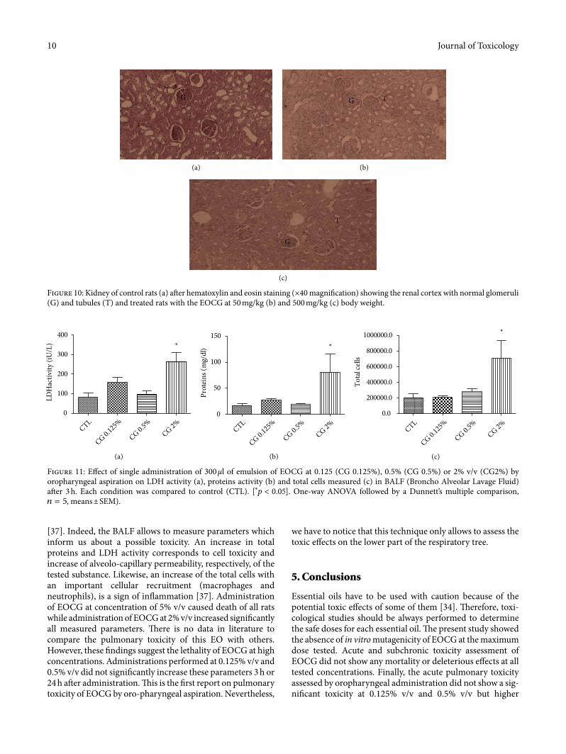
[37]. Indeed, the BALF allows to measure parameters which inform us about a possible toxicity. An increase in total proteins and LDH activity corresponds to cell toxicity and increase of alveolo-capillary permeability, respectively, of the tested substance. Likewise, an increase of the total cells with an important cellular recruitment (macrophages and neutrophils), is a sign of inflammation [37]. Administration of EOCG at concentration of 5% v/v caused death of all rats while administration of EOCG at 2% v/v increased significantly all measured parameters. �ere is no data in literature to compare the pulmonary toxicity of this EO with others. However, these findings suggest the lethality of EOCG at high concentrations. Administrations performed at 0.125% v/v and 0.5% v/v did not significantly increase these parameters 3 h or 24 h a�er administration. �is is the first report on pulmonary toxicity of EOCG by oro-pharyngeal aspiration. Nevertheless,
GT
(a)
G T
(b)
T
G
(c)
Figure 10: Kidney of control rats (a) a�er hematoxylin and eosin staining (×40 magnification) showing the renal cortex with normal glomeruli (G) and tubules (T) and treated rats with the EOCG at 50 mg/kg (b) and 500 mg/kg (c) body weight.
CTL
CG 0.125
%
CG 0.5%
CG 2%0
100
200
300
400*
LDH
activ
ity (i
U/L
)
(a)
CTL
CG 0.125
%
CG 0.5%
CG 2%0
50
100
150*
Prot
eins
(mg/
dl)
(b)
CTL
CG 0.125
%
CG 0.5%
CG 2%0.0
200000.0
400000.0
600000.0
800000.0
1000000.0 *
Tota
l cel
ls
(c)
Figure 11: Effect of single administration of 300 µl of emulsion of EOCG at 0.125 (CG 0.125%), 0.5% (CG 0.5%) or 2% v/v (CG2%) by oropharyngeal aspiration on LDH activity (a), proteins activity (b) and total cells measured (c) in BALF (Broncho Alveolar Lavage Fluid) a�er 3 h. Each condition was compared to control (CTL). [∗�푝 < 0.05]. One-way ANOVA followed by a Dunnett’s multiple comparison, �푛 = 5, means ± SEM).

11Journal of Toxicology
References
[1] WHO, Guidelines on Developing Consumer Information on Proper Use of Traditional, Complementary and Alternative Medicine, World Health Organization, p. 109, 2004.
[2] M. Vigan, “Essential oils: renewal of interest and toxicity,” European Journal of Dermatology, vol. 20, no. 6, pp. 685–692, 2010.
[3] A. C. Adomou, H. Yedomonhan, B. Djossa, Si Legba, M. Oumorou, and A. Akoegninou, “Etude Ethnobotanique des plantes médicinales vendues dans le marché d’Abomey-Calavi au Bénin,” International Journal of Biological and Chemical Sciences, vol. 6, no. 2, pp. 745–772, 2012.
[4] M. Zhu, K. T. Lew, and P.-L. Leung, “Protective effect of a plant formula on ethanol-induced gastric lesions in rats,” Phytotherapy Research, vol. 16, no. 3, pp. 276–280, 2002.
[5] S. Pale, N. Kouemo, G. S. Taiwe et al., “Antipyretic and anxiolytic properties of aqueous extract of Cymbopogon giganteus in rats model,” International Neuropsychiatric Disease Journal, vol. 11, no. 1, pp. 1–10, 2018.
[6] G. A. Alitonou, “Essential oils extracted from aromatic plants acclimated in Benin: chemical study, biological evaluation and potencial applications [PhD diss.],” vol. 2, Université de Montpellier, Montpellier, 2006.
[7] L. Popielas, C. Moulis, A. Keita, I. Fourasté, and J.-M. Bessière, “�e essential oil of Cymbopogon giganteus,” Planta Medica, vol. 57, no. 6, pp. 586–587, 1991.
[8] G. A. Alitonou, F. Avlessi, F. Tchobo et al., “Chemical composition and biological activities of essential oils from
concentrations were toxic. Taking together, our results show that EOCG have a very low potential to generate adverse effects at doses lower than 0.5% v/v but higher doses may be toxic. However, more additional toxicological studies on other organs and functions such as reproduction are needed to com-plete the safety profile of this EO.
Data Availability
�e data used to support the findings of this study are included within the article.
Conflicts of Interest
�e authors declare that there are no conflicts of interest regarding the publication of this paper.
Acknowledgments
�e authors are grateful to Roel Anthonissen (Sciensano) for his skillful assistance in Ames Test We also want to thank Honoré Zoclanclounon for his assistance in performing hema-toxylin-eosin staining. �is current study was supported by ARES-CCD (PRD project: VALTRAMED), which is deeply acknowledged.
CTL
CG 0.125
%
CG 0.5%
0
20
40
60
80
100
LDH
act
ivity
(iU
/L)
(a)
CTL
CG 0.125
%
CG 0.5%
0
5
10
15
20
Prot
eins
(mg/
dl)
(b)
CTL
CG 0.125
%
CG 0.5%
0
100000
200000
300000
400000
500000
Tota
l cel
ls
(c)
CTL
CG 0.125
%
CG 0.5%
0
50000
100000
150000
200000
250000
Mac
roph
ages
(d)
CTL
CG 0.125
%
CG 0.5%
0
20000
40000
60000
80000Ly
mph
ocyt
es
(e)
CTL
CG 0.125
%
CG 0.5%
0
50000
100000
150000
Neu
troph
ils
(f)
Figure 12: Effect of single administration of 300 µl of emulsion of EOCG at 0.125% v/v (CG 0.125%) or 0.5% v/v (CG 0.5%) by oropharyngeal aspiration on total proteins (a) and LDH activity (b), total cells (c), macrophage (d), lymphocytes (e), and neutrophils (f) measured in BAL (Broncho Alveolar Lavage) a�er 24 h; each condition was compared to control (CTL). �푝 > 0.05 for all experiments. One-way ANOVA followed by a Dunnett’s multiple comparison, �푛 = 5, means ± SEM).

Journal of Toxicology12
[23] J. Muller, F. Huaux, N. Moreau et al., “Respiratory toxicity of multi-wall carbon nanotubes,” Toxicology and Applied Pharmacology, vol. 207, no. 3, pp. 221–231, 2005.
[24] V. Sironval, L. Reylandt, P. Chaurand et al., “Respiratory hazard of Li-ion battery components: elective toxicity of lithium cobalt oxide (LiCoO2) particles in a mouse bioassay,” Archives of Toxicology, vol. 92, no. 5, pp. 1673–1684, 2018.
[25] M. Arras, F. Huaux, A. Vink et al., “Interleukin-9 reduces lung fibrosis and Type 2 immune polarization induced by silica particles in a murine model,” American Journal of Respiratory Cell and Molecular Biology, vol. 24, no. 4, pp. 368–375, 2001.
[26] M. Llana-Ruiz-Cabello, S. Pichardo, S. Maisanaba et al., “In vitro toxicological evaluation of essential oils and their main compounds used in active food packaging: a review,” Food and Chemical Toxicology, vol. 81, pp. 9–27, 2015.
[27] M. Saverini, I. Catanzaro, G. Sciandrello et al., “Genotoxicity of citrus wastewater in prokaryotic and eukaryotic cells and efficiency of heterogeneous photocatalysis by TiO2,” Journal of Photochemistry and Photobiology B: Biology, vol. 108, pp. 8–15, 2012.
[28] C. Ravichandran, P. C. Badgujar, P. Gundev, and A. Upadhyay, “Review of toxicological assessment of d-limonene, a food and cosmetics additive,” Food and Chemical Toxicology, vol. 120, pp. 668–680, 2018.
[29] Z. Gardner and M. McGuffin, American Herbal Products Association’s Botanical Safety Handbook, CRC Press, London, 2013.
[30] S. Santos, E. Rangel, J. Lima et al., “Toxicological and phytochemical studies of Aspidosperma subincanum Mart. stem bark (Guatambu),” Die Pharmazie-An International Journal of Pharmaceutical Sciences, vol. 64, no. 12, pp. 836–839
[31] R. S. Sellers, D. Mortan, B. Michael et al., “Society of toxicologic pathology position paper: organ weight recommendations for toxicology studies,” Toxicologic Pathology, vol. 35, no. 5, pp. 751–755, 2007.
[32] W. M. Kluwe, “Renal function tests as indicators of kidney injury in subacute toxicity studies,” Toxicology and Applied Pharmacology, vol. 57, no. 3, pp. 414–424, 1981.
[33] H. Olson, G. Betton, D. Robinson et al., “Concordance of the toxicity of pharmaceuticals in humans and in animals,” Regulatory Toxicology and Pharmacology, vol. 32, no. 1, pp. 56–67, 2000.
[34] R. Tisserand and R. Young, Essential Oil Safety-E-Book: A Guide for Health Care Professionals, Elsevier Health Sciences, 2013.
[35] J. P. Kehrer and S. Kacew, “Systematically applied chemicals that damage lung tissue,” Toxicology, vol. 35, no. 4, pp. 251–293, 1985.
[35] P. Goetz and K. Ghedira, Phytothérapie anti-infectieuse, Springer Science & Business Media, 2012.
[37] R. Henderson, J. Benson, F. Hahn et al., “New approaches for the evaluation of pulmonary toxicity: bronchoalveolar lavage fluid analysis,” Toxicological Sciences, vol. 5, no. 3, pp. 451–458, 1985.
the leaves of Cymbopogon giganteus Chiov. and Cymbopogon schoenanthus (L.) Spreng (Poaceae) from Benin,” International Journal of Biological and Chemical Sciences, vol. 6, no. 4, pp. 1819–1827, 2012.
[9] Fiches guide d’utilisation des Huiles essentielles, “Huile essentielle d’Ahibero BIO,” aroma-zone.com. https://www.aroma-zone.com/info/fiche-technique/huile-essentielle-dahibero?page=library (accessed July 3, 2019).
[10] I. H. Bassole, A. Lamien-Meda, B. Bayala et al., “Chemical composition and antimicrobial activity of Cymbopogon citratus and Cymbopogon giganteus essential oils alone and in combination,” Phytomedicine, vol. 18, no. 12, pp. 1070–1074, 2011.
[11] B. Bayala, I. H. Bassole, S. Maqdasy, S. Baron, J. Simpore, and J.-M. A. Lobaccaro, “Cymbopogon citratus and Cymbopogon giganteus essential oils have cytotoxic effects on tumor cell cultures. Identification of citral as a new putative anti-proliferative molecule,” Biochimie, vol. 153, pp. 162–170, 2018.
[12] S. Kpoviessi, J. Bero, P. Agbani et al., “Chemical composition, cytotoxicity and in vitro antitrypanosomal and antiplasmodial activity of the essential oils of four Cymbopogon species from Benin,” Journal of Ethnopharmacology, vol. 151, no. 1, pp. 652–659, 2014.
[13] J. B. Boti, A. Muselli, F. Tomi et al., “Combined analysis of Cymbopogon giganteus Chiov. leaf oil from Ivory Coast by GC/RI, GC/MS and 13 C-NMR,” Comptes Rendus Chimie, vol. 9, no. 1, pp. 164–168, 2006.
[14] L. Jirovetz, G. Buchbauer, G. Eller, M. B. Ngassoum, and P. M. Maponmetsem, “Composition and antimicrobial activity of Cymbopogon giganteus (Hochst.) chiov. essential flower, leaf and stem oils from Cameroon,” Journal of Essential Oil Research, vol. 19, no. 5, pp. 485–489, 2007.
[15] B. Ali, N. A. Al-Wabel, S. Shams, A. Ahamad, S. A. Khan, and F. Anwar, “Essential oils used in aromatherapy: a systemic review,” Asian Pacific Journal of Tropical Biomedicine, vol. 5, no. 8, pp. 601–611, 2015.
[16] OECD, “Bacterial reverse mutation test. Guideline 471, adopted 21.07. 1997,” Eleventh Addendum to the OECD Guidelines for the Testing of Chemicals, OECD Library, 1997.
[17] K. Mortelmans and E. Zeiger, “�e Ames Salmonella/microsome mutagenicity assay,” Mutation Research/Fundamental and Molecular Mechanisms of Mutagenesis, vol. 455, no. 1–2, pp. 29–60, 2000.
[18] E. Zeiger, K. J. Risko, and B. H. Margolin, “Strategies to reduce the cost of mutagenicity screening with the Salmonella assay,” Environmental Mutagenesis, vol. 7, no. 6, pp. 901–911, 1985.
[19] OECD, “OECD guideline for testing chemicals,” Acute Oral Toxicity-Acute Toxic Class Method, Guideline no. 423. Adopted 2001, Organisation for Economic and Cooperation Development, OECD Publishing, 2001.
[20] OECD, OECD for Test and Chemicals: Repeated Dose 28-day Oral Toxicity Study in Rodents, Guideline no. 407, OECD Publishing, 2008, Adopted: 3 October.
[21] I. Barbayianni, I. Ninou, A. Tzouvelekis, and V. Aidinis, “Bleomycin revisited,” Frontiers in Medicine, vol. 5, p. 269, 2018.
[22] C. Egger, C. Gérard, N. Vidotto et al., “Lung volume quantified by MRI reflects extracellular-matrix deposition and altered pulmonary function in bleomycin models of fibrosis: effects of SOM230,” American Journal of Physiology-Lung Cellular and Molecular Physiology, vol. 306, no. 12, pp. L1064–L1077, 2014.


![REPUBLIQUE DU BENIN - USEmbassy.govREPUBLIQUE DU BENIN [LOI N° 2010-33 DU 7 JANVIER 2011 PORTANT REGLES GENERALES POUR LES ELECTIONS EN REPUBLIQUE DU BENIN, MODIFIEE ET COMPLETEE]](https://static.fdocuments.fr/doc/165x107/5fe14b4a84a1bc33c92a8ddd/republique-du-benin-republique-du-benin-loi-n-2010-33-du-7-janvier-2011-portant.jpg)