Hedgehog Signaling Inhibition by Smoothened Antagonist BMS ...
13
Research Article Hedgehog Signaling Inhibition by Smoothened Antagonist BMS-833923 Reduces Osteoblast Differentiation and Ectopic Bone Formation of Human Skeletal (Mesenchymal) Stem Cells Nihal AlMuraikhi, 1 Nuha Almasoud, 1 Sarah Binhamdan, 1 Ghaydaa Younis, 1 Dalia Ali, 2 Muthurangan Manikandan, 1 Radhakrishnan Vishnubalaji, 3 Muhammad Atteya, 1,4 Abdulaziz Siyal, 1 Musaad Alfayez, 1 Abdullah Aldahmash, 1 Moustapha Kassem , 1,2,5 and Nehad M. Alajez 3 1 Stem Cell Unit, Department of Anatomy, College of Medicine, King Saud University, Riyadh 11461, Saudi Arabia 2 Molecular Endocrinology Unit (KMEB), Department of Endocrinology, University Hospital of Odense and University of Southern Denmark, Odense, Denmark 3 Cancer Research Center, Qatar Biomedical Research Institute (QBRI), Hamad Bin Khalifa University (HBKU), Qatar Foundation (QF), PO Box 34110, Doha, Qatar 4 Histology Department, Faculty of Medicine, Cairo University, Cairo, Egypt 5 Department of Cellular and Molecular Medicine, Danish Stem Cell Center (DanStem), University of Copenhagen, 2200 Copenhagen, Denmark Correspondence should be addressed to Nehad M. Alajez; [email protected] Received 24 June 2019; Revised 29 October 2019; Accepted 1 November 2019; Published 21 November 2019 Academic Editor: Tong-Chuan He Copyright © 2019 Nihal AlMuraikhi et al. This is an open access article distributed under the Creative Commons Attribution License, which permits unrestricted use, distribution, and reproduction in any medium, provided the original work is properly cited. Background. Hedgehog (Hh) signaling is essential for osteoblast differentiation of mesenchymal progenitors during endochondral bone formation. However, the critical role of Hh signaling during adult bone remodeling remains to be elucidated. Methods.A Smoothened (SMO) antagonist/Hedgehog inhibitor, BMS-833923, identified during a functional screening of a stem cell signaling small molecule library, was investigated for its effects on the osteoblast differentiation of human skeletal (mesenchymal) stem cells (hMSC). Alkaline phosphatase (ALP) activity and Alizarin red staining were employed as markers for osteoblast differentiation and in vitro mineralization capacity, respectively. Global gene expression profiling was performed using the Agilent® microarray platform. Effects on in vivo ectopic bone formation were assessed by implanting hMSC mixed with hydroxyapatite-tricalcium phosphate granules subcutaneously in 8-week-old female nude mice, and the amount of bone formed was assessed using quantitative histology. Results. BMS-833923, a SMO antagonist/Hedgehog inhibitor, exhibited significant inhibitory effects on osteoblast differentiation of hMSCs reflected by decreased ALP activity, in vitro mineralization, and downregulation of osteoblast-related gene expression. Similarly, we observed decreased in vivo ectopic bone formation. Global gene expression profiling of BMS-833923-treated compared to vehicle-treated control cells, identified 348 upregulated and 540 downregulated genes with significant effects on multiple signaling pathways, including GPCR, endochondral ossification, RANK-RANKL, insulin, TNF alpha, IL6, and inflammatory response. Further bioinformatic analysis employing Ingenuity Pathway Analysis revealed significant enrichment in BMS-833923-treated cells for a number of functional categories and networks involved in connective and skeletal tissue development and disorders, e.g., NFκB and STAT signaling. Conclusions. We identified SMO/Hedgehog antagonist (BMS-833923) as a powerful inhibitor of osteoblastic differentiation of hMSC that may be useful as a therapeutic option for treating conditions associated with high heterotopic bone formation and mineralization. Hindawi Stem Cells International Volume 2019, Article ID 3435901, 12 pages https://doi.org/10.1155/2019/3435901
Transcript of Hedgehog Signaling Inhibition by Smoothened Antagonist BMS ...

Research ArticleHedgehog Signaling Inhibition by Smoothened AntagonistBMS-833923 Reduces Osteoblast Differentiation and Ectopic BoneFormation of Human Skeletal (Mesenchymal) Stem Cells
Nihal AlMuraikhi,1 Nuha Almasoud,1 Sarah Binhamdan,1 Ghaydaa Younis,1 Dalia Ali,2
Muthurangan Manikandan,1 Radhakrishnan Vishnubalaji,3 Muhammad Atteya,1,4
Abdulaziz Siyal,1 Musaad Alfayez,1 Abdullah Aldahmash,1 Moustapha Kassem ,1,2,5
and Nehad M. Alajez 3
1Stem Cell Unit, Department of Anatomy, College of Medicine, King Saud University, Riyadh 11461, Saudi Arabia2Molecular Endocrinology Unit (KMEB), Department of Endocrinology, University Hospital of Odense and University ofSouthern Denmark, Odense, Denmark3Cancer Research Center, Qatar Biomedical Research Institute (QBRI), Hamad Bin Khalifa University (HBKU),Qatar Foundation (QF), PO Box 34110, Doha, Qatar4Histology Department, Faculty of Medicine, Cairo University, Cairo, Egypt5Department of Cellular and Molecular Medicine, Danish Stem Cell Center (DanStem), University of Copenhagen,2200 Copenhagen, Denmark
Correspondence should be addressed to Nehad M. Alajez; [email protected]
Received 24 June 2019; Revised 29 October 2019; Accepted 1 November 2019; Published 21 November 2019
Academic Editor: Tong-Chuan He
Copyright © 2019 Nihal AlMuraikhi et al. This is an open access article distributed under the Creative Commons AttributionLicense, which permits unrestricted use, distribution, and reproduction in any medium, provided the original work isproperly cited.
Background. Hedgehog (Hh) signaling is essential for osteoblast differentiation of mesenchymal progenitors during endochondralbone formation. However, the critical role of Hh signaling during adult bone remodeling remains to be elucidated. Methods. ASmoothened (SMO) antagonist/Hedgehog inhibitor, BMS-833923, identified during a functional screening of a stem cellsignaling small molecule library, was investigated for its effects on the osteoblast differentiation of human skeletal(mesenchymal) stem cells (hMSC). Alkaline phosphatase (ALP) activity and Alizarin red staining were employed as markers forosteoblast differentiation and in vitro mineralization capacity, respectively. Global gene expression profiling was performedusing the Agilent® microarray platform. Effects on in vivo ectopic bone formation were assessed by implanting hMSC mixedwith hydroxyapatite-tricalcium phosphate granules subcutaneously in 8-week-old female nude mice, and the amount of boneformed was assessed using quantitative histology. Results. BMS-833923, a SMO antagonist/Hedgehog inhibitor, exhibitedsignificant inhibitory effects on osteoblast differentiation of hMSCs reflected by decreased ALP activity, in vitro mineralization,and downregulation of osteoblast-related gene expression. Similarly, we observed decreased in vivo ectopic bone formation.Global gene expression profiling of BMS-833923-treated compared to vehicle-treated control cells, identified 348 upregulatedand 540 downregulated genes with significant effects on multiple signaling pathways, including GPCR, endochondralossification, RANK-RANKL, insulin, TNF alpha, IL6, and inflammatory response. Further bioinformatic analysis employingIngenuity Pathway Analysis revealed significant enrichment in BMS-833923-treated cells for a number of functional categoriesand networks involved in connective and skeletal tissue development and disorders, e.g., NFκB and STAT signaling.Conclusions. We identified SMO/Hedgehog antagonist (BMS-833923) as a powerful inhibitor of osteoblastic differentiation ofhMSC that may be useful as a therapeutic option for treating conditions associated with high heterotopic bone formation andmineralization.
HindawiStem Cells InternationalVolume 2019, Article ID 3435901, 12 pageshttps://doi.org/10.1155/2019/3435901
1. Introduction
The Hedgehog (Hh) signaling pathway plays manyimportant roles during development and postnatal tissuehomeostasis including bone formation [1]. In mammals,the hedgehog family is composed of three protein members:Sonic Hedgehog (Shh), Indian Hedgehog (Ihh), and DesertHedgehog (Dhh) [2, 3]. The negative and positive cellularregulation of Hh is controlled by two transmembrane pro-teins, Patched (Ptc), a negative regulator of Hh signaling,and Smoothened (Smo), a GPCR-like 7-transmembranereceptor that stimulates downstream signaling in responseto Hh [3, 4]. Thus, an inhibition of Hh signaling conse-quently inhibits SMO activity [3].
Human skeletal (mesenchymal) stem cells (hMSC) aremultipotent stem cells found in the bone marrow and cangive rise to various mesoderm cell types including bone-forming osteoblasts [5]. The osteoblastic differentiation ofhMSCs is controlled by multiple signaling pathways [6]including Hh signaling [1], TGFβ [7], bone morphogeneticproteins [8], and Wnt/β-catenin [9]. The Hh proteinenhances the differentiation of hMSCs into osteoblastic line-age and increases the expression of ALP and in vitro forma-tion of the mineralized matrix which are markers of matureosteoblast functions [10]. Moreover, a disruption of the Hhpathway has been associated with a number of developmen-tal bone diseases [1].
Small molecule inhibitors are currently used as valuabletools to decipher the molecular mechanisms regulating stemcell differentiation, and they have potential therapeutic use[11, 12]. Specifically, the small molecule agonist or antagonistregulators of the Hh pathway, could potentially be used totreat bone diseases. Here, we identified a small moleculeBMS-833923, an inhibitor of SMO and the Hh pathway,through a small molecule library screen [11] and character-ized its functions in vitro and in vivo on hMSC differentia-tion to bone-forming osteoblastic cells.
2. Materials and Methods
2.1. Cell Culture. A hMSC-TERT cell line was used in thisstudy as a model for hMSCs [13, 14]. hMSC-TERT expressesall features of primary hMSCs including multipotency andmolecular signature [13, 14]. The cells were cultured inDulbecco’s modified Eagle’s medium (DMEM), a basalmedium supplemented with 4mM L-glutamine, 4,500mg/lD-glucose, and 110mg/l 10% sodium pyruvate, in additionto 10% fetal bovine serum (FBS), 1% penicillin-streptomycin,and 1% nonessential amino acids. All reagents were pur-chased fromThermo Fisher Scientific Life Sciences,Waltham,MA (http://www.thermofisher.com). Cells were incubated in5% CO2 incubators at 37
°C and 95% humidity.
2.2. Osteoblast Differentiation. The cells were maintaineduntil 80%–90% confluency was achieved; then, themedium was replaced with osteoblast induction medium(DMEM containing 10% FBS, 1% penicillin-streptomycin,50mg/mL L-ascorbic acid (Wako Chemicals GmbH,Neuss, Germany,http://www.wako-chemicals. de/), 10mMb-glycerophosphate (Sigma-Aldrich), 10 nM calcitriol
(1a,25-dihydroxyvitamin D3, Sigma-Aldrich), and 10nMdexamethasone (Sigma-Aldrich). The stem cell signalingsmall molecule inhibitor library and BMS-833923 wereobtained from Selleckchem Inc. (Houston, TX, http://www.selleckchem.com). Each small molecule inhibitor was addedat 3μM concentration to the osteoblast induction medium,and cells were exposed to the inhibitor throughout the differ-entiation period. Control cells were treated with osteoblastinduction medium containing dimethyl sulfoxide (DMSO)as vehicle.
2.3. Cell Viability Assay. Cell viability assay was performedusing an alamarBlue assay according to the manufacturer’srecommendations (Thermo Fisher Scientific). Cells were cul-tured in 96-well plates in 300μL of the medium. On day 10,30μL/well of alamarBlue substrate was added (10%) andplates were incubated for 1 hr in the dark at 37°C. Readingswere obtained using the BioTek Synergy 2 Microplate Reader(BioTek Instruments Inc., Winooski, VT, US) at fluorescentmode (ex: 530 nm/em: 590nm).
2.4. Quantification of Alkaline Phosphatase Activity. Alkalinephosphatase (ALP) activity quantification was assessed usingthe BioVision ALP Activity Colorimetric Assay Kit (BioVi-sion, Inc., Milpitas, CA, http://http://www.biovision.com/)with some modifications. The cells were cultured in 96-wellplates. At day 10 of osteoblast differentiation, cells wererinsed once with PBS and fixed using 3.7% formaldehyde in90% ethanol for 30 seconds at room temperature. The fixa-tive was removed and 50μL/well of p-nitrophenyl phosphatesolution was added and incubated for 30–60minutes. Opticaldensities were then measured at 405nm using a Spectra-Max/M5 fluorescence spectrophotometer plate reader, andALP enzymatic activity was then normalized to cell number.
2.5. Alkaline Phosphatase Staining. Cells were cultured in a6-well plate in osteoblast differentiation medium. On day 10,the cells were washed in PBS and fixed in 10mM acetone/-citrate buffer at pH4.2 for 5min at room temperature. Thefix-ative was removed and Naphthol/Fast Red stain (0.2mg/mLNaphthol AS-TR phosphate substrate (Sigma-Aldrich);0.417mg/mL of Fast Red (Sigma-Aldrich)) was added for1 hr at room temperature. Then, cells were washed with water3 times and images were taken under the microscope.
2.6. Alizarin Red S Staining for Mineralized MatrixFormation. On day 21 of osteoblast differentiation, cells werewashed twice with PBS and fixed with 4% paraformaldehydefor 10min at room temperature. The fixative was rinsed, andthe cells were then washed 3 times with distilled water andstained with the 2% Alizarin Red S Staining Kit (ScienCellResearch Laboratories, Cat. No. 0223) for 10–20min at roomtemperature. Later, the cells were washed with water andimages were taken under the microscope.
2.7. RNA Extraction and cDNA Synthesis. Total RNA wasextracted from cell pellets on day 10 of osteoblast differen-tiation using the Total RNA Purification Kit (Norgen Bio-tek Corp., Thorold, ON, Canada, https://norgenbiotek.com/) according to the manufacturer’s instructions. The
2 Stem Cells International

concentrations of total RNA extracted were then measuredusing NanoDrop 2000 (Thermo Fisher Scientific Life Sci-ences). cDNAwas synthesized with 500 ng of total RNA usingthe High-Capacity cDNA Transcription Kit (Thermo FisherScientific Life Sciences) according to the manufacturer’sinstructions.
2.8. Quantitative Real Time-Polymerase Chain Reaction.Quantitative Real Time-Polymerase Chain Reaction(RT-PCR) was performed with fast SYBR Green using theApplied Biosystems ViiA™ 7 Real-Time PCR System(Thermo Fisher Scientific Life Sciences). Primers used in thisstudy are listed in Table 1. Relative expression was calculatedusing the 2ΔCT value method, and analysis was made aspreviously described [15].
2.9. Gene Expression Profiling by Microarray. One hundredfifty nanograms of total RNA from day 10 of osteoblast dif-ferentiation was labeled using a low-input Quick Amp Label-ing Kit (Agilent Technologies, Santa Clara, CA, http://www.agilent.com) and hybridized to the Agilent HumanSurePrint G3 Human GE 8 × 60 k microarray chip. Allmicroarray experiments were performed at the MicroarrayCore Facility (Stem Cell Unit, Department of Anatomy, KingSaud University College of Medicine, Riyadh, Saudi Arabia).Data were normalized and analyzed using GeneSpring 13.0software (Agilent Technologies). Pathway analyses were com-pleted using GeneSpring 13.0 as described previously [16].Twofold cutoff and PðcorrÞ < 0:05 (Benjamini-Hochbergmultiple testing corrected) were set to define significantlychanged transcripts. Pathway and functional annotation anal-yses were conducted using the Ingenuity Pathway Analysis(Ingenuity Systems, http://www.ingenuity.com/) [17, 18].Downregulated genes ≤ 2 FC (fold change) and corrected
P value < 0.05 were chosen for analysis. Enriched networkcategories were algorithmically generated based on their con-nectivity and ranked according to Z score.
2.10. In Vivo Ectopic Bone Formation Assay. Ethical approvalfor all animal experiments was obtained from the AnimalCare Committee of King Saud University. In vivo experi-ments were performed as previously described [19]. In brief,cells were trypsinized to a single-cell suspension and resus-pended in culture medium with/without the small moleculeinhibitor, BMS-833923. Around 5 × 10⁵ cells were seededonto 40mg Triosite hydroxyapatite-tricalcium phosphategranules per implant (HA/TCP, Biomatlante/Zimmer,Albertslund, Denmark) with 0.5 to 1mm granules, and keptovernight at 37°C and 5% CO2. HA/TCP granules in combi-nation with cells were then implanted subcutaneously (fourimplants per cell line) in the dorsolateral area of immune-compromised nude mice for 8 weeks. The implants wererecovered, fixed in formalin, decalcified using formic acidsolution (0.4M formic acid and 0.5M sodium formate) forthree days, embedded, and sectioned at 4μm. Staining wasperformed with hematoxylin and eosin or Sirius Red to iden-tify areas of formed bone.
2.11. Quantification of Ectopic Bone Formation. Slides weredigitized using a high-resolution whole-slide digital Scan-Scope scanner (Aperio Technologies, Inc.). The digitalimages from hematoxylin and eosin-stained slides wereviewed and quantified using the tools of ImageJ software.The whole implant was contoured to acquire the totalimplant area in pixels (TA). All areas of bone are selectedto obtain total bone area in pixels (BA). The BA/TA ratiowas calculated and reported as percentage (n = 3 sectionsper implant and 4 implants/condition). The digital images
Table 1: List of SYBR Green primers used in current study.
Gene name Forward primer Reverse primer
ACTB 5′AGCCATGTACGTTGCTA3′ 5′AGTCCGCCTAGAAGCA3′ALPL 5′GGAACTCCTGACCCTTGACC3′ 5′TCCTGTTCAGCTCGTACTGC3′COL1A1 5′GAGTGCTGTCCCGTCTGC3′ 5′TTTCTTGGTCGGTGGGTG3′ON 5′GAGGAAACCGAAGAGGAGG3′ 5′GGGGTGTTGTTCTCATCCAG3′LIF 5′GCCACCCATGTCACAACAAC3′ 5′CCCCCTGGGCTGTGTAATAG3′CXCL1 5′CCAGCTCTTCCGCTCCTC3′ 5′CACGGACGCTCCTGCTG3′CXCL2 5′GGGGTTCGCCGTTCTCGGA3′ 5′TGCGAGGAGGAGAGCTGGCAA3′CXCL3 5′CGCCCAAACCGAAGTCATAGCCA3′ 5′TGGTAAGGGCAGGGACCACCC3′IL6 5′CGAGCCCACCGGGAACGAAA3′ 5′GGACCGAAGGCGTTGTGGAG3′PLAU 5′ACTCCAAAGGCAGCAATGAAC3′ 5′GTGCTGCCCTCCGAATTTCT3′TNF 5′ACTTTGGAGTGATCGGCC3′ 5′GCTTGAGGGTTTGCTACAAC3′CCL20 5′CGAATCAGAAGCAGCAAGCAA3′ 5′TTGCGCACACAGACAACTTT3′VCAM 5′TGTTTGCAGCTTCTCAAGCTTTT3′ 5′GATGTGGTCCCCTCATTCGT3′GLI1 5′CTGGATCGGATAGGTGGTCT3′ 5′CAGAGGTTGGGAGGTAAGGA3′PTCH1 5′TGACCTAGTCAGGCTGGAAG3′ 5′GAAGGAGATTATCCCCCTGA3′
3Stem Cells International

from Sirius red-stained slides were viewed and analyzedusing Aperio’s viewing and image analysis tools. In eachslide, five rectangular fields with a fixed area of 1.18mm2
(total analysis area) were randomly selected. A color deconvo-lution algorithm (Aperio Technologies, Inc.) was employed tomeasure areas of the red color of stained collagen (positivestaining of Sirius red), and its percentage relative to the totalarea was calculated (n = 4 implants per treatment).
2.12. Statistical Analysis. Statistical analysis and graphingwere assessed using Microsoft Excel 2010 and GraphPadPrism 6 Software (GraphPad Software, San Diego, CA,U.S.A.), respectively. Results were presented as mean ± SEMfrom at least two independent experiments. An unpaired,two-tailed Student’s t-test was used to calculate statistical sig-nificance, and P values < 0.05 were considered statisticallysignificant.
3. Results
3.1. BMS-833923 Inhibited Osteoblast Differentiation ofhMSCs. BMS-833923 was initially identified as a potentinhibitor (at 3μM) of hMSC osteoblastic differentiationbased on a functional small molecule library screen [11].hMSCs exposed to BMS-833923 (3μM) exhibited a signifi-cant decrease in ALP cytochemical staining and ALP activitycompared to DMSO-vehicle control cells (Figures 1(a)–1(b))but did not exert significant effects on hMSC cell viability(Figure 1(c)). Also, hMSCs exposed to BMS-833923 exhib-ited a significant reduction in mineralized matrix formationas stained with Alizarin red, compared to vehicle-treatedcontrols (Figure 2(a)) which was associated with a significantreduction in the expression of a number of osteoblast genemarkers: ALP, COL1A1, and ON (Figure 2(b)). To confirmthat BMS-833923 is indeed targeting the hedgehog signalingpathway, hMSCs were treated with BMS-833923 at the same
concentration (3μM), and 48 hrs later, the expression of GLIFamily Zinc Finger 1 (GLI1) and Patched 1 (PTCH1), down-stream readouts of the hedgehog signaling pathway, wasassessed using qRT-PCR. Data presented in Figure 2(c) dem-onstrated significant inhibition of the hedgehog signalingpathway by BMS-833923. These data demonstrate thatBMS-833923 exerts inhibitory effects on osteoblast differenti-ation and mature osteoblastic cell functions, through theinhibition of the hedgehog signaling pathway.
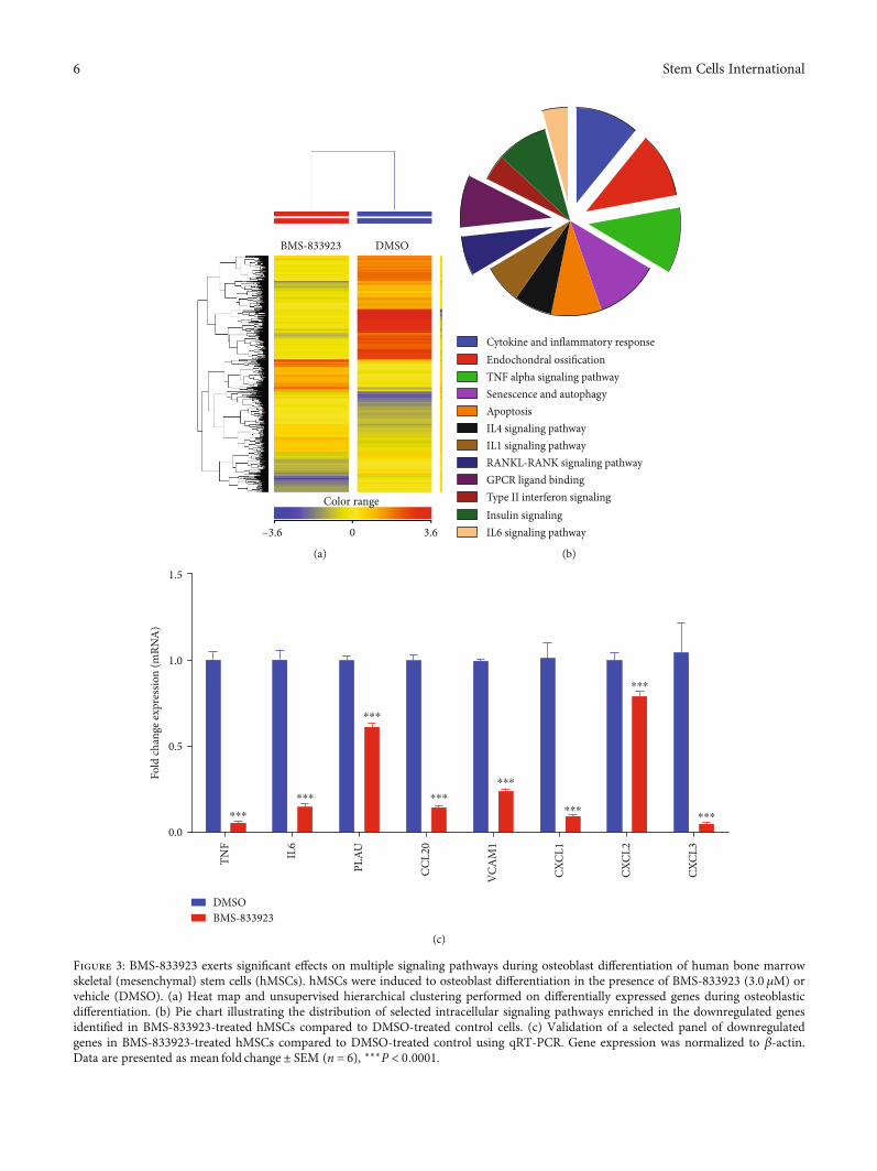
3.2. Global Gene Expression Identified Multiple AlteredSignaling Pathways in BMS-833923-Treated hMSCs. Tounderstand the molecular mechanism by which BMS-833923 reduces osteoblastic differentiation, we performedglobal gene expression profiling in BMS-833923-treatedhMSCs compared to vehicle-treated controls. Heat-map clus-tering revealed consistent changes in gene expression in BMS-833923-treated hMSCs compared to controls (Figure 3(a)).We identified 348 upregulated and 540 downregulated genes(fold change ≥ 2:0; PðcorrÞ < 0:05) (Supplementary Tables 1and 2). Pathway analysis of the downregulated genesidentified several differentially regulated signaling pathwaysthat are associated with osteoblastic differentiation includingGPCR, RANK-RANKL, insulin, TNF alpha and IL6, andcytokine and inflammatory response signaling (Figure 3(b)).A number of genes, including TNF, IL6, PLAU, CCL20,VCAM1, CXCL1, CXCL2, and CXCL3, were selected for afurther validation using qRT-PCR, which together were inconcordance with the microarray data (Figure 3(c)).
To determine the possible mechanisms of action, wedetermined the functional categories and signaling networksregulated by BMS-833923 during osteoblastic differentiationof hMSCs. The identified downregulated genes were sub-jected to core significance analysis using a manually curatedhuman functional annotation and network database
DMSO BMS-833923
50 𝜇m50 𝜇m
(a)
0
50% A
LP ac
tivity 100
⁎⁎⁎
150
DM
SO
BMS-
8339
23
(b)
0
50
Rela
tive c
ell v
iabi
lity
100n.s.
150
DM
SO
BMS-
8339
23
(c)
Figure 1: Effects of BMS-833923 treatment on the osteoblast differentiation of human bone marrow skeletal (mesenchymal) stem cells(hMSCs). (a) Representative alkaline phosphatase (ALP) staining of BMS-833923-treated hMSCs (3.0 μM) on day 10 postosteoblasticdifferentiation compared to DMSO-treated control cells. Magnification: 10x. (b) Quantification of ALP activity in BMS-833923-treatedhMSCs (3.0 μM) on day 10 postosteoblastic differentiation compared to DMSO-treated control cells. (c) Assay for cell viability usingalamarBlue assay BMS-833923-treated hMSCs (3.0 μM) on day 10 postosteoblastic differentiation compared to DMSO-treated controlcells. Data are presented as mean ± SEM (n = 16). DMSO: dimethyl sulfoxide.
4 Stem Cells International

DMSO BMS-833923
50 𝜇m50 𝜇m
(a)
0.0
0.5
Fold
chan
ge ex
pres
sion
(mRN
A)
1.0
⁎⁎⁎⁎ ⁎
1.5
COLI
AI
ALP ON
DMSOBMS-833923
(b)
0.0
0.5
Fold
chan
ge ex
pres
sion
(mRN
A)
1.0
⁎⁎⁎
1.5
GLI
1
PTCH
1
⁎⁎⁎
DMSOBMS-833923
(c)
Figure 2: Effects of BMS-833923 treatment on human bone marrow skeletal (mesenchymal) stem cell (hMSC) functions in vitro. hMSCswere induced to osteoblast differentiation in the presence of BMS-833923 (3.0μM) or vehicle (DMSO), and osteoblast differentiation wasdetermined by cytochemical staining with Alizarin red for an in vitro formed mineralized matrix (a) and expression of osteoblast-specificgenes by quantitative RT-PCR (b). Magnification: 10x. Data are presented as mean ± SEM, n = 6. ∗P < 0:05; ∗∗∗P < 0:0005. (c) Expressionof GLI1 and PCTH1 in hMSCs treated with BMS-833923 (3.0 μM) for 24 hours and measured using qRT-PCR, n = 6. Abbreviations:ALP—alkaline phosphatase; COL1A1—collagen Type I Alpha 1; ON—osteonectin; DMSO—dimethyl sulfoxide; GLI1—GLI Family ZincFinger 1; PTCH1—patched 1.
5Stem Cells International
BMS-833923 DMSO
0 3.6–3.6
Color range
(a)
Cytokine and inflammatory responseEndochondral ossificationTNF alpha signaling pathwaySenescence and autophagyApoptosisIL4 signaling pathwayIL1 signaling pathwayRANKL-RANK signaling pathwayGPCR ligand bindingType II interferon signalingInsulin signalingIL6 signaling pathway
(b)
Fold
chan
ge ex
pres
sion
(mRN
A)
0.0
0.5
1.0
1.5
DMSOBMS-833923
TNF
IL6
PLAU
CCL2
0
VCA
M1
CXCL
1
CXCL
2
CXCL
3
⁎⁎⁎
⁎⁎⁎
⁎⁎⁎
⁎⁎⁎
⁎⁎⁎
⁎⁎⁎⁎⁎⁎
⁎⁎⁎
(c)
Figure 3: BMS-833923 exerts significant effects on multiple signaling pathways during osteoblast differentiation of human bone marrowskeletal (mesenchymal) stem cells (hMSCs). hMSCs were induced to osteoblast differentiation in the presence of BMS-833923 (3.0 μM) orvehicle (DMSO). (a) Heat map and unsupervised hierarchical clustering performed on differentially expressed genes during osteoblasticdifferentiation. (b) Pie chart illustrating the distribution of selected intracellular signaling pathways enriched in the downregulated genesidentified in BMS-833923-treated hMSCs compared to DMSO-treated control cells. (c) Validation of a selected panel of downregulatedgenes in BMS-833923-treated hMSCs compared to DMSO-treated control using qRT-PCR. Gene expression was normalized to β-actin.Data are presented as mean fold change ± SEM (n = 6), ∗∗∗P < 0:0001.
6 Stem Cells International

(Ingenuity Pathway Analysis). This analysis revealed a signif-icant reduction in gene expression in several functional genecategories including those involved in tissue development,inflammatory responses, and connective tissue diseases(Figure 4(a)). The tissue development functional category isillustrated in Figure 4(b). Follow-up upstream regulator anal-ysis characterized a number of suppressed networks includ-ing TNF and NFκB and subsequent suppression of STATsignaling (Figures 4(c) and 4(d)). The Connective TissueDisorder, Organismal Injury and Abnormality, and Skeletaland Muscular Disorder networks were among the top identi-fied networks that are regulated in response to BMS-833923treatment of hMSCs (Figure 4(e)). Our data suggest thatBMS-833923 plays a role in regulating the function of a num-ber of signaling networks that are associated with the inhibi-tion of osteoblastic differentiation of hMSCs.
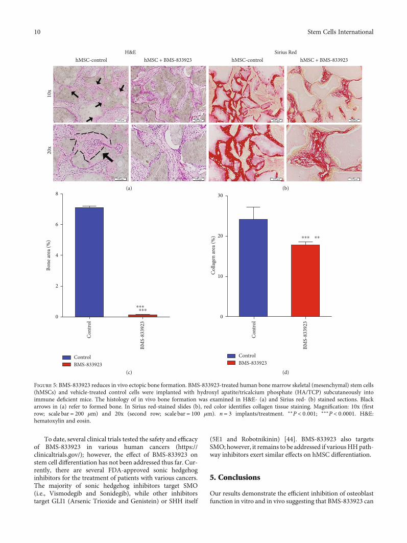
3.3. BMS-833923 Reduced In Vivo Ectopic Bone Formation.To further study the role of BMS-833923 in regulating osteo-blast differentiation in vivo, we determined the amount ofbone formed in vivo by hMSCs treated with BMS-833923or DMSO vehicle-treated control, following subcutaneousimplantation into immune deficient mice. BMS-833923-treated hMSCs exhibited a significant reduction in ectopicbone formation capacity (Figures 5(a)–5(b)). Quantitativehistological analysis revealed 90% (n = 3, P < 0:0001) reduc-tion of bone area (Figure 5(c)) and 30% reduction in collagenmatrix formation (n = 3, P < 0:001) (Figure 5(d)).
4. Discussion
MSCs are multipotent stem cells that reside in the bone mar-row and have the potential to differentiate into variousmesoderm-type cells including bone-forming osteoblasts[5]. The molecular processes and intracellular signaling path-ways regulating osteoblastic differentiation are under investi-gation with the aim of better understanding the pathogenesisof bone disorders and identifying novel approaches fortherapies [6, 20].
Chemical biology approaches employing small moleculeinhibitors targeting specific intracellular signaling pathwayshave been widely employed to dissect molecular mechanismsregulating stem cell fate due to their known mechanism ofaction and biological potency that make them suitable foruse in both in vitro and in vivo studies [11, 12]. In the currentstudy, we identified BMS-833923, a small molecule inhibitorof the Hh signaling pathway, through a small moleculelibrary screen, as a powerful inhibitor of osteoblast differenti-ation of hMSCs in vitro and in vivo.
BMS-833923 is a small-molecule Smoothened (SMO)inhibitor, and thus it inhibits the sonic hedgehog (SHH) sig-naling. In the absence of Hh, a cell-surface transmembraneprotein Patched (PTCH) prevents the high expression andactivity of SMO. When extracellular Hh is present, it bindsto and inhibits Patched, allowing SMO to accumulate andtransmit intracellular signaling [2, 21, 22]. BMS-833923 hasbeen as an experimental oral drug in advanced cancerpatients due to its antiproliferative effects. Here, we demon-strate that BMS-833923 treatment can regulate stem cell
functions and that it can reduce osteoblast differentiationboth in vitro and in vivo.
Global gene expression profiling of hMSCs treated withBMS-833923 identified significant changes in several intra-cellular signaling pathways including GPCR [23], pathwaysinvolved in endochondral ossification [24], RANK-RANKL[25], insulin [26], TNF alpha [27], IL6 [28], and cytokineand inflammatory response signaling [29] known to regulateosteoblast differentiation and function. These data suggestthat hH signaling is not only important for embryonic bonedevelopment but may also play a role in the regulationof adult bone cells. During early stages of development,Shh regulates the patterning of the future limb [30–32],while Ihh acts later in the process of endochondral boneformation in limb development. Together with parathyroidhormone-related peptide, Ihh controls the growth plateand long bone development [33–35]. Hh signaling alsoplays a crucial role in dermal bone formation and intra-membranous ossification by regulating osteogenesis ratherthan proliferation [36]. In adults, Hh signaling is requiredfor bone homeostasis [37]. Shh regulates osteoblast prolif-eration and differentiation and osteoclast formation at theremodeling site of fractures [38]. Notably, we did notobserve direct changes in HH genes based on the microar-ray data; however, we observed the suppression of theGPCR, which is downstream of the HH signaling pathway(Figure 3(b)). This could be explained by the fact that themicroarray experiment was conducted on day 10 postdiffer-entiation; therefore, at that time point, and since hMSCs havealready differentiated, we anticipate that the HH pathway isno longer active in those cells. It is plausible that BMS-833923 exerts its inhibitory effects at an early stage duringhMSC differentiation (Figure 2(c), at 48 hrs). On day 10,the most significant changes were in processes related to oste-ogenesis and cytokine production.
BMS-833923-treated hMSCs exhibited significantdownregulation in members of the two major chemokinesubfamilies—CC and CXC, i.e., CXCL1, CXCL2, CXCL3,CXCL8, CCL2, CCL20, CCL4L2, and CCL21 (Figure 3(c)and Supplementary Table 2). These chemokines are knownto play a role during bone repair through the recruitmentof progenitor cells to bone regeneration sites [39, 40]. Inaddition, we identified NFκB and STAT signaling amongthe top inhibited signaling pathways in BMS-833923-treated hMSCs; both are known to play a role in osteoblastdifferentiation and bone formation.
BMS-833923 has been suggested to be relevant for thetherapy of a number of cancer types due to its efficacy in inhi-biting hH signaling. BMS-833923 inhibited the growth oftumor cell lines derived from patients with hematologicalmalignancies such as CML, ALL, and AML [41] and it hasalso in vivo inhibitory effects on tumor growth of medullo-blastoma and pancreatic carcinoma in animal models aftera single oral dose [21]. Finally, some studies have suggestedthat BMS-833923 can be used in the management of cancerbone diseases. In vitro treatment with BMS-833923 inhibitedthe growth of myeloma cells and decreased the percentage ofALDH+ cancer stem cells in the bone marrow of patientswith multiple myeloma [42, 43].
7Stem Cells International

Decreasing < –3.636
AllCellular movement
Hematological system development and functionInflammatory response
Cell-to-cell signalling and interac... Cellular development
Tissue development
Lymphoid tissue st...
Connective tissue d...
Inflammatory diseaseGastrointestinal dise... Connective t...
Hematopoiesis
Cardiovascul...
Skeletal a...
Digestiv...
Dermatol...
Immunolo...
Neurologica...
Organismal injury and abnormalities Immune cell trafficking Cellular growth and prol... Cancer Cell death and surviv... Skeletal and ...
0.349 increasing
Organ devel... Cellular func... Cell morphol... Infectious ...
Small mo...Organism...Cell cycle Cell sig...Molecular t...
Hepatic sy...Hepatic ... Lipid ... Respir... Gene ... Cell-...
Car...
Ren...Cel...
C... F... H...
Repr...Tum...Tissu...
Endoc...
Metab... Hem...End...
Ner... D...
Oph...Ren...
Car...Psyc...Embry... Vit...
(a)
All > tissue developmentDifferentiation Proliferation Development
Formation
Accumulation
Monocytopoiesis Hematopoiesis Morphol... Branching
Morphog...GenerationActivationLeukopoiesis
Growth Cell proliferation Osteoclastogenesis Transdifferentiation
(b)
TNF
ERK1/2
Ap1
IL1B
NF𝜅B1NF𝜅BIA
NF𝜅B (complex)
RNA polymerase IIJUNRELA
STAT3 KLF6
FOXO1
TNF
ERK1/2
Ap1
IL1B
NF𝜅B1NF𝜅BIA
NF𝜅B (complex)
RNA polymerase IIJUNRELA
STAT3 KLF6
FOXO1
(c)
Figure 4: Continued.
8 Stem Cells International

Prediction legendMore extreme in dataset
Increased measurementDecreased measurement
Predicted activationPredicted inhibition
Glow indicates activitywhen oppositeof measurement
Predicted relationshipsLeads to activationLeads to inhibitionFindings inconsistentwith state of downstreammoleculeEffect not predicted
Less
Less
NF𝜅B1ESR1
NF𝜅B (complex)
RNA polymerase II
Ap1
TGFB1 IL1BTNF
STAT3 KLF6
EP300CTNNB1JUNRELANF𝜅B1ESR1
NF𝜅B (complex)
RNA polymerase II
Ap1
TGFB1 IL1BTNF
STAT3 KLF6
EP300CTNNB1JUNRELA
(d)
(e)
Figure 4: Bioinformatic analysis of signaling networks regulated in BMS-833923-treated human bone marrow skeletal (mesenchymal) stemcells (hMSCs). (a) Disease and function heat map depicting activation (red) or inhibition (blue) of the indicated functional and diseasecategories identified in the downregulated transcripts in BMS-833923-treated hMSCs. (b) Heat map illustrating the tissue developmentfunctional category. (c) Illustration of the TNF and (d) NFκB networks with predicted activated state based on transcriptome data. Figurelegend illustrates the interaction between molecules within the network. (e) Illustration of the connective tissue disorders, organismalinjury and abnormalities, and skeletal and muscular disorder network.
9Stem Cells International
To date, several clinical trials tested the safety and efficacyof BMS-833923 in various human cancers (https://clinicaltrials.gov/); however, the effect of BMS-833923 onstem cell differentiation has not been addressed thus far. Cur-rently, there are several FDA-approved sonic hedgehoginhibitors for the treatment of patients with various cancers.The majority of sonic hedgehog inhibitors target SMO(i.e., Vismodegib and Sonidegib), while other inhibitorstarget GLI1 (Arsenic Trioxide and Genistein) or SHH itself
(5E1 and Robotnikinin) [44]. BMS-833923 also targetsSMO; however, it remains to be addressed if variousHHpath-way inhibitors exert similar effects on hMSC differentiation.
5. Conclusions
Our results demonstrate the efficient inhibition of osteoblastfunction in vitro and in vivo suggesting that BMS-833923 can
hMSC-control
(a) (b)
(c) (d)
200 𝜇m 200 𝜇m 200 𝜇m 200 𝜇m
100 𝜇m 100 𝜇m 100 𝜇m 100 𝜇m
10x
20x
hMSC + BMS-833923 hMSC-control hMSC + BMS-833923H&E Sirius Red
8
6
4
2
Bone
area
(%)
0
Con
trol
BMS-
8339
23
⁎⁎⁎⁎⁎⁎
ControlBMS-833923
ControlBMS-833923
30
20
10Col
lage
n ar
ea (%
)
0
Con
trol
BMS-
8339
23
⁎⁎⁎ ⁎⁎
Figure 5: BMS-833923 reduces in vivo ectopic bone formation. BMS-833923-treated human bone marrow skeletal (mesenchymal) stem cells(hMSCs) and vehicle-treated control cells were implanted with hydroxyl apatite/tricalcium phosphate (HA/TCP) subcutaneously intoimmune deficient mice. The histology of in vivo bone formation was examined in H&E- (a) and Sirius red- (b) stained sections. Blackarrows in (a) refer to formed bone. In Sirius red-stained slides (b), red color identifies collagen tissue staining. Magnification: 10x (firstrow; scale bar = 200 μm) and 20x (second row; scale bar = 100 μm). n = 3 implants/treatment. ∗∗P < 0:001; ∗∗∗P < 0:0001. H&E:hematoxylin and eosin.
10 Stem Cells International

be employed in the treatment of bone diseases with enhancedbone formation, e.g., sclerotic bone metastases.
Data Availability
Additional data are available as supplementary data.
Conflicts of Interest
The authors declare no conflict of interest.
Acknowledgments
Wewould like to thank the Deanship of Scientific Research atKing Saud University (Research Group No. RG-1440-019)for funding this work.
Supplementary Materials
Supplementary Table 1: upregulated genes in hBMSCstreated with BMS-833923 compared to DMSO on day 10 ofosteoblastic differentiation. Supplementary Table 2: down-regulated genes in hBMSCs treated with BMS-833923 com-pared to DMSO on day 10 of osteoblastic differentiation.(Supplementary Materials)
References
[1] J. Yang, P. Andre, L. Ye, and Y. Z. Yang, “The Hedgehog sig-nalling pathway in bone formation,” International Journal ofOral Science, vol. 7, no. 2, pp. 73–79, 2015.
[2] T. L. Lin and W. Matsui, “Hedgehog pathway as a drug target:smoothened inhibitors in development,” OncoTargets andTherapy, vol. 5, pp. 47–58, 2012.
[3] F. Wu, Y. Zhang, B. Sun, A. P. McMahon, and Y. Wang,“Hedgehog signaling: from basic biology to cancer therapy,”Cell Chemical Biology, vol. 24, no. 3, pp. 252–280, 2017.
[4] M. Philipp and M. G. Caron, “Hedgehog signaling: is Smo a Gprotein-coupled receptor?,” Current Biology, vol. 19, no. 3,pp. R125–R127, 2009.
[5] A. Aldahmash, W. Zaher, M. Al-Nbaheen, and M. Kassem,“Human stromal (mesenchymal) stem cells: basic biologyand current clinical use for tissue regeneration,” Annals ofSaudi Medicine, vol. 32, no. 1, pp. 68–77, 2012.
[6] B. M. Abdallah, A. Jafari, W. Zaher, W. Qiu, and M. Kassem,“Skeletal (stromal) stem cells: an update on intracellularsignaling pathways controlling osteoblast differentiation,”Bone, vol. 70, pp. 28–36, 2015.
[7] M. Elsafadi, M. Manikandan, S. Almalki et al., “TGFβ1-Induced Differentiation of Human Bone Marrow-DerivedMSCs Is Mediated by Changes to the Actin Cytoskeleton,”Stem Cells International, vol. 2018, 14 pages, 2018.
[8] X. Cao and D. Chen, “The BMP signaling and in vivo bone for-mation,” Gene, vol. 357, no. 1, pp. 1–8, 2005.
[9] Z. Zhong, N. J. Ethen, and B. O. Williams, “WNT signaling inbone development and homeostasis,” Wiley InterdisciplinaryReviews: Developmental Biology, vol. 3, no. 6, pp. 489–500,2014.
[10] J. Q. Cai, Y. Z. Huang, X. H. Chen et al., “Sonic hedgehogenhances the proliferation and osteogenic differentiation of
bone marrow-derived mesenchymal stem cells,” Cell BiologyInternational, vol. 36, no. 4, pp. 349–355, 2012.
[11] N. AlMuraikhi, D. Ali, A. Alshanwani et al., “Stem cell libraryscreen identified ruxolitinib as regulator of osteoblastic differ-entiation of human skeletal stem cells,” Stem Cell Research &Therapy, vol. 9, no. 1, p. 319, 2018.
[12] B. Lu and A. Atala, “Small molecules and small molecule drugsin regenerative medicine,” Drug Discovery Today, vol. 19,no. 6, pp. 801–808, 2014.
[13] B. M. Abdallah, M. Haack-Sorensen, J. S. Burns et al., “Main-tenance of differentiation potential of human bone marrowmesenchymal stem cells immortalized by human telomerasereverse transcriptase gene despite (corrected) extensive prolif-eration,” Biochemical and Biophysical Research Communica-tions, vol. 326, no. 3, pp. 527–538, 2005.
[14] J. L. Simonsen, C. Rosada, N. Serakinci et al., “Telomeraseexpression extends the proliferative life-span and maintainsthe osteogenic potential of human bone marrow stromal cells,”Nature Biotechnology, vol. 20, no. 6, pp. 592–596, 2002.
[15] K. J. Livak and T. D. Schmittgen, “Analysis of Relative GeneExpression Data Using Real-Time Quantitative PCR and the2−ΔΔCT Method,” Methods, vol. 25, no. 4, pp. 402–408, 2001.
[16] R. Vishnubalaji, M. Manikandan, M. Fahad et al., “Molecularprofiling of ALDH1+ colorectal cancer stem cells reveals pref-erential activation of MAPK, FAK, and oxidative stress pro-survival signalling pathways,” Oncotarget, vol. 9, no. 17,pp. 13551–13564, 2018.
[17] S. E. Calvano, Inflammation and Host Response to InjuryLarge Scale Collaborative Research Program, W. Xiao et al.,“A network-based analysis of systemic inflammation inhumans,” Nature, vol. 437, no. 7061, pp. 1032–1037, 2005.
[18] R. Vishnubalaji, V. Sasidharan Nair, K. Ouararhni, E. Elkord,and N. M. Alajez, “Integrated transcriptome and pathwayanalyses revealed multiple activated pathways in breast can-cer,” Frontiers in Oncology, vol. 9, p. 910, 2019.
[19] N. AlMuraikhi, D. Ali, R. Vishnubalaji et al., “Notch signalinginhibition by LY411575 attenuates osteoblast differentiationand decreased ectopic bone formation capacity of human skel-etal (mesenchymal) stem cells,” Stem Cells International,vol. 2019, Article ID 3041262, 12 pages, 2019.
[20] W. Huang, S. Y. Yang, J. Z. Shao, and Y. P. Li, “Signaling andtranscriptional regulation in osteoblast commitment and dif-ferentiation,” Frontiers in Bioscience, vol. 12, no. 8-12,pp. 3068–3092, 2007.
[21] T. Rimkus, R. Carpenter, S. Qasem, M. Chan, and H. W. Lo,“Targeting the Sonic Hedgehog signaling pathway: review ofsmoothened and GLI inhibitors,” Cancers, vol. 8, no. 2, p. 22,2016.
[22] L. L. Siu, K. Papadopoulos, S. R. Alberts et al., “A first-in-human, phase I study of an oral hedgehog (HH) pathwayantagonist, BMS-833923 (XL139), in subjects with advancedor metastatic solid tumors,” Journal of Clinical Oncology,vol. 28, 15_suppl, p. 2501, 2010.
[23] J. Jules, S. Yang, W. Chen, and Y. P. Li, “Role of regulators of Gprotein signaling proteins in bone physiology and pathophys-iology,” Progress in Molecular Biology and TranslationalScience, vol. 133, pp. 47–75, 2015.
[24] H. Hojo, S. Ohba, and U.-I. Chung, “Signaling pathwaysregulating the specification and differentiation of the osteo-blast lineage,” Regenerative Therapy, vol. 1, pp. 57–62, 2015.
11Stem Cells International
[25] B. F. Boyce and L. Xing, “Functions of RANKL/RANK/OPG inbone modeling and remodeling,” Archives of Biochemistry andBiophysics, vol. 473, no. 2, pp. 139–146, 2008.
[26] S. N. Pramojanee, M. Phimphilai, N. Chattipakorn, and S. C.Chattipakorn, “Possible roles of insulin signaling in osteo-blasts,” Endocrine Research, vol. 39, no. 4, pp. 144–151, 2014.
[27] B. Zhao, “TNF and bone remodeling,” Current OsteoporosisReports, vol. 15, no. 3, pp. 126–134, 2017.
[28] Z. Xie, S.’. Tang, G. Ye et al., “Interleukin-6/interleukin-6receptor complex promotes osteogenic differentiation of bonemarrow-derived mesenchymal stem cells,” Stem Cell Research& Therapy, vol. 9, no. 1, p. 13, 2018.
[29] T. M. Skerry, “The effects of the inflammatory response onbone-growth,” European Journal of Clinical Nutrition,vol. 48, pp. S190–S198, 1994.
[30] A. Kicheva, M. Cohen, and J. Briscoe, “Developmental patternformation: insights from physics and biology,” Science,vol. 338, no. 6104, pp. 210–212, 2012.
[31] B. Wang, J. F. Fallon, and P. A. Beachy, “Hedgehog-regulatedprocessing of Gli3 produces an anterior/posterior repressorgradient in the developing vertebrate limb,” Cell, vol. 100,no. 4, pp. 423–434, 2000.
[32] J. Zhu, E. Nakamura, M. T. Nguyen, X. Bao, H. Akiyama, andS. Mackem, “Uncoupling Sonic hedgehog control of patternand expansion of the developing limb bud,” DevelopmentalCell, vol. 14, no. 4, pp. 624–632, 2008.
[33] J. M. Kindblom, O. Nilsson, T. Hurme, C. Ohlsson, andL. Savendahl, “Expression and localization of Indian hedgehog(Ihh) and parathyroid hormone related protein (PTHrP) inthe human growth plate during pubertal development,” TheJournal of Endocrinology, vol. 174, no. 2, pp. R1–R6, 2002.
[34] H. M. Kronenberg, “Developmental regulation of the growthplate,” Nature, vol. 423, no. 6937, pp. 332–336, 2003.
[35] B. St-Jacques, M. Hammerschmidt, and A. P. McMahon,“Indian hedgehog signaling regulates proliferation and differ-entiation of chondrocytes and is essential for bone formation,”Genes & Development, vol. 13, no. 16, pp. 2072–2086, 1999.
[36] A. Abzhanov, S. J. Rodda, A. P. McMahon, and C. J. Tabin,“Regulation of skeletogenic differentiation in cranial dermalbone,” Development, vol. 134, no. 17, pp. 3133–3144, 2007.
[37] R. Petrova and A. L. Joyner, “Roles for Hedgehog signaling inadult organ homeostasis and repair,” Development, vol. 141,no. 18, pp. 3445–3457, 2014.
[38] Y. Horikiri, T. Shimo, N. Kurio et al., “Sonic hedgehog regu-lates osteoblast function by focal adhesion kinase signaling inthe process of fracture healing,” PLoS One, vol. 8, no. 10,p. e76785, 2013.
[39] D. S. Amarasekara, H. Yun, S. Kim, N. Lee, H. Kim, and J. Rho,“Regulation of osteoclast differentiation by cytokine net-works,” Immune Network, vol. 18, no. 1, 2018.
[40] E. Galliera, M. Locati, A. Mantovani, and M. M. Corsi,“Chemokines and bone remodeling,” International Journalof Immunopathology and Pharmacology, vol. 21, no. 3,pp. 485–491, 2008.
[41] N. P. Shah, J. E. Cortes, G. Martinelli et al., “Dasatinib plussmoothened (SMO) inhibitor BMS-833923 in chronic myeloidleukemia (CML) with resistance or suboptimal response to aprior tyrosine kinase inhibitor (TKI): phase I studyCA180323,” Blood, vol. 124, no. 21, p. 4539, 2014.
[42] S. B. Gendreau, D. Hawkins, C.‐. P. Ho et al., “Abstract B192:Preclinical characterization of BMS‐833923 (XL139), a hedge-
hog (HH) pathway inhibitor in early clinical development,”Molecular Cancer Therapeutics, vol. 8, no. 12, 2009.
[43] C. D. Peacock, Q. Wang, G. S. Gesell et al., “Hedgehog signal-ing maintains a tumor stem cell compartment in multiplemyeloma,” Proceedings of the National Academy of Sciencesof the United States of America, vol. 104, no. 10, pp. 4048–4053, 2007.
[44] R. L. Carpenter and H. Ray, “Safety and tolerability of sonichedgehog pathway inhibitors in cancer,” Drug Safety, vol. 42,no. 2, pp. 263–279, 2019.
12 Stem Cells International

Hindawiwww.hindawi.com
International Journal of
Volume 2018
Zoology
Hindawiwww.hindawi.com Volume 2018
Anatomy Research International
PeptidesInternational Journal of
Hindawiwww.hindawi.com Volume 2018
Hindawiwww.hindawi.com Volume 2018
Journal of Parasitology Research
GenomicsInternational Journal of
Hindawiwww.hindawi.com Volume 2018
Hindawi Publishing Corporation http://www.hindawi.com Volume 2013Hindawiwww.hindawi.com
The Scientific World Journal
Volume 2018
Hindawiwww.hindawi.com Volume 2018
BioinformaticsAdvances in
Marine BiologyJournal of
Hindawiwww.hindawi.com Volume 2018
Hindawiwww.hindawi.com Volume 2018
Neuroscience Journal
Hindawiwww.hindawi.com Volume 2018
BioMed Research International
Cell BiologyInternational Journal of
Hindawiwww.hindawi.com Volume 2018
Hindawiwww.hindawi.com Volume 2018
Biochemistry Research International
ArchaeaHindawiwww.hindawi.com Volume 2018
Hindawiwww.hindawi.com Volume 2018
Genetics Research International
Hindawiwww.hindawi.com Volume 2018
Advances in
Virolog y Stem Cells International
Hindawiwww.hindawi.com Volume 2018
Hindawiwww.hindawi.com Volume 2018
Enzyme Research
Hindawiwww.hindawi.com Volume 2018
International Journal of
MicrobiologyHindawiwww.hindawi.com
Nucleic AcidsJournal of
Volume 2018
Submit your manuscripts atwww.hindawi.com


![Proxima Systems - IProx BMS - Datasheet [FR]](https://static.fdocuments.fr/doc/165x107/55c7266abb61eb004d8b46a9/proxima-systems-iprox-bms-datasheet-fr.jpg)