
Genetic Vaccines and Therapy BioMed Central · Accidentelle, LTCRA, UPRES 1632, CH U Saint Antoine,...
8
BioMed Central Page 1 of 8 (page number not for citation purposes) Genetic Vaccines and Therapy Open Access Research In vivo gene targeting of IL-3 into immature hematopoietic cells through CD117 receptor mediated antibody gene delivery Alain Chapel* 1,2 , Olivier Deas 3,4 , Morad Bensidhoum 2 , Sabine François 1,2 , Moubarak Mouiseddine 1,2 , Pascal Poncet 4 , Antoine Dürrbach 3 , Jocelyne Aigueperse 1 , Patrick Gourmelon 1 , Norbert C Gorin 2 , François Hirsch †3 and Dominique Thierry †1,2 Address: 1 Institut de Radioprotection et de Sûreté Nucléaire, Département de Protection et de santé de l'Homme et de Dosimétrie, Section Autonome de Radiobiologie Appliquée à la Médecine, Fontenay aux roses, France, 2 Laboratoire de Thérapie Cellulaire et de Radioprotection Accidentelle, LTCRA, UPRES 1632, CHU Saint Antoine, Paris, France, 3 Inserm U542 and Paris XI University, Villejuif, France and 4 Institut Pasteur, Paris, France Email: Alain Chapel* - [email protected]; Olivier Deas - [email protected]; Morad Bensidhoum - [email protected]; Sabine François - [email protected]; Moubarak Mouiseddine - [email protected]; Pascal Poncet - [email protected]; Antoine Dürrbach - [email protected]; Jocelyne Aigueperse - [email protected]; Patrick Gourmelon - [email protected]; Norbert C Gorin - [email protected]; François Hirsch - [email protected]; Dominique Thierry - [email protected] * Corresponding author †Equal contributors Abstract Background: Targeted gene transfection remains a crucial issue to permit the real development of genetic therapy. As such, in vivo targeted transfection of specific subsets of hematopoietic stem cells might help to sustain hematopoietic recovery from bone marrow aplasia by providing local production of growth factors. Methods: Balb/C mice were injected intravenously, with an anti-mouse c-kit (CD117) monoclonal antibody chemically coupled to a human IL-3 gene-containing plasmid DNA. Mice were sacrificed for tissue analyses at various days after injection of the conjugates. Results: By ELISA, the production of human IL-3 was evidenced in the sera of animals 5 days after treatment. Cytofluorometric analysis after in vivo transfection of a reporter gene eGFP demonstrated transfection of CD117+/Sca1+ hematopoietic immature cells. By PCR analysis of genomic DNA and RNA using primer specific pIL3 sequences, presence and expression of the human IL-3-transgene were detected in the bone marrow up to 10 days in transfected mice but not in control animals. Conclusions: These data clearly indicate that antibody-mediated endocytosis gene transfer allows the expression of the IL-3 transgene into hematopoietic immature cells, in vivo. While availability of marketed recombinant growth factors is restricted, this targeting strategy should permit delivery of therapeutic genes to tissues of interest through systemic delivery. In particular, the ability to specifically target growth factor expression into repopulating hematopoietic stem cells may create new opportunities for the treatment of primary or radiation-induced marrow failures. Published: 27 October 2004 Genetic Vaccines and Therapy 2004, 2:16 doi:10.1186/1479-0556-2-16 Received: 07 June 2004 Accepted: 27 October 2004 This article is available from: http://www.gvt-journal.com/content/2/1/16 © 2004 Chapel et al; licensee BioMed Central Ltd. This is an Open Access article distributed under the terms of the Creative Commons Attribution License (http://creativecommons.org/licenses/by/2.0 ), which permits unrestricted use, distribution, and reproduction in any medium, provided the original work is properly cited.
Transcript of Genetic Vaccines and Therapy BioMed Central · Accidentelle, LTCRA, UPRES 1632, CH U Saint Antoine,...
BioMed CentralGenetic Vaccines and Therapy
ss
Open AcceResearchIn vivo gene targeting of IL-3 into immature hematopoietic cells through CD117 receptor mediated antibody gene deliveryAlain Chapel*1,2, Olivier Deas3,4, Morad Bensidhoum2, Sabine François1,2, Moubarak Mouiseddine1,2, Pascal Poncet4, Antoine Dürrbach3, Jocelyne Aigueperse1, Patrick Gourmelon1, Norbert C Gorin2, François Hirsch†3 and Dominique Thierry†1,2Address: 1Institut de Radioprotection et de Sûreté Nucléaire, Département de Protection et de santé de l'Homme et de Dosimétrie, Section Autonome de Radiobiologie Appliquée à la Médecine, Fontenay aux roses, France, 2Laboratoire de Thérapie Cellulaire et de Radioprotection Accidentelle, LTCRA, UPRES 1632, CHU Saint Antoine, Paris, France, 3Inserm U542 and Paris XI University, Villejuif, France and 4Institut Pasteur, Paris, France
Email: Alain Chapel* - [email protected]; Olivier Deas - [email protected]; Morad Bensidhoum - [email protected]; Sabine François - [email protected]; Moubarak Mouiseddine - [email protected]; Pascal Poncet - [email protected]; Antoine Dürrbach - [email protected]; Jocelyne Aigueperse - [email protected]; Patrick Gourmelon - [email protected]; Norbert C Gorin - [email protected]; François Hirsch - [email protected]; Dominique Thierry - [email protected]
* Corresponding author †Equal contributors
AbstractBackground: Targeted gene transfection remains a crucial issue to permit the real developmentof genetic therapy. As such, in vivo targeted transfection of specific subsets of hematopoietic stemcells might help to sustain hematopoietic recovery from bone marrow aplasia by providing localproduction of growth factors.
Methods: Balb/C mice were injected intravenously, with an anti-mouse c-kit (CD117) monoclonalantibody chemically coupled to a human IL-3 gene-containing plasmid DNA. Mice were sacrificedfor tissue analyses at various days after injection of the conjugates.
Results: By ELISA, the production of human IL-3 was evidenced in the sera of animals 5 days aftertreatment. Cytofluorometric analysis after in vivo transfection of a reporter gene eGFPdemonstrated transfection of CD117+/Sca1+ hematopoietic immature cells. By PCR analysis ofgenomic DNA and RNA using primer specific pIL3 sequences, presence and expression of thehuman IL-3-transgene were detected in the bone marrow up to 10 days in transfected mice butnot in control animals.
Conclusions: These data clearly indicate that antibody-mediated endocytosis gene transfer allowsthe expression of the IL-3 transgene into hematopoietic immature cells, in vivo. While availability ofmarketed recombinant growth factors is restricted, this targeting strategy should permit deliveryof therapeutic genes to tissues of interest through systemic delivery. In particular, the ability tospecifically target growth factor expression into repopulating hematopoietic stem cells may createnew opportunities for the treatment of primary or radiation-induced marrow failures.
Published: 27 October 2004
Genetic Vaccines and Therapy 2004, 2:16 doi:10.1186/1479-0556-2-16
Received: 07 June 2004Accepted: 27 October 2004
This article is available from: http://www.gvt-journal.com/content/2/1/16
© 2004 Chapel et al; licensee BioMed Central Ltd. This is an Open Access article distributed under the terms of the Creative Commons Attribution License (http://creativecommons.org/licenses/by/2.0), which permits unrestricted use, distribution, and reproduction in any medium, provided the original work is properly cited.
Page 1 of 8(page number not for citation purposes)
Genetic Vaccines and Therapy 2004, 2:16 http://www.gvt-journal.com/content/2/1/16
IntroductionIn vivo gene targeting of highly specific cell subsetsremains the main challenge for gene therapy of a broadrange of conditions associated with acquired diseases,including infectious disorders, cancer and failure of thehematopoietic system [1,2]. In vivo gene transfection ismore appealing than in vitro transfection of an aliquot ofcells or tissue that would be then reinfused to the patients,because it potentially concerns the total population of tar-geted cells disseminated in the whole body; this is partic-ularly relevant to patients with primary or secondaryfailures of the hematopoietic system, since, in mostinstances, residual foci of hematopoiesis exist that cannotbe easily located and cannot be collected by a marrow har-vest procedure. In vivo targeted transfection of specificsubsets of hematopoietic stem cells (HSC) might help tosustain hematopoietic recovery from bone marrow apla-sia by providing local production of growth factors.
Systemic gene delivery systems are needed for therapeuticapplications in which the target cells are not directly acces-sible [3]. However, for several reasons including lack ofcell specificity and safety, in vivo targeted gene transfercannot use current viral vectors. Although cationic lipo-somes have been promising systems in transfecting cellsin tissue culture, it has been recognised that their in vitroefficiency does not correlate with their ability to deliverDNA after in vivo administration [4-10].
Tissue-specific targeting can be achieved through ligandreceptor interactions [11,12]. We have already described atechnique of antibody-mediated targeted gene transfec-tion termed antibody delivery system [11,12]: a ligand(capable of binding to the surface of the targeted cells)conjugated with plasmid DNA retains its ability to specif-ically interact with cognate receptors on the cell surface.
In previous studies, antibodies directed against internal-ised cell surface antigens such as the T lymphocyte-relatedCD3 molecule or the B lymphocyte-related surface IgDwere chemically coupled to purified plasmid DNA encod-ing various reporter genes. This approach was validatedboth in vitro by the transfer of G418 resistance (neor) intohuman T-cell lines [13] or human hematopoietic imma-ture cells [14] and in vivo by the transfer of β-galactosidaseactivity into mouse splenocytes [13]. We have reportedthat this strategy can be applied to targeted gene deliveryto human renal carcinoma cells [15]. More recently, invivo, we have shown a specific tumor targeting after a sin-gle intravenous injection in mice bearing tumour express-ing the renal carcinoma – related G250 tumor associatedantigen [16].
We have previously reported that the method is suitablefor the production of a functional growth factor in specif-
ically CD117+ targeted cells, mediating an in vitro biolog-ical effect on hematopoiesis [14]. As our previous reportevidenced interaction of the conjugate with hematopoi-etic cells in vitro, this study was focused on specific in vivotargeting of hematopoietic tissues.
In the present study, we used anti-CD117 (c-kit) mAb cov-alently coupled to human IL-3-encoding plasmid DNA.CD117 antigen is expressed on a CD34+ hematopoieticsubpopulation and is readily internalised upon binding toits ligand [17]. Thus, targeted-gene transfer throughCD117 may be achieved in this cell subset. We indeeddemonstrated an in vivo targeting of hematopoietic imma-ture cells via a systemic route, mediating an efficient invivo transgene expression.
MethodsAb-DNA conjugationThe human IL-3 coding sequence (R&D Systems, Minne-apolis, Minnesota) was ligated to synthetic fragments con-taining the natural leader sequence of human IL-3 andwas subcloned into pCEP4 vector (Invitrogen Corpora-tion). Transgene expression was controlled by the cytome-galovirus (CMV) enhancer-promoter sequence. TheEpstein-Barr Virus replication (oriP) and nuclear antigen(encoded by the EBNA-1 gene) were carried by this plas-mid to permit extrachromosomal replication in human,primate and canine cells [18]. pCEP4 also carries thehygromycin B resistance gene for stable selection of trans-fected cells. The resulting vector was named pIL3.
IgG mAbs were chemically coupled to plasmid DNA aspreviously described [13]. Briefly, purified IgG (3 mg/ml)in borate buffer (pH 8.2) (100 mM boric acid, 25 mMsodium tetraborate, and 75 mM NaCl) were activatedusing 3 mg/ml (final concentration) of benzoquinone(Sigma-Aldrich, St Louis, Missouri, USA). After gel filtra-tion through a G25 column (Roche Diagnostics, Man-nheim Germany) activated IgG were then covalentlylinked to pIL3 24 hours, in 0.1 M carbonate buffer (pH8.7), in a ratio of 100 µg of plasmid DNA for 10 µg of IgGantibody. IgG-plasmid conjugates were then purified byHPLC. Antibodies used was clone 2B8 a monoclonal ratanti mouse IgG reacting with the mouse p145 c-kit pro-tein (CD117) (BD Biosciences Pharmingen Tullastrasse,Heidelberg, Germany). The negative control was themouse G250 IgG1 mAb reacting with human renal cellcarcinoma (kindly provided by Dr A. Gorter, The Nether-lands) [19]. The quantities of conjugates were expressed asthe quantities of plasmid initially used for reaction.
In vivo transfection assessmentWe have previously shown that in vitro transfection ofHSC may be observed in a dose-dependent effect for up to100 µg of conjugate [14].
Page 2 of 8(page number not for citation purposes)

Genetic Vaccines and Therapy 2004, 2:16 http://www.gvt-journal.com/content/2/1/16
BalbC mice (6 weeks) were intravenously injected with adose of up to 400µg of monoclonal 2B8 (BD BiosciencesPharmingen) covalently coupled to the pIL3 plasmid(named conjugate) and as negative control the mono-clonal 2B8 and plasmid DNA uncoupled (named uncon-jugate) or irrelevant human monoclonal antibody (G250)covalently coupled to the pIL3 plasmid (named controlconjugate) or physiological serum (named controlserum).
In a set of experiments, two intraperitoneal injections ofchloroquine (32.5 mg/kg) were performed 2 hours andjust a few minutes before intravenous injection of conju-gates. The tolerance of chloroquine (used to prevent thedegradation of the plasmid for transfection assays, 20)was in the range reported in mice for the study of malariatreatment [21]. Monoclonal antibody (mAb) 2B8 (BDBiosciences Pharmingen) was covalently coupled to 100µg of the enhanced green fluorescent protein encoding plas-mid pEGFP-1 provided from Clontech and was namedeGFP conjugate.
Mice were intravenously injected twice (day 0 and day 2)and euthanasied 5, 7 or 10 days after the first injection ofthe conjugate, after proper anaesthesia.
Human IL-3 production in serum was assayed by HighSensitivity ELISA (R & D Systems). Controls were sera orcell culture supernatants of control mice (unconjugate,control conjugate, control serum).
After euthanasia, the presence of the transgene was inves-tigated in blood, brain, lungs, liver, spleen, kidneys, adre-nal glands and bone marrow. In order, to observe toxicitythe weight of mice and their organs were measured (brain,lungs, liver, spleen, kidneys).
In mice injected with eGFP conjugate, a MACS magneticcell separation systems (Miltenyi Biotec, Sunnyvale, CA)was used to enrich cells expressing CD117 and Sca1 frommononuclear bone marrow cells. Negative and positivecells were collected for experimental use. To achieve apurity greater than 50%, it was necessary to perform twosequential passes through magnetic columns. The overallrecovery of CD117 was about 30% and enrichment 40fold, as assessed by the fraction of CD117/Sca1 positivepopulation before and after separation. Cells were ana-lysed by flow cytometry to determine the purity of cellfractions. Then the presence of eGFP positive cells wasinvestigated by flow cytometry into negative fraction(CD117/Sca1 negative populations) and positive cell frac-tions (CD117/Sca1 positive populations). All experi-ments were conducted according to French regulation foranimal experimentation (Ministry of agriculture ActNo.87848, 1987).
Long-term culturesLong-term cultures of bone marrow cells were performed,as previously described [22]. At one week, 50 µg/ml ofhygromycin were added to the long-term culture, in orderto select for stably transfected cells (plasmid conferredhygromycin resistance to stably transfected cells). After 1-week selection, these cells were cultured 2 weeks in long-term culture medium. Viable cells were numbered usingtrypan blue exclusion assay.
Clonogenic hematopoietic progenitor assay5 × 105 cells from bone marrow were assayed for clono-genic hematopoietic immature cells [23]. Briefly, cellswere plated in triplicate in 35-mm dishes at a concentra-tion of 5 × 10⁵ cells/ml in complete methylcelluloseM3434 from Stem Cell Technologies (West Broadway,Vancouver, Canada). Cultures were incubated at 37°C in5% CO2 and removed at 14 days. Colonies were definedas containing more than 40 cells using an inverted micro-scope. Cells were then harvested and studied for IL-3 geneexpression. Two weeks post-transfection, semi-solid colo-nies were removed from methylcellulose culture for PCRanalysis of the presence of the pIL3 plasmid.
DNA and RNA analysesThe simultaneous isolation of total cellular RNA and DNAfrom tissues or cells was performed using TriPure Isola-tion Reagent Kit (Roche Diagnostics) [24]. Total cellularRNA was incubated 30 min in the presence of RNAse-freeDNAse (Invitrogen), heated at 90°C for 5 min andpromptly cooled at 4°C. The RT-PCR was then carried outas previously described [25]. Briefly, total cellular RNAwas first annealed with 1 mM of oligo-dT15 (Sigma-Aldrich) and then incubated at 42°C for 1 hour in thepresence of 100 units of Moloney murine leukemia virusreverse transcriptase (Invitrogen) in a final volume of 20µl. DNA or the reverse transcriptase reaction mixtureswere then subjected to PCR amplification using senseprimer (GTGGTTTGTCCAAACTCATC) and anti-senseprimer (AGAGCTCGTTTAGTGAACCG) located on bothsides of the IL-3 gene (into the multiple cloning site ofpCEP4), which resulted in a PCR product specific of thegene inserted in the pCEP4. Nested PCR was performedusing sense (CCAAACTCAATGTATCTTATCATGTCT) andanti-sense (TCAGATTCTAGAAGCTTGGGT) primerslocalized in the multiple site of clonage of pCEP4 plas-mid. These pairs of primers allow for detection of a 542 bpfragment when electrophoresed on a 2% agarose gel andvisualization with ethidium bromide. Specificity of PCRproducts was controlled using an internal 33P-5'-endlabeled oligo-probe specific of human IL3 coding sequence(ACGGCCGCACCCACGCGACA), in Southern blot anal-ysis as previously described [26]. To detect a false positivedue to plasmid contamination, we have tested RNA sam-ples by direct amplification of RNA (without the reverse
Page 3 of 8(page number not for citation purposes)
Genetic Vaccines and Therapy 2004, 2:16 http://www.gvt-journal.com/content/2/1/16
transcription step). Indeed in the absence of plasmid, Taqpol will be unable to amplified RNA whereas a PCR prod-uct would be observed if the RNA sample was contami-nated with plasmid DNA. No DNA plasmidcontamination was observed for all the assayed RNA sam-ples. As internal control a 590 bp region of the endog-enous mouse RAP-SYN gene was also amplified using asecond set of unique 30 bp primers (sense: AGGACT-GGGTGGCTTCCAACTCCCAGACAC, anti-sense: AGCT-TCTCATTGCTGCGCGCCAGGTTCAGG), which allowsthe detection of a 590 bp fragment [27].
ResultsAssessment of transgene product secretionBalb/C mice were intravenously injected twice (day 0 andday 3), with the anti-mouse CD117 (c-kit) 2B8 mAb con-jugated to pIL3 expression vector. Control animalsreceived unconjugated pIL3 expression vector and 2B8mAb (named control unconjugate) or irrelevant G250mouse mAb covalently coupled to the pIL3 plasmid(named control conjugate) or physiological serum(named control serum). To increase the transgeneprocessing into cells, mice were injected with the conju-gate up to a dose of 400µg in the presence or not of chlo-
roquine known to diminish endosomal DNA degradation[20]. Mice were euthanasied 5, 7 or 10 days after the firstinjection of the conjugate. The presence of human IL-3 inserum was measured by a human IL3 specific ELISA, from5 to 10 days. Using 400µg of conjugate in the presence ofchloroquine, we detected human IL-3 in the serum ofmice at 50 pg/ml at day 5 (table 1). No human IL-3 wasobserved in the serum of mice sacrificed at days 7 and 10nor in mice injected with lower dose of conjugate, withcontrol unconjugate or control conjugate (data notshown).
Assessment of transfection cell specificityGene targeting was then evaluated by injecting mice witheGFP conjugated or unconjugated to either 2B8 mAb or toG250 control mAb. At day 5, the presence of transfectedcells into bone marrow mononucleated cells was analysedinto the purified CD117- and CD117+ subpopulations,by flow cytometry using anti-CD117 and anti-Sca1 Abs.As shown in Table 2, 4.7% cells from the CD117+/Sca1-and 2.8% cells from the CD117+/Sca1+ subpopulationscollected from mice injected with the eGFP-2B8 conjugatewere positive. All controls were negative.
Table 1: Detection of circulating human IL-3 in mouse serum at day 5 post injection of pIL3 conjugate
Treatment (IP injection) Quantity of conjugate pg/ml of human IL-3 in mice
Chloroquine unconjugate conjugatemean sd mean Sd
0 100µg 0 0 0 00 400µg 0 0 0 0
2 × 32.5 mg/kg 100µg 0 0 0 02 × 32.5 mg/kg 400µg 0 0 50* 17
The presence of human IL-3 in serum was investigated by ELISA. The data are representative of three independent experiments and are the mean of triplicate determinations ± S.D. * indicates statistically significant differences by Student's t-test analysis; p < 0.007 as compared to 400µg of unconjugate.
Table 2: Detection of transfected cells in bone marrow mononucleated cells at 5 day postinjection of eGFP conjugate
plasmid eGFP
Cell population Control serum Unconjugate Control conjugate Conjugate
MNC 0 0 0 0CD117- 0 0 0 0
CD117-/Sca1- 0 0 0 0CD117+/Sca1- 0 0 0 4.7%CD117+/Sca1+ 0 0 0 2.8%
The presence of transfected cells (eGFP positives) in bone marrow was investigated 5 days postinjection among mononucleated cells (MNC): CD117 negative cell population (CD117-), CD117/Sca1 negative cell population (CD117-/Sca1-), CD117 positive/Sca1 negative (CD117+/Sca1-) and CD117/Sca1 positive cell population (CD117+/Sca1+). In all cases no transfected cells were observed in the controls.
Page 4 of 8(page number not for citation purposes)

Genetic Vaccines and Therapy 2004, 2:16 http://www.gvt-journal.com/content/2/1/16
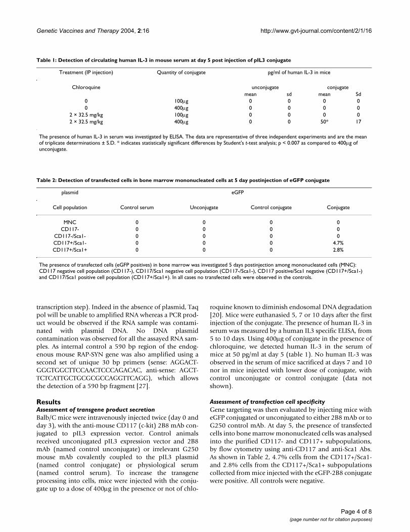
Assessment of transfection tissue specificityTo assess the tissue specificity of the targeting, presence ofpIL3 plasmid was investigated in bone marrow, bloodcells, liver, spleen, lungs, kidneys, adrenal glands, andbrain. PCR analysis of genomic DNA and RNA isolatedfrom bone marrow and blood (or serum) was performedusing primer specific pIL3 sequences. Specificity of thePCR and RT-PCR products was assessed by a Southernblot hybridised with a specific radiolabelled human IL3probe. The expected 542 bp band of the PCR product cor-responding to the IL3-transgene presence (both DNA andRNA) were was specifically detected in the bone marrowof transfected mice up to 7 days for RNA and 10 days forDNA, post transfection (figure 1). Nested PCR also was
positive for the IL3 transgene DNA in the spleen of trans-fected animals up to day 7 (not shown). In control ani-mals (control serum, unconjugate, control conjugate),pIL3 DNA but no RNA was detected in peripheral bloodbut not in serum until day 5 after the first injection andthen disappeared (figure 2); there was no detection ofDNA or RNA in bone marrow (figure 1). Aside from this,all other tissues were negative when assayed by nestedPCR on day 5, 7, 10 in transfected animals. IL3 transgeneDNA was only found in the kidney of control animalsreceiving an unconjugated mixture of Ab and DNA or thecontrol conjugate, on day 5 only (not shown).
Nested PCR detection of pIL3 plasmid in bone marrow 5, 7, and 10 days after injection of the conjugateFigure 1Nested PCR detection of pIL3 plasmid in bone marrow 5, 7, and 10 days after injection of the conjugate. Mice were intrave-nously injected twice with 100µg of anti-CD117-pIL3 conjugate (at day 0 and at day 2). Control groups corresponded to bone marrow of mice treated with unconjugated pIL3 and anti-CD117 Abs or control conjugate (G250-pIL3). IL3 DNA and RNA were detected in the bone marrow of animals receiving the pIL3-anti CD117 conjugate up to day 10. The data are representa-tive of three independent experiments.
Nested PCR detection of pIL3 plasmid in mononuclear peripheral blood cells 5, 7, and 10 days after injection of the conjugateFigure 2Nested PCR detection of pIL3 plasmid in mononuclear peripheral blood cells 5, 7, and 10 days after injection of the conjugate. Mice were intravenously injected twice with 100µg of anti-CD117-pIL3 conjugate (at day 0 and at day 2). Control group corre-sponded to mononuclear peripheral blood cells or serum of mice treated with unconjugated pIL3 and anti-CD117 Abs. pIL3 DNA was only detected in peripheral blood of control animals until day 5 after the first injection. The data are representative of three independent experiments.
PCRControl
D5D7 D10
ControlConjugate
Unconjugate
D5 D7 D10 D5 D7 D10 Neg Pos
507 pb
days
post -transfection
DNA
RNA
DNA
RNA
DNA
RNA
DNA
RNA
DNA
RNA
DNA
RNA
DNA
RNA
DNA
RNA
DNA
RNA
DNA
RNA
DNA
RNA
DNA
RNA
PCRControl
D5D7 D10
D5 D7 D10 D5 D7 D10 Neg Pos
507 pb
days
post -transfection
DNA
RNA
DNA
RNA
DNA
RNA
DNA
RNA
DNA
RNA
DNA
RNA
DNA
RNA
DNA
RNA
DNA
RNA
DNA
RNA
DNA
RNA
DNA
RNAD5D7 D10
Conjugate
D5 D7 D10 D5 D7 D10 Neg Pos
days
post -transfection
DNA
RNA
DNA
RNA
DNA
RNA
DNA
RNA
DNA
RNA
DNA
RNA
DNA
RNA
DNA
RNA
DNA
RNA
DNA
RNA
DNA
RNA
DNA
RNA
DNA
RNA
DNA
RNA
DNA
RNA
DNA
RNA
DNA
RNA
DNA
RNA
DNA
RNA
DNA
RNA
DNA
RNA
DNA
RNA
DNA
RNA
DNA
RNA
DNA
RNA
DNA
RNA
DNA
RNA
DNA
RNA
DNA
RNA
DNA
RNA
DNA
RNA
DNA
RNA
DNA
RNA
DNA
RNA
DNA
RNA
DNA
RNA
D5D7 D10
Conjugate Controls
Neg PosDNA
RNA
DNA
RNA
DNA
RNA
DNA
RNA
DNA
RNA
DNA
RNA
DNA
RNA
DNA
RNA
DNA
RNA
DNA
RNA
DNA
RNA
DNA
RNA
D5D7 D10
UnconjugateSerum D5 D7 D10 D5 D7 D10
Neg PosDNA
RNA
DNA
RNA
DNA
RNA
DNA
RNA
DNA
RNA
DNA
RNA
DNA
RNA
DNA
RNA
DNA
RNA
DNA
RNA
DNA
RNA
DNA
RNA
DNA
RNA
DNA
RNA
DNA
RNA
DNA
RNA
DNA
RNA
DNA
RNA
DNA
RNA
DNA
RNA
DNA
RNA
DNA
RNA
DNA
RNA
DNA
RNA
DNA
RNA
DNA
RNA
DNA
RNA
DNA
RNA
DNA
RNA
DNA
RNA
DNA
RNA
DNA
RNA
DNA
RNA
DNA
RNA
DNA
RNA
DNA
RNA
Unconjugate
Page 5 of 8(page number not for citation purposes)

Genetic Vaccines and Therapy 2004, 2:16 http://www.gvt-journal.com/content/2/1/16
The measurement of the weight of the mice and theirorgans (liver, kidneys spleen, brain, adrenal glands,lungs), did not reveal any change, suggesting the lack oftoxicity detected in mice receiving the conjugate (data notshown). Furthermore, since no IL3 transgene was evi-denced in these organs, further investigation of potentialtoxicity of conjugate might not be relevant.
Finally, clonogenic assay hematopoietic immature cellswere performed on cells removed from sacrificed animals.As shown in Table 3, their was no differences in micereceiving the conjugate, control unconjugate, control con-jugate and mice receiving physiological control serum.These data clearly demonstrated that our approach didnot alter the hematopoiesis.
Lack of transgene integrationLong-term cultures of bone marrow cells from micereceiving the conjugate or the controls were performed.After 1 week of selection in hygromycin-containingmedium (plasmid conferred hygromycin resistance), cellswere cultured for another 2 weeks and then viable cellswere quantified using trypan blue exclusion assay. Asillustrated on Figure 3, upon hygromycin selection, noviable cell was found in mice transfected with anti-CD117-pIL3 conjugate, suggesting that there was no inte-gration of pIL3 into host DNA.
DiscussionAlthough much progress has been accomplished in thefield of gene therapy over the last years, there is still a needto develop more effective vectors and new strategies [28].Using a non-viral gene delivery system, targeting primaryhematopoietic stem/progenitor cells in vitro can be espe-cially useful for studying the biological effects of variousgrowth factors [29]. Our conjugate linking an anti-CD117mAb to a pIL3 plasmid should be a good candidate to tar-get specifically hematopoietic stem cells. We havepreviously reported that the method is suitable for the
production of a functional growth factor in specificallyCD117+ targeted cells, mediating an in vitro biologicaleffect on hematopoiesis [14]. Since our previous reportevidenced interaction of the conjugate with hematopoi-etic cells in vitro, the present study focus on specific target-ing of hematopoietic tissues, in vivo.
We first demonstrated the efficacy of our approach sincethe transgene and its product (RNA and circulatinghuman IL3) were found in mice injected with anti-CD117/pIL3 conjugate. It is of note that although humanIL3 was only detected in plasma of chloroquine-treatedmice injected with high quantity of conjugate (400µg);human IL3 encoding RNA were evidenced in treated miceinjected with lower quantity of conjugate (100µg). Theseresults were in accordance with the design of these exper-iments aiming at observing even a transitory and localeffect (within the bone marrow).
PCR analyses of tissues evidenced the specific targeting ofthe hematopoietic system since brain, liver and lungs werenegative. Only the spleen of mice transfected with theconjugate and kidneys of control animals (transfectedwith unconjugate mixture of Ab and DNA or with the con-trol conjugate) displayed a positive PCR signal. Observedshortly after the last plasmid injection in blood, the pres-ence of plasmid might be due to the intravenous adminis-tration route used and in kidney, to a progressiveelimination of the plasmid in this organ of refinement.These results correspond to kinetic of plasmid availabilitywhen not using the specific vector (conjugate) to carryplasmid into progenitor cells. In the latter case, CD117+cells were specifically transfected, and among them, Sca1+cells were positive, suggesting a targeting of hematopieticprogenitor cells via the systemic route.
Several parameters contribute to the efficiency and specif-icity of our system such as the internalisation of theantigen targeted, the choice of the transgene used, the tis-
Table 3: Frequencies of colonies in bone marrow following transfection anti-CD117-pIL3 conjugate
Days Control serum Unconjugate Control conjugate Conjugate
mean sd mean sd mean sd mean sd
5 191 10 183 8 192 10 185 257 157 55 152 50 192 12 197 1610 187 23 173 11 182 22 187 26
Number of colonies was measured 5, 7 and 10 days following in vivo transfection with 100µg of anti-CD117-pIL3 conjugate. Control groups corresponded to mice injected with unconjugated pIL3 and anti-CD117 mAb or with the control conjugate (G250-pIL3). 5 × 10⁵ cells from bone marrow were cultured in complete methylcellulose. Colony (aggregates of more than 40 cells) numbers were evaluated under inverted light microscope. The data are representative of three independent experiments and are the mean of triplicate determinations ± S.D.
Page 6 of 8(page number not for citation purposes)

Genetic Vaccines and Therapy 2004, 2:16 http://www.gvt-journal.com/content/2/1/16
sues targeted, the conformation of the conjugate. Bonemarrow was a good candidate for gene targeting as it is ahighly proliferative tissue, as opposed to tissues whichpossess terminally differentiated cells such as hepatocytesor adipocytes, which are more resistant to transfection[30].
Factors affecting the bioavailabilty of the administeredconjugates strongly determine their in vivo performance.These include avid interaction with serum components,resulting in colloidal instability, including both aggrega-tion and dissociation of the conjugates and rapid elimina-tion from blood circulation [31,32]. Therefore, the genedelivery carrier should function as a protector of DNA dur-ing in vivo administration. Protamine has been shown tocause condensation of DNA, which promotes cellularentry [33,34]. Our complex of plasmid and antibody mayhave been sufficiently compacted to resist nuclease degra-dation and non-specific interaction with plasma proteins.Furthermore the reduced dimensions of the conjugatemay have been sufficient to allow its diffusibility throughthe extracellular space to reach bone marrow cells.
ConclusionsOur gene delivery system is specific and leads to transientgene delivery and expression. It may prove useful and safefor numerous clinical applications of gene transfer inhemato-oncology and radiopathology, whereby a stablegenetic modification is not required, in contrast to thegene therapy approaches for genetic diseases. For exam-ple, it may be of interest to facilitate the long-term recon-stitution of hematopoiesis through transient gene deliveryinto progenitor cells of patients after therapeutic and /oraccidental exposure to chemo/radiotherapy. Whether ourapproach could be used to potentate hematopoieticreconstitution following irradiation remains to bestudied.
List of Non-Standard Abbreviations UsedHSC Hematopoietic Stem Cells
Competing InterestsThe author(s) declare that they have no competinginterests.
Morphology of survival long-term bone marrow cellsFigure 3Morphology of survival long-term bone marrow cells. (a) Long-term bone marrow cells were cultured 7 days. (b) After a 1-week culture, 50µg/ml of hygromycin was added in order to select for stably transfected cells. After 1 week of selection, these cells were cultured 2 weeks in long-term culture medium. Cells observed in controls or in long-term culture in mice injected with the conjugate were viable (original magnification ×400).
Page 7 of 8(page number not for citation purposes)

Genetic Vaccines and Therapy 2004, 2:16 http://www.gvt-journal.com/content/2/1/16
Authors' contributionsAC, OD, AD, MB, SF, MM, PP carried out the studies. FH,DT participated to the designed of the study and its coor-dination. All authors read and approved the finalmanuscript.
AcknowledgementsThis work was supported by Electricité De France EDF-Comité de Radio-protection, Morad Bensidhoum was supported by a grant from Association Combattre la Leucémie. François Sabine was supported by a grant from Région Ile De France. F.H. and A.D. received support from the GDR 2352 "immunotargeting of tumors".
References1. Moen RC: Direction in gene therapy. Blood Cells 1991,
17:407-416.2. May G, Enver T: Targeting gene expression to hematopoietic
stem cells: a chromatin-dependent upstream element medi-ates cell type-specific expression of the stem cell antigenCD34. EMBO J 1995, 14:564-574.
3. Ogris M, Wagner E: Targeting tumors with non-viral genedelivery systems. Drug Discovery Today 2002, 7:479-485.
4. Wang J, Guo X, Xu Y, Barron L, Szoka FC: Synthesis and charac-terisation of long chain alkyl acyl carnitine esters. Potentiallybiodegradable cationic lipids for use in gene therapy. J MedChem 1998, 41:2207-2215.
5. Solodin I, Brown CS, Bruno MS, Chow CY, Jang EH, Debs RJ, HeathTD: A novel series of amphilic imidazolinium compounds forin vitro and in vivo gene delivery. Biochemistry 1995,34:13537-13544.
6. Gorman CM, Aikawa M, Fox B, Fox E, Lapuz C, Michaud B, NguyenH, Roche E, Sawa T, Wiener-Kronish JP: Efficient in vivo deliveryof DNA to pulmonary cells using the novel lipid EDMPC.Gene Ther 1997, 4:983-992.
7. Hong K, Zheng W, Baker A, Papahadjopoulos D: Stabilization ofcationic liposome-plasmid DNA complexes by polyaminesand poly(ethylene glycol)-phospholipid conjugates for effi-cient in vivo gene delivery. FEBS Letters 1997, 400:233-237.
8. Crook K, stevenson BJ, Dubochet M, Porteous DJ: Inclusion of cho-lesterol in DOTAP transfection complexes increases deliv-ery to DNA cells in vitro in the presence of serum. Gene Ther1998, 5:137-143.
9. Egilmez NK, Iwanuma Y, Bankert RB: Evaluation and optimiza-tion of different cationic liposome formulation for in vivogene transfer. Biochem Biophys Res Commun 1996, 221:169-173.
10. Pedroso de Lima MC, Simoes S, Pires P, Faneca H, Duzgunes N: Cat-ionic lipid-DNA complexes in gene delivery: from biophysicsto biological applications. Advanced Drug Delivery Reviews 2001,47:277-294.
11. Cristano RJ, Curiel DT: Strategies to gene delivery via thereceptor-mediated endocytosis pathway. Cancer Gene Ther1996, 3:49-57.
12. Hirsch F, Poncet P, Freeman S, Gress RE, Sachs DH, Druet P, Hirsch: Antifection : a new method to targeted gene transfection.Transplant Proc 1993, 25:138-139.
13. Poncet P, Panczak A, Goupy C, Gustafsson K, Blanpied C, ChavanelG, Hirsch R, Hirsch F: Antifection an antibody-mediatedmethod to introduce genes into lymphoid cells in vitro and invivo. Gene Ther 1996, 3:731-738.
14. Chapel A, Poncet p, Neildez-Nguyen TMA, Vetillard J, Brouard N,Goupy C, Chavanel G, Hirsch F, Thierry D: Targeted transfectionof IL-3 gene into primary human hematopoietic pogenitorcells through the c-kit receptor. Experimental Hematology 1999,27:250-258.
15. Durrbach A, Angevin E, Poncet P, Rouleau M, Chavanel G, Chapel A,Thierry D, Gorter A, Hirsch R, Charpentier B, Senik A, Hirsch F:Antibody-mediated endocytosis of G250 tumor-associatedantigen allows targeted gene transfer to human renal carci-noma in vitro. Cancer Gene Ther 1999, 6:564-571.
16. Deas O, Angevin E, Cherbonnier C, Senik A, Charpentier B, LevillainJP, Oosterwijk E, Hirsch F, Dürrbach A: In vivo targeted gene
delivery using antibody based non viral vector. Hum Gene Ther2002, 13:1101-1114.
17. Wagemaker G, Neelis KJ, Wognum AW: Surface Markers andgrowth factors receptors of immature hematopoietic cellsubsets. Stem Cells 1995, 13:165-171.
18. Yates JL, Warreb N, Sugden B: Stable replication of plasmidsderived from Epstein-Barr virus in various mammalian cells.Nature 1985, 313:812-815.
19. Oosterwijk E, Ruiter DJ, Hoedemaeker PJ, Pauwels EK, Jonas U,Zwartendijk J, Warnaar SO: Monoclonal antibody G250 recog-nizes a determinant present in renal-cell carcinoma andabsent from normal kidney. Int J Cancer 1986, 38:489-494.
20. Wolfert MA, Seymour LW: Chloroquine and amphipathic pep-tide helices show synergistic transfection in vitro. Gene Ther1998, 5:409-414.
21. Owais M, Varshney GC, Choudhury A, Chandra S, Gupta CM: Chlo-roquine encapsulated in Malaria-Infected erythrocyte-spe-cific antibody-bearing liposomes effectively controlschloroquine-resistant plasmodium berghei infection in mice.Antimicrob Agents Chemother 1995, 39:180-184.
22. Issaad C, Croisille L, Katz A, Vainchenker W, Coulombel L: A mousestroma cell line allows the proliferation of very primitivehuman CD34+/CD38- progenitor cells in long-term culturesand semisolid assays. Blood 1993, 81:2916-2964.
23. Fauser A, Messner H: Granuloerythropoietic colonies in humanmarrow, peripheral blood and cord blood. Blood 1978,52:1243-1248.
24. Chomczynski PA: Reagent for the single-step simultaneous iso-lation of RNA, DNA and proteins from cell and tissuesamples. BioTechniques 1993, 15:532-534.
25. Sharf JS, Horn GT, Erlich HA: Direct cloning analysis of enzymat-ically amplified genomic sequences. Science 1986,233:1076-1078.
26. Cunningham M: Spot Blot : a hybridization assay for specificDNA sequences in multiples samples. Anal Biochem 1983,128:415-421.
27. Brouard N, Chapel A, Neildez-Nguyen TMA, Granotier C, KahazaalI, Peault B, Thierry D: Transplantation of stromals cells trans-duced with the human IL3 gene to stimulate hematopoiesisin human fetal bone grafts in non-obese, diabetic-severecombined immunodeficiency mice. Leukemia 1998,12:1128-1135.
28. Orkin SH, Motulsky AG: Report and recommendations of thepanel to assess the NIH investment in research on genetherapy. NIH 1995, 7:.
29. Reynolds PN, Nicklin SA, Kaliberova L, Boatman B, Grizzle WE, Bal-yasnikova IV, Baker AH, Anilov SM, Curiel DT: Combined trans-ductional and transcriptional targeting improves thespecificity of transgene expression in vivo. Nature Biotechnology2001, 19:838-842.
30. Tros de Ilarduya C, Arangoa MA, Moreno-Aliaga MJ, Duzgunes N:Enhanced gene delivery in vitro and in vivo improved trans-ferrin-lipoplexes. Biochimica and Biophysica Acta 2002,1561:209-221.
31. Planck C, Mechtler K, Szoka FC, Wagner E: Activation of the com-plent system by synthetic DNA complexs: a potential barrierfor intravenous gene delivery. Human Gene Therapy 1996,7:1437-1466.
32. Zelphati O, Uyechi LS, Barron LG, Szoka FC: Effect of serum com-ponents on the physico-chemical properties of cationic lipid/ oligonucleotide complexes and on their interactions withcells. Biochem Biophys Acta 1998, 1390:119-133.
33. Sorgi FL, Bhattaacharya S, Huang L: Protamine sulfate enhancelipid mediated gene transfer. Gene therapy 1997, 4:961-968.
34. Gao X, Huang L: Potentiation of cationic liposome mediatedgene delivery by polycations. Biochemistry 1996, 35:1027-1036.
Page 8 of 8(page number not for citation purposes)
http://www.ncbi.nlm.nih.gov/entrez/query.fcgi?cmd=Retrieve&db=PubMed&dopt=Abstract&list_uids=1912600
http://www.ncbi.nlm.nih.gov/entrez/query.fcgi?cmd=Retrieve&db=PubMed&dopt=Abstract&list_uids=7532132
http://www.ncbi.nlm.nih.gov/entrez/query.fcgi?cmd=Retrieve&db=PubMed&dopt=Abstract&list_uids=7532132
http://www.ncbi.nlm.nih.gov/entrez/query.fcgi?cmd=Retrieve&db=PubMed&dopt=Abstract&list_uids=7532132
http://www.ncbi.nlm.nih.gov/entrez/query.fcgi?cmd=Retrieve&db=PubMed&dopt=Abstract&list_uids=9632353
http://www.ncbi.nlm.nih.gov/entrez/query.fcgi?cmd=Retrieve&db=PubMed&dopt=Abstract&list_uids=9632353
http://www.ncbi.nlm.nih.gov/entrez/query.fcgi?cmd=Retrieve&db=PubMed&dopt=Abstract&list_uids=9632353
http://www.ncbi.nlm.nih.gov/entrez/query.fcgi?cmd=Retrieve&db=PubMed&dopt=Abstract&list_uids=7577942
http://www.ncbi.nlm.nih.gov/entrez/query.fcgi?cmd=Retrieve&db=PubMed&dopt=Abstract&list_uids=7577942
http://www.ncbi.nlm.nih.gov/entrez/query.fcgi?cmd=Retrieve&db=PubMed&dopt=Abstract&list_uids=9349436
http://www.ncbi.nlm.nih.gov/entrez/query.fcgi?cmd=Retrieve&db=PubMed&dopt=Abstract&list_uids=9349436
http://www.ncbi.nlm.nih.gov/entrez/query.fcgi?cmd=Retrieve&db=PubMed&dopt=Abstract&list_uids=9001404
http://www.ncbi.nlm.nih.gov/entrez/query.fcgi?cmd=Retrieve&db=PubMed&dopt=Abstract&list_uids=9001404
http://www.ncbi.nlm.nih.gov/entrez/query.fcgi?cmd=Retrieve&db=PubMed&dopt=Abstract&list_uids=9001404
http://www.ncbi.nlm.nih.gov/entrez/query.fcgi?cmd=Retrieve&db=PubMed&dopt=Abstract&list_uids=9536275
http://www.ncbi.nlm.nih.gov/entrez/query.fcgi?cmd=Retrieve&db=PubMed&dopt=Abstract&list_uids=9536275
http://www.ncbi.nlm.nih.gov/entrez/query.fcgi?cmd=Retrieve&db=PubMed&dopt=Abstract&list_uids=9536275
http://www.ncbi.nlm.nih.gov/entrez/query.fcgi?cmd=Retrieve&db=PubMed&dopt=Abstract&list_uids=8660330
http://www.ncbi.nlm.nih.gov/entrez/query.fcgi?cmd=Retrieve&db=PubMed&dopt=Abstract&list_uids=8660330
http://www.ncbi.nlm.nih.gov/entrez/query.fcgi?cmd=Retrieve&db=PubMed&dopt=Abstract&list_uids=8660330
http://www.ncbi.nlm.nih.gov/entrez/query.fcgi?cmd=Retrieve&db=PubMed&dopt=Abstract&list_uids=8785712
http://www.ncbi.nlm.nih.gov/entrez/query.fcgi?cmd=Retrieve&db=PubMed&dopt=Abstract&list_uids=8785712
http://www.ncbi.nlm.nih.gov/entrez/query.fcgi?cmd=Retrieve&db=PubMed&dopt=Abstract&list_uids=8438253
http://www.ncbi.nlm.nih.gov/entrez/query.fcgi?cmd=Retrieve&db=PubMed&dopt=Abstract&list_uids=8854099
http://www.ncbi.nlm.nih.gov/entrez/query.fcgi?cmd=Retrieve&db=PubMed&dopt=Abstract&list_uids=7488942
http://www.ncbi.nlm.nih.gov/entrez/query.fcgi?cmd=Retrieve&db=PubMed&dopt=Abstract&list_uids=7488942
http://www.ncbi.nlm.nih.gov/entrez/query.fcgi?cmd=Retrieve&db=PubMed&dopt=Abstract&list_uids=7488942
http://www.ncbi.nlm.nih.gov/entrez/query.fcgi?cmd=Retrieve&db=PubMed&dopt=Abstract&list_uids=2983224
http://www.ncbi.nlm.nih.gov/entrez/query.fcgi?cmd=Retrieve&db=PubMed&dopt=Abstract&list_uids=2983224
http://www.ncbi.nlm.nih.gov/entrez/query.fcgi?cmd=Retrieve&db=PubMed&dopt=Abstract&list_uids=2428759
http://www.ncbi.nlm.nih.gov/entrez/query.fcgi?cmd=Retrieve&db=PubMed&dopt=Abstract&list_uids=2428759
http://www.ncbi.nlm.nih.gov/entrez/query.fcgi?cmd=Retrieve&db=PubMed&dopt=Abstract&list_uids=2428759
http://www.ncbi.nlm.nih.gov/entrez/query.fcgi?cmd=Retrieve&db=PubMed&dopt=Abstract&list_uids=9614562
http://www.ncbi.nlm.nih.gov/entrez/query.fcgi?cmd=Retrieve&db=PubMed&dopt=Abstract&list_uids=9614562
http://www.ncbi.nlm.nih.gov/entrez/query.fcgi?cmd=Retrieve&db=PubMed&dopt=Abstract&list_uids=7695303
http://www.ncbi.nlm.nih.gov/entrez/query.fcgi?cmd=Retrieve&db=PubMed&dopt=Abstract&list_uids=7695303
http://www.ncbi.nlm.nih.gov/entrez/query.fcgi?cmd=Retrieve&db=PubMed&dopt=Abstract&list_uids=7684620
http://www.ncbi.nlm.nih.gov/entrez/query.fcgi?cmd=Retrieve&db=PubMed&dopt=Abstract&list_uids=7684620
http://www.ncbi.nlm.nih.gov/entrez/query.fcgi?cmd=Retrieve&db=PubMed&dopt=Abstract&list_uids=7684620
http://www.ncbi.nlm.nih.gov/entrez/query.fcgi?cmd=Retrieve&db=PubMed&dopt=Abstract&list_uids=7692896
http://www.ncbi.nlm.nih.gov/entrez/query.fcgi?cmd=Retrieve&db=PubMed&dopt=Abstract&list_uids=7692896
http://www.ncbi.nlm.nih.gov/entrez/query.fcgi?cmd=Retrieve&db=PubMed&dopt=Abstract&list_uids=7692896
http://www.ncbi.nlm.nih.gov/entrez/query.fcgi?cmd=Retrieve&db=PubMed&dopt=Abstract&list_uids=3461561
http://www.ncbi.nlm.nih.gov/entrez/query.fcgi?cmd=Retrieve&db=PubMed&dopt=Abstract&list_uids=3461561
http://www.ncbi.nlm.nih.gov/entrez/query.fcgi?cmd=Retrieve&db=PubMed&dopt=Abstract&list_uids=6189420
http://www.ncbi.nlm.nih.gov/entrez/query.fcgi?cmd=Retrieve&db=PubMed&dopt=Abstract&list_uids=6189420
http://www.ncbi.nlm.nih.gov/entrez/query.fcgi?cmd=Retrieve&db=PubMed&dopt=Abstract&list_uids=9665200
http://www.ncbi.nlm.nih.gov/entrez/query.fcgi?cmd=Retrieve&db=PubMed&dopt=Abstract&list_uids=9665200
http://www.ncbi.nlm.nih.gov/entrez/query.fcgi?cmd=Retrieve&db=PubMed&dopt=Abstract&list_uids=9665200
http://www.ncbi.nlm.nih.gov/entrez/query.fcgi?cmd=Retrieve&db=PubMed&dopt=Abstract&list_uids=8844203
http://www.ncbi.nlm.nih.gov/entrez/query.fcgi?cmd=Retrieve&db=PubMed&dopt=Abstract&list_uids=8844203
http://www.ncbi.nlm.nih.gov/entrez/query.fcgi?cmd=Retrieve&db=PubMed&dopt=Abstract&list_uids=8844203
http://www.ncbi.nlm.nih.gov/entrez/query.fcgi?cmd=Retrieve&db=PubMed&dopt=Abstract&list_uids=9507083
http://www.ncbi.nlm.nih.gov/entrez/query.fcgi?cmd=Retrieve&db=PubMed&dopt=Abstract&list_uids=9507083
http://www.ncbi.nlm.nih.gov/entrez/query.fcgi?cmd=Retrieve&db=PubMed&dopt=Abstract&list_uids=9507083
http://www.ncbi.nlm.nih.gov/entrez/query.fcgi?cmd=Retrieve&db=PubMed&dopt=Abstract&list_uids=9349433
http://www.ncbi.nlm.nih.gov/entrez/query.fcgi?cmd=Retrieve&db=PubMed&dopt=Abstract&list_uids=9349433


![Cours de Compilation, Master 1, 2004-2005 - CITI …perso.citi.insa-lyon.fr/trisset/cours/compilStudent.pdf · 2013-08-23 · sity : ”Engineering a Compiler” [CT03]. Il est aussi](https://static.fdocuments.fr/doc/165x107/5b96968709d3f2501c8b679f/cours-de-compilation-master-1-2004-2005-citi-persocitiinsa-lyonfrtrissetcours.jpg)
![The Conserved and Unique Genetic Architecture of Kernel Size and Weight in Maize … · The Conserved and Unique Genetic Architecture of Kernel Size and Weight in Maize and Rice1[OPEN]](https://static.fdocuments.fr/doc/165x107/5f3da9a26ef31850087a1e16/the-conserved-and-unique-genetic-architecture-of-kernel-size-and-weight-in-maize.jpg)