Further Evidence for BRCA1 Communication with the Inactive ...
12
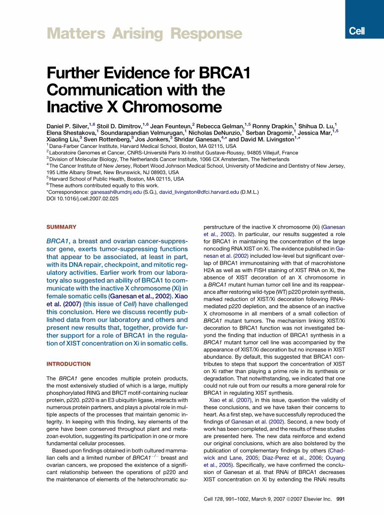
Matters Arising Response Further Evidence for BRCA1 Communication with the Inactive X Chromosome Daniel P. Silver, 1,6 Stoil D. Dimitrov, 1,6 Jean Feunteun, 2 Rebecca Gelman, 1,5 Ronny Drapkin, 1 Shihua D. Lu, 1 Elena Shestakova, 1 Soundarapandian Velmurugan, 1 Nicholas DeNunzio, 1 Serban Dragomir, 1 Jessica Mar, 1,5 Xiaoling Liu, 3 Sven Rottenberg, 3 Jos Jonkers, 3 Shridar Ganesan, 4, * and David M. Livingston 1, * 1 Dana-Farber Cancer Institute, Harvard Medical School, Boston, MA 02115, USA 2 Laboratoire Genomes et Cancer, CNRS-Universite ´ Paris XI-Institut Gustave-Roussy, 94805 Villejuif, France 3 Division of Molecular Biology, The Netherlands Cancer Institute, 1066 CX Amsterdam, The Netherlands 4 The Cancer Institute of New Jersey, Robert Wood Johnson Medical School, University of Medicine and Dentistry of New Jersey, 195 Little Albany Street, New Brunswick, NJ 08903, USA 5 Harvard School of Public Health, Boston, MA 02115, USA 6 These authors contributed equally to this work. *Correspondence: [email protected] (S.G.), [email protected] (D.M.L.) DOI 10.1016/j.cell.2007.02.025 SUMMARY BRCA1, a breast and ovarian cancer-suppres- sor gene, exerts tumor-suppressing functions that appear to be associated, at least in part, with its DNA repair, checkpoint, and mitotic reg- ulatory activities. Earlier work from our labora- tory also suggested an ability of BRCA1 to com- municate with the inactive X chromosome (Xi) in female somatic cells (Ganesan et al., 2002). Xiao et al. (2007) (this issue of Cell) have challenged this conclusion. Here we discuss recently pub- lished data from our laboratory and others and present new results that, together, provide fur- ther support for a role of BRCA1 in the regula- tion of XIST concentration on Xi in somatic cells. INTRODUCTION The BRCA1 gene encodes multiple protein products, the most extensively studied of which is a large, multiply phosphorylated RING and BRCT motif-containing nuclear protein, p220. p220 is an E3 ubiquitin ligase, interacts with numerous protein partners, and plays a pivotal role in mul- tiple aspects of the processes that maintain genomic in- tegrity. In keeping with this finding, key elements of the gene have been conserved throughout plant and meta- zoan evolution, suggesting its participation in one or more fundamental cellular processes. Based upon findings obtained in both cultured mamma- lian cells and a limited number of BRCA1 / breast and ovarian cancers, we proposed the existence of a signifi- cant relationship between the operations of p220 and the maintenance of elements of the heterochromatic su- perstructure of the inactive X chromosome (Xi) (Ganesan et al., 2002). In particular, our results suggested a role for BRCA1 in maintaining the concentration of the large noncoding RNA XIST on Xi. The evidence published in Ga- nesan et al. (2002) included low-level but significant over- lap of BRCA1 immunostaining with that of macrohistone H2A as well as with FISH staining of XIST RNA on Xi, the absence of XIST decoration of an X chromosome in a BRCA1 mutant human tumor cell line and its reappear- ance after restoring wild-type (WT) p220 protein synthesis, marked reduction of XIST/Xi decoration following RNAi- mediated p220 depletion, and the absence of an inactive X chromosome in all members of a small collection of BRCA1 mutant tumors. The mechanism linking XIST/Xi decoration to BRCA1 function was not investigated be- yond the finding that induction of BRCA1 synthesis in a BRCA1 mutant tumor cell line was accompanied by the appearance of XIST/Xi decoration but no increase in XIST abundance. By default, this suggested that BRCA1 con- tributes to steps that support the concentration of XIST on Xi rather than playing a prime role in its synthesis or degradation. That notwithstanding, we indicated that one could not rule out from our results a more general role for BRCA1 in regulating XIST synthesis. Xiao et al. (2007), in this issue, question the validity of these conclusions, and we have taken their concerns to heart. As a first step, we have successfully reproduced the findings of Ganesan et al. (2002). Second, a new body of work has been completed, and the results of these studies are presented here. The new data reinforce and extend our original conclusions, which are also bolstered by the publication of complementary findings by others (Chad- wick and Lane, 2005; Diaz-Perez et al., 2006; Ouyang et al., 2005). Specifically, we have confirmed the conclu- sion of Ganesan et al. that RNAi of BRCA1 decreases XIST concentration on Xi by extending the RNAi results Cell 128, 991–1002, March 9, 2007 ª2007 Elsevier Inc. 991
Transcript of Further Evidence for BRCA1 Communication with the Inactive ...
Matters Arising Response
Further Evidence for BRCA1Communication with theInactive X ChromosomeDaniel P. Silver,1,6 Stoil D. Dimitrov,1,6 Jean Feunteun,2 Rebecca Gelman,1,5 Ronny Drapkin,1 Shihua D. Lu,1
Elena Shestakova,1 Soundarapandian Velmurugan,1 Nicholas DeNunzio,1 Serban Dragomir,1 Jessica Mar,1,5
Xiaoling Liu,3 Sven Rottenberg,3 Jos Jonkers,3 Shridar Ganesan,4,* and David M. Livingston1,*1Dana-Farber Cancer Institute, Harvard Medical School, Boston, MA 02115, USA2Laboratoire Genomes et Cancer, CNRS-Universite Paris XI-Institut Gustave-Roussy, 94805 Villejuif, France3Division of Molecular Biology, The Netherlands Cancer Institute, 1066 CX Amsterdam, The Netherlands4The Cancer Institute of New Jersey, Robert Wood Johnson Medical School, University of Medicine and Dentistry of New Jersey,
195 Little Albany Street, New Brunswick, NJ 08903, USA5Harvard School of Public Health, Boston, MA 02115, USA6These authors contributed equally to this work.
*Correspondence: [email protected] (S.G.), [email protected] (D.M.L.)
DOI 10.1016/j.cell.2007.02.025
SUMMARY
BRCA1, a breast and ovarian cancer-suppres-sor gene, exerts tumor-suppressing functionsthat appear to be associated, at least in part,with its DNA repair, checkpoint, and mitotic reg-ulatory activities. Earlier work from our labora-tory also suggested an ability of BRCA1 to com-municate with the inactive X chromosome (Xi) infemale somatic cells (Ganesan et al., 2002). Xiaoet al. (2007) (this issue of Cell) have challengedthis conclusion. Here we discuss recently pub-lished data from our laboratory and others andpresent new results that, together, provide fur-ther support for a role of BRCA1 in the regula-tion of XIST concentration on Xi in somatic cells.
INTRODUCTION
The BRCA1 gene encodes multiple protein products,
the most extensively studied of which is a large, multiply
phosphorylated RING and BRCT motif-containing nuclear
protein, p220. p220 is an E3 ubiquitin ligase, interacts with
numerous protein partners, and plays a pivotal role in mul-
tiple aspects of the processes that maintain genomic in-
tegrity. In keeping with this finding, key elements of the
gene have been conserved throughout plant and meta-
zoan evolution, suggesting its participation in one or more
fundamental cellular processes.
Based upon findings obtained in both cultured mamma-
lian cells and a limited number of BRCA1�/� breast and
ovarian cancers, we proposed the existence of a signifi-
cant relationship between the operations of p220 and
the maintenance of elements of the heterochromatic su-
perstructure of the inactive X chromosome (Xi) (Ganesan
et al., 2002). In particular, our results suggested a role
for BRCA1 in maintaining the concentration of the large
noncoding RNA XIST on Xi. The evidence published in Ga-
nesan et al. (2002) included low-level but significant over-
lap of BRCA1 immunostaining with that of macrohistone
H2A as well as with FISH staining of XIST RNA on Xi, the
absence of XIST decoration of an X chromosome in
a BRCA1 mutant human tumor cell line and its reappear-
ance after restoring wild-type (WT) p220 protein synthesis,
marked reduction of XIST/Xi decoration following RNAi-
mediated p220 depletion, and the absence of an inactive
X chromosome in all members of a small collection of
BRCA1 mutant tumors. The mechanism linking XIST/Xi
decoration to BRCA1 function was not investigated be-
yond the finding that induction of BRCA1 synthesis in a
BRCA1 mutant tumor cell line was accompanied by the
appearance of XIST/Xi decoration but no increase in XIST
abundance. By default, this suggested that BRCA1 con-
tributes to steps that support the concentration of XIST
on Xi rather than playing a prime role in its synthesis or
degradation. That notwithstanding, we indicated that one
could not rule out from our results a more general role for
BRCA1 in regulating XIST synthesis.
Xiao et al. (2007), in this issue, question the validity of
these conclusions, and we have taken their concerns to
heart. As a first step, we have successfully reproduced the
findings of Ganesan et al. (2002). Second, a new body of
work has been completed, and the results of these studies
are presented here. The new data reinforce and extend
our original conclusions, which are also bolstered by the
publication of complementary findings by others (Chad-
wick and Lane, 2005; Diaz-Perez et al., 2006; Ouyang
et al., 2005). Specifically, we have confirmed the conclu-
sion of Ganesan et al. that RNAi of BRCA1 decreases
XIST concentration on Xi by extending the RNAi results
Cell 128, 991–1002, March 9, 2007 ª2007 Elsevier Inc. 991
from human to mouse cells using different target se-
quences. In addition, we have confirmed the relationship
of BRCA1 to XIST concentration on Xi by demonstrating
that depleting BRCA1 by Cre-mediated excision also de-
creases XIST concentration on Xi. Lastly, we demonstrate
that mouse tumor lines derived from true Brca1 null mam-
mary tumors do not show XIST RNA concentration on Xi
despite the presence of two X chromosomes.
RESULTS
A major claim of Xiao et al. (2007) (this issue of Cell) relates
to whether BRCA1 colocalizes with XIST on Xi. Xiao et al.
state that they lack consistent evidence to support our
finding that a small minority of asynchronous cultured
cells reveal colocalization of BRCA1 immunostaining and
XIST FISH signals on the inactive X chromosome (Xi)
(Ganesan et al., 2002). Second, Xiao et al. also question
whether RNAi-mediated BRCA1 depletion affects XIST.
They report that our data linking BRCA1 knockdown to de-
creased XIST signal on Xi are not reproducible. Finally,
they question whether Xi is absent in Brca1 mutant breast
cancers. Contrary to Ganesan et al. (2002), Xiao et al. re-
port that loss of BRCA1 function is not accompanied by
concomitant loss of XIST/Xi decoration in Brca1�/�murine
tumors. We will address each of these points below.
Detection of BRCA1 at Xi
In their analysis, Xiao et al. (2007) have interpreted mean-
ingful colocalization of BRCA1 and XIST to occur only
when BRCA1 completely coats the territory of Xi with per-
fect coincidence of both images. This is a narrower defini-
tion of colocalization than we used. To be scored positive
for colocalization, we required, at a minimum, a significant
degree of apposition (i.e., partial fluorescent signal over-
lap) between BRCA1 and XIST or macrohistone H2A
(mH2A), and this was repeatedly observed in a small mi-
nority of asynchronous cells in multiple cell lines.
Our work has been reproduced in part and extended by
Chadwick and Lane (2005), who demonstrated significant
costaining by both BRCA1 antibody and either mH2A or
BUdR antibody of Xi in multiple human cell strains in late
S phase. Multiple BRCA1 antibodies were used. These
findings fit with our observation that ‘‘costaining was
largely apparent in mid/late S phase cells.’’ Very recently,
significant S phase-associated apposition of BRCA1 and
Xi was also reported independently by others as part of
a more general interaction of BRCA1 and replicating het-
erochromatin-dense chromosomal structures (Pageau
et al., 2006; Pageau and Lawrence, 2006).
Despite the call by Xiao et al. (2007) for a perfect fit of
XIST and BRCA1 images as the only indicator of biological
significance, it has been shown that certain Xi territory
markers, such as H3K27 methylation, only partly overlap
other Xi markers (Chadwick and Willard, 2004; Figure S3
in Xiao et al., 2007) because Xi heterochromatic territory
is heterogeneous. Two distinct Xi regions, one marked
by XIST, MH2A, and H3mK27 and the other by H3mK9
992 Cell 128, 991–1002, March 9, 2007 ª2007 Elsevier Inc.
and HP1g (a BRCA1 partner protein; S.G. and D.M.L., un-
published data), have been detected (Chadwick and Will-
ard, 2004). BRCA1 has been said to associate with both
of these Xi territories, although likely at different times dur-
ing Xi replication (Chadwick and Lane, 2005). Thus, perfect
BRCA1/Xi marker costaining in asynchronous cells should
be very uncommon or absent, as confirmed by Xiao et al.,
while partial overlap of XIST or other Xi-associated ele-
ments and BRCA1 in a minority of cells in an asynchronous
culture would be an expected result. Notably, subsequent
to our previous publication (Ganesan et al., 2002), other
investigators published evidence of robust BRCA1/Xi cos-
taining in mouse cells (Diaz-Perez et al., 2006; Ouyang
et al., 2005). We, too, continue to detect such an associa-
tion in a minority of asynchronous cells (see Figure S1 in the
Supplemental Data available with this article online).
Given these findings, we would argue that our original
evidence of BRCA1 immunostaining on a significant frac-
tion of Xi territory in a small subset of unsynchronized cells
has been reproduced and extended by newer results of
other laboratories. The biochemical significance of these
findings requires further work.
Effects of BRCA1 RNAi-Mediated Depletion
on XIST Concentration on Xi
Xiao et al. (2007) report that attempts to knock down
BRCA1 expression with RNAi failed to change the inten-
sity of XIST/Xi signals. Ganesan et al. (2002) reported
that RNAi-mediated depletion of BRCA1 in primary human
cells led to suppression of XIST/Xi staining in many cells.
Here we discuss the possible reasons for the difference in
results with RNAi-mediated knockdown and present new
data confirming our original RNAi data in another species.
With respect to the RNAi-mediated knockdown, we, too,
have observed in some experiments using BRCA1-spe-
cific RNAi (even using the pooled, commercial BRCA1
RNAi reagent that Xiao et al. [2007] used) that S phase
BRCA1 nuclear dots largely disappear, but nothing hap-
pens to XIST. When BRCA1 protein levels were analyzed
by western blots in these experiments, there was, typically,
limited BRCA1 protein depletion (e.g., 50%–70%). How-
ever, when knockdowns approached R90% or more, as
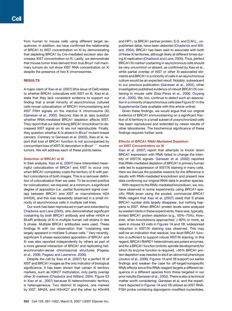
seen in mouse X3 cells in Figures 1A and 1B, widespread
reduction in XIST/Xi staining was observed. This may
well be an indication that residual, low-level BRCA1 func-
tion is sufficient to support robust XIST/Xi staining. In this
regard, BRCA1/BARD1 heterodimers are potent enzymes,
and for a BRCA1 function (mitotic spindle development) for
which its enzyme function is required, >95% BRCA1 pro-
tein depletion was needed to elicit an abnormal phenotype
(Joukov et al., 2006). Figures 1A and 1B support our earlier
findings and weaken the case for off-target/nonspecific
RNAi effects since this RNAi reagent targets a different se-
quence in a different species from those targeted in our
prior results (Ganesan et al., 2002). There is also a technical
matter worth considering. Ganesan et al. and the experi-
ment depicted in Figures 1A and 1B utilized an XIST RNA-
FISH probe containing digoxigenin-modified nucleotides,
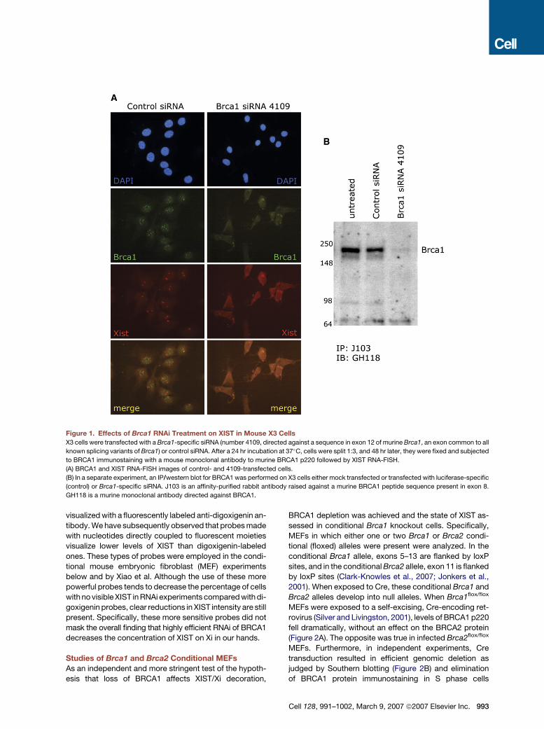
Figure 1. Effects of Brca1 RNAi Treatment on XIST in Mouse X3 Cells
X3 cells were transfected with a Brca1-specific siRNA (number 4109, directed against a sequence in exon 12 of murine Brca1, an exon common to all
known splicing variants of Brca1) or control siRNA. After a 24 hr incubation at 37�C, cells were split 1:3, and 48 hr later, they were fixed and subjected
to BRCA1 immunostaining with a mouse monoclonal antibody to murine BRCA1 p220 followed by XIST RNA-FISH.
(A) BRCA1 and XIST RNA-FISH images of control- and 4109-transfected cells.
(B) In a separate experiment, an IP/western blot for BRCA1 was performed on X3 cells either mock transfected or transfected with luciferase-specific
(control) or Brca1-specific siRNA. J103 is an affinity-purified rabbit antibody raised against a murine BRCA1 peptide sequence present in exon 8.
GH118 is a murine monoclonal antibody directed against BRCA1.
visualized with a fluorescently labeled anti-digoxigenin an-
tibody. We have subsequently observed that probes made
with nucleotides directly coupled to fluorescent moieties
visualize lower levels of XIST than digoxigenin-labeled
ones. These types of probes were employed in the condi-
tional mouse embryonic fibroblast (MEF) experiments
below and by Xiao et al. Although the use of these more
powerful probes tends to decrease the percentage of cells
with no visible XIST in RNAi experiments compared with di-
goxigenin probes, clear reductions in XIST intensity are still
present. Specifically, these more sensitive probes did not
mask the overall finding that highly efficient RNAi of BRCA1
decreases the concentration of XIST on Xi in our hands.
Studies of Brca1 and Brca2 Conditional MEFs
As an independent and more stringent test of the hypoth-
esis that loss of BRCA1 affects XIST/Xi decoration,
BRCA1 depletion was achieved and the state of XIST as-
sessed in conditional Brca1 knockout cells. Specifically,
MEFs in which either one or two Brca1 or Brca2 condi-
tional (floxed) alleles were present were analyzed. In the
conditional Brca1 allele, exons 5–13 are flanked by loxP
sites, and in the conditional Brca2 allele, exon 11 is flanked
by loxP sites (Clark-Knowles et al., 2007; Jonkers et al.,
2001). When exposed to Cre, these conditional Brca1 and
Brca2 alleles develop into null alleles. When Brca1flox/flox
MEFs were exposed to a self-excising, Cre-encoding ret-
rovirus (Silver and Livingston, 2001), levels of BRCA1 p220
fell dramatically, without an effect on the BRCA2 protein
(Figure 2A). The opposite was true in infected Brca2flox/flox
MEFs. Furthermore, in independent experiments, Cre
transduction resulted in efficient genomic deletion as
judged by Southern blotting (Figure 2B) and elimination
of BRCA1 protein immunostaining in S phase cells
Cell 128, 991–1002, March 9, 2007 ª2007 Elsevier Inc. 993
Figure 2. Cre Infection Leads to Efficient Excision of Conditional Brca1 and Brca2 Alleles, Loss of Protein Expression, and Induc-
tion of Genomic Instability
MEFs carrying conditional (floxed) or WT alleles of Brca1 or Brca2 were either infected or mock infected with a self-deleting Cre retrovirus
(HR = hit-and-run self-excising Cre retrovirus; Silver and Livingston, 2001).
(A) Western blots of protein extracts from either mock-infected or Cre-infected Brca1flox/flox and Brca1WT/WT MEFs using a BRCA1-specific antibody
are shown in the left panel. Western blots of protein extracts from mock- or Cre-infected Brca2flox/flox and Brca2WT/WT MEFs using a BRCA2-specific
antibody are shown in the right panel.
(B) Southern blots were performed on DNA extracted from Cre- or mock-treated MEFs containing Brca1flox/WT alleles (left panel) or Brca2flox/WT alleles
(right panel) and probed with Brca1- or Brca2-specific DNAs, respectively. Expected sizes of the WT, floxed, and del alleles are shown. In both cases,
two separate MEF strains of floxed/WT genotype are shown. In the right panel, additional controls of Cre-treated Brca2WT/WT MEFs (labeled strain A2)
and untreated Brca2del/flox (A11) and Brca2flox/flox MEFs (A5) are shown.
(C) Deletion of the Brca1 gene by Cre-mediated excision results in the development of chromosomal instability. Brca1 conditional MEFs of the ge-
notype Brca1flox/flox were either infected or mock infected with a self-deleting Cre retrovirus. Seventy-two hours later, chromosomal spreads were
prepared. An example of a chromosomal spread from the Cre-treated Brca1flox/flox MEFs is shown in the left panel, with quadriradial chromosomes
or fragments of quadriradial forms indicated by arrows. The number of metaphase spreads examined and number of radial forms found are shown in
the table to the right. All chromosomal spreads were analyzed with the observer blinded to the history and identity of the cells.
994 Cell 128, 991–1002, March 9, 2007 ª2007 Elsevier Inc.
(Figure S3A). The presence of a Rosa26 locus-embedded
lox-stop-lox-lacZ allele (R26R) (Soriano, 1999) in some of
these MEF strains permitted assessment of Cre activity.
Efficient Cre-mediated deletion was reflected by lacZ-
dependent X-gal staining in >90% of cells in the Cre-in-
fected MEFs containing the R26R allele (data not shown).
A different manifestation of the absence of BRCA1 or
BRCA2 function was also observed in Cre-treated condi-
tional cells (Figure 2C and Figure S3B). In both Cre-treated
Brca1flox/flox and Brca2flox/flox MEFs, significant numbers
of cells revealed quadriradial chromosome formation
(likely a manifestation of illegitimate recombination) within
72 hr after infection. No such structures were observed in
Cre-infected WT MEFs, showing that Cre alone did not
elicit them, nor were they seen in naive, untreated cells
of any genotype. Furthermore, quadriradial chromosomes
were absent after Cre infection of MEFs bearing condi-
tional R26R alleles but no Brca1 or Brca2 conditional al-
leles (data not shown). Thus, their appearance was a sign
of BRCA1 or BRCA2 depletion rather than merely of Cre
activity. Therefore, MEFs rendered Brca1�/� by Cre infec-
tion revealed independent evidence of a loss of a funda-
mental BRCA1 function, genome integrity maintenance.
Five days after Cre virus or mock infection, XIST RNA-
FISH was performed on conditional Brca1, conditional
Brca2, and control MEFs, and an investigator blinded to
the identity of the cultures being analyzed obtained multi-
ple photomicrographs from each. The photomicrographs
were scored for XIST/Xi staining intensity by four blinded
analysts (Figure 3 and Figure S2). Representative pictorial
examples of such fields and a semiquantitative analysis of
the results obtained by the blinded analysts revealed that
only cells that had lost both Brca1 alleles experienced an
overt, statistically significant (p < 0.0001) reduction in focal
XIST/Xi staining by comparison with controls analyzed in
parallel (Figure 3C). Similar results were obtained using
MEFs of the same genotypes derived from separate em-
bryos in an independent experiment (data not shown).
The reduction in XIST/Xi intensity in Brca1 null cells was
not a result of a proliferation block/defect, because no
such reduction was observed in Brca2 null cells, which ex-
perienced a proliferation effect similar to Brca1 null cells
(Patel et al., 1998; Xu et al., 1999). Indeed, FACS analyses
of Brca1�/� and Brca2�/� MEFs generated by this ap-
proach revealed no major changes in the distribution of
cell-cycle intervals from that of Cre-treated WT MEFs.
This implies that the proliferation defect in the nullizygous
cells was a result of a general slow-down in cell-cycle pro-
gression (D.P.S. and D.M.L., unpublished data).
Hence, acute homozygous Brca1, but not Brca2, loss of
function in conditional MEFs resulted in a significant dim-
inution in the intensity of XIST/Xi staining.
XIST/Xi in Breast Tumors Arising in Brca1 Germline
Mutant Mice
Xiao et al. (2007) report that cells of mammary tumors aris-
ing in mice engineered to produce a homozygous Brca1
mutation (D11/D11) in mammary tissue contain XIST/Xi
foci. The authors argue that this result runs counter to our
observations that human BRCA1�/� tumors lack robust
XIST and H3meK27/Xi foci (Ganesan et al., 2002). Here we
discuss possible reasons for the discrepancies in the re-
sults and report new data examining XIST staining and
localization in cell lines derived from Brca1 null tumors
generated in a conditional Brca1 knockout mouse model.
In our view, the discrepancy between our original results
and those of Xiao et al. (2007) may reflect the nature of the
different Brca1 mutations in the two sets of experiments.
Brca1 D11, the mouse Brca1 mutant allele studied by
Xiao et al., is a hypomorph, not a null mutation (Brodie
and Deng, 2001). It results from a clean deletion of
Brca1 exon 11 and, unlike null mutants, leaves intact a
normal, naturally occurring alternatively spliced BRCA1
protein, D11. Brca1�/� (null) mutant murine embryos die
early in embryogenesis, even when bred onto a p53
mutant background (Ludwig et al., 1997). In contrast,
Brca1D11/D11;p53+/� mice are born and become viable
adults (Xu et al., 1999, 2001). The females tend to develop
mammary tumors. Moreover, the D11 protein retains cer-
tain WT p220-like properties both in mitotic cells (Huber
et al., 2001) and in a minority of pachytene spermatocytes
(Turner et al., 2004). In addition, some organisms com-
pletely lack an exon 11-like unit; for example, the WT
C. elegans BRCA1 ortholog lacks these sequences yet
performs normal DNA damage-response functions (Boul-
ton et al., 2004). Finally, BRCA1 D11 protein retains an
ability to localize in subnuclear foci indistinguishable
from BRCA1 p220 (Huber et al., 2001). Since D11 retains
certain WT BRCA1 functions, it is difficult with the avail-
able evidence to know whether D11 mammary tumors
emerge via the same molecular and biological pathway
(or pathways) as null BRCA1�/� human tumors. In addi-
tion, mammary epithelial cell-associated p53 mutations
alone can give rise to murine breast cancer (Lin et al.,
2004), raising the question of whether some of the D11 tu-
mor cells analyzed in Xiao et al. are actually Brca1 WT, p53
mutant. We have found that cell lines derived from tumors
arising in Brca1 conditional, p53 conditional mammary ep-
ithelia exposed to Cre are not infrequently p53 mutant but
Brca1 WT. Xiao et al. offer no evidence that the tumor lines
that they analyzed harbor Brca1 mutations. In fact, at least
one of the cell lines analyzed in Xiao et al., W0069, appears
to be Brca1 WT (Figure S4). Hence, for the reasons noted
here, the presence of a normal Xi in cells derived from mu-
rine D11 mammary tumors analyzed in Xiao et al. might not
be remarkable. In this regard, the human L56Br-C1 cell line
studied by Xiao et al. and found to contain XIST/Xi struc-
tures carries a translation-terminating BRCA1 mutation
within exon 11 that should, in principle, leave the endoge-
nous D11 mRNA intact (Johannsson et al., 2003). If D11
protein positively influences XIST/Xi costaining, this line
could have retained that function.
To determine the status of XIST localization in murine
tumors carrying true null Brca1 alleles, we have employed
a conditional Brca1 mouse model in which exons 5–13 are
flanked by loxP sites (the same allele used in the Brca1
Cell 128, 991–1002, March 9, 2007 ª2007 Elsevier Inc. 995
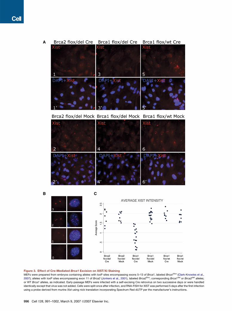
Figure 3. Effect of Cre-Mediated Brca1 Excision on XIST/Xi Staining
MEFs were prepared from embryos containing alleles with loxP sites encompassing exons 5–13 of Brca1, labeled Brca1flox (Clark-Knowles et al.,
2007); alleles with loxP sites encompassing exon 11 of Brca2 (Jonkers et al., 2001), labeled Brca2flox; corresponding Brca1del or Brca2del alleles;
or WT Brca1 alleles, as indicated. Early-passage MEFs were infected with a self-excising Cre retrovirus on two successive days or were handled
identically except that virus was not added. Cells were split once after infection, and RNA-FISH for XIST was performed 5 days after the first infection
using a probe derived from murine Xist using nick translation incorporating Spectrum Red dUTP per the manufacturer’s instructions.
996 Cell 128, 991–1002, March 9, 2007 ª2007 Elsevier Inc.
floxed MEFs described above). The Jonkers laboratory
has used these mice to generate Brca1 null mammary car-
cinomas using a cytokeratin 14 promoter-driven Cre
transgene (K14cre; Jonkers et al., 2001) in the context of
conditional p53. In a blind analysis performed in three sep-
arate laboratories, none of the cultures of six indepen-
dently derived clonal cell lines from two Brca1 null tumors
revealed focal XIST spots (Figure 4A). The absence of XIST
spots was not a result of X monosomy, given the presence
of at least two X chromosomes in cells of these tumor lines
when analyzed by DNA-FISH (Figure 4B). The Brca1 null
genotype of each line was confirmed by allele-specific
PCR, and all six lines lacked BRCA1 nuclear foci when ex-
amined by immunofluorescence (data not shown). Cells
derived from control mammary tumors in which only p53
was targeted showed both intact BRCA1 nuclear foci and
robust focal XIST staining (Figure 4 and data not shown).
To explore the XIST status of additional BRCA1 mutant
cell lines, we have recently performed XIST RNA-FISH on
the five human BRCA1 mutant cell lines available in our
laboratory, four of which were not known to be BRCA1�/�
in 2002—i.e., HCC1395, SUM149PT, SUM1315MO2 (El-
strodt et al., 2006), and L56Br-C1. HCC1937, long known
to be BRCA1�/�, was also tested. Only L56Br-C1 dis-
played focal XIST staining, whereas focal XIST could not
be detected in the other four lines (Figure S5). We agree
with Xiao et al. (2007) that there are undoubtedly BRCA1
WT cell lines and tumors that lack focal XIST staining,
most likely a result of Xi loss. However, among BRCA1�/�
carcinomas, loss of focal XIST staining was more common
than in breast carcinomas—even high grade lesions—of
other pathological subtypes (Richardson et al., 2006).
The human tumor data in our original paper have been
confirmed in a more recent analysis from our group
(Richardson et al., 2006). We report a lack of focal XIST
and H3meK27 spots in all five new human BRCA1�/�
breast carcinoma samples examined. This paper also de-
scribed a similar XIST/Xi loss phenotype in 14 out of 17
cases of sporadic basal-like breast cancer. Sporadic
basal-like carcinomas are BRCA1 WT yet are a known
BRCA1�/� breast carcinoma phenocopy, sharing a similar
gene expression pattern, a similar estrogen and proges-
terone receptor and cytokeratin expression phenotype,
a lack of HER2 amplification, and p53 mutation status
with BRCA1�/� breast cancers (Richardson et al., 2006;
Sorlie et al., 2003). Thus, it is tempting to speculate that
the sporadic basal-like breast cancers are defective in
a pathway (or pathways) in which BRCA1 participates
and may, therefore, share XIST loss with hereditary
BRCA1�/� breast cancers. This notwithstanding, given
the relatively small number of tumors analyzed, there
may well be some BRCA1�/� human tumors with XIST/Xi
structures. The nature of the relevant BRCA1 mutant ge-
notypes in settings where XIST is absent compared to
any where it is present would need to be considered. If,
as suggested by Xiao et al. (2007), low-level XIST synthe-
sis from Xa occurs, this, too, could account for XIST foci in
some tumor cells.
DISCUSSION
We believe that the data reported here help to explain the
apparent discrepancies noted by Xiao et al. (2007) and re-
inforce the view that BRCA1 influences, directly or indi-
rectly, XIST decoration of Xi in adult somatic cells. Xiao
et al. present four lines of evidence in support of their
view that BRCA1 does not influence XIST localization.
First, they argue that BRCA1 does not ‘‘coat’’ the inactive
X chromosome. However, in Ganesan et al. (2002), no
claim was made that BRCA1 perfectly coats Xi or provides
the prime force that drives XIST/Xi coating, e.g., during
embryogenesis; rather, in a small percentage of cells,
a percentage that varied among the cell types examined,
BRCA1 exhibited overlapping staining with XIST or with
a marker of Xi, macrohistone H2A. Though quantitation
likely varies among observers and cell lines and as a func-
tion of the scoring criteria used, a small minority of cycling
cells showed significant overlap of BRCA1 and markers of
Xi in Xiao et al. and Ganesan et al. Furthermore, an asso-
ciation of BRCA1 with markers of Xi has now been pub-
lished by at least three independent laboratories (Chad-
wick and Lane, 2005; Diaz-Perez et al., 2006; Pageau
et al., 2006).
Second, Xiao et al. (2007) show an analysis of XIST in
HCC1937, a BRCA1�/� tumor line, and the effect of
BRCA1 reconstitution of that line. Much of the apparent
disagreement between Ganesan et al. and Xiao et al. re-
garding these experiments is likely explained by the chro-
mosomal instability of this line. The subclone used in Ga-
nesan et al. was itself a mixture of cells containing two X
chromosomes and a minor population with many more X
chromosomes. Subsequent work with HCC1937 has
shown that the chromosomal content of this line has
evolved quickly with passage, and different cultures of
this line differ considerably in chromosomal content
(A. De Nicolo and D.M.L., unpublished data). We strongly
(A1–20) XIST RNA-FISH analysis of Brca2flox/del MEFs treated with Cre-encoding retrovirus (upper panels) or mock infected (lower panels).
(A3–40) XIST RNA-FISH staining of Brca1flox/del MEFs treated with Cre-encoding retrovirus (upper panels) or mock infected (lower panels).
(A5–60) XIST RNA-FISH staining of Brca1flox/WT MEFs treated with Cre-encoding retrovirus (upper panels) or mock infected (lower panels). The expo-
sure time for each XIST micrograph was the same throughout. Similarly, the exposure time for the DAPI micrographs was held constant.
(B) Four assigned intensity categories (0, 1, 2, and 3) used as a training slide for scoring XIST intensity.
(C) Semiquantitative analysis of the intensity of XIST signals in Brca2flox/del, Brca1flox/del, and Brca1flox/WT MEFs 5 days after infection with Cre or mock
treatment. Photomicrographs of multiple fields from each cell strain were blindly analyzed by four independent analysts, who categorized each nu-
cleus for XIST signal strength according to the standards in (B). The XIST intensity was averaged over the cells in each field for each analyst (see
Experimental Procedures), and each such average is represented as a single point on the graph. An exact two-sided Wilcoxon rank-sum test revealed
a significant difference (p < 0.0001) between XIST intensity in Brca1flox/del Cre-infected cells compared to the other five groups combined.
Cell 128, 991–1002, March 9, 2007 ª2007 Elsevier Inc. 997

Figure 4. Cell Lines Derived from Brca1�/�;p53�/� Murine Breast Cancers Lack XIST FociCell lines were obtained from tumors that arose in mice homozygous for a conditional Brca1 allele in which exons 5–13 of Brca1 were flanked by loxP
sites, homozygous for a conditional p53 allele, and carrying a cytokeratin 14 promoter-driven Cre allele. These breast cancers and derived cell lines
had a Brca1�/�;p53�/� genotype as shown by direct genotyping. A control cell line (1146) was obtained from a breast cancer that arose in a mouse
with WT Brca1 but conditional p53 and cytokeratin 14-driven Cre. Its genotype was Brca1+/+;p53�/�.
(A) All cell lines were subject to RNA-FISH for XIST RNA. The RNA-FISH signal is shown in red, and the DAPI nuclear signal is shown in blue for all cell
lines tested.
(B) X chromosome DNA-FISH analysis of murine Brca1�/� cell lines. Brca1 WT (1146) and six Brca1�/�mouse mammary tumor cell lines (30.1, 30.3,
30.5, 40.1, 40.10, and 40.11 were grown on coverslips and processed for DNA-FISH using a probe specific for the murine X chromosome. For each
cell line, the DNA-FISH signal is shown in red (left panel) and DAPI nuclear staining in blue (middle panel), and a merged image is shown in the right
panel.
suspect that the inability of Xiao et al. to restore strong,
focal XIST staining may relate to differences in the X
chromosomal content of subclones. Early passages of
BRCA1-reconstituted HCC1937 were sent to another lab-
oratory, and they reproduced our finding that, unlike either
parental HCC1937 or vector-reconstituted HCC1937, the
majority of BRCA1-reconstituted HCC1937 showed focal
XIST staining, with the majority of these cells having two X
chromosomes (Pageau et al., 2006). While another
BRCA1-reconstituted HCC1937 subclone showed no
998 Cell 128, 991–1002, March 9, 2007 ª2007 Elsevier Inc.
focal XIST, it contained only one X chromosome (Pageau
et al., 2006).
Third, the RNAi experiments of Xiao et al. (2007) were in-
terpreted by the authors to reveal no effect of BRCA1 RNAi
on XIST staining. However, close examination of their fig-
ures suggests a dimming of XIST intensity in the presence
of BRCA1 RNAi in their Figure 3H compared with their
Figure 3G and in their Figure S5B compared with their Fig-
ure S5A, in general agreement with observations made by
Ganesan et al. (2002). Perhaps some of these differences
in interpretation are based on the view that XIST coating of
Xi is an ‘‘all or nothing’’ phenomenon. Our experiments
and others that we are aware of suggest that XIST is in dy-
namic equilibrium with Xi and that the initiation and main-
tenance of XIST coating may be separable functions.
Whatever the case, the conditional knockout of BRCA1
avoids the pitfalls of incomplete RNAi and demonstrates
an effect of BRCA1 loss on XIST/Xi intensity.
Xiao et al. (2007) analyzed murine tumors and cell lines
derived from them that were all generated in a mouse
model that deletes Brca1 exon 11. As described above,
this is a hypomorphic mutation (Brodie and Deng, 2001).
Because this mutation preserves a naturally occurring
BRCA1 isoform, D11, it is difficult to predict what the out-
come of such a knockout would be on any particular
BRCA1 function. Furthermore, as discussed above, the
Brca1D11/D11 genotypic status of the tumors presented in
Xiao et al. is unclear since at least one of them examined
by us appears to be Brca1 WT (Figure S4). Xiao et al.
also presented an analysis of RNA expression of genes
along the X chromosome in both embryonic and mammary
tissue from D11 homozygous mice compared with D11
heterozygous mice to show that BRCA1 loss does not in-
fluence Xi silencing. However, Ganesan et al. (2002) did
not claim a global failure of Xi silencing or widespread reac-
tivation of X-linked genes as a result of BRCA1 loss. What
was observed was a low-level reactivation of a GFP re-
porter embedded in Xi following BRCA1 depletion and, in
later work, upregulation of a small subset of X-encoded
genes in five BRCA1�/� breast carcinomas (Richardson
et al., 2006).
Xiao et al. have also argued that our results demonstrat-
ing Xi loss in sporadic basal-like cancer (BLC), a BRCA1
WT tumor, invalidate the argument that Xi loss in
BRCA1�/� tumors is a BRCA1 mutation-associated event.
However, as discussed above, sporadic BLCs are well-
known phenocopies of BRCA1-deficient tumors (Turner
et al., 2004; Lacroix and Leclercq, 2005). Not unexpect-
edly, then, sporadic and BRCA1 mutant BLCs share
another property—i.e., loss of a normal Xi, a feature that
is rare in aggressive, nonbasaloid breast cancer cases
(Richardson et al., 2006).
Moreover, the tumor cells of a majority of the BRCA1�/�
and sporadic basal-like tumors that have been reported
have undergone X isodisomy, with the active X being du-
plicated and Xi lost (Richardson et al., 2006). On its face,
this would seem to be at odds with the notion that
BRCA1 abundance contributes to active XIST/Xi decora-
tion. However, it is worth noting that BRCA1 is a multifunc-
tional protein, one operation of which is to promote stable
mitotic spindle pole and spindle formation (Joukov et al.,
2006). Another is to participate in centrosome operations
(Parvin and Sankaran, 2006; Xu et al., 1999). In the ab-
sence of sufficient BRCA1 p220, spindle formation is com-
promised, resulting in a variety of abnormal mitotic pheno-
types. Moreover, aneuploidy is known to develop shortly
after loss-of-function mutation of both copies of the
BRCA1 gene (Shen et al., 1998), and the genomes of
BRCA1�/� and sporadic basal-like tumors are overtly un-
stable (Chappuis et al., 2000). This may help to explain why
chromosomal abnormalities (including X isodisomy) are so
prevalent in cells that have either lost BRCA1 function per
se or encountered defects in pathways in which BRCA1
normally participates.
There being no a priori or independent evidence that
BRCA1 operates as a dosage-compensation element,
we speculate that it plays a different kind of role in the bi-
ology of Xi. BRCA1 is a genome integrity maintenance
protein that contributes to DNA repair, checkpoint activa-
tion, mitotic spindle development, and possibly the cen-
trosome cell cycle (Gudmundsdottir and Ashworth, 2006;
Joukov et al., 2006; Parvin and Sankaran, 2006). Thus,
communication between BRCA1 and XIST/Xi might be a
manifestation of a BRCA1 genome integrity maintenance
function. In this regard, recent evidence from Marahrens
and coworkers (Diaz-Perez et al., 2006) strongly suggests
that engineered loss of the Xist gene is associated with
subsequent X chromosomal damage. Moreover, a repli-
cating Xi is reported to accumulate g-H2AX (Chadwick
and Lane, 2005), a signal that often reflects DNA dam-
age—e.g., double-strand breaks associated with stalled
replication forks. In this context, one wonders whether fail-
ure of XIST to concentrate on Xi in the absence of BRCA1
promotes damage to and subsequent loss of Xi and, if so,
whether loss of Xi in that setting is compensated by the
duplication of Xa. Finally, Pageau and Lawrence (2006)
have shown that BRCA1 displays a significant associa-
tion, especially during S phase, with certain heterochro-
matin-containing nuclear structures, Xi being but one of
them. This set of findings raises the interesting possibility
that communication between BRCA1 and Xi may well be
a reflection of a larger role for BRCA1 in maintaining het-
erochromatin structure or function.
EXPERIMENTAL PROCEDURES
Brca1 RNAi in Mouse Cells
2 3 104/cm2 cells were plated on glass coverslips in six-well plates and
incubated overnight at 37�C in DMEM + 10% FBS + Pen/Strep. After
24 hr, cells were transfected with either a control siRNA or a Brca1-
specific siRNA 4109 (directed against a sequence in exon 12 of murine
Brca1; 50-TCCGGATACGAGAGTGAAA-30 ) using Lipofectamine 2000
transfection reagent (Invitrogen) following the manufacturer’s protocol.
Ten microliters of each 20 mM siRNA (Dharmacon) was diluted in 250 ml
of Opti-MEM I medium (Invitrogen). Ten microliters of Lipofectamine
2000 was diluted in 250 ml of Opti-MEM I, incubated for 5 min at room
temperature, and added to the diluted siRNA. After 20 min of incuba-
tion at room temperature, samples were added to the individual wells
containing 2 ml DMEM + 10% FBS. After 24 hr of incubation at 37�C,
cells were split 1:3, plated on glass coverslips in six-well plates, and
analyzed 48 hr later.
Conditional Deletion of Brca1 and Brca2
MEFs were prepared from embryos containing alleles with loxP sites
encompassing exons 5–13 of Brca1, labeled Brca1flox (Clark-Knowles
et al., 2007); embryos containing with loxP sites encompassing exon
11 of Brca2 (Jonkers et al., 2001), labeled Brca2flox; embryos with cor-
responding germline deletions; or embryos containing WT Brca1 or
Brca2 alleles. Early-passage MEFs were infected with a self-excising
Cell 128, 991–1002, March 9, 2007 ª2007 Elsevier Inc. 999
Cre retrovirus (Silver and Livingston, 2001) on two successive days or
were handled identically, except that virus was not added. For XIST ex-
periments, cells were split once after infection, and RNA-FISH for XIST
was performed 5 days after the first infection.
Derivation of Mouse Tumor Lines
To generate Brca1�/�;p53�/� and Brca1WT/WT;p53�/� murine breast
cancer cell lines, two individual spontaneous mammary tumors (num-
bers 30 and 40) that developed in K14cre;Brca1flox/flox;p53flox/flox mice
and one that developed in a K14cre;Brca1WT/WT;p53flox/flox mouse (X.L.
et al., unpublished data) were collected aseptically by blunt dissection.
Tumors were mechanically dissociated by mincing and sorted through
a 40 mm Falcon strainer to remove larger tumor cell aggregates. Cells
were cultured in DMEM/F12-GlutaMAX (GIBCO) supplemented with
10% fetal bovine serum (Greiner Bio-One), 5 mg/ml insulin (Sigma),
5 ng/ml EGF (Invitrogen), and 5 ng/ml cholera toxin (Sigma) and incu-
bated at 37�C under 5% CO2. Four days later, fibroblasts were re-
moved by partial digestion with 13 trypsin-EDTA (GIBCO), and epithe-
lial subpopulations were selected manually. Once regrown, clonal cell
lines were derived from these subpopulations by limiting dilution, and
the clones were genotyped as described (X.L. et al., unpublished data).
Indirect Immunofluorescence
Mouse cells grown on glass coverslips were washed three times with
PBS, fixed in 3% paraformaldehyde/2% sucrose solution for 10 min at
room temperature, washed twice with PBS, and permeabilized with
PBS/0.05% Triton X-100 for 5 min at room temperature. Cells were
then incubated with primary anti-BRCA1 monoclonal antibody GH118
in 43 SSC, 1 mg/ml BSA, 0.5 mg/ml salmon sperm DNA, 10 U/ml RNasin
for 1 hr at 37�C; washed twice for 5 min in PBS; and incubated with
a FITC-conjugated goat anti-mouse secondary antibody (Jackson
ImmunoResearch) for 1 hr at 37�C, followed by three PBS washes.
Coverslips were processed further for RNA-FISH or mounted on slides
with DAPI-containing mounting medium (Vector Laboratories) and kept
at 4�C until analysis. For immuno-RNA-FISH, cells were fixed again as
above with paraformaldehyde/sucrose and processed for RNA-FISH.
RNA-FISH
Cells were washed three times in PBS, fixed in 3% paraformaldehyde/
2% sucrose solution for 10 min at room temperature, washed twice in
PBS, permeabilized with PBS/0.05% Triton X-100 for 5 min at room
temperature, washed again three times in PBS, incubated in cold
70% ethanol for 2 hr at 4�C, incubated for 10 min in ice-cold 100% eth-
anol, and air dried. For the mouse X3 cell experiments, Xist probe was
prepared by labeling with digoxigenin-11-dUTP (DIG-Nick Translation
Mix, Roche Applied Science). One microgram of plasmid encoding
mouse XIST and 4 ml DIG-Nick Translation Mix were resuspended in
double-distilled water to a final volume of 20 ml and incubated for 2.5
hr at 15�C. The reaction was stopped by addition of 2.5 ml of 10%
SDS and 5 ml of 0.5 M EDTA (pH 8.0) followed by incubation for 10
min at 90�C. After precipitation with 0.1 vol 3 M NaOAc, 2.5 vol
100% EtOH, and 2 ml salmon sperm DNA (10 mg/ml)/tRNA (10 mg/
ml) (Invitrogen), probe was washed with 70% EtOH, air dried, and re-
suspended in 80 ml double-distilled water. Five microliters of this di-
goxigenin-labeled Xist probe, 12 ml of human Cot-1 DNA (Invitrogen),
and 2 ml salmon sperm DNA/tRNA were mixed and dried in a speed
vacuum for 35 min. The pellet was resuspended in 10 ml of 100% form-
amide and denatured for 10 min at 95�C. After addition of 10 ml of 4:1
mix of RNA hybridization buffer (1 ml 7.5% BSA, 1 ml 203 SSC, 2 ml
50% dextran sulfate)/VRC (vanadyl ribonucleoside complex, Invitro-
gen), the mix was applied to the coverslip and hybridized overnight
at 37�C. Coverslips were washed for 20 min in 50% formamide/23
SSC at 37�C, 20 min in 23 SSC at 37�C, and 20 min in 13 SSC at
room temperature; incubated in anti-digoxigenin secondary antibody
(1:200 in 43 SSC, 1% BSA) for 1 hr at 37�C; and washed in 43 SSC
for 10 min at room temperature, 43 SSC/0.1% Triton X-100 for 10
min at room temperature, and 43 SSC for an additional 10 min at room
1000 Cell 128, 991–1002, March 9, 2007 ª2007 Elsevier Inc.
temp. Coverslips were mounted on slides with DAPI-containing
mounting medium (Vector Laboratories) and kept at 4�C until analysis.
For the experiments with conditional MEFs, 1 mg of a plasmid encod-
ing mouse XIST was directly labeled per the manufacturer’s instruc-
tions (Vysis). The probe was then ethanol precipitated, washed in
70% ethanol, and air dried. The probe was then resuspended in 225
ml RNA hybridization buffer (1 part 20 mg/ml BSA, 1 part 203 SSC, 2
parts 50% dextran sulfate) to make a probe stock. Four parts of probe
stock were diluted in 1 part VRC and 5 parts 100% formamide, dena-
tured at 80�C for 10 min, and hybridized overnight at 37�C. The slides
were then washed and processed as above.
DNA-FISH
Murine cell lines were grown on glass coverslips, fixed in 3% buffered
paraformaldehyde, and processed for DNA-FISH as described previ-
ously (Clemson et al., 1996; Lee and Jaenisch, 1997). To prepare
murine X chromosome-specific probes, murine X chromosome BACs
(51A16 and 23H12) validated for FISH were used (Korenberg et al.,
1999). Digoxigenin-labeled probes were generated from these BAC
templates using a DIG-Nick Translation Kit (Roche Applied Sciences).
After hybridization with labeled probe, cells were then incubated with
rhodamine-labeled anti-digoxigenin Fab fragments at a final concen-
tration of 1 mg/ml (Roche Applied Sciences). After washing, coverslips
were mounted on glass slides with Vectashield containing DAPI (Vec-
tor Labs). Staining was visualized on a Zeiss Axiophot fluorescence
microscope, and images were captured with a CCD camera.
Southern and Western Blots
Southern blotting was performed with high-salt buffer as described
(Ausubel et al., 1999). Hybond-XL membrane and Ambion ULTRAhyb
were used per the manufacturer’s instructions. A probe consisting of
exon 14 sequences from murine Brca1 was prepared by digesting the
murine Brca1 cDNA with BglII and SacI, isolating the resulting 257 bp
fragment, and labeling with 32P using a Boehringer Mannheim Random
Primed DNA Labeling Kit. A probe for murine Brca2 exon 14 was pre-
pared by PCR using the forward primer 50-GCTTCTGTCTAAAGGG
CATC-30 and the reverse primer 50-TCTTCCCTGTCTCCATCT-30. To-
tal genomic DNA was digested with EcoRV in the case of Brca1 and
KpnI in the case of Brca2. Digested genomic DNA was electrophor-
esed on a 0.7% agarose gel.
For immunoblots, cell extracts were prepared in NETN (150 mM
NaCl, 1 mM EDTA, 20 mM Tris [pH 8.0], 0.5% NP40). Semidry western
transfers were performed as described in Scully et al. (1997). Signals
were detected by ECL (Amersham). Murine BRCA1 was visualized with
monoclonal antibody GH118 (Ganesan et al., 2002) and murine BRCA2
with a polyclonal affinity-purified anti-BRCA2 antibody (D.P.S. and
D.M.L., unpublished data).
Chromosome Spreads
Brca1 conditional MEFs of the various genotypes were either infected
or mock infected with a self-deleting Cre retrovirus. Seventy-two hours
later, chromosomal spreads were prepared as described in Silver and
Livingston (2001).
Statistical Methods
Photomicrographs were made for each of the six categories of MEFs in
Figure 3. Experimenters blind to the genotype of the MEFs scored
each as having XIST intensity 0, 1, 2, or 3 based on the training slides
in Figure 3B. For each experimenter on each photomicrograph, the av-
erage XIST intensity score (over all cells in the photomicrograph) was
calculated, and the averages from the Brca1flox/del Cre were compared
to the averages from the other types of MEF using a two-sided exact
Wilcoxon rank-sum test.
Supplemental Data
Supplemental Data include five figures and can be found with this arti-
cle online at http://www.cell.com/cgi/content/full/128/5/991/DC1/.
ACKNOWLEDGMENTS
We wish to extend our thanks to J. Lawrence and G. Pageau for helpful
discussions and for sharing the results of their BRCA1/heterochroma-
tin association experiments prior to publication. We would also like to
acknowledge the contributions of S. Pathania and B. Liu (DFCI) for help
with scoring photomicrographs; K. McKinney and M. Brown (DFCI) for
critical reading of the manuscript; L. van Deemter (NKI) for expert help
with generating mouse tumor lines; and M. Yao (CINJ), S. Landini
(DFCI), and E. Briggs (DFCI) for expert technical help. We also thank
J. Lee for providing a murine Xist plasmid. D.P.S. wishes to acknowl-
edge funding from the Robert and Deborah First Family Foundation,
S.D.D. from an NSF-NATO Postdoctoral Research Fellowship, J.F.
from ARC, S.R. from the Swiss National Science Foundation and the
Schweizerische Stiftung fur medizinisch-biologische Stipendien, X.L.
and J.J. from the Dutch Cancer Society, S.G. from the NIH and the
UMDNJ Foundation, and D.M.L. from the National Cancer Institute.
D.M.L. is a research grantee of and scientific consultant to the Novartis
Institute for Biomedical Research.
Received: March 2, 2006
Revised: October 30, 2006
Accepted: February 21, 2007
Published: March 8, 2007
REFERENCES
Ausubel, F.M., Brent, R., Kingston, R.E., Moore, D.D., Seidman, J.G.,
Smith, J.A., and Struhl, K. (1999). Short Protocols in Molecular Biology
(New York: John Wiley & Sons).
Boulton, S.J., Martin, J.S., Polanowska, J., Hill, D.E., Gartner, A., and
Vidal, M. (2004). BRCA1/BARD1 orthologs required for DNA repair in
Caenorhabditis elegans. Curr. Biol. 14, 33–39.
Brodie, S.G., and Deng, C.X. (2001). BRCA1-associated tumorigene-
sis: what have we learned from knockout mice? Trends Genet. 17,
S18–S22.
Chadwick, B.P., and Lane, T.F. (2005). BRCA1 associates with the
inactive X chromosome in late S-phase, coupled with transient H2AX
phosphorylation. Chromosoma 114, 432–439.
Chadwick, B.P., and Willard, H.F. (2004). Multiple spatially distinct
types of facultative heterochromatin on the human inactive X chromo-
some. Proc. Natl. Acad. Sci. USA 101, 17450–17455.
Chappuis, P.O., Nethercot, V., and Foulkes, W.D. (2000). Clinico-path-
ological characteristics of BRCA1- and BRCA2-related breast cancer.
Semin. Surg. Oncol. 18, 287–295.
Clark-Knowles, K.V., Garson, K., Jonkers, J., and Vanderhyden, B.C.
(2007). Conditional inactivation of Brca1 in the mouse ovarian surface
epithelium results in an increase in preneoplastic changes. Exp. Cell
Res. 313, 133–145.
Clemson, C.M., McNeil, J.A., Willard, H.F., and Lawrence, J.B. (1996).
XIST RNA paints the inactive X chromosome at interphase: evidence
for a novel RNA involved in nuclear/chromosome structure. J. Cell
Biol. 132, 259–275.
Diaz-Perez, S.V., Ferguson, D.O., Wang, C., Csankovszki, G., Wang,
C., Tsai, S.C., Dutta, D., Perez, V., Kim, S., Eller, C.D., et al. (2006). A
deletion at the mouse Xist gene exposes trans-effects that alter the
heterochromatin of the inactive X chromosome and the replication
time and DNA stability of both X chromosomes. Genetics 174, 1115–
1133.
Elstrodt, F., Hollestelle, A., Nagel, J.H., Gorin, M., Wasielewski, M., van
den Ouweland, A., Merajver, S.D., Ethier, S.P., and Schutte, M. (2006).
BRCA1 mutation analysis of 41 human breast cancer cell lines reveals
three new deleterious mutants. Cancer Res. 66, 41–45.
Ganesan, S., Silver, D.P., Greenberg, R.A., Avni, D., Drapkin, R., Miron,
A., Mok, S.C., Randrianarison, V., Brodie, S., Salstrom, J., et al. (2002).
BRCA1 supports XIST RNA concentration on the inactive X chromo-
some. Cell 111, 393–405.
Gudmundsdottir, K., and Ashworth, A. (2006). The roles of BRCA1 and
BRCA2 and associated proteins in the maintenance of genomic stabil-
ity. Oncogene 25, 5864–5874.
Huber, L.J., Yang, T.W., Sarkisian, C.J., Master, S.R., Deng, C.X., and
Chodosh, L.A. (2001). Impaired DNA damage response in cells
expressing an exon 11-deleted murine Brca1 variant that localizes to
nuclear foci. Mol. Cell. Biol. 21, 4005–4015.
Johannsson, O.T., Staff, S., Vallon-Christersson, J., Kytola, S.,
Gudjonsson, T., Rennstam, K., Hedenfalk, I.A., Adeyinka, A., Kjellen,
E., Wennerberg, J., et al. (2003). Characterization of a novel breast
carcinoma xenograft and cell line derived from a BRCA1 germ-line
mutation carrier. Lab. Invest. 83, 387–396.
Jonkers, J., Meuwissen, R., van der Gulden, H., Peterse, H., van der
Valk, M., and Berns, A. (2001). Synergistic tumor suppressor activity
of BRCA2 and p53 in a conditional mouse model for breast cancer.
Nat. Genet. 29, 418–425.
Joukov, V., Groen, A.C., Prokhorova, T., Gerson, R., White, E.,
Rodriguez, A., Walter, J.C., and Livingston, D.M. (2006). The BRCA1/
BARD1 heterodimer modulates ran-dependent mitotic spindle assem-
bly. Cell 127, 539–552.
Korenberg, J.R., Chen, X.N., Devon, K.L., Noya, D., Oster-Granite,
M.L., and Birren, B.W. (1999). Mouse molecular cytogenetic resource:
157 BACs link the chromosomal and genetic maps. Genome Res. 9,
514–523.
Lacroix, M., and Leclercq, G. (2005). The ‘‘portrait’’ of hereditary breast
cancer. Breast Cancer Res. Treat. 89, 297–304.
Lee, J.T., and Jaenisch, R. (1997). Long-range cis effects of ectopic
X-inactivation centres on a mouse autosome. Nature 386, 275–279.
Lin, S.C., Lee, K.F., Nikitin, A.Y., Hilsenbeck, S.G., Cardiff, R.D., Li, A.,
Kang, K.W., Frank, S.A., Lee, W.H., and Lee, E.Y. (2004). Somatic mu-
tation of p53 leads to estrogen receptor alpha-positive and -negative
mouse mammary tumors with high frequency of metastasis. Cancer
Res. 64, 3525–3532.
Ludwig, T., Chapman, D.L., Papaioannou, V.E., and Efstratiadis, A.
(1997). Targeted mutations of breast cancer susceptibility gene homo-
logs in mice: lethal phenotypes of Brca1, Brca2, Brca1/Brca2, Brca1/
p53, and Brca2/p53 nullizygous embryos. Genes Dev. 11, 1226–1241.
Ouyang, Y., Salstrom, J., Diaz-Perez, S., Nahas, S., Matsuno, Y.,
Dawson, D., Teitell, M.A., Horvath, S., Riggs, A.D., Gatti, R.A., et al.
(2005). Inhibition of Atm and/or Atr disrupts gene silencing on the inac-
tive X chromosome. Biochem. Biophys. Res. Commun. 337, 875–880.
Pageau, G.J., and Lawrence, J.B. (2006). BRCA1 foci in normal
S-phase nuclei are linked to interphase centromeres and replication
of pericentric heterochromatin. J. Cell Biol. 175, 693–701.
Pageau, G.J., Hall, L.L., and Lawrence, J.B. (2006). BRCA1 does not
paint the inactive X to localize XIST RNA but may contribute to broad
changes in cancer that impact XIST and Xi heterochromatin. J. Cell.
Biochem., in press. Published online December 4, 2006. 10.1002/
jcb.21188.
Parvin, J.D., and Sankaran, S. (2006). The BRCA1 E3 ubiquitin ligase
controls centrosome dynamics. Cell Cycle 5, 1946–1950.
Patel, K.J., Yu, V.P., Lee, H., Corcoran, A., Thistlethwaite, F.C., Evans,
M.J., Colledge, W.H., Friedman, L.S., Ponder, B.A., and Venkitaraman,
A.R. (1998). Involvement of Brca2 in DNA repair. Mol. Cell 1, 347–357.
Richardson, A.L., Wang, Z.C., De Nicolo, A., Lu, X., Brown, M., Miron,
A., Liao, X., Iglehart, J.D., Livingston, D.M., and Ganesan, S. (2006).
X chromosomal abnormalities in basal-like human breast cancer.
Cancer Cell 9, 121–132.
Scully, R., Chen, J., Plug, A., Xiao, Y., Weaver, D., Feunteun, J., Ashley,
T., and Livingston, D.M. (1997). Association of BRCA1 with Rad51 in
mitotic and meiotic cells. Cell 88, 265–275.
Cell 128, 991–1002, March 9, 2007 ª2007 Elsevier Inc. 1001
Shen, S.X., Weaver, Z., Xu, X., Li, C., Weinstein, M., Chen, L., Guan,
X.Y., Ried, T., and Deng, C.X. (1998). A targeted disruption of the mu-
rine Brca1 gene causes gamma-irradiation hypersensitivity and
genetic instability. Oncogene 17, 3115–3124.
Silver, D.P., and Livingston, D.M. (2001). Self-excising retroviral vec-
tors encoding the Cre recombinase overcome Cre-mediated cellular
toxicity. Mol. Cell 8, 233–243.
Soriano, P. (1999). Generalized lacZ expression with the ROSA26 Cre
reporter strain. Nat. Genet. 21, 70–71.
Sorlie, T., Tibshirani, R., Parker, J., Hastie, T., Marron, J.S., Nobel, A.,
Deng, S., Johnsen, H., Pesich, R., Geisler, S., et al. (2003). Repeated
observation of breast tumor subtypes in independent gene expression
data sets. Proc. Natl. Acad. Sci. USA 100, 8418–8423.
1002 Cell 128, 991–1002, March 9, 2007 ª2007 Elsevier Inc.
Turner, N., Tutt, A., and Ashworth, A. (2004). Hallmarks of ‘BRCAness’
in sporadic cancers. Nat. Rev. Cancer 4, 814–819.
Xiao, C., Sharp, J.A., Kawahara, M., Davalos, A.R., Difilippantonio,
M.J., Hu, Y., Li, W., Cao, L., Buetow, K., Ried, T., et al. (2007). The XIST
noncoding RNA functions independently of BRCA1 in X inactivation.
Cell 128, this issue, 977–989.
Xu, X., Weaver, Z., Linke, S.P., Li, C., Gotay, J., Wang, X.W., Harris,
C.C., Ried, T., and Deng, C.X. (1999). Centrosome amplification and
a defective G2-M cell cycle checkpoint induce genetic instability in
BRCA1 exon 11 isoform-deficient cells. Mol. Cell 3, 389–395.
Xu, X., Qiao, W., Linke, S.P., Cao, L., Li, W.M., Furth, P.A., Harris, C.C.,
and Deng, C.X. (2001). Genetic interactions between tumor suppres-
sors Brca1 and p53 in apoptosis, cell cycle and tumorigenesis. Nat.
Genet. 28, 266–271.