Efficient detection and post-surgical monitoring of colon ...
8
Efficient detection and post-surgical monitoring of colon cancer with a multi-marker DNA methylation liquid biopsy Shengnan Jin a,b,1,2 , Dewen Zhu a,b,1 , Fanggui Shao a,b,1 , Shiliang Chen a,b , Ying Guo a,b , Kuan Li a,b , Yourong Wang a,b , Rongxiu Ding a,b , Lingjia Gao a,b , Wen Ma a,b , Tong Lu a,b , Dandan Li a,b , Zhengzheng Zhang a,b , Suili Cai a,b , Xue Liang a,b , Huayu Song c , Ling Ji c,d,e , Jinlei Li c,d,e , Zhihai Zheng c,d,e , Feizhao Jiang c,d,e , Xiaoli Wu f , Ju Luan a,b , Huxiang Zhang g , Zhengquan Yang a,b , Charles R. Cantor a,h,2 , Chang Xu c,d,e,2 , and Chunming Ding a,b,2 a School of Laboratory Medicine and Life Science, Wenzhou Medical University, 325035 Wenzhou, Zhejiang, China; b Key Laboratory of Laboratory Medicine, Ministry of Education, Wenzhou Medical University, 325035 Wenzhou, Zhejiang, China; c Department of Colorectal Surgery, The First Affiliated Hospital of Wenzhou Medical University, 325000 Wenzhou, Zhejiang, China; d Zhejiang Clinical Research Center of Minimally Invasive Diagnosis and Treatment of Abdominal Diseases, 325000 Zhejiang, China; e Wenzhou Clinical Research Center of Colorectal Cancer, Wenzhou Medical University, 325035 Wenzhou, Zhejiang, China; f Department of Gastroenterology, The First Affiliated Hospital of Wenzhou Medical University, 325000 Wenzhou, Zhejiang, China; g Central Laboratory, The First Affiliated Hospital of Wenzhou Medical University, 325000 Wenzhou, Zhejiang, China; and h Department of Biomedical Engineering, Boston University, Boston, MA 02215 Contributed by Charles R. Cantor, December 23, 2020 (sent for review August 21, 2020; reviewed by Sriharsa Pradhan and Bing Ren) Multiplex assays, involving the simultaneous use of multiple circulating tumor DNA (ctDNA) markers, can improve the perfor- mance of liquid biopsies so that they are highly predictive of cancer recurrence. We have developed a single-tube methylation- specific quantitative PCR assay (mqMSP) that uses 10 different methylation markers and is capable of quantitative analysis of plasma samples with as little as 0.05% tumor DNA. In a cohort of 179 plasma samples from colorectal cancer (CRC) patients, ade- noma patients, and healthy controls, the sensitivity and specificity of the mqMSP assay were 84.9% and 83.3%, respectively. In a head-to-head comparative study, the mqMSP assay also per- formed better for detecting early-stage (stage I and II) and prema- lignant polyps than a published SEPT9 assay. In an independent longitudinal cohort of 182 plasma samples (preoperative, postop- erative, and follow-up) from 82 CRC patients, the mqMSP assay detected ctDNA in 73 (89.0%) of the preoperative plasma samples. Postoperative detection of ctDNA (within 2 wk of surgery) identi- fied 11 of the 20 recurrence patients and was associated with poorer recurrence-free survival (hazard ratio, 4.20; P = 0.0005). With subsequent longitudinal monitoring, 14 patients (70%) had detectable ctDNA before recurrence, with a median lead time of 8.0 mo earlier than seen with radiologic imaging. The mqMSP as- say is cost-effective and easily implementable for routine clinical monitoring of CRC recurrence, which can lead to better patient management after surgery. colorectal cancer | circulating tumor DNA | DNA methylation | recurrence | liquid biopsy C olorectal cancer (CRC) is one of the most prevalent malig- nancies worldwide, with morbidity and mortality ranking third and second among all cancers, respectively. Recurrence and metastasis are the main causes of death (1), with ∼20 to 25% of patients recurring after surgery (2, 3). CRC recurrence is monitored by radiologic imaging, colo- noscopy, and serum carcinoembryonic antigen (CEA) level (4). CEA analysis is convenient and cost-effective but has low sen- sitivity (5). Imaging analysis improves the detection of recurrence but may miss small lesions during early recurrence. Patient com- pliance for colonoscopy is relatively low due to discomfort (6). Additionally, the quality of bowel preparation may impact detection sensitivity. Circulating tumor DNA (ctDNA) is released by apoptotic tumor cells into body fluids, such as blood. It has emerged as a promising class of minimally invasive biomarkers for early cancer screening, companion diagnosis, and prognosis (7–9). For example, detection of driver mutations in such genes as Epidermal Growth Factor Receptor (EGFR) in ctDNA are used to direct tyrosine kinase inhibitor treatment in late-stage non-small cell lung adenocarcinoma (10). Early detection of recurrence using ctDNA biomarkers is substantially more difficult than using these markers in com- panion diagnosis. Different cancer patients may carry different somatic mutations. Next-generation sequencing (NGS) can be used to analyze a tumor tissue sample from each patient, and the mutations found can be configured into a custom digital droplet PCR (ddPCR) assay to analyze the patient’s ctDNA (11). How- ever, this process is tedious and difficult to standardize. Alterna- tively, targeted NGS can be performed directly on the ctDNA without prior knowledge of which mutations each patient carries. In this case, a preselected panel of hotspot genes must be sequenced Significance Colorectal cancer recurrence is one of the main causes of death. Early prediction of recurrence by minimal residual disease de- tection and longitudinal tumor monitoring can improve patient outcomes. Next-generation sequencing methods analyzing circulating tumor DNA (ctDNA) in blood can predict recurrence with high accuracy, but these methods are too expensive and complex to be broadly deployable. We have developed a cost- effective and easily implementable single-tube qPCR method (mqMSP) for quantifying a panel of ctDNA methylation mark- ers. Using this panel to monitor patients after surgery can predict cancer recurrence with high accuracy and well in ad- vance of methods in current use. Author contributions: S.J., C.R.C., and C.D. designed research; X.L., H.S., L.J., J. Li, Z. Zheng, F.J., X.W., J. Luan, H.Z., Z.Y., and C.X. enrolled patients; D.Z., F.S., S. Chen, Y.G., K.L., Y.W., R.D., L.G., and C.X. performed research; S.J., D.Z., F.S., W.M., T.L., D.L., Z. Zhang, S. Cai, X.L., H.S., L.J., J. Li, Z. Zheng, F.J., X.W., J. Luan, H.Z., Z.Y., C.X., and C.D. analyzed data; and S.J., D.Z., F.S., C.R.C., and C.D. wrote the paper. Reviewers: S.P., New England Biolabs; and B.R., Ludwig Institute for Cancer Research. Competing interest statement: S.J., D.Z., J. Luan, and C.D. are listed as inventors on a patent application based on this study. This open access article is distributed under Creative Commons Attribution-NonCommercial- NoDerivatives License 4.0 (CC BY-NC-ND). 1 S.J., D.Z., and F.S. contributed equally to this work. 2 To whom correspondence may be addressed. Email: [email protected], ccantor5@ gmail.com, [email protected], or [email protected]. This article contains supporting information online at https://www.pnas.org/lookup/suppl/ doi:10.1073/pnas.2017421118/-/DCSupplemental. Published January 25, 2021. PNAS 2021 Vol. 118 No. 5 e2017421118 https://doi.org/10.1073/pnas.2017421118 | 1 of 8 MEDICAL SCIENCES Downloaded by guest on January 17, 2022
Transcript of Efficient detection and post-surgical monitoring of colon ...
Efficient detection and post-surgical monitoring ofcolon cancer with a multi-marker DNA methylationliquid biopsyShengnan Jina,b,1,2
, Dewen Zhua,b,1, Fanggui Shaoa,b,1
, Shiliang Chena,b, Ying Guoa,b, Kuan Lia,b,Yourong Wanga,b, Rongxiu Dinga,b, Lingjia Gaoa,b, Wen Maa,b, Tong Lua,b, Dandan Lia,b, Zhengzheng Zhanga,b,Suili Caia,b, Xue Lianga,b, Huayu Songc, Ling Jic,d,e, Jinlei Lic,d,e, Zhihai Zhengc,d,e, Feizhao Jiangc,d,e, Xiaoli Wuf,Ju Luana,b, Huxiang Zhangg, Zhengquan Yanga,b
, Charles R. Cantora,h,2, Chang Xuc,d,e,2, and Chunming Dinga,b,2
aSchool of Laboratory Medicine and Life Science, Wenzhou Medical University, 325035 Wenzhou, Zhejiang, China; bKey Laboratory of Laboratory Medicine,Ministry of Education, Wenzhou Medical University, 325035 Wenzhou, Zhejiang, China; cDepartment of Colorectal Surgery, The First Affiliated Hospital ofWenzhou Medical University, 325000 Wenzhou, Zhejiang, China; dZhejiang Clinical Research Center of Minimally Invasive Diagnosis and Treatment ofAbdominal Diseases, 325000 Zhejiang, China; eWenzhou Clinical Research Center of Colorectal Cancer, Wenzhou Medical University, 325035 Wenzhou,Zhejiang, China; fDepartment of Gastroenterology, The First Affiliated Hospital of Wenzhou Medical University, 325000 Wenzhou, Zhejiang, China; gCentralLaboratory, The First Affiliated Hospital of Wenzhou Medical University, 325000 Wenzhou, Zhejiang, China; and hDepartment of Biomedical Engineering,Boston University, Boston, MA 02215
Contributed by Charles R. Cantor, December 23, 2020 (sent for review August 21, 2020; reviewed by Sriharsa Pradhan and Bing Ren)
Multiplex assays, involving the simultaneous use of multiplecirculating tumor DNA (ctDNA) markers, can improve the perfor-mance of liquid biopsies so that they are highly predictive ofcancer recurrence. We have developed a single-tube methylation-specific quantitative PCR assay (mqMSP) that uses 10 differentmethylation markers and is capable of quantitative analysis ofplasma samples with as little as 0.05% tumor DNA. In a cohortof 179 plasma samples from colorectal cancer (CRC) patients, ade-noma patients, and healthy controls, the sensitivity and specificityof the mqMSP assay were 84.9% and 83.3%, respectively. In ahead-to-head comparative study, the mqMSP assay also per-formed better for detecting early-stage (stage I and II) and prema-lignant polyps than a published SEPT9 assay. In an independentlongitudinal cohort of 182 plasma samples (preoperative, postop-erative, and follow-up) from 82 CRC patients, the mqMSP assaydetected ctDNA in 73 (89.0%) of the preoperative plasma samples.Postoperative detection of ctDNA (within 2 wk of surgery) identi-fied 11 of the 20 recurrence patients and was associated withpoorer recurrence-free survival (hazard ratio, 4.20; P = 0.0005).With subsequent longitudinal monitoring, 14 patients (70%) haddetectable ctDNA before recurrence, with a median lead time of8.0 mo earlier than seen with radiologic imaging. The mqMSP as-say is cost-effective and easily implementable for routine clinicalmonitoring of CRC recurrence, which can lead to better patientmanagement after surgery.
colorectal cancer | circulating tumor DNA | DNA methylation | recurrence |liquid biopsy
Colorectal cancer (CRC) is one of the most prevalent malig-nancies worldwide, with morbidity and mortality ranking
third and second among all cancers, respectively. Recurrence andmetastasis are the main causes of death (1), with ∼20 to 25% ofpatients recurring after surgery (2, 3).CRC recurrence is monitored by radiologic imaging, colo-
noscopy, and serum carcinoembryonic antigen (CEA) level (4).CEA analysis is convenient and cost-effective but has low sen-sitivity (5). Imaging analysis improves the detection of recurrencebut may miss small lesions during early recurrence. Patient com-pliance for colonoscopy is relatively low due to discomfort (6).Additionally, the quality of bowel preparation may impact detectionsensitivity.Circulating tumor DNA (ctDNA) is released by apoptotic
tumor cells into body fluids, such as blood. It has emerged as apromising class of minimally invasive biomarkers for early cancerscreening, companion diagnosis, and prognosis (7–9). For example,
detection of driver mutations in such genes as Epidermal GrowthFactor Receptor (EGFR) in ctDNA are used to direct tyrosinekinase inhibitor treatment in late-stage non-small cell lungadenocarcinoma (10).Early detection of recurrence using ctDNA biomarkers is
substantially more difficult than using these markers in com-panion diagnosis. Different cancer patients may carry differentsomatic mutations. Next-generation sequencing (NGS) can beused to analyze a tumor tissue sample from each patient, and themutations found can be configured into a custom digital dropletPCR (ddPCR) assay to analyze the patient’s ctDNA (11). How-ever, this process is tedious and difficult to standardize. Alterna-tively, targeted NGS can be performed directly on the ctDNAwithout prior knowledge of which mutations each patient carries.In this case, a preselected panel of hotspot genes must be sequenced
Significance
Colorectal cancer recurrence is one of the main causes of death.Early prediction of recurrence by minimal residual disease de-tection and longitudinal tumor monitoring can improve patientoutcomes. Next-generation sequencing methods analyzingcirculating tumor DNA (ctDNA) in blood can predict recurrencewith high accuracy, but these methods are too expensive andcomplex to be broadly deployable. We have developed a cost-effective and easily implementable single-tube qPCR method(mqMSP) for quantifying a panel of ctDNA methylation mark-ers. Using this panel to monitor patients after surgery canpredict cancer recurrence with high accuracy and well in ad-vance of methods in current use.
Author contributions: S.J., C.R.C., and C.D. designed research; X.L., H.S., L.J., J. Li, Z. Zheng,F.J., X.W., J. Luan, H.Z., Z.Y., and C.X. enrolled patients; D.Z., F.S., S. Chen, Y.G., K.L., Y.W.,R.D., L.G., and C.X. performed research; S.J., D.Z., F.S., W.M., T.L., D.L., Z. Zhang, S. Cai,X.L., H.S., L.J., J. Li, Z. Zheng, F.J., X.W., J. Luan, H.Z., Z.Y., C.X., and C.D. analyzed data; andS.J., D.Z., F.S., C.R.C., and C.D. wrote the paper.
Reviewers: S.P., New England Biolabs; and B.R., Ludwig Institute for Cancer Research.
Competing interest statement: S.J., D.Z., J. Luan, and C.D. are listed as inventors on apatent application based on this study.
This open access article is distributed under Creative Commons Attribution-NonCommercial-NoDerivatives License 4.0 (CC BY-NC-ND).1S.J., D.Z., and F.S. contributed equally to this work.2To whom correspondence may be addressed. Email: [email protected], [email protected], [email protected], or [email protected].
This article contains supporting information online at https://www.pnas.org/lookup/suppl/doi:10.1073/pnas.2017421118/-/DCSupplemental.
Published January 25, 2021.
PNAS 2021 Vol. 118 No. 5 e2017421118 https://doi.org/10.1073/pnas.2017421118 | 1 of 8
MED
ICALSC
IENCE
S
Dow
nloa
ded
by g
uest
on
Janu
ary
17, 2
022
at extremely high depth, since somatic mutation allele frequen-cies may be extremely low (often two orders of magnitude lowerthan in nontreated late-stage cancer patients) in postoperative orearly recurrence plasma samples (12–14).Abnormal DNA methylation is an early and frequent event in
cancer development. Different cancer patients may have differ-ent methylation patterns, but there are common DNA methyl-ation changes within each cancer type. For example, Septin9 gene(SEPT9) hypermethylation frequently occurs in CRC and mayserve as a universal biomarker for CRC monitoring (15). Indeed,DNA methylation biomarkers have shown potential in tumorscreening, prognosis, therapeutic efficacy evaluation, and per-sonalized treatment (16, 17).DNA methylation biomarkers in ctDNA can be challenging to
detect at low levels, since additional processing steps, such asbisulfite conversion, are needed. Single-marker tests, such asSEPT9 methylation, have low sensitivity for early-stage CRC (18,19). Combinations of multiple methylation markers (even hun-dreds to thousands) can increase the sensitivity of cancer de-tection (20, 21). Others have investigated methylation haplotypeblocks for improving tumor detection and tissue-of-origin map-ping (22), potentially detecting cancer years before conventionaldiagnosis (23). In extreme cases, one can analyze the entire meth-ylome of circulating free DNA (cfDNA) for cancer detection (24). Inthis study, we developed a simple methylation-specific quantitative
PCR (mqMSP) assay for ctDNA analysis using 10 subregions ofSEPT9 for the early prediction of CRC recurrence.
ResultsSelection and Validation of Multiple DNA Methylation Biomarkerswithin the SEPT9 Gene. Single-biomarker analysis with ctDNAsuffers from low sensitivity due to the low absolute and relativeconcentrations of ctDNA in body fluids, particularly in patientswith early-stage cancer or early in recurrence. We anticipatedthat testing multiple DNA methylation markers would signifi-cantly improve the detection rate when each single biomarker ispresent at a concentration close to its detection limit. Addition-ally, due to individual variations in DNA methylation patterns, asingle marker might not be universally applicable to all. Differentmarkers may complement each other to achieve a better overalldetection rate.We chose to interrogate the entire 2-kb CpG-rich region of the
SEPT9 gene promoter, from which a small subregion has previ-ously been used for CRC screening (18, 25), to identify manymore potential subregions for ctDNA analysis. After a series ofDNA methylation analyses by bisulfite cloning and sequencing,and SYBR Green-based MSP assays using several different typesof samples (CRC and paired surrounding normal tissues, buffycoat), we identified 10 subregions to use for further analysis.TaqMan probe-based qMSP assays were designed for each of
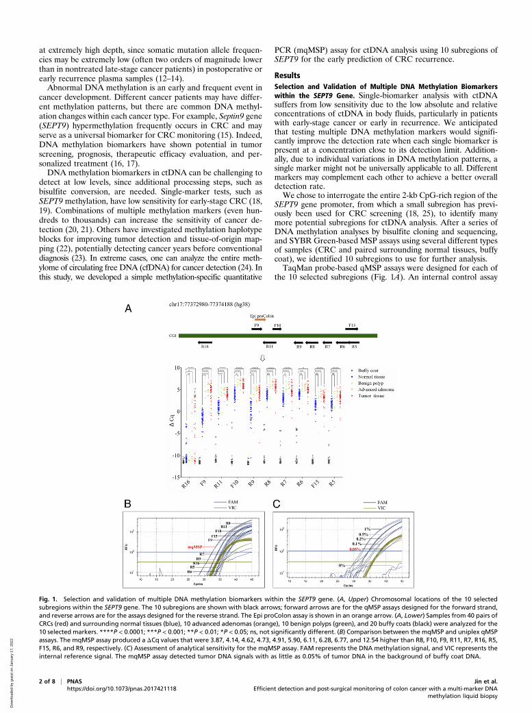
the 10 selected subregions (Fig. 1A). An internal control assay
Fig. 1. Selection and validation of multiple DNA methylation biomarkers within the SEPT9 gene. (A, Upper) Chromosomal locations of the 10 selectedsubregions within the SEPT9 gene. The 10 subregions are shown with black arrows; forward arrows are for the qMSP assays designed for the forward strand,and reverse arrows are for the assays designed for the reverse strand. The Epi proColon assay is shown in an orange arrow. (A, Lower) Samples from 40 pairs ofCRCs (red) and surrounding normal tissues (blue), 10 advanced adenomas (orange), 10 benign polyps (green), and 20 buffy coats (black) were analyzed for the10 selected markers. ****P < 0.0001; ***P < 0.001; **P < 0.01; *P < 0.05; ns, not significantly different. (B) Comparison between the mqMSP and uniplex qMSPassays. The mqMSP assay produced a ΔCq values that were 3.87, 4.14, 4.62, 4.73, 4.91, 5.90, 6.11, 6.28, 6.77, and 12.54 higher than R8, F10, F9, R11, R7, R16, R5,F15, R6, and R9, respectively. (C) Assessment of analytical sensitivity for the mqMSP assay. FAM represents the DNA methylation signal, and VIC represents theinternal reference signal. The mqMSP assay detected tumor DNA signals with as little as 0.05% of tumor DNA in the background of buffy coat DNA.
2 of 8 | PNAS Jin et al.https://doi.org/10.1073/pnas.2017421118 Efficient detection and post-surgical monitoring of colon cancer with a multi-marker DNA
methylation liquid biopsy
Dow
nloa
ded
by g
uest
on
Janu
ary
17, 2
022

targeting the ACTB gene was designed and used in the samereaction with each qMSP assay. The ΔCq value between eachqMSP assay and the internal control assay was used to representthe methylation level for each subregion, with a higher ΔCqvalue representing greater DNA methylation. We selected 40pairs of CRC and surrounding normal tissues, 10 advanced ad-enomas, 10 benign polyps, and 20 buffy coat samples for qMSPanalyses. For a qMSP assay to be applicable for plasma DNAanalysis, the signal strength (represented by the ΔCq value) forbuffy coat DNA must be extremely low, since buffy coat-derivedcfDNA is the dominant component in total plasma cfDNA. Evenweak signals from buffy coat DNA can produce false-positivesignals. As shown in Fig. 1A, all 10 qMSP assays showed eitherno or an extremely low background signal in buffy coat DNA,with typical ΔCq values <−10. In the 40 paired CRC and sur-rounding normal tissue samples, tumor DNA showed signifi-cantly higher methylation than paired normal DNA. Advancedadenoma and polyps samples also showed generally higher meth-ylation than normal tissue samples, with more subregions showing astatistically significant difference between advanced adenoma andnormal than between polyps and normal, suggesting that the DNAmethylation changes may be progressive in tumor development.
mqMSP Assay Development. Our primary aim was to develop asimple qPCR assay for simultaneously quantifying multiple DNAmethylation markers for ctDNA analysis from plasma samples.Given the limited probe fluorophore choices, it is not feasible toquantify each subregion individually in a single reaction. Thus,we designed an mqMSP assay with two different fluorophoreprobes. The FAM fluorophore was used for all 10 subregions,and the VIC fluorophore was used for the ACTB control assay.This mqMSP assay measures the total methylation of all 10subregions.We compared the 10-marker mqMSP assay with each indi-
vidual qMSP assay using 10 ng of mixed CRC and buffy coatgenomic DNA at a 1:100 ratio. The mqMSP assay produced a
ΔCq value which was 3.87, 4.14, 4.62, 4.73, 4.91, 5.90, 6.11, 6.28,6.77, and 12.54 higher than the individual R8, F10, F9, R11, R7,R16, R5, F15, R6, and R9 assays, respectively, indicating that themultiplex assay is analytically more sensitive than the individualassays (Fig. 1B).Since ctDNA is often present at low abundance in overall
plasma cfDNA, it is critical that the mqMSP assay be able todetect and quantify low proportions of ctDNA in the backgroundof normal DNA. To further evaluate the analytical sensitivity ofthe mqMSP assay, methylated tumor tissue DNA was mixed withbuffy coat DNA at 1%, 0.5%, 0.2%, 0.1%, 0.05%, and 0%, withthe total DNA amount fixed at 10 ng per reaction. The DNAmixtures were subject to bisulfite conversion and mqMSP anal-yses. We observed quantitative detection of tumor DNA at aslow as 0.05% of tumor DNA at 10 ng total DNA, mimickingplasma DNA samples from early-stage CRC patients or CRCpatients at an early period of recurrence (Fig. 1C).
mqMSP Analysis of ctDNA for CRC Detection.To test the performanceof the mqMSP assay for detecting CRC using plasma DNA, werecruited 53 CRC patients, 48 patients with advanced adenoma,30 patients with benign polyps, and 48 healthy controls (as con-firmed by colonoscopy). Using the preoperative plasma samples ofthe 53 CRC patients (Table 1, technical evaluation cohort), weobtained positive mqMSP detection in 84.9% (45/53). Amongthem, the detection rates for stage I, II, III, and IV patients were64.3% (9/14), 81.3% (13/16), 100% (9/9), and 100% (14/14), re-spectively (SI Appendix, Table S3). The detection rates for thepatients with advanced adenoma, patients with polyps, and healthycontrols were 23%, 40%, and 16.7%, respectively (SI Appendix,Table S3). The methylation signals of ctDNA were significantlyhigher in the CRC patients than in the healthy controls. The pa-tients at more advanced stages (III and IV) showed significantlyhigher methylation signals than those at earlier stages (I and II)(Fig. 2A). The area under the curve (AUC) value for the receiveroperating characteristic (ROC) curve separating the CRC patients
Table 1. Clinicopathological characteristics of CRC patients in technical evaluation, comparativestudy, and longitudinal cohorts
CharacteristicsTechnical evaluation
cohort (n = 53)Comparative
cohort (n = 43)
Longitudinal cohort
All patients(n = 82)
Preoperative ctDNApositive (n = 73)
Sex, nMale 33 24 53 46Female 20 19 29 27
Age, yMedian 69 71 66 67Range 31 to 90 51 to 89 33 to 85 33 to 85
Localization, nLeft colon 6 10 8 8Right colon 3 5 4 4Rectum 44 28 70 61
Lymph node metastasis, nYes 14 6 36 32No 34 37 46 41Unknown 5 0 0 0
Chemoradiotherapy, nYes 38 12 70 61No 15 31 12 12
Stage, nI 14 15 5 4II 16 17 30 27III 9 11 40 36IV 14 0 7 6
Jin et al. PNAS | 3 of 8Efficient detection and post-surgical monitoring of colon cancer with a multi-marker DNAmethylation liquid biopsy
https://doi.org/10.1073/pnas.2017421118
MED
ICALSC
IENCE
S
Dow
nloa
ded
by g
uest
on
Janu
ary
17, 2
022
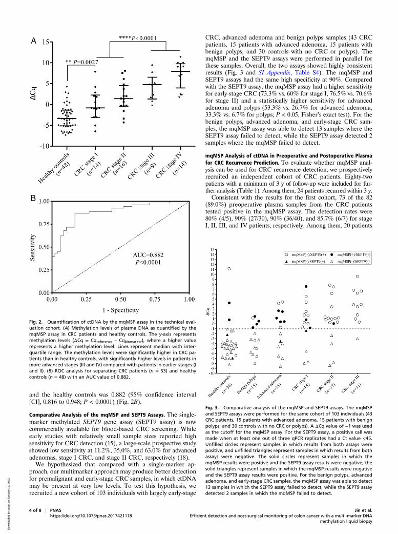
and the healthy controls was 0.882 (95% confidence interval[CI], 0.816 to 0.948; P < 0.0001) (Fig. 2B).
Comparative Analysis of the mqMSP and SEPT9 Assays. The single-marker methylated SEPT9 gene assay (SEPT9 assay) is nowcommercially available for blood-based CRC screening. Whileearly studies with relatively small sample sizes reported highsensitivity for CRC detection (15), a large-scale prospective studyshowed low sensitivity at 11.2%, 35.0%, and 63.0% for advancedadenomas, stage I CRC, and stage II CRC, respectively (18).We hypothesized that compared with a single-marker ap-
proach, our multimarker approach may produce better detectionfor premalignant and early-stage CRC samples, in which ctDNAmay be present at very low levels. To test this hypothesis, werecruited a new cohort of 103 individuals with largely early-stage
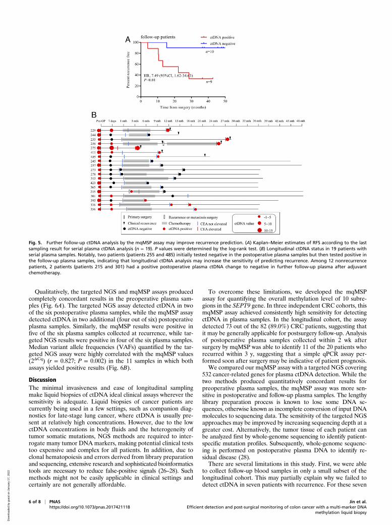
CRC, advanced adenoma and benign polyps samples (43 CRCpatients, 15 patients with advanced adenoma, 15 patients withbenign polyps, and 30 controls with no CRC or polyps). ThemqMSP and the SEPT9 assays were performed in parallel forthese samples. Overall, the two assays showed highly consistentresults (Fig. 3 and SI Appendix, Table S4). The mqMSP andSEPT9 assays had the same high specificity at 90%. Comparedwith the SEPT9 assay, the mqMSP assay had a higher sensitivityfor early-stage CRC (73.3% vs. 60% for stage I, 76.5% vs. 70.6%for stage II) and a statistically higher sensitivity for advancedadenoma and polyps (53.3% vs. 26.7% for advanced adenoma,33.3% vs. 6.7% for polyps; P < 0.05, Fisher’s exact test). For thebenign polyps, advanced adenoma, and early-stage CRC sam-ples, the mqMSP assay was able to detect 13 samples where theSEPT9 assay failed to detect, while the SEPT9 assay detected 2samples where the mqMSP failed to detect.
mqMSP Analysis of ctDNA in Preoperative and Postoperative Plasmafor CRC Recurrence Prediction. To evaluate whether mqMSP anal-ysis can be used for CRC recurrence detection, we prospectivelyrecruited an independent cohort of CRC patients. Eighty-twopatients with a minimum of 3 y of follow-up were included for fur-ther analysis (Table 1). Among them, 24 patients recurred within 3 y.Consistent with the results for the first cohort, 73 of the 82
(89.0%) preoperative plasma samples from the CRC patientstested positive in the mqMSP assay. The detection rates were80% (4/5), 90% (27/30), 90% (36/40), and 85.7% (6/7) for stageI, II, III, and IV patients, respectively. Among them, 20 patients
Fig. 2. Quantification of ctDNA by the mqMSP assay in the technical eval-uation cohort. (A) Methylation levels of plasma DNA as quantified by themqMSP assay in CRC patients and healthy controls. The y-axis representsmethylation levels (ΔCq = Cqreference – Cqbiomarker), where a higher valuerepresents a higher methylation level. Lines represent median with inter-quartile range. The methylation levels were significantly higher in CRC pa-tients than in healthy controls, with significantly higher levels in patients inmore advanced stages (III and IV) compared with patients in earlier stages (Iand II). (B) ROC analysis for separating CRC patients (n = 53) and healthycontrols (n = 48) with an AUC value of 0.882.
Fig. 3. Comparative analysis of the mqMSP and SEPT9 assays. The mqMSPand SEPT9 assays were performed for the same cohort of 103 individuals (43CRC patients, 15 patients with advanced adenoma, 15 patients with benignpolyps, and 30 controls with no CRC or polyps). A ΔCq value of −1 was usedas the cutoff for the mqMSP assay. For the SEPT9 assay, a positive call wasmade when at least one out of three qPCR replicates had a Ct value <45.Unfilled circles represent samples in which results from both assays werepositive, and unfilled triangles represent samples in which results from bothassays were negative. The solid circles represent samples in which themqMSP results were positive and the SEPT9 assay results were negative; thesolid triangles represent samples in which the mqMSP results were negativeand the SEPT9 assay results were positive. For the benign polyps, advancedadenoma, and early-stage CRC samples, the mqMSP assay was able to detect13 samples in which the SEPT9 assay failed to detect, while the SEPT9 assaydetected 2 samples in which the mqMSP failed to detect.
4 of 8 | PNAS Jin et al.https://doi.org/10.1073/pnas.2017421118 Efficient detection and post-surgical monitoring of colon cancer with a multi-marker DNA
methylation liquid biopsy
Dow
nloa
ded
by g
uest
on
Janu
ary
17, 2
022

recurred within 3 y, while 53 patients remained recurrence-freefor at least 3 y after surgery. Preoperative ctDNA status or levelwas not correlated with recurrence.We next performed mqMSP tests on postoperative plasma
samples from the 73 patients with positive preoperative ctDNA.We collected postoperative blood within 2 wk (1 to 14 d) aftersurgery. Overall, 21 of the 73 patients (28.8%) tested positive inthe mqMSP assay of postoperative plasma. Significant decreasesin ctDNA levels were observed from preoperative to postoper-ative plasma, regardless of recurrence status (SI Appendix, Fig.S1 A and B), although nonrecurrence patients had a greaterdecrease in ctDNA levels measured by ΔΔCq values [ΔΔCq =ΔCq(pre_op) − ΔCq(post_op)] (P = 0.009; SI Appendix, Fig.S1C).Among the 20 recurrence patients, 11 (55%) tested positive by
the mqMSP assay in the postoperative plasma samples. Positivedetection of ctDNA within 2 wk after surgery was associated withpoorer recurrence-free survival (RFS; HR, 4.20; 95% CI, 2.30 to18.73; P = 0.0005) (Fig. 4A). When patients were stratified bystage, significant RFS differences were observed in localized,stage II, or stage III CRC patients based on postoperative ctDNAstatus (SI Appendix, Fig. S2). Among the 20 recurrence patients,those with positive postoperative ctDNA had a poorer RFS thanthose with negative postoperative ctDNA (median RFS, 288 d vs.460 d; P = 0.008) (Fig. 4B).Adjuvant chemotherapy is often used for high-risk patients
after surgery. Thus, we stratified patients based on adjuvant che-motherapy status. Regardless of the use of adjuvant chemother-apy, positive postoperative ctDNA detection by mqMSP stronglypredicted recurrence within 3 y (with adjuvant chemotherapy: HR,5.16; 95% CI, 2.31 to 29.78; P = 0.001; without adjuvant chemo-therapy: HR, 4.08; 95% CI, 1.26 to 75.05; P = 0.037) (Fig. 4 Cand D).We were able to collect additional follow-up blood samples in
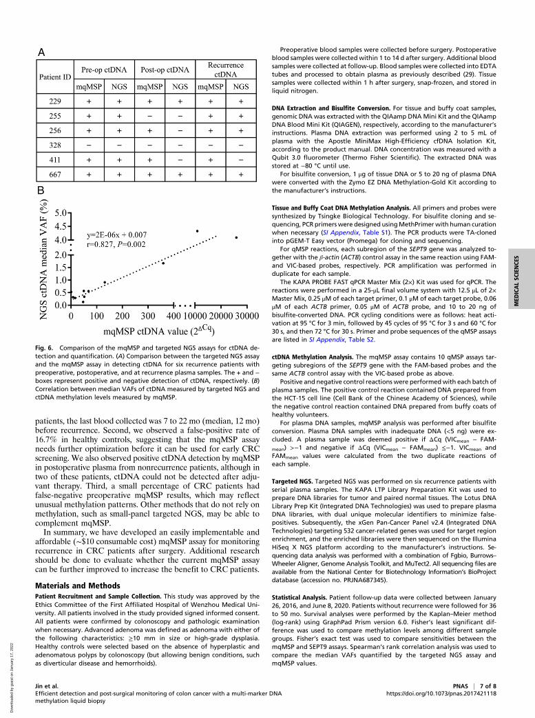
a subset of 19 patients, in which we assessed whether follow-upplasma ctDNA analysis can further improve recurrence predic-tion. Detection of ctDNA by mqMSP in serial blood samples wasassociated with poorer RFS (HR, 7.49; 95% CI, 1.62 to 34.63;
P = 0.01) (Fig. 5A). Among seven recurrence patients, fourtested positive in postoperative plasma and two additional pa-tients (patients 255 and 485) tested positive in the follow-up plasmasamples, indicating that longitudinal ctDNA analysis can in-crease the sensitivity of predicting recurrence (Fig. 5B). In 2 of12 nonrecurrence patients (patients 215 and 301), positive post-operative plasma ctDNA changed to negative in further follow-upplasma samples after adjuvant chemotherapy, suggesting that ad-juvant chemotherapy may have been effective in removing residualtumor load (Fig. 5B).Overall, the analysis of ctDNA by mqMSP had a median lead
time of 8.0 mo (range, 0 to 12.5 mo) over radiologic imaginganalysis for detecting recurrence. More than one-half (11 out of20) of the patients who experienced recurrence within 3 y ofsurgery had positive detection of ctDNA within 2 wk after surgery.
Comparison of Postoperative ctDNA and CEA for Predicting Recurrence.The relationships among postoperative ctDNA status, postoper-ative CEA level, and recurrence status are shown in SI Appendix,Table S5 (4 of the 73 patients did not have postoperative CEAdata). The CEA level was above the threshold (5 ng/mL) in thepostoperative blood of 4 of the 17 (23.5%) recurrence patientsand in 2 of the 52 (3.8%) nonrecurrence patients. While CEAhas superior specificity, it suffers from poor sensitivity comparedwith the ctDNA mqMSP assay (23.5% vs. 55%). Of the 20 re-currence patients, 11 tested positive by mqMSP in postoperativeplasma samples.
Parallel Analysis of ctDNA by mqMSP for DNA Methylation Markersand Targeted NGS for Somatic Mutations. Somatic mutation de-tection by targeted NGS is often used for ctDNA detection. Wecompared the mqMSP assay with a targeted NGS assay covering532 cancer-related genes by analyzing a trio of preoperative,postoperative and recurrence plasmas from six recurrence pa-tients. The raw sequencing depth for plasma DNA samples was14,700× on average. The corresponding tumor tissue sampleswere analyzed by the same panel, at a raw sequencing depth of∼1,800×, to confirm the somatic mutation results.
Fig. 4. Postoperative ctDNA status determined by the mqMSP assay predicts recurrence. (A) Kaplan–Meier estimates of RFS according to postoperativeplasma ctDNA status in 73 patients with positive preoperative plasma ctDNA. (B) Kaplan–Meier estimates of RFS according to postoperative plasma ctDNAstatus for the 20 patients with recurrence. (C) Kaplan–Meier estimates of RFS for the 62 patients who received adjuvant chemotherapy. (D) Kaplan–Meierestimates of RFS for the 11 patients who did not receive adjuvant chemotherapy. Without adjuvant chemotherapy, all three patients with positive post-operative ctDNA recurred within 12 mo. P values were determined by the log-rank test.
Jin et al. PNAS | 5 of 8Efficient detection and post-surgical monitoring of colon cancer with a multi-marker DNAmethylation liquid biopsy
https://doi.org/10.1073/pnas.2017421118
MED
ICALSC
IENCE
S
Dow
nloa
ded
by g
uest
on
Janu
ary
17, 2
022
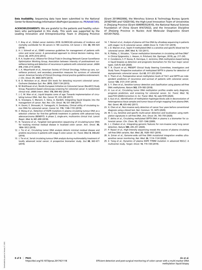
Qualitatively, the targeted NGS and mqMSP assays producedcompletely concordant results in the preoperative plasma sam-ples (Fig. 6A). The targeted NGS assay detected ctDNA in twoof the six postoperative plasma samples, while the mqMSP assaydetected ctDNA in two additional (four out of six) postoperativeplasma samples. Similarly, the mqMSP results were positive infive of the six plasma samples collected at recurrence, while tar-geted NGS results were positive in four of the six plasma samples.Median variant allele frequencies (VAFs) quantified by the tar-geted NGS assay were highly correlated with the mqMSP values(2ΔCq) (r = 0.827; P = 0.002) in the 11 samples in which bothassays yielded positive results (Fig. 6B).
DiscussionThe minimal invasiveness and ease of longitudinal samplingmake liquid biopsies of ctDNA ideal clinical assays wherever thesensitivity is adequate. Liquid biopsies of cancer patients arecurrently being used in a few settings, such as companion diag-nostics for late-stage lung cancer, where ctDNA is usually pre-sent at relatively high concentrations. However, due to the lowctDNA concentrations in body fluids and the heterogeneity oftumor somatic mutations, NGS methods are required to inter-rogate many tumor DNA markers, making potential clinical teststoo expensive and complex for all patients. In addition, due toclonal hematopoiesis and errors derived from library preparationand sequencing, extensive research and sophisticated bioinformaticstools are necessary to reduce false-positive signals (26–28). Suchmethods might not be easily applicable in clinical settings andcertainly are not generally affordable.
To overcome these limitations, we developed the mqMSPassay for quantifying the overall methylation level of 10 subre-gions in the SEPT9 gene. In three independent CRC cohorts, thismqMSP assay achieved consistently high sensitivity for detectingctDNA in plasma samples. In the longitudinal cohort, the assaydetected 73 out of the 82 (89.0%) CRC patients, suggesting thatit may be generally applicable for postsurgery follow-up. Analysisof postoperative plasma samples collected within 2 wk aftersurgery by mqMSP was able to identify 11 of the 20 patients whorecurred within 3 y, suggesting that a simple qPCR assay per-formed soon after surgery may be indicative of patient prognosis.We compared our mqMSP assay with a targeted NGS covering
532 cancer-related genes for plasma ctDNA detection. While thetwo methods produced quantitatively concordant results forpreoperative plasma samples, the mqMSP assay was more sen-sitive in postoperative and follow-up plasma samples. The lengthylibrary preparation process is known to lose some DNA se-quences, otherwise known as incomplete conversion of input DNAmolecules to sequencing data. The sensitivity of the targeted NGSapproaches may be improved by increasing sequencing depth at agreater cost. Alternatively, the tumor tissue of each patient canbe analyzed first by whole-genome sequencing to identify patient-specific mutation profiles. Subsequently, whole-genome sequenc-ing is performed on postoperative plasma DNA to identify re-sidual disease (28).There are several limitations in this study. First, we were able
to collect follow-up blood samples in only a small subset of thelongitudinal cohort. This may partially explain why we failed todetect ctDNA in seven patients with recurrence. For these seven
Fig. 5. Further follow-up ctDNA analysis by the mqMSP assay may improve recurrence prediction. (A) Kaplan–Meier estimates of RFS according to the lastsampling result for serial plasma ctDNA analysis (n = 19). P values were determined by the log-rank test. (B) Longitudinal ctDNA status in 19 patients withserial plasma samples. Notably, two patients (patients 255 and 485) initially tested negative in the postoperative plasma samples but then tested positive inthe follow-up plasma samples, indicating that longitudinal ctDNA analysis may increase the sensitivity of predicting recurrence. Among 12 nonrecurrencepatients, 2 patients (patients 215 and 301) had a positive postoperative plasma ctDNA change to negative in further follow-up plasma after adjuvantchemotherapy.
6 of 8 | PNAS Jin et al.https://doi.org/10.1073/pnas.2017421118 Efficient detection and post-surgical monitoring of colon cancer with a multi-marker DNA
methylation liquid biopsy
Dow
nloa
ded
by g
uest
on
Janu
ary
17, 2
022
patients, the last blood collected was 7 to 22 mo (median, 12 mo)before recurrence. Second, we observed a false-positive rate of16.7% in healthy controls, suggesting that the mqMSP assayneeds further optimization before it can be used for early CRCscreening. We also observed positive ctDNA detection by mqMSPin postoperative plasma from nonrecurrence patients, although intwo of these patients, ctDNA could not be detected after adju-vant therapy. Third, a small percentage of CRC patients hadfalse-negative preoperative mqMSP results, which may reflectunusual methylation patterns. Other methods that do not rely onmethylation, such as small-panel targeted NGS, may be able tocomplement mqMSP.In summary, we have developed an easily implementable and
affordable (∼$10 consumable cost) mqMSP assay for monitoringrecurrence in CRC patients after surgery. Additional researchshould be done to evaluate whether the current mqMSP assaycan be further improved to increase the benefit to CRC patients.
Materials and MethodsPatient Recruitment and Sample Collection. This study was approved by theEthics Committee of the First Affiliated Hospital of Wenzhou Medical Uni-versity. All patients involved in the study provided signed informed consent.All patients were confirmed by colonoscopy and pathologic examinationwhen necessary. Advanced adenoma was defined as adenoma with either ofthe following characteristics: ≥10 mm in size or high-grade dysplasia.Healthy controls were selected based on the absence of hyperplastic andadenomatous polyps by colonoscopy (but allowing benign conditions, suchas diverticular disease and hemorrhoids).
Preoperative blood samples were collected before surgery. Postoperativeblood samples were collected within 1 to 14 d after surgery. Additional bloodsamples were collected at follow-up. Blood samples were collected into EDTAtubes and processed to obtain plasma as previously described (29). Tissuesamples were collected within 1 h after surgery, snap-frozen, and stored inliquid nitrogen.
DNA Extraction and Bisulfite Conversion. For tissue and buffy coat samples,genomic DNA was extracted with the QIAamp DNAMini Kit and the QIAampDNA Blood Mini Kit (QIAGEN), respectively, according to the manufacturer’sinstructions. Plasma DNA extraction was performed using 2 to 5 mL ofplasma with the Apostle MiniMax High-Efficiency cfDNA Isolation Kit,according to the product manual. DNA concentration was measured with aQubit 3.0 fluorometer (Thermo Fisher Scientific). The extracted DNA wasstored at −80 °C until use.
For bisulfite conversion, 1 μg of tissue DNA or 5 to 20 ng of plasma DNAwere converted with the Zymo EZ DNA Methylation-Gold Kit according tothe manufacturer’s instructions.
Tissue and Buffy Coat DNA Methylation Analysis. All primers and probes weresynthesized by Tsingke Biological Technology. For bisulfite cloning and se-quencing, PCR primers were designed usingMethPrimer with human curationwhen necessary (SI Appendix, Table S1). The PCR products were TA-clonedinto pGEM-T Easy vector (Promega) for cloning and sequencing.
For qMSP reactions, each subregion of the SEPT9 gene was analyzed to-gether with the β-actin (ACTB) control assay in the same reaction using FAM-and VIC-based probes, respectively. PCR amplification was performed induplicate for each sample.
The KAPA PROBE FAST qPCR Master Mix (2×) Kit was used for qPCR. Thereactions were performed in a 25-μL final volume system with 12.5 μL of 2×Master Mix, 0.25 μM of each target primer, 0.1 μM of each target probe, 0.06μM of each ACTB primer, 0.05 μM of ACTB probe, and 10 to 20 ng ofbisulfite-converted DNA. PCR cycling conditions were as follows: heat acti-vation at 95 °C for 3 min, followed by 45 cycles of 95 °C for 3 s and 60 °C for30 s, and then 72 °C for 30 s. Primer and probe sequences of the qMSP assaysare listed in SI Appendix, Table S2.
ctDNA Methylation Analysis. The mqMSP assay contains 10 qMSP assays tar-geting subregions of the SEPT9 gene with the FAM-based probes and thesame ACTB control assay with the VIC-based probe as above.
Positive and negative control reactions were performedwith each batch ofplasma samples. The positive control reaction contained DNA prepared fromthe HCT-15 cell line (Cell Bank of the Chinese Academy of Sciences), whilethe negative control reaction contained DNA prepared from buffy coats ofhealthy volunteers.
For plasma DNA samples, mqMSP analysis was performed after bisulfiteconversion. Plasma DNA samples with inadequate DNA (<5 ng) were ex-cluded. A plasma sample was deemed positive if ΔCq (VICmean − FAM-
mean) >−1 and negative if ΔCq (VICmean − FAMmean) ≤−1. VICmean andFAMmean values were calculated from the two duplicate reactions ofeach sample.
Targeted NGS. Targeted NGS was performed on six recurrence patients withserial plasma samples. The KAPA LTP Library Preparation Kit was used toprepare DNA libraries for tumor and paired normal tissues. The Lotus DNALibrary Prep Kit (Integrated DNA Technologies) was used to prepare plasmaDNA libraries, with dual unique molecular identifiers to minimize false-positives. Subsequently, the xGen Pan-Cancer Panel v2.4 (Integrated DNATechnologies) targeting 532 cancer-related genes was used for target regionenrichment, and the enriched libraries were then sequenced on the IlluminaHiSeq X NGS platform according to the manufacturer’s instructions. Se-quencing data analysis was performed with a combination of Fgbio, Burrows–Wheeler Aligner, Genome Analysis Toolkit, and MuTect2. All sequencing files areavailable from the National Center for Biotechnology Information’s BioProjectdatabase (accession no. PRJNA687345).
Statistical Analysis. Patient follow-up data were collected between January26, 2016, and June 8, 2020. Patients without recurrence were followed for 36to 50 mo. Survival analyses were performed by the Kaplan–Meier method(log-rank) using GraphPad Prism version 6.0. Fisher’s least significant dif-ference was used to compare methylation levels among different samplegroups. Fisher’s exact test was used to compare sensitivities between themqMSP and SEPT9 assays. Spearman’s rank correlation analysis was used tocompare the median VAFs quantified by the targeted NGS assay andmqMSP values.
Fig. 6. Comparison of the mqMSP and targeted NGS assays for ctDNA de-tection and quantification. (A) Comparison between the targeted NGS assayand the mqMSP assay in detecting ctDNA for six recurrence patients withpreoperative, postoperative, and at recurrence plasma samples. The + and −boxes represent positive and negative detection of ctDNA, respectively. (B)Correlation between median VAFs of ctDNA measured by targeted NGS andctDNA methylation levels measured by mqMSP.
Jin et al. PNAS | 7 of 8Efficient detection and post-surgical monitoring of colon cancer with a multi-marker DNAmethylation liquid biopsy
https://doi.org/10.1073/pnas.2017421118
MED
ICALSC
IENCE
S
Dow
nloa
ded
by g
uest
on
Janu
ary
17, 2
022
Data Availability. Sequencing data have been submitted to the NationalCenter for Biotechnology Information’s BioProject (accession no. PRJNA687345).
ACKNOWLEDGMENTS. We are grateful to the patients and healthy volun-teers who participated in this study. This work was supported by theLeading Innovation and Entrepreneurship Team in Zhejiang Province
(Grant 2019R02006), the Wenzhou Science & Technology Bureau (grants2018ZY005 and Y2020156), the High-Level Innovation Team of Universitiesin Zhejiang Province (Grant 604090352/610), the National Natural SciencesFoundation of China (Grant 81672922), and the Innovation Disciplineof Zhejiang Province in Nucleic Acid Molecular Diagnostics (Grant437201702G).
1. F. Bray et al., Global cancer statistics 2018: GLOBOCAN estimates of incidence andmortality worldwide for 36 cancers in 185 countries. CA Cancer J. Clin. 68, 394–424(2018).
2. H. J. Schmoll et al., ESMO consensus guidelines for management of patients withcolon and rectal cancer. A personalized approach to clinical decision making. Ann.Oncol. 23, 2479–2516 (2012).
3. R. A. Snyder et al.; Alliance for Clinical Trials in Oncology Network Cancer SurveillanceOptimization Working Group, Association between intensity of posttreatment sur-veillance testing and detection of recurrence in patients with colorectal cancer. JAMA319, 2104–2115 (2018).
4. J. A. Meyerhardt et al.; American Society of Clinical Oncology, Follow-up care, sur-veillance protocol, and secondary prevention measures for survivors of colorectalcancer: American Society of Clinical Oncology clinical practice guideline endorsement.J. Clin. Oncol. 31, 4465–4470 (2013).
5. B. D. Nicholson et al., Blood CEA levels for detecting recurrent colorectal cancer.Cochrane Database Syst. Rev. 2015, CD011134 (2015).
6. M. Bretthauer et al.; Nordic-European Initiative on Colorectal Cancer (NordICC) StudyGroup, Population-based colonoscopy screening for colorectal cancer: A randomizedclinical trial. JAMA Intern. Med. 176, 894–902 (2016).
7. J. C. M. Wan et al., Liquid biopsies come of age: Towards implementation of circu-lating tumour DNA. Nat. Rev. Cancer 17, 223–238 (2017).
8. G. Siravegna, S. Marsoni, S. Siena, A. Bardelli, Integrating liquid biopsies into themanagement of cancer. Nat. Rev. Clin. Oncol. 14, 531–548 (2017).
9. H. Osumi, E. Shinozaki, K. Yamaguchi, H. Zembutsu, Clinical utility of circulating tu-mor DNA for colorectal cancer. Cancer Sci. 110, 1148–1155 (2019).
10. Z. Wang et al., Detection of EGFR mutations in plasma circulating tumour DNA as aselection criterion for first-line gefitinib treatment in patients with advanced lungadenocarcinoma (BENEFIT): A phase 2, single-arm, multicentre clinical trial. LancetRespir. Med. 6, 681–690 (2018).
11. N. Tarazona et al., Targeted next-generation sequencing of circulating-tumor DNAfor tracking minimal residual disease in localized colon cancer. Ann. Oncol. 30,1804–1812 (2019).
12. J. Tie et al., Circulating tumor DNA analysis detects minimal residual disease andpredicts recurrence in patients with stage II colon cancer. Sci. Transl. Med. 8, 346ra92(2016).
13. J. Tie et al., Serial circulating tumour DNA analysis during multimodality treatment oflocally advanced rectal cancer: A prospective biomarker study. Gut 68, 663–671(2019).
14. T. Reinert et al., Analysis of plasma cell-free DNA by ultradeep sequencing in patientswith stages I to III colorectal cancer. JAMA Oncol. 5, 1124–1131 (2019).
15. J. D. Warren et al., Septin 9 methylated DNA is a sensitive and specific blood test forcolorectal cancer. BMC Med. 9, 133 (2011).
16. R. Pidsley, C. Stirzaker, “Cancer methylation biomarkers in circulating cell-free DNA”in Clinical Epigenetics, L. Hesson, A. Pritchard, Eds. (Springer, 2019), pp. 217–245.
17. V. Constâncio, S. P. Nunes, R. Henrique, C. Jerónimo, DNA methylation-based testingin liquid biopsies as detection and prognostic biomarkers for the four major cancertypes. Cells 9, 624 (2020).
18. T. R. Church et al.; PRESEPT Clinical Study Steering Committee, Investigators andStudy Team, Prospective evaluation of methylated SEPT9 in plasma for detection ofasymptomatic colorectal cancer. Gut 63, 317–325 (2014).
19. C. Tham et al., Postoperative serum methylation levels of TAC1 and SEPT9 are inde-pendent predictors of recurrence and survival of patients with colorectal cancer.Cancer 120, 3131–3141 (2014).
20. S. Y. Shen et al., Sensitive tumour detection and classification using plasma cell-freeDNA methylomes. Nature 563, 579–583 (2018).
21. H. Luo et al., Circulating tumor DNA methylation profiles enable early diagnosis,prognosis prediction, and screening for colorectal cancer. Sci. Transl. Med. 12,eaax7533 (2020).Correction in: Sci. Transl. Med. 12, eabc1078 (2020).
22. S. Guo et al., Identification of methylation haplotype blocks aids in deconvolution ofheterogeneous tissue samples and tumor tissue-of-origin mapping from plasma DNA.Nat. Genet. 49, 635–642 (2017).
23. X. Chen et al., Non-invasive early detection of cancer four years before conventionaldiagnosis using a blood test. Nat. Commun. 11, 3475 (2020).
24. M. C. Liu, Sensitive and specific multi-cancer detection and localization using meth-ylation signatures in cell-free DNA. Ann. Oncol. 31, 745–759 (2020).
25. T. deVos et al., Circulating methylated SEPT9 DNA in plasma is a biomarker for co-lorectal cancer. Clin. Chem. 55, 1337–1346 (2009).
26. J. J. Chabon et al., Integrating genomic features for non-invasive early lung cancerdetection. Nature 580, 245–251 (2020).
27. P. Razavi et al., High-intensity sequencing reveals the sources of plasma circulatingcell-free DNA variants. Nat. Med. 25, 1928–1937 (2019).
28. A. Zviran et al., Genome-wide cell-free DNA mutational integration enables ultra-sensitive cancer monitoring. Nat. Med. 26, 1114–1124 (2020).
29. Z. Yang et al., Dynamics of plasma EGFR T790M mutation in advanced NSCLC: Amulticenter study. Target. Oncol. 14, 719–728 (2019).
8 of 8 | PNAS Jin et al.https://doi.org/10.1073/pnas.2017421118 Efficient detection and post-surgical monitoring of colon cancer with a multi-marker DNA
methylation liquid biopsy
Dow
nloa
ded
by g
uest
on
Janu
ary
17, 2
022