Surgical and Pathological Changes after ... - units.it and... · Surgical and Pathological Changes...
9
Research Article Surgical and Pathological Changes after Radiofrequency Ablation of Thyroid Nodules Chiara Dobrinja, 1 Stella Bernardi, 2,3 Bruno Fabris, 2,3 Rita Eramo, 1 Petra Makovac, 1,3 Gabriele Bazzocchi, 4 Lanfranco Piscopello, 5 Enrica Barro, 2,3 Nicolò de Manzini, 1,3 Deborah Bonazza, 3,6 Maurizio Pinamonti, 3,6 Fabrizio Zanconati, 3,6 and Fulvio Stacul 4 1 UCO Chirurgia Generale, Cattinara Teaching Hospital, Strada di Fiume, 34100 Trieste, Italy 2 SS Endocrinologia (UCO Medicina Clinica), Cattinara Teaching Hospital, Strada di Fiume, 34100 Trieste, Italy 3 Department of Medical, Surgical, and Health Sciences, University of Trieste, Cattinara Teaching Hospital, Strada di Fiume, 34100 Trieste, Italy 4 SC Radiologia, Maggiore Hospital, Piazza dell’Ospitale, 34100 Trieste, Italy 5 SS Endocrinologia (III Medica), Maggiore Hospital, Piazza dell’Ospitale, 34100 Trieste, Italy 6 UCO Anatomia e Istologia Patologica, Cattinara Teaching Hospital, Strada di Fiume, 34100 Trieste, Italy Correspondence should be addressed to Stella Bernardi; [email protected] Received 10 March 2015; Revised 14 May 2015; Accepted 1 July 2015 Academic Editor: Jack R. Wall Copyright © 2015 Chiara Dobrinja et al. is is an open access article distributed under the Creative Commons Attribution License, which permits unrestricted use, distribution, and reproduction in any medium, provided the original work is properly cited. Background. Radiofrequency ablation (RFA) has been recently advocated as an effective technique for the treatment of symptomatic benign thyroid nodules. It is not known to what extent it may affect any subsequent thyroid surgery and/or histological diagnosis. Materials and Methods. RFA was performed on 64 symptomatic y2 nodules (benign nodules) and 6 symptomatic y3 nodules (follicular lesions/follicular neoplasms). Two y3 nodules regrew aſter the procedure, and these patients accepted to undergo a total thyroidectomy. Here we present how RFA has affected the operation and the final pathological features of the surgically removed nodules. Results and Conclusions. RFA is effective for the treatment of y2 nodules, but it should not be recommended as first-line therapy for the treatment of y3 nodules (irrespective of their mutational status), as it delays surgery in case of malignancy. Moreover, it is unknown whether RFA might promote residual tumor progression or neoplastic progression of y3 lesions. Nevertheless, here we show for the first time that one session of RFA does not affect subsequent thyroid surgery and/or histological diagnosis. 1. Introduction yroid nodules are an extremely common occurrence [1]. Until recently international guidelines suggested either no treatment or surgery for patients with benign solid thyroid nodules, depending on their size, cytology, and symptoms [2]. In recent years, however, several nonsurgical minimally invasive approaches for treating thyroid nodules have been developed [3] and nowadays they represent an acceptable alternative to surgery for treating symptomatic benign thy- roid nodules [3]. ese techniques include percutaneous ethanol injection therapy (PEIT), laser ablation therapy (LAT), and radiofrequency ablation (RFA). Among these techniques RFA is not only an effective [4] but also a reasonably safe [5] treatment option for recurrent cysts [6], mixed or solid benign thyroid nodules causing cosmetic concerns, local pain, or hyperthyroidism [7, 8]. Nevertheless, RFA is relatively new, so some issues regarding its outcomes still exist. For example, one of the questions that remains unanswered is what would happen if a thyroid cancer were not detected before starting RFA and was leſt untreated? Would RFA jeopardize a subsequent operation and/or hinder histological diagnosis? Here we present the 2-year follow-up results of 70 patients who underwent RFA. In particular, 64 patients had y2 nodules (benign nodules) and 6 patients had y3 nodules Hindawi Publishing Corporation International Journal of Endocrinology Volume 2015, Article ID 576576, 8 pages http://dx.doi.org/10.1155/2015/576576
Transcript of Surgical and Pathological Changes after ... - units.it and... · Surgical and Pathological Changes...
Research ArticleSurgical and Pathological Changes after RadiofrequencyAblation of Thyroid Nodules
Chiara Dobrinja,1 Stella Bernardi,2,3 Bruno Fabris,2,3 Rita Eramo,1 Petra Makovac,1,3
Gabriele Bazzocchi,4 Lanfranco Piscopello,5 Enrica Barro,2,3 Nicolò de Manzini,1,3
Deborah Bonazza,3,6 Maurizio Pinamonti,3,6 Fabrizio Zanconati,3,6 and Fulvio Stacul4
1UCO Chirurgia Generale, Cattinara Teaching Hospital, Strada di Fiume, 34100 Trieste, Italy2SS Endocrinologia (UCOMedicina Clinica), Cattinara Teaching Hospital, Strada di Fiume, 34100 Trieste, Italy3Department of Medical, Surgical, and Health Sciences, University of Trieste, Cattinara Teaching Hospital,Strada di Fiume, 34100 Trieste, Italy4SC Radiologia, Maggiore Hospital, Piazza dell’Ospitale, 34100 Trieste, Italy5SS Endocrinologia (III Medica), Maggiore Hospital, Piazza dell’Ospitale, 34100 Trieste, Italy6UCO Anatomia e Istologia Patologica, Cattinara Teaching Hospital, Strada di Fiume, 34100 Trieste, Italy
Correspondence should be addressed to Stella Bernardi; [email protected]
Received 10 March 2015; Revised 14 May 2015; Accepted 1 July 2015
Academic Editor: Jack R. Wall
Copyright © 2015 Chiara Dobrinja et al.This is an open access article distributed under theCreativeCommonsAttribution License,which permits unrestricted use, distribution, and reproduction in any medium, provided the original work is properly cited.
Background. Radiofrequency ablation (RFA) has been recently advocated as an effective technique for the treatment of symptomaticbenign thyroid nodules. It is not known to what extent it may affect any subsequent thyroid surgery and/or histological diagnosis.Materials and Methods. RFA was performed on 64 symptomatic Thy2 nodules (benign nodules) and 6 symptomatic Thy3 nodules(follicular lesions/follicular neoplasms). Two Thy3 nodules regrew after the procedure, and these patients accepted to undergoa total thyroidectomy. Here we present how RFA has affected the operation and the final pathological features of the surgicallyremoved nodules. Results and Conclusions. RFA is effective for the treatment of Thy2 nodules, but it should not be recommendedas first-line therapy for the treatment of Thy3 nodules (irrespective of their mutational status), as it delays surgery in case ofmalignancy. Moreover, it is unknown whether RFA might promote residual tumor progression or neoplastic progression of Thy3lesions. Nevertheless, here we show for the first time that one session of RFA does not affect subsequent thyroid surgery and/orhistological diagnosis.
1. Introduction
Thyroid nodules are an extremely common occurrence [1].Until recently international guidelines suggested either notreatment or surgery for patients with benign solid thyroidnodules, depending on their size, cytology, and symptoms[2]. In recent years, however, several nonsurgical minimallyinvasive approaches for treating thyroid nodules have beendeveloped [3] and nowadays they represent an acceptablealternative to surgery for treating symptomatic benign thy-roid nodules [3]. These techniques include percutaneousethanol injection therapy (PEIT), laser ablation therapy(LAT), and radiofrequency ablation (RFA).
Among these techniques RFA is not only an effective [4]but also a reasonably safe [5] treatment option for recurrentcysts [6], mixed or solid benign thyroid nodules causingcosmetic concerns, local pain, or hyperthyroidism [7, 8].Nevertheless, RFA is relatively new, so some issues regardingits outcomes still exist. For example, one of the questions thatremains unanswered is what would happen if a thyroid cancerwere not detected before starting RFA and was left untreated?Would RFA jeopardize a subsequent operation and/or hinderhistological diagnosis?
Herewe present the 2-year follow-up results of 70 patientswho underwent RFA. In particular, 64 patients had Thy2nodules (benign nodules) and 6 patients had Thy3 nodules
Hindawi Publishing CorporationInternational Journal of EndocrinologyVolume 2015, Article ID 576576, 8 pageshttp://dx.doi.org/10.1155/2015/576576
2 International Journal of Endocrinology
(follicular lesions/follicular neoplasms). Because of noduleregrowth, two of these patients subsequently underwentsurgery after being treatedwithRFA. Based on our experiencethe aim of this report is to describe the surgical and patholog-ical changes after RFA of thyroid nodules and to comment onthe use of RFA for Thy3 nodules.
2. Materials and Methods
2.1. Study Population. This is a retrospective study of the2-year follow-up results of 64 patients with Thy2 thyroidnodules and 6 patients with Thy3 nodules who underwentRFA. Before the procedure, nodules were evaluated byfine needle aspiration biopsy (FNAB) twice and classifiedaccording to the British and Italian reporting systems forthyroid cytopathology [9, 10], where Thy2 corresponds toBethesda Category II (benign nodules), and Thy3 corre-sponds to Bethesda Categories III and IV nodules (follicularlesions/follicular neoplasms) [11]. A single session of RFAwas performed on 64 Thy2 solitary nodules, according tocurrent recommendations [12]. A single session of RFA wasalso performed on 6 symptomatic Thy3 nodules in surgicallyhigh-risk individuals or patients refusing surgery. Thesepatients did not have a family history of thyroid cancerand did not present suspicious lymph nodes. Their noduleswere single, unchanged over the last three years, with nocalcifications, no evidence of nuclear atypia [13, 14], andwithout BRAF or NRAS mutations. Patients were preparedfor the procedure according to current recommendations [12]and before it they all signed an informed consent form afterfull explanation of the purpose and nature of all proceduresused. Secondly, informed consent to use their clinical data forscientific purposes was obtained from each patient, accordingto the guidelines of the Azienda Ospedaliero-Universitaria diTrieste.
2.2. Radiofrequency Ablation Technique and Follow-Up. RFAwas performed in an outpatient regimen by a well-trainedradiologist experienced in US, FNAB, and RFA procedures.Before ablation, an intravenous access was obtained via anantecubital vein. Patients were placed in a supine positionwith their neck extended. They underwent local anesthesiaat the puncture site with 2% lidocaine hydrochloride. Theprocedure required conscious sedation following an intra-venous injection of 2-3mg of midazolam.We used a 450 kHzradiofrequency generator (AMICA-GENmodel AGN-H-1.0,HS Hospital Service SpA, Italy) delivering up to 200W on a50Ω load and 18-gauge, internally cooled, monopolar elec-trodes (RFAMICA-PROBEmodel RFH18100V1,HSHospitalService SpA, Italy) featuring a shaft length of 10 cm and anexposed tip length of either 7, 10, or 15mm. The exposed tiplength selectionwas based on the nodule volume: 7mmactivetips were used for the treatment of smaller nodules (<10mL),10mm active tips in intermediate size nodules (10–20mL),and 15mm active tips in larger nodules (>20mL).
The electrode was inserted into the thyroid nodule witha transisthmic approach under US-guidance by using a 5–18MHz linear probe on a real-time US system (Aplio XG,
Toshiba Corp. Medical Systems). To perform RFA themoving-shot technique was used [15]. The initial RF powersettings ranged between 30W and 50W depending on theelectrode exposed tip length: the shorter the active tip,the lower the power. The ablation procedure always startedfrom the deepest areas of the nodule. If a hyperechoic zonehad not formed at the electrode tip within 30 seconds, theRF generator output was progressively increased in 10Wsteps, up to a maximum power ranging between 60W and80W (once again depending on the active tip length). Then,once the treated zone had become hyperechoic and theelectric impedance measured by the generator increased, theelectrode was moved towards another area of the nodule.Ablation was terminated when transient hyperechoic zonescould be identified around the entire nodule. At the end of theprocedure, patients remained under observation for a coupleof hours.
To evaluate RFA results, patients underwent an ultra-sonography (US) and TSH measurement at baseline, 1, 3, 6,12, and 24 months after the procedure. To simplify the cal-culation of the nodule volumes prior to RFA (𝑉pre) and afterRFA (𝑉post) out of cross-sectional measurements taken fromthe respective US images, each nodule was approximated toan ellipsoid, both before RFA (with diameters 𝐴pre as majoraxis, 𝐵pre and 𝐶pre as minor axis, both perpendicular to𝐴pre)and after RFA (with diameters 𝐴post, 𝐵post, and 𝐶post, namedafter their pre-RFA counterparts). Therefore, the followingformulas were used: 𝑉pre = 𝜋 ∗ 𝐴pre ∗ 𝐵pre ∗ 𝐶pre/6;𝑉post = 𝜋 ∗ 𝐴post ∗ 𝐵post ∗ 𝐶post/6. The percentile volumereduction induced by the RFA treatment was calculated asΔ𝑉 = 100∗ (𝑉pre −𝑉post)/𝑉pre. Technical success was deemedto be achievedwhen a volume reductionΔ𝑉 greater than 50%at 6 months from the procedure [4] was observed.
2.3. Surgery. Two patients were operated on after RFA. Theoperations were carried out by the same surgical team. Theintraoperative nerve integrity monitoring (NIM) system wasused in order to check the laryngeal nerve functionality.Patients, under general anaesthesia, were placed in the supineposition with the neck extended. Thyroidectomy was per-formed by a cervicotomy of 2–5 cm in the middle area of theneck, approximately 2 cm above the sternal notch. Minimalsubplatysmal flaps were created.Themidline was incised andthe strap muscles were retracted laterally. The vessels of theupper thyroid peduncle were selectively ligated or closed byconventional vascular clips. Special carewas taken to preservethe parathyroid glands and the inferior laryngeal nerves.Once the thyroid had been removed, the area was examinedfor bleeding. If there was no bleeding, the incision was closedwith sutures. In both cases, a surgical drain was placed toremove fluids from the area in the following days.Anaesthesiawas discontinued and medication was given to wake thepatient. Both patients had laryngoscopic examination forvocal cord mobility before and one month after surgery.Serum calcium levels were measured on the first and secondday after surgery and weekly for the first month. Parathyroidhormone was measured 3 months after thyroid surgery.
International Journal of Endocrinology 3
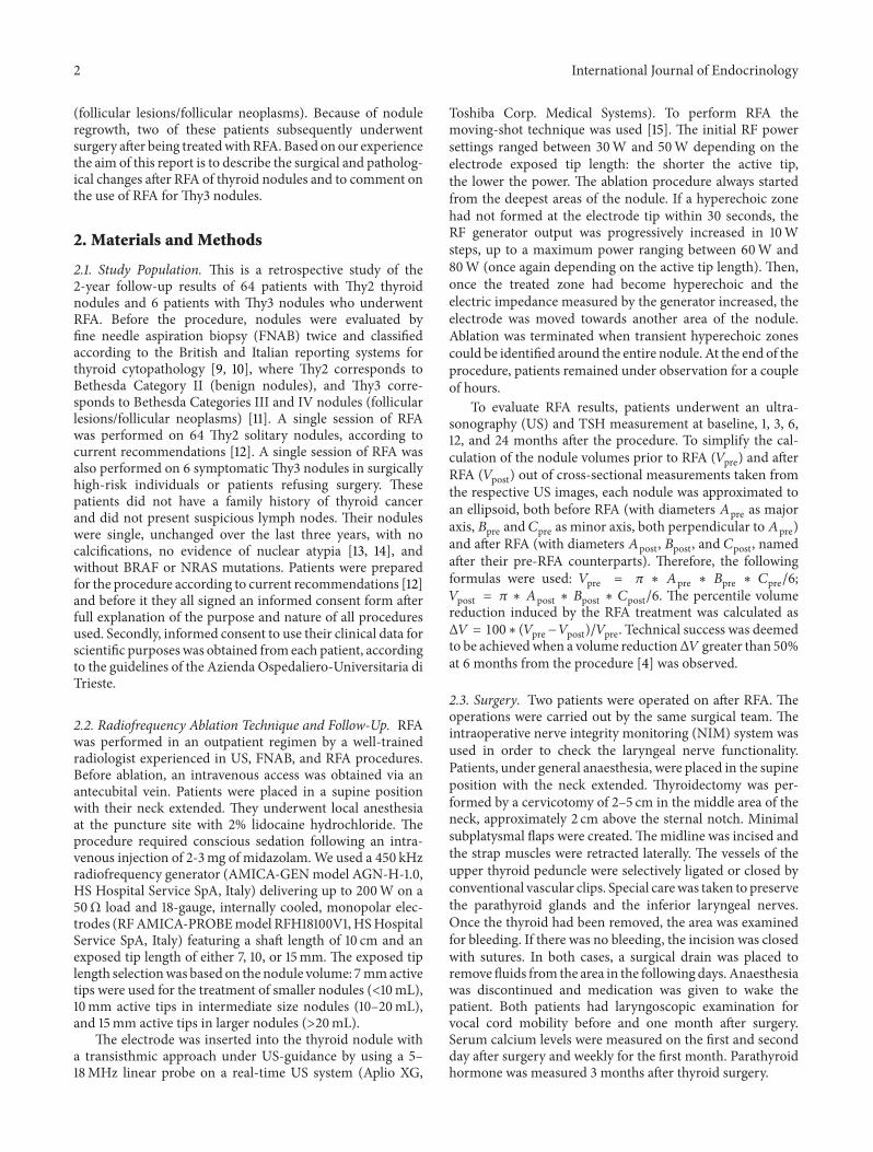
Table 1
Characteristics Thy2 (𝑛 = 64) Thy3 (𝑛 = 6) 𝑝 valueAge (years) 60.47 ± 1.89 61.16 ± 5.62 n.s.Sex (M) 17 3 n.s.Nodule max diameter (mm) 36.04 ± 2.40 38.17 ± 4.12 n.s.Nodule volume (mL) 13.81 ± 1.86 17.46 ± 5.57 n.sSolidity > 50% 55 6 n.sSolidity < 50% 9 0 n.s.BRAF mutations Not tested Absent —NRAS mutations Not tested Absent —TSH (𝜇U/mL) 1.35 ± 0.23 1.41 ± 0.47 n.s.Calcitonin (pg/mL) 1.62 ± 0.14 1.42 ± 0.36 n.s.
Table 2
Characteristics Case 1 Case 2 Case 3 Case 4 Case 5 Case 6Age (years) 71 60 50 53 79 42Sex M F F F M MNodule max diameter (mm) 53 42 41 38 32 23Nodule volume (mL) 40 24 19 11 7 2Type of nodule Solid nodule Solid nodule Solid nodule Solid nodule Solid nodule Solid noduleFNAB Thy3 Thy3 Thy3 Thy3 Thy3 Thy3BRAF mutations Absent Absent Absent Absent Absent AbsentNRAS mutations Absent Absent Absent Absent Absent AbsentTSH (𝜇U/mL) 1.38 1.94 1.22 0.65 0.02 3.35Calcitonin (pg/mL) 1.3 1 1 1 1 3.2Anti-TPO and/or Anti-TG Abs Absent Absent Absent Absent Absent Absent
2.4. Pathology. After surgery, four pathologists examined thethyroid sections stained with hematoxylin and eosin. In onecase HBME-1 staining, which is considered a good marker ofmalignancy [16, 17], was also performed and evaluated.
Although molecular biology analyses had already beenperformed on the FNA of all Thy3 nodules before RFA,they were repeated on the surgically resected nodules. BRAFmutational status (including V600E/Ec, V600D, 600K, andV600R mutations) was evaluated byTherascreen BRAF RSQPCR kit (Qiagen, UK). NRAS mutations were analysed byNRAS Pyrokit for mutations in codons 12, 13, 56, 61, 117, and146 (Qiagen, UK). DNA was extracted from paraffin blocksand tested by using real-time polymerase chain reaction onthe Rotor-Gene Q MDx 5plex HRM instrument (Qiagen,UK) and on the PyroMark Q24 System (Qiagen, UK),respectively.
2.5. Statistical Analysis. Continuous variables were evaluatedby ANOVA.The 𝜒2 test was used to compare sex and nodulesolidity between the two groups. The criterion for statisticalsignificance was 𝑝 < 0.05. All analyses were conducted withSAS (SAS Institute Inc., Cary, NC, USA).
3. Results
3.1. 2-Year Follow-Up Results after RFA. Thecharacteristics ofboth groups of nodules (Thy2 andThy3) aswell as the singular
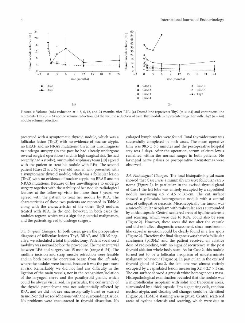
characteristics of the 6 patients with Thy3 are reported inTables 1 and 2. One session of RFA significantly reducedThy2 nodule volume by 50%, 64%, 70%, 71%, and 67% after1, 3, 6, 12, and 24 months from the procedure (Figure 1(a)).Technical success was obtained in allThy2 nodules and it wasmaintained up to 24months. On the other hand, even thoughRFA significantly reducedThy3 nodule volume by onemonth(Figure 1(a)), two of these nodules regrew. In particular, after6 months from the procedure one nodule regrew by 73%as compared to its smallest volume achieved (Figure 1(b);Case 1), while the other, after 24 months from the procedure,regrew by 57% as compared to its smallest volume achieved(Figure 1(b); Case 2). This is the reason why the curverepresenting Thy3 nodule volume reduction (Figure 1(a))deviates markedly from what we observed for Thy2 nodules.Nevertheless, in the remaining four Thy3 nodules treatedwith RFA, the procedure significantly reduced their volumeas much as what was observed in Thy2 nodules (Figure 1(b))and this was maintained throughout all the 2-year follow-up period (Figure 1(b)). Given that nodule growth is a signfor potential malignancy [18] and that this was observed intwo Thy3 nodules, where the rate of malignancy (includingmicrocarcinomas) is estimated to be between 14 and 48% [19],the patients accepted to undergo thyroid surgery.
3.2. Case Presentation of Patients Who Underwent Surgeryafter RFA. The first patient (Case 1) is a 71-year-old man who
4 International Journal of Endocrinology
0
4
8
12
16
20
24
0 3 6 9 12 15 18 21 24
Thyr
oid
nodu
le v
olum
e (m
L)
Time (months)
Thy2Thy3
−3
(a)
06
121824303642485460
0 3 6 9 12 15 18 21 24
Thyr
oid
nodu
le v
olum
e (m
L)
Time (months)
Case 1Case 2Case 3Case 4
Case 5Case 6Thy2
−3
(b)
Figure 1: Volume (mL) reduction at 1, 3, 6, 12, and 24 months after RFA. (a) Dotted line represents Thy2 (𝑛 = 64) and continuous linerepresents Thy3 (𝑛 = 6) nodule volume reduction; (b) the volume reduction of each Thy3 nodule is represented together with Thy2 (𝑛 = 64)nodule volume reduction.
presented with a symptomatic thyroid nodule, which was afollicular lesion (Thy3) with no evidence of nuclear atypia,no BRAF, and no NRAS mutations. Given his unwillingnessto undergo surgery (in the past he had already undergoneseveral surgical operations) and his high surgical risk (he hadrecently had a stroke), our multidisciplinary team [10] agreedwith the patient to treat his nodule with RFA. The secondpatient (Case 2) is a 62-year-old woman who presented witha symptomatic thyroid nodule, which was a follicular lesion(Thy3) with no evidence of nuclear atypia, no BRAF, and noNRAS mutations. Because of her unwillingness to undergosurgery together with the stability of her nodule radiologicalfeatures at the follow-up visits for more than 3 years, weagreed with the patient to treat her nodule by RFA. Thecharacteristics of these two patients are reported in Table 2along with the characteristics of the other Thy3 nodulestreated with RFA. In the end, however, in both cases thenodules regrew, which was a sign for potential malignancy,and the patients agreed to undergo surgery.
3.3. Surgical Changes. In both cases, given the preoperativediagnosis of follicular lesions Thy3, BRAF, and NRAS neg-ative, we scheduled a total thyroidectomy. Patient vocal cordmobility was normal before the procedure.Themean intervalbetween RFA and surgery was 15 months (range 6–24). Themidline incision and strap muscle retraction were feasibleand in both cases the operation began from the left side,where the nodules were located, because it was the part mostat risk. Remarkably, we did not find any difficulty in theligation of the main vessels, nor in the recognition/isolationof the laryngeal nerve and the parathyroid glands, whichcould be always visualized. In particular, the consistency ofthe thyroid parenchyma was not substantially affected byRFA, and we did not see macroscopically burnt or scarredtissue. Nor didwe see adhesionswith the surrounding tissues.No problems were encountered in thyroid dissection. No
enlarged lymph nodes were found. Total thyroidectomy wassuccessfully completed in both cases. The mean operativetime was 98.5 ± 6.5 minutes and the postoperative hospitalstay was 2 days. After the operation, serum calcium levelsremained within the normal ranges in both patients. Nolaryngeal nerve palsies or postoperative haematomas wereobserved.
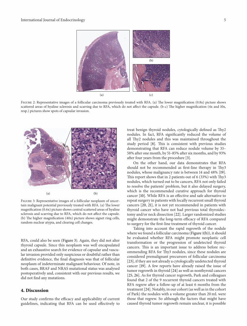
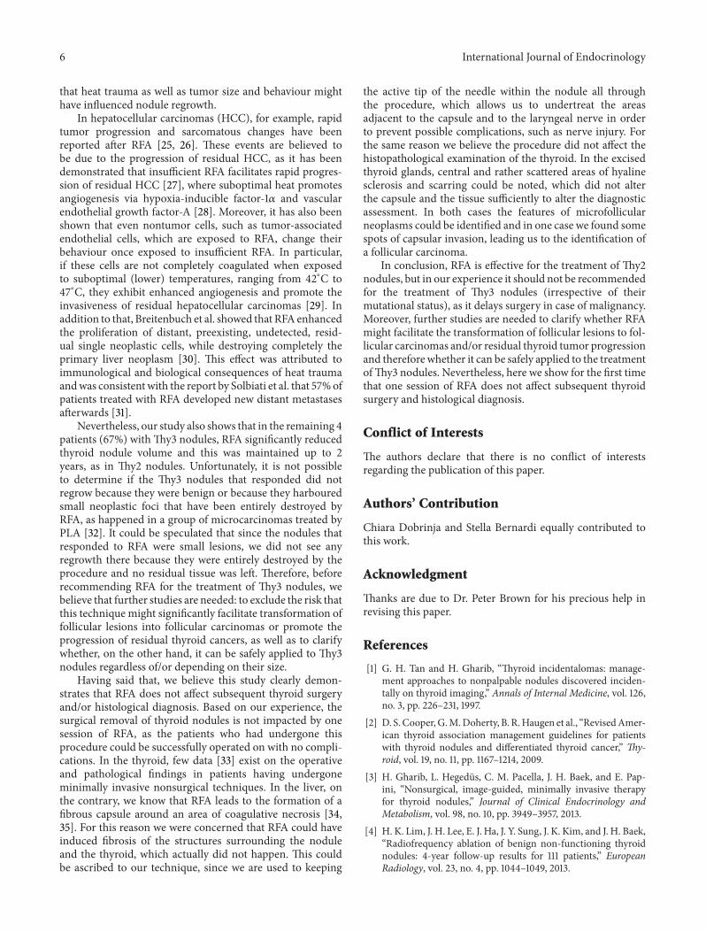
3.4. Pathological Changes. The final histopathological examshowed that Case 1 was a minimally invasive follicular carci-noma (Figure 2). In particular, in the excised thyroid glandof Case 1 the left lobe was entirely occupied by a capsulatednodule measuring 6.5 × 4.5 × 3.5 cm. The cut surfaceshowed a yellowish, heterogeneous nodule with a centralarea of colliquative necrosis. Microscopically the tumor wasa microfollicular neoplasm with trabecular areas surroundedby a thick capsule. Central scattered areas of hyaline sclerosisand scarring, which were due to RFA, could also be seen(Figure 2). However, these areas did not alter the capsuleand did not affect diagnostic assessment, since mushroom-like capsular invasion could be clearly found in a few spots(Figure 2).Therefore the final diagnosiswas that of a follicularcarcinoma (pT3Nx) and the patient received an ablativedose of radioiodine, with no signs of recurrence at the postthyroid ablation whole body scan. As for Case 2, this noduleturned out to be a follicular neoplasm of undeterminatemalignant behaviour (Figure 3). In particular, in the excisedthyroid gland of Case 2, the left lobe was almost entirelyoccupied by a capsulated lesion measuring 3.2 × 2.7 × 3 cm.The cut surface showed a grayish white homogeneous mass.Histopathological examination revealed that the nodule wasa microfollicular neoplasm with solid and trabecular areas,surrounded by a thick capsule. Few signet ring cells, randomnuclear atypia, and clearing cell changes could be identified(Figure 3). HBME-1 staining was negative. Central scatteredareas of hyaline sclerosis and scarring, which were due to
International Journal of Endocrinology 5
(a)
(b)
(c)
Figure 2: Representative images of a follicular carcinoma previously treated with RFA. (a) The lower magnification (0.8x) picture showsscattered areas of hyaline sclerosis and scarring due to RFA, which do not affect the capsule. (b-c) The higher magnification (4x and 10x,resp.) pictures show spots of capsular invasion.
(a) (b)
Figure 3: Representative images of a follicular neoplasm of uncer-tain malignant potential previously treated with RFA. (a) The lowermagnification (0.6x) picture shows central scattered areas of hyalinesclerosis and scarring due to RFA, which do not affect the capsule.(b) The higher magnification (40x) picture shows signet ring cells,random nuclear atypia, and clearing cell changes.
RFA, could also be seen (Figure 3). Again, they did not alterthyroid capsule. Since this neoplasm was well encapsulatedand an exhaustive search for evidence of capsular and vascu-lar invasion provided only suspicious or doubtful rather thandefinitive evidence, the final diagnosis was that of follicularneoplasm of indeterminate malignant behaviour. Of note, inboth cases, BRAF and NRAS mutational status was analysedpostoperatively and, consistent with our previous results, wedid not find any mutations.
4. Discussion
Our study confirms the efficacy and applicability of currentguidelines, indicating that RFA can be used effectively to
treat benign thyroid nodules, cytologically defined as Thy2nodules. In fact, RFA significantly reduced the volume ofall Thy2 nodules and this was maintained throughout thestudy period [8]. This is consistent with previous studiesdemonstrating that RFA can reduce nodule volume by 33–58% after onemonth, by 51–85% after six months, and by 93%after four years from the procedure [3].
On the other hand, our data demonstrates that RFAshould not be recommended as first-line therapy in Thy3nodules, whose malignancy rate is between 14 and 48% [19].This report shows that in 2 patients out of 6 (33%) with Thy3nodules, which turned out to be cancers, RFA not only failedto resolve the patients’ problem, but it also delayed surgery,which is the recommended curative approach for thyroidcancer [10]. While RFA is an effective and safe alternative torepeat surgery in patients with locally recurrent small thyroidcancers [20, 21], it is not yet recommended in patients withthyroid cancer who have not had previous total thyroidec-tomy and/or neck dissection [22]. Larger randomized studiesmight demonstrate the long-term efficacy of RFA comparedto surgery for the first-line treatment of thyroid cancer.
Taking into account the rapid regrowth of the nodulewhere we found a follicular carcinoma (Figure 1(b)), it shouldbe evaluated whether RFA might promote neoplastic celltransformation or the progression of undetected thyroidcancers. This is an important issue to address before rec-ommending RFA for Thy3 nodules, since these nodules areconsidered premalignant precursors of follicular carcinoma[23], if they are not already a cytologically undetected thyroidcancer [19]. A few reports have already raised the issue oftumor regrowth in thyroid [24] as well as nonthyroid cancers[25, 26]. As for thyroid cancer regrowth, Park and colleaguesfound that 2 of the 9 recurrent thyroid cancers treated withRFA regrew after a follow-up of at least 6 months from thetreatment [24]. Notably, in our cohort (as well as in the cohortof Park) the nodules with a volume greater than 20mL werethose that regrew. So although the factors that might havecaused thyroid tumor regrowth remain unclear, it is possible
6 International Journal of Endocrinology
that heat trauma as well as tumor size and behaviour mighthave influenced nodule regrowth.
In hepatocellular carcinomas (HCC), for example, rapidtumor progression and sarcomatous changes have beenreported after RFA [25, 26]. These events are believed tobe due to the progression of residual HCC, as it has beendemonstrated that insufficient RFA facilitates rapid progres-sion of residual HCC [27], where suboptimal heat promotesangiogenesis via hypoxia-inducible factor-1𝛼 and vascularendothelial growth factor-A [28]. Moreover, it has also beenshown that even nontumor cells, such as tumor-associatedendothelial cells, which are exposed to RFA, change theirbehaviour once exposed to insufficient RFA. In particular,if these cells are not completely coagulated when exposedto suboptimal (lower) temperatures, ranging from 42∘C to47∘C, they exhibit enhanced angiogenesis and promote theinvasiveness of residual hepatocellular carcinomas [29]. Inaddition to that, Breitenbuch et al. showed that RFA enhancedthe proliferation of distant, preexisting, undetected, resid-ual single neoplastic cells, while destroying completely theprimary liver neoplasm [30]. This effect was attributed toimmunological and biological consequences of heat traumaandwas consistentwith the report by Solbiati et al. that 57%ofpatients treated with RFA developed new distant metastasesafterwards [31].
Nevertheless, our study also shows that in the remaining 4patients (67%) withThy3 nodules, RFA significantly reducedthyroid nodule volume and this was maintained up to 2years, as in Thy2 nodules. Unfortunately, it is not possibleto determine if the Thy3 nodules that responded did notregrow because they were benign or because they harbouredsmall neoplastic foci that have been entirely destroyed byRFA, as happened in a group of microcarcinomas treated byPLA [32]. It could be speculated that since the nodules thatresponded to RFA were small lesions, we did not see anyregrowth there because they were entirely destroyed by theprocedure and no residual tissue was left. Therefore, beforerecommending RFA for the treatment of Thy3 nodules, webelieve that further studies are needed: to exclude the risk thatthis technique might significantly facilitate transformation offollicular lesions into follicular carcinomas or promote theprogression of residual thyroid cancers, as well as to clarifywhether, on the other hand, it can be safely applied to Thy3nodules regardless of/or depending on their size.
Having said that, we believe this study clearly demon-strates that RFA does not affect subsequent thyroid surgeryand/or histological diagnosis. Based on our experience, thesurgical removal of thyroid nodules is not impacted by onesession of RFA, as the patients who had undergone thisprocedure could be successfully operated on with no compli-cations. In the thyroid, few data [33] exist on the operativeand pathological findings in patients having undergoneminimally invasive nonsurgical techniques. In the liver, onthe contrary, we know that RFA leads to the formation of afibrous capsule around an area of coagulative necrosis [34,35]. For this reason we were concerned that RFA could haveinduced fibrosis of the structures surrounding the noduleand the thyroid, which actually did not happen. This couldbe ascribed to our technique, since we are used to keeping
the active tip of the needle within the nodule all throughthe procedure, which allows us to undertreat the areasadjacent to the capsule and to the laryngeal nerve in orderto prevent possible complications, such as nerve injury. Forthe same reason we believe the procedure did not affect thehistopathological examination of the thyroid. In the excisedthyroid glands, central and rather scattered areas of hyalinesclerosis and scarring could be noted, which did not alterthe capsule and the tissue sufficiently to alter the diagnosticassessment. In both cases the features of microfollicularneoplasms could be identified and in one case we found somespots of capsular invasion, leading us to the identification ofa follicular carcinoma.
In conclusion, RFA is effective for the treatment of Thy2nodules, but in our experience it should not be recommendedfor the treatment of Thy3 nodules (irrespective of theirmutational status), as it delays surgery in case of malignancy.Moreover, further studies are needed to clarify whether RFAmight facilitate the transformation of follicular lesions to fol-licular carcinomas and/or residual thyroid tumor progressionand thereforewhether it can be safely applied to the treatmentofThy3 nodules. Nevertheless, here we show for the first timethat one session of RFA does not affect subsequent thyroidsurgery and histological diagnosis.
Conflict of Interests
The authors declare that there is no conflict of interestsregarding the publication of this paper.
Authors’ Contribution
Chiara Dobrinja and Stella Bernardi equally contributed tothis work.
Acknowledgment
Thanks are due to Dr. Peter Brown for his precious help inrevising this paper.
References
[1] G. H. Tan and H. Gharib, “Thyroid incidentalomas: manage-ment approaches to nonpalpable nodules discovered inciden-tally on thyroid imaging,” Annals of Internal Medicine, vol. 126,no. 3, pp. 226–231, 1997.
[2] D. S. Cooper,G.M.Doherty, B. R.Haugen et al., “RevisedAmer-ican thyroid association management guidelines for patientswith thyroid nodules and differentiated thyroid cancer,” Thy-roid, vol. 19, no. 11, pp. 1167–1214, 2009.
[3] H. Gharib, L. Hegedus, C. M. Pacella, J. H. Baek, and E. Pap-ini, “Nonsurgical, image-guided, minimally invasive therapyfor thyroid nodules,” Journal of Clinical Endocrinology andMetabolism, vol. 98, no. 10, pp. 3949–3957, 2013.
[4] H. K. Lim, J. H. Lee, E. J. Ha, J. Y. Sung, J. K. Kim, and J. H. Baek,“Radiofrequency ablation of benign non-functioning thyroidnodules: 4-year follow-up results for 111 patients,” EuropeanRadiology, vol. 23, no. 4, pp. 1044–1049, 2013.
International Journal of Endocrinology 7
[5] J.H. Baek, J.H. Lee, J. Y. Sung et al., “Complications encounteredin the treatment of benign thyroid nodules with us-guidedradiofrequency ablation: a multicenter study,” Radiology, vol.262, no. 1, pp. 335–342, 2012.
[6] J. H. Lee, Y. S. Kim, D. Lee, H. Choi, H. Yoo, and J. H. Baek,“Radiofrequency ablation (RFA) of benign thyroid nodulesin patients with incompletely resolved clinical problems afterethanol ablation (EA),” World Journal of Surgery, vol. 34, no. 7,pp. 1488–1493, 2010.
[7] A. Faggiano, V. Ramundo, A. P. Assanti et al., “Thyroid nodulestreated with percutaneous radiofrequency thermal ablation:a comparative study,” Journal of Clinical Endocrinology andMetabolism, vol. 97, no. 12, pp. 4439–4445, 2012.
[8] S. Bernardi, C. Dobrinja, B. Fabris et al., “Radiofrequencyablation compared to surgery for the treatment of benignthyroid nodules,” International Journal of Endocrinology, vol.2014, Article ID 934595, 10 pages, 2014.
[9] S. Crippa and L. Mazzucchelli, “The Bethesda system forreporting thyroid fine-needle aspiration specimens,” AmericanJournal of Clinical Pathology, vol. 134, no. 2, pp. 343–344, 2010.
[10] P. Perros, K. Boelaert, S. Colley et al., “Guidelines for themanagement of thyroid cancer,” Clinical Endocrinology, vol. 81,no. s1, pp. 1–122, 2014.
[11] E. S. Cibas and S. Z. Ali, “The Bethesda system for reportingthyroid cytopathology,” American Journal of Clinical Pathology,vol. 132, no. 5, pp. 658–665, 2009.
[12] D. G. Na, J. H. Lee, S. L. Jung et al., “Radiofrequency ablationof benign thyroid nodules and recurrent thyroid cancers:consensus statement and recommendations,” Korean Journal ofRadiology, vol. 13, no. 2, pp. 117–125, 2012.
[13] C. A. Macias, D. Arumugam, R. L. Arlow et al., “A risk model todetermine surgical treatment in patients with thyroid noduleswith indeterminate cytology,” Annals of Surgical Oncology, vol.22, no. 5, pp. 1527–1532, 2015.
[14] C. Dobrinja, G. Trevisan, L. Piscopello, M. Fava, and G. Liguori,“Comparison between thyroidectomy and hemithyroidectomyin treatment of single thyroid nodules identified as indetermi-nate follicular lesions by fine-needle aspiration cytology,”AnnaliItaliani di Chirurgia, vol. 81, no. 6, pp. 403–410, 2010.
[15] J. H. Baek, J. H. Lee, R. Valcavi, C. M. Pacella, H. Rhim,and D. G. Na, “Thermal ablation for benign thyroid nodules:radiofrequency and laser,” Korean Journal of Radiology, vol. 12,no. 5, pp. 525–540, 2011.
[16] B. Cochand-Priollet, H. Dahan, M. Laloi-Michelin et al.,“Immunocytochemistry with cytokeratin 19 and anti-HumanMesothelial Cell Antibody (HBME1) increases the diagnosticaccuracy of thyroid fine-needle aspirations: preliminary reportof 150 liquid-based fine-needle aspirations with histologicalcontrol,”Thyroid, vol. 21, no. 10, pp. 1067–1073, 2011.
[17] G. Fadda, E. D. Rossi, M. Raffaelli et al., “Follicular thyroidneoplasms can be classified as low- and high-risk according toHBME-1 andGalectin-3 expression on liquid-based fine-needlecytology,” European Journal of Endocrinology, vol. 165, no. 3, pp.447–453, 2011.
[18] K. Kuma, F. Matsuzuka, A. Kobayashi et al., “Outcome of longstanding solitary thyroid nodules,”World Journal of Surgery, vol.16, no. 4, pp. 583–588, 1992.
[19] C.-C. C. Wang, L. Friedman, G. C. Kennedy et al., “A largemulticenter correlation study of thyroid nodule cytopathologyand histopathology,”Thyroid, vol. 21, no. 3, pp. 243–251, 2011.
[20] J. Kim, W. S. Yoo, Y. J. Park et al., “Efficacy and safetyof radiofrequency ablation for treatment of locally recurrentthyroid cancers smaller than 2 cm,” Radiology, 2015.
[21] S. J. Lee, S. L. Jung, B. S. Kim et al., “Radiofrequency ablationto treat loco-regional recurrence of well-differentiated thyroidcarcinoma,” Korean Journal of Radiology, vol. 15, no. 6, pp. 817–826, 2014.
[22] J. M. Monchik, G. Donatini, J. Iannuccilli, and D. E. Dupuy,“Radiofrequency ablation and percutaneous ethanol injectiontreatment for recurrent local and distant well-differentiatedthyroid carcinoma,” Annals of Surgery, vol. 244, no. 2, pp. 296–304, 2006.
[23] T. C. Brown, C. C. Juhlin, J. M. Healy, M. L. Prasad, R. Korah,and T. Carling, “Frequent silencing of RASSF1A via promotermethylation in follicular thyroid hyperplasia: a potential earlyepigenetic susceptibility event in thyroid carcinogenesis,” JAMASurgery, vol. 149, no. 11, pp. 1146–1152, 2014.
[24] K. W. Park, J. H. Shin, B.-K. Han, E. Y. Ko, and J. H. Chung,“Inoperable symptomatic recurrent thyroid cancers: prelim-inary result of radiofrequency ablation,” Annals of SurgicalOncology, vol. 18, no. 9, pp. 2564–2568, 2011.
[25] M. Koda, Y. Murawaki, Y. Hirooka et al., “Complicationsof radiofrequency ablation for hepatocellular carcinoma in amulticenter study: an analysis of 16 346 treated nodules in 13283 patients,”Hepatology Research, vol. 42, no. 11, pp. 1058–1064,2012.
[26] A. Ruzzenente, G. D. Manzoni, M. Molfetta et al., “Rapidprogression of hepatocellular carcinoma after radiofrequencyablation,” World Journal of Gastroenterology, vol. 10, no. 8, pp.1137–1140, 2004.
[27] S. Ke, X.-M. Ding, J. Kong et al., “Low temperature ofradiofrequency ablation at the target sites can facilitate rapidprogression of residual hepatic VX2 carcinoma,” Journal ofTranslational Medicine, vol. 8, article 73, 2010.
[28] J. Kong, J. Kong, B. Pan et al., “Insufficient radiofrequencyablation promotes angiogenesis of residual hepatocellular car-cinoma via HIF-1𝛼/VEGFA,” PLoS ONE, vol. 7, no. 5, Article IDe37266, 2012.
[29] J. Kong, L. Kong, J. Kong et al., “After insufficient radiofre-quency ablation, tumor-associated endothelial cells exhibitenhanced angiogenesis and promote invasiveness of residualhepatocellular carcinoma,” Journal of Translational Medicine,vol. 10, article 230, 2012.
[30] P. Von Breitenbuch, G. Kohl, M. Guba, E. Geissler, K. W. Jauch,and M. Steinbauer, “Thermoablation of colorectal liver metas-tases promotes proliferation of residual intrahepatic neoplasticcells,” Surgery, vol. 138, no. 5, pp. 882–887, 2005.
[31] L. Solbiati, T. Livraghi, S. N. Goldberg et al., “Percutaneousradio-frequency ablation of hepatic metastases from colorectalcancer: long-term results in 117 patients,”Radiology, vol. 221, no.1, pp. 159–166, 2001.
[32] R. Valcavi, S. Piana, G. S. Bortolan, R. Lai, V. Barbieri, andR. Negro, “Ultrasound-guided percutaneous laser ablation ofpapillary thyroid microcarcinoma: a feasibility study on threecases with pathological and immunohistochemical evaluation,”Thyroid, vol. 23, no. 12, pp. 1578–1582, 2013.
[33] F. Monzani, N. Caraccio, F. Basolo, P. Iacconi, V. LiVolsi, and P.Miccoli, “Surgical and pathological changes after percutaneousethanol injection therapy of thyroid nodules,” Thyroid, vol. 10,no. 12, pp. 1087–1092, 2000.
8 International Journal of Endocrinology
[34] M. C. Ries, A. J. Milligan, H.W.Merrick, and R. R. DobelbowerJr., “Biochemical and cellular effects of radiofrequency intucedinterstitial hyperthermia on normal canine liver,” InternationalJournal of Radiation Oncology, Biology, Physics, vol. 14, no. 3, pp.529–536, 1988.
[35] E. Blanco, F. Qian, B. Weinberg, N. Stowe, J. M. Anderson, andJ. Gao, “Effect of fibrous capsule formation on doxorubicin dis-tribution in radiofrequency ablated rats,” Journal of BiomedicalMaterials Research Part A, vol. 69, no. 3, pp. 398–406, 2004.

Submit your manuscripts athttp://www.hindawi.com
Stem CellsInternational
Hindawi Publishing Corporationhttp://www.hindawi.com Volume 2014
Hindawi Publishing Corporationhttp://www.hindawi.com Volume 2014
MEDIATORSINFLAMMATION
of
Hindawi Publishing Corporationhttp://www.hindawi.com Volume 2014
Behavioural Neurology
EndocrinologyInternational Journal of
Hindawi Publishing Corporationhttp://www.hindawi.com Volume 2014
Hindawi Publishing Corporationhttp://www.hindawi.com Volume 2014
Disease Markers
Hindawi Publishing Corporationhttp://www.hindawi.com Volume 2014
BioMed Research International
OncologyJournal of
Hindawi Publishing Corporationhttp://www.hindawi.com Volume 2014
Hindawi Publishing Corporationhttp://www.hindawi.com Volume 2014
Oxidative Medicine and Cellular Longevity
Hindawi Publishing Corporationhttp://www.hindawi.com Volume 2014
PPAR Research
The Scientific World JournalHindawi Publishing Corporation http://www.hindawi.com Volume 2014
Immunology ResearchHindawi Publishing Corporationhttp://www.hindawi.com Volume 2014
Journal of
ObesityJournal of
Hindawi Publishing Corporationhttp://www.hindawi.com Volume 2014
Hindawi Publishing Corporationhttp://www.hindawi.com Volume 2014
Computational and Mathematical Methods in Medicine
OphthalmologyJournal of
Hindawi Publishing Corporationhttp://www.hindawi.com Volume 2014
Diabetes ResearchJournal of
Hindawi Publishing Corporationhttp://www.hindawi.com Volume 2014
Hindawi Publishing Corporationhttp://www.hindawi.com Volume 2014
Research and TreatmentAIDS
Hindawi Publishing Corporationhttp://www.hindawi.com Volume 2014
Gastroenterology Research and Practice
Hindawi Publishing Corporationhttp://www.hindawi.com Volume 2014
Parkinson’s Disease
Evidence-Based Complementary and Alternative Medicine
Volume 2014Hindawi Publishing Corporationhttp://www.hindawi.com