Description of the sexual morph of Seimatosporium vitis
9
Cryptogamie, Mycologie, 2017, 38 (1): 3-11 © 2017 Adac. Tous droits réservés doi/10.7872/crym/v38.iss1.2017.3 Description of the sexual morph of Seimatosporium vitis Mehdi MEHRABI a* , Roghayeh HEMMATI a & Jafar ABDOLLAHZADEH b a Department of Plant Protection, Faculty of Agriculture, University of Zanjan, Zanjan, Iran b Department of Plant Protection, Faculty of Agriculture, University of Kurdistan, P.O. Box 416, Sanandaj, Iran Abstract – Seimatosporium vitis was recently described based on the collection of its coelomycetous asexual morph on Vitis vinifera in Italy. In this study Seimatosporium vitis is introduced for the first time from grapevine in Iran. The sexual morph is illustrated and a full description is provided. The connection between two different morphs was proved in culture and based on ITS sequence data. Iran / Pestalotioid fungi / Phylogeny / Vitis sp. INTRODUCTION Seimatosporium Corda is a member of family Discosiaceae in Amphisphaeriales (Senanayake et al., 2015) and typified by S. rosae (Corda, 1833). The genus is defined primarily based on conidial characteristics including size, septation, pigmentation, and presence or absence of appendages (Sutton, 1980; Nag Rag, 1993). Its members are considered so-called “pestalotioid fungi”, and their life styles range from pathogens to saprobes (Tanaka et al., 2011). This genus currently contains numerous species with more than 86 names recorded to date (Index Fungorum; http://www.indexfungorum.org/Names/Names.asp) and detailed descriptions of Seimatosporium spp. have been presented by several authors (Shoemaker, 1964; Pirozynski & Shoemaker, 1970; Sutton, 1980; Nag Raj, 1993; Hatakeyama & Harada, 2004; Tanaka et al., 2011; Barber et al., 2011; Norphanphoun et al., 2015; Senanayake et al., 2015; Goonasekara et al., 2016; Perera et al., 2016). Tanaka et al. (2011) in a phylogenetic study based on LSU and ITS sequence data confirmed Discostroma Clem. (1909) as sexual morph of Seimatosporium species, while other related genera, including Sporocadus, Sarcostroma, Diploceras and Vermisporium, which lack sexual morphs, clustered in Seimatosporium sensu stricto, suggesting that they should be placed under the genus Seimatosporium. (Tanaka et al., 2011; Barber et al., 2011). Réblová et al., (2016) proposed to use Seimatosporium over Discostroma, because the former is the oldest name, has the greater number of species, and is more commonly used. * Corresponding author: [email protected]
Transcript of Description of the sexual morph of Seimatosporium vitis
Cryptogamie, Mycologie, 2017, 38 (1): 3-11© 2017 Adac. Tous droits réservés
doi/10.7872/crym/v38.iss1.2017.3
Description of the sexual morphof Seimatosporium vitis
Mehdi MEHRABI a*, Roghayeh HEMMATI a & Jafar ABDOLLAHZADEH b
aDepartment of Plant Protection, Faculty of Agriculture,University of Zanjan, Zanjan, Iran
bDepartment of Plant Protection, Faculty of Agriculture,University of Kurdistan, P.O. Box 416, Sanandaj, Iran
Abstract – Seimatosporium vitis was recently described based on the collection of itscoelomycetous asexual morph on Vitis vinifera in Italy. In this study Seimatosporium vitis isintroduced for the first time from grapevine in Iran. The sexual morph is illustrated and a fulldescription is provided. The connection between two different morphs was proved in cultureand based on ITS sequence data.
Iran / Pestalotioid fungi / Phylogeny / Vitis sp.
INTRODUCTION
Seimatosporium Corda is a member of family Discosiaceae inAmphisphaeriales (Senanayake et al., 2015) and typified by S. rosae (Corda, 1833).The genus is defined primarily based on conidial characteristics including size,septation, pigmentation, and presence or absence of appendages (Sutton, 1980; NagRag, 1993). Its members are considered so-called “pestalotioid fungi”, and theirlife styles range from pathogens to saprobes (Tanaka et al., 2011). This genuscurrently contains numerous species with more than 86 names recorded to date(Index Fungorum; http://www.indexfungorum.org/Names/Names.asp) and detaileddescriptions of Seimatosporium spp. have been presented by several authors(Shoemaker, 1964; Pirozynski & Shoemaker, 1970; Sutton, 1980; Nag Raj, 1993;Hatakeyama & Harada, 2004; Tanaka et al., 2011; Barber et al., 2011; Norphanphounet al., 2015; Senanayake et al., 2015; Goonasekara et al., 2016; Perera et al., 2016).
Tanaka et al. (2011) in a phylogenetic study based on LSU and ITSsequence data confirmed Discostroma Clem. (1909) as sexual morph ofSeimatosporium species, while other related genera, including Sporocadus,Sarcostroma, Diploceras and Vermisporium, which lack sexual morphs, clustered inSeimatosporium sensu stricto, suggesting that they should be placed under the genusSeimatosporium. (Tanaka et al., 2011; Barber et al., 2011). Réblová et al., (2016)proposed to use Seimatosporium over Discostroma, because the former is the oldestname, has the greater number of species, and is more commonly used.
* Corresponding author: [email protected]
4 M. Mehrabi et al.
There are very few publications on pestalotioid fungi from Iran. Arzanlouet al. (2012) have provided a check list for the known pestalotioid fungi from Iran,listing 3 Seimatosporium species, viz. S. fusisporum H.J. Swart & D.A. Griffiths(Aminaee & Ershad, 2008), S. lonicerae (Cooke) Shoemaker (Gräfenhan, 2006) andS. lichenicola (Corda) Shoemaker & Müll (Aghapour et al., 2010). More recently,Crous et al. (2014) described S. pistaciae from Iran. As mentioned by Norphanphounet al. (2015) this species is similar to S. rosea based on phylogenetic analysis ofLSU and ITS sequence data, but morphologically it appears to be distinct fromS. rosea by having larger conidia, but sequence data from protein-coding genes mayshed more light on the possible synonymy of these two species. More recentlyAyoubi & Soleimani (2016) characterized a new species Neopestalotiopsis iranensisand N. mesopotamica on strawberry fruits from Kurdistan Province, Iran.
Through our work on the taxonomy of Diatrypaceae on trees native to theArasbaran forest in Iran, Seimatosporium vitis was found as a new record for Iran.In this paper it is characterized morphologically and phylogenetically and a detaileddescription and illustration for both sexual and asexual morphs is provided.
MATERIALS AND METHODS
Morphological characterization. Samples were collected from Arasbaranforest in the East Azerbaijan on dead branch of Vitis sp. Isolation were made fromsingle ascospores (IRAN2454C and IRAN2455C) and conidia (IRAN2427C). Formicroscopic examinations sections were cut freehand under an Olympus SZH stereomicroscope. A Nikon Eclipse 80i light microscope with a Canon digital camera wasused to capture micromorphological images. The measurements for each structureare in parentheses. Colony color were determined on potato dextrose agar (PDA)and malt extract agar (MEA) at 24°C using a color chart (Rayner, 1970). Dryspecimens and fresh cultures were deposited in the herbarium and culture collectionof the Iranian Research Institute of Plant Protection (IRAN, Tehran, Iran).
DNA extraction and sequencing. Isolates were grown in malt extractbroth (1.5 % MEB) at room temperature for 14 d. Genomic DNAwas extracted withan initial step of grinding the mycelia in liquid nitrogen as described by Liu et al.(2000). Polymerase chain reactions (PCR) were carried out using primer pairs ofITS1/ITS4 (White et al., 1990) to amplify the internal transcribed spacers (ITS) andLROR/LR7 (Rehner & Samuels, 1994; Vilgalys & Hester, 1990) to amplify part oflarge subunit rDNA (LSU). The PCR reaction mixtures 25 µL contained 2.5 μL 10xhigh yield PCR buffer (Jena Bioscience, Germany), 1 μL MgCl2 (25 mM), 0.5 μLdNTPs (10 mM), 1 µL of each primer (10 pmol/µL, Takapouzist Inc.), 1.5 unit Taqpolymerase (Jena Bioscience, Germany) and 1 µL genomic DNA (~30 ng/µL). PCRreactions were run on a PC-320 PCR System (ASTEC Co., Japan) machine with aninitial denaturation step of 5 min at 95°C, followed by 35 cycles of 45 s at 94°C,35 s at 58°C/57°C (ITS/LSU), 90 s at 72°C, and a final extension of 10 min at 72°C.PCR products were visualized in 1% agarose gel in 1xTBE buffer. Purification andsequencing of PCR products were performed by Macrogen (South Korea).
Phylogenetic analysis. The new sequences obtained in the present studywere read and edited with FinchTV v. 1.4.0 (Geospiza Inc.). The sequences werecompared with those in the GenBank database using the Megablast algorithm.Additional sequences were selected fromNorphanphoun et al. (2015) and Senanayake
Sexual morph of Seimatosporium vitis 5
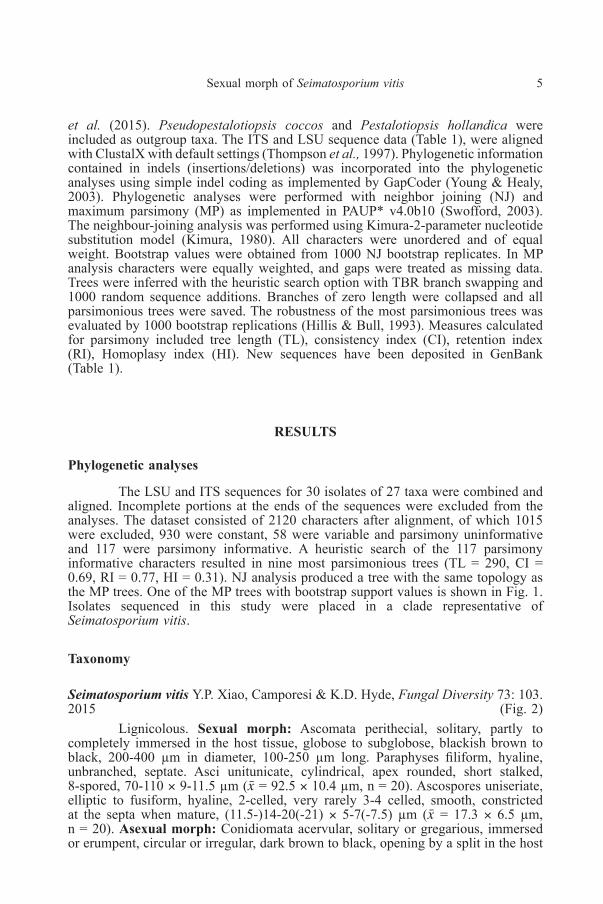
et al. (2015). Pseudopestalotiopsis coccos and Pestalotiopsis hollandica wereincluded as outgroup taxa. The ITS and LSU sequence data (Table 1), were alignedwith ClustalX with default settings (Thompson et al., 1997). Phylogenetic informationcontained in indels (insertions/deletions) was incorporated into the phylogeneticanalyses using simple indel coding as implemented by GapCoder (Young & Healy,2003). Phylogenetic analyses were performed with neighbor joining (NJ) andmaximum parsimony (MP) as implemented in PAUP* v4.0b10 (Swofford, 2003).The neighbour-joining analysis was performed using Kimura-2-parameter nucleotidesubstitution model (Kimura, 1980). All characters were unordered and of equalweight. Bootstrap values were obtained from 1000 NJ bootstrap replicates. In MPanalysis characters were equally weighted, and gaps were treated as missing data.Trees were inferred with the heuristic search option with TBR branch swapping and1000 random sequence additions. Branches of zero length were collapsed and allparsimonious trees were saved. The robustness of the most parsimonious trees wasevaluated by 1000 bootstrap replications (Hillis & Bull, 1993). Measures calculatedfor parsimony included tree length (TL), consistency index (CI), retention index(RI), Homoplasy index (HI). New sequences have been deposited in GenBank(Table 1).
RESULTS
Phylogenetic analyses
The LSU and ITS sequences for 30 isolates of 27 taxa were combined andaligned. Incomplete portions at the ends of the sequences were excluded from theanalyses. The dataset consisted of 2120 characters after alignment, of which 1015were excluded, 930 were constant, 58 were variable and parsimony uninformativeand 117 were parsimony informative. A heuristic search of the 117 parsimonyinformative characters resulted in nine most parsimonious trees (TL = 290, CI =0.69, RI = 0.77, HI = 0.31). NJ analysis produced a tree with the same topology asthe MP trees. One of the MP trees with bootstrap support values is shown in Fig. 1.Isolates sequenced in this study were placed in a clade representative ofSeimatosporium vitis.
Taxonomy
Seimatosporium vitis Y.P. Xiao, Camporesi & K.D. Hyde, Fungal Diversity 73: 103.2015 (Fig. 2)
Lignicolous. Sexual morph: Ascomata perithecial, solitary, partly tocompletely immersed in the host tissue, globose to subglobose, blackish brown toblack, 200-400 µm in diameter, 100-250 µm long. Paraphyses filiform, hyaline,unbranched, septate. Asci unitunicate, cylindrical, apex rounded, short stalked,8-spored, 70-110 × 9-11.5 µm (› = 92.5 × 10.4 µm, n = 20). Ascospores uniseriate,elliptic to fusiform, hyaline, 2-celled, very rarely 3-4 celled, smooth, constrictedat the septa when mature, (11.5-)14-20(-21) × 5-7(-7.5) µm (› = 17.3 × 6.5 µm,n = 20). Asexual morph: Conidiomata acervular, solitary or gregarious, immersedor erumpent, circular or irregular, dark brown to black, opening by a split in the host

6 M. Mehrabi et al.Table1.Isolatesusedforthephylogeneticanalyses
Spec
ies
Hos
t.Lo
catio
nC
ultu
reno
.G
enBa
nk1,
2
LSU
ITS
Dis
cost
rom
afu
scel
lum
Rosa
cani
naJa
pan
NBRC32625
AB593726
AB594794
D.s
tone
ae–
Japa
nNBRC32690
AB593729
AB594797
D.t
ostu
m–
Japa
nNBRC32626
AB593727
AB594795
Pest
alot
iops
isho
lland
ica
Scia
dopi
tys
vert
icill
ata
Netherlands
CBS265.33
KM
1162
28KM199328
Pseu
dope
stal
otio
psis
coco
sC
ocos
nuci
fera
Indo
nesi
a:Ja
vaCBS272.29
KM
1162
76KM199378
Seim
atos
pori
umbi
sept
atum
Euca
lypt
usor
esbi
aA
ustra
liaCPC13584
JN871208
JN871199
S.bo
tan
Paeo
nia
suffr
utic
osa
Japa
nH4619
AB593731
AB594799
S.co
rnii
Cor
nus
sp.
Italy
MFLUCC14-0467
KR559739
KT162918
S.di
scos
ioid
esPu
nica
gran
atum
Japa
nH4621
AB593732
AB594800
S.el
egan
sM
elal
euca
eric
ifolia
Japa
nNBRC32674
AB593733
AB594801
S.eu
caly
pti
Euca
lypt
ussm
ithii
Sout
hA
fric
aCBS115131
JN871209
JN871200
S.fa
lcat
umEu
caly
ptus
allig
atri
xA
ustra
liaCPC13580
JN871214
JN871205
S.ficeae
Ficu
ssp
.C
hina
MFLUCC15-0519
KR920686
KR092800
S.fo
liico
laJu
nipe
rus
phoe
nice
aJa
pan
NBRC32676
AB593734
AB594802
S.gl
andi
genu
mFa
gus
sylv
atic
aJa
pan
NBRC32677
AB593735
AB594803
S.gr
evill
eae
Prot
easp
.So
uth
Afr
ica
ICMP10981
AF3
8237
2AF405304
S.ha
keae
Pter
idiu
maq
uilin
umJa
pan
NBRC32678
AB593736
AB594804
S.hy
peri
cinu
mH
yper
icum
sp.
Japa
nNBRC32647
AB593737
AB594805
S.m
aria
eCorreareflexa
Japa
nNBRC32681
AB593740
AB594807
S.ob
tusu
mC
orym
bia
henr
yiA
ustra
liaCPC12935
JN871215
JN871206
S.pa
rasi
ticum
Phys
ocar
pus
amur
ensi
sJa
pan
NBRC32682
AB593741
AB594808
S.ph
ysoc
arpi
Phys
ocar
pus
opul
ifoliu
sR
ussi
aMFLUCC14-0625
KT198723
KT198722
S.pi
stac
iae
Pist
acia
vera
Iran
CBS138865
KP004491
KP004463
S.rh
ombi
spor
umVa
ccin
ium
myr
tillu
sIta
lyMFLUCC15-0543
KR092780
KR092792
S.ro
sae
Rosa
kalm
iuss
ica
Rus
sia
MFLUCC14-0621
KT198727
KT198726
S.vi
tisVi
tissp
.Ir
anIRAN2427C
KU
1629
42K
U16
2941
S.vi
tisVi
tissp
.Ir
anIRAN2454C
n.s.
KU
6484
01S.
vitis
Vitis
sp.
Iran
IRAN2455C
n.s.
KU
6484
02S.
vitis
Vitis
vini
fera
Italy
MFLUCC14-0051
KR920362
KR920363
S.w
alke
riEu
caly
ptus
sp.
Aus
tralia
CPC17644
JN871216
JN871207
1Sequencesinboldfaceweregeneratedduringthepresentstudy.
2n.s.:Notsequenced.

Sexual morph of Seimatosporium vitis 7
Fig. 1. One of the nine most parsimonious trees for Seimatosporium species based on a combined datasetof LSU and ITS sequence data. MP/NJ bootstrap support values from 1000 replicates higher than 50%are given at the nodes. Isolates from Iran are in boldface.
8 M. Mehrabi et al.
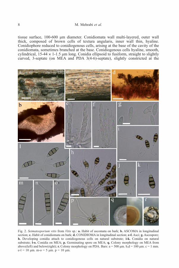
Fig. 2. Seimatosporium vitis from Vitis sp.: a. Habit of ascomata on bark; b. ASCOMA in longitudinalsection; c. Habit of conidiomata on bark; d. CONIDIOMA in longitudinal section; e-f.Asci; g.Ascospore;h. Developing conidia attach to conidiogenous cells on natural substrate; i-k. Conidia on naturalsubstrate; l-o. Conidia on MEA; p. Germinating spore on MEA; q. Colony morphology on MEA fromabove(left) and below(right); r. Colony morphology on PDA. Bars: a = 500 μm. b,d = 100 μm. c = 1 mm.e-l = 10 μm. m-o = 5 μm. p = 10 μm.
tissue surface, 100-600 μm diameter. Conidiomata wall multi-layered, outer wallthick, composed of brown cells of textura angularis, inner wall thin, hyaline.Conidiophore reduced to conidiogenous cells, arising at the base of the cavity of theconidiomata, sometimes branched at the base. Conidiogenous cells hyaline, smooth,cylindrical, 15-44 × 1-1.5 μm long. Conidia ellipsoid to fusiform, straight to slightlycurved, 3-septate (on MEA and PDA 3(4-6)-septate), slightly constricted at the
Sexual morph of Seimatosporium vitis 9
septa, (11-)13-16(-17) × 5-6 (› = 14.7 × 5.6, n=30) μm on natural substrate (onMEA; 15-25×4.5-5.5 μm, › = 18.4×4.8 μm, n=20), basal cell obconic with a truncatebase, with an appendage, hyaline to subhyaline, 2.5-3.7 (› = 3.1) μm long; 2 mediancells subcylindrical to doliiform, slightly thick-walled, smooth, brown to dark brown,with septa darker than the rest of the cell, together 7.3-9.8 (› = 8.6) μm long, (thesecond cell from the base 3.3-5(› = 3.7) μm long, the third cell 3.4-5.8 (› = 4.2) μmlong); the apical cell conical with a rounded or acute apex, hyaline to subhyaline orconcolorous with the central cells, 3-4 (› = 3.6) μm long; with or without 1 tubularapical filiform appendage ( on MEA and PDA always present), sometimes branched,arising from the apex of the apical cell, 10-22 (› = 16) μm long, basal appendagepresent, filiform, tubular, single, sometime branched, eccentric, 7-30 (› =19.7) μmlong.
Cultural characteristics: On MEA circular with regular margin, white toprimrose (23’’b), cottony, reverse sienna (13i), reaching 20 cm in 7 days at 24°C. OnPDA white to primrose (23’’b) from above, white to primrose (23’’b) from below,circular, cottony, with abundant black acervuli, reaching 10 cm in 7 days at 24°C.
Specimen examined: IRAN, East Azerbaijan Province, Arasbaran, on deadbranches of Vitis sp., 11 July 2015, M. Mehrabi, IRAN 16717F, IRAN 2427C, IRAN2454C, IRAN 2455C.
DISCUSSION
Based on phylogenetic analysis of LSU and ITS sequence data our isolateswere placed in a strongly supported clade containing the type strain of Seimatosporiumvitis. Seimatosporium vitis was recently described from Vitis plants in Italy basedsolely on the asexual morph, but although sequence data were produced, the specieswas not included in the phylogenetic analyses based on LSU and ITS sequence data(Senanayake et al. 2015). In terms of phylogeny we show here that it is completelydistinct from all other Seimatosporium species (Fig. 1). Seimatosporium vitisresembles D. ficicola by having ellipsoid and hyaline ascospores of similar size andwith one septum. D. ficicola was described based on sexual morph on leaves ofFicus pleurocarpa from Australia (Paulus et al., 2006). Since there is no informationon the asexual morph and no DNA sequence data for D. ficicola, it is not knownwhether they are conspecific. According to Paulus et al. (2006), S. vitis is alsosimilar to D. hyperboreum in terms of ascospore size. However the latter has widerascospores (14-17 × 7-8 μm vs. 14-20 × 5-7 μm).
In pestalotioid fungi conidial dimension, number of septa, shape and colorand presence and morphology of the appendages are the most important charactersto differentiate the isolates at the species level. In the absence of a standard procedurefor morphological studies, it is important to realize that morphology is affected bythe nature of substrates and environmental factors (Hatakeyama & Harada, 2004).Senanayake et al. (2015) have described S. vitis based on fungal structures on naturalsubstrate. Dimensions of conidia on natural substrate in our isolates are distinctlysmaller (13-16 × 5-6 μm) compared to the conidial dimensions (34-40 × 14-17 μm)measured by Senanayake et al. (2015). But, we strongly recommend checking themeasures presented by Senanayake et al. (2015) because there is no compliancebetween scales and conidial size. Moreover they have mentioned that conidia beara single appendage at the basal cell. In our isolates we observed both apical andbasal appendages both on conidia from natural substrate and conidia on MEA and
10 M. Mehrabi et al.
PDA culture media, even though we observed rare conidia from natural substratewithout any apical appendage. On MEA and PDA at 24°C, the fungus produced palebrown conidia with mostly 3 (rarely 4-6) septa, whereas on natural substrate conidiaonly had 3 septa. The conidia produced on artificial media were also longer thanthose on host tissue (15-25 × 4.5-5.5 µm vs. 13-16 × 5-6 µm).Our results suggesttherefore that the number of septa and size of conidia are affected by the substrate(see Figs 2 i-o).
As mentioned by Senanayake et al. (2015) this species is a firstSematosporium member reported on Vitis and to the best of our knowledge this isthe second report of this species on Vitis sp. and its first report from Iran. In thissurvey we have found the isolates as saprophyte on dead branches of Vitis sp., butit is important to examine possible pathogenicity of this species in future works.
Acknowledgments. We are grateful to the Research Institute of Modern BiologicalTechniques (University of Zanjan) for providing the laboratory equipments and facilities.
REFERENCES
AGHAPOUR B., AHMADPOUR A. & FOTOUHIFAR K.H.B., 2010— Seimatosporium lichenicola inIran. Proceedings of the 19th Iranian Plant Protection Congress, vol II, 31 Jul.-3 Aug.,Tehran, Iran: 40.
AMINAEE M.M. & ERSHAD D., 2008 — First report of Seimatosporium fusisporum from Iran.Rostaniha 9 (1): 125-127 (in Persian) & 61 (in English).
ARZANLOUM., TORBATI M., KHODAEI S. & BAKHSHI M., 2012— Contribution to the knowledgeof pestalotioid fungi of Iran. Mycosphere 3(5), 871-878.
AYOUBI N. & SOLEIMANI M.J., 2016— Strawberry Fruit Rot Caused by Neopestalotiopsis iranensissp. nov., and N. mesopotamica. Current Microbiology 72: 329-336.
BARBER P.A., CROUS P.W., GROENEWALD J.Z., PASCOE I.G. & KEANE P., 2011— ReassessingVermisporium (Amphisphaeriaceae), a genus of foliar pathogens of eucalypts. Persoonia 27:90-118.
CLEMENTS F.E., 1909 — The Genera of Fungi edn 2. i-vii. H.W. Wilson Company, New York, USA,pp. 1-496, 58 plates.
CORDA A.C.J., 1833 — In J. Sturm, Deutschlands Flora in Abbidungen nach der Natur mitBeschreibungen III. (Pilze) 3(13): 65-96.
CROUS P.W., WINGFIELDM.J., SCHUMACHER R.K., SUMMERELLB.A., GIRALDOA., GENÉ J.,GUARRO J., WANASINGHE D.N., HYDE K.D., CAMPORESI E., GARETH JONES E.B.,THAMBUGALA K.M., MALYSHEVA E.F., MALYSHEVA V.F., ACHARYA K.,ALVAREZ J., ALVARADO P., ASSEFA A., BARNES C.W., BARTLETT J.S.,BLANCHETTE R.A., BURGESS T.I., CARLAVILLA J.R., COETZEE M.P.A., DAMM U.,DECOCK C.A., DEN BREEŸEN A., DE VRIES B., DUTTA A.K., HOLDOM D.G.,ROONEY-LATHAM S., MANJÓN J.L., MARINCOWITZ S., MIRABOLFATHY M.,MORENO G., NAKASHIMA C., PAPIZADEH M., SHAHZADEH FAZELI S.A.,AMOOZEGAR M.A., ROMBERG M.K., SHIVAS R.G., STALPERS J.A., STIELOW B.,STUKELYM.J.C., SWARTW.J., TAN Y.P., VAN DER BANK M., WOODA.R., ZHANG Y.& GROENEWALD J.Z., 2014 — Fungal planet description sheets: 281-319. Persoonia 33:212-289.
GOONASEKARA I.D., MAHARACHCHIKUMBURA S.S.N., WIJAYAWARDENE N.N.,PHOOKAMSAK R., SCHUMACHER R.K., BAHKALI A.H., GARETH JONES E.B. &HYDE K.D., 2016 — Seimatosporium quercina sp. nov. (Discosiaceae) on Quercus roburfrom Germany. Phytotaxa 255 (3): 240-248.
GRÄFENHAN T., 2006 — Epidemiologie und biologische Bekämpfung latenter Rebholzkrankheite.Dissertation, Landwirtschaftlich-Gärtnerische Fakultät der Humboldt-Universität zu Berlin,138 p.
HATAKEYAMA S. & HARADA Y., 2004 — A new species of Discostroma and its anamorphSeimatosporium with two morphological types of conidia, isolated from the stems of Paeoniasuffruticosa. Mycoscience 45: 106-111.
Sexual morph of Seimatosporium vitis 11
HILLIS D.M. & BULL J.J., 1993 — An empirical test of bootstrapping as a method for assessingconfidence in phylogenetic analysis. Systematic Biology 42: 182-192.
KIMURA M., 1980 — A simple method for estimating evolutionary rate of base substitution throughcomparative studies of nucleotide sequences. Journal of Molecular Evolution 16: 111-120.
LIU D., COLOE S., BAIRD R. & PEDERSEN J., 2000 — Rapid mini-preparation of fungal DNA forPCR. Journal of Clinical Microbiology 38, 471.
NAG RAJ T.R., 1993 — Coelomycetous anamorphs with appendage-bearing conidia. MycologuePublications, Waterloo, Ontario, Canada.
NORPHANPHOUNC.,MAHARACHCHIKUMBURAS.S.N., DARANAGAMAA., BULGAKOVT.S.,BHAT D.J., BAHKALI A.H. & HYDE K.D., 2015 — Towards a backbone tree forSeimatosporium, with S. physocarpi sp. nov. Mycosphere 6 (3), 385-400.
PAGE R.D., 1996 — TreeView: an application to display phylogenetic trees on personal computers.Computer Applications in the Biosciences 12: 357-358.
PAULUS B.C., GADEK P.A. & HYDE K.D., 2006— Discostroma ficicola sp. nov. (Amphisphaeriaceae)and a key to species of Discostroma. Sydowia 58: 76-90.
PERERA R.H., MAHARACHCHIKUMBURA S.S.N., BAHKALI A.H., CAMPORESI E., GARETHJONES E.B., PHILLIPS A.J.L. & HYDE K.D., 2016 — Sexual morph of Seimatosporiumcornii found on Cornus sanguinea in Italy. Phytotaxa 257 (1): 051-060.
PIROZYNSKI K.A. & SHOEMAKER R.A., 1970 — Seimatosporium leaf spot of Ledum andRhododendron. Canadian Journal of Botany 48: 2199-2203.
RAYNER R.W., 1970 — A mycological colour chart. CMI and British Mycological Society, Kew,Surrey, UK.
RÉBLOVÁM., MILLERA.N., ROSSMANA.Y., SEIFERT K.A., CROUS P.W., HAWKSWORTH D.L.,ABDEL-WAHAB M.A., CANNON P.F., DARANAGAMA D.A., DE BEER Z.W.,HUANG S.K., HYDE K.D., JAYAWARDENAR., JAKLITSCHW., JONES E.B.G., JU Y.M.,JUDITH C., MAHARACHCHIKUMBURA S.S.N., PANG K.L., PETRINI L.E., RAJAH.A.,ROMERO A.I., SHEARER C., SENANAYAKE I.C., VOGLMAYR H., WEIR B.S. &WIJAYAWARDEN N.N., 2016— Recommendations for competing sexual-asexually typifiedgeneric names in Sordariomycetes (except Diaporthales, Hypocreales, and Magnaporthales).IMA Fungus. 7:131-153.
REHNER S.A. & SAMUELS G.J., 1994— Taxonomy and phylogeny of Gliocladium analyzed by largesubunit rDNA sequences. Mycological Research 98: 625-634.
SENANAYAKE I.C., MAHARACHCHIKUMBURA S.S.N., HYDE K.D., BHAT J.D., JONES E.B.G.,MCKENZIE E.H.C., DAI D.Q., DARANAGAMA D.A., DAYARATHNE M.C.,GOONASEKARA I.D., KONTA S., LI W.J., SHANG Q.J., STADLER M.,WIJAYAWARDENE N.N.,. XIAO Y.P., NORPHANPHOUN C., LI Q.R., LIU X.Z.,BAHKALI A.H., KANG J.C., WANG Y., WEN T.C., XU J.C. & CAMPORESI E., 2015— Towards unraveling relationships in Xylariomycetidae (Sordariomycetes). Fungal Diversity73, 73-144.
SHOEMAKER R.A., 1964 — Seimatosporium (=Cryptostictis) parasites of rosa, vitis and cornus.Canadian Journal of Botany 42: 411-421.
SUTTON B.C., 1980 — The coelomycetes: fungi imperfecti with pycnidia, acervular and stromata.Commonwealth Mycological Institute, Kew.
SWOFFORD D.L., 2003 — PAUP*. Phylogenetic Analysis Using Parsimony (*and other methods)Version 4.0b10. Sinauer Associates, Sunderland, Massachusetts.
TANAKA K., ENDO M., HIRAYAMA K., OKANE I., HOSOYA T. & SATO T., 2011— Phylogeny ofDiscosia and Seimatosporium, and introduction of Adisciso and Immersidiscosia generanova. Persoonia 26: 85-98.
THOMPSON J.D., GIBSON T.J., PLEWNIAK F., JEANMOUGIN F. & HIGGINS D.G., 1997 — TheClustalX windows interface: flexible strategies for multiple sequence alignment aided byquality analysis tools. Nucleaic Acids Research 25: 4876-4882.
VILGALYS R. & HESTER M., 1990 — Rapid genetic identification and mapping of enzymaticallyamplified ribosomal DNA from several Cryptococcus species. Journal of Bacteriology 172:4238- 4246.
WHITE T.J., BRUNS T., LEE S. & TAYLOR J., 1990— Amplification and direct sequencing of fungalribosomal RNA genes for phylogenetics. In: PCR Protocols, a Guide to Methods andApplications (eds. MA Innis, DH Gelfand, JJ Sninsky and J White) Academic Press. SanDiego, Ca, USA, 315-322.
YOUNG N.D. & HEALY J., 2003 — GapCoder automates the use of indel characters in phylogeneticanalysis. BMC Bioinformatics 4: 6.