COMPOSITION, STRUCTURE, MAGNETIC AND LUMINESCENT PROPERTIES … · 2016-02-14 · 186 between the...
12
185 COMPOSITION, STRUCTURE, MAGNETIC AND LUMINESCENT PROPERTIES OF EuFeO 3 /TiO 2 /Ti COMPOSITES V.S. Rudnev a,b,* , N.I. Steblevskaya a , K.N. Kilin a , M.A. Medkov a , I.A. Tkachenko a , M.V. Belobeletskaya a , M.V. Adigamova a , I.V. Lukiyanchuk a , P.M. Nedozorov a , K.I. Yanushkevich c a Institute of Chemistry, Far-Eastern Branch, Russian Academy of Sciences, Vladivostok, Russia b Far-Eastern Federal University, Vladivostok, Russia, c Scientific-Practical Materials Research Centre of the Belarus National Academy of Sciences, Minsk, Belarus * Corresponding author. Tel.: +7 423 2 348 356; fax: +7 423 2 312 590. E-mail address: [email protected] (V.S. Rudnev) Abstract Layered oxide coatings containing europium ferrite multiferroic have been synthesized on titanium plates by combination of plasma electrolytic oxidation and extraction pyrolysis. With the presence of EuFeO 3 , the composite acquires weak ferromagnetic properties: the coercive force attains 45–78 Oe in the temperature range 3–340 K. It has been established that EuFeO 3 /TiO 2 /Ti composites have luminescence properties characteristic of inorganic materials with europium ions. Key words: plasma electrolytic oxidation, extraction pyrolysis, EuFeO 3 /TiO 2 /Ti composites; magnetic and luminescent properties. 1. Introduction A significant attention has been devoted recently to fabrication and study of ferroelectromagnetics (multiferroics) (1-11). Multiferroics are substances characterized by at least two of three types of ordering: 1 – ferromagnetic (antiferromagnetic); 2 – ferroelectric; 3 – ferroelastic (1-4). Based on these materials, magnetic field sensors, electrically switched permanent magnets, magnetic memory cells, and other devices can be created (2). In addition to the thoroughly studied bismuth ferrite BiFeO 3 (1-3, 6, 8, 9), this class of materials includes double ferrites of bismuth and rare earth elements (REE) (6, 7) as well as REE ferrites, such as europium ferrites (EuFeO 3 and Eu 3 Fe 5 O 12 ) (10, 11). The letters are of interest because of a wide range of catalytic, electrochemical, and optical properties characteristic for these compounds (12, 13). Recently it has been shown (11) that EuFeO 3 powders obtained by the extraction–pyrolysis method are characterized by a complex behavior of the magnetization value. Up to ~230 K, the samples have low magnetization, which sharply increases at higher temperatures, reaching 2068 Oe as early as at 300 K. The extraction–pyrolysis (EP) method consisting in deposition of extracts of organic compounds containing the elements of interest at the preset stoichiometric ratio with subsequent annealing provides the possibility of both obtaining specific chemical compounds as powders and bulk samples and putting them on the surface of various substrates as separate areas or solid coatings (14). To ensure good adhesion
Transcript of COMPOSITION, STRUCTURE, MAGNETIC AND LUMINESCENT PROPERTIES … · 2016-02-14 · 186 between the...
185
COMPOSITION, STRUCTURE, MAGNETIC AND
LUMINESCENT PROPERTIES OF EuFeO3/TiO2/Ti
COMPOSITES
V.S. Rudneva,b,*
, N.I. Steblevskayaa, K.N. Kilin
a, M.A. Medkov
a, I.A.
Tkachenkoa, M.V. Belobeletskaya
a, M.V. Adigamova
a, I.V. Lukiyanchuk
a, P.M.
Nedozorova, K.I. Yanushkevich
c
aInstitute of Chemistry, Far-Eastern Branch, Russian Academy of Sciences,
Vladivostok, Russia bFar-Eastern Federal University, Vladivostok, Russia,
cScientific-Practical Materials Research Centre of the Belarus National Academy of
Sciences, Minsk, Belarus
* Corresponding author. Tel.: +7 423 2 348 356; fax: +7 423 2 312 590.
E-mail address: [email protected] (V.S. Rudnev)
Abstract
Layered oxide coatings containing europium ferrite multiferroic have been
synthesized on titanium plates by combination of plasma electrolytic oxidation and
extraction pyrolysis. With the presence of EuFeO3, the composite acquires weak
ferromagnetic properties: the coercive force attains 45–78 Oe in the temperature range
3–340 K. It has been established that EuFeO3/TiO2/Ti composites have luminescence
properties characteristic of inorganic materials with europium ions.
Key words: plasma electrolytic oxidation, extraction pyrolysis, EuFeO3/TiO2/Ti
composites; magnetic and luminescent properties.
1. Introduction
A significant attention has been devoted recently to fabrication and study of
ferroelectromagnetics (multiferroics) (1-11). Multiferroics are substances
characterized by at least two of three types of ordering: 1 – ferromagnetic
(antiferromagnetic); 2 – ferroelectric; 3 – ferroelastic (1-4). Based on these materials,
magnetic field sensors, electrically switched permanent magnets, magnetic memory
cells, and other devices can be created (2). In addition to the thoroughly studied
bismuth ferrite BiFeO3 (1-3, 6, 8, 9), this class of materials includes double ferrites of
bismuth and rare earth elements (REE) (6, 7) as well as REE ferrites, such as
europium ferrites (EuFeO3 and Eu3Fe5O12) (10, 11). The letters are of interest because
of a wide range of catalytic, electrochemical, and optical properties characteristic for
these compounds (12, 13).
Recently it has been shown (11) that EuFeO3 powders obtained by the
extraction–pyrolysis method are characterized by a complex behavior of the
magnetization value. Up to ~230 K, the samples have low magnetization, which
sharply increases at higher temperatures, reaching 2068 Oe as early as at 300 K.
The extraction–pyrolysis (EP) method consisting in deposition of extracts of
organic compounds containing the elements of interest at the preset stoichiometric
ratio with subsequent annealing provides the possibility of both obtaining specific
chemical compounds as powders and bulk samples and putting them on the surface of
various substrates as separate areas or solid coatings (14). To ensure good adhesion
186
between the EP-layer and the metal substrate, sometimes it is necessary to create
porous oxide layers on the metal substrate surface in advance (15, 16). Such porous
oxide layers of a thickness from a few up to dozens of microns can be formed on
valve metals (Al, Ti, Mg, etc.) by plasma electrolytic oxidation (PEO) technique
comprising anodic or anode–cathode electrochemical oxidation of metal or alloy
surface under spark and arc electric discharges in the near–anode area (17-19).
It can be expected that the deposition of extracts proposed in (11) on the oxide
PEO-coating followed by pyrolysis enables embedding and fixing EuFeO3
multiferroic on metal substrate. Such composites can have the properties inherent
multiferroics and be promising as catalytically or optically active materials. It is of
scientific interest to explore the possibility of application of combining the PEO and
EP methods for forming such composites and to establish their structure, magnetic,
and optical properties. Europium compounds are generally known luminophores
(20-24). The luminescent properties of europium ferrites are still studied
insufficiently.
Since the pyrolysis of extract according to procedure of (11) is carried out at a
temperature of 600°C, and refractory valve metals such as titanium, zirconium or
tantalum coated by appropriate oxides can be used as metal substrates. Note that
combining the PEO and EP methods the ‘EP–layer/PEO–coating/Ti’ layered
composites with protective or catalytic properties were obtained earlier (15, 16).
The objective of the present work was to obtain EuFeO3/TiO2/Ti composites
using the methods of plasma electrolytic oxidation and extraction–pyrolysis synthesis
and to study their composition, structure, and magnetic and luminescence properties.
2. Materials and methods
Layered combined coatings were deposited on plates made of titanium of the
technical grade (VT1–0, 99.2–99.7 % Ti) of a size of 1.50.50.1 cm3. To standardize
pre-coating sample surfaces, they were chemically polished to high luster (surface
finish class 8–9) in a mixture of HF :HNO3 = 1 : 3 (volume) at 70°С. Then the
samples were washed with distilled water and dried by air at 70°С.
Titanium-based anodic coatings were formed in the galvanostatic mode at an
effective current density of 5 A/dm2 for 10 min. The electrochemical cell comprised a
vessel of a volume of 1 L made of thermally resistant glass with the anode-polarized
treated sample, cathode in the form of a hollow coil made of nickel alloy, and
thermometer placed inside. A computer-controlled TER4-100/460N thyristor device
(Russia) working in the unipolar mode served as the current source. In the PEO
coatings formation, the aqueous electrolyte of the following composition (mol/L) was
used: 0.066 Na3PO4 + 0.034 Na2B4O7 + 0.006 Na2WO4 (PBW electrolyte [25]). The
electrolyte solution was stirred using a magnetic stirrer; it was cooled by passing cold
tap water through the coil. The electrolyte temperature during the PEO process did not
exceed 303 K. After the PEO treatment, the samples were rinsed by the distilled water
and air-dried at room temperature.
The extraction–pyrolysis method of europium and iron oxides–based coatings
deposition on TiO2/Ti substrates was implemented in the following sequence:
extraction of europium or iron from aqueous solution to obtain europium– and iron–
saturated organic phases; mixing of saturated organic phases; solvent distillation to
obtain the paste; paste deposition on the substrate with subsequent drying; finally,
thermal decomposition of the obtained precursor to yield the oxide coating. It was
established that to obtain saturated extracts for subsequent synthesis of multiferroics
187
based on mixed europium and iron oxides using the pyrolysis method, one could
successfully apply metal extraction by neutral, anion–exchange, and chelating
extractants from chloride solutions (11). To obtain saturated europium extracts, the
chloride solution containing 6.6·10-3
mol/L of Eu3+
was used. The extraction was
carried out by a mixture of 1.95 mol/L of acetylacetonate and 0.15 mol/L of
phenanthroline in benzene. To obtain saturated iron extracts, the iron chloride solution
containing 3.6·10-2
mol/L Fe3+
and 1 mol/L HCl was used. In this case, the extraction
was carried out by the benzene solution of 0.23 mol/L of tri-n-octylamine. In both
cases, the phase ratio was 1:1, the stirring time was 30 min, whereas aqueous
solutions were put three times in contact with the same organic phase. The
compositions of aqueous solutions and organic phases were controlled using the
atomic absorption and X–ray radiometric analysis as well as by luminescence and IR
spectroscopy methods. The saturated solutions contained 0.011 mol/L Eu3+
and 0.07
mol/L Fe3+
. To obtain the precursor, organic solutions of europium (III) and iron (III)
were mixed at the ratio Eu:Fe=1:1, and the paste obtained upon the solvent distillation
was deposited on the TiO2/Ti substrate with alternating deposition and drying until the
formation of a homogeneous layer. The TiO2/Ti substrate with the deposited precursor
was annealed in air in a PM–1–M muffle furnace (Russia) at 600°C for 1 h. The time
of furnace heating up to the working temperature was 40 min. The samples were taken
out muffle furnace after its cooling. Pyrolysis of organic extracts with formation of
europium ferrite took place as a result of annealing.
The thickness of the coatings was determined using an Olympus LEXT
OLS3100 confocal laser scanning microscope on their specially prepared chips.
To characterize the coatings we used X-ray spectrum (microprobe) analysis
(XSA), scanning electron microscopy (SEM), energy–dispersive X-ray analysis
(EDXA), and X-ray diffraction (XRD).
The element composition, maps of elements distribution, and coatings surface
features have been studied using a JEOL JXA 8100 electron probe micro-analyzer
(Japan) supplemented with an INCA energy dispersive X-ray Spectrometer (Oxford
Instruments, United Kingdom) (hereinafter referred to as XSA). The averaged
element composition was determined from the results of scanning five surface areas
of a size of ~300×300 µm2. Carbon was sputtered on the coatings surfaces prior to
measurements.
The coating surface was also analyzed using a Hitachi S5500 high resolution
electron microscope (Japan). In this case, we obtain SEM images of high resolution.
Using a Thermo Scientific NSS spectral imaging system (USA) for energy-dispersive
analysis (hereinafter referred to as EDXA), we determined the element composition of
some specific coating parts. Gold was preliminarily sputtered on coatings to prevent
the surface charging.
The X–ray diffraction analysis (XRD) was carried out in СuК radiation on a
D8 ADVANCE X–ray diffractometer (Bruker, Germany). The EVA search program
with the PDF–2 database was used in X–ray patterns processing.
The composites magnetic properties were investigated on a SQUID MPMS 7
(Germany) magnetometer at temperatures from 3 up to 340 K. The samples were
magnetized in parallel to the magnetic field direction. In the magnetization
calculations, the measured magnetic moment was normalized on the mass of the
sample with coating. The coatings mass fractions were ~1–3 % from that of
EuFeO3/TiO2/Ti composites.
Luminescence excitation and emission spectra were recorded at 300 K using a
Shimadzu RF-5000 spectrofluorimeter.
188
3. Results and discussion
The thickness of PEO coatings was 21±2 µm. Within the measurement errors,
the coating thickness did not change after their modification.
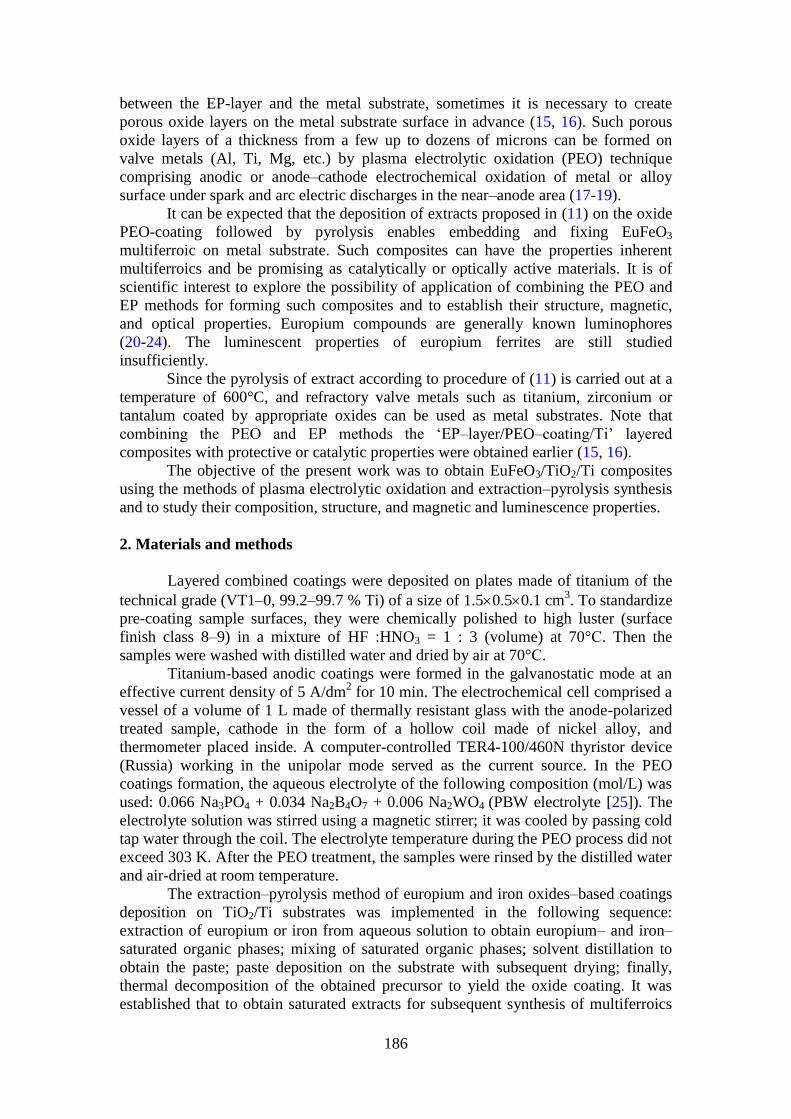
Figure 1 shows SEM images of the surface of coatings on titanium obtained in
the PBW electrolyte before (ab) and after threefold (cd) and fivefold (ef) extract
deposition with subsequent annealing. Modification results in formation of individual
areas of the new phase on the surface (threefold deposition, Figs.1cd) and filling of
larger surface areas (fivefold deposition, Figs.1ef). The increase of the number of
deposition cycles must allow formation of a solid layer of the new phase on the basic
oxide coating surface. Initially (threefold deposition, Figs.1cd), the new phase is
attached predominantly in areas with larger quantities of small pores, most probably,
the surface sites having the largest defects. Thus, the oxide layer surface sorbs the
extract selectively. Further deposition cycles (fivefold, Figs.1ef) results in as in
emergence of new areas of this type as in growth of the earlier formed ones.
Fig.1. SEM images of the PEO coating surface before (a, b) and after threefold (c, d)
and fivefold (e, f) extract deposition with subsequent annealing. Amplitude (a, c, e)
and phase (b, d, f) representation.
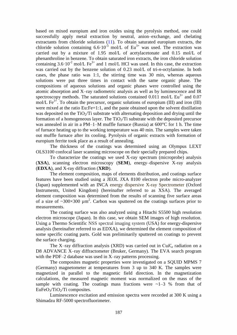
The maps of elements distribution over the surface demonstrate that the new
phase areas contain both europium and iron (Fig.2), while the titanium contents in
them are reduced. It is evident from comparison of Figs. 2b, c, and d that the titanium
189
content is clearly lower in the sites of europium and iron localization than in
neighboring areas. In other words, the PEO coating surface is closed (shielded) by a
layer of the new phase. Since one detects titanium in the deposited areas and the
analysis depth varies, depending to the nature of the analyzed material, from 2 to 5
µm, the thickness of the deposited modifying layer will be less than 2–5 µm. Here, the
modifying layer heterogeneously shields the PEO coating surface. Aside from the
areas that shield almost completely the initial coating, one also observes those with a
partial shielding, as seen from the titanium distribution maps (Fig.2).
Fig.2. SEM image (phase representation, a) of the surface of PEO coating after
fivefold extract deposition with subsequent annealing and distribution maps (white
dots) of europium (b), iron (c), and titanium (d) over the surface.
The element compositions of the outer layers of initial and modified PEO
coatings determined using XSA and averaged over the surface are shown in Table 1.
The average values in this Table were obtained for five 300×300 µm areas randomly
selected over the surface at an analysis depth of 2–5 µm. The main elements
constituting the surface of the PEO coating are oxygen, phosphorus, titanium, and
tungsten. The modified PEO coatings contain, in addition, europium and iron,
whereas the europium/iron atomic ratio is Eu/Fe =1.6 (threefold deposition) or
Eu/Fe=1.2 (fivefold deposition). Taking into account the accuracy of the elements
Table 1. Averaged element composition of the coatings outer layer as to XSA
Coating Element composition, at.%
O Na P Ti Fe Eu W
PEO 72.2 1.0 4.4 21.4 - - 0.9
PEO +3 depositions 69.9 - 5.8 18.8 1.8 2.9 0.8
PEO+5 depositions 70.4 - 5.3 15.9 3.5 4.1 0.7
190
determination by the microprobe analysis method (~15 %) and the fact that the
analysis includes not only the deposited layer, but also the initial PEO coating (with
its outer areas as well), one can conclude that the obtained values of the Eu/Fe ratio
corroborate a possible presence of EuFeO3 in the deposited layers.
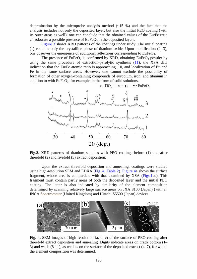
Figure 3 shows XRD patterns of the coatings under study. The initial coating
(1) contains only the crystalline phase of titanium oxide. Upon modification (2, 3),
one observes the emergence of additional reflections corresponding to EuFeO3.
The presence of EuFeO3 is confirmed by XRD, obtaining EuFeO3 powder by
using the same procedure of extraction-pyrolytic synthesis (11), the XSA data
indication that the Eu/Fe atomic ratio is approaching 1.0, and localization of Eu and
Fe in the same surface areas. However, one cannot exclude the possibility of
formation of other oxygen-containing compounds of europium, iron, and titanium in
addition to with EuFeO3, for example, in the form of solid solutions.
Fig.3. XRD patterns of titanium samples with PEO coatings before (1) and after
threefold (2) and fivefold (3) extract deposition.
Upon the extract threefold deposition and annealing, coatings were studied
using high-resolution SEM and EDXA (Fig. 4, Table 2). Figure 4a shows the surface
fragment, whose area is comparable with that examined by XSA (Figs.1cd). This
fragment must contain partly areas of both the deposited layer and the initial PEO
coating. The latter is also indicated by similarity of the element composition
determined by scanning relatively large surface areas on JXA 8100 (Japan) (with an
INCA Spectrometer (United Kingdom) and Hitachi S5500 (Japan) devices.
Fig. 4. SEM images of high resolution (a, b, c) of the surface of PEO coating after
threefold extract deposition and annealing. Digits indicate areas on crack bottom (1–
3) and walls (8-11), as well as on the surface of the deposited extract (4–7), for which
the element composition was determined.
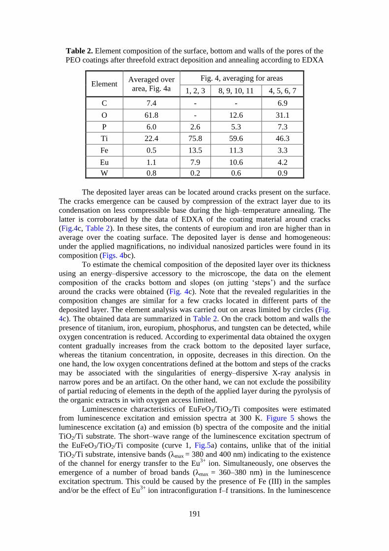
191
Table 2. Element composition of the surface, bottom and walls of the pores of the
PEO coatings after threefold extract deposition and annealing according to EDXA
Element Averaged over
area, Fig. 4a
Fig. 4, averaging for areas
1, 2, 3 8, 9, 10, 11 4, 5, 6, 7
C 7.4 - - 6.9
O 61.8 - 12.6 31.1
P 6.0 2.6 5.3 7.3
Ti 22.4 75.8 59.6 46.3
Fe 0.5 13.5 11.3 3.3
Eu 1.1 7.9 10.6 4.2
W 0.8 0.2 0.6 0.9
The deposited layer areas can be located around cracks present on the surface.
The cracks emergence can be caused by compression of the extract layer due to its
condensation on less compressible base during the high–temperature annealing. The
latter is corroborated by the data of EDXA of the coating material around cracks
(Fig.4c, Table 2). In these sites, the contents of europium and iron are higher than in
average over the coating surface. The deposited layer is dense and homogeneous:
under the applied magnifications, no individual nanosized particles were found in its
composition (Figs. 4bc).
To estimate the chemical composition of the deposited layer over its thickness
using an energy–dispersive accessory to the microscope, the data on the element
composition of the cracks bottom and slopes (on jutting ‘steps’) and the surface
around the cracks were obtained (Fig. 4c). Note that the revealed regularities in the
composition changes are similar for a few cracks located in different parts of the
deposited layer. The element analysis was carried out on areas limited by circles (Fig.
4c). The obtained data are summarized in Table 2. On the crack bottom and walls the
presence of titanium, iron, europium, phosphorus, and tungsten can be detected, while
oxygen concentration is reduced. According to experimental data obtained the oxygen
content gradually increases from the crack bottom to the deposited layer surface,
whereas the titanium concentration, in opposite, decreases in this direction. On the
one hand, the low oxygen concentrations defined at the bottom and steps of the cracks
may be associated with the singularities of energy–dispersive X-ray analysis in
narrow pores and be an artifact. On the other hand, we can not exclude the possibility
of partial reducing of elements in the depth of the applied layer during the pyrolysis of
the organic extracts in with oxygen access limited.
Luminescence characteristics of EuFeO3/TiO2/Ti composites were estimated
from luminescence excitation and emission spectra at 300 K. Figure 5 shows the
luminescence excitation (a) and emission (b) spectra of the composite and the initial
TiO2/Ti substrate. The short–wave range of the luminescence excitation spectrum of
the EuFeO3/TiO2/Ti composite (curve 1, Fig.5a) contains, unlike that of the initial
TiO2/Ti substrate, intensive bands (λmax = 380 and 400 nm) indicating to the existence
of the channel for energy transfer to the Eu3+
ion. Simultaneously, one observes the
emergence of a number of broad bands (λmax = 360–380 nm) in the luminescence
excitation spectrum. This could be caused by the presence of Fe (III) in the samples
and/or be the effect of Eu3+
ion intraconfiguration f–f transitions. In the luminescence
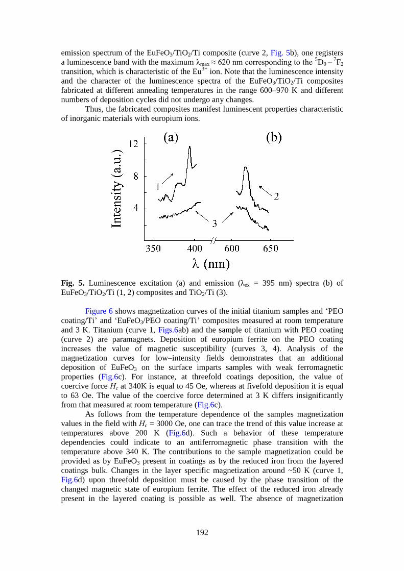
192
emission spectrum of the EuFeO3/TiO2/Ti composite (curve 2, Fig. 5b), one registers
a luminescence band with the maximum λmax ≈ 620 nm corresponding to the 5D0 –
7F2
transition, which is characteristic of the Eu3+
ion. Note that the luminescence intensity
and the character of the luminescence spectra of the EuFeO3/TiO2/Ti composites
fabricated at different annealing temperatures in the range 600–970 K and different
numbers of deposition cycles did not undergo any changes.
Thus, the fabricated composites manifest luminescent properties characteristic
of inorganic materials with europium ions.
Fig. 5. Luminescence excitation (a) and emission (λex = 395 nm) spectra (b) of
EuFeO3/TiO2/Ti (1, 2) composites and TiO2/Ti (3).
Figure 6 shows magnetization curves of the initial titanium samples and ‘PEO
coating/Ti’ and ‘EuFeO3/PEO coating/Ti’ composites measured at room temperature
and 3 K. Titanium (curve 1, Figs.6ab) and the sample of titanium with PEO coating
(curve 2) are paramagnets. Deposition of europium ferrite on the PEO coating
increases the value of magnetic susceptibility (curves 3, 4). Analysis of the
magnetization curves for low–intensity fields demonstrates that an additional
deposition of EuFeO3 on the surface imparts samples with weak ferromagnetic
properties (Fig.6c). For instance, at threefold coatings deposition, the value of
coercive force Нс at 340K is equal to 45 Oe, whereas at fivefold deposition it is equal
to 63 Oe. The value of the coercive force determined at 3 K differs insignificantly
from that measured at room temperature (Fig.6c).
As follows from the temperature dependence of the samples magnetization
values in the field with Нc = 3000 Oe, one can trace the trend of this value increase at
temperatures above 200 K (Fig.6d). Such a behavior of these temperature
dependencies could indicate to an antiferromagnetic phase transition with the
temperature above 340 K. The contributions to the sample magnetization could be
provided as by EuFeO3 present in coatings as by the reduced iron from the layered
coatings bulk. Changes in the layer specific magnetization around ~50 K (curve 1,
Fig.6d) upon threefold deposition must be caused by the phase transition of the
changed magnetic state of europium ferrite. The effect of the reduced iron already
present in the layered coating is possible as well. The absence of magnetization
193
anomalies for layers upon fivefold deposition (curve 2, Fig.6d) could result from the
increased content of europium ferrite and its predominant contribution.
Fig.6. M=f(H) dependencies (a, b): 1 – titanium; 2 – titanium with PEO coating; 3, 4 –
EuFeO3/TiO2/Ti composites with threefold and fivefold extract deposition with
subsequent annealing. M=f(H) dependencies for the samples with threefold extract
deposition at 340 and 3 K (c), insert – the low–intensity field area. Temperature
dependencies of the specific magnetization of EuFeO3/TiO2/Ti composites in the field
Н = 3000 Oe (d): M=f(T) dependencies 1 and 2 correspond to composites with
threefold and fivefold extract deposition.
In should be noted, that the dependence of the composite magnetization on
temperature differs from that of free powders of europium ferrite obtained earlier
using the extraction–pyrolysis method according to similar scheme (11) and
consisting of nanoparticles of a size of 10–20 nm. In the latter case, at temperatures
below ~230 K the powders manifested weak ferromagnetic properties, whereas at
higher temperatures their ferromagnetism increased dramatically. For instance, at Т =
300 K, the measured value of the coercive force was equal to 2068 Oe (11). The
fabricated layered metal–oxide composites with europium ferrite on the surface
manifest weak ferromagnetic properties in the temperature range 3–340 K. One
observes the trend to the increase of the value of specific magnetization at
temperatures above 200 K (Fig. 5d). The difference in temperature dependencies of
the specific magnetization of nanosized powder of europium ferrite and EuFeO3
deposited on PEO coating consisting predominantly of TiO2 must be caused by size
effects. The EuFeO3 properties could be also affected by the substrate and a complex
structure of the deposited and annealed layer, due to the presence of a small amount of
reduced iron in its composition.
As was shown earlier, in bulk multiferroic samples (for instance, BiFeO3),
observation of the magnetoelectric effect was impossible (2, 3, 6). The reason consists
in the presence of space–modulated spin structure of the cycloid type in bulk samples
(2, 3, 6). The magnetoelectric effect can be consistently observed for nanosized
194
samples (example – BiFeO3 films of a size of dozens and hundreds of nanometers
deposited on inert substrates). Destruction of the spin cycloid can be caused not only
by the size factor, but also by substitution of bismuth ions with those of rare earth
elements (6). It has been established that polycrystalline samples RxBi1-xFeO3, where
R = Nd, La or La, Gd, х = 0.05–0.2, fabricated on the basis of BiFeO3 according to the
standard ceramic technology manifest, unlike pure bulk antiferromagnetic bismuth
ferrite, weak ferromagnetic properties. Similar features were revealed in the present
work for europium ferrite deposited on the titanium oxide substrate. The available
literature data and the obtained results enable one to conclude that bulk
EuFeO3/TiO2/Ti composites fabricated through combination of the methods of plasma
electrolytic oxidation and extraction–pyrolysis can be investigated for the presence of
magnetoelectric effect in a broad temperature range.
5. Conclusions
1. The combination of extraction–pyrolysis and plasma electrolytic oxidation may be
used for obtaining EuFeO3/TiO2/Ti composites simultaneously manifesting
luminescent and weak ferromagnetic properties.
2. The magnetic properties of EuFeO3 deposited on PEO coating on titanium are
different from those of nanosized powder obtained in ref. (11). The EuFeO3/TiO2/Ti
composite is a weak ferromagnetic through the range of temperatures tested (3-340
K), whereas the ferromagnetism of the powder increases dramatically at temperatures
above 230 K.
3. The magnetic properties of deposited EuFeO3 could be affected by both the metal
substrate nature and the complex structure of the deposited and annealed layer.
4. The luminescent properties of EuFeO3/TiO2/Ti composites are typical of inorganic
materials with europium ions.
Acknowledgements
The authors are grateful to Cand. Sci. (Chem.) V.G. Kuryavyi and Cand. Sci.
(Chem.) T.A. Kaidalova for the assistance in determination of the coatings element
and phase compositions and morphology.
The study was partially supported by grants no. 15-03-03271 from Russian
Foundation for Basic Research and no. 15-I-3-034 from Presidium of FEB RAS.
References
1. Smolenskii GA, Chupis IE: 'Segnetomagnetics'. Uspekhi Fizicheskikh Nauk 1982
137 (3) 415–448 (in Russian).
2. Pyatakov AP, Zvezdin AK: 'Magnetoelectric and multiferroic media'. Physics-
Uspekhi (Advances in Physical Sciences) 2012 55 (6) 557-581.
3. Strokan’ GP: 'Nanostructured bismuth ferrite films fabricated in transverse RF
discharge'. Nanotechnologies in Russia 2009 4 (1-2) 79–84.
4. Khomskii DI: 'Multiferroics: Different ways to combine magnetism and
ferroelectricity'. Journal of Magnetism and Magnetics Materials 2006 306 (1) 1-8.
5. Wu H, Li L, Liang LZ, Liang S, Zhu YY, Zhu XH: 'Recent progress on the
structural characterizations of domain structures in ferroic and multiferroic
perovskite oxides: A review'. Journal of the European Ceramic Society 2014 35
(2) 411-441.
195
6. Ravinski AF, Makoed II, Yanushkevich KI, Galyas AI, Demidenko OF,
Poddubrovskaya AV, Lozenko VV, Moshchalkov VV: 'Magnetic properties of
multiferroic synthesized based on BiFeO3'. Phase Transitions, Ordered States and
New Materials 2012 (9) 17-21 (in Russian).
7. Fakhrul T, Mahbub R, Chowdhury N, Khosru QDM, Sharif A: 'Structural,
dielectric and magnetic properties of Ta-substituted Bi0.8La0.2FeO3 multiferroics'.
Journal of Alloys and Compounds 2015 622 471-476.
8. Song SH, Zhu QS, Weng LQ, Mudinepalli VR: 'A comparative study of dielectric,
ferroelectric and magnetic properties of BiFeO3 multiferroic ceramics synthesized
by conventional and spark plasma sintering techniques'. Journal of the European
Ceramic Society 2015 35 (1) 131-138.
9. Sando D, Barthelemy A, Bibes M: 'BiFeO3 epitaxial thin films and devices: past,
present and future' Journal of Physics: Condensed Matter 2014 26 (47) 473201.
10. Mizumaki M, Uozumi T, Agui A, Kawamura N, Nakazawa M: 'Admixture of
excited states and ground states of a Eu3+
ion in Eu3Fe5O12 by means of magnetic
circular dichroism'. Journal of the European Ceramic Society 2005 71 (13)
134416.
11. Steblevskaya NI, Medkov MA, Belobeletskaya MV, Tkachenko IA: 'Europium
oxide-based nanocomposites synthesized by extraction-pyrolytic method'. Russian
Journal of Inorganic Chemistry 2014 59 (3) 251-254.
12. Choquette AK, Colby R, Moon EJ, Schleputz CM, Scafetta MD, Keavney DJ,
May SJ: 'Synthesis, structure, and spectroscopy of epitaxial EuFeO3 thin films'.
ACS Crystal Growth & Design 2015 15 (3) 1105−1111.
13. Ciambelli P, Cimino S, De Rossi S, Lisi L, Minelli G, Porta P, Russo G: 'AFeO3
(A=La, Nd, Sm) and LaFe1−xMgxO3 perovskites as methane combustion and CO
oxidation catalysts: structural, redox and catalytic properties' Applied Catalysi B:
Environmental 2001 29 (4) 239−250.
14. Kholkin AI, Patrusheva TN: Extraction–pyrolysis method. Fabrication of
functional oxide materials. Moscow, KomKniga, 2006 (in Russian).
15. Rudnev VS, Medkov MA, Steblevskaya NI, Lukiyanchuk IV, Tyrina LM,
Belobeletskaya MV: 'Pt/SiO2 and Pt/TiO2/Ti compositions and their catalytic
properties'. Theoretical Foundations of Chemical Engineering 2011 45 (4) 496-
499.
16. Rudnev VS, Medkov MA, Yarovaya TP, Steblevskaya NI, Nedovzorov PM,
Belobeletskaya MV: 'Combination of plasma-electrolytic oxidation and
extraction-pyrolytic method for formation of metal oxide layers'. Russian Journal
of Applied Chemistry 2012 85 (4) 621-628.
17. Rakoch AG, Dub AV, Gladkova AA: Anodization of light alloys at different
electric modes. Plasma electrolytic technology. Moscow, Staraya Basmannaya,
2012 (in Russian).
18. Yerokhin AL, Nie X, Leyland A, Matthews A, Dowey SJ: 'Plasma electrolysis for
surface engineering'. Surface and Coatings Technology 1999 122 (2-3) 73-93.
19. Walsh FC, Low CTJ, Wood RJK, Stevens KT, Archer J, Poeton AR, Ryder A:
'Plasma electrolytic oxidation (PEO) for production of anodised coatings on
lightweight metal (Al, Mg, Ti) alloys'. Transactions of the Institute of Metal
Finishing 2009 87 (3) 122-135.
20. Zolin VF, Koreneva LG: Rare earth probe in chemisrty and biology. Moscow,
Nauka, 1980 (in Russian).
196
21. Petushkov AA, Shilov SM, Pak VN: 'Dimensional features of the luminescence of
europium(III) chloride nanoparticles in a porous glass matrix'. Technical Physics
Letters 2004 30 (11) 894-896.
22. Ternane R, Trabelsi-Ayedi M, Kbir-Ariguib N, Piriou B: 'Luminescent properties
of Eu3+
in calcium hydroxyapatite' Journal of Luminescence 1999 81 (3) 165-170.
23. Gasparotto G, Lima SAM, Davolos MR, Varela JA, Longo E, Zaghete MA:
'Luminescence properties of Eu3+
- and Mg2+
-doped LiTaO3 obtained via the
polymeric precursor method'. Journal of Luminescence 2008 128 (10) 1606-1610.
24. Parma A, Freris I, Riello P, Enrichi F, Cristofori D, Benedetti A: 'Structural and
photoluminescence properties of ZrO2:Eu3+
@ SiO2 nanophosphors as a function
of annealing temperature'. Journal of Luminescence 2010 130 (12) 2429-2436.
25. Rudnev VS, Yarovaya TP, Lysenko AE, Nedosorov PM, Dushina NE: 'The
influence of the conditions of forming on the characteristics of the oxide
protective films on aluminum'. Protection of Metals 2008 44 (7) 715-720.


![Presentation F. Chateauraynaud.ppt [Mode de compatibilité] · individuals and events sometimes make a difference. Like mushroom spores, they may germinate in unexpected places, reshaping](https://static.fdocuments.fr/doc/165x107/5ec75bfa1d81065d2965f699/presentation-f-mode-de-compatibilit-individuals-and-events-sometimes-make-a.jpg)