C+27.1849-
47
Cryptogamie, Mycologie, 2018, 39 (2): 211-257 © 2018 Adac. Tous droits réservés doi/10.7872/crym/v39.iss2.2018.211 Fungal Biodiversity Profiles 51-60 Xiang-hua WANG a , Kanad DAS b , Joel HORMAN c , Vladimir ANTONIN d , Abhishek BAGHELA e , Dyutiparna CHAKRABORTY b , Manoj E. HEMBROM f , Karen NAKASONE g , Beatriz ORTIZ-SANTANA g , Alfredo VIZZINI h , Valérie HOFSTETTER i & Bart BUYCK j* a Key Laboratory for Plant Diversity and Biogeography of East Asia, Kunming Institute of Botany, Chinese Academy of Sciences, Lanhei Road 132, Kunming 650201, P. R. China; e-mail: [email protected] b Cryptogamic Unit, Botanical Survey of India, P.O. Botanic Garden, Howrah 711103, India, email: [email protected] c 11 Ramblewood Road, Ridge, New York, U.S.A., 11961, email: [email protected] d Department of Botany, Moravian Museum, Zelný trh 6, CZ-659 37 Brno, Czech Republic, email: [email protected] e MACS’ Agharkar Research Institute, Biodiversity and Palaeobiology Group, National Fungal Culture Collection of India (NFCCI), G.G. Agarkar Road, Pune – 411004, India, email: [email protected] f Central National Herbarium, Botanical Survey of India, P.O. Botanic Garden, Howrah 711103, India, email: [email protected] g Center for Forest Mycology Research, Northern Research Station, One Gifford Pinchot Drive, Madison, Wisconsin, U.S.A, 53726, email: [email protected], [email protected] h Department of Life Sciences and Systems Biology, University of Torino, Viale P.A. Mattioli 25, I-10125 Torino, Italy, email: [email protected] i Agroscope Changins-Wädenswil Research Station, ACW, Department of plant protection, Rte de Duiller, 1260 Nyon, Switzerland; [email protected] j Institut de Systematique, Evolution, Biodiversité (ISYEB-UMR7205), Sorbonne Université, Museum National d’Histoire Naturelle, Case Postale 39, 12 Rue Buffon, F-75005 Paris, France, email: [email protected] Abstract. In this new series of Fungal Biodiversity Profiles, the authors describe ten species new to science, nine basidiomycetes and one ascomycete, using both morphological and molecular data. Descriptions are provided for Hydnum berkeleyanum sp. nov. (Cantharellales), for Lactarius ambiguus, L. furfuraceus, L. orientaliquietus, L. subhirtipes, Russula blennia, R. pseudociliata (Russulales), Marasmius pseudohypochrysoides, Radulomyces paumanokensis (Agaricales) and Urnula himalayana (Pezizales). Agaricales / cantharellales / Hydnum / Lactarius / Marasmius / Pezizales / phylogeny / Radulomyces / Russula / russulales / systematics / Urnula * Corresponding author B. Buyck
Transcript of C+27.1849-

Cryptogamie, Mycologie, 2018, 39 (2): 211-257© 2018 Adac. Tous droits réservés
doi/10.7872/crym/v39.iss2.2018.211
Fungal Biodiversity Profiles 51-60Xiang-hua WANG a, Kanad DAS b, Joel HORMAN c, Vladimir ANTONIN d,
Abhishek BAGHELA e, Dyutiparna CHAKRABORTY b,Manoj E. HEMBROM f, Karen NAKASONE g, Beatriz ORTIZ-SANTANA g,
Alfredo VIZZINI h, Valérie HOFSTETTER i & Bart BUYCK j*
a Key Laboratory for Plant Diversity and Biogeography of East Asia,Kunming Institute of Botany, Chinese Academy of Sciences, Lanhei Road 132,
Kunming 650201, P. R. China; e-mail: [email protected] Cryptogamic Unit, Botanical Survey of India, P.O. Botanic Garden,
Howrah 711103, India, email: [email protected] 11 Ramblewood Road, Ridge, New York, U.S.A., 11961,
email: [email protected] Department of Botany, Moravian Museum, Zelný trh 6, CZ-659 37 Brno,
Czech Republic, email: [email protected] MACS’ Agharkar Research Institute, Biodiversity and Palaeobiology Group,National Fungal Culture Collection of India (NFCCI), G.G. Agarkar Road,
Pune – 411004, India, email: [email protected] Central National Herbarium, Botanical Survey of India, P.O. Botanic Garden,
Howrah 711103, India, email: [email protected] Center for Forest Mycology Research, Northern Research Station,
One Gifford Pinchot Drive, Madison, Wisconsin, U.S.A, 53726,email: [email protected], [email protected]
h Department of Life Sciences and Systems Biology, University of Torino, VialeP.A. Mattioli 25, I-10125 Torino, Italy, email: [email protected]
i Agroscope Changins-Wädenswil Research Station, ACW,Department of plant protection, Rte de Duiller, 1260 Nyon, Switzerland;
[email protected] Institut de Systematique, Evolution, Biodiversité (ISYEB-UMR7205), Sorbonne
Université, Museum National d’Histoire Naturelle, Case Postale 39,12 Rue Buffon, F-75005 Paris, France, email: [email protected]
Abstract. In this new series of Fungal Biodiversity Profiles, the authors describe ten speciesnew to science, nine basidiomycetes and one ascomycete, using both morphological andmolecular data. Descriptions are provided for Hydnum berkeleyanum sp. nov. (Cantharellales),for Lactarius ambiguus, L. furfuraceus, L. orientaliquietus, L. subhirtipes, Russula blennia,R. pseudociliata (Russulales), Marasmius pseudohypochrysoides, Radulomyces paumanokensis(Agaricales) and Urnula himalayana (Pezizales).Agaricales / cantharellales / Hydnum / Lactarius / Marasmius / Pezizales / phylogeny /Radulomyces / Russula / russulales / systematics / Urnula
* Corresponding author B. Buyck

212 Xiang-Hua WANG et al.
51. Hydnum berkeleyanum K. Das, Hembrom, A. Baghela & Vizzini sp. nov.Figs 1-4
MycoBank: MB 824501GenBank: MG972968 (ITS), MG972970 (LSU)Systematic position: Basidiomycota, Agaricomycetes, Cantharellales,
Hydnaceae.Etymology: Commemorating Miles Joseph Berkeley for his contribution to
Indian mycobiota.Diagnosis: Distinct from all other similar looking Hydnum species mainly
by its ITS and LSU sequence data and a combination of morphological features likemoderately robust (pileus 25-80 mm in diam., stipe 30-55 × 8-14 mm) basidiomatawith orange-brown pileus, long (2-9 mm) spines, globose to subglobose or broadlyellipsoid (Q = 1.01-1.17) basidiospores, trichoderm pattern of pileipellis andoccurrence under trees of Fagaceae in mixed temperate forests.
Holotype: INDIA, Uttarakhand, Pauri district, Chourikhal, N 30° 01.553’E 79°00.477’, 2188 m a.s.l., under Quercus in temperate broadleaf forest, 1 August,2017, Kanad Das & Manoj E. Hembrom, KD-MEH 17-005 (CAL 1656, holotype!).
Basidiomata solitary to gregarious. Pileus 25-80 mm in diam., initiallyconvex, plano-convex to plane at maturity, sometimes with irregular lobes or centraldepression or umbilicate; surface dry, velutinous, sometimes with small and erectscales at the margin, non-staining, azonate, combination of pale orange to lightorange (5A3-4) with brown (6D8) centre which becoming paler gradually towardsthe margin to pale orange to light orange (5A3-4), rarely brownish orange (6C8)throughout at maturity; margin entire, becoming somewhat sinuous and lobed withage. Stipe 30-55 × 8-14 mm, eccentric to lateral, cylindrical, mainly tapering towardsbase, white (A1) at apex, gradually brownish towards the base, brown (7D8) at base.Spines adnexed to subdecurrent, 2-9 mm long (with some shorter spines on stipe),typically spinoid, not flattened, with acute (rarely subacute) apex, sometimes joinedat the base, crowded, initially white (A1), becoming orange-white to pale orange(5A3) at maturity or on bruising, gradually more brownish when drying. Contextchambered in pileus and stipe, white (A1), quickly becoming pale orange (5A3) onexposure, unchanging with 10% FeSO4, and 5% KOH but turning greenish withguaiacol. Odor pleasant, slightly fruity. Spore-print white.
Basidiospores globose to subglobose or rarely broadly ellipsoid, 7.5-8.4-8.5-9.5 × 6.9-7.9-8.0-8.8 µm (n = 40; Q = 1.01-1.06-1.08-1.17), thin-walled,inamyloid, apiculus cubic. Basidia subcylindric to clavate, 4-spored, rarely two-spored, clamped, 26-60 × 7-9 µm; sterigmata up to 9 µm long. Spine tip sterile;hyphae composed of interwoven hyphae being parallel towards the apex; eachhyphae cylindrical, septate, clamped and with subclavate to clavate apex, thin-walled, 4-6 µm wide at apex, without crystals. Hymenophoral trama composed ofhyphae in parallel to interwoven pattern, hyphae 2-11 µm wide, clamped. Pileipelliscomposed of loosely interwoven erect to suberect hyphae forming a trichodermpattern, hyphae cylindrical to swollen with pale yellow intracellular pigmentation,thin-walled, clamped, 2-9 µm wide, with cylindrical to subfusoid or appendiculatetips. Stipitipellis composed of erect to suberect hyphae forming a cutis pattern,hyphae mostly with orange intracellular pigmentation, clamped, 2-5 mm wide, withcylindrical to subfusoid or rarely appendiculate tips. Contextual hyphae interwoven,cylindrical to swollen with frequent constrictions, thin-walled, yellowish, clamped,2-12 µm wide.

Fungal Biodiversity Profiles 51-60 213
Fig. 1. Maximum Likelihood (ML) phylogram reconstructed using raxmlGUI 1.2 (Silvestro & Michalak2012) of nrITS Hydnum sequences. One thousand bootstrap replicates were analyzed to obtain the nodalsupport values. Bootstrap support values (>50%) are shown above or below the branches at nodes. Thetwo collections of the novel Indian species Hydnum berkeleyanum with GenBank accession numbersMG972968 and MG972969 are shown in red and bold in the tree. Five sequences from H. repandum(AJ547888, KU612578, KU612582, AJ547875, AY817136) and three sequences of H. vesterholtii(KU612553, KU612551, KU612552) belonging to Repandum-clade and Vesterholtii-clade respectivelywere considered as outgroup taxa.

214 Xiang-Hua WANG et al.
Specimens examined: INDIA, Uttarakhand, Pauri district, Chourikhal, N30° 01.553’ E 79°00.477’, 2188 m a.s.l., under Quercus sp. in temperate mixed(broadleaf and coniferous) forest, 1 August, 2017, Kanad Das & Manoj E. Hembrom,KD-MEH 17-005 (CAL 1656, holotype!); ibid., Sikkim, South district, Kewzing, N27°16.781’ E 088°20.300’, 1817 m a.s.l., under Castanopsis sp. in temperatebroadleaf forest, 25 August, 2017, Kanad Das, KD 17-039 (CAL 1657, paratype!).
Notes: The present new species of “hedgehog mushroom” is edible andconsumed in Western and Eastern Himalaya. Currently, based on multigenephylogeny, four well-supported lineages (Feng et al. 2016) are existing in this genus,they are Albomagnum-clade, Repandum-clade, Rufescens-clade and Versterholtii-clade (three are shown in our small ITS-based phylogenetic analyses). Our ITS-based phylogeny clearly shows that two Indian collections (KD-MEH 17-005 andKD 17-039, represented by MG972968 and MG972969 respectively in Fig. 1) of H.berkeleyanum along with four other Chinese collections (HKAS56128, HKAS77834,HKAS74570, HKAS74643) are recovered as a distinct species in the “Rufescensclade” in a strongly supported (100% bootstrap value) clade being sister to a cladeconsisting of two North American taxa: H. aerostatisporum Buyck, Lewis & V.Hofstetter (KY800344 and KY800343) and an undescribed species (KU612534 andKU612535). Similarly, in our supporting LSU-based phylogeny the two Indiancollections (KD-MEH 17-005 and KD 17-039, represented by MG972970 andMG972971 respectively in Fig. 2) along with four other undescribed Chinesecollections (KU612667, KU612665, KU612666, KU612664) are recovered as adistinct species in “Rufescens clade” in a moderately supported (65% bootstrapvalue) clade being sister to an undescribed American species (“Hydnum sp. 4”,KU612663). Our dual phylogenetic analyses with the available sequences under“Rufescens clade” in the public database GenBank also indicate the existence of acouple of undetermined taxa but kept only as “Hydnum sp. 1”, “Hydnum sp. 2” and“Hydnum species 6” in our LSU-based phylogeny (Fig. 2) as there are no supportingmorphological data. Similarly, in our ITS-based analysis at least 5 undescribedspecies (labeled as “Hydnum sp. 1”, “Hydnum sp. 2”, “Hydnum sp. 3”, Hydnum sp.4 and “Hydnum sp. 5” in Fig. 1 by us) are existing where no morphological data areavailable till date.
A number of European or North American species, namely, H.aerostatisporum, H. ellipsosporum Ostrow & Beenken, H. rufescens Pers., H.umbilicatum Peck, H. magnorufescens Vizzini, Picillo & Contu, H. ovoideisporumOlariaga, Grebenc, Salcedo & M.P. Martín are quite similar in the field and maycreate confusion with this Indian (rather Asian) species but, they all are mainlyseparated by their ITS and/or LSU sequence data as shown in Figs. 1 & 2, respectively.Moreover, in the field H. ovoideisporum (reported from Europe) differs from thepresent novel species by smaller basidiomata (pileus 12-40 mm; stipe 20-40 × 2.5-9mm) with bright orange coloured pileus (Olaraiga et al. 2012, Vizzini et al. 2013);H. aerostatisporum (from North America) has a more robust stipe (27-74 × 10-25mm) and smaller basidiospores [(7.1)7.3-7.69-8.1(8.5) × (6.5)6.7-7.07-7.5(7.9) μm](Buyck et al. 2017); H. ellipsosporum (from Europe) has typically ellipsoidbasidiospores (“Qm = 1.55-1.75”) (Ostrow & Beenken 2004, Olariaga et al. 2012);H. magnorufescens which is phylogenetically distant from H. berkeleyanum (Fig. 1)possess a distinctively paler pileus (ochre to saffron coloured) (Vizzini et al. 2013);H. umbilicatum (from North America) shows smaller basidiomata (pileus 25-50 mmin diam.; stipe 20-70 × 5-10 mm), umbilicate pileus, indistinctive odour, “oval”basidiospores (mainly globose to subglobose in the present novel species) (Bessetteet al. 1997) and H. rufescens (originally reported from Europe) has smaller
Fungal Biodiversity Profiles 51-60 215
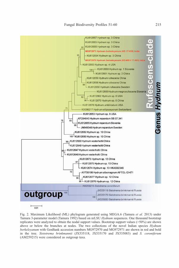
Fig. 2. Maximum Likelihood (ML) phylogram generated using MEGA.6 (Tamura et al. 2013) underTamura 3-parameter model (Tamura 1992) based on nrLSU Hydnum sequences. One thousand bootstrapreplicates were analyzed to obtain the nodal support values. Bootstrap support values (>50%) are shownabove or below the branches at nodes. The two collections of the novel Indian species Hydnumberkeleyanum with GenBank accession numbers MG972970 and MG972971 are shown in red and boldin the tree. Sistotrema brinkmannii (JX535118, JX535170 and JX535083) and S. coroniferum(AM259215) were considered as outgroup taxa.

216 Xiang-Hua WANG et al.
Fig. 3. Hydnum berkeleyanum (KD-MEH 17-005, holotype). a & b. Fresh basidiomata in the field andbasecamp. c. Spinoid hymenophore. d. Basidia bearing basidiospores. e. Radial section throughpileipellis. f. Transverse section through stipitipellis. g. Basidiospores. Scale bars: d, f & g = 10 µm,e = 50 µm

Fungal Biodiversity Profiles 51-60 217
Fig. 4. Hydnum berkeleyanum (KD-MEH 17-005, holotype). a. Basidiospores. b. Basidia. c. Transversesection through pileipellis showing hyphal arrangement. Scale bars: a-c = 10 µm.

218 Xiang-Hua WANG et al.
basidiomata (pileus up to 45 mm in diam.; stipe 10-45 × 1.5-15 mm), shorter (up to5 mm long) spines and smaller basidiospores (6.3-8.1 × 5.4-6.3 µm) (MaasGeesteranus 1971). However, Maas Geesteranus (1971) while describing H. rufescenscited a number of Asian (from Pakistan, India, Tibet or Japan) specimens in his“Collections examined and reported” part. Similarly, two collections (fromUttarakhand) of Hydnum are also reported as H. rufescens by J.R. Sharma (Sharma2012) considering their macro- and micromorphology. These two collections areseparated as well by their smaller basidiomata (pileus 30–70 mm in diam.; stipe8-30 × 3-10 mm), distinguishingly smaller (6-8.2 × 5.2-6 µm) basidiospores andshorter (1-3 mm) spines. Their identity/intercontinental conspecificity can only beconfirmed once their sequence data are available.
52. Lactarius ambiguus X.H. Wang sp. nov. Figs 5-6MycoBank: MB 826762.GenBank: MH447587 (ITS holotype); MH447582‒MH447586 and
MH447588 (ITS paratypes)Etymology: from Latin, referring to the ambiguous identity solely based on
morphology.Diagnosis: A small to medium-sized species recognized by the pileus with
much darker center and paler margin, spores with clean ornamentation, an intricate(ixo-) trichoderm as the pileipellis and presence of macrocystidia. Associated withfagaceous trees. Subtropical China.
Holotype: CHINA. HENNAN: Luanchuan Co., Laojun Mt., Tenglongyutrail, elev. 1290 m, 13 Aug. 2015, coll. X.H. Wang, no. 3656 (HKAS 89929, KUN!).
Basidiomata small to medium-sized. Pileus 30–60 mm, at first convex,soon expanding with a depressed centre to infundibuliform, papillate or not; surfacedry, glossy when wet, smooth, hygrophanous, dark olivaceous brown to brown (5E5,6E7) in the center, fading to cream ochraeus or pinkish buff with age, at margin lightyellow (4A3, 4A4), pale orange (5A3), grayish yellow (4B4, 4C5, 4C4), grayishorange (5B5), often with olivaceous tinge, with age becoming transparently striate.Context 1‒2 mm thick, pale yellow (4A3). Lamellae 3‒5 mm broad, slightlyemarginate to short ducurrent, crowded when young, remaining so or mediumcrowded with age, pale yellow (4A3), plae orange (5A3), becoming light yellow(4A4) to grayish orange (5B4) in age, slowly changing to reddish brown or brownwhen bruised. Stipe 30–90 × 4–12 mm, central to eccentrical, equal or taperingupwards, hollow; surface pruinose, nearly dry, grayish orange (6B4‒6B5), paleyellow to grayish orange (5A3‒5B4, 5B5), grayish brown (6D3, 6D4, 6D3‒6D4),brownish orange (7C4‒7C5), palest at the top; base strigose with short white hairswhen young, becoming reddish brown in age. Latex white, cream white or watery-milky, changing to watery, not discoloring, slightly bitter. Spore print not obtained.
Basidiospores (180/9/8) (6.5) 7.0–7.6–8.5 (9.0) × (5.5) 6.0–6.3–7.0 μm[Q = (1.06) 1.11–1.31 (1.45) , Q = 1.20 ± 0.06] [holotype (40/2/1): 7.0–7.7–8.5 ×(5.5) 6.0–6.3–7.0 μm, Q = (1.15) 1.17–1.27 (1.31),Q = 1.22 ± 0.03], broadly ellipsoidto ellipsoid, rarely subglobose; ornamentation 0.5–1.0 (1.3) μm high, of mediumdense elongate warts and clean ridges partly connected, often only forming closemeshes or broken reticulum, less forming nearly complete reticulum, some alignedin a subzebroid pattern, isolated warts and free ridges common; plage not amyloid,rarely slightly distally amyloid. Hymenophoral trama of hyphoid cells and scatteredcolorless or yellowish brown lactiferous hyphae. Basidia 35–50 × 10–14 μm,4-spored, clavate. Pleuromacrocystidia rare to common, more common towardslamellae base, emergent, rarely embedded in the basidia layer, protruding up to 70

Fungal Biodiversity Profiles 51-60 219
Fig. 5. Lactarius ambiguus (holotype). Photo X.H. Wang
μm, 60–100 (120) × 8–12 (14) μm, fusiform, cylindrical, some with a moniliformapex, with strongly refractive massy, crystalline or granular contents. Hymenophoralpseudocystidia rare to common, 2–4 (8) μm broad, slightly enlarged at the apex, notprotruding, with refractive content, hyaline or pale yellowish brown. Lamella edgesterile; marginal cells 10–25 (35) × 4–10 μm, similar to basidioles in shape, clavate,cylindrical, rarely ventricose, hyaline; cheilomacrocystidia rare to common, not seenin some individuals, 26–47 × 5–7 (10) μm, subfusiform, cylindrical. Pileipellis anintricate (ixo-) trichoderm, 50–100 μm thick; hyphae of suprapellis 3–8 μm broad,hyaline, often gelatinized, terminal cells 13–65 × 3–8 μm, cylindrical, equal orslightly swollen at the middle part; hyphae of subpellis strongly gelatinized, 4–13μm broad, some inflated to pyriform, ellipsoid or sausage-shaped. Stipitipellis acutis, 30–50 μm thick, of closely packed hyphae; hyphae 3–6 μm broad, slightlygelatinized or not, cylindrical, equal, hyaline. Trama of pileus and stipe of hyphaeand scattered colorless to yellowish brown lactiferous hyphae, with typical andabundant rosettes.
Additional specimens examined: HUBEI: Fang Co., Qiaoshang Town,Xihaoping, Xiping, under Quercus forest, 31 Jul. 2011, coll. X.H. Wang, no. 2941(HKAS 73501); Xingshan Co., Longmenhe forest farm, 11 Jul. 2012, coll. X.B. Liu,no. 29 (HKAS 75640). SHAANXI: Mei Co., Yingtou Town, Haopingsi, Taibai Mt.Nature Researve, under Quercus forest, 4 Sept. 2010, coll. X.H. Wang, no. 2736(HKAS 61932); Zhouzhi Co., Houzhenzi, Taibai Mt. Nature Reserve, under Quercus
220 Xiang-Hua WANG et al.
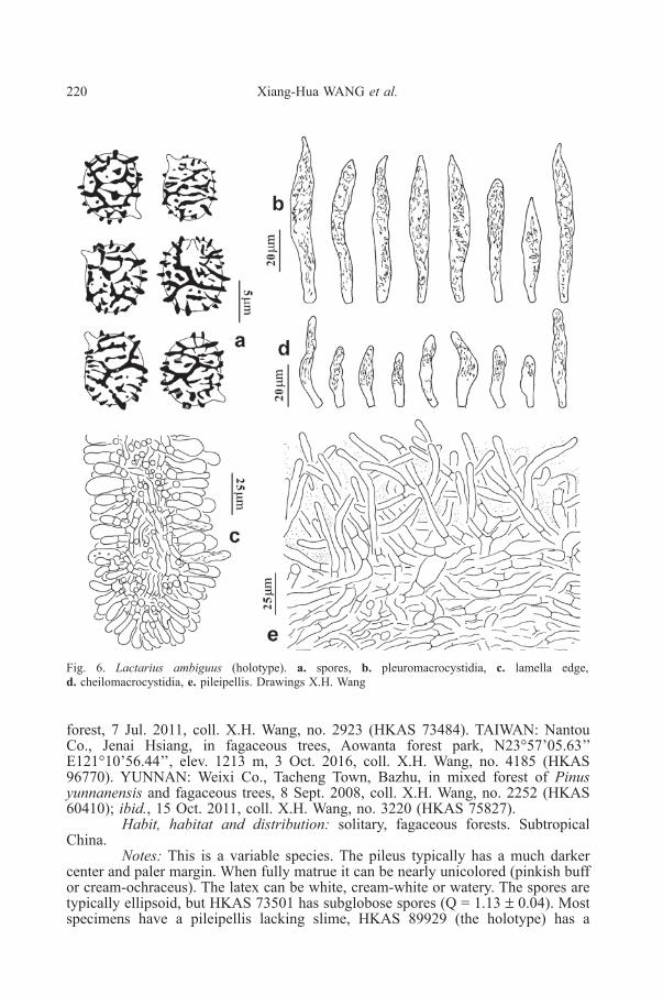
Fig. 6. Lactarius ambiguus (holotype). a. spores, b. pleuromacrocystidia, c. lamella edge,d. cheilomacrocystidia, e. pileipellis. Drawings X.H. Wang
forest, 7 Jul. 2011, coll. X.H. Wang, no. 2923 (HKAS 73484). TAIWAN: NantouCo., Jenai Hsiang, in fagaceous trees, Aowanta forest park, N23°57’05.63’’E121°10’56.44’’, elev. 1213 m, 3 Oct. 2016, coll. X.H. Wang, no. 4185 (HKAS96770). YUNNAN: Weixi Co., Tacheng Town, Bazhu, in mixed forest of Pinusyunnanensis and fagaceous trees, 8 Sept. 2008, coll. X.H. Wang, no. 2252 (HKAS60410); ibid., 15 Oct. 2011, coll. X.H. Wang, no. 3220 (HKAS 75827).
Habit, habitat and distribution: solitary, fagaceous forests. SubtropicalChina.
Notes: This is a variable species. The pileus typically has a much darkercenter and paler margin. When fully matrue it can be nearly unicolored (pinkish buffor cream-ochraceus). The latex can be white, cream-white or watery. The spores aretypically ellipsoid, but HKAS 73501 has subglobose spores (Q = 1.13 ± 0.04). Mostspecimens have a pileipellis lacking slime, HKAS 89929 (the holotype) has a

Fungal Biodiversity Profiles 51-60 221
Fig.7. Bootstrap consensus tree generated by Neighbor-Joining (NJ) phylogenetic analysis, rooted withmidpoint. NJ analysis was conducted in MEGA5, using bootstrap method (1000 replicates), p-distanceas substitution model, transitions and transversions both included, uniform rates among sites,homogeneous pattern among lineages and pairwise deletion for gaps treatment. The bootstrap proportionshigher than 70% are indicated above the nodes. New species are in bold. Initials proceeding the samplenumbers of the new species refer to the collectors and the others are GenBank accessions.
222 Xiang-Hua WANG et al.
pileipellis with mucus among the hyphae and HKAS 60410 even has a well-developed slime layer. The terminal cells of the pileipellis vary from 3‒5 µm to 5‒8µm broad among different individuals and the spore ornamentation may consist ofrelatively isolated elements or form a broken to nearly complete reticulum. Thesevariations make morphological identification difficult.
Lactarius ambiguus is similar to L. cinnamomeus W.F. Chiu ( = L. kesiyaeVerbeken & H.D. Hyde), L. inconspicuous H.T. Le & F. Hampe and L. tangerinusH.T. Le & De Crop in the general apperance and the structure of the pileipellis.Lactarius ambiguus, L. inconspicuus and L. tangerinus have a pileus with muchdarker center. The olivaceous tinge of L. ambiguus, however, is not reported on L.inconspicuus and L. tangerinus (Wisitrassameewong et al. 2015). Lactariusinconspicuus has yellowing latex and spores of L. tangerinus are more round (Q =1.02‒1.09‒1.13‒1.15). The contrast between the darker center and paler margin ofL. ambiguus and the relatively dry pileus are good characters distinguishing it fromL. cinnamomeus. Lactarius cinnamomeus and L. inconspicuus have well-developedslime on the pileus cuticle. A clear slimy layer was observed in only one specimen(HKAS 60410) of L. ambiguus.
Four sequences with 99% similarity were retrieved from GenBank. Thereare several characteristic substitutions and INDELs beween the clade formed bythese sequences and that formed by the Chinese samples. Due to the unsolvedphylogenetic relationship (Fig. 7) they are tentatively labelled as L. ambiguus. Oneof the them, KF433000 (sample KW016) was identified as L. inconspicuus byWisitrassameewong et al. (2015).
53. Lactarius furfuraceus X.H. Wang sp. nov. Figs 8-9MycoBank: MB 826763GenBank: MH447566 (ITS holotype); MH447564, MH477565 and
MH477567‒MH447581 (ITS paratypes)Systematic position: Basidiomycota, Agricomycotina, Russulales,
Rusulaceae.Etymology: from Latin, referring to the areolate-rimose pileus cuticle that
produces a furfuraceus appearance.Diagnosis: A small, slender, thin-fleshed species that can be recognized by
the orange brown basidiocarps, areolate-rimose pileus, absence of rosettes in thepileus and stipe trama and bigger spores.
Holotype: CHINA. YUNNAN: Xinping Co., Ailao Mts. Nature Reserve,roadside from Gasa to Heping, N23°57’48.17’’ E101°31’14.60’’, elev. 2223 m, 12Aug. 2017, coll. X.H. Wang, no. 4495 (HKAS 101915, KUN!).
Basidiomata small, slender, thin-fleshed. Pileus 20–30 (50) mm, at firstconvex with a conical papilla, later concave to infundibuliform, with clear sulcateswhen fully mature; surface dry, areolate-rimose, strongly hygrophanous, reddishbrown, brown (7D8–7E8) when young, then brownish orange at margin (6C6–6D6)and light brown to brown (6D7, 6E7) in the center, paler when dry. Context verythin (< 0.5 mm), paler than stipe. Lamellae 1–3 (4) mm broad, decurrent, mediumspaced, whitish with faint brownish tinge or pinkish brown when young, becominglight orange to grayish brown (5A4–5B5) when mature. Stipe 20–50 × 1–5 mm,equal, cylindrical, hollow; surface dry, smooth, reddish brown, light brown (7D6) atthe apex, pale brown (7E8) below; base strigose with long paler or light brown(7D7) hairs. Latex scanty, watery or white becoming watery, not discoloring, mild.Spore print cream-colored.

Fungal Biodiversity Profiles 51-60 223
Fig. 8. Lactarius furfuraceus (holotype). Field habit. Photo X.H. Wang
Basidiospores (420/21/17) (7.0) 8.0–8.5–9.5 (10.5) × (6.5) 7.0–7.5–8.0(9.0) μm [Q = (1.00) 1.06–1.24 (1.31) , Q = 1.14 ± 0.06] [holotype (40/2/1): (8.0)8.5–9.0–9.5 (10.5) × 7.0–7.7–8.5 μm, Q = (1.11) 1.12–1.23 (1.25), Q = 1.17 ± 0.04],subglobose to broadly ellipsoid; ornamentation 1.0–1.5 (1.8) μm high, of conicalwarts and thick ridges connected by thin lines or ridges, forming an incomplete tocomplete reticulum, isolated warts and free ends present; plage distally amyloid.Basidia (35) 40–60 × 9–14 μm, 4-spored, clavate. Pleuromacrocystidia rare tocommon, emergent, (45) 55–80 (100) × (6) 8–12 μm, fusiform, sublanceolate orsublageniform, some with long tapering ± moniliform apex often. Hymenophoralpseudocystidia uncommon, 3–5 μm broad, filiform, slightly enlarged at the apex.Lamella edge sterile, rarely fertile; marginal cells similar to basidioles in shape,clavate, hyaline; cheilomacrocystidia absent. Pileipellis an incontinuous epithelium,more as an intricate cutis between piles of round cells when mature, continuouslayer of globose cells only present in very young pileus; round cells of the epithelium20–32 × 15–20 μm, globose, ellipsoid, hyaline to pale yellowish brown; repenthyphae of the cutis hyphoid, rarely ellipsoid or sausage-shape, 6–15 (20) μm broad.Stipitipellis a cutis; of closely packed, predominantly longitudinally arranged hyphae;hyphae (3) 4–12 μm wide, some inflated to sausage-shaped, yellowish brown. Tramaof pileus and stipe composed of hyphae and scattered hyaline lactiferous hyphae,lacking rosettes, only with very few sphaerocystes in the central region of the stipecortex.

224 Xiang-Hua WANG et al.
Additional specimens examined: CHINA. GUANGDONG: Ruyuan Co.,Nanling forest park, Xiaohuangshan, 18 Sept. 2015, coll. B. Li, no. 116 (HKAS89877). GUIZHOU: Jiangkou Co., Fangjing Mt., alt. 1680 m, 11 Aug. 2006, coll.X.H. Wang, no. 1997 (HKAS 51720); Jiangkou Co., Fanjing Mt., roadside fromJinding to Tongkuangchang, 7 Jul. 1988, coll. Z.L. Yang, no. 151 (HKAS 20771);Leishan Co., Leigong Mt. Nature Reserve, N26°23’06.7’’ E108°11’39.4’’, elev. 1856m, 24 Jun. 2017, coll. X.H. Wang, no. 4257 (HKAS 101914). TAIWAN: NantouCo., Meifeng water source area, N24°06’’ E121°10’, elev. 2100 m, 22 Jun. 1994,coll. W.N. Chou, no. CWN00585 (F0002329); Chiayi Co., Chuchi, Shihcho,Fujungshan, elev. 1800 m, 14 Aug. 2003, coll. W.N. Chou, no. CWN06333(F0026116). YUNNAN: Baoshan, Gaoligong Mts. Nature Reserve, Nankang, 20 Jul.2003, coll. X.H. Wang, no. 1587 (HKAS 43953); Baoshan, Longyang District, nearLongshu chicken farm, 11 Aug. 2010, coll. T. Guo, no. 146 (HKAS 86674); Baoshan,Longyang District, Shuizhai, Haitang, 22 Jul. 2009, coll. L.P. Tang, no. 885 (HKAS56842); Chuxiong, Zixi Mt., elev. 2400 m, 13 Aug. 2004, coll. H.D. Zheng, no. 04-559 (HKAS 45070); ibid., 14 Aug. 2004, coll. H.D. Zheng, no. 04-566 (HKAS45071); Kunming, Miaogaosi, N25°06’02.73’’ E102°37’52.53’’, elev. 2178 m, 28Aug. 2005, coll. X.H. Wang, no. 1950 (HKAS 49570); Kunming, Qiongzhusi,N25°04’10.09’’ E102°37’23.91’’, elev. 2135 m, 8 Sept. 2006, coll. X.H. Wang, no.2120 (HKAS 51842); ibid., 8 Aug. 2007, coll. X.H. Wang, no. 2142 (HKAS 52364);Lincang Co., Wulao Mt., elev. 2000 m, 2 Jul. 2012, coll. X.H. Wang, no. 3378(HKAS 75947); Lüchun Co., Huanglian Mt., Huanglianshan reservoir, coll. X.H.Wang, no. 3525 (HKAS 76062); Maguan Co., Dulong Town, Laojun Mt., N22°56’38’’E104°32’29’’, elev. 1950 m, 11 Aug. 2016, coll. X.H. Wang 3916 (HKAS 96505);ibid., N 22°57’30.60’’ E104°32’16.30’’, elev. 1916 m, coll. X.H. Wang, no. 4865(HKAS 101927); ibid., N 22°58’00.3’’ E104°31’33.1’’, elev. 1873 m, coll. X.H.Wang, no. 4866 (HKAS 101928); Pingbian Co., Dawei Mt. Nature Reserve, elev.2650 m, 5 Jul. 1992, coll. P.G. Liu, no. 1324 (HKAS 25521); ibid., 18 Jul. 2005, inCastanopsis forest, elev. 2000 m, coll. X.H. Wang, no. 1893 (HKAS 49527); ibid.,9 Jul. 2012, coll. X.H. Wang, no. 3538 (HKAS 76073); Tengchong Co., GaoligongMts. Nature Reserve, roadside from Bawan to Tengchong, ca. 30 km to Bawan, 18Jul. 2003, coll. X.H. Wang, no. 1568 (HKAS 43951); Tenchong Co., Mangbang,roadside from Baoshan to Tengchong, near 52 km landmark, 9 Aug. 2011, coll. X.T.Zhu, no. 469 (HKAS 73865); Xinping Co., Ailao Mts. Nature Reserve, Jinshanvirgin forest park, N23°56’26’’ E101°30’09’’, elev. 2400 m, 13 Aug. 2017, coll.X.H. Wang, no. 4528 (HKAS 101916), no. 4529 (HKAS 101917), no. 4530 (HKAS101918), no. 4535 (HKAS 101922); ibid., N23°56’23.03’’ E101°30’13.37’’, elev.2449 m, 13 Aug. 2017, coll. X.H. Wang, no. 4531 (HKAS 101919),; Zhenyuan Co.,Ailao Mts. Nature Reserve, Heping Town, roadside from Zhenyuan to Xinping, 79km to Zhenyuan county town, elev. 2000 m, 13 Aug. 2017, coll. X.H. Wang, no.4548 (HKAS 101926).
Habit, habitat and distribution: in groups, common in fagaceous forests.Subtropical-tropical China.
Notes: This is one of the most common species of L. subg. Russularia inChinese subtropical fagaceous forests. In the field, it can be easily recognized by theslender habit, the orange brown pileus with areolate-rimose cuticle that produces afurfuraceus appearance and watery latex. It is close to Asian L. gracilis Hongo, L.squamulosus Z.S. Bi & T.H. Li (syn: L. glabrigracilis Wissitrassameewong &Nuytinck) and L. olivaceorimosellus X.H. Wang et al. All these species have anareolate-rimose pileus and (almost) lack rosettes in the trama of pileus and stipe(Wang 2007; Wisitrassameewong et al. 2014; Shi et al. 2018). Lactarius furfuraceus

Fungal Biodiversity Profiles 51-60 225
Fig. 9. Lactarius furfuraceus. a. spores, b. pleuromacrocystidia, c. pleuromacrocystidia (HKAS 51842);d. lamella edge (HKAS 52364), e. pileipellis, f. Pileipellis (HKAS 51842).a, b and e from holotype.Drawings X.H. Wang

226 Xiang-Hua WANG et al.
is the only species that has an orange tinge and it has the biggest spores among thesespecies. In North America, L. areolatus Hesler & A.H. Sm., L. highlandensis Hesler& A.H. Sm. and L. rimosellus Peck have areolate-rimulose pileus cuticle, but theyall have distinctly heteromerous tissue of pileus and stipe and spores with nearlyisolated ornamentation (Hesler & Smith 1979).
In the phylogenetic L. furfuraceus comprises two well-supported clades.However, no morphological difference was found between them.
54. Lactarius orientaliquietus X.H. Wang sp. nov. Figs 10-11MycoBank: MB 826764.GenBank: MH447589 (ITS holotype), MH447590‒MH447592 (ITS
paratypes)Systematic position: Basidiomycota, Agricomycotina, Russulales,
Rusulaceae.Etymology: from Latin, referring to the Asian origin and similarity to L.
quietus.Holotype: CHINA. SICHUAN: Emei, Hongzhushan, under fagaceous trees,
10 Sept. 2010, coll. X.H. Wang, no. 2770 (HKAS 61966, KUN!)Diagnosis: A small Lactarius with subviscid, zonate, dull brownish pileus,
ellipsoid spores with warts and thick ridges forming a reticulum and numerousmacrocystidia. The pileus tends to be subviscid and the pileipellis often has a well-delimitated slimy layer. By comparison, the spores are slightly smaller and thehyphae in the suprapellis are narrower.
Basidiomata small. Pileus 15–30 mm, at first hemispherical or convex withdecurved margin, later expanding to shallowly concave, faintly to clearly zonate,reddish brown when young, dull ochraceous brown when mature; margin sub-transparently sulcate when fully mature; surface subviscid, rarely dry, hygrophanous.Context 1 mm thick, nearly concolorous with pileus in the upper part and withlamellae in the lower part. Lamellae 1.5–3 mm broad, slightly emarginate to shortdecurrent, crowded, cream-colored when young, grayish orange (5B4) when mature.Stipe 20–45 × 4–7 mm, equal or tapering upwards, cylindrical, nearly hollow;surface dry, ochraceous brown at the apex, brown (close to 6E4) at the lower part,strongly hygrophanous. Latex cream-white or watery, not discoloring. Odor none,rarely with odor of L. quietus. Spore print not obtained.
Basidiospores (100/5/4) 6.5‒7.4‒8.5 (9.0) × (5.0) 5.5‒6.0‒7.0 μm[Q = (1.04) 1.11‒1.33 (1.45) , Q = 1.23 ± 0.06] [holotype (40/2/1): 6.5‒7.4‒8.0(8.5) × 5.5‒6.0‒6.5 (7.0) μm, Q = (1.08) 1.14‒1.27 (1.32), Q = 1.23 ± 0.07], broadlyellipsoid to ellipsoid; ornamentation 0.7–1.3 (1.5) μm high, medium acute, of ratherthick and heavy warts and ridges often connected by thin lines, forming a reticulum,often with conical warts at the crossing point of ridges, isolated warts and free endsof ridges present; plage not amyloid. Basidia 30‒50 × 10‒14 μm, 4-spored, clavate,subfusiform. Pleuromacrocystidia common to numerous, 40–65 (80) × 7‒12 μm,fusiform, cylindrical, subclavate, emergent or slightly protruding, apex acute orround, sometimes moniliform, with yellowish crystalline or massy contents.Hymenophoral pseudocystidia uncommon to common, 3‒4 μm broad, hyaline,embedded in hymenium or protruding. Lamella edge sterile; marginal cells 10–25(35) × 4–8 μm, similar to basidioles in shape, subclavate, cylindrical, hyaline;cheilomacrocystidia common to numerous, similar to pleuromacrocystidia in shapebut smaller, 20‒50 × 4‒8 μm. Hymenophoral trama composed of hyphoid cells andscattered to numerous yellowish brown lactiferous hyphae. Pileipellis a transitionfrom (ixo-) cutis to trichoderm, 50−100 μm thick, locally 160‒200 μm thick, of

Fungal Biodiversity Profiles 51-60 227
Fig. 10. Lactarius orientaliquietus (a.holotype; b. XHW 2964 ). Field habit. Photo X.H. Wang

228 Xiang-Hua WANG et al.
Fig. 11. Lactarius orientaliquietus. a. spores, b. pleuromacrocystidia, c. pleuromacrocystidia (HKAS73539); d. lamella edge, e. pileipellis. a‒c and e from holotype. Drawings X.H. Wang
loosely interwoven hyphae, (locally) covered with sparse slimy layer at someindividuals; hyphae in suprapellis 3‒5 μm broad, multi-branching, not gelatinized,pale yellowish brown, more yellowish brown near surface, terminal cells 10‒55 ×

Fungal Biodiversity Profiles 51-60 229
3‒5 μm; hyphae of subpellis 4‒7 μm broad, slightly gelatinized. Stipitipellis a cutis,30−50 μm thick; hyphae 3‒6 μm broad, closely packed, not or slightly gelatinized,hyaline to pale yellowish brown. Trama of pileus and stipe composed of hyphae,typical rosettes and scattered to abundant colorless to yellowish brown lactiferoushyphae.
Habit, habitat and distribution: in group, common in fagaceous forests.Subtropical China and South Korea.
Additional specimens examined: CHINA. YUNNAN: Maguan Co., Nanlaotown, Xiaomalipo, N23°03’21.7’’ E104°31’37.4’’, elev. 1219 m, in Quercus forest,17 Oct. 2017, coll. X.H. Wang, no. 4848 (HKAS 101925). SOUTH KOREA.SOEUL: Donggureung Nine Royal Tombs park, under Quercus trees, 15 Aug. 2011,coll. X.H. Wang, no. 2964 (HKAS 73521). INCHEON: Central Park, under Quercustrees, 16. Aug. 2011, coll. X.H. Wang, no. 2983 (HKAS 73539).
Notes: There are some variations among the specimens examined above.The holotype has a dry pileus, different from the subviscid pilei of HKAS 73521,73539 and HKAS 101925. The milk is watery in the holotype, cream-white inHKAS 101925 and white in HKAS 73521. The macrocystidia of HKAS 101925(50‒80 × 9‒12 μm) are bigger than those of the other three (40‒60 × 7‒10 μm).Only HKAS 101925 has a bug-like smell, similar to L. quietus.
This is the Asian counterpart of European L. quietus. The pileus of L.orientaliquietus tends to be subviscid and the pileipellis often has a well-delimitedslimy layer. By comparison, the spores of L. orientaliquietus are slightly smaller andthe hyphae in the suprapellis are narrower (ref. Heilmann-Clausen et al. 1998; Basso1999). It seems that the “Pentamogium bugs”-like odor, which is characteristic of L.quietus, is not common in L. orientaliquietus.
55.Lactarius subhirtipes X.H. Wang sp. nov. Figs 12-13MycoBank: MB 826767GenBank: MH447554 (ITS holotype), MH447551‒MH447553,MH447555‒
MH447563 (ITS paratypes)Systematic position: Basidiomycota, Agricomycotina, Russulales,
Rusulaceae.Etymology: from Latin, referring to the macroscopical similarity to L.
hirtipes.Holotype: CHINA. SHANXI: Xia Co., Sijiao Town, Tanghui, Zhongtiao
Mt., in Quercus forest, 15 Aug. 2015, coll. X.H. Wang, no. 3682 (HKAS 89953,KUN!)
Diagnosis: A species with slender brownish orange basidiocarps, polishedpileus with a papilla, watery latex, small globose spores with relatively isolated andacute ornamentation and absence of macrocystidia.
Basidiomata small, slender, thin-fleshed. Pileus 20–40 (50) mm, at firstconvex with a conical papilla, later concave, shallowly infundibuliform in age;surface dry, glossy when wet, sometimes finely rugose, hygrophanous, brownishorange (7C6) when young, grayish orange to brownish orange when mature (6B5–6C5, 6B6–6C6), darker at the center. Context 1 mm thick, paler than lamellae.Lamellae 1.5–2 mm broad, decurrent, extremely crowded, grayish orange (5B6),becoming reddish brown when mature. Stipe 30–60 (90) × 2–5 (10) mm, equal orgradually enlarged downwards, cylindrical, hollow; surface dry, smooth, light brown(6D6) to brown (6D7‒6E7); base often strigose with long paler hairs. Latex wateryor white soon becoming watery, not discoloring, mild. Odor none. Spore printcream-ochraceous.

230 Xiang-Hua WANG et al.
Basidiospores (220/11/10) (5.0) 5.5–6.5–7.0 (8.0) × (5.0) 5.5–6.1–6.5 (7.5)μm [Q = 1.00–1.11 (1.17) , Q = 1.05 ± 0.03] [holotype (40/2/1): (5.5) 6.0–6.6–9.5(10.5) × 5.5–6.3–7.0 (7.5) μm, Q = (1.00) 1.03–1.08, Q = 1.05 ± 0.03], subgloboseto globose; ornamentation 0.3–1.0 (1.3) μm high, of dense or medium dense conicalwarts and acute ridges partly connected, some forming closed meshes, not formingreticulum or forming very broken reticulum, isolated warts and free ridges commonto numerous; plage not amyloid, rarely distally amyloid. Basidia 30–40 (45) × (7)9–14 μm, 4-spored, clavate, cylindrical, subfusiform. Macrocystidia absent.Hymenophoral pseudocystidia uncommon to common, 3–5 μm broad, with slightlyenlarged apex, with granular, cottony or amorphous contents, hyaline. Lamella edgesterile; marginal cells 8–20 × 4–8 μm, similar to basidioles in shape, clavate,cylindrical, ellipsoid, hyaline. Pileipellis a hyphoepithelium, 30‒50 μm thick, paleyellowish brown; cells of subpellis 10–40 × 10–25 μm, pyriform, ellipsoid, globose;terminal cells of suprapellis 15–70 × 3–6 (11) μm, repent, cylindrical, rarely pyriformor fusiform, yellowish brown; hyphae beneath pileipellis 7‒15 μm broad. Stipitipellisa cutis, of closely packed, predominantly longitudinally arranged hyphae; hyphae (3)4–14 μm wide, some inflated or sausage-shaped, yellowish brown, often with slightlythickened wall (0.5-1.0 μm thick). Trama of lamellae, pileus and stipe composed ofhyphae and scattered colorless lactifers, lacking rosettes.
Additional specimens examined: CHONGQING: Jiangjin District,N28°53’1.24’’ E106°20’7.64’’, elev. 254 m, 15 Jun. 2014, coll. Y.J. Hao, no. 1051(HKAS 82842). GANSU: Cheng Co., Paosha Town, Dongying, elev. 1300 m, in
Fig. 12. Lactarius subhirtipes (holotype). Field habit. Photo X.H. Wang

Fungal Biodiversity Profiles 51-60 231
Fig. 13. Lactarius subhirtipes (holotype). a. spores, b. pleuromacrocystidia, c. pileipellis. DrawingsX.H. Wang
Fagus forest, 23 Aug. 2011, coll. X.T. Zhu, no. 554 (HKAS 73949). HENAN:Luanchuan Co., Laojun Mt., Tenglongyu trail, elev. 1200 m, 13 Aug. 2015, coll.X.H. Wang, no. 3663 (HKAS 89936); Luanchuan Co., Tantou Town, Xiaohekou,elev. 540 m, 11 Aug. 2015, coll. X.H. Wang, no. 3630 (HKAS 89904), no. 3636(HKAS 89910); Xinyang, Jigong Mt., Laoyingwo, elev. 295 m, 25 Aug. 2015, coll.X.H. Wang, no. 3784 (HKAS 90044). HUBEI: Shiyan, Maojian, Wayao, 7 Jul. 2013,coll. T. Guo, no. 682 (HKAS 81884). SHANDONG: Pingyi Co., Dawa forest farm,Dianzi section, elev. 400 m, in chestnut forest, 24 Aug. 2011, coll. X.H. Wang, no.3016 (HKAS 73561); Pingyi Co., Menshang forest park, elev. 400 m, in Quercusforest, 24 Aug. 2011, coll. X.H. Wang, no. 3017 (HKAS 73562). SICHUAN:Tongjiang Co., Chenhe Town, elev. 650 m, 23 Jul. 1996, coll. M.S. Yuan, no. 2291(HKAS 30731). TAIWAN: Nantou Co., Jenai Hsiang, Aowanta forest park, pineforest section, N23°56’41’’ E121°11’30’, elev. 1290 m, 3 Oct. 2016, coll. X.H.Wang, no. 4170 (HKAS 96758); ibid., 3 Jul. 2003, coll. W.N. Chou, no. CWN 06243(F0024489, TNM). YUNNAN: Maguan Co., Dalishu Town, Xiaonike, N23°06’06.2’’E104°07’32’’, elev. 1637 m, under fagaceous trees, 12 Oct. 2017, coll. X.H. Wang,no. 4711 (HKAS 101924); Nanjian Co., Gonglang Town, Wangjiang, 3 Aug. 2015,coll. X.X. Ding, no. 25 (HKAS 90714).
Habit, habitat and distribution: in group, common in fagaceous forests.Subtropical-tropical China.

232 Xiang-Hua WANG et al.
Notes: This is one of the most widely distributed species in Chinesesubtropical fagaceous forests. In the field it is easily confused with L. hirtipes J.Z.Ying, a species that has white, acrid latex (Wang & Liu 2002). The small globosespores with less reticulate ornamentation and absence of macrocystidia of L.subhirtipes are good microscopical characters to distinguish these two species.Lactarius subhirtipes is apparently similar to L. atlanticus Bon and L. strigosipesMontoya & Bandala, in the slender habit of the basidiocarps, strigose stipe base,globose spores and absence of macrocystidia. The European L. atlanticus has biggerspores with more reticulate ornamentation and shorter terminal cells of the pileipellis(Basso 1999; Triantafyllou et al. 2015) and the Mexican L. strigosipes has rosettesin the context (Montoya & Bandala 2008).
56. Marasmius pseudohypochroides K. Das & Antonín sp. nov. Figs 14-16MycoBank: MB 826711GenBank: MF554638 (nrITS).Systematic position: Basidiomycota, Agaricomycetes, Agaricales,
Marasmiaceae.Etymology: referring to closeness with partially similarly looking M.
hypochroides, another Asian species.Diagnosis: Marasmius pseudohypochroides is distinct from the closest
species M. hypochroides by nrITS sequence data and the combination of a distinctlylarger pileus (16-110 mm diam.) with a prominent bell-shaped umbo and aconsiderably taller stipe (80-190 × 4-8 mm).
Holotype: INDIA, Sikkim, South district, between Rabangla and Damthang,Daze, N27°16.955’ E88°21.796’, alt. 2191 m a.s.l., on the ground on leaf-litter anddead twigs of Castanopsis sp. in a temperate broadleaf forest. 24 Aug. 2017, K. DasKD 17-18 (CAL 1628!).
Pileus 16-110 mm diam., initially conic or pyramidal to convex, thenconvex with a central broad umbo, finally plano-convex to applanate with a broadbell-shaped central umbo; surface rugulose, striate to sulcate, glabrous tosubvelutinous, hygrophanous, somewhat translucent, light brown to brown (7D6-7)to reddish brown (8F7-8) at centre, reddish brown (8E7-6) towards margin, gradually,more paler, up to greyish orange (6B4) at margin; margin incurved when young,plane to uplifted in maturity; context yellowish white to cream, thin. Lamellae freeto adnexed, up to 10 mm broad, distant (5-6/cm) with 4 series of lamellulae,intricately intervenose and anastomosing, white (1A1) when young, becomingyellowish white (3-4A2) in maturity; edges light brown to brown (6D6-7). Stipe80-190 × 4-8 mm, central, cylindrical, wiry, with ridged surface, hollow, glabrous,non-insititious, translucent, apex buff, then greyish orange (5B3) with often strigosewhite basal mycelium when young, becoming reddish brown (8F8) to dark brownand with or without a whitish base at maturity. Odor and taste not distinctive. Sporeprint white when fresh, turning yellowish white (2A2) when dry.
Basidiospores (7.5-)7.6-8.8-9.1-10.7 × 4.9-5.6-6.5-7.5 μm [n = 15 x 2];Q = 1.28-1.39-1.55-1.75, broadly ellipsoid to elongate, smooth, hyaline, inamyloid,thin- to slightly thick-walled (walls up to 1.2 µm thick). Basidia 38-55 × 8-11 µm,common, cylindrical to clavate, 4-spored, basidioles abundant, cylindrical tonarrowly clavate. Lamellae edge fertile with basidia, basidioles and cheilocystidia.Cheilocystidia uncommon, like Siccus-type broom cells; broom cells main body10-30 × 5-10 μm, cylindrical to clavate or pyriform, hyaline, inamyloid, thin-walled;apical setulae often long, 5-30 × 1-1.5 μm, cylindrical, inamyloid, subacute, brownto light brown, thin- to thick-walled. Pleurocystidia absent. Hymenophoral trama

Fungal Biodiversity Profiles 51-60 233
composed of interwoven hyphae; hyphae 5-23 µm wide, often inflated. Pileipellishymeniform, composed of densely arranged Siccus-type broom cells; broom cellsmain body 6-22 × 5-12 μm, cylindrical, narrowly to broadly clavate or pyriform,often branched, hyaline, inamyloid, thin- to thick-walled (walls up to 1 µm thick);apical setulae 5-20 × 1-2.5 μm, often crowded, cylindrical, subacute to acute, brownto dark brown, thick-walled. Pileus trama composed of interwoven hyphae; hyphaeup to 20 µm wide, dextrinoid, cylindrical, sometimes inflated, smooth, hyaline, thin-walled. Stipitipellis hyphae 6-15 μm diam., cylindrical, light brown to brown,smooth, dextrinoid, thin- to thick-walled (walls 0.5-2.5 µm thick), non-gelatinous.Caulocystidia absent. Clamp connections present in all tissues.
Additional specimens examined: INDIA, Sikkim, South district, Ravangla,N27°18.086’ E88°21.606’, alt. 2011 m a.s.l., on the ground on leaf-litter and deadtwigs of Castanopsis sp. in temperate broadleaf forest. 15 Aug. 2016, K. Das KD16-001 (CAL 1627).
Notes: Marasmius pseudohypochroides is characterized by a large androbust, rugulose, striate to sulcate pileus, brownish orange to yellowish brown witha dark brown disc, distant lamellae with strong intervenation, cheilocystidia withvery long setulae (5‒30 μm), and the absence of pleurocystidia and caulocystidia. Inthe description, measurements are presented as (MIN) [KDx-2×SDx] – KDx – KDy– [KDy+2×SDy] (MAX) in which KDx = lowest mean value for the measuredcollections, KDy = greatest mean value and SDx/y = standard deviation of thelowest and greatest mean value respectively. MIN is the lowest value measured,MAX the highest value; MIN and MAX are only given when they exceed [KDx-2×SDx] or [KDy+2×SDy] respectively. Q stands for ʻquotient length/widthʼ and isgiven as MINQ – Qx – Qy – MAXQ in which Qx and Qy stand respectively for thelowest and the highest mean quotient for the measured specimens.
The morphologically similar and phylogenetically close Asian species, M.hypochroides Berk. & Broome, also known from India, differs by distinctly smallerbasidiomata (pileus 17‒56 mm diam; stipe 38-95 × 1-3 mm), larger basidiospores[(8-)9-12(-13) × 5-6(-8) μm] and sequence data of nrITS region (showing only 90%identity under 97% query coverage with M. pseudohypochroides) (Wannathes et al.2009).
Another phylogenetically close species (Fig. 14) originally reported fromEurope, M. cohaerens (Pers.) Cooke & Quél. also mostly grows in broadleaf forestand may be confused with the present species. It can, however, easily be separatedby the distinctly smaller basidiomata (pileus 9-36 mm diam., stipe 50-90 × 0.7-4mm), the unmistakable stipe with its white to yellowish brown upper half and thepresence of setae in hymenium and pileipellis (Antonín and Noordeloos 2010).Marasmius coklatus Desjardin, Retn. & E. Horak has smaller basidiomata with apileus which is 15-38(-60) mm broad and stipe 33-63(-95) × 3-5 mm; it also hasnarrower basidiospores (10-11 × 4.5-6 μm), two types of cheilocystidia and well-developed pleurocystidia of two types (Desjardin et al. 2000); M. araneocephalusWannathes, Desjardin & Lumyong differs by smaller basidiomata (pileus measuring20-42 mm broad and stipe 27-45 × 1-2 mm), smaller basidiospores (6-7(-8) × 3.5-4μm), two types of cheilocystidia, the presence of pleurosetae and caulocystidia andtwo types of pileipellis cells (Wannathes et al. 2009). All other phylogeneticallymore or less close taxa, M. nigrodiscus (Peck) Halling, M. brunneospermus Har.Takah., M. nivicola Har. Takah., M. maximus Hongo and M. oreades (Bolton: Fr.)Fr. fundamentally differ by smooth pileipellis cells (Halling 1983, Antonín et al.2010, Antonín and Noordeloos 2010).

234 Xiang-Hua WANG et al.
Fig. 14. Bayesian phylogram inferred from the ITS dataset based on MrBayes under TIMef model ofnucleotide evolution. Support values (Bayesian posterior probability) are indicated above or below cladebranches. Two collections of the new species Marasmius pseudohypochroides (GenBank nrs MG554638and MG554639) are shown in red and bold font in the phylogram.

Fungal Biodiversity Profiles 51-60 235
Fig. 15. Marasmius pseudohypochroides (KD 17-18, holotype). a & b. Fresh basidiomata in the fieldand basecamp. c. Lamellae. d. Hymenophoral trama. e & f. Transverse section through pileipellis. g.Clamped hyphae in hymenophoral trama. h. Basidiospores. Scale bars: d & e = 50 µm; f, g & h = 10µm
236 Xiang-Hua WANG et al.
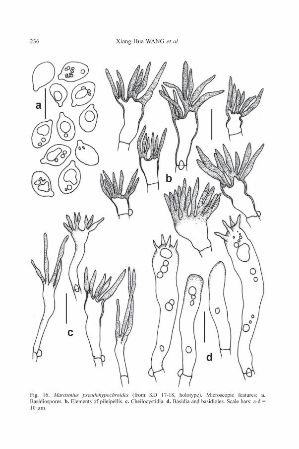
Fig. 16. Marasmius pseudohypochroides (from KD 17-18, holotype). Microscopic features: a.Basidiospores. b. Elements of pileipellis. c. Cheilocystidia. d. Basidia and basidioles. Scale bars: a-d =10 µm.

Fungal Biodiversity Profiles 51-60 237
57. Radulomyces paumanokensis J. Horman, B. Ortiz, & Nakasone, sp. nov.Figs. 17-19
MycoBank: MB 825443GenBank: MG050101 (nrLSU); MG050100 (ITS).Systematic positon: Basidiomycota,Agaricomycetes,Agaricales, PterulaceaeEtymology: Paumanok is the Native American name for Long Island, the
type locality.Diagnosis: Different from other Radulomyces species by its compact,
hemispherical to ovoid basidiocarps composed of an aggregation of fasciculate,branching, pendant spines.
Holotype: UNITED STATES, New York, Long Island, Welwyn PreserveCounty Park, Glen Cove, 40.880258, –73.639101, on bark of rotted hardwood log,19 Nov. 2016, leg. A. Norarevian (NY 03304530, holotype; CFMR, isotype).
Basidiocarps a compact, hemispherical to ovoid cluster of radiating,branching, pendant spines, up to 50 × 50 mm, white, orange white to pale orange(5A(2–3), color names from Kornerup & Wanscher 1978) when fresh, pale orange(5A3), greyish orange (5B3), or orange grey (6B2) when dried. Spines slender, upto 20 mm long, firm, terete to compressed, fused at base then branching multipletimes, finally tapering to subacute apices, brittle when dried; surface smooth tofinely farinaceous; context dense, ceraceous, yellowish brown.
Hyphal system monomitic with clamped, generative hyphae. Spinescomposed of an inner core of agglutinated, distinct hyphae arranged in parallel,surrounded by a thickening subhymenium and hymenium; terminal hyphae at apexmostly undifferentiated but occasionally slightly capitate; tramal hyphae 2.3–3.5 µmdiam, clamped, rarely branched, walls hyaline, thin to slightly thickened, smooth,weakly to distinctly cyanophilous. Subhymenium thickening, up to 90 µm thick, adense tissue of short-celled, agglutinated hyphae perpendicular to spine axis;subhymenial hyphae 3–3.5 µm diam, clamped, branched, walls hyaline, thin, smooth,acyanophilous. Hymenium up to 45 µm thick, a dense palisade of basidia andhyphidia. Hyphidia simple, filamentous, rarely subcapitate, 20–35 × 3–3.5 µm,clamped at base, walls hyaline, thin, smooth. Cystidia rare, embedded, clavate,42– 52 × 6.5–7 µm, clamped at base, walls hyaline, thin, smooth. Basidia scarce,clavate to cylindrical, sometimes nearly pleural, 25–31 × 5–7.5 µm, clamped at base,sometimes stalked, 4-sterigmate, walls hyaline, thin, smooth. Basidiospores globoseto subglobose, (5.7–) 5.8–6.9 × (5.1–) 5.2–6.4 (–6.5) µm, average of type specimens6.1–6.2 × 5.7–5.9 µm, Q = 1.1 ± 0.1, walls hyaline, thin to slightly thick, smooth,cyanophilous, not reacting in Melzer’s reagent.
Habitat and distribution: On angiospermous wood in eastern New York.Other specimen examined: New York, Long Island, E. Norwich, Muttontown
Preserve, 40.8294, –73.5330, on rotten hardwood log, 02 Oct 2016, leg. J. Horman(NY 03304531, CFMR)
Notes: Species of Radulomyces are characterized by widely effused, adnate,crust-like basidiocarps, but R. paumanokensis is unique for its compact basidiocarpscomposed of numerous branched spines. Microscopically, R. paumanokensis displaysfeatures typical of Radulomyces including simple hyphidia and large basidiosporeswith cyanophilous walls. Two other species of Radulomyces also develop largespines — R. copelandii (Pat.) Hjortstam & Spooner and R. molaris (Chaillet ex Fr.)M.P. Christ. Radulomyces copelandii produces extensive, effused basidiocarps withslender, white to yellow spines, 8–12 mm long, and subglobose basidiospores,6.4– 7 × 5.4–6.2 µm (Ginns & Millman 2011; Maekawa 1993). In contrast, R.
238 Xiang-Hua WANG et al.
molaris develops effused basidiocarps with stout, gray to cream-colored spines, 1–5mm long, and ellipsoid basidiospores, 8–12 × 6–7 (–8) µm (Bernicchia & Gorjón,2010; Martini 2016).
Some species of Mucronella Fr. and Deflexula Corner also producebasidiocarps composed of fasciculate, pendant spines. In Mucronella aggregata Fr.,the basidiospores are thin-walled and amyloid, whereas Deflexula species are dimiticwith skeletal hyphae and large basidiospores (> 9 µm diameter) (Corner 1950).
Phylogenetically, Radulomyces belongs in the Pterulaceae (Agaricales) andis most closely related to Radulotubus Y.C. Dai, S.H. He & C.L. Zhao andAphanobasidium Jülich. These three genera form a strongly supported clade that issister to the Pterula-Deflexula-Pterulicium-Merulicium-Coronicium clade asdemonstrated sequence analyses of the nrLSU or nuclear ribosomal large subunit(Zhao et al. 2016; data not shown). In Bayesian inference analyses of the ITS andnrLSU sequences alone or combined, R. paumanokensis always clustered with other
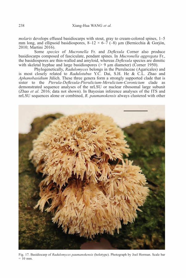
Fig. 17. Basidiocarp of Radulomyces paumanokensis (holotype). Photograph by Joel Horman. Scale bar= 10 mm.
Fungal Biodiversity Profiles 51-60 239
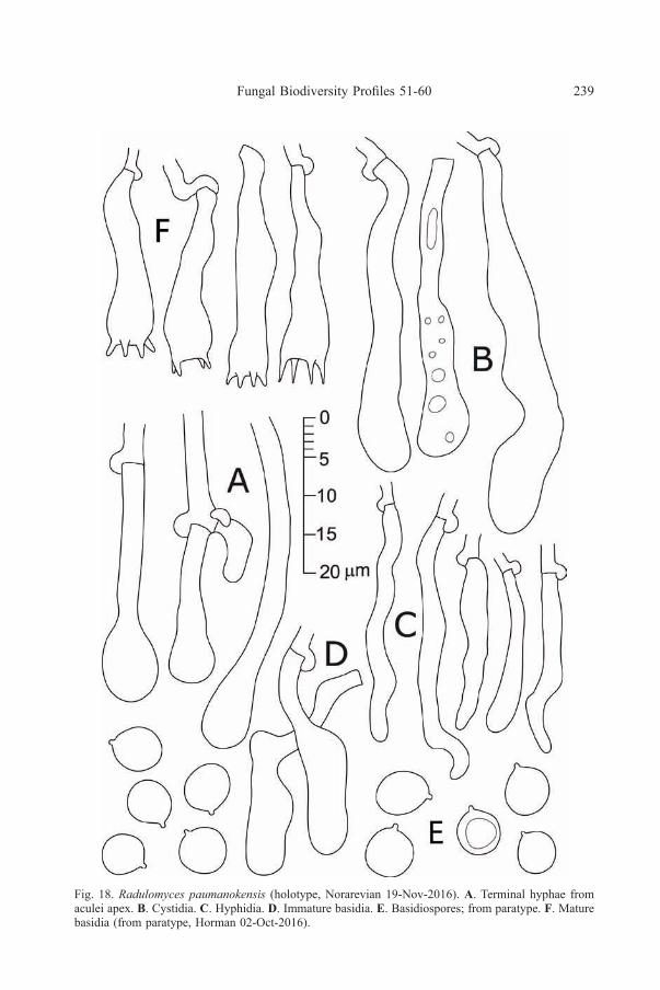
Fig. 18. Radulomyces paumanokensis (holotype, Norarevian 19-Nov-2016). A. Terminal hyphae fromaculei apex. B. Cystidia. c. Hyphidia. d. Immature basidia. e. Basidiospores; from paratype. F. Maturebasidia (from paratype, Horman 02-Oct-2016).

240 Xiang-Hua WANG et al.
Fig 19. Bayesian consensus tree from the analysis of the combined ITS+nrLSU dataset. The combineddataset consisted of 12 ingroup sequences (representing species of Radulomyces, Radulotubus andAphanobasidium) and one outgroup (Pterula echo) with a total of 1645 characters (719 ITS, 926nrLSU). Phylogenetic analysis was conducted using MrBayes 3.2.6 (Ronquist et al. 2012) on XSEDEthrough CIPRES Science Gateway V. 3.3 (Miller et al. 2010), for 1 000 000 generations in two runsand four chains with trees sampled every 100 generations. Posterior probability values are given alongbranches. GenBank no. for ITS/nrLSU sequences: Dai 15061 KU535664/KU535672, Wu 9606-5KU535663/KU535671, He 2224 KU535662/KU535670, Cui 5977 KU535661.KU535669, GEL 5394 –/AJ406571, IMG 5985-16 MG050100/MG050110, JK 951007 –/AY586706, Cui 8383 KU535660/KU535668, Dai 15315 KU535658/KU535666, HHB 822 GU187509/GU187567, A. pseudotsuage UC2023143 KP814192/NH10396 AY586696, AFTOL-ID711 DQ494693/AY629315.
Radulomyces species. With ITS alone or a combined ITS-nrLSU dataset ofRadulomyces, Radulotubus and Aphanobasidium species with Pterula echo asoutgroup, Radulomyces formed a strongly supported clade that is sister to Radulotubus(Fig. 19). In Bayesian analysis of nrLSU sequences alone, however, Radulotubuswas embedded in the Radulomyces clade (data not shown).
During our study, we discovered an ITS sequence of R. copelandii(MG722738) from northwest Georgia, near the Black Sea that is 99% identical toR. paumanokensis. We have not examined the voucher specimen from Georgia, butfrom sequence similarity alone, we conclude that these two taxa are conspecific;thus, the distribution of R. paumanokensis extends to Eurasia.

Fungal Biodiversity Profiles 51-60 241
58. Russula blennia Buyck sp. nov. Figs. 20−22Mycobank: MB 826913GenBank: KU237556 (LSU), KU237404 (mitSSU), KU237701 (RPB1),
KU237842 (RPB2), KU237987 (tef1alpha), MH545687 (ITS), all from holotypeDiagnosis: well-characterized by the combination of frequently forked
gills, a felty, brownish-gray pileus, white stipe, spore ornamentation of isolated, highwarts and spines, inamyloid but verrucose suprahilar spot and its habitat consistingof dense mountain forest types dominated by Uapaca species (mostly U. densifolia)
Holotype: MADAGASCAR. Central Plateau, special reserve ofAmbohitantely, NW of Anatanarivo, 30 km from Ankazobe, in Uapaca densifolia- Sarcolaenaceae dense dry forest, along the botanical track, 20 January 2008, Buyck08.066 (PC0124723)
Systematic position: Basidiomycota, Agaricomycetes, Russulales,Russulaceae
Etymology: named after the similarities in the field to European milk capsin the group of Lactarius blennius
Basidiomata isolated or in small groups. Pileus 24-64(75) mm diam.,regular, slightly depressed in center, not becoming deeply infundibuliform, snoothnear margin; surface felty-velutinous, becoming viscose when wet, never glutinous,separable for ½ to 2/3 from margin inwards, continuous and smooth in the pileuscenter, very finely cracked under a handlens to coarsely cracked – areolate closer tothe margin, often superficially cracking and even deeply fissuring radially close tothe very pileus margin when fully expanded, colored in tones of isabelline to dirtyochraceous, pale gray to pale or even dark brown. Gills adnate to subfree, equal butfrequently forking at different distances from stipe, ca 3 mm high, anastomosingbrittle, normally spaced (ca 1L/mm), sordid cream (4A2) but often with a clearpinkish tinge in the field, staining dark brown in old age; gill edge even, cystidioseunder a hand lens, concolorous. Stipe central, (30)43-72 x (8)11-15 mm, cylindricalto slightly narrowing downward, smooth to longitudinally wrinkled, glabrous, verypale creamy to off-white, developing brownish-grayish stains when handled or withage, not very fragile, spongy-stuffed inside, developing sometimes whitish myceliumat the base. Context firm, whitish to pale yellowish, developing pale brown to palegray punctuations or parts, turning faintly pinkish when cut, reacting yellowish toFeSO4. Taste mild or slightly like soap. Odor weak, somewhat fruity (apples) toslightly disagreable. Spore print white.
Spores (sub)globose to shortly ellipsoid, (7.7)7.9-8.27-8.6(9.0) x (6.9)7.1-7.40-7.7(7.9)µm, Q = (1.05)1.07-1.12-1.16(1.21); ornamentation composed ofstrongly amyloid, isolated, high conical warts to even spines of variable dimensions,the largest ones up to 2 µm high, mixed with a variable number of much smallerinterstitial warts; suprahilar spot not amyloid but distinctly verrucose and occupiedby gradually smaller amyloid warts, in some more irregularly developed spores withlower and denser ornamentation and sometimes partly amyloid suprahilar spot;apiculus long and distinct. Basidia widest in their median part, 34-44 x 10-13 µm,four-spored; sterigmata robust and long, 7-10 x 2-2.5 µm. Hymenial gloeocystidiasubcylindrical to fusiformous, mostly 50 - 70 x 8 – 9 µm, nearly always minutelycapitate, near the gill edge very abundant, frequently obtuse rounded at the tip andoften filled with bright yellowish contents, not shorter than on gill sides and up to85 µm long (contrary to the general pattern in Russula), thin-walled; contentsyellowing, amorphous-guttulate to shortly crystalline, SV-negative. Subhymeniumpseudoparenchymatic, composed of small cells, neither deep nor particularly well-
242 Xiang-Hua WANG et al.
developed. Lamellar trama almost entirely composed of sphaerocytes. Marginalcells not differentiated, but lamellar edge more or less sterile because of the abundantgloeocystidia. Pileipellis orthochromatic in cresyl blue, a dense trichoderm ofascending hyphal extremities, mostly aggregated in conical tufts at the surface,entirely filled with brownish-yellowish contents (concentrated in individual guttuleswhen observed in Melzer for ex., but then becoming diffused, particularly afterKOH treatment); hyphal extremities originating from strongly incrusted hyphae,composed of chains of 3 – 5 more or less inflated cells that are branching, normally4 – 7 µm wide, but some - particularly near branching points - more inflated and upto 15 µm diam., sprouting or diverticulating at nearly every cell that is not terminal;terminal cell rarely longer than 40 µm, obtuse rounded, mostly slightly tapering nearthe end, subcylindrical to conical. Pileocystidia seemingly absent but actually rareand difficult to find, easiest to locate in the pileus center and best recognized by theirtypically globose – capitate tip, mostly long and cylindrical and originating at thesame level as the hyphal extremities, measuring for ex. 100 x 5 µm, also shorter andsometimes conical when more terminal, without well-differentiated contents. Clampconnections absent in all tissues.
Additional examined material: MADAGASCAR. Central Plateau.Mandraka, on mountain crest in dense, monospecific, hygrophilous forest remnantof Uapaca densifolia, 3 Feb 1997, Buyck 97.219 (PC0125063); Central Plateau,special reserve of Ambohitantely, NW of Anatanarivo, 30 km from Ankazobe, inUapaca densifolia - Sarcolaenaceae dense dry forest, along the botanical track, 20January 2008, Buyck 08.067 (PC0125064), Buyck 08.068 (PC0125065).
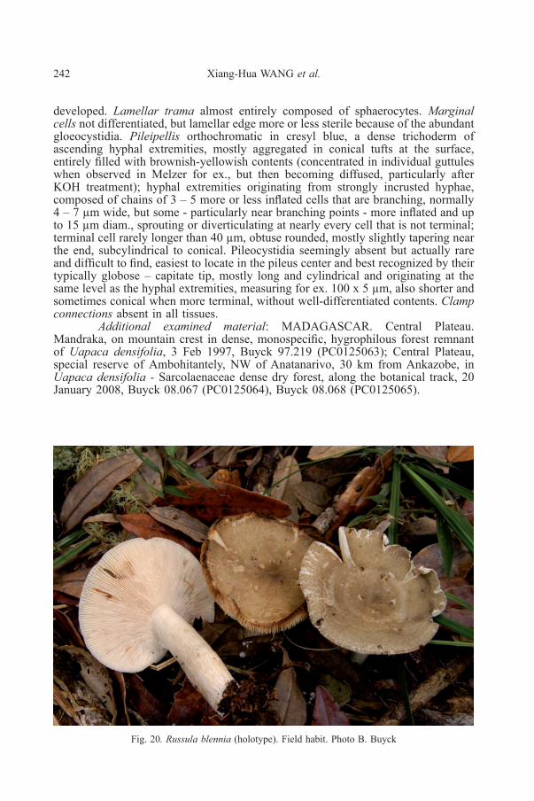
Fig. 20. Russula blennia (holotype). Field habit. Photo B. Buyck

Fungal Biodiversity Profiles 51-60 243
Fig 21. Russula blennia (holotype). Microscopic features. a. Pileocystidia (the longest ones originatingnear the base of the hyphal extremities); b. Hyphal extremities of the pileipellis; c. detail of a hyphalextremity of Buyck 97.219 showing the shorter, more inflated and more pigmented cells in that collection.Scale bar = 10 µm. Drawings B.Buyck.

244 Xiang-Hua WANG et al.
Fig 22. Russula blennia (holotype). Microscopic features. a. Spores; b. Basidia and basidiola;c. Gloeopleurocystidia; d. Cheilogloeocystidia. Cystidial contents are mostly schematic. Scale bar = 10µm, but only 5 µm for spores. Drawings B. Buyck

Fungal Biodiversity Profiles 51-60 245
Notes: This species seems well characterized by its habitat in which it canbe locally common. It has phylogenetically been placed in Russula subg. Malodora(Buyck et al. 2018). With the exception of the North American R. compacta Peck(see Adamcik et al. 2018) and R. griseobrunnea McNabb, described from NewZealand, this subgenus entirely composed of Malagasy or mainland African species.Identical gloeocystidia as the pileocystidia, and equally rare, are present on theectomycorrhizal outer mantle (see Buyck et al. 2018). This subgenus has recentlybeen split in two sections, one of which, sect. Edules Buyck & V. Hofst. (in Das etal. 2017), is composed of species with frequently forking gills. Our species belongsin the latter section.
59. Russula pseudociliata Buyck sp. nov. Figs. 23-25Mycobank: MB 826914GenBank: KU237537 (LSU), KU237383 (mitSSU), KU237686 (RPB1),
KU237823 (RPB2), KU237967 (tef1alpha), MH545688 (ITS), all from holotypeDiagnosis: differs from R. annulatolutea in the absence of a ring, the lack
of reddish or purplish-lilac tinges on stipe and pileus, the clavate pileocystidia at thevery pileus surface and in its association with trees that do not belong toCaesalpinaceae.
Holotype:MADAGASCAR. Central Plateau, Ambohitantely, in Uapacadensifolia - Sarcolaenaceae dense dry forest, 19 January 2008, Buyck 08.061(PC 0124721)
Systematic position: Basidiomycota, Agaricomycetes, Russulales,Russulaceae
Etymology: named after its resemblance in the field to the African R. ciliataBasidiomata in small groups. Pileus first strongly convex, developing
already a striate margin when still closed, at maturity becoming deeply depressed;margin strongly striate-tuberculate, even to very slightly wavy, locally radiallyfissured; surface smooth, continuous, forming a fine, rubbery pellicle in the center,detachable over ½ radius, lemon yellow with shades of olive, more brownish in thecenter. Gills equal, adnate, up to 2.5 mm high, off-white, fragile, without interstitialanastomoses or forkings; gill edge finely uneven to cystidiose, concolorous. Stipecentral, 25-37 x 6-9 mm, subcylindrical, tinged with the same yellowish olive as thepileus, particularly in the lower half, fragile, spongy inside, hollowing with age.Context whitish, unchanging, extremely thin (1 mm in the center). Odor insignificant.Taste mild. Spore print not obtained, certainly very pale.
Spores globose to subglobose, (6.7)6.9-7.20-7.5(7.9) x (6.0)6.2-6.53-6.8(7.1) µm, Q = (1.03)1.06-1.10-1.15(1.21); ornamentation strongly amyloid,reticulate to subreticulate, composed of rather spaced crests of aligned and mostlyfused cylindrical to conical warts, up to 2 µm high, practically without any isolatedwarts or spines, subtle linear connections rare to absent; suprahilar spot inamyloidto partly amyloid in ill-developed spores, verrucose or with very low reticulation inthe distal part; apiculus well-developed. Basidia rather short, (35)39-44(51) x 10-12µm, widest in the middle portion, four-spored; sterigmata robust, mostly 7-10 x 2-3µm. Hymenial gloeocystidia arising from the subhymenium or the underlyinglamellar trama, on lamellar sides mostly 50-90(100) x 10-12(15) µm, minutelymucronate, thin-walled; near the gill edge distinctly shorter and mostly with thickerwalls, generally 30-48 x 7-9 µm; contents quite abundant, crystalline. Marginal cellsonly occupying the very edge, mixed with gloeocystidia, resembling the terminalcells of the hyphal extremities of the pileipellis but somewhat larger, 7-8(10) µmdiam., conical, lageniform, to shortly clavate or almost ellipsoid. Subhymenium a

246 Xiang-Hua WANG et al.
pseudoparenchyma composed of rather small cells. Lamellar trama composed ofvoluminous sphaerocytes. Pileipellis orthochromatic in Cresyl blue, two-layered,with a distinctly delimited suprapellis of chains of inflated, short cells sitting on awell-delimited and strongly gelatinized subpellis of narrow hyphae that is abruptlyseparated from the voluminous sphaerocytes that make up nearly the entire underlyingcontext. Suprapellis a trichoderm formed of nearly vertical hyphal extremitiescomposed of chains of (3)4-5 very small, short, barrel-shaped to ellipsoid or morerarely near-cylindrical cells, 5-10 µm wide and all of more or less similar diam. orslightly narrowing upwards; the terminal cell 6-12(20) µm long, obtuse-rounded ormore rarely somewhat papillate, with some cells giving the impression of beingabruptly divided in two equal parts by a horizontal, secondary septum. Subpellis aloose tissue of very narrow hyphae, dispersed by an interstitial gelatinized matrix.Pileocystidia dispersed, not abundant but easily recognizable because of theircontents, near the surface originating from the hyphal extremities, small and short,mostly 15-25 x 5-7 µm, generally clavate and obtuse-rounded at the tip, thoseimmerged in the underlying subpellis up to 50 µm long and 4-6 µm diam., narrowlycylindrical or fusiformous to nearly conical, mostly minutely mucronate, all thin-walled; contents not abundant, concentrated in packets or part of the cystidium,finely granular to crystalline or amorphous, SV-negative. Cystidioid hyphae absentfrom subpellis and underlying context; oleiferous hyphae or hyphal fragments veryapparent and frequent, running over long distances throughout the context. Clampconnections absent.
Notes: This species from Madagascar’s Central Plateau is undeniably closeto R. annulatolutea Beeli and R. annulatosquamosa Beeli, two annulate speciesdescribed from the African central rain forest area in the Democratic Republic of theCongo (Beeli 1936). The former species was placed in R. subsect. Paradermatinae
Fig. 23. Russula pseudociliata (holotype). Field habit. Photo B. Buyck

Fungal Biodiversity Profiles 51-60 247
Fig 24. Russula pseudociliata (holotype). a. Pileocystidia (the longer ones at the bottom from thesubpellis, near the top as terminal cells of hyphal extremities); b. Hyphal extremities of the pileipellis.Scale bar = 10 µm. Drawings B.Buyck.

248 Xiang-Hua WANG et al.
Fig 25. Russula pseudociliata (holotype). Microscopic features a. Basidia and basidiola; b. Spores;c. Marginal cells; d. Gloeopleurocystidia; e. Cheilogloeocystidia. Cystidial contents are mostlyschematic. Scale bar = 10 µm, but only 5 µm for spores. Drawings B. Buyck.

Fungal Biodiversity Profiles 51-60 249
Buyck although a large part of the holotype was missing (the entire hymenophoreand most of the pileus) with the remainder of the type being strongly molded. Thesecond species, R. annulatosquamosa, was placed in R. subsect. PseudoepitheliosinaeBuyck (see also Buyck 1994), a group of small, pellicular species with brightreddish-violet-purplish tinges on stipe and pileus and wider hyphal extremitiescomposed of lesser cells giving the impression, in surface view, of an epitheliumintermixed with distinct pileocystidia. Subsect. Paradermatinae was essentiallydiffering in the larger number of cells composing the hyphal extremities and theexclusive presence of very narrow, unapparent, mucronate pileocystidia. Since then,we are more inclined to think that these differences are not that important as alsosuggested by our multigene phylogeny (Buyck et al. 2018) which places both R.pseudocarmesina (Pseudoepitheliosinae) and our new species in a fully supportedclade together with species of the R. annulata Heim and R. roseoalba Buyck complex(subsect. Heterophyllinae s.l.).
60. Urnula himalayana K. Das & D. Chakr. sp. nov. Figs. 26-28MycoBank: MB 824952GenBank: MH179122 (ITS), MH179125 (LSU)Systematic position: Ascomycota, Pezizomycetes, Pezizales,
Sarcosomataceae.Etymology: Referring to Himalaya, the type locality.Diagnosis: Distinct from all other similar looking Urnula species mainly
by its ITS and LSU sequence data and a combination of moderately robust (2-14 cmdiam.), subsessile to distinctively stalked apothecia with entire to irregularlyinterrupted cup margin, a velvety external surface without warts becoming veined-folded toward stipe, large thick-walled ascospores (24-33.6 × 10-13.8 µm) and itsoccurrence on decaying Castanopsis-trunk in temperate broadleaf forests.
Holotype: INDIA, Sikkim, South District, Pipaley, N27°14.830’ -E88°21.629’, 2082 m a.s.l., on decaying tree-trunk of Castanopsis sp. in temperatebroadleaf forest, 23 August, 2017, Kanad Das, KD 17(2)-17 (CAL 1673, holotype!).
Apothecia 2-14 cm diam., saucer- or bowl-shaped cup, subsessile todistinctively stalked; margin irregularly lobbed or interrupted, rarely entire.Hymenium surface reddish brown (9E4-5) or violet brown (10E4-10F3) to greyishbrown (11E-F3) and greyish ruby (12E4), to dark brown with age. External surfacevelvety, towards juncture to stem folded or veined, veins joined to give reticulateappearance, blackish blue (19F5-20F4) to brownish black. Stalk (when present) upto 17 mm high, usually vertically corrugated or wrinkled.
Asci 300-410 × 14-17 µm, cylindrical, operculate, inamyloid, eight-sporedwith a tapered or attenuated base. Paraphyses filiform, not or slightly extending thelength of the asci, 2-4 µm wide, cylindrical, closely septate; tips simple to branched,straight to curved, rounded to capitate with extracellular brownish, amorphouspigment in the upper part. Hymenial hairs cylindrical, as long as paraphyses, 3-5µm wide, aseptate but with a basal septum; tips rounded to slightly subcapitate,curved to almost hooked; pale brownish, especially in the upper part due to thepresence of same type of pigments as in paraphyses. Ascospores smooth, curvedwith rounded poles, allantoid to bean-shaped, wall 2 µm, 24-29.2-33.6 × 10-12.1-13.8 µm (n= 30, Q= 2.0-2.37-2.8). Subhymenium of a dense textura intricata ofclosely septate hyphae, up to 2 µm wide with thickened dark brown wall, 100-135µm thick. Medullary excipulum of loose textura intricata immersed in a gelatinousmatrix, with cylindrical, hyaline, septate hyphae, 4-6 µm wide, wall slightlythickened, 0.8 µm. Ectal excipulum of textura globosa to textura angularis, 50 µm
250 Xiang-Hua WANG et al.
thick, made up of elements 17-126 µm wide and/ or high, cells are angular toirregular, wall up to 2 µm thick, very dark brown. External hairs mainly of twotypes: 1) short, hyphoid, cylindrical, 6-7 µm wide, septate, subhyaline to very paleyellowish-brown colored, wall 1.5 µm thick, heavily encrusted by an amber brownto dark brown pigments; 2) long, true hairs, 5-6 µm, thick walled (wall up to 1 µmthick), brown to dark brown, encrusted by the brown pigments. Subiculum darkbrown. Ectal excipulum (stalk) similar to that of the cup. External hairs (stalk) long,cylindrical, septate, brown, thick-walled (up to 1.2 µm).
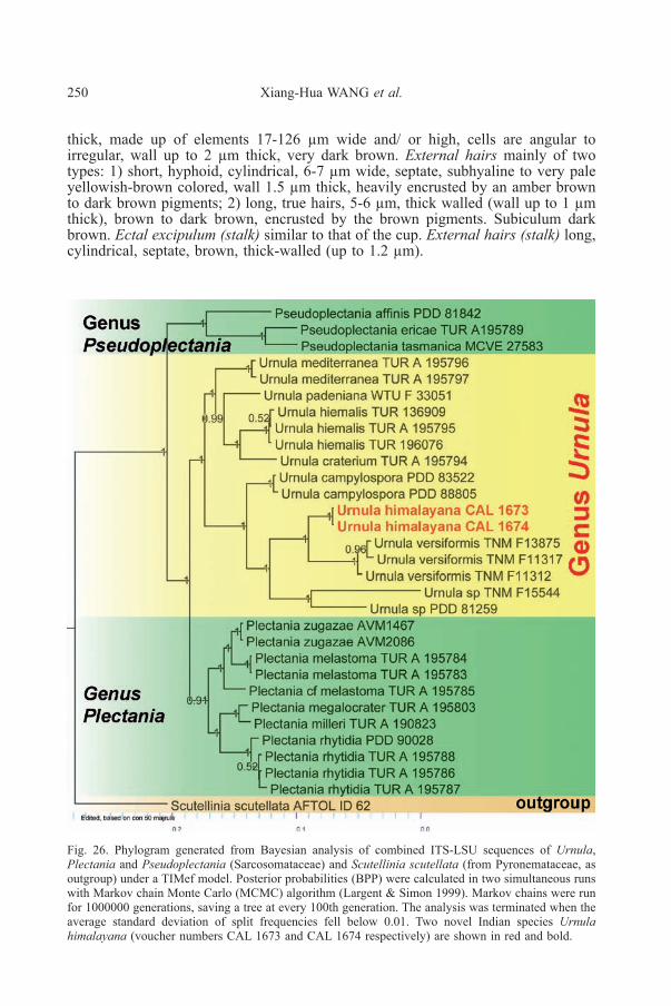
Fig. 26. Phylogram generated from Bayesian analysis of combined ITS-LSU sequences of Urnula,Plectania and Pseudoplectania (Sarcosomataceae) and Scutellinia scutellata (from Pyronemataceae, asoutgroup) under a TIMef model. Posterior probabilities (BPP) were calculated in two simultaneous runswith Markov chain Monte Carlo (MCMC) algorithm (Largent & Simon 1999). Markov chains were runfor 1000000 generations, saving a tree at every 100th generation. The analysis was terminated when theaverage standard deviation of split frequencies fell below 0.01. Two novel Indian species Urnulahimalayana (voucher numbers CAL 1673 and CAL 1674 respectively) are shown in red and bold.

Fungal Biodiversity Profiles 51-60 251
Table 1: Fungal names, collection information and GenBank accession numbers
name of the species with voucher Id lsu ITs location
Plectania rhytidia(TUR-A 195787)
JX669851 JX669815 Italy
Plectania rhytidia(TUR-A 195786)
JX669849 JX669813 Italy
Plectania rhytidia(TUR-A 195788)
JX669852 JX669816 Italy
Plectania rhytidia(PDD 90028)
JX669871 JX669832 New Zealand
Plectania megalocrater(TUR-A 195803)
JX669845 JX669809 Greece
Plectania milleri(TUR-A 190823)
JX669848 JX669812 USA
Plectania zugazae(AVM1467)
JX669854 JX669817 Spain
Plectania zugazae(AVM2086)
JX669855 JX669818 Spain
Plectania melastoma(TUR-A 195784)
JX669850 JX669814 Italy
Plectania melastoma(TUR-A 195783)
JX669841 JX669805 Italy
Plectania cf. melastoma(TUR-A 195785)
JX669840 JX669804 USA
Urnula mediterranea(TUR-A 195796)
JX669844 JX669808 Italy
Urnula mediterranea(TUR-A 195797)
JX669864 JX669824 France
Urnula padeniana(WTU-F-33051)
JX669866 JX669825 USA
Urnula campylospora(PDD 83522)
JX669869 JX669830 New Zealand
Urnula campylospora(PDD 88805)
JX669870 JX669831 New Zealand
Urnula hiemalis(TUR 136909)
JX669867 JX669827 Finland
Urnula hiemalis(TUR 196076)
JX669868 JX669828 Finland
Urnula hiemalis(TUR-A 195795)
JX669873 JX669835 Finland
Urnula himalayana(cAl 1673)
Mh179122 Mh179125 India
Urnula himalayana(cAl 1674)
Mh179123 Mh179124 India

252 Xiang-Hua WANG et al.
Specimens examined: INDIA, Sikkim, South district, Pipaley,N27°14.830’E88°21.629’, 2082 m a.s.l., on decaying tree-trunk of Castanopsis sp.in temperate broadleaf forest, 23 August, 2017, Kanad Das, KD 17(2)-17 (CAL1673, holotype!); ibid., Sikkim, South district, Kewzing, N27°16.781’E88°20.300’,2082 m a.s.l., on decaying tree-trunk of Castanopsis sp. in temperate broadleafforest, 25 August, 2017, Kanad Das, KD 17-33 (CAL 1674, paratype!).
Notes: The family Sarcosomataceae Kobayasi is divided into six distinctgenera: Plectania Fuckel, Sarcosoma Casp., Galiella Nannf. & Korf, Urnula Fr.,Pseudoplectania Fuckel and Donadinia Bellem. & Mel.-Howel. Macro- andmicromorphological features among these genera are very much overlapping andtherefore, morphology alone is not sufficient to separate them. Genetic data areneeded to correctly place these taxa.
The genus Urnula Fr. is one of the prominent and older genera in thisfamily (Fries 1849). At present, it is represented by 12-13 species (www.indexfungorum.org, Carbone & Agnello 2013), with only six species reported in thelatest (10th) edition of the Dictionary of Fungi (Kirk et al. 2008).
In our combined ITS-LSU phylogenetic (Bayesian) analysis (Fig. 26)sequences isolated from two of our Indian collections [MH179122, MH179125 asITS and LSU respectively from KD 17-2-17 and MH179123, MH179124 as ITS andLSU respectively from KD 17-33, see Table 1] clustered together and nested amongthe other Asian species of Urnula being sister to three sequences from threecollections of U. versiformis Y.Z. Wang & C.L. Huang (TNM F13875, TNM F11317,TNM F11312), however our species was recovered as a distinct taxon with strongsupport (BPP = 1). Again, the clade bearing U. himalayana (proposed novel species)and U. versiformis was sister to a clade that bears two other unknown (not yet
Urnula criterium(TUR-A 195794)
JX669857 JX669820 Italy
Urnula versiformis(TNM F13875)
Not available KJ577536 Taiwan
Urnula versiformis(TNM F11317)
Not available KJ577535 Taiwan
Urnula versiformis(TNM F11312)
Not available KJ577534 Taiwan
Urnula sp.(TNM F15544)
Not available KJ577537 Taiwan
Urnula sp.(PDD 81259)
Not available JX669829 New Zealand
Pseudoplectania affinis(PDD 81842)
JX669865 JX669826 New Zealand
Pseudoplectania ericae(TUR-A195789)
JX669862 JX669822 Spain
Pseudoplectania tasmanica(MCVE 27583)
KF305732 KF305722 Australia
Scutellinia scutellate(AFTOL-ID 62)
DQ247806 DQ491492 --

Fungal Biodiversity Profiles 51-60 253
Fig. 27. Urnula himalayana (KD 17-2-17, holotype). a. Fresh basidiomata in the field. b. Freshbasidiomata on a workout table in the basecamp.
described) Asian species. Moreover, the clade bearing these four Asian species issister to U. campylospora (Berk.) Cooke.
Morphologically, all the species of Urnula show very similar features infield. Even under microscope all have same arrangement of the excipulum, which isdivided into a medullary excipulum (showing textura intricate) and an ectalexcipulum (showing textura subglobulosa-angularis pattern with thin- to thik-walled

254 Xiang-Hua WANG et al.
Fig. 28. Urnula himalayana (KD 17-2-17, holotype). a. Cross section through apothecia. b. Ascishowing eight ascospores. c. Short external hairs. d. Long external hair. e. External hairs of stalk. f.Cells of Ectal excipulum. g. Ascospores. Scale bars: a = 100 µm, b-d, f-g = 10 µm, e = 20 µm.

Fungal Biodiversity Profiles 51-60 255
hairs rising from the latter). Further, all the species possess unbranched or branchedhymenial hairs and paraphyses (Carbone et al. 2013). Still, the combination offeatures like very large ascomata (apothecia) without any warty or horizontallycorrugated external surface, subsessile to distinctly stalked base and allantoidascospores are quite distinct. With similar morphology only known Asian species U.versiformis is separated from U. himalayana by possessing distinctively smaller(1.4–4 cm diam.) apothecia (Wang & Huang 2015). European species U.campylospora has significantly smaller (up to 5 cm) apothecia with horizontallycorrugated external surface (Carbone & Agnello 2013).
Acknowledgments. K. Das and coauthors are grateful to the Director, BotanicalSurvey of India, Kolkata and Director, MACS’ Agharkar Research Institute, Pune forproviding facilities. KD & MEH are also thankful to Aniket Ghosh (H.N.B. GarhwalUniversity, India) in many ways, and to Upendra Singh, Priyanka Uniyal and Tahir Mehmoodfor providing field assistance in Uttarakhand. The studies of Vladimír Antonín at the MoravianMuseum were supported by the Ministry of Culture of the Czech Republic as part of itslong-term conceptual development programme for research institutions (DKRVO, ref.MK000094862). X.H. Wang is grateful to Dr. S.H. Wu, Taiwan Science Museum and Prof.T.S. Lee, Incheon University, Korea for the help on field trips and loan of specimens. Thefield work in China was financed by National Natural Science Foundation of China (NSFC,grant nos. 30970020, 31093440, 31493010 and 31493011) to XHW. B. Buyck is grateful toE. Randrianjohany and staff members of CNRE in Antananarivo for logistic support and fieldassistance. The field work in Madagascar was financed in part by the National GeographicSociety (grants 6365-98, 7921-05) and in more recent years by the ATM-project “Past andpresent biodiversity” of the Muséum national d’histoire naturelle (Dirs. Ph. Janvier and S.Peigné)
reFerences
ADAMČÍK S., JANČOVIČOVÁ S. & BUYCK B., 2018— The Russulas described by Charles HortonPeck. Cryptogamie, Mycologie 39(1): 3 -108
ANTONÍN V. & NOORDELOOS M.E., 2010 — A monograph of marasmioid and collybioid fungi inEurope. Eching, Germany, 478 p.
ANTONÍN V., RYOO R. & SHIN H.-D. 2010 — Marasmioid and gymnopoid fungi of the Republic ofKorea. 2. Marasmius sect. Globulares. Persoonia 24: 49-59.
BASSO M.T., 1999 — Lactarius Pers. Fungi Europaei 7: 1–845.BEELI M., 1936 — Contribution à l’étude de la Flore mycologique du Congo. XI.- Fungi Goossensiani.
Bulletin du Jardin Botanique de Belgique 14 : 83-91, Pl.2.BERNICCHIA A. & GORJÓN S.P., 2010 — Corticiaceae s.l. Fungi Europaei n. 12. Ed. E. Candusso.
Alassio, Italy. 1007 p.BESSETTE A.E., BESSETTE A.R. & FISCHER D.W., 1997 — Mushrooms of Northeastern North
America. 1st Ed., Syracuse University Press, USA.BUYCK B., 1994 Russula II (Russulaceae). In: Rammeloo J, Heinemann P (Éds.). Flore illustrée des
Champignons d’Afrique Centrale, 16:411–542, pl 69–87BUYCK B., DUHEM B., DAS K., JAYAWARDENA R.S., NIVEIRO N., PEREIRA O.L., PRASHER
I.B., ADHIKARI S., ALBERTÓl E.O., BULGAKOV T.S., CASTAÑEDA-RUIZ R.F.,HEMBROM M.E., HYDE K.D., LEWIS D.P., MICHLIG A., NUYTINCK J., PARIHAR A.,POPOFF O.F., RAMIREZ N.A., da SILVA M., VERMA R.K. & HOFSTETTER V., 2017— Fungal Biodiversity Profiles 21-30. Cryptogamie Mycologie 38(1): 101-146.
BUYCK B., ZOLLER S. & HOFSTETTER V., 2018 — Walking the thin line… ten years later: thedilemma of above- versus below-ground features to support phylogenies in the Russulaceae(Basidiomycota). Fungal Diversity (online first), doi.org/10.1007/s13225-018-0397-5.
CARBONE M. & AGNELLO C. 2013 — Notes on Urnula campylospora (Pezizales). Ascomycete.org5 (1): 25–32.

256 Xiang-Hua WANG et al.
CARBONE M., AGNELLO C. & ALVARADO P. 2013 — Phylogenetic studies in the familySarcosomataceae (Ascomycota, Pezizales). Ascomycete.org 5 (1): 1–12.
CORNER E.J.H., 1950 — A monograph of Clavaria and allied genera. Beihefte zur Nova Hedwigia 33:1-740.
DAS K., GHOSH A., BHATT R.P., CHAKRABORTY D., HOFSTETTER V. & BUYCK B., 2017a— Fungal Biodiversity Profiles 41-50. Cryptogamie, Mycologie 38 (4): 527-547.
DESJARDIN D.E., RETNOWATI A. & HORAK E. 2000 — Agaricales of Indonesia. 2. A preliminarymonograph of Marasmius from Java and Bali. Sydowia 52: 92-193.
FENG B., WANG X-H., RATKOWSKY D., GATES G., LEE S.S., GREBENC T. & YANG Z.L. 2016— Multilocus phylogenetic analyses reveal unexpected abundant diversity and significantdisjunct distribution pattern of the Hedgehog Mushrooms (Hydnum L.). Scientific Reports.DOI: 10.1038/srep25586
FRIES E.M. 1849 — Summa Vegetabilium Scandinaviae. Sectio posterior. 259-572. Stockholm, Leipzig,Bonnier.
GINNS J. & Millman L., 2011 — Mysterious Asian beauty conquers eastern Massachusetts. Fungi 4(3):61-63.
HALLING R.E., 1983. — A synopsis of Marasmius section Globulares (Tricholomataceae) in theUnited States. Brittonia 35: 317-326.
HEILMANN-CLAUSEN J., VERBEKEN A., VESTERHOLT J., 1998 — The genus Lactarius. 2.Denmark. Svampetryk.
HESLER L.R, SMITH A.H., 1979 — North American species of Lactarius. Ann Arbor: University ofMichigan Press.
KIRK P.M., CANNON P.F., MINTER D.W. & STALPERS J.A. — 2008 Ainsworth & Bisby’s dictionaryof the fungi. 10th Ed., CABI, UK.
KORNERUP A. & WANSCHER J.H., 1978 — Methuen Handbook of Colour. London, Eyre Methuen.252 p.
KORNERUP A. & WANSCHER J.H., 1981 — Methuen handbook of colour. 3rd Ed. London, Methuen.LARGENT B. & SIMON D.L., 1999 — Markov chain Monte Carlo algorithms for the Bayesian
analysis of phylogenetic trees. Molecular Biology and Evolution 16: 750-759.MAAS GEESTERANUS R.A., 1971 — Hydnaceous fungi of the eastern old world. North-Holland
Publishing Company, UK, 582 pp.MAEKAWA N., 1993 — Taxonomic study of Japanese Corticiaceae (Aphyllophorales) I. Reports of the
Tottori Mycological Institute 31: 1-149.MARTINI E., 2016 — Radulomyces molaris. Excerpts from Crusts & Jells 1(41): 1-8.MILLER M.A., PFEIFFER W. & SCHWARTZ T., 2010 — Creating the CIPRES Science Gateway for
inference of large phylogenetic trees. Proceedings of the Gateway Computing EnvironmentsWorkshop (GCE), New Orleans, LA, pp 1-8.
MONTOYA L., BANDALA V.M., 2008 — A new species and new records of Lactarius (subgenusRussularia) in a subtropical cloud forest from eastern Mexico. Fungal Diversity 29: 61-72.
OLARIAGA I., GREBENC T., SALCEDO I. & MARTÍN M.P., 2012 — Two new species of Hydnumwith ovoid basidiospores: H. ovoideisporum and H. vesterholtii. Mycologia 104: 1443-1455.
OSTROW H. & BEENKEN L., 2004 — Hydnum ellipsosporum spec. nov. (Basidiomycetes,Cantharellales) – ein Doppelgänger von Hydnum rufescens Fr. Zeitschrift für Mykologie70(2), 137–156.
PHILLIPS R., 2006. — Mushrooms. Macmillan, EnglandRONQUIST F., TESLENKO M., VAN DER MARK P., AYRES D.L., DARLING A., HOHNA S.,
LARGET B., LIU L., SUCHARD M.A. & HUELSENBECK J.P., 2012 — MrBayes 3.2:Efficient Bayesian phylogenetic inference and model choice across a large model space.Systematic Biology 61: 539–542.
SHARMA J.R. 2012 — Aphyllophorales of Himalaya. Botanical Survey of India, Kolkata.SHI S.F., WANG X.H., BAU T., 2018 — Three new species of Lactarius from northeastern China.
Mycoscience 59: 206-217.SILVESTRO D. & MICHALAK I., 2012 — raxmlGUI: a graphical front-end for RAxML. Org Divers
Evol 12:335–337. doi:10.1007/s13127-011-0056-0TAMURA K., STECHER G., PETERSON D., FILIPSKI A. & KUMAR S. 2013. MEGA6: Molecular
Evolutionary Genetics Analysis version 6.0. Molecular Biology and Evolution 30: 2725–2729.
TRIANTAFYLLOU M., POLEMIS E., GONOU-ZAGOU Z., DIMOU D.M., DELIVORIAS P.,ZERVAKIS G.I., 2015 — Taxonomic relationships among non-macrocystidiate taxa ofLactarius subg. Russularia from Europe with special reference to species from Greece.Mycological Progress 14: 78.

Fungal Biodiversity Profiles 51-60 257
VIZZINI A., PICILLO B., ERCOLE E., VOYRON S. & CONTU M. 2013 — Detecting the variabilityof Hydnum ovoideisporum (Agaricomycetes, Cantharellales) on the basis of Italian collections,and H. magnorufescens sp. nov. Mycosphere 4(1): 32–44.
WANG X.H., 2007 — Type study on the Lactarius species published from China. Mycologia 99: 253–268.
WANG X.H., LIU P.G., 2002 — Lactarius chichuensis and L. hirtipes, two easily confused species.Mycotaxon 84: 391-400.
WANG Y.Z. & HUANG C.L. 2015 — A new species of Urnula (Sarcosomataceae, Pezizales) fromTaiwan. Nova Hedwigia 101 (1–2): 49–55.
WANNATHES N., DESJARDIN D.E., HYDE K.D., PERRY B.A. & LUMYONG S., 2009 — Amonograph of Marasmius (Basidiomycota) from Northern Thailand based on morphologicaland molecular (ITS sequences) data. Fungal Diversity 37: 209-306
WISITRASSAMEEWONG K., NUYTINCK J., FELIX H., HYDE K.D., VERBEKEN A., 2014.Lactarius subgenus Russularia (Russulaceae) in South-East Asia: 2. Species with remarkablysmall basidiocarps. Phytotaxa 188: 181−197.
WISITRASSAMEEWONG K., NUYTINCK J., LE H.T., DE CROP E., HAMPE F., HYDE K.D.,VERBEKEN A., 2015 — Lactarius subgenus Russularia (Russulaceae) in South-East Asia:3. new diversity in Thailand and Vietnam. Phytotaxa 207 (3): 215-241.
ZHAO C.-L., CHEN H., HE S.-H. & DAI Y.-C., 2016 — Radulotubus resupinatus gen. et sp. nov. witha poroid hymenophore in Pterulaceae (Agaricales, Basidiomycota). Nova Hedwigia 103(1-2):265-278.

![Rohrbacher. Dufour. Histoire universelle de l'Église Catholique. [1842-1849]. T01. histoireuniverse01rohr](https://static.fdocuments.fr/doc/165x107/577d2eaa1a28ab4e1eafadb2/rohrbacher-dufour-histoire-universelle-de-leglise-catholique-1842-1849.jpg)


![I A ,XX8U -2017/2016..] 'i 8)1426 .(2005 1849 .05 0537772043 0537771870 — •](https://static.fdocuments.fr/doc/165x107/5e6064b58fca3a475f009da1/-i-a-xx8u-20172016-i-81426-2005-1849-05-0537772043-0537771870-a-a.jpg)