Brugada Syndrome: Report of the Second Consensus...
14
Arthur Wilde Andres Ricardo Perez Riera, Wataru Shimizu, Eric Schulze-Bahr, Hanno Tan and Brugada, Domenico Corrado, Ihor Gussak, Herve LeMarec, Koonlawee Nademanee, Charles Antzelevitch, Pedro Brugada, Martin Borggrefe, Josep Brugada, Ramon Brugada Syndrome: Report of the Second Consensus Conference : ISSN: 1524-4539 Copyright © 2005 American Heart Association. All rights reserved. Print ISSN: 0009-7322. Online 72514 Circulation is published by the American Heart Association. 7272 Greenville Avenue, Dallas, TX doi: 10.1161/01.CIR.0000152479.54298.51 2005, 111:659-670: originally published online January 17, 2005 Circulation http://circ.ahajournals.org/content/111/5/659 located on the World Wide Web at: The online version of this article, along with updated information and services, is http://circ.ahajournals.org/http://circ.ahajournals.org/content/112/4/e74.full.pdf An erratum has been published regarding this article. Please see the attached page for: http://www.lww.com/reprints Reprints: Information about reprints can be found online at [email protected] 410-528-8550. E-mail: Fax: Kluwer Health, 351 West Camden Street, Baltimore, MD 21202-2436. Phone: 410-528-4050. Permissions: Permissions & Rights Desk, Lippincott Williams & Wilkins, a division of Wolters http://circ.ahajournals.org//subscriptions/ Subscriptions: Information about subscribing to Circulation is online at by guest on January 3, 2012 http://circ.ahajournals.org/ Downloaded from
Transcript of Brugada Syndrome: Report of the Second Consensus...

Arthur WildeAndres Ricardo Perez Riera, Wataru Shimizu, Eric Schulze-Bahr, Hanno Tan and
Brugada, Domenico Corrado, Ihor Gussak, Herve LeMarec, Koonlawee Nademanee, Charles Antzelevitch, Pedro Brugada, Martin Borggrefe, Josep Brugada, Ramon
Brugada Syndrome: Report of the Second Consensus Conference :
ISSN: 1524-4539 Copyright © 2005 American Heart Association. All rights reserved. Print ISSN: 0009-7322. Online
72514Circulation is published by the American Heart Association. 7272 Greenville Avenue, Dallas, TX
doi: 10.1161/01.CIR.0000152479.54298.512005, 111:659-670: originally published online January 17, 2005Circulation
http://circ.ahajournals.org/content/111/5/659located on the World Wide Web at:
The online version of this article, along with updated information and services, is
http://circ.ahajournals.org/http://circ.ahajournals.org/content/112/4/e74.full.pdf An erratum has been published regarding this article. Please see the attached page for:
http://www.lww.com/reprintsReprints: Information about reprints can be found online at
[email protected]. E-mail:
Fax:Kluwer Health, 351 West Camden Street, Baltimore, MD 21202-2436. Phone: 410-528-4050. Permissions: Permissions & Rights Desk, Lippincott Williams & Wilkins, a division of Wolters
http://circ.ahajournals.org//subscriptions/Subscriptions: Information about subscribing to Circulation is online at
by guest on January 3, 2012http://circ.ahajournals.org/Downloaded from

Brugada SyndromeReport of the Second Consensus Conference
Endorsed by the Heart Rhythm Society and the European HeartRhythm Association
Charles Antzelevitch, PhD; Pedro Brugada, MD, PhD; Martin Borggrefe, MD, PhD;Josep Brugada, MD; Ramon Brugada, MD; Domenico Corrado, MD, PhD; Ihor Gussak, MD, PhD;
Herve LeMarec, MD; Koonlawee Nademanee, MD; Andres Ricardo Perez Riera, MD;Wataru Shimizu, MD, PhD; Eric Schulze-Bahr, MD; Hanno Tan, MD, PhD; Arthur Wilde, MD, PhD
Abstract—Since its introduction as a clinical entity in 1992, the Brugada syndrome has progressed from being a raredisease to one that is second only to automobile accidents as a cause of death among young adults in some countries.Electrocardiographically characterized by a distinct ST-segment elevation in the right precordial leads, the syndrome isassociated with a high risk for sudden cardiac death in young and otherwise healthy adults, and less frequently in infantsand children. Patients with a spontaneously appearing Brugada ECG have a high risk for sudden arrhythmic deathsecondary to ventricular tachycardia/fibrillation. The ECG manifestations of Brugada syndrome are often dynamic orconcealed and may be unmasked or modulated by sodium channel blockers, a febrile state, vagotonic agents,�-adrenergic agonists, �-adrenergic blockers, tricyclic or tetracyclic antidepressants, a combination of glucose andinsulin, hypo- and hyperkalemia, hypercalcemia, and alcohol and cocaine toxicity. In recent years, an exponential risein the number of reported cases and a striking proliferation of articles defining the clinical, genetic, cellular, ionic, andmolecular aspects of the disease have occurred. The report of the first consensus conference, published in 2002, focusedon diagnostic criteria. The present report, which emanated from the second consensus conference held in September2003, elaborates further on the diagnostic criteria and examines risk stratification schemes and device andpharmacological approaches to therapy on the basis of the available clinical and basic science data. (Circulation. 2005;111:659-670.)
Key Words: arrhythmia � death, sudden � electrocardiography � diagnosis
Since its introduction as a clinical entity in 1992,1 theBrugada syndrome has attracted great interest because of
its high incidence in many parts of the world and itsassociation with high risk for sudden death in young andotherwise healthy adults and, less frequently, in infants andchildren. In recent years, an exponential rise in the number ofreported cases and a striking proliferation of articles definingthe clinical, genetic, cellular, ionic, and molecular aspects ofthe disease have occurred.2 A consensus report published in2002 focused on diagnostic criteria for the syndrome.3,4 Thepresent report, emanating from the second consensus confer-ence held in September 2003, elaborates further on thediagnostic criteria and examines risk stratification schemes
and device and pharmacological approaches to therapy. Therecommendations herein are based on available clinical andbasic science data and should be considered a work inprogress that will require modification as additional data frommolecular and clinical studies and prospective trials becomeavailable.
Clinical Characteristics and EpidemiologyThe Brugada syndrome is characterized by an ST-segmentelevation in the right precordial ECG leads (so-called type 1ECG; Figures 1 to 3) and a high incidence of sudden death inpatients with structurally normal hearts. The syndrome typi-cally manifests during adulthood, with a mean age of sudden
From the Masonic Medical Research Laboratory, Utica, NY (C.A., R.B.); Cardiovascular Center, Cardiovascular Research and Teaching Institute,Aalst, Belgium (P.B.); University of Heidelberg, University Hospital of Mannheim, Mannheim, Germany (M.B.); Cardiovascular Institute, ClinicalHospital, University of Barcelona, Barcelona, Spain (J.B.); Divisione di Cardiología, Università di Padova, Padova, Italy (D.C.); eResearch Technology,Inc, Bridgewater, NJ (I.G.); Chu de Nantes, Nantes, France (H.L.); Pacific Rim Electrophysiology Research Institute, Inglewood, Calif (K.N.); ABC’sFaculty of Medicine, ABC Foundation, Santo André, São Paulo, Brazil (A.R.P.R.); National Cardiovascular Center, Suita, Japan (W.S.); Department ofCardiology, University of Münster, and Institute for Arteriosclerosis Research, Münster, Germany (E.S.-B.); and Experimental and Molecular CardiologyGroup, Academic Medical Center, Amsterdam, and the Interuniversity Cardiology Institute, Utrecht, the Netherlands (H.T., A.W.).
This report will be copublished in Heart Rhythm.Correspondence to Dr Charles Antzelevitch, Gordon K. Moe Scholar, Masonic Medical Research Laboratory, 2150 Bleecker St, Utica, NY 13501.
E-mail [email protected]© 2005 American Heart Association, Inc.
Circulation is available at http://www.circulationaha.org DOI: 10.1161/01.CIR.0000152479.54298.51
659
Special Report
by guest on January 3, 2012http://circ.ahajournals.org/Downloaded from
death of 41�15 years. The youngest patient clinically diag-nosed with the syndrome is 2 days old and the oldest is 84years old. The syndrome is estimated to be responsible for atleast 4% of all sudden deaths and at least 20% of suddendeaths in patients with structurally normal hearts. The prev-alence of the disease is estimated to be 5/10 000 inhabitantsand, apart from accidents, is the leading cause of death in men�40 years old, particularly in countries in which the syn-drome is endemic.5 Because the ECG pattern can be dynamicand is often concealed, it is difficult to estimate the trueprevalence of the disease in the general population.6 In arecent Japanese study, a Brugada syndrome ECG (type 1) wasobserved in 12/10 000 inhabitants; type 2 and 3 ECGs, whichare not diagnostic of Brugada syndrome, were much moreprevalent, appearing in 58/10 000 inhabitants.7 The preva-lence of the Brugada syndrome among the general populationin Europe and the United States is thought to be muchlower,8,9 although among Southeast Asian immigrants it maybe as high as it is in Southeast Asia itself.10
Sudden unexplained nocturnal death syndrome (SUNDS;also known as SUDS) and Brugada syndrome have recentlybeen shown to be phenotypically, genetically, and function-ally the same disorder.11
Approximately 20% of patients with Brugada syndromedevelop supraventricular arrhythmias.12 Atrial fibrillation isassociated in 10% to 20% of cases. Atrioventricular (AV)nodal reentrant tachycardia and Wolff-Parkinson-White syn-drome also have been described.13 Prolonged sinus noderecovery time and sinoatrial conduction time,14 as well asslowed atrial conduction and atrial standstill, have beenreported in association with the syndrome.15 A recent study
reported that ventricular inducibility is positively correlatedwith a history of atrial arrhythmias.16 In patients with anindication for an implantable cardioverter defibrillator (ICD),the incidence of atrial arrhythmias was 27% versus 13% inpatients without an indication for an ICD (P�0.05), whichsuggests a more advanced disease process in patients withBrugada syndrome and spontaneous atrial arrhythmias. Inap-propriate shocks from atrial arrhythmia episodes were ob-served in 14% of cases, highlighting the need for carefulprogramming of the ICD.15
Diagnostic Criteria and RecommendationsThree ECG repolarization patterns in the right precordialleads are recognized.3,4 Type 1 is diagnostic of Brugadasyndrome and is characterized by a coved ST-segmentelevation �2 mm (0.2 mV) followed by a negative T wave(Figure 1). Brugada syndrome is definitively diagnosed whena type 1 ST-segment elevation is observed in �1 rightprecordial lead (V1 to V3) in the presence or absence of asodium channel–blocking agent, and in conjunction with oneof the following: documented ventricular fibrillation (VF),polymorphic ventricular tachycardia (VT), a family history ofsudden cardiac death at �45 years old, coved-type ECGs infamily members, inducibility of VT with programmed elec-trical stimulation, syncope, or nocturnal agonal respiration.The ECG manifestations of the Brugada syndrome, whenconcealed, can be unmasked primarily by sodium channelblockers but also during a febrile state or with vagotonicagents.17–20 Drug challenge generally is not performed inasymptomatic patients displaying the type 1 ECG underbaseline conditions because the additional diagnostic value isconsidered to be limited, the added prognostic value is not
Figure 1. Twelve-lead ECG tracings inan asymptomatic 26-year-old man withBrugada syndrome. Left, Baseline: type2 ECG (not diagnostic) displaying asaddleback-type ST-segment elevation isobserved in V2. Center, After intravenousadministration of 750 mg procainamide,the type 2 ECG is converted to the diag-nostic type 1 ECG, which consists of acoved-type ST-segment elevation. Right,A few days after oral administration ofquinidine bisulfate (1500 mg/d, serumquinidine level 2.6 mg/L), ST-segmentelevation is attenuated displaying a non-specific abnormal pattern in the rightprecordial leads. VF could be inducedduring control and procainamide infusionbut not after quinidine. Reprinted withpermission from Belhassen et al.110
Copyright 2002, Blackwell Publishing.
660 Circulation February 8, 2005
by guest on January 3, 2012http://circ.ahajournals.org/Downloaded from

clear, and the test is not without risk for provoking arrhythmicevents.
Importantly, confounding factor or factors that could ac-count for the ECG abnormality or syncope should be care-fully excluded, including atypical right bundle-branch block,left ventricular hypertrophy, early repolarization, acute peri-carditis, acute myocardial ischemia or infarction, pulmonaryembolism, Prinzmetal angina,21 dissecting aortic aneurysm,22
various central and autonomic nervous system abnormali-ties,23,24 Duchenne muscular dystrophy,25 thiamin deficien-cy,26 hyperkalemia,22,27,28 hypercalcemia,29,30 arrhythmogenicright ventricular dysplasia/cardiomyopathy,31,32 pectus exca-vatum,33 hypothermia,34,35 and mechanical compression ofthe right ventricular outflow tract (RVOT) as occurs inmediastinal tumor36 or hemopericardium.37
Of note, a Brugada-like ECG can occasionally appear for abrief period or for a period of several hours after direct-current cardioversion; it is not known whether these patientsare gene carriers for Brugada syndrome.38–40
Another prominent confounding factor is the type ofST-segment elevation encountered in well-trained athletes(Figure 4), which is distinguished by an upslope rather than a
Figure 2. Shift of right precordial leads to 2nd and 3rd intercos-tal spaces unmasks a type 1 Brugada ECG. Top, Plot of 87unipolar electrode sites (●) and of 6 precordial ECGs (✖).Eighty-seven lead points are arranged in a lattice-like pattern(13�7 matrix), except for 4 lead points on both midaxillary lines,and covered the entire thoracic surface. V1 and V2 leads of theECG are located between D5 and E5 and between E5 and F5,respectively, whereas V4, V5, and V6 are coincident with G4, H4,and I4, respectively. Bottom, Twelve-lead ECGs in a patient withBrugada syndrome. Type 2 saddleback-type ST-segment eleva-tion was observed in V1 and V2 of the standard 12-lead ECG(4th intercostal space), whereas typical type 1 coved-typeST-segment elevation was apparent in V1 and V2 recorded fromthe 2nd and 3rd intercostal spaces (4).
Figure 3. 12-lead ECG of a 40-year-oldresuscitated man. The type 1 ECGshows typical repolarization as well asdepolarization abnormalities. The formerconsists of ST-segment elevation inleads V1 and V2 (coved type, type 1).Depolarization abnormalities are presentas PQ prolongation (270 ms), prolongedQRS width (120 ms), and S waves inleads I, II, and III. P wave duration is alsowide.
Figure 4. ECG of a well-trained, asymptomatic 24-year-old soc-cer player. ST-segment elevation is observed in V2 to V6 butwith characteristics totally different from those seen in Brugadasyndrome. A coved-type ST-segment elevation is not observed.A rounded or upsloping ST elevation in seen in V2 and V3,whereas V4 to V5 show a pattern resembling that commonlyencountered in early repolarization syndrome.
Antzelevitch et al Brugada Syndrome Consensus Report 661
by guest on January 3, 2012http://circ.ahajournals.org/Downloaded from

downslope and by remaining largely unaffected by challengewith a sodium channel blocker. In addition, a variety of drugshave been reported to produce a Brugada-like ST-segmentelevation (Table 1), although it is not yet clear whether or towhat extent a genetic predisposition may be involved. Theinclusion of drug categories in Table 1 should not beinterpreted to imply that other members of the same “class”necessarily produce similar effects.
Although most cases of Brugada syndrome display rightprecordial ST-segment elevation, isolated cases of inferiorlead41 or left precordial lead42 ST-segment elevation havebeen reported in Brugada-like syndromes; in some cases theyhave been associated with SCN5A mutations.43
The type 2 ST-segment elevation has a saddleback appear-ance with a high takeoff ST-segment elevation of �2 mm, atrough displaying �1 mm ST elevation, and then eithera positive or biphasic T wave (Figure 1). Type 3 has either asaddleback or coved appearance with an ST-segment eleva-tion of �1 mm. Type 2 and type 3 ECG are not diagnostic ofthe Brugada syndrome. These 3 patterns may be observedspontaneously in serial ECG tracings from the same patient orafter the introduction of specific drugs. The diagnosis ofBrugada syndrome is also considered positive when a type 2
(saddleback pattern) or type 3 ST-segment elevation isobserved in �1 right precordial lead under baseline condi-tions and conversion to the diagnostic type 1 pattern occursafter sodium channel blocker administration (ST-segmentelevation should be �2 mm). One or more of the clinicalcriteria described above also should be present. Drug-inducedconversion of type 3 to type 2 ST-segment elevation isconsidered inconclusive for a diagnosis of Brugadasyndrome.
Placement of the right precordial leads in a superiorposition (up to the second intercostal space above normal)can increase the sensitivity of the ECG for detecting theBrugada phenotype in some patients, both in the presence orabsence of a drug challenge (Figure 2).44,45 Although previ-ous reports suggested that none of the control patientsdisplayed type 1 ST elevation when the V1 to V3 leads weredisplaced upward,44,45 a prospective study with a largernumber of controls will be required to exclude the possibilityof false-positive results via this method.
A slight prolongation of the QT interval is sometimesobserved in association with ST-segment elevation in Bru-gada syndrome.46–48 The QT interval is prolonged more inthe right precordial leads than it is in the left precordial leads,presumably because of a preferential prolongation of actionpotential duration in right ventricular epicardium secondaryto accentuation of the action potential notch.49 Depolarizationabnormalities (Figure 3), including prolongation of P waveduration and PR and QRS intervals, are frequently observed,particularly in patients linked to SCN5A mutations.50 PRprolongation likely reflects HV conduction delay.46
In addition to Brugada syndrome, ST-segment elevation isassociated with a wide variety of benign as well as malignantpathophysiological conditions. A differential diagnosis is attimes difficult, particularly when the degree of ST-segmentelevation is relatively small and the specificity of sodiumchannel blockers such as flecainide, ajmaline, procainamide,disopyramide, propafenone, and pilsicainide18,48,51 to identifypatients at risk is uncertain. The recommended dosages arelisted in Table 2. The test should be monitored with acontinuous ECG recording (a speed of 10 mm/s can be usedthroughout the test period, interposed with recordings at 25 or50 mm/s) and should be terminated when the diagnostic type1 Brugada ECG develops, the ST segment in type 2 ECGincreases by �2 mm, premature ventricular beats or otherarrhythmias develop, or QRS widens to �130% of baseline.Intravenous sodium channel blockers always should be ad-ministered with great caution and infused slowly (as recom-mended in Table 1), closely monitored, and performed in asetting that is fully equipped for resuscitation. Particularcaution should be exercised in patients with a preexisting
TABLE 1. Drug-Induced Brugada-Like ECG Patterns
I. Antiarrhythmic drugs
1. Na� channel blockers
Class IC drugs (flecainide,18,51,65,117,118 pilsicainide,119,120 propafenone121)
Class IA drugs (ajmaline,18,122 procainamide,18,19 disopyramide,4,19
cibenzoline123)
2. Ca2� channel blockers
Verapamil
3. �-Blockers
Propranolol, etc
II. Antianginal drugs
1. Ca2� channel blockers
Nifedipine, diltiazem
2. Nitrate
Isosorbide dinitrate, nitroglycerine124
3. K� channel openers
Nicorandil
III. Psychotropic drugs
1. Tricyclic antidepressants
Amitriptyline,125,126 nortriptyline,77 desipramine,75 clomipramine76
2. Tetracyclic antidepressants
Maprotiline125
3. Phenothiazine
Perphenazine,125 cyamemazine127
4. Selective serotonin reuptake inhibitors
Fluoxetine126
IV. Other drugs
1. Dimenhydrinate78
2. Cocaine intoxication79,128
3. Alcohol intoxication
TABLE 2. Drugs Used to Unmask Brugada Syndrome
Drug Dosage and Administration
Ajmaline 1 mg/kg over 5 min, IV
Flecainide 2 mg/kg over 10 min, IV (400 mg, PO)
Procainamide 10 mg/kg over 10 min, IV
Pilsicainide 1 mg/kg over 10 min, IV
662 Circulation February 8, 2005
by guest on January 3, 2012http://circ.ahajournals.org/Downloaded from

atrial or ventricular conduction (or both) disturbance (eg,suspected cases of Lev or Lenègre disease) or in the presenceof wide QRS, wide P waves, or prolonged PR intervals (ie,infranodal conduction disease) to avoid the risk of precipitat-ing complete AV block. Mechanoelectrical dissociation hasbeen encountered in isolated cases. Isoproterenol and sodiumlactate may be effective antidotes in this setting.
Patients at high risk for drug-induced AV block, such asolder adults with syncope, should be administered sodiumchannel blockers in an electrophysiological study (EPS)environment after the insertion of a temporary pacing elec-trode. For other individuals, especially younger patients,sodium blocker challenge can be safely performed as abedside test, provided the drug is discontinued as soon asexcessive ST-segment elevation, QRS widening, or ventric-ular ectopy is observed.
Differentiation From ARVC and Other StructuralHeart DiseasesA subpopulation of arrhythmogenic right ventricular cardio-myopathy (ARVC) patients have been found to display anST-segment elevation and polymorphic VT that is character-istic of Brugada syndrome.32 In addition, a case has beenreported in which a patient with a Brugada syndrome pheno-type required heart transplantation because of untreatablearrhythmias52 and in whom severe fibrosis of the rightventricle was subsequently reported. These facts notwith-standing, the vast majority of patients with Brugada syn-drome possess a structurally normal heart, which is consistentwith the notion that this is a primary electrical heart disease.53
It is not unreasonable to speculate that fibrosis and myocar-ditis, however mild, may occur and may exacerbate or indeedtrigger events in patients with Brugada syndrome, althoughdefinitive evidence in support of this hypothesis is lacking. Itis worth noting that recent studies suggest that some SCN5Adefects may be capable of causing fibrosis in the conductionsystem and ventricular myocardium.54
ARVC and Brugada syndrome are distinct clinical entitiesboth with regard to clinical presentation and genetic predis-position.4 The only gene thus far linked to Brugada syndromeis SCN5A, the gene that encodes for the � subunit of thecardiac sodium channel, whereas ARVC has been linked to10 different chromosomal loci and 3 putative genes indepen-dent of those responsible for Brugada syndrome.55,56 Only theARVC5 locus has been mapped to a region that overlaps withthe second locus for Brugada syndrome, but no gene has beenidentified as yet.57,58 Imaging techniques such as ECG,angiography, MRI, and radionuclide scintigraphy show noevidence of overt structural heart disease in patients withBrugada syndrome, whereas ARVC patients characteristi-cally display right ventricular morphological and functionalchanges (eg, global dilatation, bulgings/aneurysms, and wallmotion abnormalities). Ventricular arrhythmias in ARVC aremost commonly monomorphic VT (left bundle-branch blocktype), which is often precipitated by catecholamines orexercise and accounts for sudden death in young competitiveathletes.59 In contrast, ST-segment elevation and arrhythmiasin patients with Brugada syndrome are enhanced by va-gotonic agents or �-adrenergic blockers, and polymorphic VT
occurs most commonly during rest or sleep.60 In contrast tothose in Brugada syndrome, the ECG abnormalities in ARVCare not dynamic and display a constant T-wave inversion,epsilon waves, and, in the progressive stage, reduction of theR amplitude. These are largely unaffected by sodium channelblocker administration.4
Electron beam computed tomography has uncovered wallmotion abnormalities in a series of patients with Brugadasyndrome.61 Although these contractile abnormalities arecommonly considered pathognomonic of structural disease,recent studies62,63 suggest that such contractile dysfunctioncan result from loss of the action potential dome in regions ofthe right ventricular epicardium and thus may be unrelated toany type of morphological defect. Loss of the dome leads tocontractile dysfunction because the entry of calcium into thecells is greatly diminished and sarcoplasmic reticulum cal-cium stores are depleted. Signal-averaged ECG recordingshave demonstrated late potentials in patients with Brugadasyndrome, especially in the anterior wall of the RVOT,64,65
and recordings from the epicardial surface of the anterior wallof the RVOT have revealed delayed potentials.66 Althoughthese types of potentials are commonly considered to berepresentative of the delayed activation of the myocardiumsecondary to structural defects, recent studies suggest that inthe case of Brugada syndrome these late and delayed poten-tials may represent the delayed second upstroke of theepicardial action potential or local phase 2 reentry.63 Latepotentials also may reflect intraventricular conduction delaysassociated with SCN5A defects. Delayed contractile activa-tion of the right ventricle in patients with Brugada syn-drome67 likewise may reflect delayed impulse propagation or,alternatively, a delayed second upstroke and action potentialdome in the right ventricular epicardium.
Genetic Factors Underlying Brugada SyndromeInheritance of Brugada syndrome occurs via an autosomaldominant mode of transmission. The first and only gene to belinked to Brugada syndrome is SCN5A, the gene that encodesfor the � subunit of the cardiac sodium channel gene.68 Morethan 80 mutations in SCN5A have been linked to the syn-drome since 2001.69–73 About 2 dozen of these mutationshave been studied in expression systems and shown to resultin loss of function because of failure of the sodium channel toexpress; a shift in the voltage and time dependence of sodiumchannel current (INa) activation, inactivation, or reactivation;entry of the sodium channel into an intermediate state ofinactivation from which it recovers more slowly; or acceler-ated inactivation of the sodium channel. A second locus onchromosome 3, close to but apart from the SCN5A locus, waslinked recently to Brugada syndrome57 in a large pedigree inwhich the syndrome is autosomal dominant inherited andassociated with progressive conduction disease, a low sensi-tivity to procainamide, and a relatively benign prognosis.SCN5A mutations account for �18% to 30% of Brugadasyndrome cases. A higher incidence of SCN5A mutations hasbeen reported in familial than in sporadic cases.74 Of note,negative SCN5A results do not rule out causal gene mutationsbecause, in general, the promoter region, cryptic splicing
Antzelevitch et al Brugada Syndrome Consensus Report 663
by guest on January 3, 2012http://circ.ahajournals.org/Downloaded from

mutations, or the presence of gross rearrangements is notinvestigated.
At present, knowledge of a specific mutation may notprovide guidance in formulating a diagnosis or determining aprognosis. Genetic testing is recommended, however, tosupport the clinical diagnosis, for early detection of relativesat potential risk, and to advance through research our under-standing of the genotype–phenotype relationship.
Modulating and Precipitating FactorsThe ECG manifestations of congenital Brugada syndrome areoften concealed but can be unmasked or modulated bysodium channel blockers, a febrile state, vagotonic agents,�-adrenergic agonists, �-adrenergic blockers, tricyclic ortetracyclic antidepressants, a combination of glucose andinsulin, hyperkalemia, hypokalemia, hypercalcemia, and al-cohol and cocaine toxicity (Figure 5).17–19,75–81 These agentsmay also induce acquired forms of Brugada syndrome (Table1). Until a definitive list of drugs to avoid in Brugadasyndrome is formulated, the list of agents in Table 1 mayprovide some guidance.
Acute myocardial infarction or ischemia from vasospasminvolving the RVOT mimics ST-segment elevation similar tothat in Brugada syndrome. This effect is likely the result of adepression of calcium channel current (ICa) and the activationof ATP-sensitive potassium channel current (IK-ATP) duringischemia, and it suggests that patients with congenital andpossibly acquired forms of Brugada syndrome may be at ahigher risk for ischemia-related sudden cardiac death.82
VF and sudden death in Brugada syndrome usually occurat rest and at night. Figure 6 shows the circadian pattern of 64VF episodes in 19 SUNDS patients treated with ICD. Circa-dian variation of sympathovagal balance, hormones, and
other metabolic factors are likely to contribute to this circa-dian pattern. Bradycardia resulting from altered autonomicbalance or other factors may contribute to the initiation ofarrhythmia.83–85
Wichter et al demonstrated an abnormal 123I-m-iodobenzylguanidine (123I-MIBG) uptake in 8 (47%) of 17patients with Brugada syndrome, but 0 in the control group.86
Segmental reduction of 123I-MIBG occurred in the inferiorand the septal left ventricular walls, indicating presynapticsympathetic dysfunction. It is noteworthy that imaging of theright ventricle, particularly the RVOT, is difficult with this
Figure 5. Factors predisposing to theECG and arrhythmic manifestations ofBrugada syndrome.
Figure 6. Circadian pattern of VF episodes in patients with Bru-gada syndrome. A nocturnal increase of VF episodes was foundin 19 Thai-SUNDS patients with Brugada syndrome whoreceived an ICD. All VF episodes were detected and docu-mented by ICD interrogation. Interestingly, a significant numberof VF episodes were asymptomatic because these episodesoccurred while the patients were asleep (between 10 PM andearly-morning hours) and they did not experience ICDdischarges.
664 Circulation February 8, 2005
by guest on January 3, 2012http://circ.ahajournals.org/Downloaded from

technique, so insufficient information is available aboutsympathetic function in the regions known to harbor thearrhythmogenic substrate. Moreover, it remains unclear whatrole the reduced uptake function plays in the arrhythmogen-esis of Brugada syndrome. If the RVOT is similarly affected,then this defect may indeed alter the sympathovagal balancein favor of the development of an arrhythmogenicsubstrate.87,88
Hypokalemia has been implicated as a contributing causeof the prevalence of SUNDS in northeastern Thailand, wherepotassium deficiency is endemic.81,89 Serum potassium in thisnortheastern population is significantly lower than that of thepopulation in Bangkok, which lies in the central part ofThailand, where potassium is abundant in food.
The 1990 report of the Thai Ministry of Public Healthfound an association between a large meal of glutinous(“sticky”) rice or carbohydrates ingested on the night of deathin patients with SUNDS.89 Consistent with this observation, arecent study by Nogami et al found that glucose and insulincould unmask the Brugada ECG.80
Dumaine et al first demonstrated that premature inactiva-tion of the sodium channel in SCN5A mutations associatedwith Brugada syndrome is a function of temperature90 andsuggested that a febrile state may unmask Brugada syndrome.Indeed, several case reports have emerged recently demon-strating that febrile illness could unmask Brugada syndromeand precipitate VF.20,91–95 Anecdotal data point to hot baths asa possible precipitating factor. Of note, northeastern Thai-land, where Brugada syndrome is most prevalent, is knownfor its hot climate.
Risk Stratification and Current RecommendationsRisk stratification aimed at the identification of patients atrisk for sudden death is an important goal of research teamsworldwide.71,96–98 Brugada et al96 found that patients initiallypresenting with aborted sudden death are at the highest riskfor a recurrence (69% at 54�54 months of follow-up),whereas patients presenting with syncope and a spontane-ously appearing type 1 ECG have a recurrence rate of 19% at26�36 months of follow-up. An 8% occurrence of cardiacevents was observed in initially asymptomatic patients. Thisadverse prognosis was not observed in a population of similarsize by Priori et al,70 although the diagnostic criteria appliedin the 2 studies may have been different in that the report byPriori et al does not specify a requirement for a coved-typeECG (type 1) in �1 precordial leads as a means to diagnoseBrugada syndrome. Among asymptomatic patients, those athighest risk displayed the type 1 ECG spontaneously; patientsin whom ST-segment elevation appeared only after provoca-tion with sodium channel blockers appeared to be at minimalor no risk for arrhythmic events. Taken together, the dataindicate that asymptomatic Brugada patients at highest riskare men with inducible VT/VF and a spontaneously elevatedST segment (type 1 ECG).96
Recent studies have suggested that combined ECG markersmay be helpful in risk stratification. Atarashi et al used thewidth of the S wave and the ST-segment elevation magnitude,whereas Morita et al combined ST-segment elevation and the
presence of late potentials.99,100 The value of these combinedmarkers remains to be tested in a prospective study.
Brugada et al96 suggested that among asymptomatic pa-tients, the inducibility of VT/VF during EPS may forecastrisk. Studies by Priori et al,70 Kanda et al,97 and Eckardt etal,98 however, failed to find an association between induc-ibility and recurrence of VT/VF among both asymptomaticand symptomatic patients with Brugada syndrome. Thesediscrepancies may result from differences in patient charac-teristics and the use of nonstandardized or noncomparablestimulation protocols.13 The adverse prognosis and higherpredictive value of inducibility by Brugada et al may, at leastin part, be due to more demanding criteria for diagnosingpatients with Brugada syndrome.
It is noteworthy that programmed electrical stimulation–induced VF is observed in 6% to 9% of apparently healthyindividuals and may represent a false-positive and nonspe-cific response, particularly when aggressive stimulation pro-tocols are used.101
A protocol involving up to 3 extrastimuli applied to theright ventricular apex at cycle lengths �200 ms is recom-mended. If not inducible from the right ventricular apex, thenstimulation may be applied to the RVOT. The predictivevalue of EPS is based largely on right ventricular apexstimulation; the value of RVOT pacing for risk stratificationis not known. Although inducibility in experimental models ismost readily achieved with epicardial stimulation,88,102 clini-cal data involving this approach are limited.103 Clearly,additional studies are needed to define further the riskstratification strategy for asymptomatic patients.
A recent study by Brugada et al104 reported on 547individuals diagnosed with Brugada syndrome who had hadno previous cardiac arrest. In 124 patients, the abnormal ECGwas identified after �1 episode of syncope, and in 423individuals, the abnormal ECG was identified during routineECG screening or during study because they were familymembers of patients with the syndrome. Structural diseasewas ruled out in all patients. This study, which evaluated theclinical outcome of the largest population of patients withBrugada syndrome thus far reported, reached the followingconclusions:
1. Patients have a relatively high risk for sudden arrhythmicdeath, even in the absence of a history of cardiac arrest:8.2% experienced sudden death or at least one documentedepisode of VF during a mean follow-up of 24�33 months.Individuals with a spontaneously abnormal type 1 ECGcarried a 7.7-fold higher risk of developing an arrhythmicevent during a lifetime as compared with individuals inwhom the ECG diagnostic of Brugada syndrome wasevident only after sodium channel blocker challenge.
2. Male gender is another risk factor for sudden death. Menhad a 5.5-fold higher risk of sudden death than did women.
3. Programmed electrical stimulation that induces a sustainedventricular arrhythmia is the strongest marker of risk,associated with an 8-fold higher risk of (aborted) suddendeath than in noninducible patients.
4. Familial forms of the disease are not associated with aworse prognosis than are sporadic cases because a positive
Antzelevitch et al Brugada Syndrome Consensus Report 665
by guest on January 3, 2012http://circ.ahajournals.org/Downloaded from
family history of Brugada syndrome did not predictoutcome.
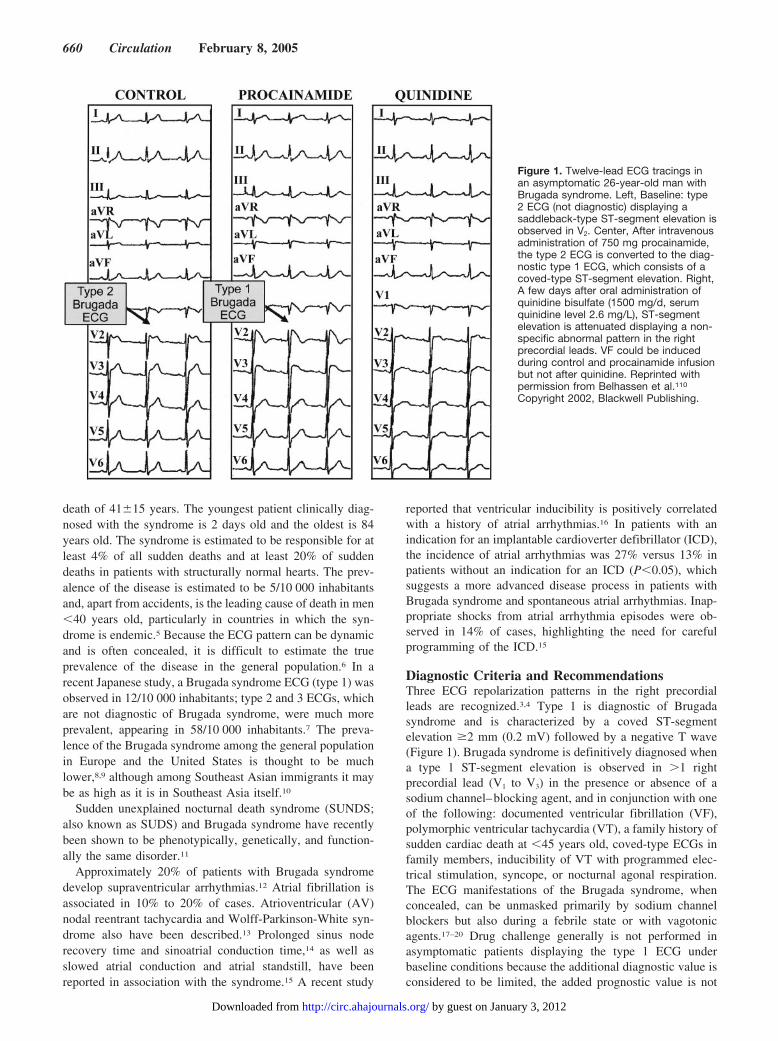
Therapeutic Recommendations forBrugada SyndromeThe important strides in the identification and characteriza-tion of Brugada syndrome during the past decade notwith-standing, progress relative to therapy has been less impres-sive. The various device and pharmacological therapies testedclinically or suggested on the basis of experimental evidenceare listed in Table 3. Currently, an ICD is the only proveneffective treatment for the disease.105,106 Of 690 patients withBrugada syndrome included in a multicenter registry, 258received an ICD because of a suspected high risk of suddenarrhythmic death. The stored electrograms were reviewed toassess the efficacy of the device by analyzing the number ofpatients that had an appropriate defibrillation of at least oneepisode of VF. The patients’ mean age at implantation was42�13.5 years, and 210 (81.3%) of these were men. A totalof 160 (62%) patients were symptomatic before establishingthe diagnosis; 120 patients (48.4%) had a family history ofsudden death, a familial Brugada ECG pattern, or both. Asustained ventricular arrhythmia was induced during the EPSin 198 patients (76.7%). During a mean follow-up of 2.5years (median 2), 1 patient died during an electrical storm,but 69 (26.7%) patients had at least one appropriate defibril-lation. The cumulative efficacy of the device was 18%, 24%,32%, 36%, and 38% at 1, 2, 3, 4, and 5 years of follow-up,respectively (Figure 7).
Recommendations for ICD implantation are summarized inFigure 8. Symptomatic patients displaying the type 1 BrugadaECG (either spontaneously or after sodium channel blockade)who present with aborted sudden death should receive an ICDwithout additional need for EPS. Similar patients presentingwith related symptoms such as syncope, seizure, or nocturnalagonal respiration also should undergo ICD implantation
after noncardiac causes of these symptoms have been care-fully ruled out. EPS is recommended in symptomatic patientsonly for the assessment of supraventricular arrhythmias.Asymptomatic patients displaying a type 1 Brugada ECG(either spontaneously or after sodium channel blockade)should undergo EPS if a family history of sudden cardiacdeath is suspected to be the result of Brugada syndrome. EPSis justified when the family history is negative for suddencardiac death if the type 1 ECG occurs spontaneously. Ifinducible for ventricular arrhythmia, then the patient shouldreceive an ICD. Asymptomatic patients who have no familyhistory and who develop a type 1 ECG only after sodiumchannel blockade should be closely followed up.
As additional data become available, these recommenda-tions will require further refinement. Until more specific dataare available, our recommendation with regard to patientswho manifest a spontaneous type 1 ECG only after placementof the right precordial leads in superior positions is to treatthem no differently from patients exhibiting a spontaneoustype 1 ECG with the leads in the standard positions.
ICD implantation may not be an adequate solution forinfants and young children or for patients who reside inregions of the world where an ICD is cost prohibitive.Although, in general, arrhythmias and sudden cardiac deathoccur during sleep or at rest and have been associated withslow heart rates, a potential therapeutic role for cardiacpacing remains largely unexplored. Data relative to a cryo-surgical approach or the use of ablation therapy are limited. Arecent report by Haissaguerre and coworkers107 points tofocal radiofrequency ablation as a potentially valuable tool incontrolling arrhythmogenesis by focal ablation of the ventric-ular premature beats that trigger VT/VF in Brugadasyndrome.
The pharmacological approach to therapy, based on exper-imental data, has been tailored to a rebalancing of currentsthat are active during the early phases of the epicardial actionpotential in the right ventricle to reduce the magnitude of the
TABLE 3. Device and Pharmacological Considerations forTherapy in Brugada Syndrome
Devices
� ICD—only established effective therapy
? Ablation or cryosurgery
? Pacemaker
Drugs
� Amiodarone: does not protect108
� �-Blockers: do not protect108
� �-Adrenergic agonists (isoproterenol19,44)
� Phosphodiesterase inhibitors (cilostazol116)
� Class IC antiarrhythmics (flecainide,propafenone): contraindicated
Class IA antiarrhythmics
� Procainamide: contraindicated
? Disopyramide129
� Quinidine88,110,111,130
? Tedisamil
� Ito blockers: cardioselective and ionchannel–specific
Figure 7. Kaplan-Meier curve of effectiveness of the ICD in 258patients with ECG pattern of Brugada syndrome according tosymptoms. Data are from the multicenter registry of 690patients with Brugada syndrome.
666 Circulation February 8, 2005
by guest on January 3, 2012http://circ.ahajournals.org/Downloaded from

action potential notch, restore the action potential dome, orboth (Table 3). Antiarrhythmic agents such as amiodaroneand �-blockers have been shown to be ineffective.108 Class ICantiarrhythmic drugs (eg, flecainide and propafenone) andclass IA agents (eg, procainamide) are contraindicated forreasons enumerated previously. Specific class IA agents suchas quinidine and tedisamil, however, may exert a therapeuticaction because of their Ito-blocking properties. Because thepresence of a prominent transient outward current, Ito, in theright ventricle is at the heart of the mechanism underlyingBrugada syndrome, any agent that inhibits this current may beprotective. Cardioselective and Ito-specific blockers are notavailable. The only agent on the US market with significantIto-blocking properties is quinidine. It is for this reason that itwas suggested that this agent may be of therapeutic value inBrugada syndrome.109 Studies have shown quinidine to beeffective in restoring the epicardial action potential dome,thus normalizing the ST segment and preventing phase 2reentry and polymorphic VT in experimental models ofBrugada syndrome.87 Clinical evidence of the effectiveness ofquinidine in normalizing ST-segment elevation in patientswith Brugada syndrome has been reported (see Figure
1),110,111 although clinical trials designed to assess the effi-cacy of this agent are limited.112 Relatively high doses ofquinidine are recommended (1200 to 1500 mg/d). Agents thatboost the L-type calcium current, such as isoproterenol, maybe useful as well.69,87 Both types of agents (Ito blocker andagents that augment ICa) have been shown to be effective innormalizing ST-segment elevation in patients with Brugadasyndrome and in controlling “electrical storms,” particularlyin children.44,110,111,113,114 Other than the studies by Belhassenand coworkers involving quinidine, none have as yet demon-strated long-term efficacy in the prevention of sudden cardiacdeath.110,115 The most recent addition to the pharmacologicalarmamentarium is a phosphodiesterase III inhibitor, cilosta-zol,116 which normalizes the ST segment most likely byaugmenting the calcium current (ICa), as well as by reducingIto secondary to an increase in heart rate. Finally, an experi-mental antiarrhythmic agent, tedisamil, with potent action toblock Ito among other outward currents has been suggested asa therapeutic candidate.69 Tedisamil may be more potent thanquinidine because it lacks the relatively strong inward cur-rent–blocking actions of quinidine. The development of acardioselective and Ito-specific blocker would be a mostwelcome addition to the limited therapeutic armamentariumavailable to combat this disease. Appropriate clinical trialsare needed to establish the effectiveness of all of the abovepharmacological agents as well as the possible role ofpacemakers in some forms of the disease.117–130
DisclosuresThis study was supported by unrestricted educational grants fromMedtronic Inc, Guidant Inc, St. Jude Medical, and the RamonBrugada Senior Foundation, and by grants HL47678 and HL61669from the National Heart, Lung, and Blood Institute (C.A., R.B.), theAmerican Heart Association (R.B., C.A.), the DFG (Schu1082/3-1;E.S.-B.), and the New York State and Florida Grand Lodges F. &A. M.
References1. Brugada P, Brugada J. Right bundle branch block, persistent ST segment
elevation and sudden cardiac death: a distinct clinical and electrocar-diographic syndrome. A multicenter report. J Am Coll Cardiol. 1992;20:1391–1396.
2. Antzelevitch C, Brugada P, Brugada J, Brugada R, Shimizu W, GussakI, Perez Riera AR. Brugada syndrome: a decade of progress. Circ Res.2002;91:1114–1118.
3. Wilde AA, Antzelevitch C, Borggrefe M, Brugada J, Brugada R,Brugada P, Corrado D, Hauer RN, Kass RS, Nademanee K, Priori SG,Towbin JA; Study Group on the Molecular Basis of Arrhythmias of theEuropean Society of Cardiology. Proposed diagnostic criteria for theBrugada syndrome. Eur Heart J. 2002;23:1648–1654.
4. Wilde AA, Antzelevitch C, Borggrefe M, Brugada J, Brugada R,Brugada P, Corrado D, Hauer RN, Kass RS, Nademanee K, Priori SG,Towbin JA; Study Group on the Molecular Basis of Arrhythmias of theEuropean Society of Cardiology. Proposed diagnostic criteria for theBrugada syndrome: consensus report. Circulation. 2002;106:2514–2519.
5. Nademanee K, Veerakul G, Nimmannit S, Chaowakul V, Bhuripanyo K,Likittanasombat K, Tunsanga K, Kuasirikul S, Malasit P,Tansupasawadikul S, Tatsanavivat P. Arrhythmogenic marker for thesudden unexplained death syndrome in Thai men. Circulation. 1997;96:2595–2600.
6. Brugada P, Brugada R, Antzelevitch C, Nademanee K, Towbin J,Brugada J. The Brugada syndrome. In: Gussak I, Antzelevitch C, eds.Cardiac Repolarization. Bridging Basic and Clinical Science. Totowa,NJ: Humana Press; 2003:427–446.
Figure 8. Indications for ICD implantation in patients with Bru-gada syndrome. Class I designation indicates clear evidencethat the procedure or treatment is useful or effective; Class II,conflicting evidence about usefulness or efficacy; Class IIa,weight of evidence is in favor of usefulness or efficacy; andClass IIb, usefulness or efficacy is less well established. BS indi-cates Brugada syndrome; NAR, nocturnal agonal respiration;and SCD, sudden cardiac death.
Antzelevitch et al Brugada Syndrome Consensus Report 667
by guest on January 3, 2012http://circ.ahajournals.org/Downloaded from

7. Miyasaka Y, Tsuji H, Yamada K, Tokunaga S, Saito D, Imuro Y,Matsumoto N, Iwasaka T. Prevalence and mortality of the Brugada-typeelectrocardiogram in one city in Japan. J Am Coll Cardiol. 2001;38:771–774.
8. Greer RW, Glancy DL. Prevalence of the Brugada electrocardiographicpattern at the Medical Center of Louisiana in New Orleans. J La StateMed Soc. 2003;155:242–246.
9. Hermida JS, Lemoine JL, Aoun FB, Jarry G, Rey JL, Quiret JC.Prevalence of the Brugada syndrome in an apparently healthy popu-lation. Am J Cardiol. 2000;86:91–94.
10. Sudden, unexpected, nocturnal deaths among Southeast Asian refugees.MMWR Morb Mortal Wkly Rep. 1981;30:581–584, 589.
11. Vatta M, Dumaine R, Varghese G, Richard TA, Shimizu W, Aihara N,Nademanee K, Brugada R, Brugada J, Veerakul G, Li H, Bowles NE,Brugada P, Antzelevitch C, Towbin JA. Genetic and biophysical basis ofsudden unexplained nocturnal death syndrome (SUNDS), a diseaseallelic to Brugada syndrome. Hum Mol Genet. 2002;11:337–345.
12. Morita H, Kusano-Fukushima K, Nagase S, Fujimoto Y, Hisamatsu K,Fujio H, Haraoka K, Kobayashi M, Morita ST, Nakamura K, Emori T,Matsubara H, Hina K, Kita T, Fukatani M, Ohe T. Atrial fibrillation andatrial vulnerability in patients with Brugada syndrome. J Am CollCardiol. 2002;40:1437–1444.
13. Eckardt L, Kirchhof P, Johna R, Haverkamp W, Breithardt G, BorggrefeM. Wolff-Parkinson-White syndrome associated with Brugadasyndrome. Pacing Clin Electrophysiol. 2001;24:1423–1424.
14. Morita H, Fukushima-Kusano K, Nagase S, Miyaji K, Hiramatsu S,Banba K, Nishii N, Watanabe A, Kakishita M, Takenaka-Morita S,Nakamura K, Saito H, Emori T, Ohe T. Sinus node function in patientswith Brugada-type ECG. Circ J. 2004;68:473–476.
15. Takehara N, Makita N, Kawabe J, Sato N, Kawamura Y, Kitabatake A,Kikuchi K. A cardiac sodium channel mutation identified in Brugadasyndrome associated with atrial standstill. J Intern Med. 2004;255:137–142.
16. Bordachar P, Reuter S, Garrigue S, Cai X, Hocini M, Jais P,Haissaguerre M, Clementy J. Incidence, clinical implications andprognosis of atrial arrhythmias in Brugada syndrome. Eur Heart J.2004;25:879–884.
17. Brugada P, Brugada J, Brugada R. Arrhythmia induction by antiar-rhythmic drugs. Pacing Clin Electrophysiol. 2000;23:291–292.
18. Brugada R, Brugada J, Antzelevitch C, Kirsch GE, Potenza D, TowbinJA, Brugada P. Sodium channel blockers identify risk for sudden deathin patients with ST-segment elevation and right bundle branch block butstructurally normal hearts. Circulation. 2000;101:510–515.
19. Miyazaki T, Mitamura H, Miyoshi S, Soejima K, Aizawa Y, Ogawa S.Autonomic and antiarrhythmic drug modulation of ST segment ele-vation in patients with Brugada syndrome. J Am Coll Cardiol. 1996;27:1061–1070.
20. Antzelevitch C, Brugada R. Fever and Brugada syndrome. Pacing ClinElectrophysiol. 2002;25:1537–1539.
21. Wang K, Asinger RW, Marriott HJ. ST-segment elevation in conditionsother than acute myocardial infarction. N Engl J Med. 2003;349:2128–2135.
22. Myers GB. Other QRS-T patterns that may be mistaken for myocardialinfarction; IV. Alterations in blood potassium; myocardial ischemia;subepicardial myocarditis; distortion associated with arrhythmias. Cir-culation. 1950;2:75–93.
23. Abbott JA, Cheitlin MD. The nonspecific camel-hump sign. JAMA.1976;235:413–414.
24. Hersch C. Electrocardiographic changes in head injuries. Circulation.1961;23:853–860.
25. Perloff JK, Henze E, Schelbert HR. Alterations in regional myocardialmetabolism, perfusion, and wall motion in Duchenne muscular dys-trophy studied by radionuclide imaging. Circulation. 1984;69:33–42.
26. Read DH, Harrington DD. Experimentally induced thiamine deficiencyin beagle dogs: clinical observations. Am J Vet Res. 1981;42:984–991.
27. Merrill JP, Levine HD, Somerville W, Smith S. Clinical recognition andtreatment of acute potassium intoxication. Ann Intern Med. 1950;33:797–830.
28. Ortega-Carnicer J, Benezet J, Ruiz-Lorenzo F, Alcazar R. TransientBrugada-type electrocardiographic abnormalities in renal failurereversed by dialysis. Resuscitation. 2002;55:215–219.
29. Douglas PS, Carmichael KA, Palevsky PM. Extreme hypercalcemia andelectrocardiographic changes. Am J Cardiol. 1984;54:674–675.
30. Sridharan MR, Horan LG. Electrocardiographic J wave of hyper-calcemia. Am J Cardiol. 1984;54:672–673.
31. Corrado D, Nava A, Buja G, Martini B, Fasoli G, Oselladore L, TurriniP, Thiene G. Familial cardiomyopathy underlies syndrome of rightbundle branch block, ST segment elevation and sudden death. J Am CollCardiol. 1996;27:443–448.
32. Corrado D, Basso C, Buja G, Nava A, Rossi L, Thiene G. Right bundlebranch block, right precordial ST-segment elevation, and sudden deathin young people. Circulation. 2001;103:710–717.
33. Kataoka H. Electrocardiographic patterns of the Brugada syndrome inright ventricular infarction/ischemia. Am J Cardiol. 2000;86:1056.
34. Osborn JJ. Experimental hypothermia; respiratory and blood pH changesin relation to cardiac function. Am J Physiol. 1953;175:389–398.
35. Noda T, Shimizu W, Tanaka K, Chayama K. Prominent J wave and STsegment elevation: serial electrocardiographic changes in accidentalhypothermia. J Cardiovasc Electrophysiol. 2003;14:223.
36. Tarin N, Farre J, Rubio JM, Tunon J, Castro-Dorticos J. Brugada-likeelectrocardiographic pattern in a patient with a mediastinal tumor.Pacing Clin Electrophysiol. 1999;22:1264–1266.
37. Tomcsanyi J, Simor T, Papp L. Images in cardiology. Haemoperi-cardium and Brugada-like ECG pattern in rheumatoid arthritis. Heart.2002;87:234.
38. Kok LC, Mitchell MA, Haines DE, Mounsey JP, DiMarco JP. TransientST elevation after transthoracic cardioversion in patients with hemody-namically unstable ventricular tachyarrhythmia. Am J Cardiol. 2000;85:878–881, A9.
39. Gurevitz O, Glikson M. Cardiac resynchronization therapy: a newfrontier in the management of heart failure. Isr Med Assoc J. 2003;5:571–575.
40. Gurevitz O, Lipchenca I, Yaacoby E, Segal E, Perel A, Eldar M, GliksonM. ST-segment deviation following implantable cardioverter defi-brillator shocks: incidence, timing, and clinical significance. Pacing ClinElectrophysiol. 2002;25:1429–1432.
41. Kalla H, Yan GX, Marinchak R. Ventricular fibrillation in a patient withprominent J (Osborn) waves and ST segment elevation in the inferiorelectrocardiographic leads: a Brugada syndrome variant? J CardiovascElectrophysiol. 2000;11:95–98.
42. Horigome H, Shigeta O, Kuga K, Isobe T, Sakakibara Y, Yamaguchi I,Matsui A. Ventricular fibrillation during anesthesia in association withJ waves in the left precordial leads in a child with coarctation of theaorta. J Electrocardiol. 2003;36:339–343.
43. Potet F, Mabo P, Le Coq G, Probst V, Schott JJ, Airaud F, Guihard G,Daubert JC, Escande D, Le Marec H. Novel Brugada SCN5A mutationleading to ST segment elevation in the inferior or the right precordialleads. J Cardiovasc Electrophysiol. 2003;14:200–203.
44. Shimizu W, Matsuo K, Takagi M, Tanabe Y, Aiba T, Taguchi A,Suyama K, Kurita T, Aihara N, Kamakura S. Body surface distributionand response to drugs of ST segment elevation in Brugada syndrome:clinical implication of eighty-seven-lead body surface potential mappingand its application to twelve-lead electrocardiograms. J CardiovascElectrophysiol. 2000;11:396–404.
45. Sangwatanaroj S, Prechawat S, Sunsaneewitayakul B, Sitthisook S,Tosukhowong P, Tungsanga K. New electrocardiographic leads and theprocainamide test for the detection of the Brugada sign in suddenunexplained death syndrome survivors and their relatives. Eur Heart J.2001;22:2290–2296.
46. Alings M, Wilde A. “Brugada” syndrome: clinical data and suggestedpathophysiological mechanism. Circulation. 1999;99:666–673.
47. Bezzina C, Veldkamp MW, van Den Berg MP, Postma AV, Rook MB,Viersma JW, Van Langen IM, Tan-Sindhunata G, Bink-Boelkens MT,van Der Hout AH, Mannens MM, Wilde AA. A single Na(�) channelmutation causing both long-QT and Brugada syndromes. Circ Res.1999;85:1206–1213.
48. Priori SG, Napolitano C, Gasparini M, Pappone C, Della Bella P,Brignole M, Giordano U, Giovannini T, Menozzi C, Bloise R, Crotti L,Terreni L, Schwartz PJ. Clinical and genetic heterogeneity of rightbundle branch block and ST-segment elevation syndrome: a prospectiveevaluation of 52 families. Circulation. 2000;102:2509–2515.
49. Pitzalis MV, Anaclerio M, Iacoviello M, Forleo C, Guida P, Troccoli R,Massari F, Mastropasqua F, Sorrentino S, Manghisi A, Rizzon P.QT-interval prolongation in right precordial leads: an additional elec-trocardiographic hallmark of Brugada syndrome. J Am Coll Cardiol.2003;42:1632–1637.
50. Smits JP, Eckardt L, Probst V, Bezzina CR, Schott JJ, Remme CA,Haverkamp W, Breithardt G, Escande D, Schulze-Bahr E, LeMarec H,Wilde AA. Genotype-phenotype relationship in Brugada syndrome:
668 Circulation February 8, 2005
by guest on January 3, 2012http://circ.ahajournals.org/Downloaded from

electrocardiographic features differentiate SCN5A-related patients fromnon-SCN5A-related patients. J Am Coll Cardiol. 2002;40:350–356.
51. Shimizu W, Antzelevitch C, Suyama K, Kurita T, Taguchi A, Aihara N,Takaki H, Sunagawa K, Kamakura S. Effect of sodium channel blockerson ST segment, QRS duration, and corrected QT interval in patients withBrugada syndrome. J Cardiovasc Electrophysiol. 2000;11:1320–1329.
52. Ayerza MR, de Zutter M, Goethals M, Wellens F, Geelen P, Brugada P.Heart transplantation as last resort against Brugada syndrome. J Car-diovasc Electrophysiol. 2002;13:943–944.
53. Remme CA, Wever EF, Wilde AA, Derksen R, Hauer RN. Diagnosisand long-term follow-up of the Brugada syndrome in patients withidiopathic ventricular fibrillation. Eur Heart J. 2001;22:400–409.
54. Bezzina CR, Rook MB, Groenewegen WA, Herfst LJ, van der Wal AC,Lam J, Jongsma HJ, Wilde AA, Mannens MM. Compound heterozy-gosity for mutations (W156X and R225W) in SCN5A associated withsevere cardiac conduction disturbances and degenerative changes in theconduction system. Circ Res. 2003;92:159–168.
55. Ahmad F. The molecular genetics of arrhythmogenic right ventriculardysplasia-cardiomyopathy. Clin Invest Med. 2003;26:167–178.
56. Antzelevitch C. Molecular genetics of arrhythmias and cardiovascularconditions associated with arrhythmias. J Cardiovasc Electrophysiol.2003;14:1259–1272.
57. Weiss R, Barmada MM, Nguyen T, Seibel JS, Cavlovich D, KornblitCA, Angelilli A, Villanueva F, McNamara DM, London B. Clinical andmolecular heterogeneity in the Brugada syndrome: a novel gene locus onchromosome 3. Circulation. 2002;105:707–713.
58. Ahmad F, Li D, Karibe A, Gonzalez O, Tapscott T, Hill R, WeilbaecherD, Blackie P, Furey M, Gardner MJ, Bachinski LL, Roberts R. Local-ization of a gene responsible for arrhythmogenic right ventricular dys-plasia to chromosome 3p23. Circulation. 1998;98:2791–2795.
59. Corrado D, Basso C, Rizzoli G, Schiavon M, Thiene G. Does sportsactivity enhance the risk of sudden death in adolescents and youngadults? J Am Coll Cardiol. 2003;42:1959–1963.
60. Matsuo K, Kurita T, Inagaki M, Kakishita M, Aihara N, Shimizu W,Taguchi A, Suyama K, Kamakura S, Shimomura K. The circadianpattern of the development of ventricular fibrillation in patients withBrugada syndrome. Eur Heart J. 1999;20:465–470.
61. Takagi M, Aihara N, Kuribayashi S, Taguchi A, Shimizu W, Kurita T,Suyama K, Kamakura S, Hamada S, Takamiya M. Localized rightventricular morphological abnormalities detected by electron-beamcomputed tomography represent arrhythmogenic substrates in patientswith the Brugada syndrome. Eur Heart J. 2001;22:1032–1041.
62. Antzelevitch C. Brugada syndrome: historical perspectives and obser-vations. Eur Heart J. 2002;23:676–678.
63. Antzelevitch C. Late potentials and the Brugada syndrome. J Am CollCardiol. 2002;39:1996–1999.
64. Futterman LG, Lemberg L. Brugada. Am J Crit Care. 2001;10:360–364.65. Fujiki A, Usui M, Nagasawa H, Mizumaki K, Hayashi H, Inoue H. ST
segment elevation in the right precordial leads induced with class ICantiarrhythmic drugs: insight into the mechanism of Brugada syndrome.J Cardiovasc Electrophysiol. 1999;10:214–218.
66. Nagase S, Kusano KF, Morita H, Fujimoto Y, Kakishita M, NakamuraK, Emori T, Matsubara H, Ohe T. Epicardial electrogram at the rightventricular outflow tract in patients with the Brugada syndrome: usingthe epicardial lead. J Am Coll Cardiol. 2002;39:1992–1995.
67. Tukkie R, Sogaard P, Vleugels J, de Groot IK, Wilde AA, Tan HL.Delay in right ventricular activation contributes to Brugada syndrome.Circulation. 2004;109:1272–1277.
68. Chen Q, Kirsch GE, Zhang D, Brugada R, Brugada J, Brugada P,Potenza D, Moya A, Borggrefe M, Breithardt G, Ortiz-Lopez R, WangZ, Antzelevitch C, O’Brien RE, Schultze-Bahr E, Keating MT, TowbinJA, Wang Q. Genetic basis and molecular mechanisms for idiopathicventricular fibrillation. Nature. 1998;392:293–296.
69. Antzelevitch C. The Brugada syndrome: ionic basis and arrhythmiamechanisms. J Cardiovasc Electrophysiol. 2001;12:268–272.
70. Priori SG, Napolitano C, Gasparini M, Pappone C, Della Bella P,Giordano U, Bloise R, Giustetto C, De Nardis R, Grillo M, Ronchetti E,Faggiano G, Nastoli J. Natural history of Brugada syndrome: insights forrisk stratification and management. Circulation. 2002;105:1342–1347.
71. Balser JR. The cardiac sodium channel: gating function and molecularpharmacology. J Mol Cell Cardiol. 2001;33:599–613.
72. Tan HL, Bezzina CR, Smits JP, Verkerk AO, Wilde AA. Genetic controlof sodium channel function. Cardiovasc Res. 2003;57:961–973.
73. Fondazione Salvatore Maugeri, Istituto di Ricovero e Cura a CarattereScientifico. Brugada syndrome (BrS) mutations. Gene: SCN5A.
Available at: http://pc4.fsm.it:81/cardmoc/SCN5A_bruada_mut.htm.Accessed November 30, 2004.
74. Schulze-Bahr E, Eckardt L, Breithardt G, Seidl K, Wichter T, WolpertC, Borggrefe M, Haverkamp W. Sodium channel gene (SCN5A)mutations in 44 index patients with Brugada syndrome: different inci-dences in familial and sporadic disease. Hum Mutat. 2003;21:651–652.
75. Babaliaros VC, Hurst JW. Tricyclic antidepressants and the Brugadasyndrome: an example of Brugada waves appearing after the adminis-tration of desipramine. Clin Cardiol. 2002;25:395–398.
76. Goldgran-Toledano D, Sideris G, Kevorkian JP. Overdose of cyclicantidepressants and the Brugada syndrome. N Engl J Med. 2002;346:1591–1592.
77. Tada H, Sticherling C, Oral H, Morady F. Brugada syndrome mimickedby tricyclic antidepressant overdose. J Cardiovasc Electrophysiol. 2001;12:275.
78. Pastor A, Nunez A, Cantale C, Cosio FG. Asymptomatic Brugadasyndrome case unmasked during dimenhydrinate infusion. J CardiovascElectrophysiol. 2001;12:1192–1194.
79. Ortega-Carnicer J, Bertos-Polo J, Gutierrez-Tirado C. Aborted suddendeath, transient Brugada pattern, and wide QRS dysrrhythmias aftermassive cocaine ingestion. J Electrocardiol. 2001;34:345–349.
80. Nogami A, Nakao M, Kubota S, Sugiyasu A, Doi H, Yokoyama K,Yumoto K, Tamaki T, Kato K, Hosokawa N, Sagai H, Nakamura H,Nitta J, Yamauchi Y, Aonuma K. Enhancement of J-ST-segment ele-vation by the glucose and insulin test in Brugada syndrome. Pacing ClinElectrophysiol. 2003;26:332–337.
81. Araki T, Konno T, Itoh H, Ino H, Shimizu M. Brugada syndrome withventricular tachycardia and fibrillation related to hypokalemia. Circ J.2003;67:93–95.
82. Noda T, Shimizu W, Taguchi A, Satomi K, Suyama K, Kurita T, AiharaN, Kamakura S. ST-segment elevation and ventricular fibrillationwithout coronary spasm by intracoronary injection of acetylcholineand/or ergonovine maleate in patients with Brugada syndrome. J AmColl Cardiol. 2002;40:1841–1847.
83. Kasanuki H, Ohnishi S, Ohtuka M, Matsuda N, Nirei T, Isogai R, ShodaM, Toyoshima Y, Hosoda S. Idiopathic ventricular fibrillation inducedwith vagal activity in patients without obvious heart disease. Circu-lation. 1997;95:2277–2285.
84. Proclemer A, Facchin D, Feruglio GA, Nucifora R. Recurrent ventric-ular fibrillation, right bundle-branch block and persistent ST segmentelevation in V1-V3: a new arrhythmia syndrome? A clinical case report[in Italian]. G Ital Cardiol. 1993;23:1211–1218.
85. Mizumaki K, Fujiki A, Tsuneda T, Sakabe M, Nishida K, Sugao M,Inoue H. Vagal activity modulates spontaneous augmentation of STelevation in the daily life of patients with Brugada syndrome. J Car-diovasc Electrophysiol. 2004;15:667–673.
86. Wichter T, Matheja P, Eckardt L, Kies P, Schafers K, Schulze-Bahr E,Haverkamp W, Borggrefe M, Schober O, Breithardt G, Schafers M.Cardiac autonomic dysfunction in Brugada syndrome. Circulation.2002;105:702–706.
87. Litovsky SH, Antzelevitch C. Differences in the electrophysiologicalresponse of canine ventricular subendocardium and subepicardium toacetylcholine and isoproterenol. A direct effect of acetylcholine in ven-tricular myocardium. Circ Res. 1990;67:615–627.
88. Yan GX, Antzelevitch C. Cellular basis for the Brugada syndrome andother mechanisms of arrhythmogenesis associated with ST-segmentelevation. Circulation. 1999;100:1660–1666.
89. Nimmannit S, Malasit P, Chaovakul V, Susaengrat W, Vasuvattakul S,Nilwarangkur S. Pathogenesis of sudden unexplained nocturnal death(lai tai) and endemic distal renal tubular acidosis. Lancet. 1991;338:930–932.
90. Dumaine R, Towbin JA, Brugada P, Vatta M, Nesterenko DV, Nest-erenko VV, Brugada J, Brugada R, Antzelevitch C. Ionic mechanismsresponsible for the electrocardiographic phenotype of the Brugadasyndrome are temperature dependent. Circ Res. 1999;85:803–809.
91. Gonzalez Rebollo JM, Hernandez Madrid A, Garcia A, Garcia de CastroA, Mejias A, Moro C. Recurrent ventricular fibrillation during a febrileillness in a patient with the Brugada syndrome [in Spanish]. Rev EspCardiol. 2000;53:755–757.
92. Madle A, Kratochvil Z, Polivkova A. The Brugada syndrome [inCzech]. Vnitr Lek. 2002;48:255–258.
93. Saura D, Garcia-Alberola A, Carrillo P, Pascual D, Martinez-Sanchez J,Valdes M. Brugada-like electrocardiographic pattern induced by fever.Pacing Clin Electrophysiol. 2002;25:856–859.
Antzelevitch et al Brugada Syndrome Consensus Report 669
by guest on January 3, 2012http://circ.ahajournals.org/Downloaded from

94. Porres JM, Brugada J, Urbistondo V, Garcia F, Reviejo K, Marco P.Fever unmasking the Brugada syndrome. Pacing Clin Electrophysiol.2002;25:1646–1648.
95. Kum LC, Fung JW, Sanderson JE. Brugada syndrome unmasked byfebrile illness. Pacing Clin Electrophysiol. 2002;25:1660–1661.
96. Brugada J, Brugada R, Antzelevitch C, Towbin J, Nademanee K,Brugada P. Long-term follow-up of individuals with the electrocardio-graphic pattern of right bundle-branch block and ST-segment elevationin precordial leads V1 to V3. Circulation. 2002;105:73–78.
97. Kanda M, Shimizu W, Matsuo K, Nagaya N, Taguchi A, Suyama K,Kurita T, Aihara N, Kamakura S. Electrophysiologic characteristics andimplications of induced ventricular fibrillation in symptomatic patientswith Brugada syndrome. J Am Coll Cardiol. 2002;39:1799–1805.
98. Eckardt L, Kirchhof P, Schulze-Bahr E, Rolf S, Ribbing M, Loh P,Bruns HJ, Witte A, Milberg P, Borggrefe M, Breithardt G, Wichter T,Haverkamp W. Electrophysiologic investigation in Brugada syndrome;yield of programmed ventricular stimulation at two ventricular sites withup to three premature beats. Eur Heart J. 2002;23:1394–1401.
99. Atarashi H, Ogawa S; Idiopathic Ventricular Fibrillation Investigators.New ECG criteria for high-risk Brugada syndrome. Circ J. 2003;67:8–10.
100. Morita H, Takenaka-Morita S, Fukushima-Kusano K, Kobayashi M,Nagase S, Kakishita M, Nakamura K, Emori T, Matsubara H, Ohe T.Risk stratification for asymptomatic patients with Brugada syndrome.Circ J. 2003;67:312–316.
101. Viskin S. Inducible ventricular fibrillation in the Brugada syndrome:diagnostic and prognostic implications. J Cardiovasc Electrophysiol.2003;14:458–460.
102. Fish JM, Antzelevitch C. Role of sodium and calcium channel block inunmasking the Brugada syndrome. Heart Rhythm. 2004;1:210–217.
103. Carlsson J, Erdogan A, Schulte B, Neuzner J, Pitschner HF. Possiblerole of epicardial left ventricular programmed stimulation in Brugadasyndrome. Pacing Clin Electrophysiol. 2001;24:247–249.
104. Brugada J, Brugada R, Brugada P. Determinants of sudden cardiac deathin individuals with the electrocardiographic pattern of Brugadasyndrome and no previous cardiac arrest. Circulation. 2003;108:3092–3096.
105. Brugada J, Brugada R, Brugada P. Pharmacological and device approachto therapy of inherited cardiac diseases associated with cardiac arrhyth-mias and sudden death. J Electrocardiol. 2000;33:41–47.
106. Brugada P, Brugada R, Brugada J, Geelen P. Use of the prophylacticimplantable cardioverter defibrillator for patients with normal hearts.Am J Cardiol. 1999;83:98D–100D.
107. Haissaguerre M, Extramiana F, Hocini M, Cauchemez B, Jais P, CabreraJA, Farre G, Leenhardt A, Sanders P, Scavee C, Hsu LF, WeerasooriyaR, Shah DC, Frank R, Maury P, Delay M, Garrigue S, Clementy J.Mapping and ablation of ventricular fibrillation associated with long-QTand Brugada syndromes. Circulation. 2003;108:925–928.
108. Brugada J, Brugada R, Brugada P. Right bundle-branch block andST-segment elevation in leads V1 through V3: a marker for suddendeath in patients without demonstrable structural heart disease. Circu-lation. 1998;97:457–460.
109. Antzelevitch C, Brugada P, Brugada J, Brugada R, Nademanee K,Towbin JA. Clinical Approaches to Tachyarrhythmias. The BrugadaSyndrome. Armonk, NY: Futura Publishing Co, 1999.
110. Belhassen B, Viskin S, Antzelevitch C. The Brugada syndrome: is animplantable cardioverter defibrillator the only therapeutic option?Pacing Clin Electrophysiol. 2002;25:1634–1640.
111. Alings M, Dekker L, Sadee A, Wilde A. Quinidine induced electrocar-diographic normalization in two patients with Brugada syndrome.Pacing Clin Electrophysiol. 2001;24:1420–1422.
112. Hermida JS, Denjoy I, Clerc J, Extramiana F, Jarry G, Milliez P,Guicheney P, Di Fusco S, Rey JL, Cauchemez B, Leenhardt A.Hydroquinidine therapy in Brugada syndrome. J Am Coll Cardiol.2004;43:1853–1860.
113. Suzuki H, Torigoe K, Numata O, Yazaki S. Infant case with a malignantform of Brugada syndrome. J Cardiovasc Electrophysiol. 2000;11:1277–1280.
114. Tanaka H, Kinoshita O, Uchikawa S, Kasai H, Nakamura M, Izawa A,Yokoseki O, Kitabayashi H, Takahashi W, Yazaki Y, Watanabe N,Imamura H, Kubo K. Successful prevention of recurrent ventricularfibrillation by intravenous isoproterenol in a patient with Brugadasyndrome. Pacing Clin Electrophysiol. 2001;24:1293–1294.
115. Belhassen B, Viskin S, Fish R, Glick A, Setbon I, Eldar M. Effects ofelectrophysiologic-guided therapy with Class IA antiarrhythmic drugson the long-term outcome of patients with idiopathic ventricular fibril-lation with or without the Brugada syndrome. J Cardiovasc Electro-physiol. 1999;10:1301–1312.
116. Tsuchiya T, Ashikaga K, Honda T, Arita M. Prevention of ventricularfibrillation by cilostazol, an oral phosphodiesterase inhibitor, in a patientwith Brugada syndrome. J Cardiovasc Electrophysiol. 2002;13:698–701.
117. Krishnan SC, Josephson ME. ST segment elevation induced by class ICantiarrhythmic agents: underlying electrophysiologic mechanisms andinsights into drug-induced proarrhythmia. J Cardiovasc Electrophysiol.1998;9:1167–1172.
118. Gasparini M, Priori SG, Mantica M, Napolitano C, Galimberti P,Ceriotti C, Simonini S. Flecainide test in Brugada syndrome: a repro-ducible but risky tool. Pacing Clin Electrophysiol. 2003;26:338–341.
119. Takenaka S, Emori T, Koyama S, Morita H, Fukushima K, Ohe T.Asymptomatic form of Brugada syndrome. Pacing Clin Electrophysiol.1999;22:1261–1263.
120. Shimizu W, Aiba T, Kurita T, Kamakura S. Paradoxic abbreviation ofrepolarization in epicardium of the right ventricular outflow tract duringaugmentation of Brugada-type ST segment elevation. J CardiovascElectrophysiol. 2001;12:1418–1421.
121. Matana A, Goldner V, Stanic K, Mavric Z, Zaputovic L, Matana Z.Unmasking effect of propafenone on the concealed form of the Brugadaphenomenon. Pacing Clin Electrophysiol. 2000;23:416–418.
122. Rolf S, Bruns HJ, Wichter T, Kirchhof P, Ribbing M, Wasmer K, PaulM, Breithardt G, Haverkamp W, Eckardt L. The ajmaline challenge inBrugada syndrome: diagnostic impact, safety, and recommendedprotocol. Eur Heart J. 2003;24:1104–1112.
123. Tada H, Nogami A, Shimizu W, Naito S, Nakatsugawa M, Oshima S,Taniguchi K. ST segment and T wave alternans in a patient withBrugada syndrome. Pacing Clin Electrophysiol. 2000;23:413–415.
124. Matsuo K, Shimizu W, Kurita T, Inagaki M, Aihara N, Kamakura S.Dynamic changes of 12-lead electrocardiograms in a patient withBrugada syndrome. J Cardiovasc Electrophysiol. 1998;9:508–512.
125. Bolognesi R, Tsialtas D, Vasini P, Conti M, Manca C. Abnormalventricular repolarization mimicking myocardial infarction after hetero-cyclic antidepressant overdose. Am J Cardiol. 1997;79:242–245.
126. Rouleau F, Asfar P, Boulet S, Dube L, Dupuis JM, Alquier P, Victor J.Transient ST segment elevation in right precordial leads induced bypsychotropic drugs: relationship to the Brugada syndrome. J CardiovascElectrophysiol. 2001;12:61–65.
127. Antzelevitch C. The Brugada syndrome. J Cardiovasc Electrophysiol.1998;9:513–516.
128. Littmann L, Monroe MH, Svenson RH. Brugada-type electrocardio-graphic pattern induced by cocaine. Mayo Clin Proc. 2000;75:845–849.
129. Chinushi M, Aizawa Y, Ogawa Y, Shiba M, Takahashi K. Discrepantdrug action of disopyramide on ECG abnormalities and induction ofventricular arrhythmias in a patient with Brugada syndrome. J Electro-cardiol. 1997;30:133–136.
130. Belhassen B, Viskin S, Fish R, Glick A, Setbon I, Eldar M. Effects ofelectrophysiologic-guided therapy with Class IA antiarrhythmic drugson the long-term outcome of patients with idiopathic ventricular fibril-lation with or without the Brugada syndrome. J Cardiovasc Electro-physiol. 1999;10:1301–1312.
670 Circulation February 8, 2005
by guest on January 3, 2012http://circ.ahajournals.org/Downloaded from

In the article, “Brugada Syndrome: Report of the Second Consensus Conference,” by Antzelevitchet al (simultaneously published online in Circulation and Heart Rhythm on January 17, 2005;subsequently published in the February 8, 2005, issue of Circulation [Circulation. 2005;111:659–670] and the April 2005 issue of Heart Rhythm [Heart Rhythm. 2005;2:429–440]), the legend forFigure 4 failed to acknowledge the prior publication of this ECG trace. The figure, illustrating anECG of a well-trained athlete, was reproduced from Figure 1, case B, of the article by Bianco etal, “Does Early Repolarization in the Athlete Have Analogies with the Brugada Syndrome?” (EurHeart J. 2001;22:504–510). Appropriate permission for use of the figure has been obtained fromOxford University Press, publisher of the European Heart Journal.
DOI: 10.1161/CIRCULATIONAHA.105.167944
(Circulation. 2005;112:e74.)© 2005 American Heart Association, Inc.
Circulation is available at http://www.circulationaha.org
e74
Correction
by guest on January 3, 2012http://circ.ahajournals.org/Downloaded from