


Langages
Pages
Légal


- 2009 -
COMMUNAUTE FRANCAISE DE BELGIQUE
UNIVERSITE DE LIEGE
GEMBLOUX AGRO-BIO TECH
Facteurs agronomiques et moléculaires influençant la sensibilité des bananes (Musa
acuminata, AAA, cv ‘Grande-Naine’) aux pourritures de la couronne
Agronomical and molecular factors influencing bananas (Musa acuminata, AAA, cv
‘Grande-Naine’) susceptibility to crown rot disease
Ludivine LASSOIS
Essai présenté en vue de l’obtention du grade de docteur en sciences agronomiques et ingénierie
biologique
Promoteurs : M. Haïssam Jijakli
Luc de Lapeyre de Bellaire
Rapporteurs : Philippe Lepoivre
Didier Mbéguié-A-Mbéguié

© Copyright. Aux termes de la loi belge du 30 juin 1994, sur le droit d’auteur et les droits voisins, seul l’auteur a le droit de reproduire partiellement ou complètement cet ouvrage de quelque façon et forme que ce soit ou d’en autoriser la reproduction partielle ou complète de quelque manière et sous quelque forme que ce soit. Toute photocopie ou reproduction sous une autre forme est donc faite en violation de la dite loi et des modifications ultérieures.

I
Summary
Ludivine Lassois (2009): Agronomical and molecular factors influencing bananas (Musa acuminata, AAA, cv ‘Grande-Naine’) susceptibility to crown rot disease. University of Liege, Gembloux Agro-Bio Tech, Belgium. 162 pages, 15 tables, 19 figures.
Crown rot affects export bananas in all producing countries and is considered to be one of the main export banana post-harvest disease. Variations are observed in the expression of crown rot symptoms. An original approach of the disease is proposed and consists on presenting the fruit quality potential at harvest as a key factor in crown rot development. This potential develops during growth of bananas in the field and depends on a physiological and a parasitical component. The physiological component refers here to the level of fruit susceptibility to crown rot and reflects the physiological state of the fruit. The aim of this study was to clarify the role of the fruit physiological component at harvest in the post-harvest crown rot development.
It appears that the fruit physiological component at harvest greatly influence the post-harvest disease development and thus the fruit susceptibility. Seasonal variations in disease severity were shown in two production area and are related to a variation of the fruit physiological component. In Guadeloupian conditions, the internal necrotic surface of the crown was nearly multiplied by 4 during 11 successive weeks. Two pre-harvest factors that could influence the fruit physiological component by modifying their susceptibility to crown rot, were identified: (i) hand position on the bunch and (ii) source-sink ratio of the banana plant (hand considered as sink and leaves as source). It was shown that within a bunch, there is a gradient of susceptibility to crown rot (r= -0.95), the hands initiated first (the upper ones) being more susceptible than those initiated last (the lower ones). These results also confirmed that source-sink ratio changes have a significant effect on fruit morphology and demonstrated that there is also an effect on fruit susceptibility to crown rot disease. When the sink is decreased by artificial removal of many hands, the level of fruit susceptibility to crown rot decreases.
However, the molecular mechanisms underlying these quantitative host-pathogen relationships were still unknown. A study was designed to compare gene expression, by cDNA-AFLP, between crowns of bananas showing a high susceptibility (S+) and crowns of bananas showing a low susceptibility (S-) to Colletotrichum musae responsible for crown rot disease. This comparison was performed at two situation time: (i) between crowns (S+ and S-) collected one hour before infection and (ii) between crowns (S+ and S-) collected 13 days after infection. Genes implied in signaling pathway and proteolytic machinery were identified. It also appears that a cellulose synthase, a CAF1 gene, 2 glycolipid transfer protein and a dopamine-β-monooxygenase were differently expressed between bananas showing different levels of susceptibility.
This is the first study of the characterization of the banana physiological component at harvest which influences the crown rot post-harvest disease development. In addition, to our knowledge, this work is the first to address both pre- and post-infection gene expression with the same host-pathogen combination and different susceptibility levels.

II
Ludivine Lassois (2009): Facteurs agronomiques et moléculaires influençant la sensibilité des bananes (Musa acuminata, AAA, cv ‘Grande-Naine’) aux pourritures de la couronne. Université de Liège, Gembloux Agro-Bio Tech, Belgique. 162 pages, 15 tableaux, 19 figures.
La maladie des pourritures de la couronne est considérée comme l’une des principales maladies post-récolte des bananes d’exportation. Elle se rencontre dans toutes les zones de production et des variations du niveau d’expression des symptômes sont observées. Une approche originale de l’étude du développement de la maladie est proposée dans ce travail et consiste à présenter le potentiel de qualité du fruit à la récolte comme un facteur déterminant dans le développement post-récolte de la maladie. Le potentiel de qualité s’élabore durant la phase de croissance du bananier et dépend d’une composante physiologique et parasitaire. La composante physiologique caractérise ici le niveau de sensibilité du fruit aux pourritures de la couronne. L’objectif de cette thèse est de clarifier le rôle de la composante physiologique du fruit à la récolte sur le développement post-récolte de la maladie.
Au terme de cette étude, il apparaît que la composante physiologique du fruit à la récolte influence le développement post-récolte de la maladie et donc la sensibilité des fruits aux pourritures de la couronne. L’existence de variations saisonnières de sensibilité des fruits a été démontrée dans deux zones de production. En Guadeloupe, la surface de nécroses internes des couronnes a été presque multipliée par 4 durant 11 semaines successives. Deux facteurs pré-récolte pouvant influencer la composante physiologique du fruit en modifiant leur sensibilité aux pourritures ont été identifiés : (i) le niveau d’insertion de la main de banane sur le régime et (ii) le ratio source-puits du bananier (les mains étant considérées comme des puits et les feuilles comme des sources d’assimilats). En effet, une relation linéaire (r=-0.95) a été établie entre le niveau d’insertion du fruit sur le régime et sa sensibilité aux pourritures de la couronne. Les mains initiées en premier sont plus sensibles que les dernières sorties. Les essais menés ont confirmé que le ratio source-puits du bananier avait un effet significatif sur la morphologie des fruits mais ont également démontré un effet significatif sur la sensibilité des fruits aux pourritures de la couronne. Lorsque le nombre de puits diminue, suite à l’ablation de mains, la sensibilité des fruits restant sur le régime diminue également.
Néanmoins, les processus sous-jacents ne sont pas connus et une approche moléculaire a été envisagée afin d’identifier les déterminants génétiques qui sous-tendent les réactions de sensibilité des fruits aux pourritures de la couronne. L’expression différentielle des gènes a été comparée, via cDNA-AFLP, entre des couronnes de bananes présentant une sensibilité élevée (S+) et des couronnes de bananes présentant une faible sensibilité (S-) à Colletotrichum musae, agent responsable des pourritures de la couronne. Cette comparaison a été réalisée à deux moments différents : (i) entre couronnes (S+ et S-) collectées une heure avant l’infection et, (ii) entre couronnes (S+ et S-) collectées 13 jours après l’infection. Des gènes impliqués dans la transduction du signal et dans la protéolyse ont été identifiés. D’autres gènes ont également été mis en évidence: une cellulose synthase, un gène CAF1, deux protéines de transfert de glycolipides et une dopamine-β-monooxygenase.
Ce travail constitue la première étude concernant l’incidence de la composante physiologique des bananes à la récolte sur le développement post-récolte des pourritures de la couronne. De plus, à notre connaissance, ce travail est le premier qui évalue l’expression des gènes en situation de pré- et post-infection avec la même combinaison hôte-pathogène présentant des niveaux de sensibilité différents.

III
Remerciements…
Seule, ce travail n’aurait pas été possible. Merci à tous.
Plus particulièrement, je tiens à exprimer mes remerciements à mon promoteur Haïssam Jijakli pour m’avoir accordé sa confiance dès le début et pour m’avoir laissé la liberté d’identifier et de développer un sujet de recherche qui fut passionnant pour moi. Merci aussi pour ton encadrement et pour m’avoir toujours encouragée à valoriser les résultats de ma recherche.
Mes remerciements vont également à Luc de Lapeyre, co-promoteur de cette thèse, pour son dynamisme, son intérêt porté au sujet de recherche, son implication dans la relecture de nos publications, les discussions toujours très constructives et les bons moments passés ensemble depuis notre rencontre en Guadeloupe.
J’adresse également mes remerciements à Philippe Lepoivre pour la confiance qu’il m’a accordée, pour sa disponibilité, sa franchise et les échanges constructifs que nous avons eus à propos de ma thèse ou d’autres sujets de phytopathologie.
Je ne peux oublier les membres de mon comité de thèse : Messieurs Baudoin, du Jardin, Kettmann, Paul, qui ont suivi ces travaux depuis le début ainsi que Didier Mbéguié-A-Mbéguié, rapporteur, pour ses conseils avisés.
Je remercie toute l’équipe de Phytopathologie (passée et actuelle) pour son aide, ses conseils et sa bonne humeur qui font de l’Unité un endroit où il fait bon travailler. Merci à Jean-Pierre Busogoro pour ces six années partagées, pour nos fous-rires, les moments plus sérieux, les moments passés dans les avions, les congrès, les cachaças,…
Je pense également à toutes les personnes que j’ai rencontrées au cours de ce travail et qui m’ont aidée occasionnellement en le faisant toujours dans la bonne humeur : l’équipe du CARBAP au Cameroun, le CIRAD en France, le CRA-W de Gembloux, Yves Brostaux, Patrick Frettinger, Edouardo Purgatto, Alexandra Jullien, Marc Chillet,…. Merci aussi aux stagiaires qui ont travaillé sur ce projet.
Je remercie mes parents qui m’ont toujours encouragée dans la poursuite de mes études mais surtout pour leur amour et leur confiance accordée durant toutes ces années. Cette confiance m’a permis de grandir librement et d’effectuer mes propres choix. Je remercie aussi particulièrement Caro ma grande sœur, ma famille au sens large et Nenette et Freddy. Mes pensées émues vont aussi à ‘Parrain André’ et ‘Bon-papa Paul’ qui auraient été très heureux de partager ces moments avec nous.
Merci à tous mes amis, les agros, les non-agros, les cdk, les valhoch, les quakers et tous les autres pour les bons moments passés et vos encouragements quand ma motivation semblait baisser. Une pensée particulière pour les ‘expats’…Vous nous manquez terriblement mais vos expériences et les discussions partagées lors de nos retrouvailles intenses nous aident à relativiser nos difficultés et à construire notre vie.
Enfin, John, merci d’être toi. Merci pour ton aide, ton soutien, merci pour la vie que nous partageons depuis 10 ans…Je pense souvent que sans toi je ne serais pas arrivée là. Et c’est de circonstance : merci pour ton soutien logistique et moral lors de mes périodes difficiles (de début ?)de grossesses….Merci à Léa et Lilly pour le bonheur qu’elles me procurent et pour les moments parfois sacrifiés à cette thèse. Merci aussi au petit dernier pour sa patience face au stress que je lui communique ces dernières semaines…
Ludivine

IV
List of abbreviations
ABA Abscissic acid
ACP Afrique, Caraïbes, Pacifique
AFLP Amplified Fragment Length Polymorphism
ANOVA Analysis of variance
AtGLTP Arabidopsis thaliana Glycolipid transfer protein
BASE Biotechnologie, agronomie, société et environnement
BORAX Sodium borate decahydrate
°C Celsius degree
CA Controlled atmosphere
CAF CCR4-associated factor
CARBAP Centre Africain de Recherche sur Bananiers et Plantains
CB Cytochrome B561
cDNA-AFLP Complementary DNA-Amplified Fragment Length Polymorphism
CIRAD Centre de Coopération Internationale en Recherche Agronomique pour le Développement
cm Centimeter
Ct Treshold cycle
CTAB Cetyl Trimethyl Ammonium Bromide
dATP Deoxyadenosine triphosphate
Dd Degree day
DDV Durée de Vie Verte
DEPC Diethylpyrocarbonate
Df Degrees of freedom
DIECA Diethyldithiocarbamate
DNA Deoxyribonucleic acid
DoH Dopamine-β-hydroxylase
DSP Dual specificity phosphatase
DTT Dithiothreitol
EC European Commission
EDTA Ethylenediaminetetraacetic acid
Ef1-α Elongation factor 1-alpha
EGTA Ethylene glycol tetraacetic acid
FHIA Fundacion Hondurena de Investigation Agricola

V
FUSAGx Faculté Universiraire des Sciences Agronomiques de Gembloux
FW Fresh weight
g Gram
GHCL Guanidium hydrochloride
GITC Guanidium isothiocyanate
GO Gene ontology
GSLs Glycosphingolipids
H Hand
Ha Hectare
HMW-PEG High molecular weight polyethylene glycol
HTS High-throughput sequencing
i.e. id est
IFC Intervalle fleur-coupe
INS Internal necrotic surface
Kg Kilograms
kGy Kilogray
L Leaf
LTP Lipid transfer protein
MA Modified atmosphere
Min Minute
ml Milliliter
mM Milimolar
mm Millimeter
mm2 Square millimeter
mRNA Messenger RNA
NCBI National Center for Biotechnology Information
Nm Nanometer
OCMB Organisation Commune du Marché de la Banane
OMC Organisation Mondiale du Commerce
PCR Polymerase chain reaction
PDA Potato dextrose agar
PHP Plantations du Haut Penja
PR Pathogenesis-related
PVP Polyvinylpyrolidone
RING Really interesting new gene
VI
RNA Ribonucleic acid
rRNA Ribosomal RNA
RT Reverse transcription
So-Si ratio Source-sink ratio
ss cDNA Single-strand cDNA
Td Daily average temperature
TDF Transcription-derived fragments
Tmax Maximum temperature
Tmin Minimum temperature
UE Union Européenne
UK United Kingdom
USD United states dollar
UV Ultra-violet
W Watt
µ Micron
µl Microlitre
µM Micromolar
µg Microgram
1hbi 1 hour before inoculation
13dpi 13 days post-inoculation
VII
Introduction
Crown rot is a major post-harvest disease of export bananas (Krauss and Johanson,
2000; Reyes et al., 1998). This rot develops during transport, ripening, and storage of
bananas, and constitutes a fatal defect when it comes to selling them. It results from the
development of several non-specific fungi, including Colletotrichum musae, which is often
considered as the most highly pathogenic (Finlay and Brown, 1993; Greene and Goos, 1963;
Lassois et al., 2008; Lukezic et al., 1967; Shillingford, 1976).
For six years, the Plant Pathology Unit of the University of Gembloux has been
conducting research on crown rot disease of bananas in collaboration with two partners. The
first is the CIRAD (Centre de coopération internationale en recherche agronomique pour le
développement), where crown rot and other diseases involving Colletotrichum musae have
been a research focus for many years. The second partner is CARBAP (Centre Africain de
Recherche sur Bananiers et Plantains), based in Cameroon. This thesis is part of this research
program aimed at a better understanding of the conditions and mechanisms determining the
post-harvest crown rot development.

VIII
Table of Contents
Summary I
Résumé II
Remerciements III
List of abbreviations IV
Introduction VII
CHAPTER I. LITERATURE REVIEW ................................................................................................... 1
1. La banane : de son origine à sa commercialisation ............................................................................ 2 1.1. Origine et classification des bananiers ................................................................................................ 3
1.2. La production mondiale de banane ..................................................................................................... 8
1.3. Le commerce international de la banane ............................................................................................. 9
1.4. La variété Cavendish ......................................................................................................................... 12
1.4.1. Morphologie de la plante ..................................................................................................... 12
1.4.1.1. Description de l’appareil végétatif ................................................................................... 12
1.4.1.2. L’inflorescence ................................................................................................................ 14
1.4.2. Le fruit ................................................................................................................................. 15
1.4.2.1. Développement du fruit ................................................................................................... 15
1.4.2.2. Physiologie du fruit ......................................................................................................... 16
1.4.3. Itinéraire technique .............................................................................................................. 17
1.4.3.1. De la plantation à la floraison .......................................................................................... 17
1.4.3.2. De la floraison à la récolte ............................................................................................... 18
1.4.3.3. La récolte ......................................................................................................................... 20
1.4.3.4. De la récolte au conditionnement .................................................................................... 20
1.4.3.5. De la station d’emballage à la mûrisserie ........................................................................ 21
1.4.4. Avantages et limites de l’utilisation exclusive de la Cavendish .......................................... 21
1.5. Conclusion ........................................................................................................................................ 23
1.6. Références ......................................................................................................................................... 25
2. Crown rot of bananas: pre-harvest factors involved in post-harvest disease development and
integrated control methods ................................................................................................................. 28
2.1. Introduction ....................................................................................................................................... 29
2.2. Crown rot symptoms ......................................................................................................................... 32
2.3. Fruit quality potential as a key factor in crown rot infection patterns .............................................. 33
2.3.1. Parasitic component of the fruit quality potential ................................................................ 33
2.3.1.1. Etiology of crown rot and pathogenicity of the fungal complex ..................................... 33
2.3.1.2. Factors influencing the level of crown contamination by the fungal complex ................ 36
2.3.2. Physiological component of the fruit quality potential ........................................................ 37
2.4. Crown rot control methods ............................................................................................................... 38
2.4.1. Field control methods .......................................................................................................... 38
2.4.1.1. Sanitation of banana plantations ...................................................................................... 38

IX
2.4.1.2. Plastic sleeving to protect bananas .................................................................................. 38
2.4.1.3. Controlling the banana harvest stage ............................................................................... 39
2.4.1.4. Genetic control ................................................................................................................ 40
2.4.2. Postharvest control methods ................................................................................................ 40
2.4.2.1. Chemical control .............................................................................................................. 40
2.4.2.2. Preventive measures in packing stations ......................................................................... 41
2.4.2.3. Banana and crown trimming ............................................................................................ 42
2.4.2.4. Banana storage techniques ............................................................................................... 43
2.4.2.5. Physical control methods ................................................................................................. 45
2.4.2.6. Biological control ............................................................................................................ 45
2.5. Conclusion ........................................................................................................................................ 46
2.6. Literature cited .................................................................................................................................. 48
3. Contributions of molecular biology to understanding the mechanisms involved in variation of
banana susceptibility to crown rot disease ........................................................................................ 56 3.1. Introduction ....................................................................................................................................... 56
3.2. cDNA-AFLP analysis ....................................................................................................................... 56
3.2.1. Introduction .......................................................................................................................... 56
3.2.2. Principle ............................................................................................................................... 56
3.2.3. Advantages ........................................................................................................................... 57
3.2.4. Disadvantages ...................................................................................................................... 58
3.2.5. Application of the method.................................................................................................... 59
3.3. High-throughput sequencing for transcriptomics .............................................................................. 59
3.4. References ......................................................................................................................................... 61
CHAPTER II. AIMS AND PROPOSED RESEARCH ........................................................................... 66
References ............................................................................................................................................... 69
CHAPTER III. SIGNIFICANCE OF THE FRUIT PHYSIOLOGICAL COMPONENT A T
HARVEST ON THE POST-HARVEST CROWN ROT DEVELOPMENT A ND
IDENTIFICATION OF PRE-HARVEST FACTORS INFLUENCING T HE FRUIT
SUSCEPTIBILITY ........................................................................................................... 70
1. Temporal variation of fruit susceptibility ......................................................................................... 71
1.1. Introduction ....................................................................................................................................... 71
1.2. Materials and methods ...................................................................................................................... 71
1.2.1. Fruit sampling and assessment of banana susceptibility to crown rot disease ..................... 71
1.2.2. Artificial inoculation of crown rot agents ............................................................................ 72
1.3. Results and discussion ...................................................................................................................... 73
1.4. References ......................................................................................................................................... 74
2. Hand position on the bunch and source-sink ratio influence the banana susceptibility to crown
rot disease ............................................................................................................................................. 75 2.1. Introduction ....................................................................................................................................... 78

X
2.2. Materials and Methods ...................................................................................................................... 79
2.2.1. Plant material ....................................................................................................................... 80
2.2.2. Evaluation of susceptibility to crown rot ............................................................................. 80
2.2.3. Intra-bunch variation of fruit susceptibility to crown rot ..................................................... 81
2.2.4. Effect of modifying the source-sink ratio on the crown rot susceptibility ........................... 82
2.3. Results ............................................................................................................................................... 83
2.3.1. Intra-bunch variation of fruit susceptibility to crown rot ..................................................... 83
2.3.2. Effect of the source-sink ratio modification on the crown rot susceptibility ....................... 84
2.3.3. Effect of the source-sink ratio modification on fruit grade and length ................................ 86
2.4. Discussion ......................................................................................................................................... 86
2.5. Conclusion ........................................................................................................................................ 88
2.6. References ......................................................................................................................................... 90
CHAPTER IV. IDENTIFICATION OF GENES POTENTIALLY IMPLIED IN QUAN TITATIVE
BANANA RESPONSE TO CROWN ROT DISEASE ................................................... 94
1. Development of molecular biology techniques for the identification of genes differently
expressed .............................................................................................................................................. 95
1.1. General introduction ......................................................................................................................... 95
1.2. Introduction ....................................................................................................................................... 98
1.3. Materials and methods ...................................................................................................................... 99
1.3.1. Plant materials ...................................................................................................................... 99
1.3.2. RNA extraction methods ...................................................................................................... 99
1.3.3. Evaluation of RNA quality, quantity and integrity ............................................................ 101
1.3.4. Evaluation of the RNA adequacy for downstream molecular analyses ............................. 101
1.3.5. In situ RNA preservation methods ..................................................................................... 102
1.4. Results and discussion .................................................................................................................... 103
1.4.1. Compared performances of the RNA extraction methods ................................................. 103
1.4.2. In situ RNA preservation methods ..................................................................................... 107
1.5. Conclusion ...................................................................................................................................... 110
1.6. References ....................................................................................................................................... 111
2. Identification of genes that may influence quantitatively the banana response to crown rot
disease. ................................................................................................................................................ 115 2.1. Introduction ..................................................................................................................................... 118
2.2. Materials and methods .................................................................................................................... 119
2.2.1. Fruit sampling .................................................................................................................... 119
2.2.2. Inoculation of fruits for the evaluation of susceptibility to crown rot ............................... 122
2.2.3. RNA isolation and reverse transcription ............................................................................ 123
2.2.4. Identification of genes differently expressed between crowns of high and low susceptibility
to crown rot disease .................................................................................................................... 124
2.2.4.1. cDNA-AFLP .................................................................................................................. 124
2.2.4.2. Isolation and reamplification of differently expressed transcription-derived fragments
(TDFs) .................................................................................................................................... 124

XI
2.2.4.3. Cloning and sequencing of reamplified differently expressed TDFs ............................ 124
2.2.4.4. Sequence analysis .......................................................................................................... 125
2.2.5. cDNA-AFLP fragment validation by real-time RT-PCR .................................................. 125
2.2.5.1. Experimental design ...................................................................................................... 125
2.2.5.2. Primer design ................................................................................................................. 125
2.2.5.3. Real time RT-PCR ......................................................................................................... 126
2.2.5.4. Functional annotation of differently expressed genes ................................................... 126
2.3. Results ............................................................................................................................................. 126
2.3.1. Banana samples .................................................................................................................. 126
2.3.2. Isolation of differently expressed genes ............................................................................. 127
2.3.3. Confirmation of differently expressed fragments by real-time RT-PCR ........................... 134
2.3.3.1. TDFs isolated 1 hbi ........................................................................................................ 134
2.3.3.2. TDFs isolated 13 dpi ...................................................................................................... 138
2.4. Discussion ....................................................................................................................................... 138
2.5. References ....................................................................................................................................... 144
CHAPTER V. CONCLUSIONS AND PERSPECTIVES ..................................................................... 156

Literature Review
1
CHAPTER I
Literature ReviewLiterature ReviewLiterature ReviewLiterature Review

Literature Review
2
1. La banane : de son origine à sa commercialisation
Lassois L., Busogoro JP. and Jijakli H. 2009. La banane : de son origine à sa
commercialisation. Biotechnologie, Agronomie, Société et Environnement, 13 : 575-586.
La banane: de son origine à sa commercialisation
Ludivine Lassois, Jean-Pierre Busogoro, Haïssam Jijakli
University of Liege, Gembloux Agro-Bio Tech, Plant Pathology Unit. Passage des Déportés 2, B-5030 Gembloux, Belgium
Reçu le 2 mars 2009
Accepté le 22 avril 2009
*Corresponding author: Tel : + 32(0)81-622430; Fax: + 32(0)81-610126; e-mail: [email protected]

Literature Review
3
Abstract
Cultivated bananas are giant herbaceous plants within the genus Musa. They are both
sterile and parthenocarpic. There are well over a thousand domesticated Musa cultivars, they
are mostly triploid (a few are diploid or tetraploid) and are derived from crosses between two
wild species, Musa acuminata and Musa balbisiana. In terms of production, bananas are the
fourth agricultural product after rice, wheat, and maize. They constitute the basis of food
security for many people. Cropping systems vary widely around the world and contrasting
objectives are encountered: consumption by the producer, sale on local or national markets,
export, etc. Cooking bananas, including plantains, must be distinguished from dessert
bananas, which constitute a major international trade. This international trade started only in
the early 1900s but it has since grown continuously. Banana is currently the most exported
fruit, in terms of both value and quantity. Despite the high genetic diversity found within the
genus Musa, the export market is mainly based on single Cavendish. There are major
challenges to banana production from biotic or abiotic stresses to continue to meet the criteria
of sustainability, quality and yield that are imposed.
Keywords: Banana, origin, genetic diversity, production, international trade, Cavendish

Literature Review
4
1.1. Origine et classification des bananiers
Le bananier est originaire de l’Asie du Sud-Est, où il est retrouvé de l’Inde à la
Polynésie (Simmonds, 1962), et son centre de diversification semble être la Malaysie ou
l’Indonésie (Daniells et al., 2001). Il s’est propagé vers l’Afrique de l’Ouest il y a au mois
2500 ans (Mbida Mindzie et al., 2001). Son implantation aux Amériques s’est d’abord faite
par la République Dominicaine, en 1516 grâce à des plants en provenance des îles Canaries, et
s’est poursuivie vers l’Amérique Centrale et du Sud. Ainsi, depuis des millénaires, les
migrations humaines et les échanges de matériel végétal ont introduit le bananier dans des
situations écologiques très différentes sur tous les continents (Lassoudière, 2007).
Les bananiers appartiennent à l’ordre des scitaminales, ou zingibérales, et à la famille
des Musaceae. Ce sont des monocotylédones aux pièces florales par 3 ou multiple de 3,
asymétriques zygomorphes avec nervation secondaire des limbes parallèles, absence de
formation vasculaire secondaire au sein de la tige et des racines. La famille des Musaceae
comporte 3 genres à savoir (i) Musella, très peu représenté et localisé en Asie , (ii) Ensete, ne
comportant pas d’espèces parthénocarpiques et (iii) le genre Musa, présentant une forte
variabilité et caractérisé par des inflorescences avec des bractées insérées séparément des
fleurs, à l’inverse du genre Ensete. Les premières classifications du genre Musa sont apparues
à la fin du 19ème siècle. Le genre Musa a été divisé en 4 sections (Cheesman, 1947 cité par
Heslop-Harrison et al., 2007) sur base du nombre de chromosomes et de caractéristiques
morphologiques : Les Australimusa (n=10) ; les Callimusa (n=10) ; les Rhodochlamys (n=11)
et enfin les Eumusa (n=11) avec 10 à 12 espèces qui constituent le genre le plus diversifié et
comprend plus de 1000 variétés dont les plantains (Lassoudière, 2007). La section Eumusa
regroupe presque tous les bananiers cultivés et se caractérise par des bractées sillonnées
longitudinalement sur leur face externe et de nombreuses fleurs par bractées disposées en
deux rangées. Une étude récente basée sur l’AFLP (Amplified fragment length
polymorphism) propose de réduire de 4 à 2 groupes le genre Musa en regroupant les
Rhodochlamys avec les Eumusa et les Australimusa avec les Callimusa (Wong et al., 2002).
Cependant d’autres regroupements ont été proposés et la classification précise des espèces et
sous-espèces reste toujours débattue (Heslop-Harrison et al., 2007). De plus, de nombreuses
régions du centre de diversification du genre Musa en Asie du Sud-Est n’ont pas encore été
explorées et de nouvelles variétés et espèces continuent d’être découvertes (Häkkinen, 2009).
D’un point de vue botanique, le genre Musa se divise en deux grands types : les variétés
comestibles à fruits charnus et les espèces sauvages. Ces dernières, séminifères à fruits non
Literature Review
5
comestibles sont toutes diploïdes (AA et BB). Actuellement, on en compte environ 180,
toutes originaires d’Asie du Sud-Est , mais leur recensement n’est pas encore définitif surtout
pour BB (Cirad-Flhor, 2003). Ces variétés fertiles sont cependant importantes car elles
présentent différents niveaux de résistance aux maladies et ravageurs. De plus, en se croisant
naturellement entre eux, ces bananiers sauvages et séminifères ont contribué à l’élargissement
de la diversité génétique. Ils sont donc la base des différents programmes d’amélioration
génétique et de créations variétales actuels et futurs. C’est à partir de croisements entre ces
espèces que sont apparues des variétés sans graine. Ces bananes qui possèdent des qualités
alimentaires ont rapidement intéressé l’homme qui les a intégrées dans son agriculture en
utilisant leur potentiel de multiplication végétative par enracinement de leurs ramifications
latérales. Le nombre de cultivars ou de variétés comestibles à fruits charnus stériles et
parthénocarpiques de part le monde est estimé à 1200 (Cirad-Flhor, 2003) et représente une
diversité génétique non négligeable. Aujourd’hui, les variétés cultivées sont classées en
groupes selon leur constitution génétique et leur niveau de ploïdie, puis en sous groupes en
rassemblant les différents cultivars dérivant les uns des autres par mutations naturelles à partir
d’un ancêtre génétiquement commun (Cirad-Flhor, 2003).
Si les bananiers sauvages sont tous diploïdes, les variétés cultivées actuellement sont
généralement des clones triploïdes stériles et aspermes (AAB et ABB), issus soit de
croisements interspécifiques entre les 2 espèces séminifères sauvages diploïdes principales
Musa acuminata et Musa balbisiana, soit de la seule espèce M. acuminata (AAA) (Figure
1).On rencontre plus rarement des variétés diploïdes (AA et AB) et des clones tétraploïdes de
nature interspécifique (Lassoudière, 2007). La contribution haploïde de M. acuminata et M.
balbisiana aux bananiers cultivés est indiquée respectivement par A et B (Simmonds et al.,
1955).

Literature Review
6
Figure 1 : Evolution des principaux groupes génomiques de la série Eumusa (Tiré de Jones, 2000). W : Sauvage.
Le tableau 1 reprend la classification et la répartition géographique des principaux
bananiers cultivés. Au sein des bananiers cultivés, il faut différencier deux grands types de
bananes comestibles : les bananes qui se consomment à l’état frais, dites « dessert » et les
bananes consommées cuites dites « à cuire » comprenant notamment les plantains. Ces
derniers (AAB) comprennent de nombreux cultivars variant par leur forme, leur taille, leur
couleur, leur goût etc... Produits de manière traditionnelle, leur productivité n’est pas très
élevée (10T/ha) mais la culture nécessite peu de soins (Lescot, 2004). Les bananes à cuire
constituent souvent l’un des produits essentiels de l’alimentation de base des populations de la
zone tropicale humide. Source de carbo-hydrates, elles sont l’équivalent de la pomme de terre
en pays tempérés (Lescot, 2004).

Literature Review
7
Tableau 1 : Classification et répartition géographique des principaux bananiers cultivés (Bakry et al., 1997).
Sous groupe Cultivars Type de fruit Distribution
Groupe AA
Sucrier Pisang Mas, Fayssinette,
Figue sucrée
dessert sucré Tous continents
Psang Lilin - dessert Indonésie, Malaisie
Pisang Berangan - dessert Indonésie, Malaisie
Lakatan - dessert Philippines
Groupe AAA
Cavendish Lacatan, Poyo,
Williams, Grande Naine,
Petite Naine
dessert Pays exportateurs
Gros Michel Gros Michel, Highgate,
Cocos
dessert Tous continents
Philippines
Figue Rose Figue Rose rose, Figue
Rose verte
dessert Pacifique, Antilles,
Afrique de l’Est
Lujugira Intuntu, Mujuba à bière, à cuire Indonésie, Afrique
Ibota Yangambi km5 dessert
Groupe AB
Ney Poovan Sait Velchi, Sukari dessert acide Inde, Afrique de l’Est
Groupe AAB
Figue Pomme Maça, Silk dessert acide Tous continents
Pome Prata dessert acide Inde, Malaisie,
Australie, Brésil,
Afrique de l’Ouest
Mysore Pisang Ceylan dessert acide Inde
Pisang Kelat Pisang Kelat dessert Inde, Malaisie
Pisang Rajah Pisang Rajah Bulu à cuire Malaisie, Indonésie
Plantains French corne, Faux
corne
à cuire Afrique du Centre et de
l’Ouest, Caraïbe,
Amérique latine
Popoulou Popoulou à cuire Pacifique
Laknao Laknao à cuire Philippines
Pisang Nangka Pisang Nangka à cuire Malaisie
Groupe ABB
Bluggoe Bluggoe, Matavia,
Poteau, Cacambou
à cuire Philippines, Caraïbe,
Amérique latine
Pelipita Pelipita à cuire Philippines, Amérique
latine
Pisang Awak Fougamou dessert Thaïlande, Inde,
Philippines, Afrique de
l’Est
Peyan - à cuire Philippines, Thaïlande
Saba Saba à cuire Philippines, Indonésie,
Malaisie

Literature Review
8
1.2. La production mondiale de banane
Les bananiers sont cultivés dans plus de 120 pays sur les 5 continents (Bakry et al.,
1997) et sur plus de 10 millions d’hectares (Lassoudière, 2007). Les bananes offrent de
multiples usages. Elles sont consommées principalement sous forme de fruit frais ou comme
légume cuit ou frit mais font également l’objet de nombreuses transformations : chips, frites,
beignets, purée, confiture, ketchup, alcool, vin, bière etc.... D’autres parties de la plante sont
utilisées comme fibre textile, pour la construction d’abris, la fabrication de couvertures ou
comme emballages de cuisson. En termes de production mondiale, la banane est le quatrième
produit agricole après le riz, le blé et le maïs (Lassoudière, 2007). Elle occupe le premier rang
de la production fruitière, avec un peu plus de 106 millions de tonnes produites annuellement
à l’échelle mondiale (Lescot, 2006). Les systèmes culturaux sont très diversifiés dans le
monde et les objectifs très contrastés : autoconsommation, ventes sur les marchés locaux ou
nationaux, exportation vers des régions proches ou vers les pays industrialisés du Nord. Près
de 90% de la production sont issus de petits agriculteurs, produisant pour la consommation
domestique et les marchés locaux. Seuls un peu plus de 10% de la production mondiale est
destinée à l’exportation. On distingue cependant deux grandes filières de production : celle
des bananiers en culture pure, dont une partie des fruits est destinée à l’exportation et celle des
bananiers en polyculture, destinés à l’approvisionnement des marchés locaux ou à
l’autoconsommation familiale.
Dans les statistiques, il faut distinguer :
Les bananes à cuire comprenant notamment les plantains (AAB) séparés des autres
types de bananes à cuire.
Les bananes dessert dominées par les variétés du sous-groupe Cavendish (AAA)
séparées des autres bananes dessert pouvant appartenir au groupe AAB (Prata), AA (Figue
sucrée) ou AAA (Gros Michel,…).
Les bananes à cuire correspondent à 43% de la production mondiale des bananes et les
plantains (AAB) représentent 40% de bananes à cuire.
Le reste de la production mondiale (57%) concerne les bananes dessert, avec la majorité
de leur production issue du groupe des Cavendish. L’Inde et le Brésil en sont les deux plus
gros producteur et écoulent la quasi-totalité de leur récolte sur les marchés intérieurs
(Lassoudière, 2007). En termes de production, ils sont suivis par l’Equateur, la Chine, la

Literature Review
9
Colombie et le Costa Rica. De 1985 à 2000, la production est passée de 42.5 à 63.4 millions
de tonnes, les surfaces ayant augmenté corrélativement de 1 million d’hectares (Lassoudière,
2007). Les études d’impact sur la production bananière sont peu nombreuses. Toutefois, cette
industrie est d’une importance vitale pour l’ensemble des pays producteurs. Elle joue non
seulement un rôle important dans l’alimentation, mais aussi aux niveaux social, économique
et écologique.
1.3. Le commerce international de la banane
La culture de la banane pour l’exportation n’a vraiment débuté qu’à la fin du XIXème
siècle. Dès 1870, la Jamaïque organise les premières exportations de bananes Gros-Michel
vers les marchés d’Amérique du Nord. Quelques années plus tard, une filière en provenance
des Canaries approvisionne le marché anglais avec une autre variété, Petite Naine, du sous
groupe Cavendish (Bakry et al., 1997). Ce n’est qu’au début du XXème siècle que des
exportations sur de plus longues distances ont débuté grâce aux premiers navires réfrigérés.
C’est au cours de cette période pionnière que les méthodes de cultures industrielles et
d’exportation massive d’un fruit fragile ont été mises en place. Depuis lors, la banane, qu’il
s’agisse de production, d’exportation ou d’importation, n’a eu sur le long terme qu’une
croissance continue et constitue à l’heure actuelle le 4ème produit d’exportation mondiale
(Wilson et al., 2004).
Malgré la grande diversité existant au niveau des variétés de bananier, le commerce
international repose essentiellement sur un seul groupe variétal : les bananes Cavendish dont
plus de 30% de la production sont destinés à l’exportation. Les Cavendish fournissent 97% du
marché international (Loeillet, 2005). Pourtant, l’offre de bananes de par le monde est riche
de variétés quasi totalement inconnues sur les grands marchés d’importations. Seuls 2% de la
production de bananes à cuire sont destinés à un commerce international. Cela concerne
principalement les bananes plantains (Lassoudière, 2007). Ces dernières sont présentes sur les
marchés d’importation depuis des décennies mais les volumes sont limités et leur croissance
minime. L’UE a importé environ 23 000 tonnes de plantains en 2000 (EUROSTAT, 2000). En
un peu moins de 10 ans, les quantités importées sont restées quasiment inchangées
(ODEADOM/Cirad-Flhor, 2000).
Alors que de très nombreux pays produisent la banane, très peu participent de manière
substantielle au marché international. Pour ces derniers, la dépendance vis-à-vis de la filière
banane est grande. C’est une activité qui occupe toute l’année une main d’œuvre nombreuse
et relativement peu qualifiée, jouant ainsi un rôle crucial dans la lutte contre la pauvreté

Literature Review
10
(Loeillet, 2005). Grâce aux exportations hebdomadaires régulières, des services de fret
maritime réguliers ont été créés. Ils ont favorisé les importations de marchandises nécessaires
au développement de ces pays et à la vie quotidienne de leurs habitants. Ces exportations
régulières ont aussi permis de stabiliser des lignes maritimes sur lesquelles peuvent se
construire d’autres filières d’exportation dans les domaines agricole et industriel (Loeillet,
2005).
Sur les 10 exportateurs mondiaux, 7 sont situés en Amérique Latine, 2 en Afrique et un
en Asie (Loeillet, 2005). Ils totalisent 95% de l’offre mondiale (Loeillet, 2005). Les
exportations américaines sont très largement dominantes, les Philippines s’intercalant au 4ème
rang (Lassoudière, 2007). L’Equateur, le Costa Rica et la Colombie fournissent environ 65%
du marché international, ce qui illustre le poids de la filière banane dans ces pays, tant au
niveau économique que dans la vie sociale et politique (Lassoudière, 2007). Le premier
producteur mondial, l’Equateur, exporte chaque année l’équivalent de la consommation de
bananes de l’Union Européenne (4,5 millions de tonnes) (Loeillet, 2005). Les pays ACP
(Afrique, Caraïbes, Pacifique) et l’Europe ne pèsent que 15% dans le commerce mondial
(Lassoudière, 2007). La zone Europe, constituée de l’Espagne et du Portugal, participe au
commerce mondial à hauteur de 3% des exportations.
Les exportations en provenance des Caraïbes décroissent alors que celles d’Afrique
augmentent, notamment en provenance du Cameroun et de la Côte d’Ivoire. Depuis 1990 la
Côte d’Ivoire a multiplié par deux ses exportations et celles du Cameroun ont plus que triplé
(Lassoudière, 2007).
Le commerce mondial de la banane dessert est estimé à 14 millions de tonnes (Loeillet,
2005), pour un chiffre d’affaire à l’exportation de plus de 4.9 milliards d’USD (Lescot et al.,
2008). Sur les 40 dernières années le marché s’est fortement développé. La production
mondiale de bananes dessert à plus que doublé mais cette croissance est principalement due à
l’augmentation des surfaces cultivées et non à une meilleure productivité (Picq et al., 2002).
Sur la même période, les exportations ont été multipliées par 3,5 (Loeillet, 2005) et la valeur
de ces exportations multipliée par 11. La croissance du marché a été de 7% par an entre 1985
et 1995 mais a ralenti ces dernières années (Loeillet, 2005).
La banane est le fruit le plus exporté aussi bien en valeur qu’en quantité. Cinq
compagnies aux structures très intégrées contrôlent les ¾ des exportations du marché
mondial : Chiquita Brands International (22%), Dole Food Company (21%), Del Monte Fresh
Produce (16%), Noboa (7%) et Fyffes (7%) (Lassoudière, 2007).

Literature Review
11
Les 4 marchés d’importation mondiaux que sont l’Union Européenne, les Etats-Unis, le
Japon et la Russie captent 78% de l’offre mondiale de banane dessert (Loeillet, 2005). A noter
que certains marchés émergent en Afrique du Nord et au Moyen-Orient (6% de la production
mondiale) et en Chine (4% de la production mondiale) (Lassoudière, 2007). Avec 4.6 millions
de tonnes d’importation et une consommation moyenne de bananes aux alentours de
10.1kg/hab/an, le marché européen est le premier marché mondial d’importation (Loeillet,
2005). La structure de l’approvisionnement se répartit comme suit : en 2004, l’UE a reçu des
bananes de trois origines différentes à savoir (i) Communautaires (16%), (ii) ACP (17%) et
(iii) latino-américaine dite « dollar » (67%) (Loeillet, 2005).
Le commerce international de la banane est très complexe et on ne peut en parler sans
évoquer le différend qui oppose quelques pays européens, particulièrement la France, et les
Etats-Unis. En effet, depuis des années, les Européens et les Américains se livrent une guerre
commerciale autour de la banane. Avant la mise en place du marché unique européen,
l’approvisionnement en bananes résultait d’une gestion nationale au cas par cas. Les pays
ayant des attaches avec des zones de productions, comme la France avec la zone Antillaise et
Africaine ou l’Espagne avec les Canaries, privilégiaient ces productions. Les autres pays, sans
attache à une zone de production particulière, s’approvisionnaient en bananes « dollars » qui
étaient importées sans frais de douane à travers les filières intégrées des sociétés américaines.
Cette exonération de droits permit aux dites entreprises de réaliser des bénéfices colossaux, le
fruit étant produit à très bas prix en Amérique Latine (Loeillet, 2005). Lors de la mise en
place du marché unique Européen (1er janvier 1993), l’approvisionnement en bananes devait
passer à une gestion commune à douze membres. Effective depuis le 1er juillet 1993,
l'Organisation Commune du Marché de la Banane (OCMB) créée dans le cadre de la mise en
place du marché unique européen instaure des quotas spécifiques d’importation et institue un
régime d'aides compensatoires destiné à assurer un revenu minimum aux producteurs
européens et de la zone ACP. Les bananes européennes et celles des pays ACP, plus chères,
bénéficient alors d’importantes aides de l’Union Européenne. Mais la commission dut
immédiatement faire face à une double pression: celle des multinationales américaines et celle
du front de refus des principaux importateurs de bananes "dollars": l'Allemagne et le Benelux
(Loeillet, 2005). En avril 1994, l’Organisation Mondiale du Commerce (OMC) est créée et
dénonce rapidement le principe des quotas spécifiques mis en place par l’Union Européenne.
Le système de l’OCMB privilégiant les bananes communautaires et ACP est jugé
discriminatoire et non conforme aux règles du commerce international par l’OMC (Maillard,
2002). Aussi, malgré une première modification de l’OCMB, le 1er janvier 1999 sous la
pression des producteurs de « bananes dollars » et une diminution consécutive des droits de

Literature Review
12
douane pour ces pays, le nouveau régime européen d’importation est à nouveau dénoncé par
l’OMC qui conteste, en particulier, le principe des quotas spécifiques. Depuis le 1er janvier
2006, bien que toutes les négociations ne soient pas achevées, les nouvelles règles sont mises
en application dans les grandes lignes. Les quotas d’importation sont abandonnés au profit
d’un système uniquement tarifaire, c’est-à-dire fondé sur un droit de douane et sur le principe
« premier arrivé = premier servi ». Des clauses particulières sont mises en place pour les pays
ACP qui bénéficient de l’absence de droit de douane pour un quota donné. Le volet interne de
l’OCMB qui régit l’aide aux producteurs de bananes européens est en cours de réexamen
(Lassoudière, 2007).
1.4. La variété Cavendish
1.4.1. Morphologie de la plante
1.4.1.1. Description de l’appareil végétatif
Le bananier est une herbe géante dont le pseudo-tronc est formé par l’emboîtement des
gaines foliaires (Champion, 1963) (Figure 2). Les feuilles sont émises par le méristème
terminal de la tige vraie souterraine improprement appelée « bulbe ». Les nouvelles feuilles se
déroulent au sommet du pseudo-tronc et sont donc de plus en plus jeunes en se rapprochant du
sommet. Par convention, elles sont numérotées de la plus jeune à la plus âgée (Bakry et al.,
1997). Le nombre de feuilles varie selon le cultivar et les conditions environnementales
(Jones, 2000). Les feuilles, dont la durée de vie varie entre 70 et 200 jours, présentent une
surface pouvant aller jusqu’à 2 m2 fournissant ainsi à la plante une surface foliaire importante
au moment de la floraison et permettant de canaliser les eaux de pluie (Stover et al., 1987).
Toutefois, la longueur et la largeur du limbe s’accroissent au cours du cycle. Au moment de la
sortie de l’inflorescence, il reste 11 à 15 feuilles fonctionnelles (Lassoudière, 2007). Pour un
développement correct des fruits jusqu’à la récolte, il faut au minimum 8 feuilles
fonctionnelles à la floraison et au moins 4 à la récolte. Le bourgeon situé à l’aisselle de
chaque feuille donne éventuellement naissance à un rejet. A la fin de la phase végétative, le
changement de fonctionnement du méristème central provoque la croissance et l’allongement
de la tige vraie au cœur du pseudo-tronc puis l’émergence de l’inflorescence.

Literature Review
13
Figure 2: Représentation de l’organisation du bananier et de ses rejets (tiré de Champion, 1963).
Literature Review
14
1.4.1.2. L’inflorescence
Les étapes du développement végétatif ont des répercussions capitales sur la croissance
et le développement de l’inflorescence (Lassoudière, 2007). Dans le cas des variétés
Cavendish comme la Grande-Naine, la floraison intervient dès qu’une trentaine de feuilles ont
été émises. Les premières phases du développement de l’inflorescence ont lieu à l’intérieur du
pseudo-tronc pendant la montée de la tige. L’inflorescence du bananier (appelée régime) se
caractérise par un pédoncule robuste d’environ 1m recourbé vers le bas (Figure 2). Elle est
constituée de spathes pourpres, déhiscentes, imbriquées, disposées selon 3 hélices qui se
soulèvent avant de tomber rapidement et à l’aisselle desquelles naissent les rangées simples
ou doubles de fleurs. Ce sont les premières rangées de fleurs, appelées mains, qui forment les
régimes de fruits. Ces premières rangées sont constituées de fleurs femelles avec un ovaire
infère comprenant trois loges carpellaires à l’intérieur desquelles deux rangées d’ovules sont
insérées sur un placenta axilaire et des étamines non fonctionnelles. Les ovaires se
remplissent de pulpe pour former le fruit sans pollinisation ni formation de graines. Les mains
sont composées de 10 à 30 fleurs ou doigts insérés sur le coussinet selon deux rangées et sont
numérotées à partir de la première main dégagée. A l’anthèse, les doigts sont dirigés vers le
bas et se redressent progressivement pour atteindre, en plus ou moins 15 jours, le stade appelé
« stade doigts horizontaux ».
Après les fleurs femelles, apparaissent deux à trois mains de fleurs neutres avec toutes
les pièces florales avortées, suivies par les mains de fleurs mâles constituées d’ovaires réduits
et d’étamines bien développées. Les fleurs mâles tombent au fur et à mesure de leur
libération, dénudant ainsi la partie inférieure de la hampe. La croissance de l’inflorescence se
poursuit indéfiniment pour former le bourgeon mâle, constitué de la superposition des
bractées. S’il n’est pas coupé, ce bourgeon mâle prolongera sa croissance jusqu’à la maturité
des fruits et la fanaison de la tige. Pour le groupe des AAA notamment, une disproportion
entre le nombre de mains femelles (4 à 18 mains) et le nombre de mains mâles (200 à 500
mains) est observée (Lassoudière, 2007).

Literature Review
15
1.4.2. Le fruit 1
1.4.2.1. Développement du fruit
Deux grandes phases peuvent-être distinguées dans le développement des fruits : une
première phase qui se déroule à l’intérieur du pseudo-tronc et au cours de laquelle les
différentes structures du futur fruit se mettent en place.
La deuxième phase se déroule après la sortie de l’inflorescence et correspond
essentiellement au développement de la pulpe. Les bananes se développent de manière
parthénocarpique à partir des fleurs femelles et sont formées de la peau (le péricarpe) et de la
pulpe (endocarpe). Le doigt est relié au coussinet par un pédicelle. Le péricarpe est composé
d’un épiderme stomatifère avec cuticule ne permettant que peu d’échanges gazeux, d’une
couche parenchymatique sous-épidermique et d’une zone profonde à parenchyme lâche
(Omoaka, 2000). La couche parenchymatique sous-épidermique contient des chloroplastes.
Les fruits possèdent une grande proportion de peau qui diminue avec leur maturité. Les ovules
avortés se retrouvent dans l’endocarpe et les grosses cellules ovoïdes amylifères des 3
carpelles constituent l’essentiel de la pulpe (Lassoudière, 2007).
Quatre périodes essentielles de croissance du fruit sont à retenir pour les variétés
Cavendish (Lassoudière, 2007) :
• Croissance faible jusqu’au début de l’allongement de la hampe florale ;
• Divisions cellulaires très actives de 10 jours avant à 30 jours après la sortie de
l’inflorescence à l’extérieur. Les divisions cellulaires sont à l’origine du
développement de la pulpe et correspondent à une phase de forte élongation du
fruit et de faible augmentation du poids sec de la pulpe ;
• Accroissement cellulaire de 30 à 80 jours après la sortie de l’inflorescence à
l’extérieur. Cette période correspond au remplissage des cellules de la pulpe par
accumulation d’amidon qui est la forme principale de stockage. Les assimilats
sont amenés jusqu’aux fruits par le pédoncule dont le rôle est uniquement
conducteur ;
• Phase finale de maturation et d’hydrolyse de l’amidon.
1 L’entièreté de ce paragraphe (1.4.2. le fruit) a été rajouté à la thèse après acceptation de la publication « La banane : de son origine à sa commercialisation » par la revue B.A.S.E. et ne se retrouve donc pas inséré dans la publication.
Literature Review
16
La phase finale de maturation commence avant la récolte et avant le remplissage
maximal du fruit.
Au sein d’un même régime, des différences de développement sont observées. Les fruits
initiés les premiers sont par exemple de 30 à 40% plus gros que ceux initiés les derniers
(Robinson, 1996). Les écarts de longueur entre la deuxième et la dernière main s’accroissent
du début de relèvement des doigts jusqu’à la récolte mais la valeur relative reste constante
(20%) (Lassoudière, 2007). En revanche, pour le grade, les écarts ne deviennent importants
qu’au cours du mois précédant la récolte (Lassoudière, 2007). Il a été suggéré que le
développement différentiel des fruits résulterait des différences au niveau des divisions
cellulaires et des caractéristiques de remplissage des fruits causées par la différence d’âge
observée entre les fruits. Lassoudière (2007) précise que si la différence d’âge entre les fleurs
d’une même main n’est que de deux jours en moyenne, elle est de plus de 15 jours entre les
mains 1 et 8. Jullien et al. (2001) ont estimé que la différence entre les premières et dernières
mains était de 70 degrés.jour.
1.4.2.2. Physiologie du fruit
Il existe deux groupes de fruits : (i) les fruits climactériques, comme la banane, la
pomme et l’avocat et (ii) les fruits non-climactériques qui comprennent les agrumes, les
fraises ou les cerises. Le processus de maturation des fruits climactériques se caractérise par
une forte augmentation du taux de respiration (appelé pic climactérique) et par une production
endogène d’éthylène. La période post-récolte des bananes d’exportation comprend trois étapes
principales (John & Marchal, 1995): premièrement, la phase pré-climactérique au cours de
laquelle le fruit reste immature ; deuxièmement, la phase de maturation accompagnée d’une
intense activité respiratoire et enfin, la phase de sénescence du fruit.
La phase pré-climactérique, aussi appelée la durée de vie verte, est particulièrement
importante pour les mûrisseurs qui recherchent une durée de vie verte la plus longue possible
afin de commercialiser des fruits de qualité. Durant cette période, les fruits verts matures
présentent une faible activité catabolique et métabolique. Le taux de respiration est faible et la
production d’éthylène presque indétectable (Marriott & Lancaster, 1983). La durée de vie
verte peut être allongée en modifiant certains paramètres environnementaux de stockage tels
que la température, l’humidité relative et la composition atmosphérique.
La période climactérique se caractérise par 3 principaux processus (Seymour et al.,
1993). Premièrement, une augmentation de la respiration du fruit indiquée par une

Literature Review
17
augmentation de la production de CO2. Deuxièmement, une diminution du niveau d’oxygène
dans la pulpe et finalement une augmentation rapide et transitoire de la production d’éthylène
par la pulpe. Ce pic climactérique peut se produire sur le plant mais dans le cas des bananes
d’exportation, celui-ci est induit après la récolte par un apport exogène d’éthylène avant la
production naturelle. L’éthylène est physiologiquement actif à faible dose (Peacock, 1972). Il
s’agit d’une hormone végétale naturelle qui est synthétisée par la pulpe (Dominguez &
Vendrell, 1994) à partir de L-méthionine. Celle-ci sera convertie en S-Adenosylméthionine
(SAM) qui sera à son tour transformée en acide 1-aminocyclopropane-1-carboxylique (ACC),
précurseur immédiat de l’éthylène. Toutes formes de dommages physiologiques au cours de la
croissance du fruit, de la récolte ou de la maturation peuvent aboutir à un stress, à une
stimulation de la maturation et de la sénescence et donc influencer la qualité des bananes
(Omoaka, 2000). De plus, les maladies parasitaires, telles que les pourritures de la couronne,
sont reconnues comme réduisant la durée de vie verte et la qualité des fruits (Jones, 2000).
La maturation des bananes se caractérise par de nombreuses modifications de la pulpe et
de la peau permettant l’obtention d’un fruit comestible. Les principales modifications sont la
transformation des réserves amylacées en sucres, une chute brutale de la teneur en
chlorophylle de la peau et un ramollissement de la pulpe.
La dernière phase du développement post-récolte des bananes d’exportation est la phase
de sénescence qui se caractérise par une importante perte de fermeté du fruit.
1.4.3. Itinéraire technique
De la plantation à la consommation, la banane dessert d’exportation de type Grande
Naine (sous-groupe Cavendish AAA) exige de nombreuses opérations techniques pouvant
être très différentes en fonction des zones de production et des systèmes de culture. A titre
d’exemple, certains aspects de la culture tels que rencontrés dans la région de Njombé au
Cameroun sont présentés ci-après.
1.4.3.1. De la plantation à la floraison
Le premier cycle de culture est mis en place au champ par la plantation, en ligne ou en
touffes, de rejets, de souches ou de plants issus de la culture in vitro. L’objectif principal de
l’utilisation de vitroplants est de disposer au champ d’un matériel sain, en particulier indemne
de nématodes, de virus et de bactéries. Au cours de sa croissance végétative, le bananier émet
des rejets latéraux. Un unique rejet sera sélectionné, par une technique appelée œilletonnage,
Literature Review
18
afin d’assurer le cycle de culture suivant tout en conservant au maximum une structure de
population constante. Le rejet successeur sera sélectionné le plus tôt possible pour favoriser
son développement. L’objectif est un retour de cycle de durée minimale, afin d’augmenter le
nombre de régimes récoltés par bananier et par an.
1.4.3.2. De la floraison à la récolte
Dès l’émergence de l’inflorescence commencent les soins aux régimes. Ces soins vont
conditionner la qualité des fruits au moment de la récolte. Les feuilles susceptibles de gêner le
développement du régime, ou risquant d’abîmer les fruits par frottements, sont dégagées.
Cette opération consiste à découper ou écarter les feuilles en contact avec l’inflorescence.
Dans la mesure du possible, cette pratique est limitée au maximum afin de ne pas diminuer le
potentiel photosynthétique du bananier.
Au stade « doigts horizontaux » le bourgeon mâle et les dernières mains sont supprimés
afin de privilégier la croissance des mains supérieures. Seuls deux doigts, appelés « tire-
sèves », sont préservés. Ces derniers permettent d’arrêter les remontées de pourritures dans le
rachis.
Les restes des pièces florales sénescentes présentes à l’extrémité des fruits sont
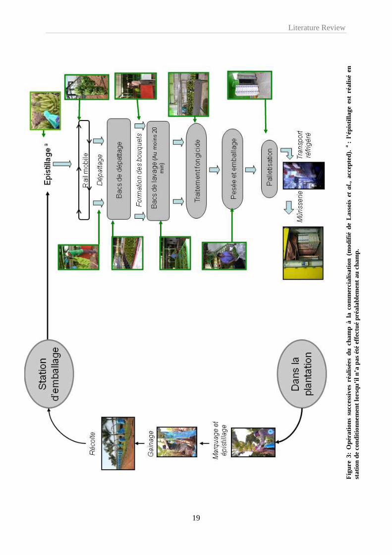
également supprimés (Figure 3). Cette opération, nommée épistillage, permet d’éviter une
source importante d’inoculum pathogène et de limiter les blessures par contact avec les autres
doigts.
Les régimes sont ensuite gainés à l’aide d’un film de polyéthylène permettant de
tamponner les variations de température, d’assurer une meilleure croissance des fruits, de
présenter une barrière mécanique contre les parasites et de protéger les fruits contre les
agressions mécaniques dues, par exemple, aux frottements des feuilles (Figure 3).
Le marquage des régimes se fait également au stade « doigts horizontaux » et permet les
prévisions de récolte (Figure 3). En effet, les différents régimes arrivés à ce stade sont
marqués d’une bande de couleur spécifique dans le but de connaître leur âge et de prévoir la
date de récolte à un âge physiologique déterminé. En fonction des plantations, 9 à 12 couleurs
de marquage sont utilisées dans une succession hebdomadaire.
Literature Review
19
Fig
ure
3: O
péra
tions
suc
cess
ives
réa
lisée
s du
cha
mp
à l
a co
mm
erci
alis
atio
n (m
odifi
é de
Las
sois
et a
l., a
ccep
ted)
. a : l’é
pist
illag
e es
t ré
alis
é en
st
atio
n de
con
ditio
nnem
ent l
orsq
u’il
n’a
pas
été
effect
ué p
réal
able
men
t au
cham
p.
Literature Review
20
1.4.3.3. La récolte
La récolte des régimes ne s’improvise pas. L’objectif est de récolter au grade le plus
élevé possible compatible avec l’absence de mûrs d’arrivage à l’entrée en mûrisserie. Le stade
de récolte sera donc fonction des délais et des conditions prévalant entre la coupe et l’entrée
en mûrisserie. Traditionnellement, la récolte s’effectue lorsque le grade commercial est
atteint. C’est-à-dire lorsque le fruit de référence, représenté par le doigt médian du rang
externe de la deuxième ou de la quatrième main, a un diamètre de respectivement 36 ou 34
mm. Les fruits sont à ce stade remplis au ¾ et sont encore verts et durs. Le seul critère du
grade n’est pas suffisant pour décider du stade optimal de récolte. En l’absence de facteurs
limitant, le grade de coupe est atteint lorsque le fruit a accumulé 900°C jours au seuil de 14°C
depuis le marquage au stade doigts horizontaux. A cet âge physiologique les fruits ont une
durée de vie verte (DVV) qui correspond au temps écoulé entre la coupe des fruits et le début
de leur crise climactérique et qui est compatible avec leur transport maritime et leur
acheminement vers la mûrisserie. Il est ainsi possible de prévoir la récolte à partir de la date
de floraison et de l’utilisation de données météorologiques (Jullien et al., 2008). L’intervalle
de temps entre la floraison du bananier et la récolte du régime, appelé « intervalle fleur-
coupe » (IFC), est donc théoriquement constant lorsqu’il est exprimé en somme de
températures. Il est par contre très variable en jours en fonction de la zone de production, de la
saison et surtout des pratiques culturales.
La récolte s’effectue à la machette avec toutes les précautions nécessaires pour éviter les
chocs et meurtrissures aux fruits. Les régimes sont portés à l’extérieur des parcelles dans des
berceaux matelassées positionnés sur la tête. Le régime est alors déposé avec le berceau dans
une remorque ou accroché à un système de câbles qui traverse la bananeraie jusqu’au hangar
d’emballage (Figure 3). La récolte du régime marque le début du dépérissement du pied-mère
qui est alors coupé. Sa suppression enlève la dominance apicale sur le rejet préalablement
sélectionné et permet de poursuivre la culture.
1.4.3.4. De la récolte au conditionnement
A la station d’emballage, les régimes sont accrochés à un rail et les mains sont séparées
de la hampe florale à l’aide d’un couteau (Figure 3). Les mains sont ensuite plongées dans un
bac d’eau enrichi en chlore et en alun appelé bac de dépattage afin de permettre l’écoulement
du latex (Figure 3). A la sortie de ces bacs, les mains de bananes sont récupérées, parfois
frottées à l’aide d’une éponge savonneuse, et sont découpées en bouquets de 3 à 8 fruits. Ces
derniers sont alors placés dans un second bac, appelé bac de lavage, pendant au moins 20

Literature Review
21
minutes (Figure 3). Ils sont ensuite acheminés sur des tapis roulants vers la zone de traitement
fongicide avant d’être pesés et conditionnés dans des emballages plastiques (sacs en
polyéthylène perforé ou non, avec ou sans vide d’air) et disposés dans des cartons
d’exportation (Figure 3). Les techniques de traitement chimique sont très variées: trempage,
tunnel de pulvérisation, pulvérisateurs, cascades, badigeonnage manuel, etc…Mais il semble
qu’un bon mouillage des fruits soit essentiel pour assurer une bonne efficacité des traitements
fongicides (de Lapeyre de Bellaire et al., 1994).
1.4.3.5. De la station d’emballage à la mûrisserie
Les cartons de bananes sont regroupés sur des palettes et sont stockés dans un container
refroidi à 13°C (Figure 3). La mise au froid permet d’une part, de minimiser la production
d’éthylène et de retarder le processus de maturation et d’autre part, de réduire le
développement de champignons éventuellement présents (Krauss et al., 2000). Ces containers
sont acheminés par camions vers le port de Douala où les palettes sont débarquées et
entreposées dans les cales de navires. Par la maîtrise de la température, de l’hygrométrie et de
la composition de l’atmosphère, ces cales assurent la conservation des bananes durant la
traversée maritime. Au bout d’une dizaine de jours, les palettes sont débarquées dans le port
de destination et sont acheminées par voies terrestres vers les mûrisseries où s’effectuera la
maturation artificielle des bananes (Figure 3). Cette maturation est initiée par un apport
exogène d’éthylène durant 24h à une température de 20°C. Au terme de ces 24h, les fruits
sont ventilés et peuvent être commercialisés.
1.4.4. Avantages et limites de l’utilisation exclusive de la Cavendish
Le sous-groupe homogène des Cavendish (AAA) a pu être adopté dans presque toutes
les régions tropicales humides pour son énorme potentiel productif (jusqu’à 60 tonnes/ha)
associé à une bonne précocité (récolte en 10 mois) et une taille réduite (moins de 3 m)
facilitant sa culture (Lescot, 1998). Ainsi, les acteurs de la filière ont fortement investi et se
sont organisés exclusivement autour du standard Cavendish. Les efforts de recherche et
développement ont été dirigés vers l’optimisation des modes de production, emballage,
transport, mûrissage et marketing des bananes Cavendish. A l’heure actuelle, le processus et
l’équipement industriel de production et de distribution sont adaptés à la Cavendish. Ce
schéma industriel ne laisse que très peu de place à l’introduction d’autres variétés et aux
changements. Elle est soumise à une forte pression normative qui pousse à banaliser le produit
et à rendre ce marché monolithique : la banane dessert au « format » Cavendish,
correspondant aux normes Dole ou Chiquita. Dans l’ensemble des fruits et légumes, il

Literature Review
22
n’existe pas d’exemple semblable. L’offre du marché pour un fruit est, dans tout les cas,
constitué d’au moins deux variétés.
Ainsi, malgré la diversité génétique des bananiers, il n’est pas rare dans certaines
régions de production destinée à un commerce d’exportation, de ne rencontrer que de la
Cavendish. La diversité des cultivars existant au sein d’une même structure de production est
d’autant plus réduite qu’on s’éloigne du centre d’origine du complexe d’espèces pour aller
vers des régions ou seuls quelques exemplaires de bananiers ont été introduits. Cette forte
spécialisation variétale et géographique s’accompagne d’une concentration du pouvoir de
marché entre quelques grandes firmes (Loeillet, 2005).
Il existe évidemment des contraintes liées à la monoculture intensive de type agro-
industriel, sans rotation, faisant appel à d’importantes quantités d’intrants et pratiquée durant
de nombreuses années (parfois plus de 40 ans) (Lescot, 2004). Ces contraintes sont
notamment d’ordre environnemental et phytosanitaire. Ce type de culture peu respectueuse de
l’environnement aboutit notamment à une évolution et dégradation des sols. Des déséquilibres
biologiques apparaissent rendant la culture plus sujette aux maladies et ravageurs. De plus les
populations de pathogènes aériens ou telluriques, inféodés à la culture, ont tendance à
s’accroître s’il n’y a pas rupture de leur cycle biologique due à la suppression de l’hôte. Les
risques de résistance aux pesticides sont également accrus dans ces conditions et l’apparition
de nouvelles maladies est une réalité. Ainsi, le problème majeur des productions bananières
sont les lourdes menaces parasitaires qui pèsent sur ces productions face à l’étroitesse de la
gamme variétale cultivée à l’heure actuelle en monoculture intensive.
Chez la banane, le premier problème est apparu lorsque la Gros-Michel a été cultivée de
manière intensive et que ce premier cultivar commercial a été détruit avec l’apparition de
Fusarium oxysporum cubense ou maladie de Panama. Après 1960 le commerce international
de la banane a été dominé par le cultivar de type Cavendish. Plus récemment, une forme
extrêmement virulente du pathogène F. oxysporum, la race T4, et pouvant s’attaquer aux
Cavendish a été décrite (Hwang et al., 2004). D’autres problèmes parasitaires associés aux
Cavendish sont apparus et ont été largement développés par divers auteurs (Jones, 2000;
Ploetz et al., 2003). Parmi les différentes contraintes liées aux parasites et ravageurs, il faut
signaler que les maladies d’origine fongique, très répandues dans les plantations industrielles,
constituent la principale perte de rendement et affectent tous les organes de l’hôte (Ploetz et
al., 2003). A l’heure actuelle, les plus néfastes, affectant de manière significative la
production des Cavendish d’exportation, sont les cercosporioses. D’autres maladies
Literature Review
23
fongiques, propres aux marchés d’exportation et causant d’importants dégâts et pertes
économiques, méritent qu’on les souligne. Il s’agit des maladies de conservation comme les
pourritures de la couronne (Lassois et al., accepted) et l’anthracnose.
1.5. Conclusion
Malgré l’importante diversité génétique existant au sein du genre Musa le marché
d’exportation est dominé par la culture presque exclusive de la variété Cavendish. Cependant,
la production de banane d’exportation doit être capable de répondre aux exigences de
durabilité, qualité et rendement qui lui sont imposées. Ceci constitue un véritable défi pour les
variétés existantes et ce de part l’étroitesse de la gamme variétale utilisée et l’apparition de
nouvelles maladies ou de nouvelles souches virulente. Ainsi, d’importantes stratégies de lutte
doivent être élaborées.
En bananeraies intensives, le système de culture dominant a longtemps reposé sur une
large utilisation systématique des produits phytosanitaires. Au début des années 90, les
bananeraies étaient traitées avec des quantités très élevées de matières actives : de 17 à 22
kg/ha/an (Lassoudière, 2007). Actuellement on ne peut plus concevoir la protection
phytosanitaire uniquement sous le seul aspect de la lutte chimique. Celle-ci pose de gros
problèmes tels que la toxicité des produits, l’accumulation de résidus dans les fruits, la
destruction d’organismes non cibles, le risque de pollution des eaux, les pertes d’efficacité des
produits. De plus, la protection phytosanitaire doit faire face à une législation de plus en plus
restrictive. Pour certaines maladies, comme la maladie de Panama, aucun produit
phytosanitaire n’a jamais apporté de réponse satisfaisante.
Ainsi, l’ensemble du système de culture et de l’itinéraire technique est à adapter. Il faut
envisager une approche globale de la filière banane dans un but de durabilité de la production.
Des solutions techniques ont permis de réduire de plus de 50% l’utilisation des pesticides aux
Antilles Françaises au cours de la dernière décennie (Chabrier et al., 2005). Parmi les
techniques alternatives à la lutte chimique, on retrouve également la sélection et
l’amélioration variétale. C’est aujourd’hui la voie privilégiée pour maintenir à plus ou moins
long terme la culture de la banane dans les zones actuelles de production mais il s’agit d’une
action de longue haleine. Les premiers travaux d’amélioration des bananiers par croisement
ont été amorcés dès les années 20 à la suite de l’extension de la maladie de Panama.
Parallèlement à ces activités de croisement, d’autres équipes ont concentré leurs efforts, à
partir des années 80, sur la mutagenèse et sur la sélection de variants somaclonaux qui sont
apparus à la suite du développement des techniques de cultures in vitro pour la multiplication

Literature Review
24
rapide et industrielle des vitroplants de bananiers. Le comportement de variétés issues de
mutations induites par l’application de rayons ionisants sur les bourgeons végétatifs est
également évalué. Enfin, l’avènement des techniques de biologie cellulaire et moléculaire a
favorisé l’émergence d’équipes qui travaillent sur la transformation génétique des bananiers.
Cependant, les réponses sur la durabilité de la culture bananière ne relèvent pas d’une
seule solution miracle, à savoir la recherche d’un produit phytosanitaire plus performant ou
même uniquement la mise au point d’un bananier résistant. Elle dépend plutôt d’une mutation
des systèmes de culture par l’amélioration d’itinéraires techniques complets basée sur une
connaissance approfondie de l’agronomie de la plante, de la biologie des parasites et des
relations hôte-pathogènes.

Literature Review
25
1.6. Références
Bakry F., Carreel F., Caruana M.L., Côte F.X., Jenny C. & Tezenas du Montcel H., 1997. Les
bananiers. In: Charrier A., Hamon S., Jacquot M. & Nicolas D., eds. L'amélioration des
plantes tropicales. CIRAD. ORSTOM, Montpellier, France, 109-139.
Chabrier C., Mauléon H., Bertrand P., Lassoudière A. & Quénéhervé P., 2005. La défense des
végétaux. Phytoma 584 : 12-16.
Champion J., 1963. Le bananier. Paris: Maisonneuve et Larose.
Cheesman, E.E., 1947. Classification of the bananas. II. The Genus Musa L. Kew Bulletin 2:
106-117.
Cirad-Flhor, 2003. Bananes for ever - La diversité génétique des bananiers. FruiTrop 99:5.
Daniells J.W., Jenny C., Karamura D.A., Tomekpe K., Arnaud E. & Sharrock S. , 2001.
Musalogue: a catalogue of Musa germplasm. Diversity in the genus Musa. Montpellier,
France, INIBAP.
de Lapeyre de Bellaire L. & Nolin J., 1994. Amélioration du contrôle du chancre sur les
bananes d'exportation et traitements post-récolte. Fruits 49:179-185.
Dominguez M. & Vendrell M., 1994. Effect of ethylene treatment on ethylene production,
EFE activity and ACC levels in peel and pulp of banana fruit. Postharvest Biology and
Technology 4: 167-177.
EUROSTAT, 2000. Statistiques d'importations européennes de plantains. FruitTrop 82:3.
Häkkinen M., 2009. Musa chunii Häkkinen, a new species (Musacea) from Yunnan, China
and taxonomic identity of Musa rubra. Journal of Systematics and Evolution 47, 87-91.
Heslop-Harrison J.S. & Schwarzacher T., 2007. Domestication, genomics and the future for
banana. Annals of Botany 100:1073-1084.
Hwang S.C. & Ko W.H., 2004. Cavendish banana cultivars resistant to Fusarium wilt
acquired through somaclonal variation in Taiwan. Plant Disease 88: 580-588.
John P. & Marchal J., 1995. Ripening and biochemistry of the fruit. In: Gowen S. Bananas
and plantains. Natural Resources Institute and Department of Agriculture University of
Reading. Chapman & Hall, London, UK.
Jones D.R., 2000. Diseases of Banana, Abaca and Enset, Wallingford, UK.
Jullien A., Chillet M. & Malezieux E., 2008. Pre-harvest growth and development, measured
as accumulated degree days, determine the post-harvest green life of banana fruit.
Journal of Horticultural Science and Biotechnology 83:506-512.

Literature Review
26
Krauss U. & Johanson A., 2000. Recent advances in the control of crown rot of banana in the
Windward Islands. Crop Protection 19:151-160.
Lassois L., Jijakli M.H., Chillet M. and de Lapeyre de Bellaire L., Accepted. Crown rot of
bananas: pre-harvest factors involved in post-harvest disease development and
integrated control methods. Plant Disease.
Lassoudière A., 2007. Le bananier et sa culture. Ed. Quæ, coll. Savoir-faire
Lescot T., 1998. Les bananiers: une diversité méconnue. FruiTrop 51:8.
Lescot T., 2004. Banane: Production, commerce et variétés. FruiTrop 118:5.
Lescot T., 2006. La banane en chiffres: le fruit préféré de la planète. FruiTrop 140: 5.
Lescot T. & Loeillet D., 2008. Banane et environnement: vers une production plus propre à
l'horizon 10 ans. FruiTrop 153: 3-4.
Loeillet D., 2005. Le commerce international de la banane: entre évoltion et révolution.
FruiTrop 129:2-19.
Maillard J.C., 2002. Le commerce international de la banane: marché, filière, système.
Cahiers d'outre-mer 220.
Mbida Mindzie C., Doutrelepont H., Vrydaghs L., Swennen R., Swennen R.J., Beeckman H.,
DeLanghe E. & DeMaret P., 2001. First archaeological evidence of banana cultivation
in central Africa during the third millenium before present. Veg. Hist. Archaeobot. 10:1-
6.
Marriott J. & Lancaster P.A., 1983. Bananas and Plantains. In: Harvey T. & Chan J.R.
Handbook of tropical foods. U.S.D.A.
ODEADOM/Cirad-Flhor. 2000. Bilan de l'approvisionnement du marché bananier européen.
Info Banane 38.
Omoaka P.O., 2000. Postharvest physiology, ripening and quality evaluation in banana (Musa
sp.) fruits. Doctoraatsproefschrift. Katholieke Universiteit Leuven. Faculteit
Landbouwkundige en Toegepaste Biologishe Wetenschappen Departement Toegepaste
Plantwetenschappen. N°445.
Peacock B.C., 1972. Role of ethylen in the initiation of fruit ripening. Queensland Journal of
Agricultural and Animal Sciences 29: 137-145.
Picq C., Lipman E., Sharrock S. & Frison E., 2002. La culture du bananier. Biofutur 222:30-
33.
Ploetz R.C., Thomas J.E. & Slabaugh W.R., 2003. Disease of banana and plantain. In: Ploetz
RC, eds. Diseases of tropical fruit crops, Cambridge, USA.

Literature Review
27
Robinson, J.C., 1996. Bananas and plantains. In: Atherton J, Rees A, eds. Cambridge:
Cambridge University Press.
Seymour G., 1993. Banana. In: Seymour G., Taylor J.E. & Tucker G.A. Biochemistry of fruit
ripening. Chapman and Hall, London, UK.
Simmonds N.W. & Shepherd K., 1955. Taxonomy and origins of cultivated bananas. Journal
of the Linnaean Society of Botany 55:302-312.
Simmonds, N., 1962. The evolution of the Bananas. John Willey & Sons Inc., New-York,
USA.
Stover R.H. & Simmonds N.W., 1987. Bananas. Longman Scientific & Technical, London,
UK.
Wilson J.S. & Otsuki T., 2004. To spray or not to spray: pesticides, banana exports, and food
safety. Food Policy 29:131-146.
Wong C., Kiew R. Argent G., Set O., Lee S.K. & Gan Y.Y., 2002. Assessment of validity of
the sections in Musa (Musaeceae) using AFLP. Annals of Botany 90: 231-238.

Literature Review
28
2. Crown rot of bananas: pre-harvest factors involved in post-harvest disease development and integrated control methods
Lassois L., Jijakli H., Chillet M. and de Lapeyre de Bellaire L. Accepted. Crown rot of
bananas: pre-harvest factors involved in post-harvest disease development and integrated
control methods. Plant Disease.
Crown rot of bananas: pre-harvest factors involved in post-harvest disease development
and integrated control methods
Ludivine Lassoisa, M. Haïssam Jijaklia, Marc Chilletb and Luc de Lapeyre de Bellairec*
a University of Liege, Gembloux Agro-Bio Tech, Plant Pathology Unit. Passage des Déportés 2, B-5030 Gembloux, Belgium; bCIRAD-PERSYST, UMR Qualisud, Departamento de Alimentos e Nutriçao Experimental – Faculdade de Ciencias Farmaceuticas – Universidade de Sao Paulo, Avenida Lineu Prestes, 580 – Bloco 14 – 05508-900, Sao Paulo, Brazil c CIRAD-PERSYST, UPR Systèmes de cultures bananes et ananas, Boulevard de la Lironde - TA B-26 / PS4, 34398 Montpellier Cedex 5, France Received 5 February 2009 Accepted 27 April 2009 *Corresponding author: Tel: (33) 467615828; Fax: (33) 467615868; e-mail: [email protected]

Literature Review
29
Abstract
Crown rot is a complex disease which affects export bananas in all producing countries.
Usually invisible when the fruits are packed for transportation from tropical countries to
distant destinations, disease symptoms occur during shipment, ripening, and storage. This
disease, characterised by rot and necrosis, affects tissues joining the fingers with each other,
called the crown. It may reach the pedicel and ultimately the banana pulp when the crown rot
is severe. Losses from 10% to 86% have been recorded for bananas respectively treated and
untreated.
In this paper, we proposed to inventory current knowledge on crown rot disease and
associated control measures which must be considered throughout the production channel in
order to be effective. We suggest a new, approach to consider this post-harvest disease of
bananas. In this approach we consider that bananas elaborate in the field a fruit quality
potential. We consider that this new concept of preharvest quality potential is a key factor in
understanding crown rot development, since is it responsible for most of disease incidence
variations observed in the industry. The fruit quality potential depends on both a physiological
and a parasitic component, both of which depend on agro-technic and pedo-climatic factors of
the crop production area. The physiological component is defined as the sensitivity of the
fruits to crown rot, while the parasitic component reflects the capacity of the parasitic
complex to induce a level of disease.
The content of this review is divided into two parts. First the fruit quality potential at
field level is addressed, with special interest on its physiological and parasitic components.
Secondly, the control methods are examined at different steps of the channel, in order to give
an overview of a possible integrated control strategy.

Literature Review
30
2.1. Introduction
Bananas are grown in over 120 countries and rank second in world fruit production,
after oranges and before grapes. The banana industry, which is based on a small number of
cultivars belonging to the Cavendish subgroup, is a vital source of income, employment, and
export revenue for most exporting countries, which are mainly developing countries in Latin
America, the West Indies, Southeast Asia, and Africa. However, major losses often occur
during shipping of bananas to their final market mainly because of ripening incidents,
appearance defects, and storage decay such as anthracnose and crown rot that occur during
shipping. Such post-harvest diseases negatively impact the market value of bananas,
especially when they are assessed at the port of arrival or in ripening facilities, thus
contributing to banana quality depreciation and constraining export trade. Anthony et al. (9)
reported that post-harvest diseases were responsible for 20% of harvest losses in Sri Lanka in
1997.
Crown rot affects export bananas in all producing countries and is considered to be one
of the main export banana post-harvest diseases (61, 83). This became a major problem in the
banana industry during varietal reconversion initiatives in the 1960s. Up until 1960, only
Gros-Michel subgroup cultivars were cropped for export, and for economic and practical
reasons bananas were shipped in complete bunches consisting of double layers of fruits called
hands which are arranged helicoidally around a central axis called the stalk. These cultivars
were, however, susceptible to Panama disease (Fusarium oxysporum f. sp. Cubense) and thus
replaced by Cavendish subgroup cultivars because of their resistance to Panama disease.
However, Cavendish bananas are more fragile during shipping (33) and this triggered a
revolution in the banana industry as bunch shipping was discontinued. Bananas were instead
cut into clusters consisting of several banana fruits joined by the crown tissues (Fig. 1) and
boxed for shipping.

Literature Review
31
Fig. 1: Crown rot symptoms. (A) Healthy crowns after maritime shipping before being placed in ripening rooms; (B) Crowns diseased with a superficial mycelium after maritime shipping; (C) Rot noted on bananas after maritime shipping; (D) Rot on the peduncles inducing banana ripening upon their arrival after maritime shipping; (E) External crown rot symptoms after fruit ripening; (F) Internal crown rot sympto ms after fruit ripening.
The crown became a prime site for infection by different pathogens (41, 52, 64). The
incidence of crown rot periodically increases during the rainy season, and losses of over 10%
have been recorded in the UK in bananas coming from the Windward Islands that were
harvested during this period (61). Losses of up to 86% have also been reported in non-
chemically treated bananas from the Philippines (6).
The first studies on crown rot focused on (i) the etiology of this disease, especially on
the identification of the most pathogenic fungal species involved; and (ii) on post-harvest
control methods, mainly chemical. Nevertheless, these studies provided neither a good
understanding of the conditions most conducive for disease expression nor adequate control
of this disease at all spatio-temporal scales. There is now some evidence that fruit physiology
at harvest influences crown rot development and that all interactions among the
microorganisms implicated should be considered. Taking these into account, a banana quality
build-up scheme was proposed (Fig. 2).

Literature Review
32
Crown rot at commercial level
Factors affecting crown rot development duringbanana commodity channel
Related crown rot control
Artificial fruit ripening
Shipping and storage: fruit conservation
Harvestand packing station
Field plotHarvest stage
Fruit quality potential
Pathological component
Physiological component
Agro-technical factors
Pedo-climatic factors
Field prophylaxy, sleeving, …
Genetic control
Chemical, biological and physical control
Prophylaxy, deflowering, water quality, fruit handling, cleaning
Storage conditions (T°, RH, CA or MA)
Fig. 2: Diagram representing different key factors that arise throughout the commodity chanel concerning crown rot development at the commercial stage, and related control methods. T°: temperature; RH: relative humidity; CA: controlled atmosphere; MA: modified atmosphere.
In this scheme, the fruit quality potential at harvest is presented as a key factor in crown
rot development. The quality potential elaborates during the banana growth phase in the field
and depends on two components: (i) a fruit physiological component, which determines the
fruit susceptibility to the disease; and (ii) a parasitic component which reflects a level of
crown contamination by the fungal complex, as well as the pathogenicity of this complex. The
influence of environmental and agrotechnical pre-harvest factors on these two components of
the fruit quality potential is a new approach in the post-harvest disease research, and we will
therefore discuss their importance in this review.
2.2. Crown rot symptoms
Crown rot affects tissues of the so-called “crown”, which unites the peduncles (Fig. 1).
The rot is not visible when the bananas are boxed, and symptoms generally appear only after
maritime shipping. The rot begins with mycelial development on the surface of the crown
(Fig. 1B), followed by the peduncles (Fig. 1D) and fruit (Fig. 1C). The bananas may detach
from the peduncle during severe infections. Crown rot results from the development in the
Literature Review
33
crown of several common fungi, which constitute a fungal complex, and leads to softening
and blackening of tissues at the site of the wound left when the cluster was cut from the
bunch. The role of bacteria in the complex (64) has not been confirmed by detailed research.
The symptoms develop rapidly during ripening when the physiology of the fruit undergoes
modifications that facilitate fungal development (46).
Crown rot affects the fruit quality because of the development of necrosis on the fruit,
and it can also trigger early ripening of bananas during shipping (77) (Fig. 1D). Ripening may
be caused by ethylene released by stressed and necrotic tissues (31, 65, 89), but also by
ethylene produced by mycelia of fungi such as Colletotrichum musae (23, 78). Onset of the
disease cannot be predicted and it spreads in an irregular pattern on all clusters in a shipping
box. The same box may contain both healthy and infected clusters.
2.3. Fruit quality potential as a key factor in crown rot infection patterns
Geographical and seasonal variations have been noted in the incidence of crown rot
disease of bananas (61, 64, 87). Lukezic et al. (64) showed that the incidence of this disease
varied throughout the year in Honduras. It is generally higher during the summer (March–
September) and declines during the coldest period (October–February). They also
demonstrated that this pattern did not seem to be correlated with variations in the fungal
complex isolated from banana crowns. Moreover, in Jamaica, a high disease incidence was
found to be correlated with periods during the year when temperatures were highest (87),
whereas in the Windward Islands, incidence was reported to be highest during the rainy
period (61). These spatiotemporal fluctuations reflect the variations in the banana fruit quality
potential that depends both on a parasitic and physiological component (Fig. 2).
2.3.1. Parasitic component of the fruit quality potential
In crown rot, the parasitic component reflects a level of crown contamination by the
fungal complex, as well as the pathogenicity of this complex.
2.3.1.1. Etiology of crown rot and pathogenicity of the fungal complex
Crown rot is the result of the activity of a fungal complex. The microorganisms most
commonly isolated in crown rot are: Musicillium theobromae, Colletotrichum musae,
Ceratocystis paradoxa, Lasiodoplodia theobromae, Nigrospora sphaerica, Cladosporium sp.,
Acremonium sp., Penicillium sp. and Aspergillus sp., as well as many Fusarium spp.,

Literature Review
34
including F. semitectum, F. verticillioides, F. sporotrichoides, F. oxysporum, and F. solani (9,
39, 41, 51, 64, 66, 71, 76, 85, 96, 97).
Several organisms may be involved in disease development. Moreover, disease severity
and the nature of the complex may vary substantially, depending on the production area
(Table 1) and season. Lukezic and Kaiser (63) showed that fungal populations may differ
between banana plants and even between crowns.
Fungi of the complex do not all have the same pathogenicity and variations have been
reported between regions (Table 1). Knight (58) considered that F. oxysporum, F.
verticillioides and F. graminearum, which have been isolated frequently from crowns of
Windward Island bananas, are primary pathogens, whereas L. theobromae, M. theobromae,
and N. sphaerica are considered to be relatively nonpathogenic species. According to the
findings of Marin et al. (66) in Costa-Rica, F. verticillioides and F. semitectum are the most
pathogenic species. According to Griffee (40), C. musae, L. theobromae, C. paradoxa, F.
semitectum, and F. graminearum are major pathogens involved in this disease. However,
many authors agree on the strong pathogenicity of C. musae, which may trigger an infection
from a very small amount of inoculum (34, 39, 62, 64, 85). Other pathogens require larger
amounts of inocula to induce crown rot symptoms (34, 40, 59, 60, 62).
A wide range of fungal complex compositions have been noted in natural
contaminations. The nature of this composition, the specific pathogenicity of the different
microorganisms involved as well as interactions among them may even alter the pathogenicity
of the complex. C. musae was more pathogenic when it was inoculated alone than when it
was coinoculated with other species (62); however, Anthony et al. (9) found that L.
theobromae, F. Verticillioides, and C. musae were more highly pathogenic when coinoculated
than when they were inoculated separately.
The respective roles of the different species that could belong to the fungal complex has
been thoroughly studied, whereas little information is available on the antagonistic or
synergistic relationships among these different species. It is essential to gain greater insight
into these interactions so in order to better characterise the pathogenicity of the fungal
complex.

Literature Review
35
Ho
nd
ura
s6
43
%8
0%
14
%1
2%
81
%9
3%
Ce
ntr
al a
nd
So
uth
A
me
rica
39
FM
FX
RF
R
66
0-1
0%
4-5
0%
0-2
8%
0-3
0%
0-8
%0
-33
%
Win
dw
ard
Isl
an
ds
(WI)
97
23
-33
%5
-7%
5%
41
36
%2
7%
6%
2%
8%
<1
%
96
24
%1
8%
3%
3%
2%
2%
3%
2%
<1
%2
%<
1%
51
26
-44
%7
-23
%6
-21
%3
-9%
4-1
8%
0-1
0%
24
-26
%0
-10
%
62
II
I
34
XX
XX
57
II
58
II
II
II
I
40
II
II
II
I
Jam
aic
a8
51
1%
FX
X3
%1
3%
1%
3%
Sri
La
nka
9X
XX
Nig
eria
76
27
%3
%1
0%
4%
6%
26
%2
%
So
ma
lia, W
I,
Gu
ate
ma
la7
13
8%
XX
20
%2
%4
%
Re
fere
nce
s
Co
untr
ies
Tab
le 1
: B
iblio
grap
hic
data
on
the
diffe
rent
mai
n fu
ngal
spe
cies
isol
ated
fro
m c
row
n ro
t in
diff
eren
t ban
ana-
prod
ucin
g re
gion
s. P
erce
ntag
e va
lues
cor
resp
ond
to is
olat
ion
freq
uenc
ies
whe
n gi
ven b
y th
e au
thor
s. “
X”
indi
cate
s th
at th
e pa
thog
en h
as b
een
iden
tifie
d on
cro
wn
by t
he
auth
ors,
but
with
out p
rovi
ding
any
isol
atio
n fr
eque
ncy.
F: f
requ
ently
isol
ated
; MF
: mos
t fre
quen
tly is
olat
ed; R
: rar
ely
isol
ated
. “I”
indi
cate
s th
at a
utho
rs h
ave
only
eva
luat
ed t
he p
atho
geni
city
of d
iffer
ent
know
fun
gal
spec
ies
with
out
info
rmat
ion
abou
t th
e iso
latio
n fr
eque
ncie
s.
Info
rmat
ions
abo
ut t
he p
atho
geni
city
are
als
o sh
owed
whe
n gi
ven
by a
utho
rs. …
.. : H
ighl
y pa
thog
enic
; …
.. :
Med
ium
pat
hoge
nici
ty;
…..
: S
light
ly o
r no
n pa
thog
enic
.
Literature Review
36
2.3.1.2. Factors influencing the level of crown contamination by the fungal complex
Very little information is available on the epidemiology of crown rot. The fact that the
characteristics of each species involved in the disease complex differ complicates studies on
the infection cycle. There are nevertheless some general features. Badger (11) showed that the
relative humidity generally has to be over 86% for germination of conidia of most fungi
involved in the complex. In banana plantations, this group of fungal species sporulates
abundantly on all putrescent organs such as leaves (68, 88), floral parts (2, 28), and bracts (26,
63). C. musae and some Fusarium species are primary colonizers of decomposing leaves (69)
and floral parts (26). Banana contamination by spores of C. musae and various Fusarium spp.
mainly occurs during the first 40 days following bunch emergence, and these are gradually
replaced by another fungal complex (28). Spores of some species such as Colletotrichum spp.
are mainly disseminated by rainwater (26, 28, 47), whereas others are airborne (63).
The fungus C. musae, which is also a causal agent of anthracnose, establishes quiescent
infections in the field during the first month following banana flowering (26). These quiescent
infections could also contribute to the onset of crown rot if the pathogen has an opportunity to
colonize the crown region (42); however, for the other pathogens, even though field infections
cannot be excluded, infections mainly occur during harvest and when clusters are trimmed
from bunches (70). Contamination generally takes place when hands are cut with a
contaminated knife (35, 39, 94, 96), or when clusters are cleaned with contaminated washing
water (86). At harvest, the fruits and senescent floral organs bear high quantities of spores that
could potentially contaminate the crowns. Some of these spores could be removed by washing
and then accumulate in the washing water (91), while others, such as C. musae appressoria,
may adhere tightly to the fruit surface (84). The washing tanks are the main source of inocula
according to Shillingford (86). After the banana clusters are dipped in the washing water, the
spores can penetrate a few millimetres into the vascular vessels of the crown, and disease is
then hard to control with a fungicide spray treatment (33). Green and Goos (39) showed that a
suspension of C. musae spores could penetrate 5–7 mm into the crown tissues within only 3
min. The crowns may also be contaminated by airborne spores before the fruit are placed in
boxes in the packing station. The risk is especially high when the facilities are dirty or if
debris (pistils, stalks and fruits) is piled up nearby. Conidia can survive for several months
under extreme temperature and humidity conditions before germination (70).
Literature Review
37
2.3.2. Physiological component of the fruit quality potential
The physiological component refers to the level of fruit susceptibility to crown rot,
which reflects the physiological state of the fruit and is dependent on agrotechnical and
pedoclimatic factors during plant growth. Only a few studies have been conducted on factors
influencing the level of banana susceptibility to crown rot. In Guadeloupian conditions, it was
shown that marked variations in fruit susceptibility could occur at the same production site
over a period of 10 successive weeks (62). Variations in crown rot susceptibility between
different Guadeloupian production areas during the same period have also been observed (L.
Lassois, unpublished).
Fruit age as expressed in accumulated degree day (dd) (53) also seems to affect crown
rot development. An intra-bunch banana crown rot susceptibility gradient has been
documented. Clusters growing on the first hands of a bunch, which are, on average, 70 dd
more advanced than the hands initiated last (54), are more susceptible than clusters
developing on the last hands (L. Lassois, H. Bastiaanse, M. Chillet, M.H. Jijakli, A. Jullien,
and L. de Lapeyre de Bellaire, unpublished). Moreover, a linear relationship between fruit age
(in dd) and the susceptibility of the fruit to crown rot was reported, with the oldest fruit being
most susceptible to this disease (36).
Some agricultural practices have a bearing on crown rot development. Modifying the
source-sink ratio by trimming leaves and hands (where leaves are considered as sources and
fruits as sinks) may induce variations in fruit susceptibility. Severe bunch trimming leads to a
sharp drop in banana crown rot susceptibility when the bananas are harvested at a constant
physiological age of 900 dd (L. Lassois, H. Bastiaanse, M. Chillet, M.H. Jijakli, A. Jullien,
and L. de Lapeyre de Bellaire, unpublished).
The mechanisms underlying variations in fruit crown rot susceptibility have yet to be
investigated. The plant mineral status has an effect on banana susceptibility to various
diseases (18, 45). Preformed fungitoxic polyphenolic compounds could be involved in banana
resistance to post-harvest diseases (1, 4, 19, 72, 74). Nevertheless, all factors potentially
involved in plant resistance mechanisms could have an impact on the level of banana crown
rot susceptibility. Plants are equipped with a series of defense mechanisms controlled through
the expression of different genes. Genes governing observed susceptibility variations could be
identified by assessing differences in gene expression between bananas with different levels
of susceptibility. Studies are currently under way to identify the underlying mechanisms and
key genetic factors involved in crown rot susceptibility variations using a differential

Literature Review
38
expression analysis technique (L. Lassois, P. Frettinger, L. de Lapeyre de Bellaire, P.
Lepoivre and H. Jijakli, unpublished).
2.4. Crown rot control methods
Banana crown rot, like other storage diseases, has an especially detrimental impact on
export produce. A routine postharvest treatment with a fungicide is the main method currently
used to control this disease. Problems may, however, arise from differences in fungicide
efficacy associated with the level of susceptibility of the fruit to crown rot or of the pathogens
to the different fungicides. Finally, discharge of fungicidal slurries can also lead to
environmental pollution, and residues of fungicides may subsequently be detected in the
marketed bananas. Research focused on alternative nonchemical control methods is of
considerable interest in the current increasingly prohibitive social and legal setting. It is
essential to implement sound integrated control strategies throughout the commodity chain
considering the complexity of the disease and the difficulty of its control (33, 91).
2.4.1. Field control methods
We have a set of methods that can usually be implemented in an integrated way to
achieve efficient crown rot control (Fig. 2). These are mainly preventive measures aimed at
limiting fungal contamination of bananas in the field.
2.4.1.1. Sanitation of banana plantations
Most species involved in the fungal complex are saprophytes that occur on senescent
banana organs, especially on decomposing leaves (69). Old leaves present in the banana
plantation may harbour inocula that could be responsible for severe contamination of bananas
by this fungal complex (90). The inoculum pressure, and thus the disease development rate,
may be reduced through regular elimination of senescent leaves around the fruit (91). Banana
floral parts are also inoculum sources, especially for C. musae and several Fusarium species
(28). In light of the potential role of bunch stalk contamination in the development of crown
rot (35), early elimination of flower parts in the field is also essential for reducing bunch
contamination by the pathogens (26).
2.4.1.2. Plastic sleeving to protect bananas
No accurate studies have been carried out to assess the impact of sleeving on the crown
rot development rate. It is nevertheless known that bunch sleeving (Fig. 3) with perforated

Literature Review
39
plastic film protects bunches from fungal contamination, thus especially curbing the
development of pitting disease (44) and speckling disease (52). Moreover, it has been shown
that sleeving can reduce contamination of banana bunches by C. musae by over 80% (26).
These findings suggest that sleeving directly reduces crown contamination in the field, or that
sleeved fruits release fewer spores in washing water during packing operations.
Fig. 3: Successive operations carried out from the field to the banana marketing stage. a: deflowering is done in the packing station when it has not been done in the field.
2.4.1.3. Controlling the banana harvest stage
Bananas are exported when they have grown to a commercial grade specified by
European regulations (Commission Regulation (EC) No 2257/94 of 16 September 1994,
setting quality standards for bananas) and market requirements. However, bananas should be
harvested at an age that will ensure a sufficient conservation. It has also been shown that the
best estimator of fruit shelf life is physiological age, expressed as a sum of accumulated
temperatures from flowering to harvest rather than in days (53). It was shown that the
physiological age of bananas (expressed in dd) has an impact on crown rot susceptibility (36).
Literature Review
40
The fruit physiological age should thus be taken into account at harvest. Some practices
such as field trimming of false hands, some true hands, male buds and external fruits of hands
can accelerate the fruit pulp filling rate. These practices can thus reduce the physiological age
that bananas need at harvest to enable them to reach a sufficient commercial grade (61, L.
Lassois, H. Bastiaanse, M. Chillet, M.H. Jijakli, A. Jullien and L. de Lapeyre de Bellaire,
unpublished).
2.4.1.4. Genetic control
The range of varieties grown for dessert banana export is very narrow as all clones
belong to the Cavendish subgroup (12). Moreover, breeding is complicated because triploid
banana varieties are generally sterile. In the past, banana genetic improvement programs were
mainly focused on obtaining varieties resistant to Sigatoka and Panama diseases. Resistance
to crown rot of improved varieties has not been considered in any breeding programs to date.
According to Marin et al. (66), FHIA-01 and FHIA-02, two hybrids produced by the
FHIA breeding program, are partially resistant to crown rot. Conversely, Perez Vicente and
Hernandez (79) consider that these two varieties are more susceptible to crown rot caused by
F. semitectum and C. musae as compared with Cavendish varieties. According to these
authors, only FHIA-23 is more resistant to crown rot than Grande-Naine. Finally, the first
FHIA hybrids were introduced in the late 1980s, but their characteristics differ from those of
Cavendish bananas and consumers have shown little interest in them, thus limiting their
distribution.
2.4.2. Postharvest control methods
2.4.2.1. Chemical control
Routine post-harvest fungicide treatment is still the most efficient crown rot control
method. This strategy was introduced in the late 1960s with the discovery of systemic
benzimidazole derivative fungicides (thiabendazole and benomyl). They are classified as
antimitotic compounds. Gradually, other fungicides that inhibit ergosterol biosynthesis, such
as imazalil and bitertanol, were also introduced (29, 37). The application methods vary
markedly: dipping, spraying (Fig. 3), cascade treatment, etc., but in every case the bananas
must be thoroughly wetted to ensure the fungicide treatment efficacy (29, 50). Alum is often
combined with fungicide slurries to neutralise latex residue remaining on crowns when the
bananas are removed from the washing bath (73); however, this mixture seems to have a
negative impact on the performance of some active ingredients such as thiabendazole (50). The

Literature Review
41
time between crown trimming and fungicide application is critical. Crown rot severity seems to
increase when fungicide application is delayed (39). Bananas are nevertheless usually treated in
time in packing stations, i.e. just after they are removed from the washing bath. Problems
concerning the development of resistant strains may arise with this practice. Postharvest
fungicides generally have the same mode of action as those sprayed in the field to control
Sigatoka disease. C. musae strains resistant to thiabendazole, and generally to all antimitotic
compounds, have been detected in many banana-producing countries (27, 46, 51, 61). In
Guadeloupe, it has been shown that thiabendazole-resistant C. musae strains developed after
exclusive foliar applications of benomyl over a decade (1972-1982) for Sigatoka Disease
control (27). Postharvest use of chemicals could ultimately be prohibited because the number
of active substances registered for postharvest treatment has been reduced by current
regulations, and legislation is becoming increasingly restrictive. Moreover, the efficacy of
intensive fungicide treatments is not always adequate. In some areas where very few resistant
strains are present, the efficacy of fungicides for postharvest disease control was found to vary
substantially depending on season and production area (16). Finally, consumers are now highly
selective with respect to food safety concerns, and demand is increasing for produce that has not
undergone any postharvest chemical treatments. Alternative non-fungicide solutions are
therefore being sought.
2.4.2.2. Preventive measures in packing stations
Deflowering in packing stations
Deflowering (Fig. 3) involves the removal of all floral parts that could potentially
harbour inocula before the dehanding operation. When the floral parts have not been removed
in the field, deflowering is done in the packing station before bunch trimming, thus reducing
the risk of contamination of the different washing baths (61, 85).
Packing station water quality
Fruit dehanding is a risky operation because the resulting wound is the main portal for
infection by crown rot pathogens. Contamination of crowns when the bananas are processed in
the dehanding tanks and then in the washing tanks is a key step in the crown-rot development
process (Fig. 3). Spore accumulation in the water can be reduced by regularly changing the
washing water in the washing and delatexing baths (91) (Fig. 3). These baths can host a high
quantity of spores, mainly Fusarium sp. and Verticillum sp., that detach from the peel surface
(86), whereas C. musae conidia are less often found (85). Care should be taken to avoid

Literature Review
42
contaminating the baths with plant debris (pistils, leaves, trimming waste, etc.) and the water
should be regularly refreshed.
It is also recommended that the bath water be treated with active chlorine (10, 33, 91) or
quaternary ammonium disinfectants in order to hamper contamination (86), but the efficacy of
this disinfection procedure is controversial (33, 91). The chlorine concentration should be
regularly adjusted to offset the high observed losses by volatilization or through redox
reactions with latex or other organic matter in the washing baths (86). It is especially hard to
maintain the bath water quality when the water is recirculated in a closed system. In such
situations, latex contamination gradually increases in the tanks, so it is hard to control the water
quality simply through chlorine treatment. Note also that, due to changes in market standards, it
is important to reduce health risks associated with the development of human pathogens in the
washing baths.
Packing station sanitation
Protective measures implemented in the packing station are aimed at keeping the crowns
of freshly trimmed bananas away from all inoculum sources. To ensure efficient crown rot
control, it is thus essential to keep the packing station and the adjoining facilities clean. Waste
(stalks, low-grade fruit, etc.) located in the vicinity of the banana packing area, which could
contaminate the air in the station, must be eliminated (35, 61). It has been shown that trimming
clusters in a clean environment rather than in the field can reduce the crown rot incidence by
50% (35).
The fruits should also be cleaned before being trimmed from the bunch. A contaminated
trimming knife could spread inocula from the peel into the crown tissues (64). This may be
avoided by washing the bunches in lightly chlorinated water before they are trimmed into
clusters (61).
2.4.2.3. Banana and crown trimming
Bananas should be trimmed with a clean stainless steel blade. Finlay and Brown (34)
showed that roughly trimming the crowns, or ripping them off the hands, significantly increased
the level of fruit contamination. Tissue fragments on the surface of the crowns dry out and
quickly become senescent, thus providing an ideal site for rot development (34). Moreover,
banana trimming knife tips are rounded to avoid banana fruit wounds (61). It is also important
to cut wide crown sections containing as much crown tissue as possible, a technique that seems
Literature Review
43
to enhance crown resistance to rot and seldom leads to the spread of rot into the fruit pedicels
(73).
2.4.2.4. Banana storage techniques
The temperature, relative humidity and atmospheric composition are the main
environmental factors that impact storage disease development. These factors may directly
affect the biology of the pathogens, but they can also have an indirect effect by slowing down
the fruit metabolism. These environmental parameters can thus be modified, especially to
extend the banana greenlife. The greenlife represents the period between the fruit harvest and
the beginning of the climacteric phase and seems to be a prime factor in the development of
storage diseases since a direct relationship has been noted between the degree of banana
ripeness and susceptibility to disease (73). Fruit resistance also seems to decrease at the onset of
the ripening process. It is therefore crucial to carefully manage these factors to ensure efficient
banana crown rot control (61).
Storage temperature
One way to slow down the banana metabolism, and thus to delay crown rot development,
is to refrigerate the fruit in boxes at the lowest possible temperature without changing fruit
physiology. Cooling should be continuous throughout the shipping phase, which in turn should
be as short as possible. Containers designed for maritime shipping of bananas are climate
controlled at 13-14 °C, because temperatures below 12 °C are detrimental to fruit quality (73).
Fungal growth is slowed down at 13 °C (34), which is a good tradeoff with respect to
preserving the bananas and hampering fungal colonization of the crowns. However, this is
much lower than the optimal temperatures for germination and growth of fungi responsible
for crown rot, but it is not low enough to slow down the activity of these fungi, which can
take place within a broad temperature range, i.e. 8 to 36 °C (90). As a result, crown rot
incidence and severity are higher in bananas exposed to temperatures over 16°C (91).
Relative humidity
Although most pathogens require a high relative humidity for their in vitro development
(11), bananas are less susceptible to crown rot under these conditions. A high relative humidity
seems to hinder transpiratory water loss from the fruit, which is essential to ensure a long
greenlife. Indeed, banana greenlife is markedly reduced under low relative humidity
atmospheric conditions (30 to 40%) as a result of ethylene production from the fruit peel (77).

Literature Review
44
The senescence of banana crown tissues, which is conducive to crown rot development, can be
hampered by maintaining their turgidity (73).
Atmosphere composition
The composition of the atmosphere around bananas during shipping can be manipulated
to slow down metabolic activity. Modified atmospheres (MA) can be achieved by packing
bananas in sealed plastic bags (polybags), and controlled atmospheres (CA) are obtained by
injecting nitrogen in the storage rooms. It was shown that crown rot can be partially
controlled by packing bananas in MA (H. Bastiaanse, L. de Lapeyre de Bellaire, L. Lassois,
C. Misson, and M. H. Jijakli, unpublished). For MA, the balance of an atmosphere with a
lower O2 and a higher CO2 content depends on the extent of fruit respiration, bag permeability,
and the composition of the air outside the bag (95). The O2 and CO2 contents generally range
from 1 to 10% and 2 to 14%, respectively, depending on the quality and thickness of the
plastic packaging (67, 95). This modification in the gas composition reduces the fruit
respiratory intensity and hampers endogenous ethylene synthesis, which can considerably
increase the length of the preclimacteric phase. MA also inhibits the metabolism of some
pathogenic agents. These plastic bags must also have a high mechanical strength because even
small punctures will upset the MA (16).
High CO2 (>15%) and low O2 (<1%) levels are toxic to many fungi (3, 38).
Unfortunately, bananas cannot be stored under these conditions because major alterations
generally occur during ripening when the atmospheric CO2 level is above 7 to 12% or when
the O2 level is below 1 to 2% (22, 95, 100).
Another way to modify gas exchange is to coat the peel with a wax composed of
sucrose esters combined with cellulose or fatty acids. This wax blocks the stomatal pores, thus
reducing gas exchange (14). The fruits have higher permeability to CO2 than to O2, and wax
coatings accentuate this difference. This leads to a substantial decrease in the fruit internal O2
content without increasing the CO2 to an excessively high level, thus creating an ideal
atmosphere for preserving bananas without alterations (13).

Literature Review
45
2.4.2.5. Physical control methods
Hot water treatments
Hot water treatments destroy the pathogens (15) and modify the fruit environment by
activating antimicrobial compounds in the peel (24). de Costa and Erabadupitiya (24) showed
that the optimal temperature and exposure time for controlling crown rot was 50 °C for 3 min.
Higher temperatures lead to pale fruit, and exposure times of over 5 min reduce Brix values
(24) and damage the fruit peel (101). A 20 min longer treatment at a temperature under 45°C is
effective for controlling C. paradoxa, with the percentage of infected bananas decreasing from
100% to less than 15% (83). However, commercial tests on naturally infected fruit have not
achieved crown rot control, and ripening delays have also been noted. There has been no
commercial adaptation of this technique to date, except in other tropical fruits such as papaya
(21, 75) and mango (20, 92). Hot water treatments have also been combined with the
application of antagonistic bacteria in order to increase the biological control efficacy (24).
UV and gamma radiation treatment
Ultraviolet light (UV) treatments have considerable potential for fruit and vegetable
post-harvest diseases control. Stevens et al. (93) discussed the possibility of inducing apple
resistance to Colletotrichum gloeosporioides through UV-C treatments. In bananas, however,
the peel seems to be too sensitive to UV for the use of this technique for postharvest control
of some pathogens of the complex like C. musae (49).
Kanapathipillai et al. (55) pointed out that gamma ray treatment (38 min at 4 kGy)
inhibited spore germination, the formation of C. musae appressoria, and all fungal
development on the surface of fruit pieces. However, these authors did not investigate the
effects of gamma rays on whole fruit, or on their complex components. Although dosages of
around 0.5 kGy can extend banana greenlife (67, 99), Marriott and Palmer (67) noted that
irradiation can alter the banana peel; the maximum dose tolerated by the fruits is likely around
0.5 kGy (99). Finally, the development of postharvest fruit irradiation has been hampered
especially by the high cost of such treatments and their low consumer acceptance (99).
2.4.2.6. Biological control
Postharvest biological control is very promising because the crown rot infection site on
the fruit is limited, the environmental conditions during storage are clearly defined and stable,

Literature Review
46
and bananas have a high added value (48). The results of many studies have suggested that the
use of microorganisms such as fungi, bacteria and yeasts could provide partial crown rot
control (7, 24, 25, 32, 43, 60, 62, 80, 98). It has also been shown that biological control
efficacy increases with the antagonistic agent concentration and with the incubation time
between the application of the antagonist and crown contamination by the fungal complex
(62, 80). Antagonists can be used to significantly reduce lesions induced by the fungal
complex that causes crown rot, but the control efficacy is limited and variable (62). This type
of control, when used alone, cannot provide total crown rot control, and so should be
combined with other control tactics such as calcium additives and MA packaging (H.
Bastiaanse, L. de Lapeyre de Bellaire, L. Lassois, C. Misson, and M. H. Jijakli, unpublished ).
The impact of several natural substances or nonsynthetic fungicides, such as
preparations of calcium, plant extracts or organic acids, on crown rot development has also
been evaluated. Allium sativum extracts (61), and essential oils of Cinnamomum zeylanicum,
Syzygium aromaticum (81, 82), Cymbopogon nardus, and Ocimum basilicum (9) have also been
found to have fungicidal activity. Win et al. (101) showed that cinnamon extracts reduced
crown rot, increased greenlife, and had no negative effects on postharvest banana quality. These
plant extracts have fungistatic and fungitoxic activity and inhibit conidial germination and
mycelial growth of C. musae, Fusarium spp., and L. theobromae. However, cases of
phytotoxicity have been reported and the level of control provided by these natural substances
is not sufficient to meet market requirements. The use of antioxidants (56) and organic salts
(5), sometimes combined with surfactants (8), can also enhance crown rot control. Finally,
treatments with Biocto 6 (seed extract from citrus) combined with a wax-based additive
(Verdiol), was found to provide the same level of crown rot control as fungicide treatments of
export bananas (30).
2.5. Conclusion
Crown rot studies and control are especially complex because of the observed diversity
and variability in the composition and pathogenicity of the complex involved in the
development of this disease. The broad range of possible situations complicates studies on both
the parasitic and physiologic components of fruit quality. Further studies are thus required to
gain better insight into this disease, especially since little documentation is available on certain
aspects such as the epidemiology of crown rot. It is also very important to enhance the overall
understanding of banana physiological mechanisms involved in the induction of fruit resistance
to crown rot pathogens with the aim of improving control.

Literature Review
47
There is considerable growing interest in alternative methods to reduce or even
completely eliminate fungicide treatments. To be efficient, these alternative control methods
should not be too specific considering the broad spectrum of pathogens involved in the
complex. No alternative methods to chemical control are currently efficient enough by
themselves to match the efficacy of fungicide treatments. However, the results of some
experimental trials have shown that crown rot can actually be managed by combining different
nonchemical control methods. Crown rot research should enhance the overall understanding of
this disease and thus lead to the development of an effective integrated control strategy.

Literature Review
48
2.6. Literature cited
1. Abdel-Saatar, M., and Nawwar, M. 1986. 3,4-dimethoxybenzaldehyde, a fungistatic
substance in peel of green banana fruits in relation to resistance at different degrees of
maturity. Horticultural science 21:812.
2. Agati, J. A. 1992. Banana stem and fruit rot. Philippine Agriculture 10:411-422.
3. Al Zaemey, A. B., Magan, N., and Thompson, A. K. 1994. In vitro studies of the effect
of environmental conditions on the anthracnose pathogen of bananas, Colletotrichum
musae. Int. Biodeterior. Biodegrad. 33:369-381.
4. Alam, M. S., Alam, S., Islam, S., and Alam, N. 1993. Biochemical changes in banana
fruits in response to crown rot pathogens. Bangladesh J. Bot. 22:143-148.
5. Alvindia, D. G., Kobayashi, T., Natsuaki Keiko, T., and Tanda, S. 2004. Inhibitory
influence of inorganic salts on banana postharvest pathogens and preliminary
application to control crown rot. Journal of General Plant Pathology 70:61-65.
6. Alvindia, D. G., Kobayashi, T., Yaguchi, Y., and Natsuaki, K. T. 2000. Symptoms and
the associated fungi of postharvest diseases on non-chemical bananas imported from the
Philippines. Japanese Journal of Tropical Agriculture 44:87-93.
7. Alvindia, D. G., and Natsuaki, K. T. 2008. Evaluation of fungal epiphytes isolated from
banana fruit surfaces for biocontrol of banana crown rot disease. Crop Prot. 27:1200-
1207.
8. Alvindia, D. G., and Natsuaki, K. T. 2007. Control of crown rot-causing fungal
pathogens of banana by inorganic salts and a surfactant. Crop Prot. 26:1667-1673.
9. Anthony, S., Abeywickrama, K., Dayananda, R., Wijeratnam Shanthi, W., and
Arambewela, L. 2004. Fungal pathogens associated with banana fruit in Sri Lanka, and
their treatment with essential oils. Mycopathologia 157:91-97.
10. Arneson, P. A. 1971. Sensitivity of postharvet rot fungi of bananas to chlorine.
Phytopathology 61:344-345.
11. Badger, A. M. 1965. Influence of relative humidity on fungi causing crown rot of boxed
bananas. Phytopathology 55:688-692.
12. Bakry, F., Carreel, F., Caruana, M. L., Côte, F. X., Jenny, C., and Tezenas du Montcel,
H. 1997. Les bananiers. Pages 109-139 in: L'amélioration des plantes tropicales, A.
Charrier, S. Hamon, M. Jacquot and D. Nicolas, eds. CIRAD. ORSTOM, Montpellier.
13. Banks, N. H. 1984. Some effects of TAL Pro-long coating on ripening bananas. J. Exp.
Bot. 35:127-137.

Literature Review
49
14. Banks, N. H. 1984. Studies of the banana fruit surface in relation to the effects of Tal
Pro-long coating on gaseous exchange. Sci. Hortic. 24:279-286.
15. Burden, O. J. 1968. Reduction of banana anthracnose following hot treatment of the
green fruit. Queensland Journal of Agriculture and Animal Science 25:135-144.
16. Chillet, M., and de Lapeyre de Bellaire, L. 1996. Conditionnement en polybag pour le
contrôle de l'anthracnose de blessure des bananes. Fruits 51:163-172.
17. Chillet, M., and de Lapeyre de Bellaire, L. 1996. Elaboration de la qualité des bananes
au champ. Détermination de critères de mesure. Fruits 51:317-326.
18. Chillet, M., de Lapeyre de Bellaire, L., Dorel, M., Joas, J., Dubois, C., Marchal, J., and
Perrier, X. 2000. Evidence for the variation in susceptibility of bananas to wound
anthracnose due to Colletotrichum musae and influence of edaphic conditions. Sci.
Hortic. 86:33-47.
19. Chillet, M., Hubert, O., and de Lapeyre de Bellaire, L. 2007. Relationship between
physiological age, ripening and susceptibility of banana to wound anthracnose. Crop
Prot. 26:1078-1082.
20. Coates, L. M., Johnson, G. I., and Cooke, A. W. 1993. Postharvest disease control in
mangoes using high humidity hot air and fungicide treatments. Ann. Appl. Biol.
123:441-448.
21. Couey, H. M., Alvarez, A. M., and Nelson, M. G. 1984. Comparison of hot-water spray
and immersion treatments for control of pstharvest decay of papaya. Plant Dis. 68:436-
437.
22. Daun, H., Gilbert, S. G., Ashkenazi, Y., and Henig, Y. 1973. Storage quality of bananas
packaged in selected permeability films. J. Food Sci. 38:1247-1250.
23. Daundasekera, M., Joyce, D. C., Aked, J., and Adikaram, N. K. B. 2003. Ethylene
production by Colletotrichum musae in vitro. Physiol. Mol. Plant. Pathol. 62:21-28.
24. De Costa, D. M., and Erabadupitiya, H. R. U. T. 2005. An integrated method to control
postharvest diseases of banana using a member of the Burkholderia cepacia complex.
Postharvest Biol. Technol. 36:31-39.
25. De Costa, D. M., and Subasinghe, S. S. N. S. 1998. Antagonistic bacteria associated
with the fruit skin of banana in controlling its post-harvest diseases. Tropical Science
38:206-212.
26. de Lapeyre de Bellaire, L., Chillet, M., Dubois, C., and Mourichon, X. 2000.
Importance of different sources of inoculum and dispersal methods of conidia of
Colletotrichum musae, the causal agent of banana anthracnose, for fruit contamination.
Plant Pathol. 49:782-790.
Literature Review
50
27. de Lapeyre de Bellaire, L., and Dubois, C. 1997. Distribution of thiabendazole-resistant
Colletotrichum isolates from Guadeloupe banana plantations. Plant Dis. 81:1378-1383.
28. de Lapeyre de Bellaire, L., and Mourichon, X. 1997. The pattern of fungal
contamination of the banana bunch during its development and potential influence on
incidence of crown-rot and anthracnose diseases. Plant Pathol. 46:481-489.
29. de Lapeyre de Bellaire, L., and Nolin, J. 1994. Amélioration du contrôle du chancre sur
les bananes d'exportation et traitements post-récolte. Fruits 49:179-185.
30. Demerutis, C., Quiros, L., Martinuz, A., Alvarado, E., Williams, R. N., and Ellis, M. A.
2008. Evaluation of an organic treatment for post-harvest control of crown rot of
banana. Ecol. Eng. 34:324-327.
31. Dominguez, M., and Vendrell, M. 1993. Wound ethylene biosynthesis in preclimacteric
banana slices. Acta Horticulturae 343:270-274.
32. East, L., and Kenyon, L. 1998. Development of biological control methods for post-
harvest rots of banana. Pages 549-554 in: Proceedings of the Brighton Crop Prot.
Conference: Pests and Diseases, B. C. P. Council, ed. Farnham (United Kingdom),
Brighton, UK, 16-19 November 1998.
33. Eckert, J. W., and Ogawa, J. M. 1985. The chemical control of post-harvest diseases :
subtropical and tropical fruits. Annu. Rev. Phytopathol. 23:421-454.
34. Finlay, A. R., and Brown, A. E. 1993. The relative importance of Colletotrichum musae
as a crown-rot pathogen on Windward Island bananas. Plant Pathol. 42:67-74.
35. Finlay, A. R., Lubin, C., and Brown, A. E. 1992. The banana stalk as a source of
inoculum of fungal pathogens which cause crown rot. Tropical Science 32:343-352.
36. Forret, M. 2008. Etude de la variation de sensibilité des bananes d'exportation aux
pourritures de couronne en fonction du stade de récolte. Travail de fin d'étude, Faculté
des Sciences Agronomiques de Gembloux:93p.
37. Frossard, P., Laville, E., and Plaud, G. 1977. Etude des traitements fongicides appliqués
aux bananes après récolte. III/. Action de l'imazalil. Fruits 32:673-676.
38. Goos, R. D., and Tschirsch, M. 1962. Effect of environmental factors on spore
germination, spore survival, and growth of Gloeosporium musarum. Mycologia 54:353-
366.
39. Greene, G. L., and Goos, R. D. 1963. Fungi associated with crown rot of boxed
bananas. Phytopathology 53:271-275.
40. Griffee, P. J. 1976. Pathogenicity of some fungi isolated from diseased crowns of
banana hands. J. Phytopathol. 85:206-216.
Literature Review
51
41. Griffee, P. J., and Burden, O. J. 1976. Fungi associated with crown rot of boxed bananas
in the Windward Islands. J. Phytopathol. 85:149-158.
42. Griffee, P. J., and Pinegar, J. A. 1974. Fungicides for control of the crown rot complex:
in vivo and in vitro studies. Tropical Sciences 16:107-120.
43. Gunasinghe, R. N., Ikiriwatte, C. J., and Karunaratne, A. M. 2004. The use of Pantoea
agglomerans and Flavobacterium sp. to control banana pathogens. Journal of
Horticultural Science and Biotechnology 79:1002-1006.
44. Guyon, M. 1970. Essais de lutte chimique contre la "Johnson fruit spot" au Nicaragua.
Fruits 25:685-691.
45. Hecht Buchholz, C., Borges Perez, A., Fernandez Falcon, M., and Borges, A. A. 1998.
Influence of zinc nutrition on Fusarium wilt of banana - an electron microscopic
investigation. Proceedings of the International Banana Symposium on Bananas in the
Subtropics. 10-14 november, Puerto de la cruz, Tenerife, Spain 490:277-283.
46. Hostachy, B., Vegh, I., Leroux, P., Jacquemot, E., Foucher, S., and Pigou, R. 1990.
Bananes de la Martinique. Incidence des problèmes fongiques sur la qualité. Phytoma
420:37-44.
47. Jeffries, P., Dodd, J. C., Jeger, M. J., and Plumbey, R. A. 1990. The biology and control
of Colletotrichum species on tropical fruit crops. Plant Pathol. 39:343-366.
48. Jijakli, M., Lepoivre, P., and C, G. 1999. Yeast species for biocontrol of apple
postharvest diseases: an encouraging case of study for practical use. Pages 31-49 in:
Biotechnological approaches in biocontrol of plant pathogens, R. Upadhyay and K.
Mukerij, eds. Kluwer academic/Plenum publishers, NY.
49. Joas, J. 1997. Programme qualité de la banane d'exportation. Rapport d'essai sur
l'emploi des UV en post récolte. Page 2 p in: Rapport interne. Cirad-Flhor, Fort de
France.
50. Joas, J., and Malisart, S. 2001. Incidence des conditions d' application sur l' efficacite
des fongicides utilises en post-recolte pour la banane - Effect of the application
techniques on the effectiveness of the fungicides used in the banana postharvest. Fruits
56:383-394.
51. Johanson, A., and Blasquez, B. 1992. Fungi associated with banana crown-rot on field-
packed fruit from the Windward Islands and assessment of their sensitivity to the
fungicides thiabendazole, prochloraz, and imazalil. Crop Prot. 11:79-83.
52. Jones, D. R. 2000. Diseases of Banana, Abaca and Enset., Wallingford, UK.

Literature Review
52
53. Jullien, A., Chillet, M., and Malezieux, E. 2008. Pre-harvest growth and development,
measured as accumulated degree days, determine the post-harvest green life of banana
fruit. Journal of Horticultural Science and Biotechnology 83:506-512.
54. Jullien, A., Malezieux, E., Michaux-Ferriere, N., Chillet, M., and Ney, B. 2001b.
Within-bunch Variability in Banana Fruit Weight: Importance of Developmental Lag
Between Fruits. Ann. Bot. 87:101-108.
55. Kanapathipillai, V. S., Ahmad, R., and Mahamad, M. I. 1987. The effect of sterile
filtrates of Trichoderna spp and Penicillium spp and 4 KGy irradiation on the spore
germination of Colletotrichum musae. Pages 283-292 in: Movements of pests and
control strategies, Singh K.G., Manalo P.L., Sastrontomo S.S., Chan K.C., Lim L.G.,
Ganapathi A.N., Rahim M.A.A., Durai P.S.S., and Doss M.C., eds., Kuala Lumpur,
Malaysia.
56. Khan, S. H., Aked, J., and Magan, N. 2001. Control of the anthracnose pathogen of
banana (Colletotrichum musae) using antioxidants alone and in combination with
thiabendazole or imazalil. Plant Pathol. 50:601-608.
57. Knight, C., Cutts, D.F., Colhoun, J. 1977. The role of Fusarium semitectum in causing
crown rot of bananas. J. Phytopathol. 89: 170-176.
58. Knight, C. 1982. Pathogenicity of some fungi associated with crown rot of bananas. J.
phytopathol. 104:13-18.
59. Krauss, U. 1996. Establishment of a bioassay for testing control measures against crown
rot of banana. Crop Prot. 15:269-274.
60. Krauss, U., Bidwell, R., and Ince, J. 1998. Isolation and preliminary evaluation of
mycoparasites as biocontrol agents of crown rot of banana. Biol. control 13:111-119.
61. Krauss, U., and Johanson, A. 2000. Recent advances in the control of crown rot of
banana in the Windward Islands. Crop Prot. 19:151-160.
62. Lassois, L., de Lapeyre de Bellaire, L., and Jijakli, M. H. 2008. Biological control of
crown rot of bananas with Pichia anomala strain K and Candida oleophila strain O.
Biol. Control 45:410-418.
63. Lukezic, F. L., and Kaiser, W. J. 1966. Aerobiology of Fusarium roseum 'Gibbosum'
associated with crown rot of boxed bananas. Phytopathology 56:545-548.
64. Lukezic, F. L., Kaiser, W. J., and Martinez, M. M. 1967. The incidence of crown rot of
boxed bananas in relation to microbial populations of the crown tissus. Can. J. Bot.
45:413-421.
Literature Review
53
65. MacCracken, A. R., and Swinburne, T. R. 1980. Effect of bacteria isolated from surface
of banana fruits on germination of Colletotrichum musae conidia. Transactions of the
British Mycological Society 74:212-214.
66. Marin, D. H., Sutton, T. B., Blankenship, S. M., and Swallow, W. H. 1996.
Pathogenicity of fungi associated with crown rot of bananas in Latin America on
Grande Naine and disease-resistant hybrid bananas. Plant Dis. 80:525-528.
67. Marriott, J., and Palmer, J. K. 1980. Bananas - Physiology and biochemistry of storage
and ripening for optimum quality. Crit. Rev. Food Sci. Nutr. 13:41-88.
68. Meredith, D. S. 1962. Some components of the air-spora in Jamaican banana
plantations. Ann. Appl. Biol. 50:577-594.
69. Meredith, D. S. 1962. Some fungi on decaying banana leaves in Jamaica. Transactions
of the British Mycological Society 45:335-347.
70. Meredith, D. S. 1971. Transport and storage diseases of bananas: biology and control.
Tropical Agriculture 48:35-50.
71. Mesturino, L., and Ragazzi, A. 1988. Microorganismi fungini associati ai frutti di Musa
spp., presenti sul mercato italiano. Rivista di Agricoltura Subtropicale e Tropicale
82:503-515.
72. Muirhead, I. F., and Deverall, B. J. 1984. Evaluation of 3,4-dihydrobenzaldehyde,
dopamine and its oxydation products as inhibitors of Colletotrichum musae (Berk. et
Curt.) Arx in green banana fruits. Aust. J. Bot. 32:575-582.
73. Muirhead, I. F., and Jones, D. R. 2000. Fungal diseases of banana fruit. Postharvest
diseases. Pages 190-206 in: Diseases of banana, abaca and ensete, D. R. Jones, ed.
CABI, Wallingford.
74. Mulvena, D., Webb, E. C., and Zerner, B. 1969. 3,4-Dihydrobenzaldehyde, a fungistatic
substance from green cavendish bananas. Phytochemistry 8:393.-395.
75. Nishijima, K. A., Miura, C. K., Armstrong, J. W., Brown, S. A., and Hu, B. K. S. 1992.
Effect of forced, hot-air treatment of papaya fruit on fruit quality and incidence of
postharvest diseases. Plant Dis. 76:723-727.
76. Ogundero, V. W. 1987. Crown rot fungi of Nigerian bananas cv. Robusta and the
effects of benomyl on their exo-enzymes. J. Basic Microb. 27:43-47.
77. Peacock, B. C. 1973. Effect of Colletotrichum musae infections on the preclimacteric
life of bananas. Queensland Journal of Agriculture and Animal Sciences 30:239-246.
78. Peacock, B. C., and Muirhead, I. F. 1974. Ethylene production by Colletotrichum
musae. Queensland Journal of Agriculture and Animal Sciences 31:249-252.

Literature Review
54
79. Perez Vicente, L., and Hernandez, A. 2002. Reaction of Black Sigatoka resistant FHIA
hybrids to Fusarium pallidoroseum and Colletotrichum musae causal agents of crown
rot and anthracnose diseases of bananas. Phytopathology 92:S136.
80. Postmaster, A., Kuo, J., Sivasithamparam, K., and Turner, D. W. 1997. Interaction
between Colletotrichum musae and antagonistic microorganisms on the surface of
banana leaf discs. Sci. Hortic. 71:113-125.
81. Ranasinghe, L., Jayawardena, B., and Abeywickrama, K. 2002. Fungicidal activity of
essential oils of Cinnamomum zeylanicum (L.) and Syzygium aromaticum (L.) Merr et
L.M.Perry against crown rot and anthracnose pathogens isolated from banana. Lett.
Appl. Microbiol. 35:208-211.
82. Ranasinghe, L., Jayawardena, B., and Abeywickrama, K. 2005. An intergrated strategy
to control post-harvest decay of embul banana by combining essential oils with
modified atmosphere packaging. Int. J. Food Sci. Technol. 40:97-103.
83. Reyes, M. E. Q., Nishijima, W., and Paull, R. E. 1998. Control of crown rot in `Santa
Catarina Prata' and `Williams' banana with hot water treatments. Postharvest Biol.
Technol. 14:71-75.
84. Sela-Buurlage, M. B., Epstein, L., and Rodriguez, R. J. 1991. Adhesion of
ungerminated Colletotrichum musae conidia. Physiol. Mol. Plant. Pathol. 39:345-352.
85. Shillingford, C. A. 1976. Occurence of banana fruit-rot fungi in Jamaican boxing plants.
Plant Disease Reporter 60:788-793.
86. Shillingford, C. A. 1977. Control of banana fruit rots and of fungi that contaminate
washing water. Tropical Science 19:197-203.
87. Shillingford, C. A. 1978. Climatic factors affecting post-harvest decay of jamaican
bananas. J. Agric. Univ. P. R.:45-49.
88. Simmonds, J. H. 1941. Latent infection in tropical fruits discussed in relation to the part
played by species of Gloeosporium and Colletotrichum. Proc. R. Soc. Queensl. 52:92-
120.
89. Simmonds, J. H. 1963. Studies in the latent phase of Colletotrichum species causing
ripe rots of tropical fruits. Queensland Journal of Agricultural and Animal Science
20:373-424.
90. Simmonds, J. H., and Mitchell, R. S. 1940. Black end and anthracnose of the banana
with special reference to Gloeosporium musarum Cke. and Mass. Page 63 p in: Bulletin
n° 131. Council for Scientific and Industrial Research of Australia, Melbourne.
91. Slabaugh, W. R., and Grove, M. D. 1982. Postharvest diseases of bananas and their
control. Plant Dis. 66:746-750.

Literature Review
55
92. Spalding, D. H., and Reeder, W. F. 1978. Controlling market diseases of mangoes with
heated benomyl. Proceedings of the Florida State Horticultural Society 91:186-187.
93. Stevens, C., Khan, V. A., Wilson, C. L., Lu, J. Y., Chalutz, E., and Droby, S. 2005. The
effect of fruit orientation of postharvest commodities following low dose ultraviolet
light-C treatment on host induced resistance to decay. Crop Prot. 24:756-759.
94. Stover, R. H. 1972. Banana, plantain and abaca diseases. Kew, Surrey, England.
95. Thompson, A. K. 1998. Controlled atmosphere storage of fruits and vegetables.
Wallingford.
96. Wallbridge, A. 1981. Fungi associated with crown-rot disease of boxed bananas from
the Windward Islands during a two year survey. Transactions of the British Mycological
Society 77:567-577.
97. Wallbridge, A., and Pinegar, J. A. 1975. Fungi associated with crown-rot disease of
bananas from St Lucia in the Windward Islands. Transactions of the British
Mycological Society 64:247-254.
98. Williamson, S. M., Guzmán, M., Marin, D. H., Anas, O., Jin, X., and Sutton, T. B.
2008. Evaluation of Pseudomonas syringae strain ESC-11 for biocontrol of crown rot
and anthracnose of banana. Biol. Control 46:279-286.
99. Wills, R., McGlasson, W. B., Graham, D., and Joyce, D. C. 1998. Postharvest. An
introduction to the physiology and handling of fruit, vegetables and ornamentals.
Wallingford.
100. Wilson, L. G. 1976. Handling of postharvest tropical fruit. Horticultural science 11:120-
121.
101. Win, N., Jitareerat, P., Kanlayanarat, S., and Sangchote, S. 2007. Effects of cinnamon
extract, chitosan coating, hot water treatment and their combinations on crown rot
disease and quality of banana fruit. Postharvest Biol. Technol. 45:333-340.

Literature Review
56
3. Contributions of molecular biology to understanding the mechanisms involved in variation of banana susceptibility to crown rot disease
3.1. Introduction
Biological responses of fruits, and notably physiological changes and susceptibility to
fungi, are controlled and regulated by the expression of certain genes associated with specific
metabolic pathways. Although the genome is identical in every cell of an organism, genes
may be expressed differently over time (development-stage-specific expression), in space
(cell-type-specific expression), and according to the state of the fruit (which may be, for
example, more or less susceptible to a disease). One way to understand the phenomena and
reactions involved in variation of banana susceptibility to crown rot disease in relation to the
physiological state of the fruit at harvest is to identify genes involved in these processes via
the study of their expression. Transcriptomic analysis is an important tool for evaluating gene
expression. Both molecular biology and genetic engineering have spawned a wide range of
techniques for studying the differential expression of genes in a given tissue on the basis of
transcript levels.
3.2. cDNA-AFLP analysis
3.2.1. Introduction
cDNA-AFLP derives from AFLP (amplified fragment length polymorphism) analysis,
based on PCR amplification and developed by Vos et al. (1995) primarily for mapping
genomic DNA. AFLP was adapted for analysis of mRNA levels in plants, and cDNA-AFLP
was first described by Bachem et al. (1996). Since its introduction in 1996, this technique has
become a reference tool for the study of differently expressed genes (Botton et al., 2008). The
technique consists in using RT-PCR to compare mRNA populations produced by different
populations of cells. This wide-range screening has revealed differential transcription of genes
whose function is "a priori" unknown, by comparison of the transcriptomes of similar cells or
tissues (Bachem et al. 1998; Bachem et al. 2001; Van der Biezen et al. 2000; Durrant et al.,
2000).
3.2.2. Principle
After reverse transcription of the mRNA, the synthesized double-stranded cDNA is
digested by a pair of restriction enzymes, one with a 4-basepair recognition site and one with

Literature Review
57
a 6-basepair recognition site. The ends of the digested fragments are then ligated to specific
adapters. All digested, ligated cDNAs are amplified with a pair of primers, one hybridizing
with each adapter. This step, called pre-amplification, yields many different fragments that
cannot be separated on a polyacrylamide gel. A second amplification, called selective
amplification, is performed with selective primers containing, in addition to the sequence of
the adapter, one, two, or three additional bases at the 3' end and allowing selective
amplification of cDNA fragments with complementary 5’ nucleotides. Thus, only a sub-
population of fragments, separated on a polyacrylamide gel, are amplified. Radiolabeling of
one of the two primers used makes it possible to visualize the fragments by autoradiography.
Figure 1 : Schematic representation of the cDNA-AFLP applied with Eco RI and Mse I like restriction enzymes and radiolabelled with 33P. The digestion of cDNA by Eco RI and Mse I and the adaptator ligation result in the generation of fragments flanked by (i) identical adaptators and by (ii) non-identical adaptators. Fragments that are flanked by identical adaptators are excluded from further amplification due the formation of a hairpin structure.
3.2.3. Advantages
The cDNA-AFLP method does not require prior sequence information and can thus be
used with any biological system, especially when genomic resources are lacking (Bachem et

Literature Review
58
al., 1996 ; Ditt et al., 2001). Secondly, the technique allows identification of new genes
(Botton et al., 2008; Cappelli et al., 2007; Ramonell and Somerville, 2002; Reijans et al.,
2003). These features are particularly interesting because the factors responsible for
quantitative variation of symptoms in the case of compatible interactions linked to infection
are still poorly understood and most likely remain to be discovered. More than two
populations of mRNAs can be compared simultaneously, and by using all combinations of
specific primers, the cDNA-AFLP makes it possible in principle to amplify all cDNAs
(Donson et al., 2002). Its reproducibility is very high (Campalans et al. 2001; Lassois et al.,
2009; Matz and Luckyanov, 1998), having been estimated at 100% (Khun, 2001). Its
sensitivity is comparable to that obtained with microarrays (Reijans et al., 2003), allowing
detection of transcripts present in very low quantity (Decorosi et al., 2005; Lievens et al.,
2001; Ramonell and Somerville, 2002; Reijans et al., 2003; Savelkoul, 1999) and even of
transcripts that are considered rare (Fukumura et al. 2003). Bachem et al. (1998) evaluated the
sensitivity of cDNA-AFLP at one copy of mRNA per cell. In addition, the expression profiles
obtained correlate well with those obtained by Northern blotting (Albertini et al., 2004;
Bachem et al. 1996; Breyne et al., 2003; Cnudde et al., 2003; Donson et al., 2002; Durrant et
al. 2000, Jones et al., 2000). The false positive rate is low, lower than with other similar
techniques such as differential display (Matz and Luckyanov 1998, Kuhn, 2001; Campalans et
al., 2001). Moreover, cDNA-AFLP analysis requires only a very small amount of sample
(Cappelli, 2005) and allows discrimination of homologous genes (Decorosi et al., 2005). As
its cost is low and it does not require specific hardware, it is affordable for smaller
laboratories (Ramonell and Somerville, 2002).
3.2.4. Disadvantages
Despite its many advantages, cDNA-AFLP analysis has a few disadvantages. Firstly,
the interpretation of results can be complicated by the fact that a single initial mRNA can give
rise to amplification of several fragments (Matz and Luckyanov, 1998; Lorkowski and Cullen,
2004). Secondly, as it is tedious to use all primer combinations, cDNA-AFLP is generally
performed with only a few primer pairs. Thus, cDNAs that doe not have the restriction sites
recognized by the enzymes used are not analyzed by the method (Habu et al., 1997;
Lorkowski and Cullen, 2004). In addition, information on differently expressed genes is
limited to bands that are sequenced, and the technique does not provide quantitative data on
the abundance of transcripts (Ramonell and Somerville, 2002). Since cDNA-AFLP analysis
requires radiolabeling PCR primers, safety precautions must be taken and the work must be
carried out in a laboratory accredited for work with radioisotopes (Decorosi et al., 2005).
Literature Review
59
Finally, a weakness of the technique lies in the possibility of obtaining false positives due to
co-isolation of sequences of the same size as the fragment of interest. This is why it is
imperative to confirm the results obtained by means of an independent technique such as real-
time RT-PCR.
3.2.5. Application of the method
The cDNA-AFLP technique and its application to plants were first described by
Bachem et al. (1996), who analyzed the differential expression of genes in Solanum
tuberosum in vitro tuberization systems. Other authors subsequently applied the cDNA-AFLP
analysis to the same model (Trindade et al., 2003, Trindade et al., 2004) and since then, the
technique has been applied to many other models, among which cotton plants (Ma et al.,
2007), apple (Yao et al., 2007), rice (Akihiro et al., 2006), Arabidopsis thaliana (de Diego et
al., 2006), and sugarcane (Borras-Hidalgo et al., 2005) are just a few examples. The technique
has revealed interesting parallels between the responses of plants to pathogens and
environmental constraints and the expression of genes that establish these responses (Lin et
al., 2003, Zheng et al., 2009; Ramonell and Somerville, 2002; Simoes-Araujo et al., 2002;
Nimbalkar et al., 2006; Ditt et al., 2001; Ditt et al., 2005; Chen et al., 2003). cDNA-AFLP
analysis has also been applied to other models besides plants: to mammals such as horses
(Cappelli et al., 2005; Capelli et al., 2007), rats (Fukuda et al., 1999), dogs (Vandeput et al.,
2005), and humans (Egert et al., 2006), but and also to fungi (Avrova et al., 2003; Botton et
al. , 2008), yeast (Massart and Jijakli, 2006; Reijans et al., 2003), bacteria (Decorosi et al.,
2005; Dellagi et al., 2000), and nematodes (Qin et al., 2000).
3.3. High-throughput sequencing for transcriptomics
Recently developed high-throughput sequencing (HTS) technology has made possible
powerful strategies for comprehensively interrogating nucleic-acid-based information in a cell
at unparalleled resolution and depth (Qui et al., 2009; Lister et al., 2009). The availability of
relatively low-cost sequencing techniques rapidly producing huge amounts of sequence
information has triggered a paradigm shift in genomics (Lister et al., 2009). The diversity of
applications to which such techniques have been applied demonstrates the immense range of
cell processes and properties that can now be studied at single-base resolution. Transcriptome
sequencing is among these applications, as high-throughput sequencing has become a real
alternative to other classical techniques for studying the transcriptome. It offers many
advantages: quantitative detection of messengers, detection of rare transcripts and small
RNAs, and no a priori dependence on annotation of the genome. This approach thus offers

Literature Review
60
the plant biologist unprecedented opportunities to probe the functions and dynamics of plant
cells and populations. Currently and in planning future research, these techniques cannot be
ignored. They will notably make it possible to obtain information that will help to elucidate
plant responses to pathogen attack.

Literature Review
61
3.4. References
Akihiro T., T. Umezawa, C. Ueki, B.M. Lobna, K. Mizuno, M. Ohta and T. Fujimura (2006).
Genome wide cDNA-AFLP analysis of genes rapidly induced by combined sucrose and
ABA treatment in rice cultured cells. FEBS Letters 580:5947-5952.
Albertini E., G. Marconi, G. Barcaccia, L. Raggi and M. Falcinelli (2004). Isolation of
candidate gene for apomixis in Poa pratensis L. Plant Molecular Biology 56:879–894.
Avrova A., E. Venter, P. Birch and S. Whisson (2003). Profiling and quantifying differential
gene transcription in Phytophthora infestans prior to and during the early stages of
potato infection. Fungal Genetics and Biology. 40: 4-14.
Bachem C. (1996). Visualization of differential gene expression using a novel method of
RNA fingerprinting based on AFLP: Analysis of gene expression during potato tuber
development. The Plant Journal 9: 745-753.
Bachem C., R. Oomen and R. Visser (1998). Transcript imaging with cDNA-AFLP: a step by
step protocol. Plant molecular biology reporter 16:157-173.
Bachem C., B. Horvath, L. Trindade, M. Claassens, E. Davelaar, W. Jordi and R. Visser
(2001). A potato tuber-expressed mRNA with homology to steroid dehydrogenases
affectes gibberellin levels and plant development. The Plant Journal. 25: 595-604.
Borrás-Hidalgo O., B. Thomma, E. Carmona, C.J. Borroto, M. Pujol, A. Arencibia and J.
Lopez (2005). Identification of sugarcane genes induced in disease-resistant somaclones
upon inoculation with Ustilago scitaminea or Bipolaris sacchari. Plant Physiology and
Biochemistry 43:1115-1121.
Botton A., D. Ferrigo, C. Scopel, R. Causin, C. Bonghi, and A. Ramina (2008). A cDNA-
AFLP approach to study ochratoxin A production in Aspergillus carbonarius.
International Journal of Food Microbiology 127:105-115.
Breyne P., R. Dreesen, B. Cannoot, D. Rombauts, K. Vandepoele, S. Rombauts, R.
Vanderhaeghen, D.M. Inze and M. Zabeau (2003). Quantitative cDNA-AFLP analysis
for genome-wide expression studies. Molecular Genetics and Genomics 2692:173-179.
Campalans A., M. Pagès and R. Messeguer (2001). Identification of differentially expressed
genes by the cDNA-AFLP technique during dehydration of almond. Tree physiology
21:633-643.
Cappelli K., A. Porceddu, A. Verini-Supplizi, S. Capomaccio, F.D. Marchis, M. Falcinelli, A.
Gaiti and M. Silvestrelli (2005). cDNA AFLP-based techniques for studying transcript
profiles in horses. Research in Veterinary Science 79:105-112.

Literature Review
62
Cappelli K., A. Verini-Supplizi, S. Capomaccio and M. Silvestrelli (2007). Analysis of
peripheral blood mononuclear cells gene expression in endurance horses by cDNA-
AFLP technique. Research in Veterinary Science 82:335-343.
Chen G.P., W.S. Ma, Z.J. Huang, T. Xu, Y.B. Xue and Y.Z. Shen (2003). Isolation and
characterization of TaGSK1 involved in wheat salt tolerance. Plant Science 165:1369-
1375.
Cnudde F., C. Moretti, A. Porceddu, M. Pezzotti and T. Gerats (2003). Transcript profiling on
developing Petunia hybrida floral organs Sexual. Plant Reproduction 16:77-85.
de Diego J.G., F. David Rodríguez, J.L. Rodríguez Lorenzo, P. Grappin and E. Cervantes
(2006). cDNA-AFLP analysis of seed germination in Arabidopsis thaliana identifies
transposons and new genomic sequences. Journal of Plant Physiology 163:452-462.
Decorosi F., C. Viti, A. Mengoni, M. Bazzicalupo and L. Giovannetti (2005). Improvement of
the cDNA-AFLP method using fluorescent primers for transcription analysis in bacteria.
Journal of Microbiological Methods 63:211-215.
Dellagi A., P.R.J. Birch, J. Heilbronn, G.D. Lyon and I.K. Toth (2000). cDNA-AFLP analysis
of differential gene expression in the prokaryotic plant pathogen Erwinia amylovora.
Microbiology 146:165-171.
Ditt R.F., E.W. Nester and L. Comai (2001). Plant gene expression response to
Agrobacterium tumefaciens. Procceding of the national academy of sciences 98:10954-
10959.
Ditt R.F., E. Nester and L. Comai (2005). The plant cell defense and Agrobacterium
tumefaciens. FEMS Microbiology Letters 247:207-213.
Donson J., Y. Fang, G. Espiritu-Santo, W. Xing, A. Salzar, S. Miyamoto, V. Armandarez and
W. Volkmuth (2002). Comprehensive expression analysis by transcript profiling. Plant
molecular biology 48:75-97.
Durrant W., O. Rowland, P. Piedras, K. Hammond-Kosack, J. Jones (2000). CDNA-AFLP
reveals a striking overlap in race specific resistance and wound response gene
expression profiles. The Plant Cell 12: 963-977.
Egert M., A.A. de Graaf, H. Smidt, W.M. de Vos and K. Venema (2006). Beyond diversity:
functional microbiomics of the human colon. Trends in Microbiology 14:86-91.
Fukuda T., A. Kido, K. Kajino, M. Tsutsumi, Y. Miyauchi, T. Tsujiuchi, Y. Konishi and O.
Hino (1999). Cloning of Differentially Expressed Genes in Highly and Low Metastatic
Rat Osteosarcomas by a Modified cDNA-AFLP Method. Biochemical and Biophysical
Research Communications 261: 35-40.

Literature Review
63
Fukumura R., H. Takahashi, T. Saito, Y. Tsutsumi, A. Fujimori, S. Sato, K. Tatsumi, R. Araki
and M. Abe (2003). A sensitive transcriptome analysis method that can detect unknown
transcripts. Nucleic Acids Research 31: 1-7.
Habu Y., S. Fukada-Tanaka, Y. Hisatomi and S. Iida (1997). Amplified Restriction Fragment
Length Polymorphism-Based mRNA Fingerprinting Using a Single Restriction Enzyme
That Recognizes a 4-bp Sequence. Biochemical and Biophysical Research
Communications. 234: 516-521.
Jones C.S., H.V. Davies and M.A. Taylor (2000). Profiling of changes in gene expression
during raspberry (Rubus idaeus) fruit ripening by application of RNA fingerprinting
techniques. Planta 211:708-714.
Kuhn E. (2001). From library screening to microarray technology: strategies to determine
gene expression profiles and to identify differentially regulated genes in plants. Annals
of botany 87:139-155.
Lassois L., L. De Lapeyre de Bellaire, and M.H. Jijakli. 2009. Combining an original method
for preserving RNA expression in situ with an effective RNA extraction method makes
it possible to study gene expression in any banana fruit tissue. Fruits 64 : 127-137.
Lievens,S., S. Gooriachtig and M. Holsters (2001). A critical evaluation of differential display
as a tool to identify genes involved in legume nodulation: looking back and looking
forward. Nucleic Acids Research 29:3459-3468.
Lin Z., H. Meakin and M. Dickinson (2003). Isolation of genes expressed during compatible
interactions between leaf rust (Puccinia triticina) and wheat using cDNA-AFLP.
Molecular Plant Pathology 4:469-477.
Lister R., B.D. Gregory and J.R. Ecker (2009). Next is now: new technologies for sequencing
of genomes, transcriptomes, and beyond. Current Opinion in Plant Biology 12:107-118.
Lorkowski S., and P. Cullen (2004). High-throughput analysis of mRNA expression:
microarrays are not the whole story. Expert Opinion on Therapeutic patents 14:377-
403.
Ma X., C. Xing, L. Guo, Y. Gong, H. Wang, Y. Zhao and J. Wu (2007). Analysis of
Differentially Expressed Genes in Genic Male Sterility Cotton (Gossypium hirsutum L.)
Using cDNA-AFLP. Journal of Genetics and Genomics 34:536-543.
Massart S., and M.H. Jijakli (2006). Identification of Differentially Expressed Genes by
cDNA-Amplified Fragment Length Polymorphism in the Biocontrol Agent Pichia
anomala (Strain Kh5). Phytopathology 96:80-86.
Matz M. and S.A. Luckyanov (1998). Different strategies of Differential display: areas of
application. Nucleic Acids Research 26: 5537-5543.

Literature Review
64
Nimbalkar S.B., A.M. Harsulkar, A.P. Giri, M.N. Sainani, V. Franceschi and V.S. Gupta
(2006). Differentially expressed gene transcripts in roots of resistant and susceptible
chickpea plant (Cicer arietinum L.) upon Fusarium oxysporum infection. Physiological
and Molecular Plant Pathology 68:176-188.
Qin L., Overmars H., Helder J., Poeijus H., Rouppe van der Voort J., Groenik W., van Koert
P., Schots A., Bakker J. and Smant G. (2000). An efficient cDNA-AFLP based strategy
for the identification of putative pathogenicity factors from the potato cyst nematode
Globodera rostochiensis. Molecular Plant-Microbe Interactions 13: 830-836.
Qiu D., X. Pan, I.W. Wilson, F. Li, M. Liu, W. Teng and B. Zhang (2009). High throughput
sequencing technology reveals that the taxoid elicitor methyl jasmonate regulates
microRNA expression in Chinese yew (Taxus chinensis). Gene 436:37-44.
Ramonell K. and S. Somerville (2002). The genomics parade of defense responses: to infinity
and beyond. Current Opinion in Plant Biology 5: 291-294.
Reijans M., R. Lascaris, A.O. Groeneger, A. Wittenberg, E. Wesselink, J. van Oeveren, E.d.
Wit, A. Boorsma, B. Voetdijk and H. van der Spek (2003). Quantitative comparison of
cDNA-AFLP, microarrays, and genechip expression data in Saccharomyces cerevisiae.
Genomics 82:606-618.
Savelkoul P., H. Aarts, J. De Haas, L. Dijkshoorn, B. Duim, M. Otsen, J. Rademaker, L.
Schouls and J. Lenstra (1999). Amplified fragment length polymorphism analysis: the
state of an art. Journal of clinical microbiology 37: 3083-3091.
Simoes-Araujo J.L., R.L. Rodrigues, L.B. A. Gerhardt, J.M.C. Mondego, M. Alves-Ferreira,
N.G. Rumjanek, and M. Margis-Pinheiro (2002). Identification of differentially
expressed genes by cDNA-AFLP technique during heat stress in cowpea nodules. FEBS
Letters 515:44-50.
Trindade L., B. Horvath, C. Bachem, E. Jacobsen and R. Visser (2003). Isolation and
functional characterization of a stolon specific promoter from potato (Solanum
tuberosum L.). Gene, 303: 77-87.
Trindade L.M., B. Horvath, R. van Berloo and R. Visser (2004). Analysis of genes
differentially expressed during potato tuber life cycle and isolation of their promoter
regions. Plant Science 166:423-433.
Van der Biezen E., H. Juwana, J. Parker, J. Jones (2000). cDNA-AFLP display for the
isolation of Peronospora parasitica genes expressed during infection in Arabidopsis
thaliana. Molecular Plant Microbe Interaction 13: 895-898.
Vandeput,F., M. Zabeau and C. Maenhaut (2005). Identification of differentially expressed
genes in thyrotropin stimulated dog thyroid cells by the cDNA-AFLP technique.
Molecular and Cellular Endocrinology 243:58-65.

Literature Review
65
Vos P., R. Hogers, M. Bleeker, M. Reijans, T. van de Lee, M. Hornes, A. Frijters (1995).
AFLP: a new techniques for DNA fingerprinting. Nucleic acids research 23: 4407-
4414.
Yao Y.-X., M. Li, Z. Liu, Y.-J. Hao and H. Zhai (2007). A novel gene, screened by cDNA-
AFLP approach, contributes to lowering the acidity of fruit in apple. Plant Physiology
and Biochemistry 45:139-145.
Zheng L., J. Huang and D. Yu (2009). Isolation of genes expressed during compatible
interactions between powdery mildew (Blumeria graminis) and wheat. Physiological
and Molecular Plant Pathology 73: 61-66.

Aims and proposed research
66
CHAPTER II
Aims and proposed researchAims and proposed researchAims and proposed researchAims and proposed research
Aims and proposed research
67
As show in figure 1 hereafter, crown rot development at commercial level depends on
several post-harvest factors. The first researches on crown rot focused on (i) the etiology of
this disease, especially on the identification of the most pathogenic fungal species involved;
and (ii) on post-harvest control methods, mainly chemical. Nevertheless, these researches did
not provide neither a good understanding of the most conducive conditions for disease
expression, nor a perfect control of this disease at all spatio-temporal scales. Variations were
observed in the expression of crown rot symptoms when post-harvest conditions were
assumed to have been reasonably constant, although not actively controlled (Griffee, 1976;
Lukezic et al., 1967; Shillingford, 1978).
We suggest a new, interesting and original approach to consider the crown rot post-
harvest development. It consists to present a fruit quality potential at harvest as a key factor in
crown rot development (Lassois et al., accepted) (chapter 1.2). This potential develops during
growth of bananas in the field and depends on a physiological and a parasitical component
which both are function on agro-technical and pedo-climatic factors of the crop production
area.
The parasitic component reflects both a level of crown contamination by the parasitic
complex and the pathogenicity of this parasitic complex.
The physiological component refers here to the level of fruit susceptibility to crown rot.
This level of banana crown rot susceptibility reflects the physiological state of the fruit. Yet
little is known about the influence of the fruit physiological component in post-harvest
development of the disease and on the pre-harvest factors influencing the level of banana
susceptibility to crown rot. Although pedo-climatic conditions and agro-technical factors are
known to influence the development of this post-harvest disease (Krauss and Johanson, 2000;
Lukezic et al. 1967; Shillingford, 1978), there are few studies linking such fluctuations to the
field susceptibility level (Lassois et al., 2008). In addition, the cellular mechanisms and
genetic determinants underlying the susceptibility to crown rot disease variation are not yet
known.
The aim of this study was to clarify the role of the fruit physiological component at
harvest in the observed crown rot disease incidence variations on Musa acuminata, AAA, cv
‘Grande-Naine’. To evaluate the influence of the fruit physiological component in post-
harvest disease development it is necessary to overcome the influence of the parasitic
component. To do this, fruit susceptibility is measured by lesion size after standardized
artificial inoculations (de Lapeyre de Bellaire et al., 2008). In figure 1 the general research

Aims and proposed research
68
outline of the study is depicted. Specific aims were first to evaluate the influence of the fruit
physiological component at harvest on the crown rot post-harvest development (chapter 3.1)
and to characterize the importance of some pre-harvest factors in determining fruit
susceptibility to crown rot (chapter 3.2). Then, the genetic determinants of fruit (Musa
acuminata, AAA, cv ‘Grande-Naine’) susceptibility to crown rot caused by C. musae were
studied. For this part of the work, the molecular techniques necessary from sample
conservation to differential gene expression analyses had to be developed, tested and
compared (chapter 4.1). The cDNA-AFLP technique was applied in order to identify the pre-
and post-infection genes differently expressed between bananas with different levels of
susceptibility to crown rot (chapter 4.2).
Finally, in chapter 5, conclusions and perspectives for further studies have been
formulated.
Crown rot at commercial level
Factors affecting crown rot development duringbanana commodity channel
Artificial fruit ripening
Shipping and storage: fruit conservation
Harvestand packing station
Field plot
Harvest stage
Fruit quality potential
Pathological component
Physiological component
Agro-technical factors
Pedo-climatic factors
Hand position on the bunch
Source-sink ratio at flowering stage
Elaboration of necessary techniques (chapter 4.1)
Quantification of the seasonal susceptibility variation (chapter 3.1)
Evaluation of the importance of thefruit physiological component atharvest in post-harvest diseasedevelopment
Evaluation of the influence of somepre-harvest factors in physiologicalcomponent at harvest (chapter3.2.)
Study of the genetic determinants offruit susceptibility
Genes identification (chapter 4.2)
Research conducted
Figure 1: Diagram representing different key factors that arise throughout the commodity chain concerning crown rot development at commercial stage and research outline of the study on the physiological component of the fruit quality potential at harvest.

Aims and proposed research
69
References
de Lapeyre de Bellaire L., M. Chillet and Y. Chilin-Charles (2008). Determination of banana
fruit susceptibility to post-harvest diseases: wound anthracnose, quiescent anthracnose
and crown-rot. Fruits 63: 183-186.
Griffee P. J. (1976). Pathogenicity of some fungi isolated from diseased crowns of banana
hands. Journal of. Phytopathology 85:206-216.
Krauss U. and A. Johanson (2000). Recent advances in the control of crown rot of banana in
the Windward Islands. Crop Protection 19:151-160.
Lassois L., L. de Lapeyre de Bellaire and M.H. Jijakli (2008). Biological control of crown rot
of bananas with Pichia anomala strain K and Candida oleophila strain O. Biological
Control 45:410-418.
Lassois L., M.H. Jijakli, M. Chillet and L. de Lapeyre de Bellaire (Accepted). Crown rot of
bananas: pre-harvest factors involved in post-harvest expression and integrated control
methods. Plant Disease.
Lukezic F. L., W. J Kaiser and M. M. Martinez (1967). The incidence of crown rot of boxed
bananas in relation to microbial populations of the crown tissus. Canadian Journal of
Botany 45:413-421.
Shillingford C. A. (1978). Climatic factors affecting post-harvest decay of jamaican bananas.
Journal of Agriculture of University of Puerto Rico :45-49.

Significance of the fruit physiological component at harvest on the post-harvest crown rot development and identification of pre-harvest factors influencing the fruit susceptibility
70
CHAPTER III
Significance of the fruit physiological component at harvest on Significance of the fruit physiological component at harvest on Significance of the fruit physiological component at harvest on Significance of the fruit physiological component at harvest on
tttthe posthe posthe posthe post----harvest crown rot development and identification of harvest crown rot development and identification of harvest crown rot development and identification of harvest crown rot development and identification of
prepreprepre----harvest factors influencing the fruit susceptibilityharvest factors influencing the fruit susceptibilityharvest factors influencing the fruit susceptibilityharvest factors influencing the fruit susceptibility

Significance of the fruit physiological component at harvest on the post-harvest crown rot development and identification of pre-harvest factors influencing the fruit susceptibility
71
1. Temporal variation of fruit susceptibility
1.1. Introduction
As explained in the above review of the literature, a banana quality build-up scheme has
been designed for the anthracnose model (Chillet and de Lapeyre de Bellaire, 1996a). In the
light of the similarities between anthracnose and crown rot, this scheme was transposed to the
crown rot model (Lassois et al., accepted), postulating that crown rot disease development
depends on a physiological and a parasitic component. In crown rot disease, however, the
relative importance of the physiological and parasitical components in disease development is
unknown. Seasonal fluctuations in crown rot severity have been reported in situations of
natural contamination (Griffee, 1976; Lukezic et al., 1967; Shillingford, 1978) and it have
been suggest that fruit susceptibility variation could occur but this has never been confirmed
by detailed research conducted in controlled inoculation situation. In the present study, fruit
susceptibility to crown rot disease was monitored for 11 weeks in order to identify the role of
the physiological component in disease development. As in our other experiments and to
avoid dealing with the complex parasitic component, the susceptibility level of the fruit was
measured by the size of the lesions after standardized artificial inoculations (de Lapeyre de
Bellaire et al., 2008). For the first time in crown rot disease, we have demonstrated that fruit
physiological component at harvest greatly influence the post-harvest crown rot development.
The results obtained are included in the following publication and are presented hereafter the
way they have been published.
Lassois L., de Lapeyre de Bellaire L. and Jijakli H. 2008. Biological control of crown
rot of bananas with Pichia anomala strain K and Candida oleophila strain O, Biological
Control, 45: 410-418.
1.2. Materials and methods
1.2.1. Fruit sampling and assessment of banana susceptibility to crown rot
disease
The banana cultivar used was Grande-Naine (Musa acuminata AAA, Cavendish group).
As susceptibility to some postharvest diseases depends on banana physiological age (Chillet
et al., 2006), all fruits were harvested at the same physiological age of 900°C.days according
to the method described by Ganry (1978). One cluster of 4 bananas from either the 2nd or 3rd
hand of each of 5 homogenous bunches was harvested in the morning on the day each
experiment began in order to evaluate the average susceptibility of the bananas at this harvest
Significance of the fruit physiological component at harvest on the post-harvest crown rot development and identification of pre-harvest factors influencing the fruit susceptibility
72
time. Thirteen experiments like this were conducted over 11 weeks from March to June 2003
at the CIRAD experimental station (Neufchâteau) in Guadeloupe, France. Assessment of rot
progression was carried out 13 days after artificial inoculation (i.e. 3 days after ethylene
treatment). The internal progression of rot from the original inoculation point was determined
by cutting the crown longitudinally and measuring the rot area in the crown. This “internal
necrotic surface” (INS) was measured and expressed in mm2. The INS average of 5 banana
clusters was calculated for each experiment.
1.2.2. Artificial inoculation of crown rot agents
The clusters of 4 bananas were placed in tap water for 20 minutes, for latex elimination,
before refreshing the crown surface with a knife. These cuttings were square, with regular and
clean-cut sections in order to obtain similar crowns. The crowns were surface-sterilized by
immersion in 50% ethanol. Three fungal pathogens frequently observed in the complex from
Guadeloupe were used to inoculate the clusters, namely C. musae, F. moniliforme, and
Cephalosporium sp. These pathogens had been isolated in Guadeloupe from different organs
of the banana plant (crown rots, floral remnants) and identified as being frequently involved
in the development of crown rot. They were conserved at -80°C in glycerol solution (50%).
Before use, they were grown at 25°C on Potato Dextrose Agar (PDA) (BioMérieux, Lyon,
France) for 7 to 10 days. Conidia were removed by flooding the plates with sterile distilled
water and filtered through a 40-µm sieve. Conidia concentrations were determined with a
Mallassez cell. F. moniliforme, Cephalosporium sp, and C. musae were mixed and the
respective final concentrations of these species were 104, 104, and to only 103 conidia/ml for
C. musae because of its strong pathogenicity. One-hundred microlitres of conidial suspension
was applied to the centre of the freshly exposed crown tissue and covered with a small filter
paper, which was withdrawn 15 minutes later. The 5 clusters of 4 bananas were packed in
punched polyfilms normally used in the industry and placed in a small cardboard box
(24*23*23cm) in order to simulate commercial packing. To simulate shipment, the boxes
were stored on shelves in a conditioned room (15m3) at 13°C for 10 days. Then artificial
ripening was initiated by exposing the bananas to 1000 ppm ethylene (Azethyl, AIR
LIQUIDE, France) for 24 h at 20°C. After strong ventilation they remained at 20°C for
another two days before the assessment of crown rot.
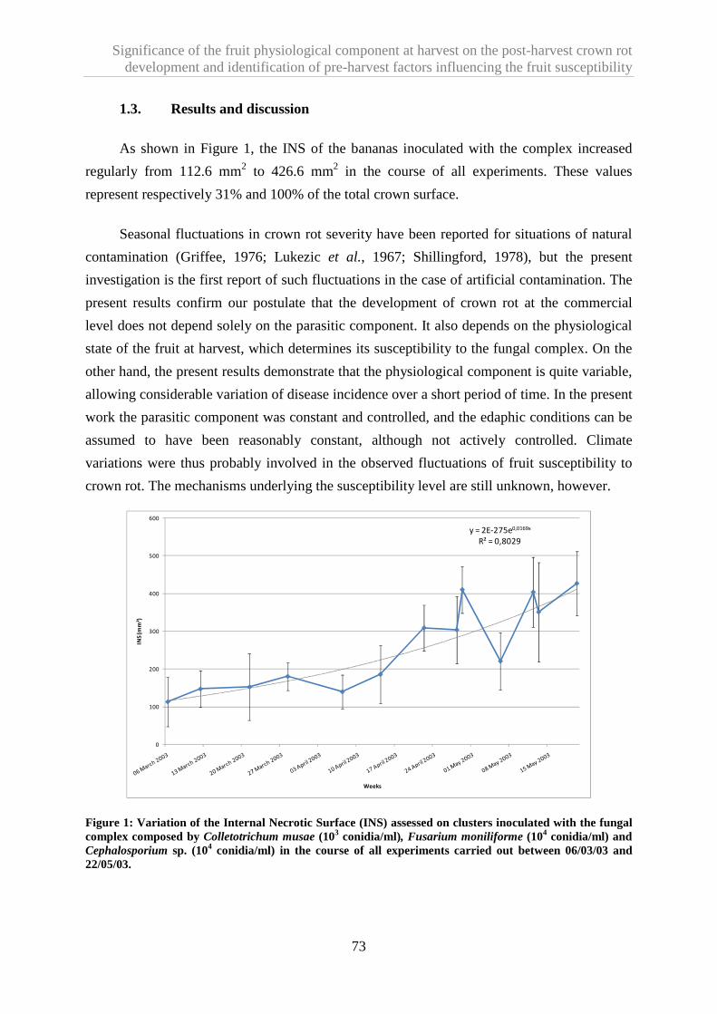
Significance of the fruit physiological component at harvest on the post-harvest crown rot development and identification of pre-harvest factors influencing the fruit susceptibility
73
1.3. Results and discussion
As shown in Figure 1, the INS of the bananas inoculated with the complex increased
regularly from 112.6 mm2 to 426.6 mm2 in the course of all experiments. These values
represent respectively 31% and 100% of the total crown surface.
Seasonal fluctuations in crown rot severity have been reported for situations of natural
contamination (Griffee, 1976; Lukezic et al., 1967; Shillingford, 1978), but the present
investigation is the first report of such fluctuations in the case of artificial contamination. The
present results confirm our postulate that the development of crown rot at the commercial
level does not depend solely on the parasitic component. It also depends on the physiological
state of the fruit at harvest, which determines its susceptibility to the fungal complex. On the
other hand, the present results demonstrate that the physiological component is quite variable,
allowing considerable variation of disease incidence over a short period of time. In the present
work the parasitic component was constant and controlled, and the edaphic conditions can be
assumed to have been reasonably constant, although not actively controlled. Climate
variations were thus probably involved in the observed fluctuations of fruit susceptibility to
crown rot. The mechanisms underlying the susceptibility level are still unknown, however.
y = 2E-275e0,0169x
R² = 0,8029
0
100
200
300
400
500
600
INS
(m
m²)
Weeks
Figure 1: Variation of the Internal Necrotic Surface (INS) assessed on clusters inoculated with the fungal complex composed by Colletotrichum musae (103 conidia/ml), Fusarium moniliforme (104 conidia/ml) and Cephalosporium sp. (104 conidia/ml) in the course of all experiments carried out between 06/03/03 and 22/05/03.
Significance of the fruit physiological component at harvest on the post-harvest crown rot development and identification of pre-harvest factors influencing the fruit susceptibility
74
1.4. References
Chillet M., de Lapeyre de Bellaire L., 1996a. Elaboration de la qualité des bananes au champ.
Détermination de critères de mesure. Fruits, 51, 317-326.
Chillet M., Hubert O., Rives M. J., de Lapeyre de Bellaire L., 2006. Effects of the
Physiological Age of Bananas on Their Susceptibility to Wound Anthracnose Due to
Colletotrichum musae. Plant Disease, 90, 1181-1185.
de Lapeyre de Bellaire L., Chillet M., Chilin-Charles Y., 2008. Determination of banana fruit
susceptibility to post-harvest diseases: wound anthracnose, quiescent anthracnose and
crown-rot. Fruits, 63, 183-186.
Ganry J., 1978. Recherche d’une méthode d’estimation de la date de récolte du bananier à
partir de données climatiques dans les conditions des Antilles. Fruits, 33, 669-680.
Griffee P. J., 1976. Pathogenicity of some fungi isolated from diseased crowns of banana
hands. Journal of Phytopathology, 85, 206-216.
Lassois L., Jijakli H., Chillet M., de Lapeyre de Bellaire L., Accepted. Crown rot of bananas:
pre-harvest factors involved in post-harvest expression and integrated control methods.
Plant Disease.
Lukezic F. L., Kaiser J., Martinez M.M., 1967. The incidence of crown rot of boxed bananas
in relation to microbial population of the crown tissue. Canadian Journal of Botany, 45,
413-421.
Shillingford, C.A., 1978. Climatic factors affecting post-harvest decay of jamaican bananas.
Journal of Agriculture of the University of Puerto Rico, 63, 45-49.

Significance of the fruit physiological component at harvest on the post-harvest crown rot development and identification of pre-harvest factors influencing the fruit susceptibility
75
2. Hand position on the bunch and source-sink ratio influence the banana susceptibility to crown rot disease
It was showed there was a lack of information and researches about the role of the fruit
physiological component that influence the fruit quality potential at harvest. In the same way,
no information was available about the pre-harvest factors implicate in the observed variation
in susceptibility. We first tried, but without success, to identify simultaneously parameters
that influencing the fruit susceptibility to crown rot in field conditions (results not show). It
quickly became apparent that the environment-plant relationships determining susceptibility
of fruit were very complex. We therefore concentrated on the study of 2 individual pre-
harvest factors. We identified that fruit position on the bunch and source-sink ratio at
flowering stage have an impact on the fruit susceptibility level.
The results obtained were accepted for publication.
Lassois L., Bastiaanse H., Chillet M., Jullien A., Jijakli M.H. and de Lapeyre de
Bellaire. Hand position on the bunch and source-sink ratio influence the banana susceptibility
to crown rot disease. Annals of Applied Biology. DOI: 10.1111/j.1744-7348.2009.00381.x

Significance of the fruit physiological component at harvest on the post-harvest crown rot development and identification of pre-harvest factors influencing the fruit susceptibility
76
Hand position on the bunch and source-sink ratio influence the banana fruit
susceptibility to crown rot disease
Lassois Ludivine a, Bastiaanse Heloïse a, Chillet Marc b,c, Jullien Alexandrad, Jijakli M.
Haissam a and de Lapeyre de Bellaire Luc e,*
a University of Liege, Gembloux Agro-Bio Tech, Plant Pathology Unit. Passage des Déportés 2, B-5030 Gembloux, Belgium; b CIRAD, UMR Qualisud, F-34398 Montpellier France ; c Departamento de Alimentos e Nutriçao Experimental – Faculdade de Ciencias Farmaceuticas – Universidade de Sao Paulo, Brasil; d AgroParisTech, UMR INRA/AgroParisTech Environnement et Grandes Cultures, F- 78850 Thiverval-Grignon, France. e CIRAD, UPR Systèmes bananes et ananas, F-34398 Montpellier, France *Corresponding author: Tel : +33 (0)467615828 ; Fax: +33 (0)467615688 ; e-mail: [email protected] Received 25 March 2009 Accepted 28 September 2009
Running title: Banana susceptibility to crown rot disease

Significance of the fruit physiological component at harvest on the post-harvest crown rot development and identification of pre-harvest factors influencing the fruit susceptibility
77
Abstract
The post-harvest development of crown rot of bananas depends notably on the fruit
susceptibility to this disease at harvest. It has been shown that fruit susceptibility to crown rot
is variable and it was suggested that this depends on environmental pre-harvest factors.
However, little is known about the pre-harvest factors influencing this susceptibility. The aim
of this work was to evaluate the extent to which fruit filling characteristics during growth and
the fruit development stage influence the banana susceptibility to crown rot. This involved
evaluating the influence of (i) the fruit position at different levels of the banana bunch (hands)
and (ii) changing the source-sink ratio, on the fruit susceptibility to crown rot. The fruit
susceptibility was determined by measuring the internal necrotic surface (INS) after artificial
inoculation of Colletotrichum musae. A linear correlation (r = -0.95) was found between the
hand position on the bunch and the INS. The source-sink ratio was found to influence the
pomological characteristics of the fruits and their susceptibility to crown rot. Fruits of
bunches from which six hands were removed (2 hands remaining on the bunch) proved to be
significantly less susceptible to crown rot (INS = 138.3 mm2) than those from bunches with 8
hands (INS = 237.9 mm2). The banana susceptibility to crown rot is thus likely to be
influenced by the fruit development stage and filling characteristics. The present results
highlight the importance of standardizing hand sampling on a bunch when testing fruit
susceptibility to crown rot. They also show that hand removal in the field has advantages in
the context of integrated pest management, making it possible to reduce fruit susceptibility to
crown rot while increasing fruit size.
Keywords: Colletotrichum musae, Musa sp., plant-pathogen interactions, post-harvest
disease, fruit quality.

Significance of the fruit physiological component at harvest on the post-harvest crown rot development and identification of pre-harvest factors influencing the fruit susceptibility
78
2.1. Introduction
Crown rot disease affects export bananas in all producing countries and is viewed as one
of the main post-harvest diseases of bananas. The disease develops during shipping, ripening,
and storage and has a negative impact on the market value of bananas (Slabaugh and Grove,
1982). It results from the development of several relatively nonspecific pathogens, but many
authors agree on the high pathogenicity of Colletotrichum musae, which can trigger an
infection from a very small inoculum (Finlay and Brown, 1993; Lassois et al., 2008).
Geographic and seasonal variations have been noted in the incidence of banana post-
harvest diseases (Chillet and de Lapeyre de Bellaire, 1996; Chillet et al., 2007; Krauss and
Johanson, 2000; Lassois et al., 2008; Lukezic et al., 1967; Shillingford, 1978). It has been
suggested that these spatiotemporal fluctuations may reflect variations in the banana fruit
quality potential that develops in the field and which determines the post-harvest onset or
absence of diseases (Chillet and de Lapeyre de Bellaire, 1996; Lassois et al., accepted). The
quality potential comprises a physiological and parasitic component, which both depend on
agro-technical factors and on soil and climate environment conditions (names pedo-climatic
factors).
The physiological component here refers to the fruit susceptibility to crown rot. In order
to overcome the parasitic component, this susceptibility level of the fruit is measured by the
size of lesions in standardized artificial inoculations (de Lapeyre de Bellaire et al., 2008). In
the case of anthracnose it has been shown that fruit susceptibility at harvest is a key factor in
the development of this disease and its control (Chillet et al., 2007; Chillet et al., 2006). Little
is known, however, about the factors influencing the banana susceptibility to crown rot.
Although pedo-climatic conditions and agro-technical factors are known to influence the
development of this post-harvest disease (Krauss and Johanson, 2000; Lukezic et al. 1967;
Shillingford, 1978), there are few studies linking such fluctuations to the field susceptibility
(Lassois et al., 2008).
The aim of this work was to characterize the importance of some pre-harvest factors in
determining fruit susceptibility to crown rot. In particular, we were interested in the influence
of the fruit development stage and fruit filling characteristics. First we examined whether the
position of a hand in a banana bunch influences the susceptibility of its fruits to crown rot,
taking advantage of the fact that the hands of a bunch differ as regards to both their
development stage and filling status (Jullien et al. 2001a). Next we evaluated how a change in

Significance of the fruit physiological component at harvest on the post-harvest crown rot development and identification of pre-harvest factors influencing the fruit susceptibility
79
the source-sink ratio during growth of the bunch affects fruit susceptibility to crown rot
(leaves being the source and fruit, the sink) considering that source-sink ratio modification
leads, among others, to changes in the rate of fruit filling (Jullien et al., 2001b). The source-
sink terms are commonly used in plant characterization. Leaves were considered as source
tissues because they produce excess of assimilate, while fruits were sink organs. With respect
to metabolism, plant organs are generally divided into source and sink tissues. Source tissues
like mature leaves produce excess of assimilates which are transported via the phloem to the
different sink tissues not able to produce themselves sufficient amounts of assimilates.
2.2. Materials and Methods
A banana plant produces an inflorescence called a bunch. Fruits on a bunch are grouped
into female and male hands arranged helicoidally around a central axis called the stalk. The
hand at the top of the bunch is the first to be initiated and hands are traditionally numbered
from this one downward. Each hand can be divided into clusters consisting of several banana
fruits called fingers (Figure 1).
Hand
Cluster
Finger
BUNCH
1st hand
2d hand
3rd hand
…
Figure 1: Organization of a banana bunch

Significance of the fruit physiological component at harvest on the post-harvest crown rot development and identification of pre-harvest factors influencing the fruit susceptibility
80
2.2.1. Plant material
For each performed test, homogeneous sets of banana plants (Musa acuminata [AAA
group, Cavendish subgroup] cv Grand Nain) which have grown under the same agro-technical
practices were randomly selected at the Dia-Dia commercial plantation (PHP, Njombe,
Cameroon (altitude: 80 m; annual mean temperature: 26.5°C; annual mean rainfall: 3500
mm). Non-systemic fungicide applications were used to control foliar diseases. The crown
tissues have never been in contact with fungicides. The date of flowering was indicated by
tying a colored belt to each bunch at the horizontal finger stage in order to predict the time of
harvest. Bunches were also covered with a plastic sleeve at this stage. Bunches were
harvested at a constant physiological age (Jullien et al., 2008), i.e. when the mean daily
temperature sum accumulated by the fruit at the 14°C threshold between flowering and
harvest reached 900 degree days (dd). Temperatures were recorded at a weather station on the
plantation. The daily average temperature (Td) was estimated from measurements of
maximum temperature (Tmax) and minimum temperature (Tmin).
2.2.2. Evaluation of susceptibility to crown rot
Hands of bananas collected on the day of the experiment and transported to the
laboratory were cut into clusters of 4 fingers without defects. The crown surfaces were
refreshed with a knife. These cuttings were square, with regular, clean-cut sections in order to
obtain similar crowns. Smoothly cut crowns were obtained with a sharp knife, leaving as
much crown tissue as possible. Latex from crown tissues was dried with absorbent paper and
the crowns were surface-sterilized by submersion in 50% ethanol. Fifty microlitres of C.
musae conidial suspension containing 104 conidia/ml was applied to the centre of the freshly
exposed crown tissue and covered with a small paper filter. The C. musae strain was isolated
in Njombe, Cameroon. It is sensitive to thiabendazole and was stored at –20°C in a glycerol
solution (30%). C. musae cultures were grown at 25°C in Mathur medium (MgSO4.7H2O:
2.5g/l; KH2PO4: 2.7g/l; peptone : 1 g/l; yeast extract: 1g/l; saccharose: 10 g/l; agar: 15 g/l) for
10 days. Conidia were removed by flooding the plates with sterile distilled water and filtration
through a 45µ sieve. Their concentrations were determined with a Mallassez cell. Two hours
after application of the conidial suspension, the clusters were packed in punched polyfilms
normally used in the industry, placed in commercial boxes, and stored at 13°C for 10 days to
simulate shipment. Artificial ripening was then initiated by dipping the bananas for 5 seconds
in an ethrel solution (480 g/l), after which the clusters remained at 20°C for another 3 days
before crown rot assessment. The internal progression of rot was determined by cutting the
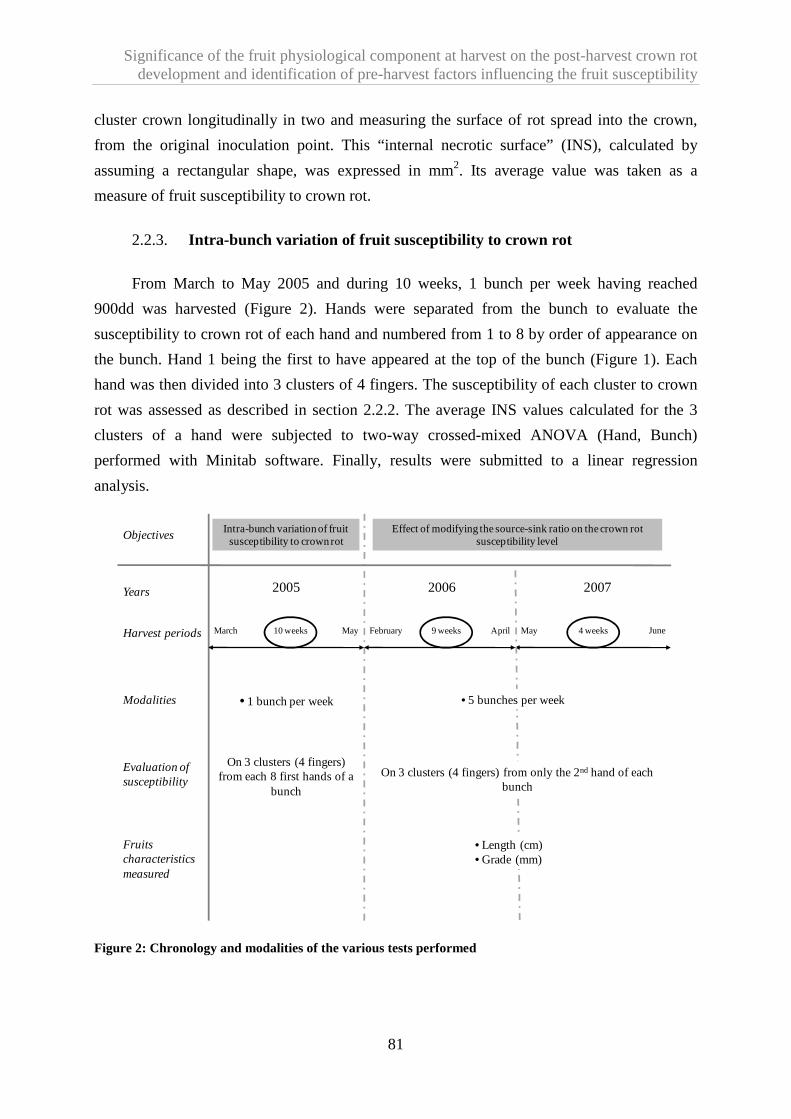
Significance of the fruit physiological component at harvest on the post-harvest crown rot development and identification of pre-harvest factors influencing the fruit susceptibility
81
cluster crown longitudinally in two and measuring the surface of rot spread into the crown,
from the original inoculation point. This “internal necrotic surface” (INS), calculated by
assuming a rectangular shape, was expressed in mm2. Its average value was taken as a
measure of fruit susceptibility to crown rot.
2.2.3. Intra-bunch variation of fruit susceptibility to cr own rot
From March to May 2005 and during 10 weeks, 1 bunch per week having reached
900dd was harvested (Figure 2). Hands were separated from the bunch to evaluate the
susceptibility to crown rot of each hand and numbered from 1 to 8 by order of appearance on
the bunch. Hand 1 being the first to have appeared at the top of the bunch (Figure 1). Each
hand was then divided into 3 clusters of 4 fingers. The susceptibility of each cluster to crown
rot was assessed as described in section 2.2.2. The average INS values calculated for the 3
clusters of a hand were subjected to two-way crossed-mixed ANOVA (Hand, Bunch)
performed with Minitab software. Finally, results were submitted to a linear regression
analysis.
2006 20072005
ObjectivesIntra-bunch variation of fruit susceptibility to crown rot
Effect of modifying the source-sink ratio on the crown rot susceptibility level
JuneHarvest periods March May10 weeks February April9 weeks May 4 weeks
Modalities • 1 bunch per week • 5 bunches per week
Evaluation of susceptibility
On 3 clusters (4 fingers) from each 8 first hands of a
bunch
On 3 clusters (4 fingers) from only the 2nd hand of each bunch
Years
Fruits characteristics measured
• Length (cm)• Grade (mm)
Figure 2: Chronology and modalities of the various tests performed

Significance of the fruit physiological component at harvest on the post-harvest crown rot development and identification of pre-harvest factors influencing the fruit susceptibility
82
2.2.4. Effect of modifying the source-sink ratio on the crown rot susceptibility
The source-sink ratio was modified as follows during fruit growth by removal of leaves
(L) and hands (H) at the flowering stage (horizontal finger stage) so as to obtain the following
treatments:
12L/2H: 12 leaves and two hands (the first two to have appeared on the bunch)
remaining at the flowering stage;
12L/8H: 12 leaves and eight hands (the first eight to have appeared on the bunch)
remaining at the flowering stage;
5L/2H: 5 leaves (the last to have emerged) and two hands (the first two to have
appeared on the bunch) remaining at the flowering stage;
5L/8H: 5 leaves (the last to have emerged) and eight hands (the first eight to have
appeared on the bunch) remaining at the flowering stage;
An empirical source-sink ratio was also calculated. As the banana plants with 12 leaves
and bunches of 8 hands (12L/8H) did not undergo leaf or hand removal, they were considered
as reference to calculate an empirical source-sink ratio (So-Si ratio). The empirical So-Si
ratios for the other treatments including leaf or hand trimming were estimated with the
following formula: [(Number of leaves/number of hands) for the treatment] / [(Number of
leaves/number of hands) of the reference treatment]
For example, the empirical source-sink ratio calculated for the plant with the 12L/2H
configuration was [(12/2)/(12/8)] = 4.
Five banana plants per week were selected for each ratio, and bunches were harvested
when the fruits had accumulated 900 dd (Jullien et al., 2008). Two test series were conducted
(Figure 2): 9 successive repetitions (weeks) between February and April 2006 and 4
successive repetitions (weeks) between May and June 2007. Fruit susceptibility was assessed
on 3 clusters of 4 fingers on hand 2 of each bunch, as described in 2.2. The average INS
values calculated for the 3 clusters were subjected to partially hierarchical mixed four-way
ANOVA (Treatment, Bunch, Week, Year), each cluster being taken as an experimental unit.
This was followed by the contrast hypothesis testing to separate leaves and hands effects.

Significance of the fruit physiological component at harvest on the post-harvest crown rot development and identification of pre-harvest factors influencing the fruit susceptibility
83
In addition to assessment of fruit susceptibility, some fruit pomological characteristics
were measured. On each harvested hand 2, the lengths (cm) and diameters name grades (mm)
of the fruits were systematically measured. Statistical analysis consisted of two partially
hierarchical mixed three-way ANOVAs (Treatment, Week, Year) carried out with Minitab
software followed by the contrast hypothesis testing to separate leaves and hands effects.
2.3. Results
2.3.1. Intra-bunch variation of fruit susceptibility to cr own rot
The position of fruit on the bunch was found to influence fruit susceptibility to crown
rot very highly significantly (p <0.001) (Table 1). Although very highly significant
differences were observed between bunches (weeks) (p <0.001), the trend was the same
whatever the bunch, as no effect of the interaction ‘hand x bunch’ was observed (p = 0.218).
Table 1: Results of a two-way crossed-mixed ANOVA (hand, bunch) on Internal Necrotic Surface (mm2).
Internal Necrotic Surface
Source d.f. F-value P-value
Hand 7 17.85 <0.001
Bunch 9 6.95 <0.001
Hand * Bunch 63 1.17 0.218
A gradient of susceptibility was observed between hand 8, with an INS average of 73.6
mm2, and hand 1, with an INS average of 150.9 mm2 (Figure 3). Hand rank is an equidistant
ordinal variable that can be compared to a quantitative value in the calculations. Hence, a
linear correlation was found (r = -0.95) between the hand position on the bunch and the 8
corresponding INS means.

Significance of the fruit physiological component at harvest on the post-harvest crown rot development and identification of pre-harvest factors influencing the fruit susceptibility
84
1 2 3 4 5 6 7 8
Mean 150,9 135,0 129,1 93,7 97,1 88,8 84,7 73,6
y = -10,83x + 155,3
R² = 0,910
0
50
100
150
200
250
INS
(m
m2
)
Hands position in the bunch
Figure 3: Mean values and standard deviations of internal necrotic surface (INS mm2) as a function of the hand position on the bunch.
2.3.2. Effect of the source-sink ratio modification on the crown rot susceptibility
The change in the source-sink ratio imposed during flowering by removal of both leaves
and hands from the bunch had a highly significant effect on the fruit susceptibility to crown
rot (p = 0.004) (Table 2). Although there was also a highly significant effect of the year factor
(p = 0.004), the trend was the same in both years because no interaction was observed
between the treatments and the years (p = 0.225) leading to analyze the results of both years
together. The fruit susceptibility to crown rot showed a very highly significant variation
(p<0.001) from week to week within a year without consequence on the preceding
conclusions.

Significance of the fruit physiological component at harvest on the post-harvest crown rot development and identification of pre-harvest factors influencing the fruit susceptibility
85
Table 2: Results of a partially hierarchical mixed four-way ANOVA (Bunch, Treatment, Week, Year) on Internal Necrotic Surface (mm2) and of two partially hierarchical mixed three-way ANOVAs (Treatment, Week, Year) on fruit grade and length.
Internal Necrotic Surface Grade Length
Source d.f. F-value P-value F-value P-value F-value P-value
Treatment 3 57.87 0.004 140.46 <0.001 103.49 <0.001 Year 1 13.29 0.004 10.85 0.001 102.91 <0.001 Treatment * Year 3 1.52 0.225 4.80 0.003 2.44 0.065 Week (Year) 11 10.14 <0.001 7.65 <0.001 2.58 0.005 Treatment * Week (Year) 33 1.77 0.009 1.14 0.283 1.64 0.022
Bunch (Treatment Year Week) 191 2.32 <0.001 / / / /
The ranking of different treatments (Table 3) shows that the fruits of bunches from
which 6 hands were removed (treatments 12L/2H and 5L/2H) were significantly less
susceptible to crown rot (INS = 138.3 mm2) than those of bunches with 8 hands (12L/8H
treatment and 5L/8H, INS = 237.9 mm2). There appeared no susceptibility difference,
however, between treatments 12L/8H and 5L/8H or between treatments 12L/2H and 5L/2H.
Thus, the removal of 6 out of 8 hands had a significant effect on fruit susceptibility to crown
rot but the removal of 7 out of 12 leaves did not. Table 3 also shows the classification of the
INS by order of decreasing empirical source-sink ratio. It is noteworthy that in trend terms,
the crown susceptibility increased as the empirical source-sink ratio decreased.

Significance of the fruit physiological component at harvest on the post-harvest crown rot development and identification of pre-harvest factors influencing the fruit susceptibility
86
Table 3: Mean values and standard deviations of Internal Necrotic Surfaces (INS) mm2, fruit grade (mm), and length (cm) after treatments affecting the source-sink ratio. All fruits were obtained from hand 2. The letters a, b, c, d, e and f represent groups of statistically similar fruits based on contrast testing. 12L/2H: banana plant with 12 leaves and 2 hands; 5L/2H: banana plant with 5 leaves and 2 hands; 12L/8H: banana plant with 12 leaves and 8 hands; 5L/8H: banana plant with 5 leaves and 8 hands. The source-sink ratio was estimated according to the number of leaves and fruits removed in each treatment, as compared to unaltered banana plants (12L/8H).
Treatment Source-sink ratio INS (mm2) Grade (mm) Length (cm)
12L/2H 4 130.2±65.4a 37.9 ± 0.3c 22.0 ± 0.2e
5L/2H 1.7 146.4±60.2a 36.8 ± 0.3c 21.3 ± 0.2e
12L/8H 1 227.4±66.7b 33.3 ± 0.2d 19.5 ± 0.2f
5L/8H 0.4 248.4±72.0b 32.0 ± 0.2d 18.6 ± 0.2 f
2.3.3. Effect of the source-sink ratio modification on fruit grade and length
The source-sink ratio change imposed during flowering by removal of both leaves and
hands had a very highly significant effect on fruit length (p < 0.001) and grade (p < 0.001)
(Table 2). The ranking of the different treatments (Table 3) shows that the fruits of bunches
from which 75% of the hands were removed (treatments 12L/2H and 5L/2H) were
significantly longer and had a significantly higher grade (average length = 21.6 cm and
average grade = 37.3 mm) than those of bunches with 8 hands (treatments 12L/8H and 5L/8H,
average length = 19 cm and average grade = 32.6 mm). Removal of many hands thus has a
significant effect on the morphometric characters of the fruit. No difference in fruit grade or
length was observed, however, between treatments 12L/8H and 5L/8H or treatments 12L/2H
and 5L/2H. Thus, leaf removal had no significant effect on these morphometric characteristics
(Table 3). It is noteworthy that the fruit pomological characteristics and banana yield
increased as the empirical source-sink ratio decreased (Table 3). The various changes in the
source-sink ratio had an effect on fruit filling.
2.4. Discussion
We show here that fruit development stage and filling characteristics are parameters
influencing post-harvest susceptibility of bananas to crown rot. Within a bunch, there is a
gradient of susceptibility to crown rot, the hands initiated first (the upper ones) being more
susceptible than those initiated last (the lower ones). It has already been established that
morphological differences between hands of a same bunch result from differential
development associated with cell division and fruit filling characteristics (Jullien et al.
Significance of the fruit physiological component at harvest on the post-harvest crown rot development and identification of pre-harvest factors influencing the fruit susceptibility
87
2001a). Here it appears that competition within a bunch affects not only the morphological
characteristics of the fruits but also the fruit quality potential. Taking into account the
observation of Jullien et al. (2001a) that the hands initiated first are approximately 70dd ahead
those initiated last, the susceptibility gradient might be due to this physiological age gap
between fruit in the same bunch. Effectively, in the case of anthracnose of banana, the fruit
susceptibility has been shown to increase with the physiological age of the fruit expressed in
dd (Chillet et al., 2007; Chillet et al., 2006).
The position of a hand on the bunch has also been shown to influence fruit filling. This
results in gradients of pulp dry weight, cell number per fruit, and starch grain number per cell
(Jullien et al. 2001a) and also in differences in sap concentration and composition within the
same bunch (Kurien et al., 2000). Furthermore, it is well known that partitioning of
assimilates between various sink organs are complex and not equally distributed (Kozlowski,
1992). These differences in filling characteristics within a bunch may be involved in the
observed susceptibility changes.
We confirm here that source-sink ratio changes have a significant effect on fruit
morphology, as previously demonstrated in several studies (Chillet et al., 2006, Daniells et
al., 1987, Daniells et al. 1994; Israeli et al., 1995, Johns 1996, Jullien et al. 2001b; Kurien et
al. 2000; Mouen Bedimo et al., 2003). We reveal, furthermore, a new effect of a source-sink
ratio change on the fruit quality potential: when the sink is decreased by removal of many
hands, the fruit susceptibility to crown rot decreases. Few studies on various plants have
linked the importance of source-sink ratio, and thus photosynthetic assimilate distribution, in
plant-pathogen interactions (Barrière, 1985; Barrière et al., 1981; Dodd, 1980; Pegg, 1986;
Seetharama et al., 1991). However, in the case of bananas, the present results contrast with
the previously reported observation that modifying the source-sink ratio has no effect on fruit
susceptibility to anthracnose or on the fruit conservation potential (Chillet et al., 2006). This
suggests that different mechanisms govern the susceptibility of fruit to crown rot and
anthracnose. Jullien et al. (2001b) have also highlighted the impact of this source-sink ratio
on fruit filling characteristics, and notably on the rate of cell filling. In our study, we consider
that hand and leaf removal result in a modification of the empirical source-sink ratio. In
keeping with the observation of Jullien et al. (2001b), we assume that an increased empirical
source-sink ratio results in an increased fruit filling rate. One might hypothesize that this
increased cell filling rate is involved in the observed reduction of the fruit susceptibility to
crown rot. On the other hand, the impact of the source-sink ratio on nutrient availability,
distribution, storage, and assimilative transformation has been extensively documented in

Significance of the fruit physiological component at harvest on the post-harvest crown rot development and identification of pre-harvest factors influencing the fruit susceptibility
88
many models (Noquet et al., 2004; Savin et al., 2006; Famiani et al. 2000; Dordas, 2009;
Zhenming et al., 2008). We suggest that a modification of the source-sink ratio lead to a
change in the partitioning of assimilates between various sink which influences the formation
of secondary metabolites involved in plant-pathogen interaction. When hands are removed,
the competition between sinks is reduced and the availability of mobile assimilates for
remaining hands is more important.
It is noteworthy that in our study source reduction (leaf removal) had a lesser effect on
fruit susceptibility to crown rot than sink reduction, since removal of about 60% of the leaves
(7 out of 12) had little effect on fruit susceptibility, in contrast to removal of 75% of the fruits
(6 out of 8). It is recognized that the impact of defoliation on the qualitative and quantitative
development of bananas is variable and highly dependent on (i) when the defoliation is
performed (Arcila et al., 1995), (ii) the intensity of defoliation (Israeli et al. 1995, Robinson
and Anderson, 1990, Rodriguez et al., 2005), and (iii) how defoliation is done: mechanically
or through the action of pathogens (Robinson et al., 1992). Here, mechanical defoliation
appears not to have been sufficiently early and/or severe to influence the processes
determining fruit susceptibility to crown rot. On the other hand, a compensatory phenomenon
reducing the impact of defoliation might also be involved, as the development of a bunch
results from the distribution of dry matter not only from the leaves but also from other parts of
the plant. In this way, the rachis and pseudostem may partially compensate for late-occurring
defoliation (Eckstein et al., 1995). It has also been shown that an increased photosynthetic
capacity of the remaining leaves may partially compensate for losses caused by defoliation
(Robinson et al., 1992).
2.5. Conclusion
The susceptibility of bananas to crown rot is thus likely to be influenced by the stage of
fruit development and by filling characteristics, these parameters being in close interaction
and dependent on the soil-climate conditions and agro-technical factors of the production
area. It is essential not to lose sight of the fact that the regulation of plant susceptibility is, in
all cases, the result of nutritional balance established during plant growth. This balance is the
consequences of all physiological relations of the whole plant and the environmental factors
and might affect plant-pathogen interaction by two ways. First by influencing the ability of
the plant to establish defense mechanisms, notably through changes in secondary metabolism.
Secondly by altering the bioavailability of nutrients necessary for pathogen development.
Significance of the fruit physiological component at harvest on the post-harvest crown rot development and identification of pre-harvest factors influencing the fruit susceptibility
89
However, the molecular underlying mechanisms implied in the susceptibility variations
observed in this study still unknown.
The fact that the fruit susceptibility depends on the hand position in the bunch shows the
importance of standardizing the sampling method when measuring the susceptibility of
bananas to crown rot. The hands collected for an experiment should be collected
systematically from the same position on each bunch. As the hands in the upper portion have
more fruits and allow division into more 4-fruit clusters, it has been recommended to use the
third hand of the bunch, which is more stable from one bunch to another than the first two
hands (Jannoyer, 1995). If more than one hand per bunch is needed to carry out the
experiment, it is essential to work with successive hands like demonstrated in this paper.
Lastly, the main method used to control crown rot is a systematic chemical post-harvest
treatment. Apart from the environmental, social, and legislative problems resulting from this
chemical control strategy, growers also face problems of treatment efficiency which are
notably related to the fruit susceptibility in some specific areas. Only a truly integrated pest
management strategy applied to the whole chain can provide effective alternatives to chemical
treatment (Lassois et al., accepted). We have shown that an increase in the empirical source-
sink ratio makes it possible to reduce fruit susceptibility to crown rot while increasing fruit
size at harvest. Thus, early hand removal in the field might be used as part of an integrated
pest management scheme.
Acknowledgements
The authors wish to thank Yves Brostaux, of the Applied Statistics, Computer Science
and Mathematics Unit of University of Liege, Gembloux Agro-Bio Tech, for his assistance in
the statistical analysis.

Significance of the fruit physiological component at harvest on the post-harvest crown rot development and identification of pre-harvest factors influencing the fruit susceptibility
90
2.6. References
Arcila M.I., Belalcázar S., Valencia J.A, Cayón G. (1995) Influencia del número de hojas en
postfloración sobre el llenado de los frutos del clon de plátano Dominico Hartón, Musa
AAB Simmonds. In : ICA-CORPOICA (Ed.) Mejoramiento de la Producción del
Cultivo del Plátano. Segundo informe técnico 1984-1994. Región 9 ICA-CORPOICA.
Creced-Quindio, Armenia, Colombia, 90-93.
Barrière Y. (1985) Aspects physiologiques de la pourriture des tiges du maïs. Agronomie, 5,
361-368.
Barrière Y., Panouillé A., Cassini R. (1981) Relations source-puits et sélection du maïs pour
la résistance à la pourriture des tiges. Agronomie, 8, 707-711.
Chillet M., de Lapeyre de Bellaire L. (1996) Elaboration de la qualité des bananes au champ.
Détermination de critères de mesure. Fruits, 51, 317-326.
Chillet M., Hubert O., de Lapeyre de Bellaire L. (2007) Relationship between physiological
age, ripening and susceptibility of banana to wound anthracnose. Crop Protection, 26,
1078-1082.
Chillet M., Hubert O., Rives M.J., de Lapeyre de Bellaire L. (2006) Effects of the
physiological age of bananas on their susceptibility to wound anthracnose due to
Colletotrichum musae. Plant Disease, 90, 1181-1185.
Daniells J.W, O'Farrell P.J., Mulder J.C., Campbell S.J. (1987) Effects of bunch covering and
bunch trimming on bananas in North Queensland. Queensland Journal of Agricultural
and Animal Science, 44, 259-265.
Daniells J.W., Lisle A.T., Bryde N.J. (1994) Effect of bunch trimming and leaf removal at
flowering on maturity bronzing, yield and other aspects of fruit quality of bananas in
North Queensland. Australian Journal of Experimental Agriculture, 34, 259-265.
de Lapeyre de Bellaire L., Chillet M., Chilin-Charles Y. (2008) Determination of banana fruit
susceptibility to post-harvest diseases: wound anthracnose, quiescent anthracnose and
crown-rot. Fruits, 63, 183-186.
Dood J.L. (1980) Grain size and predisposition of Zea mays to stalk rot. Phytopathology, 70,
534-535.
Dordas C. (2009) Dry matter, nitrogen and phosphorus accumulation, partitioning and
remobilization as affected by N and P fertilization and source-sink relations. European
Journal of Agronomy, 30, 129-139.
Eckstein K., Robinson J., Davie J. (1995) Physiological responses of banana (Musa AAA;
Cavendish sub-group) in the subtropics. III gas exchange, growth analysis and source-

Significance of the fruit physiological component at harvest on the post-harvest crown rot development and identification of pre-harvest factors influencing the fruit susceptibility
91
sink interaction over a complete crop cycle. Journal of Horticultural Science, 70, 169-
180.
Famiani F., Proietti P., Palliotti A., Ferranti F., Antognozzi E. (2000) Effects of leaf to fruit
ratios on fruit growth in chestnut. Scientia Horticulturae, 85, 145-152.
Finlay A.R., Brown A.E. (1993) The relative importance of Colletotrichum musae as a crown-
rot pathogen on Windward Island bananas. Plant Pathology, 42, 67-74.
Israeli Y., Plaut Z., Schwartz A. (1995) Effect of shade on banana morphology, growth and
production. Scientia Horticulturae, 62, 45-56.
Jannoyer M. (1995) Determinism of reproductive organ number for banana (Musa acuminata
cv `Grande Naine'). PhD Thesis in agronomy, Paris, INAPG. 199 p.
Johns G.G. (1996) Effects of bunch trimming and double bunch covering on yield of bananas
during winter in New South Wales. Australian Journal of Experimental Agriculture, 36,
229-235.
Jullien A., Malezieux E., Michaux-Ferriere N., Chillet M., Ney B. (2001a) Within-bunch
Variability in Banana Fruit Weight: Importance of Developmental Lag Between Fruits.
Annals of Botany, 87, 101-108.
Jullien A., Munier-Jolain N., Malézieux E., Chillet M., Ney, B. (2001b) Effect of pulp cell
number and assimilate availability on dry matter accumulation rate in a banana fruit
[Musa sp. AAA group 'Grande Naine' (Cavendish subgroup)]. Annals of Botany, 88,
321-330.
Jullien A., Chillet M., Malezieux E. (2008) Pre-harvest growth and development, measured as
accumulated degree days, determine the post-harvest green life of banana fruit. Journal
of Horticultural Science and Biotechnology, 8, 506-512.
Kozlowski T.T. (1992) Carbohydrate sources and sinks in woody plants. The Botanical
Review, 58, 107-222.
Krauss U., Johanson A. (2000) Recent advances in the control of crown rot of banana in the
Windward Islands. Crop Protection, 19, 151-160.
Kurien S., Anil B.K., Rajeevan P.K., Bharathan V., Krishnan S. (2000) Phosphorus
mobilisation to uneconomic tissues and effects of bunch trimming regimes in banana.
Scientia Horticulturae, 83, 25-32.
Lassois L., de Lapeyre de Bellaire L., Jijakli M.H. (2008) Biological control of crown rot of
bananas with Pichia anomala strain K and Candida oleophila strain O. Biological
Control, 45, 410-418.

Significance of the fruit physiological component at harvest on the post-harvest crown rot development and identification of pre-harvest factors influencing the fruit susceptibility
92
Lassois L., Jijakli H., Chillet M., de Lapeyre de Bellaire L. Accepted. Crown rot of bananas:
pre-harvest factors involved in post-harvest expression and integrated control methods.
Plant Disease.
Lukezic F.L., Kaiser W.J., Martinez M.M. (1967) The incidence of crown rot of boxed
bananas in relation to microbial populations of the crown tissus. Canadian Journal of
Botany, 45, 413-421.
Mouen Bedimo J., Chillet M., Jullien A., de Lapeyre de Bellaire L. (2003) Le gainage précoce
des régimes de bananes améliore la croissance des fruits et leur état sanitaire vis-à-vis
de l'anthracnose (Colletotrichum musae). Fruits, 58, 71-81.
Noquet C., Avice J.-C., Rossato L., Beauclair P., Henry M.-P., Ourry A. (2004) Effects of
altered source-sink relationships on N allocation and vegetative storage protein
accumulation in Brassica napus L. Plant Science, 166, 1007-1018.
Pegg G.F. (1986) The effect of assimilate demand on Phytophthora infection and disease
development in NFC-grown tomato plants. Acta Horticulturae, 178, 128a-128l.
Robinson J., Anderson T. (1990) Banana bunch development in relation to leaf pruning at
flowering. Citrus and subtropical fruit research institute, South Africa. Information
Bulletin, 216, 4.
Robinson J.C., Anderson T., Eckstein K. (1992) The influence of functionnal leaf removal at
flower emergence on components of yield and photosynthetic compensation in banana.
Journal of Horticultural Science, 67, 403-410.
Rodriguez V., da Silva A., Rodriguez O. (2005) Balance nutritional y numero de hojas como
variables de prediccion del rendimiento del platano Harton. Pesquisa Agropecuaria
Brasileira, 40, 175-177.
Savin R., Prystupa P., Araus J.L. (2006) Hordein composition as affected by post-anthesis
source-sink ratio under different nitrogen availabilities. Journal of Cereal Science, 44, 113-116.
Seetharama N., Sachan R.C., Huda A.K.S., Gill K.S., Rao K.N., Bidinger F.R. and Reddy
D.M. (1991) Effect of pattern and severity of moisture-deficit stress on stalk-rot
incidence in sorghum. II. Effect of source-sink relationships. Field Crops Research, 26,
355-374.
Shillingford C A. (1978) Climatic factors affecting post-harvest decay of jamaican bananas.
Journal of Agriculture of the University of Puerto Rico, 45-49.
Slabaugh W.R., Grove M.D. (1982) Postharvest diseases of bananas and their control. Plant
Disease, 66, 746-750.

Significance of the fruit physiological component at harvest on the post-harvest crown rot development and identification of pre-harvest factors influencing the fruit susceptibility
93
Zhenming N., Xuefeng X., Yi W., Tianzhong L., Jin K., Zhenhai H. (2008) Effects of leaf-
applied potassium, gibberellin and source-sink ratio on potassium absorption and
distribution in grape fruits. Scientia Horticulturae, 115, 164-167.

Identification of genes potentially implied in quantitative banana response to crown rot disease
94
CHAPTER IV
IdentificaIdentificaIdentificaIdentification of tion of tion of tion of genes potentially implied in quantitative genes potentially implied in quantitative genes potentially implied in quantitative genes potentially implied in quantitative
banana response to crown rot diseasebanana response to crown rot diseasebanana response to crown rot diseasebanana response to crown rot disease

Identification of genes potentially implied in quantitative banana response to crown rot disease
95
1. Development of molecular biology techniques for the identification of genes differently expressed
1.1. General introduction
Prior to identify genes which could be implicated in banana quantitative defence-
response to crown rot, it was necessary to develop all techniques necessary for the study of
differently expressed genes.
The first difficulty was in the crown collected conservation in order to transport sample
from Njombé, Cameroun to Gembloux, Belgium without affecting the RNA expression profil
and quality. Indeed, for technical reasons, RNA extraction could not be realized in Cameroon.
Among the various tested solutions, the most efficient and practical in our conditions was to
freeze-dried our freshly collected sample. It is an original method because it has not been used
extensively in plant tissues.
Another difficulty was to obtain good quality RNA from banana crown tissues which is
a prerequisite to studying gene expression. Because of its high level in polysaccharides and
polyphenols, banana RNA could not be extracted with standard methods. We have tested
several methods, from simple to more time-consuming, and comparing them to choice the
best one.
Finally the cDNA-AFLP was evaluated like method which could be used for studying
gene expression between bananas showing various level of susceptibility.
All these results have been published:
Lassois L., de Lapeyre de Bellaire L. and Jijakli H. 2009. Combining an original
method for preserving RNA expression in situ with an effective RNA extraction method makes
it possible to study gene expression in any banana fruit tissue. Fruits, 64: 127-137.

Identification of genes potentially implied in quantitative banana response to crown rot disease
96
Combining an original method for preserving RNA expression in situ with an effective
RNA extraction method makes it possible to study gene expression in any banana fruit
tissue
Ludivine Lassois 1, Luc de Lapeyre de Bellaire 2, Haïssam Jijakli 1*
1 Gembloux Agricultural University, Plant Pathology Unit, Passage des Déportés 2, B-5030 Gembloux, Belgium, [email protected]* 2 CIRAD, Persyst, UPR Syst. Banan. Ananas, TA B-26 / PS4, Blvd. de la Lironde, 34398 Montpellier Cedex 5, France Received 24 October 2008 Accepted 5 December 2008 * Correspondence and reprints
Running title: RNA extraction from banana fruit tissue

Identification of genes potentially implied in quantitative banana response to crown rot disease
97
Abstract
Introduction. RNA isolation is a prerequisite to studying gene expression in banana
and to understanding changes occurring in response to the environment. Standard extraction
methods do not efficiently extract RNA from plants such as banana, with high levels of
phenolics, carbohydrates, or other compounds that bind to and/or coprecipitate with RNA.
Materials and methods. Five to seven RNA extraction methods were compared. Four crown-
tissue storage methods were also compared. cDNA-AFLP was used to ensure that the
obtained RNA was of sufficient quality for molecular applications and that RNA expression
was unaltered by in situ storage. Results and discussion. The modified hot-borate method
proved to be the best RNA extraction method, allowing high yields of good quality,
undegraded RNA from the crown, fruit peel and pulp at all stages of ripening. The RNA
obtained by this method was of sufficient quality for molecular applications such as cDNA-
AFLP that give highly reproducible results. Freeze-drying of fresh tissues and tissue
conservation in hot-borate buffer, two original storage methods, appear appropriate for
preserving RNA in situ without ultra-low temperature. The RNA obtained was of high
quality, undegraded, and useful for all downstream applications. The genome expression
profile obtained by cDNA-AFLP analysis was unaltered by these methods for storing
collected tissues. Conclusion. By applying all the suggested procedures in this work, it is
possible to store and study gene expression in any banana fruit tissue, whatever the maturity
stage, without affecting the RNA expression level.
Keywords: Belgium / Musa sp. / bananas / freeze-drying / RNA / storage / extraction

Identification of genes potentially implied in quantitative banana response to crown rot disease
98
1.2. Introduction
Banana is cultivated in more than 120 countries and holds the second place in world
fruit production, just behind orange and in front of grape. Exported dessert bananas belong to
the Cavendish subgroup. Gene expression studies are essential to understanding the
physiological processes of this plant species [1, 2]. Expression profiling, however, requires
adequate sample conservation and high-quality RNA isolation for cDNA library construction
and molecular analysis. The total RNA and mRNA obtained must notably be free of protein,
genomic DNA and secondary metabolites. RNA extraction is thus a crucial step, but it can be
problematic because of the relative instability of RNA, largely due to RNA degradation by
ribonucleases [3]. Conservation and isolation of RNA from plant material, particularly from
fruit tissues, presents special challenges [4, 5]. First, most plant materials contain relatively
high levels of RNase activity, mostly located in the vacuoles [6]. They also contain various
plant metabolites, such as polysaccharides and polyphenols. They bind to and/or co-
precipitate with RNA, affecting the yield and quality of RNA [7] and rendering it unsuitable
for cDNA synthesis. Thus, qualitative and quantitative differences in polysaccharide and
polyphenol content among different plant tissues significantly influence the efficiency of
nucleic acid extraction and purification procedures [3, 8]. Many procedures [4, 5, 8–14] have
been developed to cope with endogenous polysaccharides, phenolics and RNases during RNA
isolation from various plants, but the efficiency of an extraction method depends largely on
the kind of plant or tissue.
Another potential problem with RNA is its rapid degradation or alteration during tissue
storage. In some cases, plant tissues must be preserved as collected, especially when a
genome-wide expression profile is to be studied. The most commonly used method to store
tissues for preparation of RNA is to remove fresh tissues and to maintain them at a strictly
ultra-low temperature [(–50 to –70) °C] [15]. However, other efficient storage methods
without ultra-low temperature could be of interest but have not been extensively documented.
Freeze-drying is proposed in some cases to conserve animal tissues [16–20], human tissues
[21, 22] and bacteria [23] in order to further RNA extraction. However, in plant tissues,
freeze-drying has not been used extensively to preserve RNA [4, 24] and contradictory results
have been reported [25, 26].
The first aim of our study was to evaluate various techniques of RNA extraction from
fresh banana tissues for quantity, quality and integrity, but also its adequacy for downstream
molecular analyses such as cDNA-Amplified Fragment Length Polymorphism analysis
Identification of genes potentially implied in quantitative banana response to crown rot disease
99
(cDNA-AFLP). This RNA fingerprinting technique derives from AFLP (Amplified Fragment
Length Polymorphism), a method described by Vos et al. [27] for genomic DNA
fingerprinting, which was applied for the first time like cDNA-AFLP to plants by Bachem et
al. [28].
Classical RNA isolation techniques were compared with methods specifically
elaborated for banana or other plants rich in polyphenols and polysaccharides.
As collected materials sometimes have to be stored prior to RNA extraction, the second
aim of our study was to assess different methods for preserving RNA in situ. This means both
avoiding RNA degradation between sample collection and RNA extraction and ensuring that
nothing happens, during storage, to alter the expression profile obtained. Checking that this
latter criterion is met requires obtaining pre- and post-storage differential expression profiles
by cDNA-AFLP.
1.3. Materials and methods
1.3.1. Plant materials
Ripe yellow bananas of the Cavendish subgroup were bought on the market.
Preclimacteric green bananas of the Cavendish subgroup were obtained from a commercial
source in Belgium (Van Damme, Brussels) just before the ethylene treatment that triggers
ripening.
1.3.2. RNA extraction methods
RNA extraction methods were tested on various fruit tissues from green and ripe
bananas. Samples of peel, pulp and crown tissue were sliced, immediately frozen in liquid
nitrogen, and ground to a fine powder with a pre-cooled mortar and pestle before extraction.
Five extractions were performed for each tested method. All materials and reagents were
treated so as to be RNase-free. Glassware was oven-baked at 240°C overnight. Sterile
disposable plastic wares were used when it was possible and non-disposable plastic wares
were rinsed with 0.1 M NaOH, 1 mM EDTA, followed by RNase-free water. Solutions (water
and other solutions) were treated with 0.1% DEPC (diethyl pyrocarbonate), incubated
overnight at 37 °C, then autoclaved to eliminate residual DEPC.
Five RNA extraction methods were applied to peel and pulp from green bananas.
Methods based on commercial products were carried out according to the supplied
Identification of genes potentially implied in quantitative banana response to crown rot disease
100
instructions. Methods taken from the literature were carried out according to the authors’
protocols. These methods were:
1. The commercial ready-to-use RNA isolation product TRIZOL® Reagent (Invitrogen,
Carlsbad, CA, USA), according to the protocol proposed for extraction from plant tissues: this
is an improved version of the single-step RNA isolation method developed by Chomczynski
and Sacchi [29]. During sample homogenization or lysis, TRIZOL Reagent maintains RNA
integrity while disrupting cells and dissolving cell components.
2. The ‘SV Total RNA Isolation System’ developed by Promega (Madison, WI, USA):
this is a quick and simple technique for preparing purified, intact total RNA. The system
combines the disruptive and protective properties of guanidine thiocyanate and β-
mercaptoethanol to inactivate the ribonucleases present in cell extract. The system also
incorporates a DNase treatment step, and purification is achieved without any
phenol/chloroform extraction.
3. The method elaborated by Liu et al. [30] to isolate total RNA from banana fruit
tissues: RNA was extracted at room temperature with a high-ionic-strength buffer. Proteins,
genomic DNA and secondary metabolites were then removed from the extract by
precipitation with pre-cooled potassium acetate and repeated phenol/chloroform/isoamyl
alcohol extractions. RNA was recovered by ethanol precipitation without LiCl. This
procedure can be completed in less than 4 h.
4. The hot-borate method [31], adapted from that described for cotton [32]: RNA
extraction was performed in a basic hot-borate/proteinase K buffer. A RNA-enriched pellet is
precipitated from the supernatant with lithium chloride and dissolved in DEPC water. Two
phenol/chloroform/isoamyl alcohol (25/24/1) extractions and one chloroform/isoamyl alcohol
(24/1) extraction are incorporated into the RNA purification protocol to remove contaminant
proteins. Total RNA is precipitated with ethanol.
5. The simple procedure developed by Asif et al. [33] for RNA isolation from banana
tissue: these authors have modified the CTAB/NaCl method [11] by removing PVP from the
extraction buffer and including a simple polysaccharide precipitation step. A RNA-enriched
pellet is precipitated from the supernatant with lithium chloride and dissolved in DEPC water
before phenol/chloroform extraction. Total RNA is precipitated with ethanol.
Identification of genes potentially implied in quantitative banana response to crown rot disease
101
On peel and pulp from ripe bananas, in addition to these five protocols, two additional
ones among those proposed by Gehrig et al. [7] were tested. Their techniques based on GITC
(guanidium isothiocyanate) (for technique 6) and GHCL (guanidium hydrochloride) (for
technique 7) buffers were selected. Both methods use a high-molecular-weight polyethylene
glycol (HMW-PEG). The hot-borate method [31] was also tested on crown tissues. All
extractions were performed on a small scale in a 1.5- or 2-mL tube, by using the protocols
proportionally.
1.3.3. Evaluation of RNA quality, quantity and integrity
After extraction, RNA was recovered by centrifugation for 20 min at 16,000 g and 4 °C.
The pellet was washed once in 1 mL of 70% cold ethanol, vortexed, and centrifuged for 5 min
at 16,000 g and 4 °C. The pellet was air-dried at 37 °C, and then resuspended in RNase-free
water. The purified RNA was quantified by measuring the absorbance at 260 nm (A260) with a
Nanodrop (ND-1000 spectrophotometer, Isogen Life Sciences, Maarssen, Netherlands). An
A260 of 1 is equivalent to 40 ng RNA•µL–1. The absorbance at 280 nm was also measured, and
sample purity was estimated by the [A260 / A280] absorbance ratio. RNA integrity was
evaluated by the sharpness of the bands corresponding to 28S and 18S ribosomal RNA
(rRNA) visualized by ethidium bromide staining on a 1% agarose gel.
1.3.4. Evaluation of the RNA adequacy for downstream molecular analyses
RNA extracts were treated for 30 min at 37 °C with 20 units of RNase-free DNase
(Roche, Basel, Switzerland) in the presence of 40 units of RNase inhibitor (Roche
Diagnostics, Mannheim, Germany). The cDNA-AFLP technique involves three steps: (1)
restriction of cDNA and ligation of oligonucleotide adapters; (2) selective amplification of
sets of restriction fragments using PCR primers bearing selective nucleotides at the 3’ end; (3)
gel analysis of the amplified fragments. Double-stranded cDNA was synthesized from total
RNA according to the instructions for use of the Superscript Double-Stranded cDNA
Synthesis kit (Invitrogen, Carlsbad, CA, USA). The double-stranded cDNA was digested with
EcoRI and MseI, ligated with EcoRI and MseI adapters, and pre-amplified with the Eco (5’-
GACTGCGTACCAATTC-3’) and Mse (5’-GATGAGTCCTGAGTAA-3’) primers,
following the instructions of the AFLP Analysis System kit for microorganisms (Invitrogen,
Carlsbad, CA, USA). After twenty-fold dilutions of the PCR fragments, specific
amplifications were carried out with combinations of an Eco primer and a Mse primer
containing two additional bases at their 3’ ends. The Eco primers were labeled with γ33P

Identification of genes potentially implied in quantitative banana response to crown rot disease
102
dATP. Amplification products were separated by electrophoresis at 50 W on a vertical
denaturing polyacrylamide gel (6%) containing 7 M urea. Gels were dried on a Whatman
paper before screening with a phosphoimager (BioRad, Richmond, CA, USA). To evaluate
the reproducibility of the cDNA-AFLP, a single banana crown was ground in liquid nitrogen
to a fine powder and divided into two pools. RNA was extracted according the hot-borate
method [31]. After DNase treatment, two different pre-amplifications were performed per
sample and two different selective amplifications were performed per pre-amplified sample
(Figure 1). Two replicates were performed.
Figure 1. Protocol applied to test the reproducibility of the cDNA-AFLP analyses based on RNA extracted from banana fruit tissues.
1.3.5. In situ RNA preservation methods
Four tissue storage methods were compared on crown tissue material. The first two
methods involved placing the tissues on RNAlater (Ambion, Austin, TX, USA) according to
the manufacturer’s instructions. Here, crown tissue was first either sliced into tiny lamellas
before storage in RNAlater at 4 °C or ground in liquid nitrogen before storage in RNAlater at
4 °C.
The third conservation method (buffer storage) consisted of storing crown tissues
(ground to a fine powder in liquid nitrogen) in the hot-borate buffer [31] (200 mM BORAX,
pH 9.0; 30 mM EGTA; 1% SDS; 10 mM DTT; 2% PVP 40; 2% DIECA; 0.5% Igepal) to
which 0.5 mg proteinase K•mL–1 buffer was added. This was stored at room temperature.
Identification of genes potentially implied in quantitative banana response to crown rot disease
103
The last method consisted of freeze-drying the crown sample at –80 °C for 24 h
(Telstar, Cryodos, Barcelona, Spain) and storing it at room temperature before RNA
extraction (freeze-drying storage). The freeze-dried crown samples were stored with silica gel
to avoid an aqueous environment.
In each case, RNA extraction was performed 21 d later by the hot-borate method [31].
The extraction efficiency was assessed by analysis of RNA quality, quantity and integrity as
described in section 1.3.2..
The adequacy of the RNA, obtained after a buffer or a freeze-drying storage, for
downstream molecular application was evaluated by cDNA-AFLP as described in
section 1.3.4. Furthermore, this technique allows evaluating the effect of the storage
conditions on gene expression by comparing pre- and post-storage differential expression
profiles. To do this, three samples were collected from a single crown. One of these served as
control and RNA extraction was performed immediately; both of the others were stored either
by freeze-drying or in hot-borate storage buffer [31] as described above prior to RNA
extraction and cDNA-AFLP. This experiment was repeated twice.
1.4. Results and discussion
1.4.1. Compared performances of the RNA extraction methods
Among the RNA extraction methods tested, the hot-borate method [31] emerges as
particularly adequate for banana pulp and peel, whatever the maturity stage, and for the
crown, which is particularly fibrous. Total RNA isolated by this method was of high purity
and integrity and of sufficient yield, making it suitable for molecular analysis. This was
confirmed in several ways.
First, spectrophotometric analysis revealed an [A260 / A280] ratio ranging consistently
from 2.0 to 2.2, indicating that the RNA isolated by this approach was largely free of
contaminating proteins. This was true whatever the tissue type and maturity stage (Table 1).
Pure RNA has an [A260 / A280] ratio of 2.0, but, for most applications, a lower or higher ratio
probably will not affect the results [3]. As a result of variations between individual starting
materials and in performing the procedure, the expected range of the [A260 / A280] ratio for
RNA is 1.7-2.2. Three other methods gave this ratio: Liu et al. [30] and both methods
described by Gehrig et al. [7]. Both commercial kits failed to extract RNA from banana fruit
tissues. The TRIZOL reagent was developed for animal tissues, especially those rich in
RNase, but it seems unsuitable for recalcitrant plant tissues such as banana. This is in
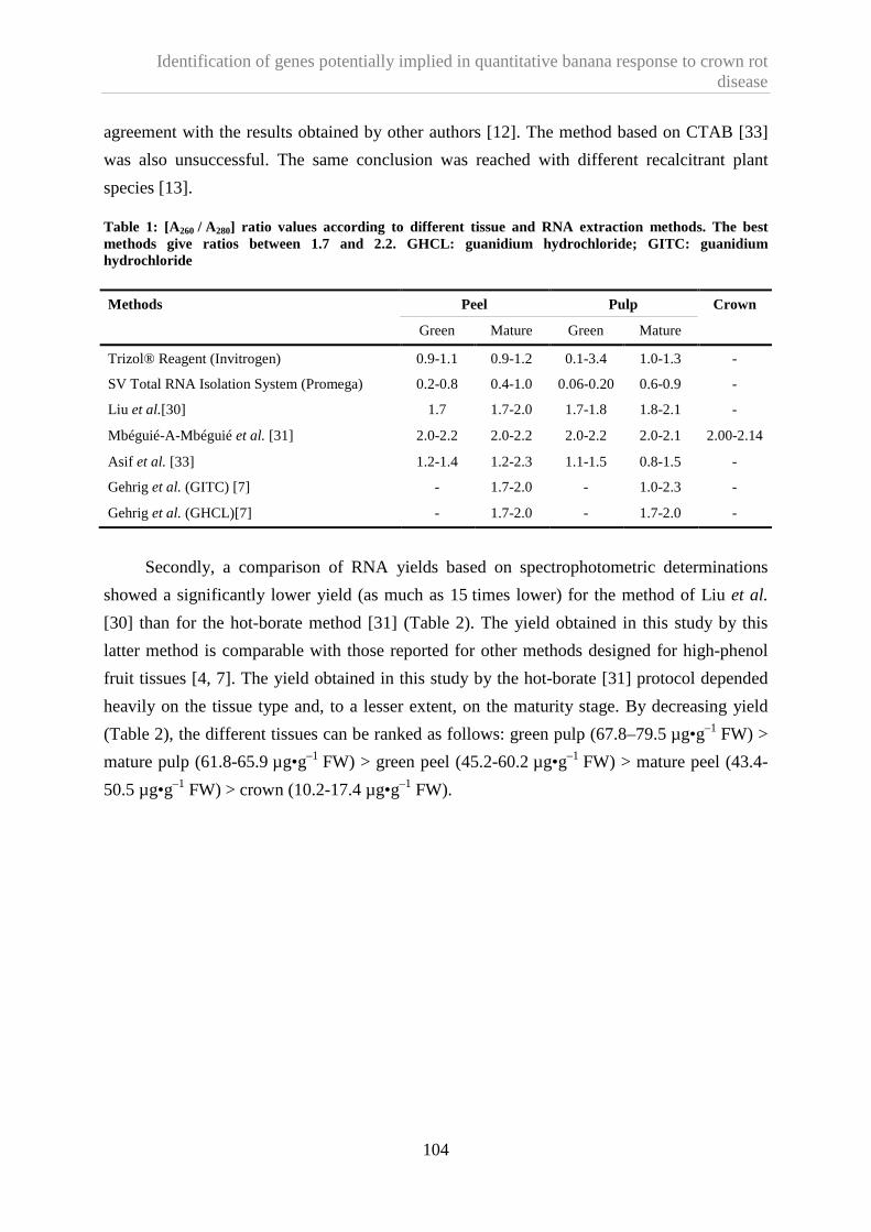
Identification of genes potentially implied in quantitative banana response to crown rot disease
104
agreement with the results obtained by other authors [12]. The method based on CTAB [33]
was also unsuccessful. The same conclusion was reached with different recalcitrant plant
species [13].
Table 1: [A260 / A280] ratio values according to different tissue and RNA extraction methods. The best methods give ratios between 1.7 and 2.2. GHCL: guanidium hydrochloride; GITC: guanidium hydrochloride
Methods Peel Pulp Crown
Green Mature Green Mature
Trizol® Reagent (Invitrogen) 0.9-1.1 0.9-1.2 0.1-3.4 1.0-1.3 -
SV Total RNA Isolation System (Promega) 0.2-0.8 0.4-1.0 0.06-0.20 0.6-0.9 -
Liu et al.[30] 1.7 1.7-2.0 1.7-1.8 1.8-2.1 -
Mbéguié-A-Mbéguié et al. [31] 2.0-2.2 2.0-2.2 2.0-2.2 2.0-2.1 2.00-2.14
Asif et al. [33] 1.2-1.4 1.2-2.3 1.1-1.5 0.8-1.5 -
Gehrig et al. (GITC) [7] - 1.7-2.0 - 1.0-2.3 -
Gehrig et al. (GHCL)[7] - 1.7-2.0 - 1.7-2.0 -
Secondly, a comparison of RNA yields based on spectrophotometric determinations
showed a significantly lower yield (as much as 15 times lower) for the method of Liu et al.
[30] than for the hot-borate method [31] (Table 2). The yield obtained in this study by this
latter method is comparable with those reported for other methods designed for high-phenol
fruit tissues [4, 7]. The yield obtained in this study by the hot-borate [31] protocol depended
heavily on the tissue type and, to a lesser extent, on the maturity stage. By decreasing yield
(Table 2), the different tissues can be ranked as follows: green pulp (67.8–79.5 µg•g–1 FW) >
mature pulp (61.8-65.9 µg•g–1 FW) > green peel (45.2-60.2 µg•g–1 FW) > mature peel (43.4-
50.5 µg•g–1 FW) > crown (10.2-17.4 µg•g–1 FW).

Identification of genes potentially implied in quantitative banana response to crown rot disease
105
Table 2: Yield values (µg RNA•g–1 fresh material), based on A260, according to different tissue and RNA extraction methods. (x) means that the yield could not be calculated because the [A260 / A280] ratio was not between 1.7 and 2.2. GHCL: guanidium hydrochloride; GITC: guanidium hydrochloride.
Methods Peel Pulp Crown
Green Mature Green Mature
Trizol® Reagent (Invitrogen) (x) (x) (x) (x) -
SV Total RNA Isolation System (Promega) (x) (x) (x) (x) -
Liu et al.[30] 3.9-5.7 7.6-8.9 3.6-9.9 50.3-56.8 -
Mbéguié-A-Mbéguié et al. [31] 45.2-60.2 43.4-50.5 67.8-79.5 61.8-65.9 10.2-17.4
Asif et al. [33] (x) (x) (x) (x) -
Gehrig et al. (GITC) [7] - 72.0-75.8 - (x) -
Gehrig et al. (GHCL)[7] - 102.4-109.7 - 117.5-125.2 -
Thirdly, denaturing agarose gel electrophoresis yielded clear rRNA bands with a 28s
rRNA band equal to or more abundant than the 18s rRNA band (Figure 2), indicating that
little or no degradation had occurred during extraction with the hot-borate [31] and Liu et al.'s
[30] methods. In high-quality RNA, the 28s band should be approximately twice as intense as
the 18s band [3]. In degraded RNA samples, the 18s band will be enhanced, since the 28s
ribosomal RNA is typically degraded to an 18s-like species. No rRNA was observed with any
other technique, and notably not with either of those described by Gehrig et al. [7]. The latter
methods did give, however, a good [A260 / A280] ratio (Table 1) and thus a high estimated
yield (Table 2). An explanation might be that A260 could be falsely boosted by contaminants
such as salt, organic solvent or protein, or by one or more of the many chemicals used in
nucleic acid purification which absorb at 260 nm [34]. Thus, the [A260 / A280] ratio can
accurately describe nucleic acid purity, but it can also be misleading [35]. For this reason, it is
essential to have both the [A260 / A280] ratio and an agarose gel result to evaluate a RNA
extraction method.

Identification of genes potentially implied in quantitative banana response to crown rot disease
106
Figure 2. Band patterns obtained by ethidium bromide staining after electrophoresis, through a 1% agarose gel, of total RNA extracted from green banana peel (1 and 3) or pulp (2 and 4). The extraction protocols used were that of Liu et al., [30] (1 and 2) and the hot-borate method [31] (3 and 4). (5) is a RNA ladder and the positions of of the 28S and 18S rRNAs are shown. Similar results were obtained for both protocols with peel and pulp from ripe banana. No rRNAs were observed with any other method tested in this work (resumts not shown).
Finally, the adequacy of the hot-borate method [31] for extracting total RNA from
banana tissues in order to generate cDNA for downstream molecular analyses was
successfully performed by cDNA-AFLP after DNase treatment (Figures 3 and 4).
For these reasons, although the hot-borate method [31] is labor-intensive and time-
consuming, it seems to be the most suitable for extracting RNA from banana tissues in order
to carry out genome-wide expression analyses. This RNA extraction technique is applicable to
large samples of up to 25 g fresh material [36], but also to small samples of 0.24 g of material
in a 2-mL tube, as in this work. It could be more convenient to extract RNA from a small
sample, because many samples can be extracted at the same time, and the yield obtained is
still sufficient for subsequent molecular applications.

Identification of genes potentially implied in quantitative banana response to crown rot disease
107
Figure 3. Reproducibility of cDNA-AFLP based on total RNA. cDNA-AFLP was applied to various pools of total RNA from from a single crown according to figure 1. (1) to (8) represent the various cDNA-AFLP pools and (9) is a blank.
1.4.2. In situ RNA preservation methods
On the basis of quality, quantity and integrity, it appears that all four methods tested can
preserve RNA (Table 3). The choice of a method must thus be based on the experimental
constraints and the equipment available. Yet, although all four techniques appeared to
preserve RNA quality and integrity, their yields were different. RNAlater is an aqueous tissue
storage reagent that stabilizes and protects cellular RNA in intact, unfrozen tissue samples.
According to Ambion (Austin, TX, USA), it eliminates the need to process tissue samples
immediately or to freeze them in liquid nitrogen for later processing. Tissue pieces can be
harvested and submerged in RNAlater for storage without jeopardizing the quality or quantity
of RNA obtained by subsequent RNA isolation.
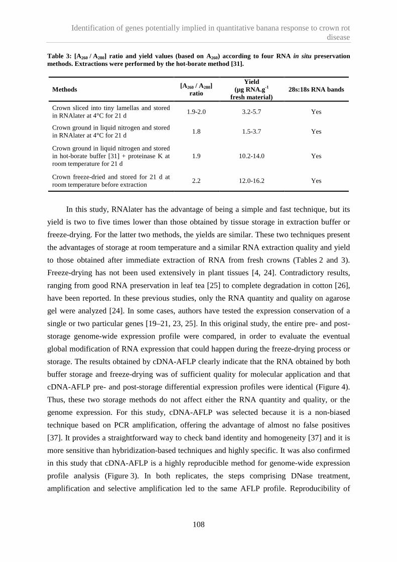
Identification of genes potentially implied in quantitative banana response to crown rot disease
108
Table 3: [A260 / A280] ratio and yield values (based on A260) according to four RNA in situ preservation methods. Extractions were performed by the hot-borate method [31].
Methods [A 260 / A280]
ratio
Yield (µg RNA.g-1
fresh material) 28s:18s RNA bands
Crown sliced into tiny lamellas and stored in RNAlater at 4°C for 21 d
1.9-2.0 3.2-5.7 Yes
Crown ground in liquid nitrogen and stored in RNAlater at 4°C for 21 d
1.8 1.5-3.7 Yes
Crown ground in liquid nitrogen and stored in hot-borate buffer [31] + proteinase K at room temperature for 21 d
1.9 10.2-14.0 Yes
Crown freeze-dried and stored for 21 d at room temperature before extraction
2.2 12.0-16.2 Yes
In this study, RNAlater has the advantage of being a simple and fast technique, but its
yield is two to five times lower than those obtained by tissue storage in extraction buffer or
freeze-drying. For the latter two methods, the yields are similar. These two techniques present
the advantages of storage at room temperature and a similar RNA extraction quality and yield
to those obtained after immediate extraction of RNA from fresh crowns (Tables 2 and 3).
Freeze-drying has not been used extensively in plant tissues [4, 24]. Contradictory results,
ranging from good RNA preservation in leaf tea [25] to complete degradation in cotton [26],
have been reported. In these previous studies, only the RNA quantity and quality on agarose
gel were analyzed [24]. In some cases, authors have tested the expression conservation of a
single or two particular genes [19–21, 23, 25]. In this original study, the entire pre- and post-
storage genome-wide expression profile were compared, in order to evaluate the eventual
global modification of RNA expression that could happen during the freeze-drying process or
storage. The results obtained by cDNA-AFLP clearly indicate that the RNA obtained by both
buffer storage and freeze-drying was of sufficient quality for molecular application and that
cDNA-AFLP pre- and post-storage differential expression profiles were identical (Figure 4).
Thus, these two storage methods do not affect either the RNA quantity and quality, or the
genome expression. For this study, cDNA-AFLP was selected because it is a non-biased
technique based on PCR amplification, offering the advantage of almost no false positives
[37]. It provides a straightforward way to check band identity and homogeneity [37] and it is
more sensitive than hybridization-based techniques and highly specific. It was also confirmed
in this study that cDNA-AFLP is a highly reproducible method for genome-wide expression
profile analysis (Figure 3). In both replicates, the steps comprising DNase treatment,
amplification and selective amplification led to the same AFLP profile. Reproducibility of

Identification of genes potentially implied in quantitative banana response to crown rot disease
109
cDNA-AFLP has already been reported by many authors [28, 38–40]. Khun [37] even
evaluates the reproducibility of cDNA-AFLP at 100%.
Figure 4. Reproducibility of the cDNA-AFLP gene expression profile whatever the in situ preservation method. (10)-(13) = replicate 1; (14)-(17) = replicate 2. (10) and (14) are blanks; (11) and (15) are profiles obtained after immediate RNA extraction from fresh material; (12) and (16) are profiles obtained with RNA from freeze-dried tissues; (13) and (17) are profiles obtained with RNA from tissue samples stored in the hot-borate buffer [31].
Both storage techniques are thus perfectly suitable for cDNA-AFLP applied to the study
of differential gene expression in different cell populations. However, freeze-drying, which
preserves enzymatic activity, offers further advantages highlighted in this study; they can be
summarized: (1) the extraction yield, quality and integrity equal those obtained by fresh tissue
RNA extraction or by conventional storage techniques; (2) the freeze-dried RNA can be
stored even at room temperature, without any alteration of the expression profile, for further
RNA extraction; (3) freeze-drying is an easy way to obtain a fine dry power without using
liquid nitrogen, which is not always available or economically feasible in all banana-
producing countries; (4) freeze-dried tissues can be easily transported from one laboratory to
another and even between countries; (5) a final advantage of freeze-drying is the large amount

Identification of genes potentially implied in quantitative banana response to crown rot disease
110
of RNA that can be obtained in a single extraction carried out in a 2-mL tube. Such a tube can
hold only about 0.25 g fresh sample material, but if the material is freeze-dried, a mere 0.2 g
is equivalent to about 2.4 g fresh weight. Assuming the same yield from freeze-dried material
as from fresh material, the amount of RNA that can be obtained per tube is ten to twelve times
higher when the material is freeze-dried. In applications requiring a lot of RNA, it could be
very convenient to meet this requirement while working on a small scale with a 2-mL tube.
These advantages make freeze-drying an attractive alternative to conventional storage.
Moreover, the two efficient storage methods proposed in this study to conserve fruit
banana RNA in situ without altering the quality, quantity or expression profile could probably
be applied to other plant tissues in particular fruits which are particularly recalcitrant.
1.5. Conclusion
We thus evaluated and made choices among various techniques spanning all steps
required to study differential gene expression in banana fruit. Our results highlight two good,
reliable and practical storage methods that preserve RNA in situ before extraction without
requiring ultra-low temperature. We also identified a RNA extraction method yielding high-
quality RNA suitable for gene expression profiling. To illustrate this, we used the extracted
RNA successfully to generate cDNA-AFLP differential expression profiles. Thus, it is
possible to study gene expression in any banana fruit tissue, whatever the maturity stage, by
applying all the procedures selected in our work.
Acknowledgements
The authors wish to thank Van Damme (Brussels), an independent banana importer for
Benelux, for its interest in this project. Thanks are also due to Didier Mbéguié-A-Mbéguié of
CIRAD, UMR 1270 QUALITROP, Guadeloupe, France, for his assistance with our extraction
protocol research and to Frettinger Patrick from the Plant Biology Unit of Agricultural
University of Gembloux, Belgium, for his advice.

Identification of genes potentially implied in quantitative banana response to crown rot disease
111
1.6. References
[1] Xu B.Y., Su W., Liu J. H., Wang J.B., Jin Z.Q., Differentially expressed cDNAs at the
early stage of banana ripening identified by suppression subtractive hybridization and
cDNA microarray, Planta 226 (2007) 529–539.
[2] Choudhury S.R., Roy S., Sengupta D.N., Characterization of transcriptional profiles of
MA-ACS1 and MA-ACO1 genes in response to ethylene, auxin, wounding, cold and
different photoperiods during ripening in banana fruit, J. Plant Physiol. 165 (2008)
1865–1878.
[3] Martin L.A., Smith T.J., Obermoeller D., Bruner B., Kracklauer M., Dharmaraj S., RNA
purification, in: Gerstein A.S. (Ed.), Molecular biology problem solver, Wiley-Liss,
NY, USA, 2001.
[4] Hu C., Honda C., Kita M., Zhang Z., Tsuda T., Moriguchi T., A simple protocol for
RNA isolation from fruit trees containing high levels of polysaccharides and polyphenol
compounds, Plant Mol. Biol. Rep. 20 (2002) 69a–69g.
[5] Ding L., Sun Q., Wang Z., Sun Y., Xu Z., Using silica particles to isolate total RNA
from plant tissues recalcitrant to extraction in guanidine thiocyanate, Anal. Biochem.
374 (2008) 426–428.
[6] Claros M., Canovas F., RNA isolation from plant tissues: a practical experience for
biological undergraduates, Biochem. Educ. 27 (1999) 110–113.
[7] Gehrig H., Winter K., Cushman J., Borland A., Taybi T., An improved RNA isolation
method for succulent plant species rich in polyphenols and polysaccharides, Plant Mol.
Biol. Rep. 18 (2000) 369–376.
[8] Liu W., Wang B., Duan C., Li B., A method for isolating functional RNA from callus of
Dendrobium candidum contented rich polysaccharides, Colloid Surf. B-Biointerfaces 42
(2005) 259–262.
[9] Venugopalan C., Kapoor H., Single step isolation of plant RNA, Phytochem. 46 (1997)
1303–1305.
[10] Wu Y., Llewellyn D., Dennis E., A quick and easy method for isolating good-quality
RNA from cotton (Gossypium hirsutum L.) tissues, Plant Mol. Biol. Rep. 20 (2002)
213–218.
[11] Chang S., Puryear J., Cairney J., A simple and efficient method for isolating RNA from
pine trees, Plant Mol. Biol. Rep. 11 (1993) 113–116.

Identification of genes potentially implied in quantitative banana response to crown rot disease
112
[12] Gao J., Liu J., Li B., Li Z., Isolation and purification of functional total RNA from blue-
grained wheat endosperm tissues containing high levels of starches and flavonoids,
Plant Mol. Biol. Rep. 19 (2001) 185a–185i.
[13] Salzman R.A., Fujita T., Zhu-Salzman K., Hasegawa P.M., Bressan R.A., An improved
RNA isolation method for plant tissues containing high levels of phenolic compounds or
carbohydrates, Plant Mol. Biol. Rep. 17 (1999) 11–17.
[14] Zeng Y., Yang T., RNA isolation from highly viscous samples rich in polyphenols and
polysaccharides, Plant Mol. Biol. Rep. 20 (2002) 417a–417e.
[15] Maniatis T., Fritsch E., Sambrook J., eds. Molecular Cloning: A laboratory Manual.
Cold Spring Harbor Laboratory Press, NY, USA,1982.
[16] Chen T., Gagliardo R., Walker B., Zhou M., Shaw C., Partial structure of the phylloxin
gene from the giant monkey frog, Phyllomedusa bicolor: Parallel cloning of precursor
cDNA and genomic DNA from lyophilized skin secretion, Pept. 26 (2005) 2624–2628.
[17] Chen T., Xue Y., Zhou M., Shaw C., Molecular cloning of mRNA from toad granular
gland secretion and lyophilized skin: identification of Bo8 a novel prokineticin from
Bombina orientalis, Pept. 26 (2005) 377–383.
[18] Gadbois D., Salo W., Ann D., Downing S., Carlson D., The preparation of
poly(A)+mRNA from the hagfish slime gland, Prep. Biochem. Biotechnol. 18 (1988)
67–76.
[19] Huang Z., Ortmeyer H., Hansen B., Shuldiner R., Preparation of RNA from lyophilized
tissue: a stable and reliable method for long term storage, Biotech. 17 (1994) 4.
[20] Matsuo S., Sugiyama T., Okuyama T., Yoshikawa K., Honda K., Takahashi R., Maeda
S., Preservation of pathological tissue specimens by freeze-drying for
immunohistochemical staining and various molecular biological analyses, Pathol. Int.
49 (1999) 383–390.
[21] Tsuka H., Mori H., Okada K., Matsukawa S., Utilization of the freeze-drying method in
the preparation of biologically active, intact RNA from hard frozen tissues of human
prostate, Anal. Biochem. (1997) 458–461.
[22] Vaughan H., Chalker V., Mee Z., Rossouw A., James V., Stability of lyophilised
specimens for the molecular detection of viral DNA/RNA, J. Clin. Virol. 35 (2006)
135–140.
[23] Sessitsch A., Gyamfi S., Stralis-Pavese N., Weilharter A., Pfeifer U., RNA isolation
from soil for bacterial community and functional analysis: evaluation of different
extraction and soil conservation protocols, J. Microbiol. Methods 51 (2002) 171–179.

Identification of genes potentially implied in quantitative banana response to crown rot disease
113
[24] Drouet A., Hartmann C., Polyribosomes from pear fruit, Plant Physiol. 69 (1982) 885–
887.
[25] Jaiprakash M., Pillai B., Venkatesh P., Subramanian N., Sinkar V., Sadhale P., RNA
isolation from high-phenolic freeze-dried tea (Camelia sinensis) leaves, Plant Mol. Biol.
Rep. 21 (2003) 465a–465g.
[26] Saha S., Callahan F., Dollar D., Creech J., Effect of lyophilisation of cotton tissue on
quality of extractable DNA, RNA, and protein, J. Cotton Sci. 1 (1997) 10–14.
[27] Vos P., Hogers R., Bleeker M., Reijans M., van de Lee T., Hornes M., Frijters A.,
AFLP: a new techniques for DNA fingerprinting, Nucleic Acids Res. 23 (1995) 4407–
4414.
[28] Bachem C., Visualization of differential gene expression using a novel method of RNA
fingerprinting based on AFLP: analysis of gene expression during potato tuber
development, Plant J. 9 (1996) 745–753.
[29] Chomczynski P., Sacchi N., Single step method of RNA isolation by acid guanidinium
thiocyanate-phenol-chloroform extraction, Anal. Biochem. 162 (1987) 156–159.
[30] Liu J., Goh C., Loh C., Liu P., Pua E., A method for isolation of total RNA from fruit
tissues of banana, Plant Mol. Biol. Rep. 16 (1998) 1–6.
[31] Mbéguié-A-Mbéguié D., Fils-Lycaon B., Chillet M., Hubert O., Galas C., Gomez R.-
M., Extraction and purification of total RNA from banana tissues (small scale), Fruits
63 (4) (2008) 255–261.
[32] Wan C., Wilkins T., A modified hot borate method significantly enhances the yield of
high-quality RNA from cotton (Gossypium hirsutum L.), Anal. Biochem. 233 (1994) 7–
12.
[33] Asif M.H., Dhawan P., Nath P., A simple procedure for isolation of high quality RNA
from ripening banana fruit, Plant Mol. Biol. Rep. 18 (2000) 109–115.
[34] Huberman J.A., Importance of measuring nucleic acid absorbance at 240 nm as well as
at 260 and 280 nm, Biotech. 18 (1995) 636.
[35] Troutman T., Prasauckas K., Kennedy M., Stevens J., Davies M., Dadd A., How to
properly use and maintain laboratory equipment, in: Gerstein A.S. (Ed.), Molecular
biology problem solver, Wiley-Liss, NY, USA, 2001.
[36] Mbeguie-A-Mbeguie D., Hubert O., Sabau X., Chillet M., Fils-Lycaon B., Baurens
F.C., Use of suppression subtractive hybridization approach to identify genes
differentially expressed during early banana fruit development undergoing changes in
ethylene responsiveness, Plant Sci. 172 (2007) 1025–1036.

Identification of genes potentially implied in quantitative banana response to crown rot disease
114
[37] Kuhn E., From library screening to microarray technology: strategies to determine gene
expression profiles and to identify differentially regulated genes in plants, Ann. Bot. 87
(2001) 139–155.
[38] Matz M., Lukyanov S.A., Different strategies of differential display: areas of
application, Nucleic Acids Res. 26 (1998) 5537–5543.
[39] Campalans A., Pagès M., Messeguer R., Identification of differentially expressed genes
by the cDNA-AFLP technique during dehydration of almond, Tree Physiol. 21 (2001)
633–643.
[40] Brugmans B., Fernadez del Carmen A., Bachem C., van Hos H., van Eck H.J., Visser
R., A novel method for the construction of genome wide transcriptome maps, Plant J. 31
(2002) 1–15.

Identification of genes potentially implied in quantitative banana response to crown rot disease
115
2. Identification of genes that may influence quantitatively the banana response to crown rot disease.
To study the molecular mechanisms underlying the susceptibility variation it was
important to develop a sampling that allows selecting at harvest two kinds of banana. On one
hand fruits with a high susceptibility level to crown rot and on the other hand fruits with a low
susceptibility level to compare gene expression between them. The difficulty of such
sampling was that crown for molecular analysis should be collected at harvest time whereas
the fruit susceptibility can only be assessed once the symptoms appeared on ripe fruit. This
corresponds in our essays at 13 days post-harvest. The results presented in chapter 3.2 show
that by modifying the source-sink ratio at flowering it could be expected that fruit
susceptibility would be highly different. Thus to identify gene expression implied in banana
quantitative defence-response to crown rot disease, we have decided to selected our sample on
base of their source-sink ratio. cDNA-AFLP method was applied on these samples and results
obtain were confirmed by real-time RT-PCR. By this way, genes potentially implied in
banana quantitative defence response to crown rot disease were identified.
Results are presented hereafter and will be soon submitted for publication to
Physiological and Molecular Plant Pathology.
Lassois Ludivine, Frettinger Patrick, de Lapeyre de Bellaire Luc, Lepoivre Philippe and
Jijakli Haissam. Identification of genes that may influence quantitatively the banana response
to crown rot disease.

Identification of genes potentially implied in quantitative banana response to crown rot disease
116
Identification of genes that may influence quantitatively the banana response to crown
rot disease
Lassois Ludivine a, Frettinger Patrick b, de Lapeyre de Bellaire Luc c, Lepoivre Philippe a and
Jijakli Haissam a
a University of Liege, Gembloux Agro-Bio Tech, Plant Pathology Unit. Passage des Déportés 2, B-5030 Gembloux, Belgium; b Unité Mixte de Recherches INRA-CNRS-Université de Bourgogne, Plante-Microbe Environnement, INRA, 17 Rue Sully, BP 86510, 21065 Dijon cedex, France ; c CIRAD, Persyst, UPR Syst. Banan. Ananas, TA B-26 / PS4, Blvd. de la Lironde, 34398 Montpellier Cedex 5, France

Identification of genes potentially implied in quantitative banana response to crown rot disease
117
Abstract
Variations in banana fruit susceptibility to the crown rot post-harvest disease have been
observed but the molecular mechanisms underlying these quantitative host-pathogen
relationships were still unknown. The present study was designed to compare gene expression
between crowns of bananas (Musa acuminata, AAA, ‘Grande-Naine’) showing a high
susceptibility (S+) and crowns of bananas showing a low susceptibility (S-) to Colletotrichum
musae responsible for crown rot disease. This comparison was performed at two situation
time: (i) between crowns (S+ and S-) collected one hour before infection and (ii) between
crowns (S+ and S-) collected 13 days after infection. Gene’s expression comparisons were
performed with cDNA-AFLP technique and differential expression results obtained were
confirmed by real-time RT-PCR for two biological replicate. The limitations of the cDNA-
AFLP and the importance of the results validation by an independent method and for two
biological replicates are discussed. Among the 3161 transcript-derived fragments (TDFs)
screened by cDNA-AFLP, 443 were differently expressed between bananas showing different
susceptibility. Only 10% were selected, successfully recovered from the gel, cloned,
sequenced and showed non redundant similarities with the data base. Among those, only
eleven TDFs differently expressed were finally confirmed for both biological replicates. Two
identified genes were involved in signal transduction and three in proteolytic machinery. In
addition, two TDFs were similar to pathogenesis-related protein 14, one was similar with a
CCR4-associated factor (CAF1) protein, one with a cellulose synthase. Paradoxically, the
over-expression of the cellulose synthase gene is associated with banana showing a high
susceptibility both in pre- and post-inoculation situation. Finally, the cDNA-AFLP allows
identifying that a gene, coding for a dopamine-β-monooxygenase, involved in catecholamine
pathway seems to be associated in the quantitative banana responses to crown rot disease. To
our knowledge, this work is the first to address both pre- and post-infection gene expression
with the same host-pathogen combination and different susceptibility.

Identification of genes potentially implied in quantitative banana response to crown rot disease
118
2.1. Introduction
Crown rot disease affects export bananas in all producing countries and is considered as
one of the main post-harvest diseases of bananas (for a review see Lassois et al., accepted a).
The symptoms appear on the crown, i.e. the tissue joining the fruit pedicels with each other.
The disease develops during shipping, ripening, and storage and has a negative impact on the
market value of bananas (Slabaugh and Grove, 1982).
Variations, notably geographic and seasonal, have been reported in the post-harvest
development of crown rot disease symptoms (Krauss and Johanson, 2000; Lassois et al.,
2008; Lukezic et al., 1967; Shillingford, 1978). It has been suggested that these
spatiotemporal fluctuations reflect the variations in the banana fruit quality potential that
elaborates during preharvest stages (Lassois et al., accepted a). This quality potential depends
both on a physiological and a parasitic components, which in turn are influenced by
agrotechnical and pedoclimatic factors. The parasitic component reflects the level of crown
contamination by the parasitic complex, as well as the pathogenicity of this parasitic complex.
Effectively, crown rot results from an unspecific parasitic complex from which
Colletotrichum musae is the most pathogenic species (Finlay and Brown, 1993). The term
“physiological component” refers here to the level of fruit susceptibility to crown rot. To
assess the level of fruit susceptibility it is necessary to overcome the influence of the parasitic
component. It is then estimated through lesion size after standardized artificial inoculations
with C. musae (de Lapeyre de Bellaire et al., 2008). Very few studies have linked the spatio-
temporal fluctuations of crown rot disease to preharvest factors influencing the level of fruit
susceptibility (Lassois et al., 2008). Nevertheless, it has been shown recently that source-sink
ratio and the hand position in the bunch affect the banana susceptibility to crown rot disease
(Lassois et al., acceptedb). The genetic and molecular mechanisms underlying these
quantitative host-pathogen relationships have not been identified.
Plant defense mechanisms to diseases are mediated by both preformed and inducible
responses which lead to significant transcriptional changes in the host (Lucas, 1998). One way
to understand the reactions involved in the variation of banana responses to crown rot is then
to identify genes involved in these processes via the study of their expression. The cDNA-
AFLP technique (Bachem, 1996) has been used to compare genome-wide expression patterns
and to identify differently expressed genes potentially related to plant defense mechanisms.
This technique does not require prior hypothesis on defense mechanisms and allows the
identification of genes without a priori. It can thus be used for any biological system,

Identification of genes potentially implied in quantitative banana response to crown rot disease
119
especially when genomic resources are lacking. In addition, the mechanisms responsible for
compatibility interactions and for the quantitative variation of symptoms after infection
remain poorly understood, and it is likely that some of the factors involved remain to be
discovered. The sensitivity and specificity of cDNA-AFLP analysis are comparable to those
of a microarray approach (Reijans et al., 2003). Its reproducibility has also been proved
(Kuhn, 2001; Lassois et al., 2009).
The present study was designed to identify genes influencing the susceptibility to crown
rot disease in a same banana cultivar. A cDNA-AFLP technique has been used to compare
gene expression in banana crowns from plants (Musa acuminata, AAA, cv ‘Grande-Naine’)
showing different susceptibility to crown rot disease. In this study, the variation of source-
sink ratio previously described (Lassois et al., acceptedb) has been used to obtain banana
plants showing different susceptibility to crown rot disease. Samples were collected before
and after infection in order to differentiate constitutive and induced defense responses. The
differential expression deduced from cDNA-AFLP data was latter confirmed by an
independent technique, the real-time RT-PCR. This latter technique is quick and, thanks to its
high sensitivity, requires only a small amount of starting material (Massart and Jijakli, 2006).
2.2. Materials and methods
2.2.1. Fruit sampling
Banana fruits were harvested from plants (Musa acuminata, AAA, cv ‘Grande-Naine’)
grown on the Dia-Dia commercial plantation (PHP) in Njombé, Cameroon (altitude: 80 m;
annual mean temperature: 26.5°C; annual mean rainfall: 3500 mm). The date of flowering
was indicated by tying a colored belt to each bunch (banana inflorescence) at the horizontal
finger stage in order to predict the time of harvest. Bunches were also covered with a plastic
sleeve at this stage. Bunches were harvested at a constant physiological age (Jullien et al.,
2008), i.e. when the mean daily temperature sum accumulated by the fruit at the 14°C
threshold between flowering and harvest reached 900 dd. Temperatures were recorded at a
weather station on the plantation. The daily average temperature (Td) was estimated from
measurements of maximum temperature (Tmax) and minimum temperature (Tmin). To obtain
bananas with different levels of susceptibility to crown rot, fruits were harvested from plants
characterized by different source/sink ratios (Lassois et al., acceptedb) established at the
flowering stage, leaves being viewed as sources and fruits as sinks:

Identification of genes potentially implied in quantitative banana response to crown rot disease
120
− Plants showing a low susceptibility to crown rot disease were called “S-“. These plants
were obtained through severe trimming of bunches at flowering stage. These plants had
12 leaves and 2 hands (a hand is a group of bananas) left on the bunch. Two plants of
this treatment were used, namely, “S- plant 1” and “S- plant 2” and constitute the 2
biological replicates.
− Plants showing a high susceptibility to crown rot disease were called “S+“. These plants
were reference plants without severe trimming of bunch. At flowering, bunches were
not trimmed, so these plants had 12 leaves and 8 hands left on the bunch. Two plants of
this treatment were used, namely, “S+ plant 1” and “S+ plant 2” and constitute the 2
biological replicates.
Figure 1 outlines the experimental sampling design for fruit susceptibility evaluation
and molecular analysis, while table 1 identifies the samples, with their respective sources and
processing modalities. Only the second hand of each bunch was collected and each one was
divided into 4 clusters of 4 fingers (fruits) without defects. Each sample group consisted of
clusters from the same hand, so as to avoid variability due to susceptibility variations among
different hands (Lassois et al., acceptedb). At harvest for each bunch, 1 cluster was used for
sampling crown 1 hour before inoculation of C. musae (1 hbi - clusters A, C, E and G
according to the treatment and the biological replicate) and 3 clusters were inoculated with C.
musae (figure 1). These 3 clusters were used for fruit susceptibility evaluation in order to
confirm the expected S+ or S- status of each treatment. One of these 3 was collected for crown
sampling 13 days after inoculation when symptoms are developed (13 dpi - clusters B, D, F
and H according to the treatment and the biological replicate). The crowns collected at each
period (1hbi and 13dpi) for molecular analyses were immediately frozen in liquid nitrogen,
freeze-dried at -80°C for 24h (Telstar, Cryodos, Barcelona, Spain), and stored at room
temperature before RNA extraction (Lassois et al., 2009) and molecular analysis.

Identification of genes potentially implied in quantitative banana response to crown rot disease
121
Banana plant with 12Leaves/2Hands
S- Plants (Low fruit susceptibility to C. musae)Harvest
900dd
2d hand
Susceptibility evaluation
Waste
10 days at13°C
3 days at20°C
cDNA
C. musae
Artificial ripening
Molecular
experiment
2d hand
C. musae
Artificial ripening
Molecular
experiment
Susceptibility evaluation
Waste
Crown freeze dried
RNA extraction
cDNA
A
B
RNA extraction
Crown freeze dried
A B
C
D
C D
Banana plant with 12Leaves/8Hands
S+ Plants (high fruit susceptibility to C. musae)
1hbi
13dpi
cDNA-AFLP(Comparison A with
C and B with D)
Real time RT-PCR
S- Plant 1
cDNA samples
E (1hbi) and F
(13dpi)
S- Plant 2cDNA-AFLP(Comparison A with
C and B with D)
Real time RT-PCR
S+ Plant 1
cDNA samples
E (1hbi) and F
(13dpi)
S+ Plant 2
Figure 1: Experimental procedure. As bananas from plants with 12 leaves and 2 hands are waiting to be less susceptible than bananas from plants with 12 leaves and 8 hands (Lassois et al., acceptedb), two plants with each configuration were used as source material (S- plant 1, S- plant 2, S+ plant 1 and S+ plant 2). Only the second hand of each bunch was collected and each one was divided into 4 clusters of 4 fingers without defects. The “S- plant 1” (S- for low susceptibility) and “S+ plant 1” (S+ for high susceptibility) were used as sources of material for cDNA-AFLP analysis and for one replicate of the real time RT-PCR. Samples A and B were collected from “S- plant 1” respectively 1 hour before inoculation (1hbi) and 13 days post-inoculation (13dpi). Likewise, samples C and D were collected from the “S+ plant 1” respectively 1 hour before inoculation (1hbi) and 13 days post-inoculation. Following the same procedure, samples E (1 hbi) and F (13 dpi) were collected from “S- plant 2” and samples G (1 hbi) and H (13 dpi) from “S+ plant 2”. These samples provided the second biological replicate used for result confirmation by real time RT-PCR. Furthermore, 3 clusters of each selected hand were used to evaluate fruit susceptibility to Colletotrichum musae at 13 dpi to confirm the expected S+ or S- status of each source.

Identification of genes potentially implied in quantitative banana response to crown rot disease
122
Table 1: Presentation of the various collected samples used for molecular analysis in order to identify genes differently expressed in banana crowns showing high and low susceptibility to crown rot disease. S-
stands for low susceptibility and S+ for high susceptibility. 1 hbi: Crowns collected 1 hour before inoculation of clusters from the second hand; 13 dpi: Crowns collected 13 days after inoculation of clusters from the second hand. The cDNA pools obtained after RNA extraction and reverse transcription are labeled A to H. In the cDNA-AFLP analysis, sample A was compared with C (two susceptibility levels, 1 hbi) and sample B with D (two susceptibility levels, 13 dpi). In the real-time RT-PCR assays, A and E yielded biological replicates, as did B and F, C and G, D and H.
Biological replicate 1 Biological replicate 2
Susceptibility level S- plant 1 S+ plant 1 S- plant 2 S+ plant 2
Source/sink ratio 12leaves/2hands 12leaves/8hands 12leaves/2hands 12leaves/8hands
Collecting time 1hbi 13dpi 1hbi 13dpi 1hbi 13dpi 1hbi 13dpi
Crown identification (cfr Figure 1)
A B C D E F G H
cDNA-AFLP comparison
2.2.2. Inoculation of fruits for the evaluation of susceptibility to crown rot
The C. musae strain was isolated in Njombé, Cameroon. It is sensitive to thiabendazole
and was stored at –20°C in a glycerol solution (30%). Before use, it was grown at 25°C in
Mathur medium (MgSO4.7H2O: 2.5g/l; KH2PO4: 2.7g/l; peptone: 1 g/l; yeast extract: 1g/l;
saccharose: 10 g/l; agar: 15 g/l) for 10 days. Conidia were removed by flooding the plates
with sterile distilled water and filtration through a 45-µm sieve. The concentrations of the
conidial suspension was determined with a Mallassez cell.
Smoothly and similarly cut crowns were obtained with a sharp knife, leaving as much
crown tissue as possible. The latex from the crown tissue was eliminated with absorbent paper
and the crowns were surface-sterilized by immersion in 50% ethanol. Fifty microliters of C.
musae conidial suspension containing 104 conidia/ml was applied to the centre of the freshly
exposed crown tissue and covered with a small paper filter. Two hours after application of the
conidial suspension, the clusters were packed in punched polyfilms normally used in the
industry, placed in commercial boxes, and stored at 13°C for 10 days to simulate shipment.
Artificial ripening was then initiated by dipping the bananas for 5 seconds in an ethrel
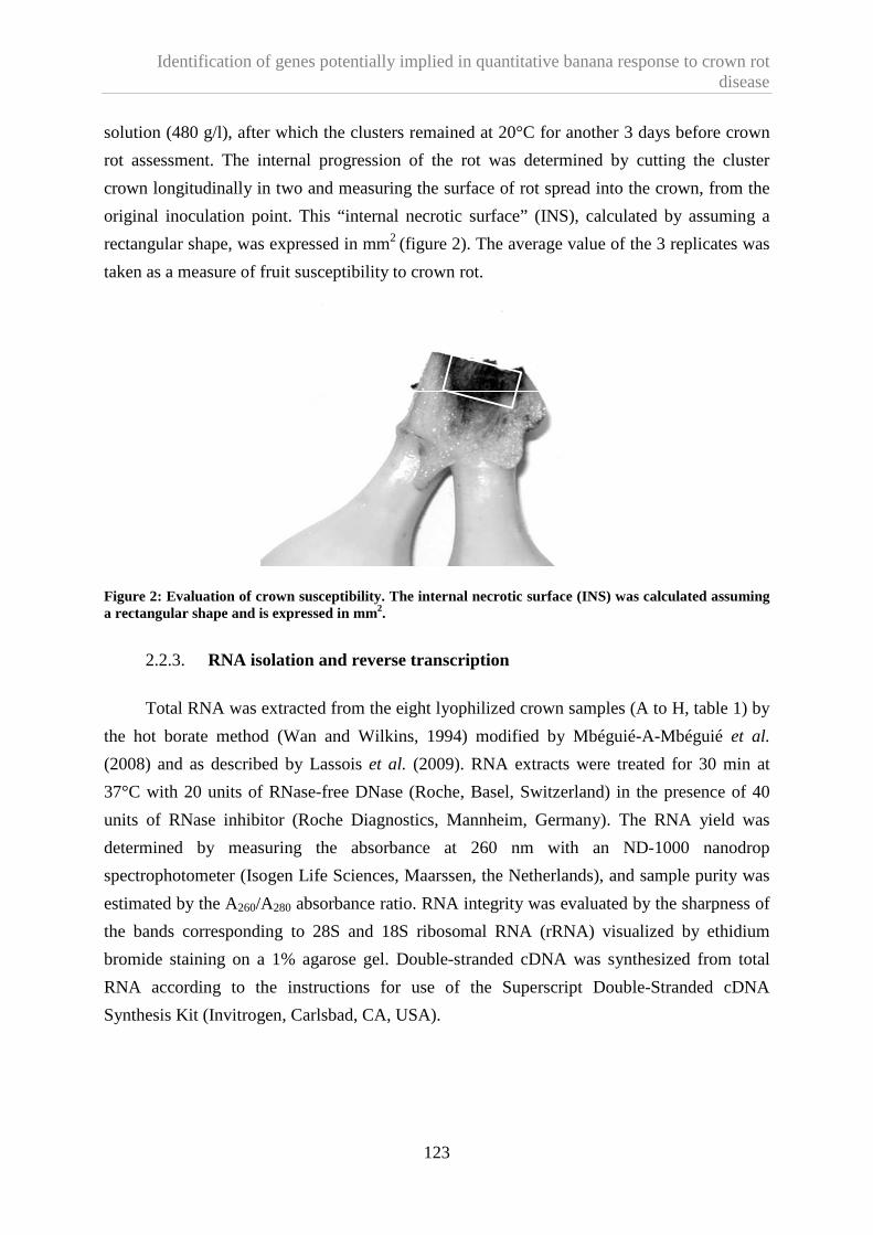
Identification of genes potentially implied in quantitative banana response to crown rot disease
123
solution (480 g/l), after which the clusters remained at 20°C for another 3 days before crown
rot assessment. The internal progression of the rot was determined by cutting the cluster
crown longitudinally in two and measuring the surface of rot spread into the crown, from the
original inoculation point. This “internal necrotic surface” (INS), calculated by assuming a
rectangular shape, was expressed in mm2 (figure 2). The average value of the 3 replicates was
taken as a measure of fruit susceptibility to crown rot.
Figure 2: Evaluation of crown susceptibility. The internal necrotic surface (INS) was calculated assuming a rectangular shape and is expressed in mm2.
2.2.3. RNA isolation and reverse transcription
Total RNA was extracted from the eight lyophilized crown samples (A to H, table 1) by
the hot borate method (Wan and Wilkins, 1994) modified by Mbéguié-A-Mbéguié et al.
(2008) and as described by Lassois et al. (2009). RNA extracts were treated for 30 min at
37°C with 20 units of RNase-free DNase (Roche, Basel, Switzerland) in the presence of 40
units of RNase inhibitor (Roche Diagnostics, Mannheim, Germany). The RNA yield was
determined by measuring the absorbance at 260 nm with an ND-1000 nanodrop
spectrophotometer (Isogen Life Sciences, Maarssen, the Netherlands), and sample purity was
estimated by the A260/A280 absorbance ratio. RNA integrity was evaluated by the sharpness of
the bands corresponding to 28S and 18S ribosomal RNA (rRNA) visualized by ethidium
bromide staining on a 1% agarose gel. Double-stranded cDNA was synthesized from total
RNA according to the instructions for use of the Superscript Double-Stranded cDNA
Synthesis Kit (Invitrogen, Carlsbad, CA, USA).

Identification of genes potentially implied in quantitative banana response to crown rot disease
124
2.2.4. Identification of genes differently expressed between crowns of high and
low susceptibility to crown rot disease
The cDNA pools derived from S+ plant 1 and S- plant 1 (samples A to D in table 1 and
figure 1) were subjected to cDNA-AFLP. For each collection time (1 hbi or 13dpi) the S+ and
S- samples were compared. A was thus compared with C (1 hbi) and B with D (13 dpi).
2.2.4.1. cDNA-AFLP
Double-stranded cDNA was digested with EcoRI and MseI, ligated with EcoRI and
MseI adapters, and pre-amplified with Eco (5’-GACTGCGTACCAATTC-3’) and Mse (5’-
GATGAGTCCTGAGTAA-3’) primers according to the instructions of the AFLP Analysis
System kit for microorganisms (Invitrogen, Carlsbad, CA, USA). After 20-fold dilution of the
PCR fragments, specific amplifications were carried out with 8 primer combinations of an
Eco primer and an Mse primer containing two additional bases at their 3’ ends. The Eco
primers were labeled with γ33P dATP. Amplification products were separated by
electrophoresis at 50 W on a vertical denaturing polyacrylamide gel (6%) containing 7 M
urea. Gels were dried on Whatman paper before autoradiography.
2.2.4.2. Isolation and reamplification of differently expressed transcription-derived
fragments (TDFs)
The film and gel were aligned and the fragments of interest were excised from the gel
with a sterile razor blade and immersed overnight in 100 µl distilled water. Of this solution
5µl was used in a standard PCR with the same EcoRI and MseI primers as used in the
preamplification step described above. After separation of the amplified products in a 1.2%
agarose gel, the bands were excised and purified as recommended with the QiaEx II gel
extraction Kit (Quiagen, Venlo, Pays-Bas).
2.2.4.3. Cloning and sequencing of reamplified differently expressed TDFs
The DNA fragments were ligated to the pJET1.2 vector according to the instructions of
the Clonejet PCR Cloning Kit (Fermentas, Vilnius, Lithuania) and cloned into Escherichia
coli (UltracompTM INVαF’, Invitrogen, Carlsbad, CA, USA). After transformation, bacterial
cells were spread onto NZY medium (2.1% NZY, 1.5% agar; pH 7.0) containing 100 µg/ml
ampicillin. After incubation for at least 30 h at 37°C, 5 colonies per fragment were chosen and
cultured overnight at 37°C in liquid NZY medium containing ampicillin. Plasmids containing
Identification of genes potentially implied in quantitative banana response to crown rot disease
125
the PCR product were extracted with the GeneJet Plasmid miniprep Kit (Fermentas, Vilnius,
Lithuania), according to the instructions. The presence of inserts in plasmids was checked by
standard PCR amplification with pJET Forward and Reverse primers (Fermenta,Vilnius,
Lithuania).
2.2.4.4. Sequence analysis
Three colonies were selected for sequence analysis for each TDF. Sequencing was
carried out with an automated sequencer (AbiPrism3730XL, Applied Biosystems, Foster City,
CA, USA) by Macrogen Inc. (Seoul, South Corea) with the pJET Forward primer of the
ClonjetTM PCR Cloning Kit (Fermentas, Vilnius, Lithuania). Sequence alignments were
performed and the sequence of each fragment corrected by means of the Bioedit sequence
alignment editor software developed by the Microbiology Department of North Carolina
University (USA). Sequence homology was determined with the Basic Local Alignment and
Search Tool (BlastX program) from the National Center for Biotechnology Information
(NCBI). The identified putative protein sequences were used in queries against the UniProt
database. Homologies with E-value scores below 10-8 were considered significant.
2.2.5. cDNA-AFLP fragment validation by real-time RT-PCR
2.2.5.1. Experimental design
The results obtained by cDNA-AFLP were confirmed for selected TDFs by real-time
RT-PCR (with two independent biological replicates, table 1). In these confirmation tests,
expression-level differences between identically processed S- and S+ samples were
determined for both collection times (1 hbi and 13 dpi) and expressed as described in the
legend of table 5. Hence, samples A and E yielded biological replicate, as did B and F, C and
G, D and H. Template-free controls (water instead of cDNA) were included. Three technical
replicates were done for each biological replicate.
2.2.5.2. Primer design
For each TDF, the forward and reverse primers were designed on the basis of the
isolated sequence, with the online Primer3 software under default settings. Newly designed
primers were ordered from and synthesized at Eurogentec (Seraing, Belgium).
Identification of genes potentially implied in quantitative banana response to crown rot disease
126
2.2.5.3. Real time RT-PCR
Real-time RT-PCR was carried out with StepOne+ Real-Time PCR systems (Applied
Biosystems, Foster City, CA, USA). Each PCR amplification was performed in 20 µl reaction
mixture consisting of 10 µl Maxima Sybr Green qPCR Master Mix 2X (Fermentas), 2 µl each
of the forward and reverse primers (10 µM), 1 µl cDNA template (1ng/µl), and 5µl PCR-
grade water. The cycling conditions were: pre-incubation for 10 min at 95°C, followed by 40
cycles, each consisting of 30s denaturing at 95°C, 40s annealing at 52°C, and 45s elongation
at 72°C, the last cycle ending with a final 10-min extension at 72°C. Melting curve analysis
(60 to 95°C) was performed to validate the gene specificity of the primers. Only genes with
clear melting curves were taken for further data analysis. Samples showing irregular melting
peaks were excluded from the quantification procedure. Differences in expression were
calculated according to the “Delta-delta method” (Pfaffl, 2001), elongation factor 1-alpha
(Ef1a) being used as an internal control for normalization (Czechowski et al., 2005;
Eungwanichayapant and Popluechai, 2009). RT-PCR analyses and subsequent calculations
were performed with the StepOneTM software (Applied Biosystems, Foster City, CA, USA).
2.2.5.4. Functional annotation of differently expressed genes
Genes identified by cDNA-AFLP analysis and confirmed in both real time RT-PCR
biological replicates to be differently expressed to a significant degree in S+ and S- banana
crown tissues were grouped into gene ontology (GO) categories according to biological
process and molecular function. To obtain the GO annotations of our proteins, we
downloaded those of Arabidopsis proteins from the TAIR website as reference annotations.
The Arabidopsis protein GO annotations were extended to the proteins found in this study.
2.3. Results
2.3.1. Banana samples
As expected, by modification of the source-sink ratio at flowering we were able to
obtain banana plants with different susceptibility levels. As shown in figure 3, crowns
obtained from both 12-leaf/2-hand banana plants (average INS values: 123 mm2 and 168
mm2) were less susceptible to crown rot disease than the crowns obtained from both 12-
leaf/8-hand banana plants (INS values: 369mm2 and 395mm2). RNA was successfully
extracted from all 8 freeze-dried crown samples (results not shown).

Identification of genes potentially implied in quantitative banana response to crown rot disease
127
S- plant 1 S+ plant 1 S- plant 2 S+ plant 2
Biological replicate 1 Biological replicate 2
Mean 123 369 168 395
0
50
100
150
200
250
300
350
400
450
500IN
S (
mm
²)
Susceptibility level
Figure 3: Susceptibility of the banana crowns used for molecular analyses, as determined by measuring the internal necrotic surface (INS, expressed in mm2). The INS average and standard deviation of the 3 clusters of the same second hand are given for the 2 independent biological replicates and for the 2 different susceptibility level banana plants.
2.3.2. Isolation of differently expressed genes
The cDNA pools from S- plant 1 (less susceptible to crown rot) and S+ plant 1 (more
susceptible) at both collection times were subjected to cDNA-AFLP screening. A total of 8
combinations of EcoRI+2/MseI+2 primers were used and more than 3100 bands were
amplified from the cDNA pools. The cDNA-AFLP technique revealed (table 2, a and b) a
total of 157 amplicons differentially expressed between the high- (S+) and low- (S-)
susceptibility samples collected before inoculation (1 hbi) (i.e. about 10% of the total
amplicons on the corresponding profile); and 286 amplicons differentially expressed between
the high- and low-susceptibility samples collected 13 days after inoculation (13 dpi) (about
14% of the total amplicons). Only bands showing a clear intensity difference between S+ and
S- upon visual inspection, without other bands nearby and with a size superior to 100 bp, were
excised from the polyacrylamide gels (figure 4), cloned and sequenced.

Identification of genes potentially implied in quantitative banana response to crown rot disease
128
(1)
(2)
(3)
Eco RI+cc/Mse I+ag
S- (1hbi) S+ (1hbi) S- (13dpi) S+ (13dpi)
Figure 4: Autoradiography showing a typical result for one selective Eco RI/MseI primer combination for comparing high-susceptibility (S+) banana tissues with low-susceptibility banana tissues (S-) at two times: 1 hour before inoculation (1 hbi) and 13 days post-inoculation (13 dpi). Arrows indicate examples of: (1) a transcripts of an induced gene; (2) transcripts of an upregulated gene; and (3) transcripts of an unaffected gene. Differentially expressed transcript-derived fragments (TDFs) were excised from the gel, reamplified, cloned and sequenced.
A total of 32 and 99 bands (TDF) were selected respectively from samples collected
before inoculation (1hbi) and 13 days after inoculation (13 dpi). Among them, 16 (for 1 hbi)
and 62 (for 13 hpi) were successfully recovered from the gels, reamplified, cloned, and
sequenced (table 2, a and b). These 78 distinct nucleotide sequences were then subjected to a
BlastX homology search. This analysis revealed non-redundant significant homology with
plant gene sequences in the public database for 15 of the 16 selected TDFs from the “ 1 hbi ”
profile (table 3a); and 31 of the 62 TDFs selected from the “13 dpi” profile (table 3b). The
remaining 31 TDFs from “13 dpi” profile showed no significant homology to any known
plant sequence.

Identification of genes potentially implied in quantitative banana response to crown rot disease
129
Table 2a: Overall results of cDNA-AFLP analysis (with 8 primer combinations) and real-time RT-PCR confirmation for material harvested 1 hour before inoculation (1hbi).
Expression profile (1hbi) Number
cDNA fragments displayed (100-450bp) in S- and S+ 822 and 683
Differently expressed fragments 157
Upregulated in S- 62
Downregulated in S- 95
TDFs selected and excised 32
TDFs successfully recovered from the gels, reamplified, cloned and sequenced
16
Non redundant significant similarities with the database after analysis 15
Selected TDFs for real time RT-PCR confirmation 7
cDNA-AFLP result confirmation by real time RT-PCR/ Biological replicate 1
7
cDNA-AFLP result confirmation by real time RT-PCR/ Biological replicate 2
6
Table 2b: Overall results of cDNA-AFLP analysis (with 8 primer combinations) and real-time RT-PCR confirmation for material harvested 13 days post-inoculation (13dpi).
Expression profile (13dpi) Number
cDNA fragments displayed (100-450bp) in S- and S+ 811 and 845
Differently expressed fragments 286
Upregulated in S- 166
Downregulated in S- 120
TDFs selected and excised 99
TDFs successfully recovered from the gels, reamplified, cloned and sequenced
62
Non redundant significant similarities with the database after analysis 31
Selected TDFs for real time RT-PCR confirmation 21
cDNA-AFLP result confirmation by real time RT-PCR/ Biological replicate 1
17
cDNA-AFLP result confirmation by real time RT-PCR/ Biological replicate 2
5

Identification of genes potentially implied in quantitative banana response to crown rot disease
130
Table 3a: BlastX identities of non-redundant clones identified by comparing the cDNA-AFLP profiles of highly susceptible and less susceptible banana crown tissues collected 1 hour before inoculation. The table also shows the number of cases in which differential expression was confirmed by real-time RT-PCR (two independent biological replicates). BR1: biological replicate 1; BR2: biological replicate 2.
cDNA-AFLP
Fragment
Size (bp)
cDNA-AFLP regulation S
-
Homology Annotation Organism origin E-value (%similarity)
Real Time RT-PCR confirmation
BR1 BR2
48b.1 447 + ABF71990 Putative protein kinase Musa acuminata 8e-29 (82%) yes yes
44b.2 620 - ABF70116 dual specificity protein
phosphatase family protein Musa balbisiana 2e-18 (57%) yes yes
31.1 411 - NP_195309 RING-type ubiquitin ligase Arabidopsis thaliana 8e-21 (79%) yes yes
33.2 348 + EEF29330 dopamine beta-monooxygenase Ricinus communis 5e-34 (74%) yes yes
44.1 607 - NP_001053150 Hypothetical protein Oriza sativa 5e-65 (87%) yes yes
47.1 490 - NP_565766 Glycolip transfer protein Arabidopsis thaliana 7e-39 (87%) yes yes
48.1 454 - CAN81047 Hypothetical protein Vitis vinifera 6e-65 (91%) yes no
73.1 245 - CAO60899 Hypothetical protein Vitis vinifera 3e-06 (61%) Not done
73.2 248 - CAN81194 Hypothetical protein Vitis vinifera 7e-33 (98%) Not done
32.1 402 + NP_187791 Unknown protein Arabidopsis thaliana 1e-20 (68%) Not done
36.1 193 - NP_001060105 Hypothetical protein Oriza sativa 6e-19 (85%) Not done
37.1 186 - EEC73391 Hypothetical protein Oriza sativa 2e-16 (80%) Not done
51b.1 366 - NP_001055242 Hypothetical protein Oriza sativa 9e-15 (56%) Not done
70.2 426 - CAN66568.1| Hypothetical protein Vitis vinifera 3e-14 (65%) Not done
5.2 282 + EEE54897 Hypothetical protein Oriza sativa 7e-07 (54%) Not done

Identification of genes potentially implied in quantitative banana response to crown rot disease
131
Table 3b: BlastX identities of non-redundant clones identified by comparing the cDNA-AFLP profiles of highly susceptible and less susceptible banana crown tissues collected 13 days post-inoculation. The table also shows the number of cases in which differential expression was confirmed by real-time RT-PCR (two independent biological replicates). BR1: biological replicate 1; BR2: biological replicate 2.
cDNA-AFLP
Fragment
Size (bp)
cDNA-AFLP regulation S
-
Homology Annotation Organism origin E-value (%similarity)
Real Time RT-PCR confirmation
BR1 BR2
294.2 298 + NP_001104954 Cellulose synthase Zea mays 3e-43 (96%) yes yes
283.1 444 + EEF32539 Putative glycolipid transfer
protein Ricinus communis 4e-20 (92%) yes yes
232.2 119 + ACG29071 Serine carboxypeptidase II-3 Zea mays 1e-12 (89%) yes yes
317.1 425 - EEF47930 Putative ubiquitin carboxyl-
terminal hydrolase Ricinus communis 3e-52 (78%) yes yes
190.2 618 + ABG66307 CCR4 associated factor 1-
related protein Capsicum annuum 1e-58 (77%) yes yes
284.1 434 - EEF44770 Putative lactoylglutathione
lyase Ricinus communis 5e-28 (84%) yes no
197.2 418 + AAF23074 Heat shock protein 70 Triticum aestivum 6e-53 (99%) yes no
145.2 298 - EEF32493 Putative ribose-5-phosphate
isomerase Ricinus communis 1e-10 (81%) yes no
220.2 390 + EEF37576 Putative sulfate transporter Ricinus communis 1e-27 (86%) yes no
220.3 395 + BAE99290 β-N-acetylhexosaminidase -like
protein Arabidopsis thaliana 2e-59 (90%) yes no

Identification of genes potentially implied in quantitative banana response to crown rot disease
132
cDNA-AFLP
Fragment
Size (bp)
cDNA-AFLP regulation S
-
Homology Annotation Organism origin E-value (%similarity)
Real Time RT-PCR confirmation
BR1 BR2
178.1 270 + A2XDD6 Homeobox-leucine zipper
protein HOX13 Oryza sativa 9e-12 (64%) yes no
233.2 129 + ABC72694 Granule-bound starch synthase Cephalostachyum
mannii 3e-12 (97%) yes no
231.3 147 + NP_195906 Pentatricopeptide (PPR) repeat-
containing protein Arabidopsis thaliana 3e-09 (82%) yes no
241.1 458 + AAL59042 Putative tetratricopeptide repeat
protein Oryza sativa 5e-45 (86%) yes no
243.2 343 + BAD30585 Putative 2-amino-4-hydroxy-6- hydroxymethyldihydropteridine
diphosphokinase Oryza sativa 3e-31 (83%) yes no
291.2 348 + EEC70392 Hypothetical protein Oryza sativa 6e-27 (73%) yes no
216.1 466 + AAX07420 Actin 2 Musa acuminata 1e-74 (98%) yes no
198.1 291 + ACG32616 expp1 protein precursor Zea mays 1e-25 (84%) no
170.2 405 + ABF98518 Putative protein kinase Oryza sativa 3e-59 (89%) no
182.2 169 - AAO43609 Caffeic acid O-
methyltransferase Sorghum bicolor 2e-09 (76%) no
288.1 357 + Q9XF47 Cytosolic fructose 1,6
biphosphatase Musa acuminata 4e-42 (97%) no

Identification of genes potentially implied in quantitative banana response to crown rot disease
133
cDNA-AFLP
Fragment
Size (bp)
cDNA-AFLP regulation S
-
Homology Annotation Organism origin E-value (%similarity)
Real Time RT-PCR confirmation
BR1 BR2
285.1 417 + Q0JDM0 Zinc finger CCCH domain-
containing protein 27 Oryza sativa 9e-41 (81%) Not done
283.2 448 - ACG28752 Serine/threonine-protein kinase
NAK Zea mays 1e-46 (81%) Not done
183.1 170 + CAN60313 Putative serine/threonine
protein kinase Vitis vinifera 5e-13 (79%) Not done
182.1 167 - EEF50185 Putative serine/threonine
protein kinase Ricinus communis 4e-14 (80%) Not done
302.1 205 - CAO46913 Unnamed protein product Vitis vinifera 2e-09 (89%) Not done
143.1 334 + CAO45614 Unnamed protein product Vitis vinifera 1e-31 (88%) Not done
318.2 419 + CAO66159 Unnamed protein product Vitis vinifera 3e-38 (82%) Not done
312.2 120 - CAN71825 Hypothetical protein Vitis vinifera 7e-10 (88%) Not done
290.1 361 - EEE60281 Hypothetical protein Oryza sativa 8e-32 (74%) Not done
171.1 373 + CAN72176 Hypothetical protein Vitis vinifera 5e-37 (82%) Not done

Identification of genes potentially implied in quantitative banana response to crown rot disease
134
2.3.3. Confirmation of differently expressed fragments by real-time RT-PCR
All TDFs from tables 3a and 3b, for which specific primer pairs were successfully
designed, were selected for confirmation of their differential expression by real time RT-PCR:
Here, cDNA pools from all four plants were used, providing two independent biological
replicates.
2.3.3.1. TDFs isolated 1 hbi
Primer pairs were successfully designed, amplifying the expected products for only 7 of
the 15 TDFs (table 4).
Table 4: Nucleotide sequences of the specific primers used in real-time RT-PCR analyses. *collection time of the sample from which the fragment was isolated.
Target cDNA Isolation* Forward primer sequence (5’-3’) Reverse primer sequence (5’-3’) Product
size (Bp)
44.1 1hbi GTGAATAAGCCCCGAGTTGA GAATCCCTGCAGTTGGATGT 208
44B.2 1hbi ATGGTGATGAGGAGGCTTTG GCTGGTAAAAGGGGGAAAAC 192
48B.1 1hbi TTTACTCGGGGAAGGAGGTT TCTGTCAGCCATTCCTTGTG 150
31.1 1hbi CAACAGGGGAGAAGAATTGC CGCCTACACAGTGGACAAGA 151
47.1 1hbi TGTTCACTCCCTCATTGGAAG CCAGCCTCGTTATGTTACCC 168
48.1 1hbi GCTATGATGCTTTCACTCTTG GTTCTTCACGGGGATTCG 119
33.2 1hbi GACGCAACAGGGGAGAAG CGAAAGAGAGGAAAGCAAGC 218
317.1 13dpi CCATCAGGAAGCATTCCATT GTGCCCTCAACAACAGTTCC 203
170.2 13dpi TGATGGATTCCAAGAAAAACG AAGACTTGGAGCCAATGGTG 160
197.2 13dpi AACCCTGATGAGGTTGTTGC TGTTGTGTTCCTTGGGATGA 156
216.1 13dpi GTTTGGATCTTGCTGGTCGT GATCAAGGATGGCTGGAAGA 272
190.2 13dpi TCCGCTACATCTGGGAGTTC ACCAGTCCCGACGACATTAG 169
220.3 13dpi AGGTACACGGTTGAGGATGC GCTCACATCTAGGGGCTCAG 162
291.2 13dpi TCGAGGAAGACTGCCAAGAT GTCAGATGCCCGATCGTTAT 169
288.1 13dpi TGGCAAATGCTGTCAAATGT GCGATGGATCCACAAAAGTT 242
294.2 13dpi GAATGAACCACATTCCAGCA CTTTTGGAGAGGGTGGCATA 151
241.1 13dpi CGAGGCTATTGCTGACATGA ACCAATTCCCCATCTCCTTC 155
243.2 13dpi AAAGCTTGGCTGCTTCACAT CATTGCATCACAGACCTTCG 203
283.1 13dpi ACCAATTCCCCGTATCCTTC AACCTCCTTCCCCGAGTAAA 256
178.1 13dpi GCTGCTCCTAGCCGAGATAA CGCCGTCCTTGTAGATCAAT 125
284.1 13dpi TGCTGAGGAAGAAGGCTCAT CCTGAGTAACCCCAAGGACA 258
198.1 13dpi TTGTGTGTCTCAGGGAGCAG CAGCTTGGAAACCTCGTACC 155

Identification of genes potentially implied in quantitative banana response to crown rot disease
135
The real-time RT-PCR results for biological replicate 1 (material from S+ plant 1 and S-
plant 1) confirmed the changes in expression of all 7 tested TDFs (table 3a). Only one TDF
(48.1) coding for a hypothetical protein did not show the same regulation in the biological
replicate 2 (table 3a). The 6 TDFs which had the same pattern of expression for the two
biological replicates were found to encode proteins showing similarity to 1 putative protein
kinase (48b.1), 1 protein of the dual specificity protein phosphatase family (44b.2), 1 protein
of the RING-type ubiquitin ligase family (31.1), 1 dopamine-β-monooxygenase (33.2), 1
hypothetical protein (44.1), and 1 glycolipid transfer protein (47.1) (table 3a). The 6
corresponding primer combinations were also used in real-time RT-PCR experiments on the
cDNA pools obtained from the samples collected 13 dpi from all four plants (table 5). The
putative protein kinase and the dopamine-β-monooxygenase appeared upregulated in the S-
tissues collected 1 hbi, becoming highly upregulated in the tissues collected 13 dpi. The
member of the dual specificity phosphatase family, the RING-type ubiquitin ligase family,
and the glycolipid transfer protein appeared downregulated in the S- tissues collected 1 hbi,
becoming highly upregulated in the S- tissues collected 13 dpi. The last protein, a hypothetical
one, showed high downregulation in the tissues collected at both times (table 5).
231.3 13dpi GCAGGCCAAATGAGAATACC GCGTACCAATTCCACGATCT 120
232.2 13dpi TAATGGAGGTCCCGGGTGT CCAATTCCAAGCATAAGGATTC 115
182.2 13dpi CTCACAGCGATGGAGGTAGG GCGGCGAAGAGATAAGTGAC 100
145.2 13dpi TGGTTGCTTTTGTGTTGGTT CCTCAAATTTTCTCCGTCCA 113
220.2 13dpi AGGTACACGGTTGAGGATGC GCTCACATCTAGGGGCTCAG 162
233.2 13dpi ATGCCCTGAAGCTGGATAAG TCCTGAGTAATGCACCTTTGG 103

Identification of genes potentially implied in quantitative banana response to crown rot disease
136
Table 5: Genes differently expressed to a significant degree in S+ and S- banana crown tissues and results of real-time RT-PCR confirmations of differential transcription (two biological replicates). Genes identified by cDNA-AFLP were grouped into gene ontology categories according to biological process and molecular function. The leftmost column shows the time of collection (1 hbi: 1 hour before pathogen inoculation; 13 dpi: 13 days post-inoculation) of the tissues in which the genes were identified by cDNA-AFLP analysis. The rightmost column provides, on a semi-quantitative scale, an estimate of the extent of differential transcription of the selected genes, as measured in confirmatory real-time RT-PCR assays performed on material collected at both times. A “+” sign means upregulation in S- as compared to S+ crown tissue, and a “-”sign means downregulation in S- as compared to S+ crown tissue. The “regulation levels” defined for S- tissue are ±±±±1: less than 2-fold up- or downregulation; ±±±±2: 2- to 5-fold up- or downregulation; ±±±±3: 5- to 10-fold up- or downregulation; ±±±±4: more than 10-fold up- or downregulation. When only one level appears, it is the mean of the results of the two biological replicates. When two levels appear, the two replicates showed different regulation trends and the values obtained for both replicates are given separately.”
Isolation TDF Annotation GO biological process GO molecular function Regulation level 1 hbi 13 dpi
1hbi 48b1 Putative protein kinase GO: 0006468 Protein amino acid phosphorylation
GO: 0004672 Protein kinase
activity +2 +4
1hbi 44b2 Dual specificity
phosphatase family protein GO: 0006470
Protein amino acid dephosphorylation
GO: 0016791 Phosphatase activity -4 +4
1hbi 31.1 RING-type ubiquitin ligase (C3H4 RING zinc finger
family protein) GO: 0006511
Ubiquitin-dependent protein catabolic process
GO: 0005515 Protein binding -2 +3
13dpi 317.1 Putative ubiquitin carboxyl-terminal
hydrolase GO: 0006511
Ubiquitin-dependent protein catabolic process
GO: 0004221 Ubiquitin
thiolesterase activity -1; +1 +2
13dpi 232.2 Serine carboxypeptidase GO: 0006508 Proteolysis GO: 0004185 Serine-type
carboxypeptidase activity
+2 +3
1hbi 47.1 Glycolipid transfer protein GO: 0046836 Glycolipid transport GO: 0017089 Glycolipid transporter
activity -1 +3
13dpi 283.1 Glycolipid transfer protein GO: 0046836 Glycolipid transport GO: 0017089 Glycolipid transporter
activity -1 +3
1hbi 33.2 Dopamine beta-monooxygenase
GO: 0042420 Dopamine catabolic
process GO: 0004500
Dopamine monooxygenase
activity +1 +4
13dpi 294.2 Cellulose synthase GO: 0030244 Cellulose biosynthetic
process GO: 0016760
Cellulose synthase activity
-2 -2
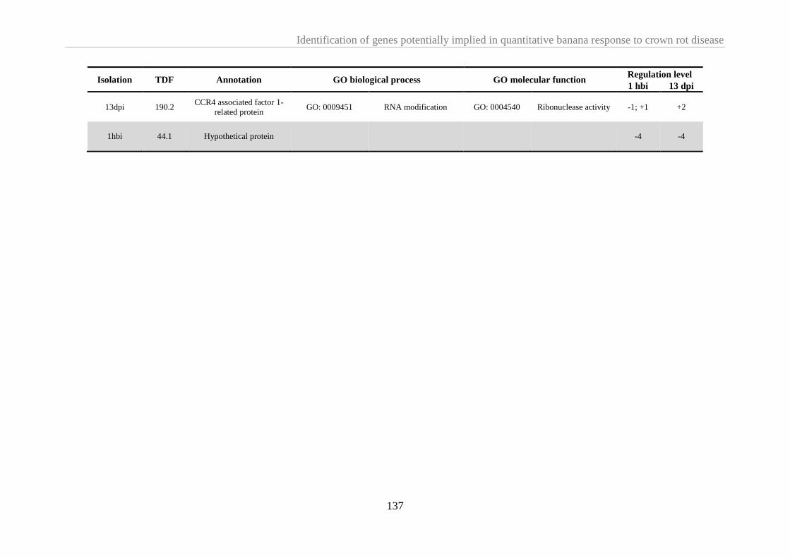
Identification of genes potentially implied in quantitative banana response to crown rot disease
137
Isolation TDF Annotation GO biological process GO molecular function Regulation level 1 hbi 13 dpi
13dpi 190.2 CCR4 associated factor 1-
related protein GO: 0009451 RNA modification GO: 0004540 Ribonuclease activity -1; +1 +2
1hbi 44.1 Hypothetical protein -4 -4

Identification of genes potentially implied in quantitative banana response to crown rot disease
138
2.3.3.2. TDFs isolated 13 dpi
Twenty-one primer pairs were successfully designed and they amplified the expected
products (table 4). Among the corresponding 21 TDFs, only 5 had the same pattern of
expression in both real-time RT-PCR biological replicates (table 3b). They were found to
encode proteins showing similarity to 1 cellulose synthase (294.2), 1 putative glycolipid
transfer protein (283.1), 1 serine carboxypeptidase (232.2), 1 putative ubiquitin carboxyl-
terminal hydrolase (317.1), and 1 protein related to the CCR4-associated factor 1 (190.2)
(table 3b). The 5 corresponding primer combinations were also used in real-time RT-PCR
experiments performed on the cDNA pools derived from samples collected 1 hbi from all four
plants (table 5). Serine caroboxypeptidase appeared upregulated in S- tissues collected 1 hbi
and slightly more so in tissues collected 13 dpi. Cellulose synthase showed similar
downregulation in S- tissues collected 1 hbi and 13 dpi. The putative glycolipid transfer
protein appeared downregulated in S- tissues collected 1 hbi, becoming highly upregulated 13
dpi. For both the putative ubiquitin carboxyterminal hydrolase and the protein related to
CCR4-associated factor 1, the biological duplicates showed similar results for 13 dpi
(upregulation in S- tissues) but different results for 1 hbi: slight downregulation in tissues
from S- plant 1 but slight upregulation in tissues from S- plant 2.
2.4. Discussion
Transcriptome analysis is a common way of discovering differences in gene expression
between two samples because regulation of gene activity occurs primarily on transcription
level. cDNA-AFLP presents the advantage to be an open-end methods requiring only standart
instrumentation and incurring low costs. But its drawback is a high fraction of cDNA
molecules escaping detection. Indeed, although in theory the cDNA-AFLP technique allows
analysis of the whole transcriptome, in practice the estimated transcriptome coverage ranges
from less than 20% to 73% (Venkatesh et al., 2005). Means reasons are because (i) of the lack
of suitable restriction sites (Breyne et al., 2003) ; (ii) gene discovery is based on subjective
band selection ; and (iii) because this is a laborious and time-consuming technique requiring
some selection at various steps. Furthermore, in the present study, we have retained only
fragments showing homology with sequences in a database, amounting to only 60% of our
fragments. Similar or lower percentages are reported in the literature for cDNA-AFLP (Botton
et al., 2008; Chen et al., 2003; Dong et al., 2004; Dilger et al., 2003; Guo et al., 2006;
Trindade et al., 2004). Results obtained in this study provide a supplementary proof of the
escaping detection. Indeed, different genes were put in evidence between 1hbi and 13dpi
Identification of genes potentially implied in quantitative banana response to crown rot disease
139
detection while the real-time RT-PCR shows that these genes were differently expressed in
both stages. It is thus essential to bear in mind that some interesting genes involved in banana
response to crown rot disease might not have been identified in our study and may remain to
be discovered.
This study also highlights the importance of confirming results with an independent
technique. In the runs performed on material derived from S+ plant 1 and S- plant 1 (the same
material as used for cDNA-AFLP analysis), the real-time RT-PCR confirmed the initially
differential expression for 86% of the cDNA-AFLP fragments. Non-confirmation is likely due
to the fact that a band in a gel can contain a mixture of several fragments (Chen et al., 2006;
Hsu et al., 2008; Zhang, 2003). This introduces artefactual cloning (Bachem et al., 1998) and
can also possibly lead to misinterpretation (overestimation) of cDNA-AFLP fragment
expression levels in the gel. In the literature, investigators report confirmation rates of 30%
(Campalans et al., 2001; Fukuda et al., 1999) to 100% (Ditt et al., 2001) for genes identified
by cDNA-AFLP analysis and tested with an independent technique. Moreover, our results
also demonstrate the necessity of confirming results with biological replicates before
concluding that a gene is involved in a molecular process, since the initial differential
expression was confirmed in both biological replicates for only 46% of our selected genes.
Despite these limitations, the cDNA-AFLP technique has provided useful information
allowing a step towards understanding the mechanisms involved in the variations of banana
susceptibility to crown rot disease and this is the first work carried on this pathosystem.
Because of the compatibility of the interaction, we expected significant transcriptional
changes to occur among genes associated with cellular changes involved in general defense
responses. It was no surprise to find genes involved in signal transduction or proteolytic
pathways among those potentially involved in banana defense responses. It is well known that
these pathways are complex but required to regulate defense responses, even though the exact
regulatory mechanisms are not yet known.
The sequences encoded by fragments 48b.1 and 44b.2 respectively show homology with
a putative protein kinase and a protein of the dual specificity phosphatase (DSP) family.
Proteins of both types catalyze reversible protein phosphorylations involved in signaling
pathways. It is known that synthesis of such proteins is triggered by an array of stimuli and
that they target a broad range of downstream effectors, thereby regulating various processes
such as defense responses (Agrawal et al., 2002; Agrawal et al., 2003; Lecourieux-Quaked et
al., 2000; Nurnberger et al., 2004; Rakwal et al., 2001).
Identification of genes potentially implied in quantitative banana response to crown rot disease
140
The proteins corresponding to fragments 31.1 (a RING-type ubiquitin ligase) and 317.1
(a putative ubiquitin carboxyl-terminal hydrolase) belong the proteolytic machinery that plays
key roles in the regulation of biological processes in plants (Edelmann and Kessler, 2008),
including ones mediating responses to pathogens (Devoto et al., 2003; Ellis et al., 2002; van
der Hoorn and Jones, 2004; Zeng et al., 2006). More specifically, proteins of this type are
involved in the 26S proteasome pathway, considered to be the main proteolytic pathway in
eukaryotes. Over the past years, approximately a dozen ubiquitinylation-related components
have been identified as being involved in plant-pathogen interactions (Zeng et al., 2006), but
in most cases, the exact nature of the regulation is not known. Most proteins of this type are
induced in response to pathogen or elicitor challenges (Boyes et al., 1998; Ramonell et al.,
2005; Salinas-Mondragón et al., 1999; Serrano and Guzman, 2004; Takizawa et al., 2005;
Zeng et al., 2006).
In addition to the 26S proteasome pathway, there are other proteases that may be
involved in plant defense (Avrova et al., 1999; Kruger et al., 2002; Pautot et al., 1993;
Tornero et al., 1996; Xia et al., 2004). Serine carboxypeptidase (232.2) is a protease with a
well-established function in protein turnover for the mobilization of N-resources, notably
during seed germination (Dal Degan et al., 1994; Granat et al, 2003) and wound stress (Moura
et al., 2001). Our results suggest that serine carboxypeptidase could be also implied in plant-
pathogen response. This protein appears to be upregulated in S- crown both pre- and post-
infection, but to a lesser extent 1hbi.
The proteins corresponding to fragments 47.1 and 283.1 show high similarity to
glycolipid transfer protein type 1 of Arabidopsis thaliana (AtGLTP1). They belong to the
lipid-transfer protein 1 (LTP1) family, classified as pathogenesis-related family 14 (PR-14).
Such proteins can be activated by elicitation and accumulate in plants, mounting a defensive
response against pathogens, including fungi (Van Loon et al., 2006; Buhot et al., 2004). Yet
the precise role of LTPs in defense responses remains to be discovered. AtGLTP1 has been
shown in vitro to enhance the intervesicular trafficking of glycosphingolipids (GSLs), but the
precise in vivo biological function is still unknown (Brown and Mattjus, 2007). Both banana
genes showed the same regulation profile: slightly lower expression in S- than in S+ crown
tissue before pathogen inoculation and much higher expression in the former than in the latter
13 days post-inoculation.
On the other hand, cDNA-AFLP providing clues that may contribute to understanding
the mechanisms involved in banana responses to crown rot disease. One is CCR4-associated
Identification of genes potentially implied in quantitative banana response to crown rot disease
141
factor 1 (CAF1). CAF1 proteins are required for regulated deadenylation of a broad spectrum
of stress-responsive mRNAs (Liang et al., 2009), but the biochemical and physiological
functions of CAF1 proteins are not clearly established (Liang et al., 2009). They have been
implicated in the regulation of plant growth (Sarowar et al., 2007) and defense responses to
biotic (Lee et al., 2004; Liang et al., 2009; Sarowar et al., 2007) and abiotic stress (Liang et
al., 2009; Walley et al., 2007). In our study, as in all known cases, CAF1 overexpression
appears to result in enhanced resistance. No constitutive differential expression was noted
prior to inoculation. Investigators have linked up-regulation of multiple pathogenesis-related
genes to over-expression of CAF1 genes (Liang et al., 2009; Sarowar et al., 2007). It will be
interesting to study in our model the expression levels of some pathogenesis-related proteins
in order to corroborate previous results and to attempt to better understand the role of CAF1
in resistance mechanisms.
Expression of the gene encoding a protein similar to cellulose synthase (294.2) is down-
regulated in S- banana crowns. This is somewhat surprising, as cellulose is a cell-wall
component expected to play a role in forming a physical barrier against fungal penetration.
Nevertheless there is evidence that when cellulose synthesis is reduced by mutation, lignin
synthesis (Cano-Delgado et al., 2003) and defense responses are activated through various
signaling pathways (Cano-Delgado et al., 2003; Ellis et al., 2002). Here we provide no
information on lignin synthesis, but we do show a correlation between reduced expression of
the cellulose synthase gene and enhanced resistance. This downregulation seems constitituve,
as S- crowns showed the same level of downregulation both pre- and post-infection. Perhaps a
plant more exposed to pathogen attack because of a low cellulose synthase level produces
compensating defense mechanisms that are more efficient.
Finally, cDNA-AFLP analysis has enabled us to identify a gene showing homology
with a dopamine-β-monooxygenase (DoH) combined with a cytochrome B561 (CB) (33.2).
DoH-CB proteins form a recently identified group of proteins, likely to play a key role in
catecholamine biosynthesis pathway in plants (Tsubaki et al., 2005; Verelst and Asard, 2004).
Cathecholamines are biogenic amines possessing a 3,4-dihydroxy-substituted phenyl ring.
Among them are phenylethylamine, tyramine, dopamine, norepinephrine, and epinephrine
(Kuklin and Conger, 1995). In banana fruits, dopamine is formed only through hydroxylation
of tyramine (Smith, 1980). Dopamine hydroxylation by dopamine-β-monooxygenase leads to
synthesis of norepinephrine, which is subsequently methylated to epinephrine. From a broad
range of plants, bananas, and particularly Cavendish bananas, have the richest content in
cathecolamines and particularly in dopamine : 100 µg/g FW in Cavendish banana peel as

Identification of genes potentially implied in quantitative banana response to crown rot disease
142
compared with 7 µg/g FW for potato, the second richest plant of this list (Kulma and Szopa,
2007). This observation suggests that cathecolamines might play an important role in banana
physiology.
Various functions, including plant pathogen resistance, have been proposed for
catecholamines (Swiedrych et al., 2004; reviewed in Kulma and Szopa, 2007). Effects are
probably due to the antioxidant properties of catecholamines themselves and also to cell-wall
deposition and to the antioxidant properties of derivatives (Kulma and Szopa, 2007). Whether
catecholamines can stimulate some defense responses more directly remains to be seen
(Kulma and Szopa, 2007). However, catecholamine, dopamine, or oxidation products are
suggested to be involved in banana resistance mechanisms (Mace, 1963; Muirhead and
Deverall, 1984; Valette et al., 1998; Wuyts et al., 2006; Wuyts et al., 2007). Particularly, it
has been shown that dopamine oxidation products have a brown-red color and show a strong
antifungal activity against C. musae (Muirhead and Deverall, 1984). These authors suggested
that four of the five antifungal products reported (Brown and Swinburne, 1980; Brown and
Swinburne, 1981; Swinburne, 1978) subsequently to the infection of green bananas by hyaline
appressoria of Colletotrichum musae were dopamine oxidation products (contrarily to
melanized appressoria that do not germinate on green bananas, these infections from hyaline
appressoria are rapidly blocked and a brown-red coloration of neighboring tissues is
observed). It is noteworthy that the peripheral necrotic tissues observed here in the S- banana
crowns showed a specific red-brown color (data not shown). Then, our results suggest that
dopamine oxidation products might play an important role in susceptibility to crown rot
disease.
The factors involved in overexpression of dopamine oxidation in S- plants might be
complex. Effectively, catecholamine biosynthesis and content depend on various factors:
plant development stage (Lyte, 1997; Swiedrych et al., 2004b; Szopa et al., 2001); growth
conditions (Swiedrych et al., 2004b); environmental factors (Kulma and Szopa, 2007) such as
the pH (Homeyer and Roberts, 1984), brightness (Endress et al., 1984; Protacio et al., 1992),
temperature (Swiedrych et al., 2004); and stress conditions including (i) biotic factors such as
pathogen infection (Facchini et al., 1996), and (ii) abiotic stresses such as drought, ABA
treatment, UV light, and wounding (Swiedrych et al., 2004b; Swiedrych et al., 2004; Szopa et
al., 2001). In the case of cactus (Steelink et al., 1967) and potato (Szopa et al., 2001)
wounding is accompanied by a rise in the dopamine concentration, and wounded tissues
appear to stimulate dopamine production in the surrounding healthy tissue.

Identification of genes potentially implied in quantitative banana response to crown rot disease
143
Similarly, in the case of crown rot of bananas we can hypothetize that wounding at
harvest could also influence cathecolamine biosynthetic pathway.
It would be interesting to learn more about the role of dopamine-β-monooxygenase and
other catecholamines in the response of bananas to crown rot. We have shown that the
dopamine-β-monooxygenase gene is overexpressed in bananas less susceptible to the disease.
One should make sure, however, that this enzyme and some others are active and that their
substrates are present in the crowns. By determining the levels of various catecholamines in
crowns with different levels of susceptibility it might be possible to gain interesting
information about their respective roles. Furthermore, by measuring the potential antifungal
activities of catecholamines one could learn whether they are active by themselves in defense
mechanisms or whether they are precursors of other active compounds.
Finally, it is necessary to evaluate the expression levels of all these genes in other
situations of susceptibility level variation. To be sure that they are really involved in
quantitative regulation of banana defense responses and not in other pathways that might be
triggered by a change in the source-sink ratio quite independently of any plant defense
response. Furthermore, because plant defense responses invariably are by nature
multicomponent processes, it is not easy to determine which components are both necessary
and sufficient to confer protection.

Identification of genes potentially implied in quantitative banana response to crown rot disease
144
References
Agrawal, G.K., R. Rakwal, and H. Iwahashi. 2002. Isolation of novel rice (Oryza sativa L.)
multiple stress responsive MAP kinase gene, OsMSRMK2, whose mRNA accumulates
rapidly in response to environmental cues. Biochemical and Biophysical Research
Communications 294:1009-1016.
Agrawal, G.K., H. Iwahashi, and R. Rakwal. 2003. Rice MAPKs. Biochemical and
Biophysical Research Communications 302:171-180.
Albertazzi, G., J. Milc, A. Caffagni, E. Francia, E. Roncaglia, F. Ferrari, E. Tagliafico, E.
Stefani, and N. Pecchioni. 2009. Gene expression in grapevine cultivars in response to
Bois Noir phytoplasma infection. Plant Science 176:792-804.
Alvindia, D.G., T. Kobayashi, Y. Yaguchi, and K.T. Natsuaki. 2000. Symptoms and the
associated fungi of postharvest diseases on non-chemical bananas imported from the
Philippines. Japanese Journal of Tropical Agriculture 44:87-93.
Appel, H.M. 1993. Phenolics in ecological interactions: The importance of oxidation. Journal
of Chemical Ecology 19:1521-1552.
Avrova, A.O., H.E. Stewart, W. De Jong, J. Heilbronn, G.D. Lyon, and P.R.J. Birch. 1999. A
Cysteine Protease Gene Is Expressed Early in Resistant Potato Interactions with
Phytophthora infestans. Molecular Plant-Microbe Interactions 12:1114-1119.
Bachem, C. 1996. Visualization of differential gene expression using a novel method of RNA
fingerprinting based on AFLP: Analysis of gene expression during potato tuber
development. The Plant Journal 9:745-753.
Bachem, C., R. Oomen, and R. Visser. 1998. Transcript imaging with cDNA-AFLP: a step by
step protocol. Plant Molecular Biology Reporter 16:157-173.
Botton, A., D. Ferrigo, C. Scopel, R. Causin, C. Bonghi, and A. Ramina. 2008. A cDNA-
AFLP approach to study ochratoxin A production in Aspergillus carbonarius.
International Journal of Food Microbiology 127:105-115.
Boyes, D.C., J. Nam, and J.L. Dangl. 1998. The Arabidopsis thaliana RPM1 disease
resistance gene product is a peripheral plasma membrane protein that is degraded
coincident with the hypersensitive response. Proceedings of the National Academy of
Sciences USA 95:15849-15854.
Breyne, P., R. Dreesen, B. Cannoot, D. Rombaut, K. Vandepoele, S. Rombauts, R.
Vanderhaeghen, D. Inze, M. Zabeau. 2003. Quantitative cDNA-AFLP analysis for
genome-wide expression studies. Molecular Genetics and Genomics 269: 173-179.

Identification of genes potentially implied in quantitative banana response to crown rot disease
145
Brodersen, P., M. Petersen, H.M. Pike, B. Olszak, S.r. Skov, N. Ødum, L.B. Jørgensen,
R.E. Brown, and J. Mundy. 2002. Knockout of Arabidopsis ACCELERATED-CELL-
DEATH11 encoding a sphingosine transfer protein causes activation of programmed
cell death and defense. Genes & Development 16:490-502.
Brown, A.E., and T.R. Swinburne. 1980. The resistance of immature banana fruits to
anthracnose Colletotrichum musae (Berk. & Curt.) Arx. Journal of Phytopathology
99:70-80.
Brown, A.E., and T.R. Swinburne. 1981. Influence of iron and iron chelators on formation of
progressive lesions by Colletotrichum musae on banana fruits. Transactions of the
British Mycological Society 77:119-124.
Brown, R.E., and P. Mattjus. 2007. Glycolipid transfer proteins. Biochimica et Biophysica
Acta 1771:746-776.
Campalans, A., M. Pagès, and R. Messeguer. 2001. Identification of differentially expressed
genes by the cDNA-AFLP technique during dehydration of almond. Tree physiology
21:633-643.
Cano-Delgado, A., S. Penfield, C. Smith, M. Catley, and M. Bevan. 2003. Reduced cellulose
synthesis invokes lignification and defense responses in Arabidopsis thaliana. The Plant
Journal 34:351-362.
Chen, G.P., W.S. Ma, Z.J. Huang, T. Xu, Y.B. Xue, and Y.Z. Shen. 2003. Isolation and
characterization of TaGSK1 involved in wheat salt tolerance. Plant Science 165:1369-
1375.
Chen, W.H., I.C. Tseng, W.C. Tsai, M.S. Chiang, Y.H. Chen, and H.H. Chen. 2006. AFLP
fingerprinting and conversion to sequence-tag site markers for the identification of
Oncidium cultivars. Journal of horticultural science & biotechnology 81:791-796
Cheung, M.-Y., N.-Y. Zeng, S.-W. Tong, F. Wing-Yen Li, K.-J. Zhao, Q. Zhang, S. Sai-Ming
Sun, and H.-M. Lam. 2007. Expression of a RING-HC protein from rice improves
resistance to Pseudomonas syringae pv. tomato DC3000 in transgenic Arabidopsis
thaliana. Journal of Experimental Botany 58:4147-4159.
Christou, P., and K.A. Barton. 1989. Cytokinin Antagonist Activity of Substituted
Phenethylamines in Plant Cell Culture. Plant Physiology 89:564-568.
Collart, M.A., and H.T. Timmers. 2004. The eukaryotic Ccr4-not complex: a regulatory
platform integrating mRNA metabolism with cellular signaling pathways? Progress in
Nucleic Acid Research and Molecular Biology 77:289-322.

Identification of genes potentially implied in quantitative banana response to crown rot disease
146
Costa Mondego, J.M., J.L. Simões-Araújo, D.E. de Oliveira, and M. Alves-Ferreira. 2003. A
gene similar to bacterial translocase I (mra Y) identified by cDNA-AFLP is expressed
during flower bud development of Arabidopsis thaliana. Plant Science 164:323-331.
Czechowski, T., M. Stitt, T. Altmann, M. K. Udvardi, and W. Scheible. 2005. Genome-Wide
Identification and Testing of Superior Reference Genes for Transcript Normalization in
Arabidopsis. Plant Physiology 139:5-17.
Dai, Y., P. Michaels, and H. Flores. 1993. Stimulation of ethylene production by
catecholamines and phenylethylamine in potato cell suspension cultures. Plant Growth
Regulation 12:219-222.
Dal Degan, F., A. Rocher, V. Cameron-Mills, and D. von Wettstein. 1994. The expression of
serine carboxypeptidases during maturation and germination of the barley grain.
Proceedings of the National Academy of Sciences USA 91:8209-8213.
de Lapeyre de Bellaire, L., M. Chillet, and Y. Chilin-Charles. 2008. Determination of banana
fruit susceptibility to post-harvest diseases: wound anthracnose, quiescent anthracnose
and crown-rot. Fruits 63:183-186.
Delauré, S.L., W. Van Hemelrijck, M.F.C. De Bolle, B.P.A. Cammue, and B.M.A. De
Coninck. 2008. Building up plant defenses by breaking down proteins. Plant Science
174:375-385.
Denis, C.L., and J. Chen. 2003. The CCR4-NOT complex plays diverse roles in mRNA
metabolism. Progress in Nucleic Acid Research and Molecular Biology 73:221-250.
Devoto, A., P.R. Muskett, and K. Shirasu. 2003. Role of ubiquitination in the regulation of
plant defence against pathogens. Current Opinion in Plant Biology 6:307-311.
Dilger, M., F.G. Felsenstein, and G. Schwarz. 2003. Identification and quantitative expression
analysis of genes that are differentially expressed during conidial germination in
Pyrenophora teres. Molecular Genetic and Genomic 270:147-155.
Ditt, R.F., E.W. Nester, and L. Comai. 2001. Plant gene expression response to
Agrobacterium tumefaciens. Proceeding of the National Academy of Sciences USA
98:10954-10959.
Dixon, R.A., and N.L. Paiva. 1995. Stress-Induced Phenylpropanoid Metabolism. Plant Cell
7:1085-1097.
Dong, W., M. Latijnhouwers, R.H.Y. Jiang, H.J.G. Meijer, and F. Govers. 2004. Downstream
targets of the Phytophthora infestans Gα subunit PiGPA1 revealed by cDNA-AFLP.
Molecular plant pathology 5:483-494.
Dreher, K., and J. Callis. 2007. Ubiquitin, Hormones and Biotic Stress in Plants. Annals of
Botany :mcl255.

Identification of genes potentially implied in quantitative banana response to crown rot disease
147
Edelmann, M.J., and B.M. Kessler. 2008. Ubiquitin and ubiquitin-like specific proteases
targeted by infectious pathogens: Emerging patterns and molecular principles.
Biochimica et Biophysica Acta (BBA) - Molecular Basis of Disease 1782:809-816.
Ellis, C., J.G. Turner, and A. Devoto. 2002. Protein complexes mediate signalling in plant
responses to hormones, light, sucrose and pathogens. Plant Molecular Biology 50:971-
980.
Elstner, E.F., J.R. Konze, B.R. Selman, and C. Stoffer. 1976. Ethylene formation in sugar beet
leaves. Plant Physiology 58:163-168.
Endress, R., A. Jager, and W. Kreis. 1984. Catecholamines biosynthesis dependent on the
dark in betacyanin-forming Portulaca callus. Journal of Plant Physiology 115:291-295.
Eungwanichayapant P.D., and S. Popluechai. 2009. Accumulation of catechins in tea in
relation to accumulation of mRNA from genes involved in catechin biosynthesis.
Plant Physiology and Biochemistry 47 : 94-97.
Facchini, P.J., A.G. Johnson, J. Poupart, and V. De Luca. 1996. Uncoupled Defense Gene
Expression and Antimicrobial Alkaloid Accumulation in Elicited Opium Poppy Cell
Cultures. Plant Physiology 111:687-697.
Finlay, A.R., and A.E. Brown. 1993. The relative importance of Colletotrichum musae as a
crown-rot pathogen on Windward Island bananas. Plant Pathology 42:67-74.
Fukuda, T., A. Kido, K. Kajino, M. Tsutsumi, Y. Miyauchi, T. Tsujiuchi, Y. Konishi, and O.
Hino. 1999. Cloning of differentially expressed genes in highly and low metastatic rat
osteosarcomas by a modified cDNA-AFLP method. Biochemical and biophysical
Research Communications 261:35-40.
Gonzalez-Lamothe, R., D.I. Tsitsigiannis, A.A. Ludwig, M. Panicot, K. Shirasu, and J.D.G.
Jones. 2006. The U-Box Protein CMPG1 Is Required for Efficient Activation of
Defense Mechanisms Triggered by Multiple Resistance Genes in Tobacco and Tomato.
Plant Cell 18:1067-1083.
Granat, S.J., K.A. Wilson and A.L. Tan-Wilson. 2003. New serine carboxypeptidases in mung
bean seedling cotyledons. Journal of Experimental Botany 160:1263–1266.
Grandmaison, J., G.M. Olah, M.R. Van Calsteren, and V. Furlan. 1993. Characterisation and
localisation of plant phenolics likely involved in the pathogen resistance expressed by
endomycorrizal roots. Mycorhizza 3:155-164.
Griffiths, L.A. 1959. Detection and identification of polyphenol oxidase substrate of banana.
Nature 184:58.
Griffiths, L.A. 1961. Relationship between 3,4-dihydroxyphenylethylamine content and the
genome of Musa acuminata. Nature 192:84-85.

Identification of genes potentially implied in quantitative banana response to crown rot disease
148
Guo, J., R.H.Y. Jiang, L.G. Kamphuis, and F. Govers. 2006. A cDNA-AFLP based strategy to
identify transcripts associated with avirulence in Phytophthora infestans. Fungal
Genetics and Biology 43:111-123.
Hershko, A., and A. Ciechanover. 1998. The ubiquitin system. Annual Review of
Biochemistry 67:425-479.
Hohlfeld, H., W. Schurmann, D. Scheel, and D. Strack. 1995. Partial Purification and
Characterization of Hydroxycinnamoyl-Coenzyme A: Tyramine Hydroxycinnamoyl-
transferase from Cell Suspension Cultures of Solanum tuberosum. Plant Physiology
107:545-552.
Homeyer, B., and M. Roberts. 1984. Dopamine accumulation in Papaver somniferum latex.
Journal of Phytopathology 39c:1034–1037.
Hsu, T.-W., W.-C. Tsai, D.-P. Wang, S. Lin, Y.-Y. Hsiao, W.-H. Chen, and H.-H. Chen.
2008. Differential gene expression analysis by cDNA-AFLP between flower buds of
Phalaenopsis hsiang fei cv. H. F. and its somaclonal variant. Plant Science 175:415-
422.
Huang, Y.-C., Y.-L. Chang, J.-J. Hsu, and H.-w. Chuang. 2008. Transcriptome analysis of
auxin-regulated genes of Arabidopsis thaliana. Gene 420:118-124.
Jullien, A., M. Chillet, and E. Malezieux. 2008. Pre-harvest growth and development,
measured as accumulated degree days, determine the post-harvest green life of banana
fruit. Journal of Horticultural Science and Biotechnology 83:506-512.
Kanazawa, K., and H. Sakakibara. 2000. High Content of Dopamine, a Strong Antioxidant, in
Cavendish Banana. Journal of Agricultural and Food Chemistry 48:844-848.
Kaur, A., and A. Thurkral. 1990. Effect of animal hormones on the growth, protein and sugar
contents of Vigna unguiculata L. seedlings. Indian Journal of Plant Physiology 33:259-
261.
Khurana, J.P., B.K. Tamot, N. Maheshwari, and S.C. Maheshwari. 1987. Role of
catecholamines in promotion of flowering in a short-day duckweed, Lemna paucicostata
6746. Plant Physiology 85:10-12.
Krauss, U., and A. Johanson. 2000. Recent advances in the control of crown rot of banana in
the Windward Islands. Crop Protection 19:151-160.
Kruger, J., C.M. Thomas, C. Golstein, M.S. Dixon, M. Smoker, S. Tang, L. Mulder, and
J.D.G. Jones. 2002. A Tomato Cysteine Protease Required for Cf-2-Dependent Disease
Resistance and Suppression of Autonecrosis. Science 296:744-747.

Identification of genes potentially implied in quantitative banana response to crown rot disease
149
Kuhn, E. 2001. From library screening to microarray technology: strategies to determine gene
expression profiles and to identify differentially regulated genes in plants. Annals of
Botany 87:139-155.
Kuklin, A., and B. Conger. 1995. Catecholamines in plants. Journal of Plant Growth
Regulation 14:91-97.
Kuklin, A., and B. Conger. 1995b. Enhancement of somatic embryogenesis in orchardgrass
leaf cultures by epinephrine. Plant Cell Reports 14:641-644.
Kulma, A., and J. Szopa. 2007. Catecholamines are active compounds in plants. Plant Science
172:433-440.
Lassois, L., L. de Lapeyre de Bellaire, and M.H. Jijakli. 2008. Biological control of crown rot
of bananas with Pichia anomala strain K and Candida oleophila strain O. Biological
Control 45:410-418.
Lassois, L., L. De Lapeyre de Bellaire, and M.H. Jijakli. 2009. Combining an original method
for preserving RNA expression in situ with an effective RNA extraction method makes
it possible to study gene expression in any banana fruit tissue. Fruits 64 : 127-137.
Lassois, L., M.H. Jijakli, M. Chillet, and L. De Lapeyre de Bellaire. Accepteda. Crown rot of
bananas : pre-harvest factors involved in post-harvest expression and integrated control
methods. Plant Disease.
Lassois, L., H. Bastiaanse, M. Chillet, A. Jullien, H. Jijakli, and L. de Lapeyre de Bellaire.
Acceptedb. Hand position on the bunch and source-sink ratio influence the level of
banana fruit susceptibility to crown rot disease. Annals of Applied Biology.
Lecourieux-Quaked, F., A. Pugin, and A. Lebrun-Garcia. 2000. Phosphoproteins involved in
the signal transduction of cryptogein, an elicitor of defense reactions in tobacco.
Molecular Plant Microbe Interaction 13:821-829.
Lee, S., S.-Y. Kim, E. Chung, Y.-H. Joung, H.-S. Pai, C.-G. Hur, and D. Choi. 2004. EST and
microarray analyses of pathogen-responsive genes in hot pepper (Capsicum annuum L.)
non-host resistance against soybean pustule pathogen (Xanthomonas axonopodis pv.
glycines). Functional & Integrative Genomics 4:196-205.
Liang, W., C. Li, F. Liu, H. Jiang, S. Li, J. Sun, X. Wu, and C. Li. 2009. The Arabidopsis
homologs of CCR4-associated factor 1 show mRNA deadenylation activity and play a
role in plant defence responses. Cell Research 19:307-316.
Lu, Z., D. Liu, A. Hornia, W. Devonish, M. Pagano, and D.A. Foster. 1998. Activation of
Protein Kinase C Triggers Its Ubiquitination and Degradation. Molecular and Cellular
Biology 18:839-845.

Identification of genes potentially implied in quantitative banana response to crown rot disease
150
Lucas, J.A. 1998. Plant pathology and plant pathogens. Blackwell Science, Oxford, U.K.
(Malden, MA, USA).
Lukezic, F.L., W.J. Kaiser, and M.M. Martinez. 1967. The incidence of crown rot of boxed
bananas in relation to microbial populations of the crown tissus. Canadian Journal of
Botany 45:413-421.
Lyte, M. 1997. Induction of Gram-negative bacterial growth by neurochemical containing
banana (Musa x paradisiaca) extracts. FEMS Microbiology Letters 154:245-250.
Mace, M.E. 1963. Histochemical localization of phenols in healthy and diseased banana roots.
Physiologia plantarum 16:915-925.
Massart, S., and H. Jijakli. 2006. Identification of Differentially Expressed Genes by cDNA-
Amplified Fragment Length Polymorphism in the Biocontrol Agent Pichia anomala
(Strain Kh5). Phytopathology 96:80-86.
Mattjus, P., B. Turcq, H.M. Pike, J.G. Molotkovsky, and R.E. Brown. 2003. Glycolipid
Intermembrane Transfer Is Accelerated by HET-C2, a Filamentous Fungus Gene
Product Involved in the Cell—Cell Incompatibility Response. Biochemistry 42:535-542.
Mbéguié-A-Mbéguié, D., B. Fils-Lycaon, M. Chillet, O. Hubert, C. Galas, and R. Gomez.
2008. Extraction and purification of total RNA from banana tissues (small scale). Fruits
63:255-261.
Meredith, D.S. 1971. Transport and storage diseases of bananas: biology and control. Tropical
Agriculture 48:35-50.
Montesano, M., G. Brader, and E.T. Palva. 2003. Pathogen derived elicitors: searching for
receptors in plants. Molecular Plant Pathology 4: 73-79.
Moura, D.S., D.R. Bergey, and C.A. Ryan. 2001. Characterization and localization of a
wound-inducible type I serine-carboxypeptidase from leaves of tomato plants
(Lycopersicon esculentum Mill.). Planta 212:222-230.
Muirhead, I.F., and B.J. Deverall. 1984. Evaluation of 3,4-dihydrobenzaldehyde, dopamine
and its oxydation products as inhibitors of Colletotrichum musae (Berk. et Curt.) Arx in
green banana fruits. Australian Journal of Botany 32:575-582.
Newman, M.-A., E. von Roepenack-Lahaye, A. Parr, M.J. Daniels, and J.M. Dow. 2001.
Induction of Hydroxycinnamoyl-Tyramine Conjugates in Pepper by Xanthomonas
campestris, a Plant Defense Response Activated by hrp Gene-Dependent and hrp Gene-
Independent Mechanisms. Molecular Plant-Microbe Interactions 14:785-792.
Nishimura, M.T., M. Stein, B.-H. Hou, J.P. Vogel, H. Edwards, and S.C. Somerville. 2003.
Loss of a Callose Synthase Results in Salicylic Acid-Dependent Disease Resistance.
Science 301:969-972.

Identification of genes potentially implied in quantitative banana response to crown rot disease
151
Nurnberger, T., F. Brunner, B. Kemmerling, and L. Piater. 2004. Innate immunity in plants
and animals: striking similarities and obvious differences. Immunological reviews
198:249-266.
Ohn, T., Y.-C. Chiang, D.J. Lee, G. Yao, C. Zhang, and C.L. Denis. 2007. CAF1 plays an
important role in mRNA deadenylation separate from its contact to CCR4. Nucleic
Acids Research 35:3002-3015.
Palmer, J.K. 1963. Banana polyphenoloxydase: preparation and properties. Plant Physiology
38:508-513.
Pautot, V., F.M. Holzer, B. Reisch, and L.L. Walling. 1993. Leucine aminopeptidase: an
inducible component of the defense response in Lycopersicon esculentum (tomato).
Proceedings of the National Academy of Sciences USA 90:9906-9910.
Peng, M., C. Hannam, H. Gu, Y. Bi, and S.J. Rothstein. 2007. A mutation in NLA, which
encodes a RING-type ubiquitin ligase, disrupts the adaptability of Arabidopsis to
nitrogen limitation. Plant Journal 50:320-327.
Pfaffl, M.W. 2001. A new mathematical model for relative quantification in real-time RT-
PCR. Nucleic Acids Research 29:2002-2007.
Promyou, S., S. Ketsa, and W.G. van Doorn. 2008. Hot water treatments delay cold-induced
banana peel blackening. Postharvest Biology and Technology 48:132-138.
Protacio, C.M., Y.-r. Dai, E.F. Lewis, and H.E. Flores. 1992. Growth Stimulation by
Catecholamines in Plant Tissue/Organ Cultures. Plant Physiology 98:89-96.
Rakwal, R., K. Shii, G.K. Agrawal, and M. Yonekura. 2001. Protein phosphatase inhibitors
activate defense responses in rice (Oryza sativa) leaves. Physiologia Plantarum
111:151-157.
Ramakers, C., J.M. Ruijter, R.H.L. Deprez, and A.F.M. Moorman. 2003. Assumption-free
analysis of quantitative real-time polymerase chain reaction (PCR) data. Neuroscience
Letters 339:62-66.
Ramonell, K., M. Berrocal-Lobo, S. Koh, J. Wan, H. Edwards, G. Stacey, and S. Somerville.
2005. Loss-of-Function Mutations in Chitin Responsive Genes Show Increased
Susceptibility to the Powdery Mildew Pathogen Erysiphe cichoracearum. Plant
Physiology 138:1027-1036.
Reijans M., R. Lascaris, A.O. Groeneger, A. Wittenberg, E. Wesselink, J. van Oeveren, E.d.
Wit, A. Boorsma, B. Voetdijk and H. van der Spek (2003). Quantitative comparison of
cDNA-AFLP, microarrays, and genechip expression data in Saccharomyces cerevisiae.
Genomics 82:606-618.

Identification of genes potentially implied in quantitative banana response to crown rot disease
152
Roshchina, V.V. 1990. Biomediators in chloroplasts of higher plants.4. Reception by
photosynthetic membranes. Photosynthetica 24:539-549.
Salinas-Mondragón, R., C. Garcidueñas-Piña, and P. Guzmán. 1999. Early elicitor induction
in members of a novel multigene family coding for highly related RING-H2 proteins in
Arabidopsis thaliana. Plant Molecular Biology 40:579-590.
Sarowar, S., H.W. Oh, H.S. Cho, K.H. Baek, E.S. Seong, Y.H. Joung, G.J. Choi, S. Lee, and
D. Choi. 2007. Capsicum annuum CCR4-associated factor CaCAF1 is necessary for
plant development and defence response. The Plant Journal 51:792-802.
Schenk, R.U., and A.C. Hildebrandt. 1972. Medium and techniques for induction and growth
of monocotyledonous and dicotyledonous plant cell cultures. Canadian Journal of
Botany 50:199-204.
Serrano, M., and P. Guzman. 2004. Isolation and Gene Expression Analysis of Arabidopsis
thaliana Mutants With Constitutive Expression of ATL2, an Early Elicitor-Response
RING-H2 Zinc-Finger Gene. Genetics 167:919-929.
Shillingford, C.A. 1978. Climatic factors affecting post-harvest decay of jamaican bananas.
Journal of Agriculture of the University of Puerto Rico:45-49.
Simmonds, J.H. 1963. Studies in the latent phase of Colletotrichum species causing ripe rots
of tropical fruits. Queensland Journal of Agricultural and Animal Science 20:373-424.
Slabaugh, W.R., and M.D. Grove. 1982. Postharvest diseases of bananas and their control.
Plant Disease 66:746-750.
Smith, T.A. 1977. Phenethylamine and related compounds in plants. Phytochemistry 16:9-18.
Smith, T.A. 1980. Plant amines, p. 433-460, In C. B. e. Bell IA, ed. Secondary Plant Products,
Encyclopedia of Plant Physiology New Series, Vol. 8. Springer Verlag, Berlin.
Steelink, C., M. Yeung, and R.L. Caldwell. 1967. Phenolic constituents of healthy and wound
tissues in the giant cactus (Caregiea gigantea). Phytochemistry 6:1435-1440.
Su, K., M.D. Roos, X. Yang, I. Han, A.J. Paterson, and J.E. Kudlow. 1999. An N-terminal
Region of Sp1 Targets Its Proteasome-dependent Degradation in Vitro. The journal of
Biological Chemistry 274:15194-15202.
Swiedrych, A., J. Stachowiak, and J. Szopa. 2004b. The catecholamine potentiates starch
mobilization in transgenic potato tubers. Plant Physiology and Biochemistry 42:103-
109.
Swiedrych, A., K. Lorenc-Kukula, A. Skirycz, and J. Szopa. 2004. The catecholamine
biosynthesis route in potato is affected by stress. Plant Physiology and Biochemistry
42:593-600.

Identification of genes potentially implied in quantitative banana response to crown rot disease
153
Swinburne, T.R. 1978. Post-infection antifungal compounds in quiescent or latent infections.
Annals of Applied Biology 89:322-325.
Szopa, J., G. Wilczynski, O. Fiehn, A. Wenczel, and L. Willmitzer. 2001. Identification and
quantification of catecholamines in potato plants (Solanum tuberosum) by GC-MS.
Phytochemistry 58:315-320.
Takizawa, M., A. Goto, and Y. Watanabe. 2005. The tobacco ubiquitin-activating enzymes
NtE1A and NtE1B are induced by tobacco mosaic virus, wounding and stress
hormones. Molecules and Cells 19:228-231.
Tornero, P., V. Conejero, and P. Vera. 1996. Primary structure and expression of a pathogen-
induced protease (PR-P69) in tomato plants: Similarity of functional domains to
subtilisin-like endoproteases. Proceedings of the National Academy of Sciences USA
93:6332-6337.
Trindade, L.M., B.M. Horvath, R. van Berloo, and R.G.F. Visser. 2004. Analysis of genes
differentially expressed during potato tuber life cycle and isolation of their promoter
regions. Plant Science 166:423-433.
Tsubaki, M., F. Takeuchi, and N. Nakanishi. 2005. Cytochrome b561 protein family:
Expanding roles and versatile transmembrane electron transfer abilities as predicted by
a new classification system and protein sequence motif analyses. Biochimica and
Biophysica Acta (BBA) - Proteins & Proteomics 1753:174-190.
Valette, C., C. Andary, J.P. Geiger, J.L. Sarah, and M. Nicole. 1998. Histochemical and
Cytochemical Investigations of Phenols in Roots of Banana Infected by the Burrowing
Nematode Radopholus similis. Phytopathology 88:1141-1148.
Van Der Biezen, E.A., and J.D.G. Jones. 1998. Plant disease-resistance proteins and the gene-
for-gene concept. Trends in Biochemical Sciences 23:454-456.
van der Hoorn, R.A.L., and J.D.G. Jones. 2004. The plant proteolytic machinery and its role
in defence. Current Opinion in Plant Biology 7:400-407.
Vaughn, K.C., and S.O. Duke. 1984. Function of polyphenol oxidase in higher plants.
Physiologia Plantarum 60:106-112.
Venkatesh, B., U. Hettwer, B. Koopmann, and P. Karlovsky. 2005. Conversion of cDNA
differential display results (DDRT-PCR) into quantitative transcription profiles. BMC
Genomics 6:Art. No. 51.
Verelst, W.i.m., and H.a.n. Asard. 2004. Analysis of an Arabidopsis thaliana protein family,
structurally related to cytochromes b 561 and potentially involved in catecholamine
biochemistry in plants. Journal of Plant Physiology 161:175-181.

Identification of genes potentially implied in quantitative banana response to crown rot disease
154
von Roepenack-Lahaye, E., M.-A. Newman, S. Schornack, K.E. Hammond-Kosack, T.
Lahaye, J.D.G. Jones, M.J. Daniels, and J.M. Dow. 2003. p-coumaroylnoradrenaline, a
novel plant metabolite implicated in tomato defense against pathogens. The Journal of
Biological Chemistry 278: 43373-43383.
Waalkes, T.P., A. Sjoerdsma, C.R. Creveling, H. Weissbach, and S. Udenfriend. 1958.
Serotonin, Norepinephrine, and Related Compounds in Bananas. Science 127:648-650.
Walley, J.W., S. Coughlan, M.E. Hudson, M.F. Covington, R. Kaspi, G. Banu, S.L. Harmer,
and K. Dehesh. 2007. Mechanical Stress Induces Biotic and Abiotic Stress Responses
via a Novel cis-Element. PLoS Genetics 3:e172.
Wan, C., and T. Wilkins. 1994. A modified hot borate method significantly enhances the yield
of high-quality RNA from cotton (Gossypium hirsutum L.). Analytical Biochemistry
223:7-12.
West, G., L. Viitanen, C. Alm, P. Mattjus, T.A. Salminen, and J. Edqvist. 2008. Identification
of a glycosphingolipid transfer protein GLTP1 in Arabidopsis thaliana. The FEBS
Journal 275:3421-3437.
Wilkinson, K.D. 1999. Ubiquitin-Dependent Signaling: The Role of Ubiquitination in the
Response of Cells to Their Environment. Journal of Nutrition 129:1933-1936.
Wuyts, N., D. De Waele, and R. Swennen. 2006. Extraction and partial characterization
of polyphenol oxidase from banana (Musa acuminata Grande naine) roots. Plant
Physiology and Biochemistry 44:308-314.
Wuyts, N., G. Lognay, M. Verscheure, M. Marlier, D. De Waele, and R. Swennen. 2007.
Potential physical and chemical barriers to infection by the burrowing nematode
Radopholus similis in roots of susceptible and resistant banana (Musa spp.). Plant
Pathology 56:878-890.
Xia, Y., H. Suzuki, J. Borevitz, J. Blount, Z. Guo, K. Patel, R.A. Dixon, and C. Lamb. 2004.
An extracellular aspartic protease functions in Arabidopsis disease resistance signaling.
The EMBO Journal 23:980-988.
Yang, C.-P., S. Fujita, K. Kohno, A. Kusubayashi, M.D. Ashrafuzzaman, and N. Hayashi.
2001. Partial Purification and Characterization of Polyphenol Oxidase from Banana
(Musa sapientum L.) Peel. Journal of Agricultural and Food Chemistry 49:1446-1449.
Yang, C.-P., Z.-R. Nong, J.-L. Lu, L. Lu, J.-S. Xu, Y.-Z. Han, Y.-J. Li, and S. Fujita. 2004.
Banana Polyphenol Oxidase: Occurrence and Change of Polyphenol Oxidase Activity in
Some Banana Cultivars during Fruit Development. Food Science and Technology
Research 10:75-78.

Identification of genes potentially implied in quantitative banana response to crown rot disease
155
Yang, Y., J. Shah, and D.F. Klessig. 1997. Signal perception and transduction in plant disease
responses. Genes and development 11:1621-1639.
Zeng, L.-R., M.E. Vega-Sanchez, T. Zhu, and G.-L. Wang. 2006. Ubiquitination-mediated
protein degradation and modification: an emerging theme in plant-microbe interactions.
Cell Research 16:413-426.
Zhang, L.H.M.a.M.D. 2003. Isolation of genes expressed during compatible interactions
between leaf rust (Puccinia triticina) and wheat using cDNA-AFLP. Molecular Plant
Pathology 4:469-477.

Conclusions and Perspectives
156
CHAPTER V
Conclusions and PerspectivesConclusions and PerspectivesConclusions and PerspectivesConclusions and Perspectives
Conclusions and Perspectives
157
We propose a new way to view the post-harvest development of banana crown rot
disease. The idea is that that crown rot development depends notably on fruit physiology at
harvest, which in turn depends on pre-harvest factors. Although it has been suggested
previously that fruit physiology may affect disease incidence, this has never been investigated
in detail. Two aspects of the fruit physiology were considered in this study. In the first part (i),
the importance of the fruit physiological component at harvest in post-harvest crown rot
disease was highlighted (a) and the influence of some environmental pre-harvest factors on
this component was studied (b). The second part of the study (ii) focused on the genetic
determinant implied in the observed variation of susceptibility.
(i) a. The conducted assays have shown that the fruit physiological component at
harvest has a significant effect on post-harvest crown rot development. In two
production areas (Cameroon and Guadeloupe), fruit susceptibility was found to
fluctuate significantly over time. Under Guadeloupian growth conditions, bananas
harvested over an 11-week period interval showed a trend towards increasing
susceptibility by 4-fold. However, the pre-harvest factors affecting fruit physiology
and determining the susceptibility remained unknown. In both cases we suggested
that climate conditions during banana growth were probably the most important pre-
harvest parameter influencing the post-harvest susceptibility to crown rot. Indeed,
climate conditions can vary from week to week whereas other parameters can be
assumed to be reasonably constant in our assays (although they were not actively
monitored). It quickly became apparent, however, that the environment-plant
relationship determining fruit susceptibility is very complex. It was notably difficult
to correlate fruit susceptibility with any particular environmental parameter, as
different parameters tended to vary simultaneously in the field (results not shown).
b. Because of the apparent complexity of plant-environment interactions in determining
susceptibility, we chose to focus on two pre-harvest factors: the source-sink ratio at
flowering and the hand position on the bunch. Both were found to influence
significantly the susceptibility of banana to crown rot. Within a bunch, there
appeared a gradient of susceptibility to crown rot (r=-0.95), the hands initiated first
(the upper ones) being more susceptible than those initiated last (the lower ones).
Furthermore, when the sink was decreased by removal of many hands, the fruit
susceptibility decreased. These observations suggest that susceptibility is influenced
by the stage of fruit development and by fruit filling characteristics. These two
parameters are in close interaction with − and dependent on − the soil-climate

Conclusions and Perspectives
158
conditions and agro-technical factors of the production area. We propose that plant
susceptibility is regulated according to the nutritional balance established during
plant growth. This balance varies between plants and also between fruits of the same
bunch. It should result from all physiological interactions between the whole plant
and environmental factors, however slight. Macro-environmental stress factors such
as cloudiness, drought, fertilizer imbalance, poor soil water-holding capacity, the
presence of pathogens, etc. probably have a greater impact on this balance than
micro-environmental factors. Yet because plants growing close to one another can
show very different susceptibility, we hypothesize that the nutritional balance of
each plant, and even of each fruit, is affected by soil and leaf micro-environmental
factors and by plant competition. The nutritional balance might affect the plant-
pathogen interaction in two ways: by influencing the ability of the plant to establish
defence mechanisms (notably through changes in secondary metabolism) and by
altering the bioavailability of nutrients necessary for pathogen development.
Prospects: The ubiquitous nature of the fungi encountered in the disease complex, their
diversity, and their highly variable occurrence and pathogenicity complicate the
study of the parasitic component of the fruit quality potential. To reduce the disease,
a better understanding of the pre-harvest factors that predispose bananas to crown
rot would be helpful, rather than focusing on the fungi involved. Physiological
studies should be designed under standardized and controlled conditions throughout
the banana growth phase. These conditions should allow to examine separatly the
effect of each parameter of the complex environment-host-pathogen interactions. On
the basis of such knowledge, it should be possible to reduce crown rot disease by
controlling environmental factors, rather than through systematic chemical control
with its known drawbacks.
Practical applications: As a first measure liable both to reduce fruit susceptibility and
to increase fruit size, we recommend early hand removal on the field. This practice
should be easy to apply on a large scale on industrial plantations. It is already
applied in some areas to increase fruit size.
On the basis of our results, we further propose advices when evaluating fruit
susceptibility to crown rot disease. It is important to standardize the hand sampling
method. The hands to be used in an experiment should be collected from the same
position on each bunch, with a preference for the third one. If more than one hand

Conclusions and Perspectives
159
per bunch is needed, it is essential to work with successive hands. Furthermore,
samples should be collected over the shortest possible time period, to avoid the high
week-to-week variability described in this study.
(ii) The second part of this work focused on genes whose expression-level variations
may be linked to the fruit susceptibility variation observed. cDNA-AFLP analysis
was used, so as to keep our approach as broad and as unbiased as possible. We
obtained fundamental information about genes that may quantitatively influence
banana defence mechanisms. To our knowledge, this work is the first to address
both pre- and post-infection gene expression with the same host-pathogen
combination and different susceptibility. Some of the genes identified as potentially
influencing the defence-response participate in signalling pathways or are part of the
plant’s proteolytic machinery. These mechanisms are nonspecific regulatory
processes known to respond to environmental modifications generally. Their
identification thus sheds no light on the mechanisms involved in susceptibility
variation. The products of other identified genes show homology with glycolipid
transfer proteins, a CAF1 protein, and a cellulose synthase. These proteins have
already been linked to responses to pathogens in other models, but their precise role
remains to be discovered. Expression of the CAF1 gene seems to be induced by the
pathogen, as no constitutive differential expression was noted prior to inoculation.
Overexpression of the CAF1 gene is associated with enhanced resistance, and we
found this overexpression to persist 13 days post-inoculation. Paradoxically,
overexpression of the cellulose synthase gene is associated with high banana
susceptibility both pre- and post-inoculation. cDNA-AFLP analysis, furthermore,
highlighted a gene whose predicted product shows homology with a dopamine-β-
monooxygenase (DoH) combined with a cytochrome B561. Such genes play a key
role in catecholamine action in plants. This seems particularly interesting, because
other investigators have previously suggested that catecholamines, dopamine, or
their oxidation products play a key role in banana resistance mechanisms. More
detailed research is necessary to determine whether catecholamines contribute to
determining the strength of the banana defence-response to crown rot disease.
Our results confirm the reproducibility of the cDNA-AFLP method. It is a
sensitive method allowing the discovery of new genes (40% of the identified
fragments showed no similarity to any database gene) and of genes whose
expression is low. Our work also shows the limitations of cDNA-AFLP analysis.
Conclusions and Perspectives
160
Although in theory this technique allows visualizing and analysing the whole
transcriptome, in practice it allows only partial coverage, because it is laborious and
time-consuming and requires selection at various steps. In our case, selection
resulted from using only one restriction enzyme pair on the cDNA pool (genes
lacking the corresponding restriction site could not be detected), from using only 8
primer combinations for the amplifications, and from selecting only some of the
differently expressed fragments for further analyses. We retained only fragments
that were successfully recovered from the gels, re-amplified, cloned, and sequenced,
for which specific primers could be designed and which showed homology to
database genes. It is worth stressing that only 60% of the selected fragments showed
homology to database genes. All these selection steps were necessary and they are
frequently applied (Costa Mondego et al., 2003), but it is essential to bear in mind
that interesting genes involved in plant stress responses may have escaped
identification in our study and would thus remain to be discovered.
This study also illustrates the importance of validating cDNA-AFLP results by
an independent method and on two biological replicates before concluding that a
gene is involved in a biological process. The expression-level differences observed
by cDNA-AFLP analysis were confirmed by an independent technique for 86% of
the identified fragments, and only 46% of them were validated on the second
biological replicate.
Prospects: It is necessary to confirm the involvement of the identified genes in banana
defence responses to be sure that the observed expression differences are not merely
a side effect of the change in the source-sink ratio and of the mechanical injury of
the bunch. This could be done by studying the expression of these genes under other
conditions of susceptibility variation. Furthermore, it would be interesting to
evaluate the expression level of these genes between varieties showing different
degrees of resistance to crown rot. This would make it possible to assess the
potential utility of these genes as selection markers for varietal improvement. Then
it would be interesting to measure the catecholamine level and associated enzyme
activities in fruits showing different susceptibility levels so as to gain interesting
information about their respective roles in defence responses. Furthermore, by
testing catecholamines for antifungal activity, one could learn whether they are
active by themselves in defence mechanisms or whether they are precursors of other
active compounds.

Conclusions and Perspectives
161
The present results give fundamental informations about mechanisms which
govern the fruit susceptibility to crown rot disease. However, no one could be use,
in the current state of knowledge, to direct the crop management. Nevertheless,
further characterization and functional analyses of the genes that are identified in
this study should lead to a more comprehensive understanding of plant-pathogen
interactions.
Another important contribution may come from the fragments (40% of the
total) that showed no similarity to any database gene. Although we excluded them
from the present study, their isolation constitutes a first step to a better
understanding of the plant-pathogen interactions. However, further caracterization is
needed and could allow to identify original and/or specific mechanisms that govern
the banana-crown rot interactions.
Practical applications: To perform our gene expression study we had to solve two
major problems. Firstly, we wanted to study banana gene expression at the harvest
stage and 13 days post-harvest on samples collected in Cameroon, where the
necessary materials for extracting RNA are not available. We demonstrated that by
freeze-drying our samples it was possible to preserve RNA without affecting its
quality, and hence without compromising the results of our expression studies. This
technique offers several advantages and can probably be transposed to other plant
models.
The second difficulty was to extract efficiently, from bananas containing high
levels of polyphenols and polysaccharides, RNA of sufficient quality to allow
differential gene expression analysis. The proposed methods in this work, from in
vivo sample RNA conservation to differentially expressed gene analysis by passing
throught the RNA extraction step, allow studying differently expressed genes in any
banana fruit tissue. These methods should prove useful wathever the transcriptome
analysis method applied.
The results of this original study constitute the first demonstration of the importance of
the fruit physiological component at harvest in post-harvest crown rot development. This
observation is probably transferable to other plant-pathogen systems. Indeed plant stress
responses depend on the plant-environment interactions that determine the physiological state
of the plant. This should encourage researchers to view disease development more broadly, as
a process that depends not only of a parasitic component but also on the plant physiological

Conclusions and Perspectives
162
status. Characterizing the banana fruit physiological component is complex, but there is
evidence that integrated control management must take into account the pre-harvest factors
that influence fruit susceptibility. We hope our pioneering research will make possible more
detailed studies leading to practical agronomic transfer and to developing a banana crop
management system that is more sustainable in terms of human and environmental constraints
and needs.
Top Related