


Langages
Pages
Légal

Cell-free reconstitution reveals the molecularmechanisms for the initiation of secondarysiRNA biogenesis in plantsYuriki Sakuraia,b,1, Kyungmin Baega,1
, Andy Y. W. Lama,b, Keisuke Shojia, Yukihide Tomaria,b,2,
and Hiro-oki Iwakawaa,c,2
aInstitute for Quantitative Biosciences, The University of Tokyo, Bunkyo-ku, Tokyo 113-0032, Japan; bDepartment of Computational Biology and MedicalSciences, Graduate School of Frontier Sciences, The University of Tokyo, Bunkyo-ku, Tokyo 113-0032, Japan; and cPrecursory Research for Embryonic Scienceand Technology (PRESTO), Japan Science and Technology Agency (JST), Saitama 332-0012, Japan
Edited by R. Scott Poethig, University of Pennsylvania, Philadelphia, PA, and approved June 23, 2021 (received for review February 11, 2021)
Secondary small interfering RNA (siRNA) production, triggered byprimary small RNA targeting, is critical for proper development andantiviral defense in many organisms. RNA-dependent RNA poly-merase (RDR) is a key factor in this pathway. However, how RDRspecifically converts the targets of primary small RNAs into double-stranded RNA (dsRNA) intermediates remains unclear. Here, we de-velop an in vitro system that allows for dissection of the molecularmechanisms underlying the production of trans-acting siRNAs, aclass of plant secondary siRNAs that play roles in organ developmentand stress responses. We find that a combination of the dsRNA-binding protein, SUPPRESSOR OF GENE SILENCING3; the putativenuclear RNA export factor, SILENCING DEFECTIVE5, primary smallRNA, and Argonaute is required for physical recruitment of RDR6to target RNAs. dsRNA synthesis by RDR6 is greatly enhanced bythe removal of the poly(A) tail, which can be achieved by the cleavageat a second small RNA-binding site bearing appropriate mismatches.Importantly, when the complementarity of the base pairing at thesecond target site is too strong, the small RNA–Argonaute complexremains at the cleavage site, thereby blocking the initiation of dsRNAsynthesis by RDR6. Our data highlight the light and dark sides ofdouble small RNA targeting in the secondary siRNA biogenesis.
RNA silencing | microRNA | secondary siRNA | trans-acting siRNA |phased siRNA
MicroRNAs (miRNAs) and small interfering RNAs (siRNAs)are critical for the regulation of a broad range of biological
functions across the kingdoms of life. Such small RNAs are pro-cessed from hairpin RNAs or double-stranded RNAs (dsRNAs)by the RNase III Dicer or Dicer-like proteins (DCLs) (1, 2). SmallRNAs cannot function alone and form effector ribonucleoproteincomplexes called RNA-induced silencing complexes (RISCs) withArgonaute (AGO) proteins to exert their functions (1, 3). RISCsbind to target RNAs via base complementarity. When the centralregion of the base pairing between small RNA and the target siteis complementary, RISCs cleave the target RNA through theintrinsic slicing activity of AGO. Even in the presence of centralmismatches, RISCs can recruit additional regulatory factors, therebyinducing messenger RNA (mRNA) decay and/or translationalrepression (2, 4–6).In addition, many organisms, including plants, fungi, and worms,
harness a powerful mechanism that amplifies the initial RNAsilencing signal, in which target RNAs of primary small RNAs triggerthe production of secondary siRNAs (7). RNA-dependent RNApolymerase (RDR), which converts target RNAs into dsRNAs, iscentral to secondary siRNA production (1, 8–11). RDR-mediateddsRNA production and subsequent Dicer-mediated processingtrigger the spreading of silencing from the initial cleavage sitetargeted by primary small RNA-loaded RISC toward flankingsequences that lie 5′ or 3′ in the target transcript. Production ofsecondary siRNAs is required for regulation of endogenous geneexpression as well as defense against viruses and transposons
(1, 2, 12). However, the molecular details of these critical silencingamplification steps are not well understood due to their complexity.Phased siRNAs (phasiRNAs) are plant secondary siRNAs that
regulate development and stress responses. PhasiRNA biogenesisis triggered by the recruitment of primary miRNA-loaded RISCs,the majority of which are 22-nt miRNA-loaded AGO1-RISCs, tothe phasiRNA generating (PHAS) precursor transcripts (PHAStranscripts) (13–15). Trans-acting siRNAs (tasiRNAs) are a sub-type of phasiRNAs. As the name suggests, tasiRNAs predomi-nantly suppress mRNAs other than the original tasiRNA generating(TAS) precursor transcripts (TAS transcripts) in trans. After RISC-mediated cleavage or binding, RDR6 synthesizes the complemen-tary strand of the precursor transcripts (16, 17). The resultinglong dsRNA is then processed by DCLs into ∼21- or 24-nt sec-ondary siRNAs, with the phase determined by the initial miRNA-guided cleavage site (15, 18, 19). In addition to these factors, twoproteins, known as the dsRNA-binding protein, SUPPRESSOROF GENE SILENCING3 (SGS3) and a putative RNA exportprotein, SILENCING DEFECTIVE5 (SDE5), are required fortasiRNA biogenesis (20–23). It has been reported that SGS3 formsmembrane-associated cytoplasmic foci named siRNA bodies with
Significance
Double-stranded RNA (dsRNA) synthesis by RNA-dependentRNA polymerase (RDR) is a critical step in secondary small in-terfering RNA (siRNA) biogenesis. However, how RDR specifi-cally converts the targets of primary small RNAs into dsRNAintermediates remains unclear. Here, we developed an in vitrosystem that recapitulates the production of secondary siRNAsthat are physiologically important in plants. Leveraging thissystem, we showed that a combination of four plant factorspromotes physical recruitment of RDR6 to the target RNA.Moreover, we found that dsRNA synthesis by RDR6 is en-hanced by the removal of the poly(A) tail, which is achieved bycleavage at another small RNA-binding site bearing appropri-ate mismatches. Our data elucidate the molecular events nec-essary for secondary siRNA biogenesis in plants.
Author contributions: Y.S., K.B., and H.-o.I. designed research; Y.S., K.B., and A.Y.W.L.performed research; Y.S., K.B., A.Y.W.L., K.S., Y.T., and H.-o.I. analyzed data; Y.S., K.B.,A.Y.W.L., Y.T., and H.-o.I. wrote the paper; and Y.T. and H.-o.I. supervised the project.
The authors declare no competing interest.
This article is a PNAS Direct Submission.
This open access article is distributed under Creative Commons Attribution-NonCommercial-NoDerivatives License 4.0 (CC BY-NC-ND).1Y.S. and K.B. contributed equally to this work.2To whom correspondence may be addressed. Email: [email protected] [email protected].
This article contains supporting information online at https://www.pnas.org/lookup/suppl/doi:10.1073/pnas.2102889118/-/DCSupplemental.
Published July 30, 2021.
PNAS 2021 Vol. 118 No. 31 e2102889118 https://doi.org/10.1073/pnas.2102889118 | 1 of 10
PLANTBIOLO
GY
Dow
nloa
ded
by g
uest
on
Feb
ruar
y 26
, 202
2
RDR6 and AGO7, a special AGO protein able to trigger tasiRNAbiogenesis by forming RISC with 21-nt miR390 (24–27). SGS3 isalso known to physically interact with RISCs in the presence ofprecursor transcripts, including miR390-loaded AGO7 (28) and22-nt miR173-loaded AGO1-RISC (29). Although no physicalinteraction has been detected between SDE5 and RISCs, epis-tasis analysis places SDE5 functions between SGS3 and RDR6during tasiRNA biogenesis (30). However, due to the lack of abiochemical framework that allows dissection of the complex phasi/tasiRNA pathway, the molecular mechanism of phasi/tasiRNAbiogenesis driven by RISCs, SDE5, and SGS3 remains obscure.PHAS/TAS loci are divided into two classes—dubbed “one hit”
and “two hit”—based on the number of primary miRNA bindingsites. Most PHAS/TAS transcripts, including TAS1/2 RNA inArabidopsis, have a single 22-nt miRNA binding site (15). Al-though a few two-hit precursors have been predicted, only TAS3 isexperimentally validated, which is evolutionarily conserved frommoss to flowering plants and considered the origin of PHAS/TASprecursors (31). TAS3 carries two 21-nt miR390 target sites (31);the 5′ proximal site has a central mismatch in most species, whereasthe central region of the 3′ proximal site is perfectly complementaryto miR390 and is thereby cleaved by AGO7-RISC (31–33) (Fig. 1A).Although the 3′ miR390-binding site is not essential for secondarysiRNA biogenesis (34), it is important for determining the correctphase to generate functional tasiRNAs. For example, the seventh(5′D7 [+]) and eighth (5′D8 [+]) tasiRNAs relative to the 3′miR390-guided cleavage site regulate AUXIN RESPONSE FACTOR3 and 4(35, 36) (Fig. 1A). The base pairing between the 3′ site andmiR390 has evolutionarily conserved mismatches at the 3′ endregion of miR390 (Fig. 1A) (31). However, the significance ofthese mismatches remains unclear.Another unsolved issue is how the 3′ poly(A) tail impacts the
initiation of phasi/tasiRNA biogenesis. Recombinant RDR6 hasa strong preference for non–poly(A)-tailed 3′ ends as initiation sites(17). This is supported by several in vivo observations that ineffi-cient termination of transcription, which produces “read-through”mRNAs lacking poly(A) tail or with a short poly(A) tail, inducesstrong posttranscriptional gene silencing (PTGS) (37, 38). In con-trast, the complementary strands of one-hit precursors, TAS1/2,with 5′ poly(U) accumulate in the dcl2/3/4 mutant (39), indicatingthat RDR6 can start dsRNA conversion from the 3′ end of pol-yadenylated TAS1/2. These conflicting results leave the role of thepoly(A) tail in tasiRNA biogenesis unclear.Here, we reconstitute the tasiRNA biogenesis pathway in a
tobacco cell-free system, allowing for dissection of this pathway.We showed that a combination of 1) initiator RISCs, 2) SGS3,and 3) SDE5 is required for physical recruitment of RDR6 to thetemplate RNA. Furthermore, we find that the removal of the poly(A)tail from the template RNAs greatly enhances the efficiency ofcomplementary RNA synthesis by RDR6 in both the TAS3 andTAS1 tasiRNA biogenesis pathways. Moreover, we find that theevolutionarily conserved mismatches between miR390 and the 3′binding site are essential for the initiation of dsRNA synthesis bypromoting rapid release of AGO7-RISC from the cleavage site.Our study provides mechanistic understanding of RDR-mediatedsecondary siRNA biogenesis in plants.
ResultsReconstitution of the TAS3 tasiRNA Biogenesis Pathway In Vitro. Wedeveloped an in vitro system to dissect the plant secondary siRNAbiogenesis pathway. We selected the TAS3 pathway as a modelbecause this pathway is well conserved and essential for properplant development (31, 40). We started with a tobacco BrightYellow 2 (BY-2) cell lysate, specifically the 17,000 × g supernatant,which has been successfully used to study plant RISC assembly andfunction (29, 41–45). In order to check whether endogenoustasiRNA biogenesis factors in the naïve tobacco cell extract aresufficient for secondary siRNA production, we added TAS3 RNA
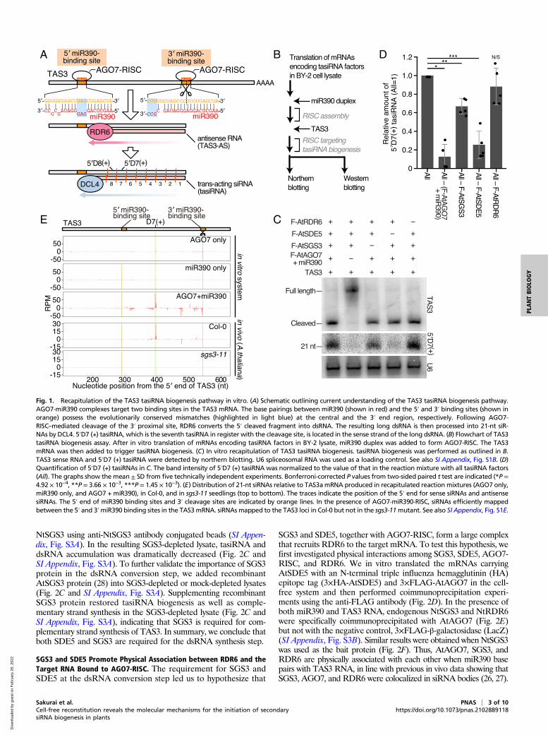
to the extract (SI Appendix, Fig. S1A). However, 5′D7 (+)—themost abundant TAS3 tasiRNA—was not detected by northernblotting (SI Appendix, Fig. S1A), suggesting a lack of tasiRNA factorsin the naïve tobacco cell extract. Indeed, TAS3 mRNA remainedintact without cleavage (SI Appendix, Fig. S1A), suggesting thatthis naïve extract contains insufficient levels of functional AGO7-RISC. We therefore in vitro translated mRNAs carrying tasiRNAfactors fused to Flag tags (AtAGO7, AtSGS3, AtSDE5, andAtRDR6) in the cell-free system. We added miR390 duplexes,which are bound to AtAGO7 to form RISC, before incubationwith TAS3 RNA (Fig. 1 B and C and SI Appendix, Fig. S1B). Wedid not supplement DCLs since previous reports observed en-dogenous DCL activities in BY-2 lysate (46, 47). Supplementa-tion of tasiRNA factors efficiently triggered tasiRNA productionfrom cleaved TAS3 fragments (Fig. 1C and SI Appendix, Fig.S1C). To check the contribution of each supplemented factor, wesystematically omitted miR390 or mRNAs corresponding to eachtasiRNA factor, one at a time, from the reaction. Removal of TAS3mRNA, AtAGO7 mRNA, miR390 duplex, or AtSDE5 mRNAgreatly compromised tasiRNA production (Fig. 1 C and D andSI Appendix, Fig. S1 A and D), so we supplemented naïve BY-2cell extract with all of these factors as we moved forward withfurther analysis.Although we successfully detected two TAS3 tasiRNAs, 5′D7
(+) and 5′D8 (+) (Fig. 1C and SI Appendix, Fig. S1 C and D), itwas still unclear whether the pattern of in vitro expressed tasiRNAswas similar to in vivo production. To ascertain this, we sequencedthe small RNAs produced in our in vitro system and comparedthem to TAS3a tasiRNAs produced in wild-type Arabidopsis seed-lings (Col-0) but lost in sgs3mutant plants (sgs3-11) (Fig. 1E). SmallRNAs generated in vitro mapped between the two miR390 bindingsites, but only in the presence of both miR390 duplex and AGO7(Fig. 1E), suggesting that the mapped small RNAs were bonafide miR390-AGO7-triggered tasiRNAs. Interestingly, althoughrelatively high levels of 24-nt small RNAs mapped to the TAS3aRNA due to robust DCL3-like activity in BY-2 cell extracts (SI Ap-pendix, Fig. S1E) (46, 47), the position of the most prominent 21-nttasiRNA hotspot was common between in vivo and in vitro pro-cessing (Fig. 1E). Taken altogether, the system we developed ac-curately recapitulates TAS3 tasiRNA biogenesis in vitro. We alsotested that this in vitro system also synthesizes dsRNAs and pro-duces tasiRNAs from the one-hit precursor TAS1a that has abinding site for 22-nt miR173-AGO1 RISC (SI Appendix, Fig. S2).
SDE5 and SGS3 Function at the dsRNA Conversion Step.We next soughtto investigate the exact steps at which SDE5 and SGS3 functionin tasiRNA biogenesis. Given that the removal of SGS3 or SDE5mRNA from the reaction had no effect on AGO7-mediated targetcleavage (Fig. 1C), we hypothesized that both factors functionupstream of dsRNA synthesis. However, the antisense strand ofTAS3 RNA was barely detected by northern blotting even in thepresence of exogenous AGO7-RISC and SDE5 (Fig. 2A), presum-ably due to rapid processing of dsRNAs into siRNAs by endogenousDCL activities. To stabilize dsRNA intermediates so that we coulddetect them and resolve the role of SGS3 and SDE5, we addedFlock House virus (FHV) B2 protein, which is known to inhibitdsRNA processing by DCLs (48–50) (Fig. 2B). Addition of FHVB2 protein strongly inhibited tasiRNA processing, leading to ac-cumulation of dsRNA intermediates (Fig. 2 A and B), while havingno effect on RISC-mediated target cleavage. The size of the anti-sense strand is in line with a previous report, having the sequencefrom the third nucleotide of the 3′ cleaved site to the one nucleo-tide upstream of the 3′ end of the 5′ miR390 binding site (Fig. 2B)(51). FHV B2 exposure in the absence of SDE5 supplementationshowed decreased accumulation of dsRNA intermediates (Fig. 2A),indicating that SDE5 functions at the dsRNA conversion step. Sinceendogenous SGS3 in BY-2 cell extract sufficiently promoted tasiRNAproduction (Fig. 1 C and D), we immunodepleted endogenous
2 of 10 | PNAS Sakurai et al.https://doi.org/10.1073/pnas.2102889118 Cell-free reconstitution reveals the molecular mechanisms for the initiation of secondary
siRNA biogenesis in plants
Dow
nloa
ded
by g
uest
on
Feb
ruar
y 26
, 202
2
NtSGS3 using anti-NtSGS3 antibody conjugated beads (SI Appen-dix, Fig. S3A). In the resulting SGS3-depleted lysate, tasiRNA anddsRNA accumulation was dramatically decreased (Fig. 2C andSI Appendix, Fig. S3A). To further validate the importance of SGS3protein in the dsRNA conversion step, we added recombinantAtSGS3 protein (28) into SGS3-depleted or mock-depleted lysates(Fig. 2C and SI Appendix, Fig. S3A). Supplementing recombinantSGS3 protein restored tasiRNA biogenesis as well as comple-mentary strand synthesis in the SGS3-depleted lysate (Fig. 2C andSI Appendix, Fig. S3A), indicating that SGS3 is required for com-plementary strand synthesis of TAS3. In summary, we conclude thatboth SDE5 and SGS3 are required for the dsRNA synthesis step.
SGS3 and SDE5 Promote Physical Association between RDR6 and theTarget RNA Bound to AGO7-RISC. The requirement for SGS3 andSDE5 at the dsRNA conversion step led us to hypothesize that
SGS3 and SDE5, together with AGO7-RISC, form a large complexthat recruits RDR6 to the target mRNA. To test this hypothesis, wefirst investigated physical interactions among SGS3, SDE5, AGO7-RISC, and RDR6. We in vitro translated the mRNAs carryingAtSDE5 with an N-terminal triple influenza hemagglutinin (HA)epitope tag (3×HA-AtSDE5) and 3×FLAG-AtAGO7 in the cell-free system and then performed coimmunoprecipitation experi-ments using the anti-FLAG antibody (Fig. 2D). In the presence ofboth miR390 and TAS3 RNA, endogenous NtSGS3 and NtRDR6were specifically coimmunoprecipitated with AtAGO7 (Fig. 2E)but not with the negative control, 3×FLAG-β-galactosidase (LacZ)(SI Appendix, Fig. S3B). Similar results were obtained when NtSGS3was used as the bait protein (Fig. 2F). Thus, AtAGO7, SGS3, andRDR6 are physically associated with each other when miR390 basepairs with TAS3 RNA, in line with previous in vivo data showing thatSGS3, AGO7, and RDR6 were colocalized in siRNA bodies (26, 27).
BA
C
D
E
Fig. 1. Recapitulation of the TAS3 tasiRNA biogenesis pathway in vitro. (A) Schematic outlining current understanding of the TAS3 tasiRNA biogenesis pathway.AGO7-miR390 complexes target two binding sites in the TAS3 mRNA. The base pairings between miR390 (shown in red) and the 5′ and 3′ binding sites (shown inorange) possess the evolutionarily conserved mismatches (highlighted in light blue) at the central and the 3′ end region, respectively. Following AGO7-RISC–mediated cleavage of the 3′ proximal site, RDR6 converts the 5′ cleaved fragment into dsRNA. The resulting long dsRNA is then processed into 21-nt siR-NAs by DCL4. 5′D7 (+) tasiRNA, which is the seventh tasiRNA in register with the cleavage site, is located in the sense strand of the long dsRNA. (B) Flowchart of TAS3tasiRNA biogenesis assay. After in vitro translation of mRNAs encoding tasiRNA factors in BY-2 lysate, miR390 duplex was added to form AGO7-RISC. The TAS3mRNA was then added to trigger tasiRNA biogenesis. (C) In vitro recapitulation of TAS3 tasiRNA biogenesis. tasiRNA biogenesis was performed as outlined in B.TAS3 sense RNA and 5′D7 (+) tasiRNA were detected by northern blotting. U6 spliceosomal RNA was used as a loading control. See also SI Appendix, Fig. S1B. (D)Quantification of 5′D7 (+) tasiRNAs in C. The band intensity of 5′D7 (+) tasiRNA was normalized to the value of that in the reaction mixture with all tasiRNA factors(All). The graphs show the mean± SD from five technically independent experiments. Bonferroni-corrected P values from two-sided paired t test are indicated (*P =4.92 × 10⁻⁴, **P = 3.66 × 10⁻³, ***P = 1.45 × 10⁻³). (E) Distribution of 21-nt siRNAs relative to TAS3amRNA produced in recapitulated reaction mixtures (AGO7 only,miR390 only, and AGO7 + miR390), in Col-0, and in sgs3-11 seedlings (top to bottom). The traces indicate the position of the 5′ end for sense siRNAs and antisensesiRNAs. The 5′ end of miR390 binding sites and 3′ cleavage sites are indicated by orange lines. In the presence of AGO7-miR390-RISC, siRNAs efficiently mappedbetween the 5′ and 3′miR390 binding sites in the TAS3mRNA. siRNAs mapped to the TAS3 loci in Col-0 but not in the sgs3-11mutant. See also SI Appendix, Fig. S1E.
Sakurai et al. PNAS | 3 of 10Cell-free reconstitution reveals the molecular mechanisms for the initiation of secondarysiRNA biogenesis in plants
https://doi.org/10.1073/pnas.2102889118
PLANTBIOLO
GY
Dow
nloa
ded
by g
uest
on
Feb
ruar
y 26
, 202
2
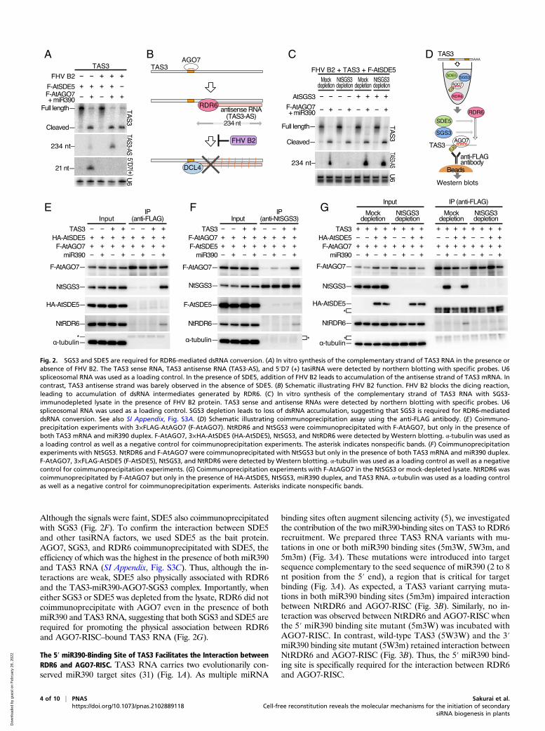
Although the signals were faint, SDE5 also coimmunoprecipitatedwith SGS3 (Fig. 2F). To confirm the interaction between SDE5and other tasiRNA factors, we used SDE5 as the bait protein.AGO7, SGS3, and RDR6 coimmunoprecipitated with SDE5, theefficiency of which was the highest in the presence of both miR390and TAS3 RNA (SI Appendix, Fig. S3C). Thus, although the in-teractions are weak, SDE5 also physically associated with RDR6and the TAS3-miR390-AGO7-SGS3 complex. Importantly, wheneither SGS3 or SDE5 was depleted from the lysate, RDR6 did notcoimmunoprecipitate with AGO7 even in the presence of bothmiR390 and TAS3 RNA, suggesting that both SGS3 and SDE5 arerequired for promoting the physical association between RDR6and AGO7-RISC–bound TAS3 RNA (Fig. 2G).
The 5′ miR390-Binding Site of TAS3 Facilitates the Interaction betweenRDR6 and AGO7-RISC. TAS3 RNA carries two evolutionarily con-served miR390 target sites (31) (Fig. 1A). As multiple miRNA
binding sites often augment silencing activity (5), we investigatedthe contribution of the two miR390-binding sites on TAS3 to RDR6recruitment. We prepared three TAS3 RNA variants with mu-tations in one or both miR390 binding sites (5m3W, 5W3m, and5m3m) (Fig. 3A). These mutations were introduced into targetsequence complementary to the seed sequence of miR390 (2 to 8nt position from the 5′ end), a region that is critical for targetbinding (Fig. 3A). As expected, a TAS3 variant carrying muta-tions in both miR390 binding sites (5m3m) impaired interactionbetween NtRDR6 and AGO7-RISC (Fig. 3B). Similarly, no in-teraction was observed between NtRDR6 and AGO7-RISC whenthe 5′ miR390 binding site mutant (5m3W) was incubated withAGO7-RISC. In contrast, wild-type TAS3 (5W3W) and the 3′miR390 binding site mutant (5W3m) retained interaction betweenNtRDR6 and AGO7-RISC (Fig. 3B). Thus, the 5′ miR390 bind-ing site is specifically required for the interaction between RDR6and AGO7-RISC.
B C DA
E F G
Fig. 2. SGS3 and SDE5 are required for RDR6-mediated dsRNA conversion. (A) In vitro synthesis of the complementary strand of TAS3 RNA in the presence orabsence of FHV B2. The TAS3 sense RNA, TAS3 antisense RNA (TAS3-AS), and 5′D7 (+) tasiRNA were detected by northern blotting with specific probes. U6spliceosomal RNA was used as a loading control. In the presence of SDE5, addition of FHV B2 leads to accumulation of the antisense strand of TAS3 mRNA. Incontrast, TAS3 antisense strand was barely observed in the absence of SDE5. (B) Schematic illustrating FHV B2 function. FHV B2 blocks the dicing reaction,leading to accumulation of dsRNA intermediates generated by RDR6. (C) In vitro synthesis of the complementary strand of TAS3 RNA with SGS3-immunodepleted lysate in the presence of FHV B2 protein. TAS3 sense and antisense RNAs were detected by northern blotting with specific probes. U6spliceosomal RNA was used as a loading control. SGS3 depletion leads to loss of dsRNA accumulation, suggesting that SGS3 is required for RDR6-mediateddsRNA conversion. See also SI Appendix, Fig. S3A. (D) Schematic illustrating coimmunoprecipitation assay using the anti-FLAG antibody. (E) Coimmuno-precipitation experiments with 3×FLAG-AtAGO7 (F-AtAGO7). NtRDR6 and NtSGS3 were coimmunoprecipitated with F-AtAGO7, but only in the presence ofboth TAS3 mRNA and miR390 duplex. F-AtAGO7, 3×HA-AtSDE5 (HA-AtSDE5), NtSGS3, and NtRDR6 were detected by Western blotting. α-tubulin was used asa loading control as well as a negative control for coimmunoprecipitation experiments. The asterisk indicates nonspecific bands. (F) Coimmunoprecipitationexperiments with NtSGS3. NtRDR6 and F-AtAGO7 were coimmunoprecipitated with NtSGS3 but only in the presence of both TAS3 mRNA and miR390 duplex.F-AtAGO7, 3×FLAG-AtSDE5 (F-AtSDE5), NtSGS3, and NtRDR6 were detected by Western blotting. α-tubulin was used as a loading control as well as a negativecontrol for coimmunoprecipitation experiments. (G) Coimmunoprecipitation experiments with F-AtAGO7 in the NtSGS3 or mock-depleted lysate. NtRDR6 wascoimmunoprecipitated by F-AtAGO7 but only in the presence of HA-AtSDE5, NtSGS3, miR390 duplex, and TAS3 RNA. α-tubulin was used as a loading controlas well as a negative control for coimmunoprecipitation experiments. Asterisks indicate nonspecific bands.
4 of 10 | PNAS Sakurai et al.https://doi.org/10.1073/pnas.2102889118 Cell-free reconstitution reveals the molecular mechanisms for the initiation of secondary
siRNA biogenesis in plants
Dow
nloa
ded
by g
uest
on
Feb
ruar
y 26
, 202
2
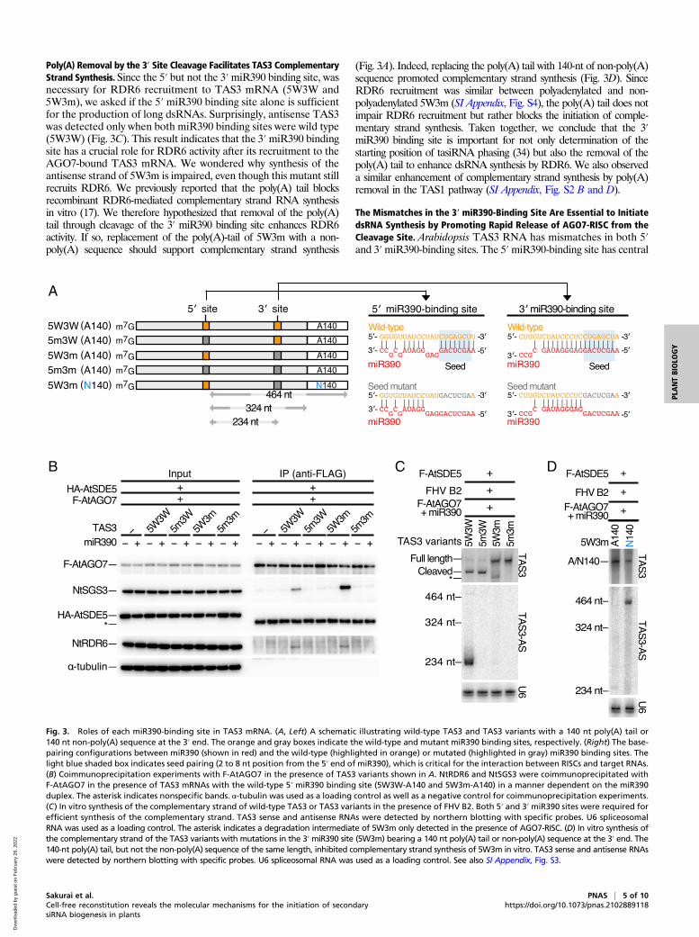
Poly(A) Removal by the 3′ Site Cleavage Facilitates TAS3 ComplementaryStrand Synthesis. Since the 5′ but not the 3′miR390 binding site, wasnecessary for RDR6 recruitment to TAS3 mRNA (5W3W and5W3m), we asked if the 5′ miR390 binding site alone is sufficientfor the production of long dsRNAs. Surprisingly, antisense TAS3was detected only when both miR390 binding sites were wild type(5W3W) (Fig. 3C). This result indicates that the 3′miR390 bindingsite has a crucial role for RDR6 activity after its recruitment to theAGO7-bound TAS3 mRNA. We wondered why synthesis of theantisense strand of 5W3m is impaired, even though this mutant stillrecruits RDR6. We previously reported that the poly(A) tail blocksrecombinant RDR6-mediated complementary strand RNA synthesisin vitro (17). We therefore hypothesized that removal of the poly(A)tail through cleavage of the 3′ miR390 binding site enhances RDR6activity. If so, replacement of the poly(A)-tail of 5W3m with a non-poly(A) sequence should support complementary strand synthesis
(Fig. 3A). Indeed, replacing the poly(A) tail with 140-nt of non-poly(A)sequence promoted complementary strand synthesis (Fig. 3D). SinceRDR6 recruitment was similar between polyadenylated and non-polyadenylated 5W3m (SI Appendix, Fig. S4), the poly(A) tail does notimpair RDR6 recruitment but rather blocks the initiation of comple-mentary strand synthesis. Taken together, we conclude that the 3′miR390 binding site is important for not only determination of thestarting position of tasiRNA phasing (34) but also the removal of thepoly(A) tail to enhance dsRNA synthesis by RDR6. We also observeda similar enhancement of complementary strand synthesis by poly(A)removal in the TAS1 pathway (SI Appendix, Fig. S2 B and D).
The Mismatches in the 3′ miR390-Binding Site Are Essential to InitiatedsRNA Synthesis by Promoting Rapid Release of AGO7-RISC from theCleavage Site. Arabidopsis TAS3 RNA has mismatches in both 5′and 3′miR390-binding sites. The 5′miR390-binding site has central
A
B C D
Fig. 3. Roles of each miR390-binding site in TAS3 mRNA. (A, Left) A schematic illustrating wild-type TAS3 and TAS3 variants with a 140 nt poly(A) tail or140 nt non-poly(A) sequence at the 3′ end. The orange and gray boxes indicate the wild-type and mutant miR390 binding sites, respectively. (Right) The base-pairing configurations between miR390 (shown in red) and the wild-type (highlighted in orange) or mutated (highlighted in gray) miR390 binding sites. Thelight blue shaded box indicates seed pairing (2 to 8 nt position from the 5′ end of miR390), which is critical for the interaction between RISCs and target RNAs.(B) Coimmunoprecipitation experiments with F-AtAGO7 in the presence of TAS3 variants shown in A. NtRDR6 and NtSGS3 were coimmunoprecipitated withF-AtAGO7 in the presence of TAS3 mRNAs with the wild-type 5′ miR390 binding site (5W3W-A140 and 5W3m-A140) in a manner dependent on the miR390duplex. The asterisk indicates nonspecific bands. α-tubulin was used as a loading control as well as a negative control for coimmunoprecipitation experiments.(C) In vitro synthesis of the complementary strand of wild-type TAS3 or TAS3 variants in the presence of FHV B2. Both 5′ and 3′ miR390 sites were required forefficient synthesis of the complementary strand. TAS3 sense and antisense RNAs were detected by northern blotting with specific probes. U6 spliceosomalRNA was used as a loading control. The asterisk indicates a degradation intermediate of 5W3m only detected in the presence of AGO7-RISC. (D) In vitro synthesis ofthe complementary strand of the TAS3 variants with mutations in the 3′miR390 site (5W3m) bearing a 140 nt poly(A) tail or non-poly(A) sequence at the 3′ end. The140-nt poly(A) tail, but not the non-poly(A) sequence of the same length, inhibited complementary strand synthesis of 5W3m in vitro. TAS3 sense and antisense RNAswere detected by northern blotting with specific probes. U6 spliceosomal RNA was used as a loading control. See also SI Appendix, Fig. S3.
Sakurai et al. PNAS | 5 of 10Cell-free reconstitution reveals the molecular mechanisms for the initiation of secondarysiRNA biogenesis in plants
https://doi.org/10.1073/pnas.2102889118
PLANTBIOLO
GY
Dow
nloa
ded
by g
uest
on
Feb
ruar
y 26
, 202
2
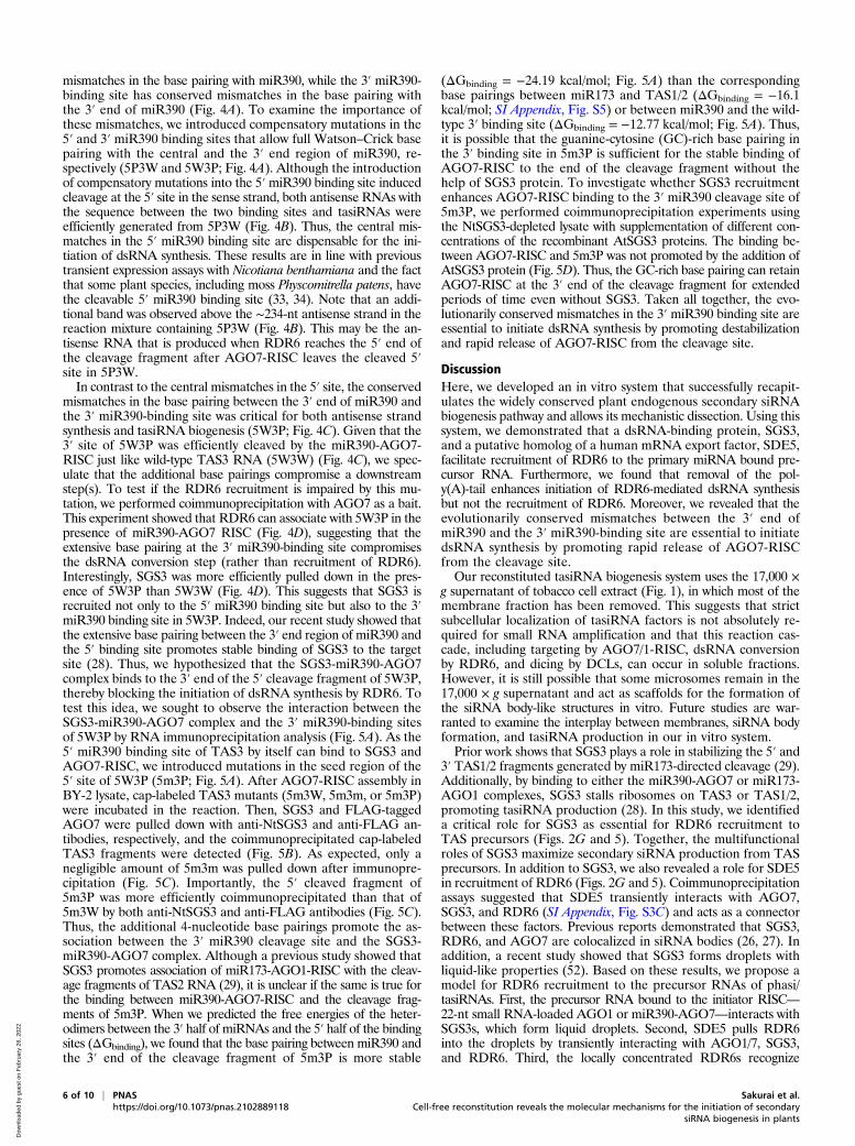
mismatches in the base pairing with miR390, while the 3′ miR390-binding site has conserved mismatches in the base pairing withthe 3′ end of miR390 (Fig. 4A). To examine the importance ofthese mismatches, we introduced compensatory mutations in the5′ and 3′ miR390 binding sites that allow full Watson–Crick basepairing with the central and the 3′ end region of miR390, re-spectively (5P3W and 5W3P; Fig. 4A). Although the introductionof compensatory mutations into the 5′miR390 binding site inducedcleavage at the 5′ site in the sense strand, both antisense RNAs withthe sequence between the two binding sites and tasiRNAs wereefficiently generated from 5P3W (Fig. 4B). Thus, the central mis-matches in the 5′ miR390 binding site are dispensable for the ini-tiation of dsRNA synthesis. These results are in line with previoustransient expression assays with Nicotiana benthamiana and the factthat some plant species, including moss Physcomitrella patens, havethe cleavable 5′ miR390 binding site (33, 34). Note that an addi-tional band was observed above the ∼234-nt antisense strand in thereaction mixture containing 5P3W (Fig. 4B). This may be the an-tisense RNA that is produced when RDR6 reaches the 5′ end ofthe cleavage fragment after AGO7-RISC leaves the cleaved 5′site in 5P3W.In contrast to the central mismatches in the 5′ site, the conserved
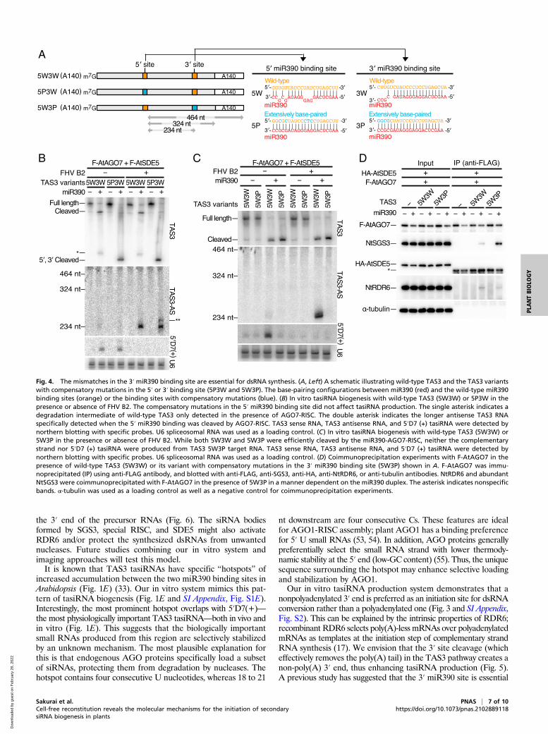
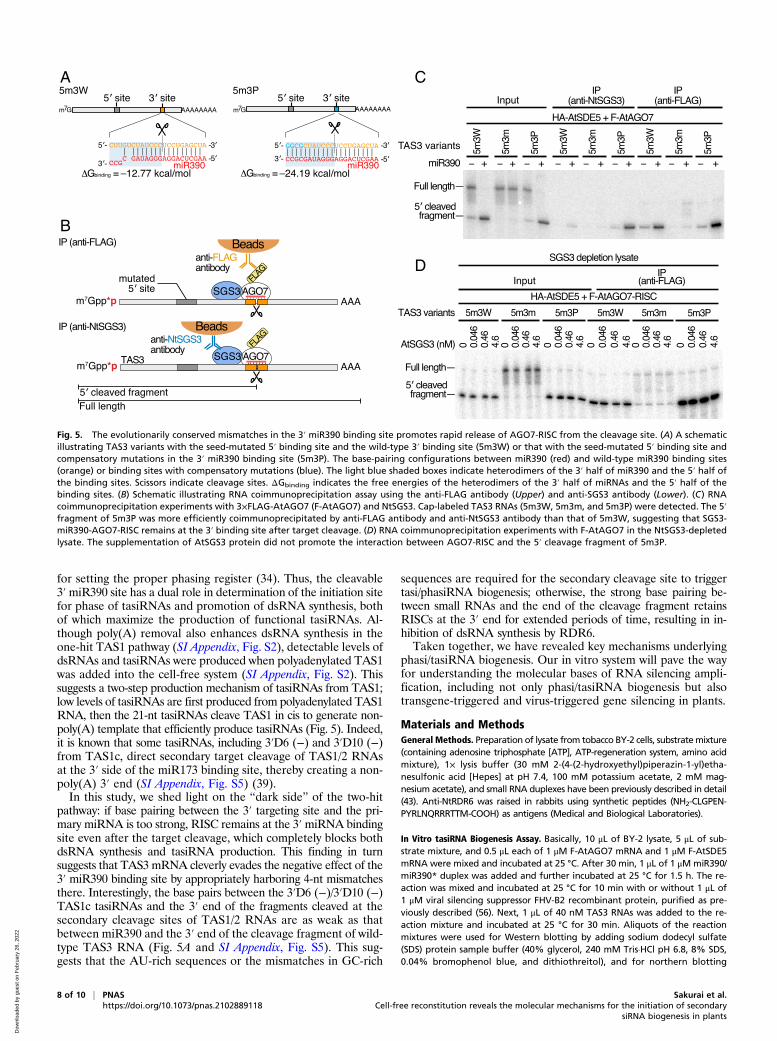
mismatches in the base pairing between the 3′ end of miR390 andthe 3′ miR390-binding site was critical for both antisense strandsynthesis and tasiRNA biogenesis (5W3P; Fig. 4C). Given that the3′ site of 5W3P was efficiently cleaved by the miR390-AGO7-RISC just like wild-type TAS3 RNA (5W3W) (Fig. 4C), we spec-ulate that the additional base pairings compromise a downstreamstep(s). To test if the RDR6 recruitment is impaired by this mu-tation, we performed coimmunoprecipitation with AGO7 as a bait.This experiment showed that RDR6 can associate with 5W3P in thepresence of miR390-AGO7 RISC (Fig. 4D), suggesting that theextensive base pairing at the 3′ miR390-binding site compromisesthe dsRNA conversion step (rather than recruitment of RDR6).Interestingly, SGS3 was more efficiently pulled down in the pres-ence of 5W3P than 5W3W (Fig. 4D). This suggests that SGS3 isrecruited not only to the 5′ miR390 binding site but also to the 3′miR390 binding site in 5W3P. Indeed, our recent study showed thatthe extensive base pairing between the 3′ end region of miR390 andthe 5′ binding site promotes stable binding of SGS3 to the targetsite (28). Thus, we hypothesized that the SGS3-miR390-AGO7complex binds to the 3′ end of the 5′ cleavage fragment of 5W3P,thereby blocking the initiation of dsRNA synthesis by RDR6. Totest this idea, we sought to observe the interaction between theSGS3-miR390-AGO7 complex and the 3′ miR390-binding sitesof 5W3P by RNA immunoprecipitation analysis (Fig. 5A). As the5′ miR390 binding site of TAS3 by itself can bind to SGS3 andAGO7-RISC, we introduced mutations in the seed region of the5′ site of 5W3P (5m3P; Fig. 5A). After AGO7-RISC assembly inBY-2 lysate, cap-labeled TAS3 mutants (5m3W, 5m3m, or 5m3P)were incubated in the reaction. Then, SGS3 and FLAG-taggedAGO7 were pulled down with anti-NtSGS3 and anti-FLAG an-tibodies, respectively, and the coimmunoprecipitated cap-labeledTAS3 fragments were detected (Fig. 5B). As expected, only anegligible amount of 5m3m was pulled down after immunopre-cipitation (Fig. 5C). Importantly, the 5′ cleaved fragment of5m3P was more efficiently coimmunoprecipitated than that of5m3W by both anti-NtSGS3 and anti-FLAG antibodies (Fig. 5C).Thus, the additional 4-nucleotide base pairings promote the as-sociation between the 3′ miR390 cleavage site and the SGS3-miR390-AGO7 complex. Although a previous study showed thatSGS3 promotes association of miR173-AGO1-RISC with the cleav-age fragments of TAS2 RNA (29), it is unclear if the same is true forthe binding between miR390-AGO7-RISC and the cleavage frag-ments of 5m3P. When we predicted the free energies of the heter-odimers between the 3′ half of miRNAs and the 5′ half of the bindingsites (ΔGbinding), we found that the base pairing between miR390 andthe 3′ end of the cleavage fragment of 5m3P is more stable
(ΔGbinding = −24.19 kcal/mol; Fig. 5A) than the correspondingbase pairings between miR173 and TAS1/2 (ΔGbinding = −16.1kcal/mol; SI Appendix, Fig. S5) or between miR390 and the wild-type 3′ binding site (ΔGbinding = −12.77 kcal/mol; Fig. 5A). Thus,it is possible that the guanine-cytosine (GC)-rich base pairing inthe 3′ binding site in 5m3P is sufficient for the stable binding ofAGO7-RISC to the end of the cleavage fragment without thehelp of SGS3 protein. To investigate whether SGS3 recruitmentenhances AGO7-RISC binding to the 3′ miR390 cleavage site of5m3P, we performed coimmunoprecipitation experiments usingthe NtSGS3-depleted lysate with supplementation of different con-centrations of the recombinant AtSGS3 proteins. The binding be-tween AGO7-RISC and 5m3P was not promoted by the addition ofAtSGS3 protein (Fig. 5D). Thus, the GC-rich base pairing can retainAGO7-RISC at the 3′ end of the cleavage fragment for extendedperiods of time even without SGS3. Taken all together, the evo-lutionarily conserved mismatches in the 3′ miR390 binding site areessential to initiate dsRNA synthesis by promoting destabilizationand rapid release of AGO7-RISC from the cleavage site.
DiscussionHere, we developed an in vitro system that successfully recapit-ulates the widely conserved plant endogenous secondary siRNAbiogenesis pathway and allows its mechanistic dissection. Using thissystem, we demonstrated that a dsRNA-binding protein, SGS3,and a putative homolog of a human mRNA export factor, SDE5,facilitate recruitment of RDR6 to the primary miRNA bound pre-cursor RNA. Furthermore, we found that removal of the pol-y(A)-tail enhances initiation of RDR6-mediated dsRNA synthesisbut not the recruitment of RDR6. Moreover, we revealed that theevolutionarily conserved mismatches between the 3′ end ofmiR390 and the 3′ miR390-binding site are essential to initiatedsRNA synthesis by promoting rapid release of AGO7-RISCfrom the cleavage site.Our reconstituted tasiRNA biogenesis system uses the 17,000 ×
g supernatant of tobacco cell extract (Fig. 1), in which most of themembrane fraction has been removed. This suggests that strictsubcellular localization of tasiRNA factors is not absolutely re-quired for small RNA amplification and that this reaction cas-cade, including targeting by AGO7/1-RISC, dsRNA conversionby RDR6, and dicing by DCLs, can occur in soluble fractions.However, it is still possible that some microsomes remain in the17,000 × g supernatant and act as scaffolds for the formation ofthe siRNA body-like structures in vitro. Future studies are war-ranted to examine the interplay between membranes, siRNA bodyformation, and tasiRNA production in our in vitro system.Prior work shows that SGS3 plays a role in stabilizing the 5′ and
3′ TAS1/2 fragments generated by miR173-directed cleavage (29).Additionally, by binding to either the miR390-AGO7 or miR173-AGO1 complexes, SGS3 stalls ribosomes on TAS3 or TAS1/2,promoting tasiRNA production (28). In this study, we identifieda critical role for SGS3 as essential for RDR6 recruitment toTAS precursors (Figs. 2G and 5). Together, the multifunctionalroles of SGS3 maximize secondary siRNA production from TASprecursors. In addition to SGS3, we also revealed a role for SDE5in recruitment of RDR6 (Figs. 2G and 5). Coimmunoprecipitationassays suggested that SDE5 transiently interacts with AGO7,SGS3, and RDR6 (SI Appendix, Fig. S3C) and acts as a connectorbetween these factors. Previous reports demonstrated that SGS3,RDR6, and AGO7 are colocalized in siRNA bodies (26, 27). Inaddition, a recent study showed that SGS3 forms droplets withliquid-like properties (52). Based on these results, we propose amodel for RDR6 recruitment to the precursor RNAs of phasi/tasiRNAs. First, the precursor RNA bound to the initiator RISC—22-nt small RNA-loaded AGO1 or miR390-AGO7—interacts withSGS3s, which form liquid droplets. Second, SDE5 pulls RDR6into the droplets by transiently interacting with AGO1/7, SGS3,and RDR6. Third, the locally concentrated RDR6s recognize
6 of 10 | PNAS Sakurai et al.https://doi.org/10.1073/pnas.2102889118 Cell-free reconstitution reveals the molecular mechanisms for the initiation of secondary
siRNA biogenesis in plants
Dow
nloa
ded
by g
uest
on
Feb
ruar
y 26
, 202
2
the 3′ end of the precursor RNAs (Fig. 6). The siRNA bodiesformed by SGS3, special RISC, and SDE5 might also activateRDR6 and/or protect the synthesized dsRNAs from unwantednucleases. Future studies combining our in vitro system andimaging approaches will test this model.It is known that TAS3 tasiRNAs have specific “hotspots” of
increased accumulation between the two miR390 binding sites inArabidopsis (Fig. 1E) (33). Our in vitro system mimics this pat-tern of tasiRNA biogenesis (Fig. 1E and SI Appendix, Fig. S1E).Interestingly, the most prominent hotspot overlaps with 5′D7(+)—the most physiologically important TAS3 tasiRNA—both in vivo andin vitro (Fig. 1E). This suggests that the biologically importantsmall RNAs produced from this region are selectively stabilizedby an unknown mechanism. The most plausible explanation forthis is that endogenous AGO proteins specifically load a subsetof siRNAs, protecting them from degradation by nucleases. Thehotspot contains four consecutive U nucleotides, whereas 18 to 21
nt downstream are four consecutive Cs. These features are idealfor AGO1-RISC assembly; plant AGO1 has a binding preferencefor 5′ U small RNAs (53, 54). In addition, AGO proteins generallypreferentially select the small RNA strand with lower thermody-namic stability at the 5′ end (low-GC content) (55). Thus, the uniquesequence surrounding the hotspot may enhance selective loadingand stabilization by AGO1.Our in vitro tasiRNA production system demonstrates that a
nonpolyadenylated 3′ end is preferred as an initiation site for dsRNAconversion rather than a polyadenylated one (Fig. 3 and SI Appendix,Fig. S2). This can be explained by the intrinsic properties of RDR6;recombinant RDR6 selects poly(A)-less mRNAs over polyadenylatedmRNAs as templates at the initiation step of complementary strandRNA synthesis (17). We envision that the 3′ site cleavage (whicheffectively removes the poly(A) tail) in the TAS3 pathway creates anon-poly(A) 3′ end, thus enhancing tasiRNA production (Fig. 5).A previous study has suggested that the 3′ miR390 site is essential
A
B C D
Fig. 4. The mismatches in the 3′miR390 binding site are essential for dsRNA synthesis. (A, Left) A schematic illustrating wild-type TAS3 and the TAS3 variantswith compensatory mutations in the 5′ or 3′ binding site (5P3W and 5W3P). The base-pairing configurations between miR390 (red) and the wild-type miR390binding sites (orange) or the binding sites with compensatory mutations (blue). (B) In vitro tasiRNA biogenesis with wild-type TAS3 (5W3W) or 5P3W in thepresence or absence of FHV B2. The compensatory mutations in the 5′ miR390 binding site did not affect tasiRNA production. The single asterisk indicates adegradation intermediate of wild-type TAS3 only detected in the presence of AGO7-RISC. The double asterisk indicates the longer antisense TAS3 RNAspecifically detected when the 5′ miR390 binding was cleaved by AGO7-RISC. TAS3 sense RNA, TAS3 antisense RNA, and 5′D7 (+) tasiRNA were detected bynorthern blotting with specific probes. U6 spliceosomal RNA was used as a loading control. (C) In vitro tasiRNA biogenesis with wild-type TAS3 (5W3W) or5W3P in the presence or absence of FHV B2. While both 5W3W and 5W3P were efficiently cleaved by the miR390-AGO7-RISC, neither the complementarystrand nor 5′D7 (+) tasiRNA were produced from TAS3 5W3P target RNA. TAS3 sense RNA, TAS3 antisense RNA, and 5′D7 (+) tasiRNA were detected bynorthern blotting with specific probes. U6 spliceosomal RNA was used as a loading control. (D) Coimmunoprecipitation experiments with F-AtAGO7 in thepresence of wild-type TAS3 (5W3W) or its variant with compensatory mutations in the 3′ miR390 binding site (5W3P) shown in A. F-AtAGO7 was immu-noprecipitated (IP) using anti-FLAG antibody, and blotted with anti-FLAG, anti-SGS3, anti-HA, anti-NtRDR6, or anti-tubulin antibodies. NtRDR6 and abundantNtSGS3 were coimmunoprecipitated with F-AtAGO7 in the presence of 5W3P in a manner dependent on the miR390 duplex. The asterisk indicates nonspecificbands. α-tubulin was used as a loading control as well as a negative control for coimmunoprecipitation experiments.
Sakurai et al. PNAS | 7 of 10Cell-free reconstitution reveals the molecular mechanisms for the initiation of secondarysiRNA biogenesis in plants
https://doi.org/10.1073/pnas.2102889118
PLANTBIOLO
GY
Dow
nloa
ded
by g
uest
on
Feb
ruar
y 26
, 202
2
for setting the proper phasing register (34). Thus, the cleavable3′miR390 site has a dual role in determination of the initiation sitefor phase of tasiRNAs and promotion of dsRNA synthesis, bothof which maximize the production of functional tasiRNAs. Al-though poly(A) removal also enhances dsRNA synthesis in theone-hit TAS1 pathway (SI Appendix, Fig. S2), detectable levels ofdsRNAs and tasiRNAs were produced when polyadenylated TAS1was added into the cell-free system (SI Appendix, Fig. S2). Thissuggests a two-step production mechanism of tasiRNAs from TAS1;low levels of tasiRNAs are first produced from polyadenylated TAS1RNA, then the 21-nt tasiRNAs cleave TAS1 in cis to generate non-poly(A) template that efficiently produce tasiRNAs (Fig. 5). Indeed,it is known that some tasiRNAs, including 3′D6 (−) and 3′D10 (−)from TAS1c, direct secondary target cleavage of TAS1/2 RNAsat the 3′ side of the miR173 binding site, thereby creating a non-poly(A) 3′ end (SI Appendix, Fig. S5) (39).In this study, we shed light on the “dark side” of the two-hit
pathway: if base pairing between the 3′ targeting site and the pri-mary miRNA is too strong, RISC remains at the 3′miRNA bindingsite even after the target cleavage, which completely blocks bothdsRNA synthesis and tasiRNA production. This finding in turnsuggests that TAS3 mRNA cleverly evades the negative effect of the3′ miR390 binding site by appropriately harboring 4-nt mismatchesthere. Interestingly, the base pairs between the 3′D6 (−)/3′D10 (−)TAS1c tasiRNAs and the 3′ end of the fragments cleaved at thesecondary cleavage sites of TAS1/2 RNAs are as weak as thatbetween miR390 and the 3′ end of the cleavage fragment of wild-type TAS3 RNA (Fig. 5A and SI Appendix, Fig. S5). This sug-gests that the AU-rich sequences or the mismatches in GC-rich
sequences are required for the secondary cleavage site to triggertasi/phasiRNA biogenesis; otherwise, the strong base pairing be-tween small RNAs and the end of the cleavage fragment retainsRISCs at the 3′ end for extended periods of time, resulting in in-hibition of dsRNA synthesis by RDR6.Taken together, we have revealed key mechanisms underlying
phasi/tasiRNA biogenesis. Our in vitro system will pave the wayfor understanding the molecular bases of RNA silencing ampli-fication, including not only phasi/tasiRNA biogenesis but alsotransgene-triggered and virus-triggered gene silencing in plants.
Materials and MethodsGeneral Methods. Preparation of lysate from tobacco BY-2 cells, substratemixture(containing adenosine triphosphate [ATP], ATP-regeneration system, amino acidmixture), 1× lysis buffer (30 mM 2-(4-(2-hydroxyethyl)piperazin-1-yl)etha-nesulfonic acid [Hepes] at pH 7.4, 100 mM potassium acetate, 2 mM mag-nesium acetate), and small RNA duplexes have been previously described in detail(43). Anti-NtRDR6 was raised in rabbits using synthetic peptides (NH2-CLGPEN-PYRLNQRRRTTM-COOH) as antigens (Medical and Biological Laboratories).
In Vitro tasiRNA Biogenesis Assay. Basically, 10 μL of BY-2 lysate, 5 μL of sub-strate mixture, and 0.5 μL each of 1 μM F-AtAGO7 mRNA and 1 μM F-AtSDE5mRNA were mixed and incubated at 25 °C. After 30 min, 1 μL of 1 μM miR390/miR390* duplex was added and further incubated at 25 °C for 1.5 h. The re-action was mixed and incubated at 25 °C for 10 min with or without 1 μL of1 μM viral silencing suppressor FHV-B2 recombinant protein, purified as pre-viously described (56). Next, 1 μL of 40 nM TAS3 RNAs was added to the re-action mixture and incubated at 25 °C for 30 min. Aliquots of the reactionmixtures were used for Western blotting by adding sodium dodecyl sulfate(SDS) protein sample buffer (40% glycerol, 240 mM Tris·HCl pH 6.8, 8% SDS,0.04% bromophenol blue, and dithiothreitol), and for northern blotting
A
B
C
D
Fig. 5. The evolutionarily conserved mismatches in the 3′ miR390 binding site promotes rapid release of AGO7-RISC from the cleavage site. (A) A schematicillustrating TAS3 variants with the seed-mutated 5′ binding site and the wild-type 3′ binding site (5m3W) or that with the seed-mutated 5′ binding site andcompensatory mutations in the 3′ miR390 binding site (5m3P). The base-pairing configurations between miR390 (red) and wild-type miR390 binding sites(orange) or binding sites with compensatory mutations (blue). The light blue shaded boxes indicate heterodimers of the 3′ half of miR390 and the 5′ half ofthe binding sites. Scissors indicate cleavage sites. ΔGbinding indicates the free energies of the heterodimers of the 3′ half of miRNAs and the 5′ half of thebinding sites. (B) Schematic illustrating RNA coimmunoprecipitation assay using the anti-FLAG antibody (Upper) and anti-SGS3 antibody (Lower). (C) RNAcoimmunoprecipitation experiments with 3×FLAG-AtAGO7 (F-AtAGO7) and NtSGS3. Cap-labeled TAS3 RNAs (5m3W, 5m3m, and 5m3P) were detected. The 5′fragment of 5m3P was more efficiently coimmunoprecipitated by anti-FLAG antibody and anti-NtSGS3 antibody than that of 5m3W, suggesting that SGS3-miR390-AGO7-RISC remains at the 3′ binding site after target cleavage. (D) RNA coimmunoprecipitation experiments with F-AtAGO7 in the NtSGS3-depletedlysate. The supplementation of AtSGS3 protein did not promote the interaction between AGO7-RISC and the 5′ cleavage fragment of 5m3P.
8 of 10 | PNAS Sakurai et al.https://doi.org/10.1073/pnas.2102889118 Cell-free reconstitution reveals the molecular mechanisms for the initiation of secondary
siRNA biogenesis in plants
Dow
nloa
ded
by g
uest
on
Feb
ruar
y 26
, 202
2
after deproteinization with proteinase K (Nacalai Tesque, Inc.) and ethanolprecipitation followed by dissolving the pellet in formamide dye (49% form-amide, 5 mM ethylenediamine tetraacetic acid [EDTA], 0.01% xylene cyanol,and 0.01% bromophenol blue). For Fig. 1 C and D and SI Appendix, Fig. S1B,12 μL of BY-2 lysate, 6 μL of substrate mixture, 0.6 μL of 1 μM F-AtAGO7mRNA,0.6 μL of 1 μM F-AtSDE5 mRNA, 0.5 μL of 0.25 μM F-AtSGS3 mRNA, and 1 μL of0.5 μM F-AtRDR6 mRNA were used.
Coimmunoprecipitation Assay. The anti-FLAG antibody (Sigma) was immobilizedusing Dynabeads protein G (Invitrogen) in 1× lysis buffer and rotated at 4 °C for1 h. After immobilization, the beads were washed 3 times with lysis washbuffer containing 200 mM NaCl and 1% Triton X-100 [polyoxyethylene (10)octylphenyl ether, Wako]. Afterward, the tasiRNA biogenesis assay reactionmixture was added to the beads and rotated at 4 °C for 1 h. The beads werewashed with lysis wash buffer three times, and finally, proteins were resus-pended in SDS protein sample buffer. For SGS3 immunoprecipitation (Fig. 2F),
the anti-NtSGS3 antibody (29) was added directly to the tasiRNA biogenesisreaction mixture and incubated for 30 min on ice. After incubation, Dynabeadsprotein G (Invitrogen), washed with lysis wash buffer, was added to the reac-tion mixture and rotated at 4 °C for 30 min. The wash step was performed asoutlined. For RNA immunoprecipitation, cap-labeled TAS3 target RNAs wereused (Fig. 4D). After wash step, the beads were incubated with 1× lysis buffercontaining proteinase K (200 ng/uL) at 55 °C for 10 min. Finally, the sampleswere resuspended in the same volume of formamide dye (10 mM EDTA pH 8.0,98% [wt/vol] deionized formamide, 0.025% [wt/vol] xylene cyanol, and 0.025%bromophenol blue). After incubation at 95 °C for 2 min, the coimmunopreci-pitated RNAs were separated on an 5% polyacrylamide denaturing gel.
Immunodepletion of Endogenous SGS3. Immunodepletion of NtSGS3 from BY-2 lysate was performed as previously described (28).
Western Blot Assay. The reactions, which were resuspended in 1× SDS proteinsample buffer, were heated at 95 °C for 5 min. After electrophoresis, theproteins were transferred onto polyvinylidene difluoride membrane (Merck)via a semidry transfer method. The membrane was incubated with blockingbuffer (1% skim milk and 1× Tris buffered saline with Tween 20 [TBS-T]) atroom temperature for 1 h and supplemented with anti-FLAG antibodies(Sigma, #F1804; 1:10,000 dilution) or anti-HA antibodies (abcam, #ab130275;1:10,000 dilution), anti-tubulin antibodies (Sigma, #T6074; 1:10,000 dilution),anti-NtSGS3 antibodies (29) (1:10,000 dilution), and anti-NtRDR6 antibodies(1:5,000 dilution) at 4 °C overnight. The membrane was washed with 1× TBS-T and then incubated with blocking buffer (1% skim milk and 1× TBS-T) withanti-mouse immunoglobulin G (IgG)–horseradish peroxidase (HRP) antibodies(MBL, #330; 1:25,000 dilution) or anti-rabbit IgG-HRP antibodies (Jackson,#111-035-003; 1:20,000 dilution) at room temperature for 1 h. After washingwith 1× TBS-T, the membrane was treated with Immobilon Western Chemi-luminescent HRP Substrate (Millipore). Images were acquired using the Imager600 (Amersham) or FUSION FX (VILBER).
Preparation of Probes for Northern Blot Assay. DNA oligo probes (SI Appendix,Table S4) were phosphorylated using T4 polynucleotide kinase (TaKaRa)with [γ-32P]-ATP. Multiple DNA oligos (SI Appendix, Table S4) were mixedand radiolabeled as described for the detection of sense, antisense TAS3a,and antisense TAS1a.
Northern Blot Assay. To detect U6 spliceosomal RNA and tasiRNAs, RNAs wereseparated on an 18% polyacrylamide denaturing gel and transferred onto aHybond-N membrane (GE Healthcare) using semidry transfer. The 1-ethyl-3-(3-dimethylaminopropyl)carbodiimide (EDC) cross-linking was used to fixRNA to the Hybond-N membrane, as described previously (57). Hybond-Nmembrane was placed onto filter paper soaked in EDC solution and incu-bated at 60 °C for 1 h. To detect TAS3 mRNA, antisense TAS3 mRNA, TAS1mRNA, and antisense TAS1 mRNA, a 5% polyacrylamide denaturing gel and aHybond-N+ membrane (GE Healthcare) were used. RNAs were fixed to theHybond-N+ membrane by ultraviolet cross-linking. Membranes were placed ina hybridization bottle with 10 mL of PerfectHyb Plus Hybridization buffer(Sigma). The bottle was then preincubated at 42 °C for more than 5 min in thehybridization oven. After preincubation, a radiolabeled probe was hybridizedwith membranes overnight. The membrane was washed with low stringencybuffer (2× saline-sodium citrate [SSC] and 0.1% SDS) at room temperature formore than 5 min and then washed with high stringency buffer (0.1× SSC and0.1% SDS) at the hybridization temperature for 20 min. Images were acquiredusing the Typhoon FLA 7000 IP (GE Healthcare). The markers for northernblotting used in this study are listed in SI Appendix, Table S5.
Small RNA-seq. Total RNA was extracted from 3-d-old Col-0 and sgs3-11 (21)Arabidopsis seedlings and BY-2 lysate with TRIzol reagent. Followed by gelpurification, 19 to 28 nt length small RNAs were gel extracted from totalRNAs. Small RNA libraries were prepared by using NEBNext Multiplex SmallRNA Library Prep Set for Illumina (NEB) and analyzed by Illumina HiSEq 4000.
Sequence Analysis of tasiRNAs. After removal of adaptor sequences by cutadapt(58), small RNA sequence reads of 20 to 29 nt length were mapped to theTAS3a sequence used in the tasiRNA biogenesis assay using the FASTX-Toolkit(http://hannonlab.cshl.edu/fastx_toolkit/) and Bowtie (59) allowing for up toone mismatch. Sequence Alignment Map (SAM) files were converted to BAMfiles using SAMtools (60) and then to BED files with BEDTools (61). Processeddata were transferred to R, and length distribution and the 5′-end positionfor each siRNA were mapped onto the TAS3 mRNA using the ggplot2package.
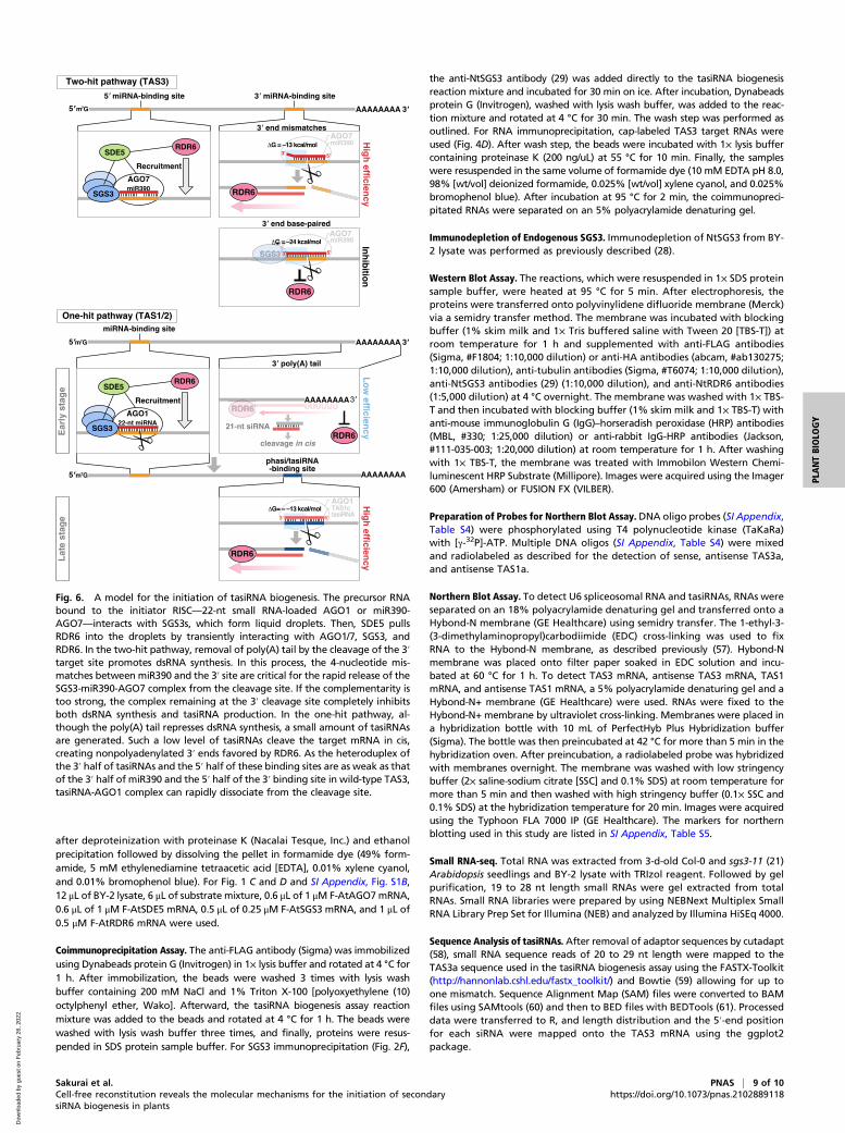
Fig. 6. A model for the initiation of tasiRNA biogenesis. The precursor RNAbound to the initiator RISC—22-nt small RNA-loaded AGO1 or miR390-AGO7—interacts with SGS3s, which form liquid droplets. Then, SDE5 pullsRDR6 into the droplets by transiently interacting with AGO1/7, SGS3, andRDR6. In the two-hit pathway, removal of poly(A) tail by the cleavage of the 3′target site promotes dsRNA synthesis. In this process, the 4-nucleotide mis-matches between miR390 and the 3′ site are critical for the rapid release of theSGS3-miR390-AGO7 complex from the cleavage site. If the complementarity istoo strong, the complex remaining at the 3′ cleavage site completely inhibitsboth dsRNA synthesis and tasiRNA production. In the one-hit pathway, al-though the poly(A) tail represses dsRNA synthesis, a small amount of tasiRNAsare generated. Such a low level of tasiRNAs cleave the target mRNA in cis,creating nonpolyadenylated 3′ ends favored by RDR6. As the heteroduplex ofthe 3′ half of tasiRNAs and the 5′ half of these binding sites are as weak as thatof the 3′ half of miR390 and the 5′ half of the 3′ binding site in wild-type TAS3,tasiRNA-AGO1 complex can rapidly dissociate from the cleavage site.
Sakurai et al. PNAS | 9 of 10Cell-free reconstitution reveals the molecular mechanisms for the initiation of secondarysiRNA biogenesis in plants
https://doi.org/10.1073/pnas.2102889118
PLANTBIOLO
GY
Dow
nloa
ded
by g
uest
on
Feb
ruar
y 26
, 202
2
Prediction of the Free Energies of Heterodimers. The free energies of heter-odimers of the 3′ half of small RNAs and the 5′ half of binding sites werepredicted at 25 °C via the Vienna RNA Websuite (62).
Data Availability. The sequencing data reported in this paper are publiclyavailable in DNA Data Bank of Japan (DDBJ; http://www.ddbj.nig.ac.jp/) underthe accession numbers DRA009601 for in vitro data and DRA009602 forin vivo data.
ACKNOWLEDGMENTS. We thank Manabu Yoshikawa for sharing unpub-lished data and providing anti-NtSGS3 and anti-NtRDR6, Mariko Watanabe
for production of FHV B2 protein, Kaori Kiyokawa for experimental assistance,and Life Science Editors for editorial assistance. We also thank all the membersof the Y.T. laboratory for discussion and critical comments on the manuscript.This work was supported in part by JST, PRESTO (Grant JPMJPR18K2 to H.-o.I.),Grant-in-Aid for Young Scientists (A) (Grant 16H06159 to H.-o.I.), Grant-in-Aidfor Scientific Research on Innovative Areas (“Nascent-chain Biology”) (Grant26116003 to H.-o.I.), Grant-in-Aid for Challenging Exploratory Research (Grant15K14444 to H.-o.I.), Grant-in-Aid for Japan Society for the Promotion of Sci-ence (JSPS) Fellows 16J07290 (to K.B.), and Grant-in-Aid for JSPS Fellows20J11529 (to Y.S.). DNA libraries were sequenced by the Vincent J. Coates Ge-nomics Sequencing Laboratory at University of California, Berkeley, supportedby an NIH S10 OD018174 Instrumentation Grant.
1. M. Ghildiyal, P. D. Zamore, Small silencing RNAs: An expanding universe. Nat. Rev.Genet. 10, 94–108 (2009).
2. N. G. Bologna, O. Voinnet, The diversity, biogenesis, and activities of endogenoussilencing small RNAs in Arabidopsis. Annu. Rev. Plant Biol. 65, 473–503 (2014).
3. H. Kobayashi, Y. Tomari, RISC assembly: Coordination between small RNAs and Ar-gonaute proteins. Biochim. Biophys. Acta 1859, 71–81 (2016).
4. S. L. Ameres, P. D. Zamore, Diversifying microRNA sequence and function. Nat. Rev.Mol. Cell Biol. 14, 475–488 (2013).
5. D. P. Bartel, MicroRNAs: Target recognition and regulatory functions. Cell 136,215–233 (2009).
6. H. O. Iwakawa, Y. Tomari, The functions of MicroRNAs: mRNA decay and translationalrepression. Trends Cell Biol. 25, 651–665 (2015).
7. D. C. Baulcombe, Molecular biology. Amplified silencing. Science 315, 199–200 (2007).8. T. Dalmay, A. Hamilton, S. Rudd, S. Angell, D. C. Baulcombe, An RNA-dependent RNA
polymerase gene in Arabidopsis is required for posttranscriptional gene silencingmediated by a transgene but not by a virus. Cell 101, 543–553 (2000).
9. Y. Dang, Q. Yang, Z. Xue, Y. Liu, RNA interference in fungi: Pathways, functions, andapplications. Eukaryot. Cell 10, 1148–1155 (2011).
10. H. Nakayashiki, N. Kadotani, S. Mayama, Evolution and diversification of RNA si-lencing proteins in fungi. J. Mol. Evol. 63, 127–135 (2006).
11. T. Sijen et al., On the role of RNA amplification in dsRNA-triggered gene silencing.Cell 107, 465–476 (2001).
12. S. E. Castel, R. A. Martienssen, RNA interference in the nucleus: Roles for small RNAs intranscription, epigenetics and beyond. Nat. Rev. Genet. 14, 100–112 (2013).
13. J. T. Cuperus et al., Unique functionality of 22-nt miRNAs in triggering RDR6-dependent siRNA biogenesis from target transcripts in Arabidopsis. Nat. Struct.Mol. Biol. 17, 997–1003 (2010).
14. H. M. Chen et al., 22-Nucleotide RNAs trigger secondary siRNA biogenesis in plants.Proc. Natl. Acad. Sci. U.S.A. 107, 15269–15274 (2010).
15. Y. Liu, C. Teng, R. Xia, B. C. Meyers, PhasiRNAs in plants: Their biogenesis, genicsources, and roles in stress responses, development, and reproduction. Plant Cell 32,3059–3080 (2020).
16. J. Curaba, X. Chen, Biochemical activities of Arabidopsis RNA-dependent RNA poly-merase 6. J. Biol. Chem. 283, 3059–3066 (2008).
17. K. Baeg, H. O. Iwakawa, Y. Tomari, The poly(A) tail blocks RDR6 from converting selfmRNAs into substrates for gene silencing. Nat. Plants 3, 17036 (2017).
18. M. Yoshikawa, A. Peragine, M. Y. Park, R. S. Poethig, A pathway for the biogenesis oftrans-acting siRNAs in Arabidopsis. Genes Dev. 19, 2164–2175 (2005).
19. Z. Xie, E. Allen, A. Wilken, J. C. Carrington, DICER-LIKE 4 functions in trans-actingsmall interfering RNA biogenesis and vegetative phase change in Arabidopsis thali-ana. Proc. Natl. Acad. Sci. U.S.A. 102, 12984–12989 (2005).
20. I. Hernandez-Pinzon et al., SDE5, the putative homologue of a human mRNA exportfactor, is required for transgene silencing and accumulation of trans-acting endog-enous siRNA. Plant J. 50, 140–148 (2007).
21. A. Peragine, M. Yoshikawa, G. Wu, H. L. Albrecht, R. S. Poethig, SGS3 and SGS2/SDE1/RDR6 are required for juvenile development and the production of trans-actingsiRNAs in Arabidopsis. Genes Dev. 18, 2368–2379 (2004).
22. F. Vazquez et al., Endogenous trans-acting siRNAs regulate the accumulation ofArabidopsis mRNAs. Mol. Cell 16, 69–79 (2004).
23. V. Gasciolli, A. C. Mallory, D. P. Bartel, H. Vaucheret, Partially redundant functions ofArabidopsis DICER-like enzymes and a role for DCL4 in producing trans-acting siRNAs.Curr. Biol. 15, 1494–1500 (2005).
24. T. Elmayan et al., A neomorphic sgs3 allele stabilizing miRNA cleavage products re-veals that SGS3 acts as a homodimer. FEBS J. 276, 835–844 (2009).
25. E. Glick et al., Interaction with host SGS3 is required for suppression of RNA silencing bytomato yellow leaf curl virus V2 protein. Proc. Natl. Acad. Sci. U.S.A. 105, 157–161 (2008).
26. N. Kumakura et al., SGS3 and RDR6 interact and colocalize in cytoplasmic SGS3/RDR6-bodies. FEBS Lett. 583, 1261–1266 (2009).
27. V. Jouannet et al., Cytoplasmic Arabidopsis AGO7 accumulates in membrane-associatedsiRNA bodies and is required for ta-siRNA biogenesis. EMBO J. 31, 1704–1713 (2012).
28. H. O. Iwakawa et al., Ribosome stalling caused by the Argonaute-microRNA-SGS3 com-plex regulates the production of secondary siRNAs in plants. Cell Rep. 35, 109300 (2021).
29. M. Yoshikawa et al., 3′ fragment of miR173-programmed RISC-cleaved RNA is pro-tected from degradation in a complex with RISC and SGS3. Proc. Natl. Acad. Sci. U.S.A.110, 4117–4122 (2013).
30. M. Yoshikawa et al., A short open reading frame encompassing the microRNA173target site plays a role in trans-acting small interfering RNA biogenesis. Plant Physiol.171, 359–368 (2016).
31. R. Xia, J. Xu, B. C. Meyers, The emergence, evolution, and diversification of themiR390-TAS3-ARF pathway in land plants. Plant Cell 29, 1232–1247 (2017).
32. M. D. Howell et al., Genome-wide analysis of the RNA-DEPENDENT RNA POLYMER-ASE6/DICER-LIKE4 pathway in Arabidopsis reveals dependency on miRNA- andtasiRNA-directed targeting. Plant Cell 19, 926–942 (2007).
33. M. J. Axtell, C. Jan, R. Rajagopalan, D. P. Bartel, A two-hit trigger for siRNA biogenesisin plants. Cell 127, 565–577 (2006).
34. F. F. de Felippes, A. Marchais, A. Sarazin, S. Oberlin, O. Voinnet, A single miR390targeting event is sufficient for triggering TAS3-tasiRNA biogenesis in Arabidopsis.Nucleic Acids Res. 45, 5539–5554 (2017).
35. E. Allen, Z. Xie, A. M. Gustafson, J. C. Carrington, microRNA-directed phasing duringtrans-acting siRNA biogenesis in plants. Cell 121, 207–221 (2005).
36. N. Fahlgren et al., Regulation of AUXIN RESPONSE FACTOR3 by TAS3 ta-siRNA affectsdevelopmental timing and patterning in Arabidopsis. Curr. Biol. 16, 939–944 (2006).
37. Z. Luo, Z. Chen, Improperly terminated, unpolyadenylated mRNA of sense transgenes istargeted by RDR6-mediated RNA silencing in Arabidopsis. Plant Cell 19, 943–958 (2007).
38. F. F. de Felippes et al., The key role of terminators on the expression and post-transcriptional gene silencing of transgenes. Plant J. 104, 96–112 (2020).
39. R. Rajeswaran et al., Sequencing of RDR6-dependent double-stranded RNAs revealsnovel features of plant siRNA biogenesis. Nucleic Acids Res. 40, 6241–6254 (2012).
40. E. Marin et al., miR390, Arabidopsis TAS3 tasiRNAs, and their AUXIN RESPONSEFACTOR targets define an autoregulatory network quantitatively regulating lateralroot growth. Plant Cell 22, 1104–1117 (2010).
41. Y. Endo, H. O. Iwakawa, Y. Tomari, Arabidopsis ARGONAUTE7 selects miR390 throughmultiple checkpoints during RISC assembly. EMBO Rep. 14, 652–658 (2013).
42. H. O. Iwakawa, Y. Tomari, Molecular insights into microRNA-mediated translationalrepression in plants. Mol. Cell 52, 591–601 (2013).
43. Y. Tomari, H. O. Iwakawa, In vitro analysis of ARGONAUTE-mediated target cleavageand translational repression in plants. Methods Mol. Biol. 1640, 55–71 (2017).
44. T. Iki et al., In vitro assembly of plant RNA-induced silencing complexes facilitated bymolecular chaperone HSP90. Mol. Cell 39, 282–291 (2010).
45. K. Komoda, S. Naito, M. Ishikawa, Replication of plant RNA virus genomes in a cell-free extract of evacuolated plant protoplasts. Proc. Natl. Acad. Sci. U.S.A. 101,1863–1867 (2004).
46. T. Iki, M. A. Tschopp, O. Voinnet, Biochemical and genetic functional dissection of theP38 viral suppressor of RNA silencing. RNA 23, 639–654 (2017).
47. J. Schuck, T. Gursinsky, V. Pantaleo, J. Burgyán, S. E. Behrens, AGO/RISC-mediated an-tiviral RNA silencing in a plant in vitro system. Nucleic Acids Res. 41, 5090–5103 (2013).
48. J. A. Chao et al., Dual modes of RNA-silencing suppression by Flock House virusprotein B2. Nat. Struct. Mol. Biol. 12, 952–957 (2005).
49. J. K. Seo, S. J. Kwon, A. L. Rao, Molecular dissection of Flock house virus protein B2reveals that electrostatic interactions between N-terminal domains of B2 monomersare critical for dimerization. Virology 432, 296–305 (2012).
50. G. Singh et al., Suppression of RNA silencing by Flock house virus B2 protein is mediatedthrough its interaction with the PAZ domain of Dicer. FASEB J. 23, 1845–1857 (2009).
51. R. Rajeswaran, M. M. Pooggin, RDR6-mediated synthesis of complementary RNA is ter-minated by miRNA stably bound to template RNA. Nucleic Acids Res. 40, 594–599 (2012).
52. E. Y. Kim et al., Ribosome stalling and SGS3 phase separation prime the epigeneticsilencing of transposons. Nat. Plants 7, 303–309 (2021).
53. S. Mi et al., Sorting of small RNAs into Arabidopsis argonaute complexes is directed bythe 5′ terminal nucleotide. Cell 133, 116–127 (2008).
54. A. Takeda, S. Iwasaki, T. Watanabe, M. Utsumi, Y. Watanabe, The mechanism se-lecting the guide strand from small RNA duplexes is different among argonauteproteins. Plant Cell Physiol. 49, 493–500 (2008).
55. A. Khvorova, A. Reynolds, S. D. Jayasena, Functional siRNAs and miRNAs exhibitstrand bias. Cell 115, 209–216 (2003).
56. M. Watanabe, H. O. Iwakawa, H. Tadakuma, Y. Tomari, Biochemical and single-molecule analyses of the RNA silencing suppressing activity of CrPV-1A. NucleicAcids Res. 45, 10837–10844 (2017).
57. G. S. Pall, A. J. Hamilton, Improved northern blot method for enhanced detection ofsmall RNA. Nat. Protoc. 3, 1077–1084 (2008).
58. M. Martin, Cutadapt removes adapter sequences from high-throughput sequencingreads. EMBnet. J. 17, 10–12 (2011).
59. B. Langmead, C. Trapnell, M. Pop, S. L. Salzberg, Ultrafast and memory-efficient align-ment of short DNA sequences to the human genome. Genome Biol. 10, R25 (2009).
60. H. Li et al.; 1000 Genome Project Data Processing Subgroup, The sequence alignment/map format and SAMtools. Bioinformatics 25, 2078–2079 (2009).
61. A. R. Quinlan, I. M. Hall, BEDTools: A flexible suite of utilities for comparing genomicfeatures. Bioinformatics 26, 841–842 (2010).
62. A. R. Gruber, R. Lorenz, S. H. Bernhart, R. Neuböck, I. L. Hofacker, The Vienna RNAwebsuite. Nucleic Acids Res. 36, W70–W74 (2008).
10 of 10 | PNAS Sakurai et al.https://doi.org/10.1073/pnas.2102889118 Cell-free reconstitution reveals the molecular mechanisms for the initiation of secondary
siRNA biogenesis in plants
Dow
nloa
ded
by g
uest
on
Feb
ruar
y 26
, 202
2
Top Related