Université de Montréal COMPARISON OF A … · université de montréal comparison of a leukocyte...
101
Université de Montréal COMPARISON OF A LEUKOCYTE ESTERASE TEST WITH ENDOMETRIAL CYTOLOGY FOR THE DIAGNOSIS OF SUBCLINICAL ENDOMETRITIS AND CORRELATION WITH FIRST SERVICE PREGNANCY RATE IN POSTPARTUM HOLSTEIN COWS par GABRIEL BORGES COUTO Département de sciences cliniques Faculté de médecine vétérinaire Mémoire présenté à la Faculté de médecine vétérinaire en vue de l’obtention du grade Maître ès sciences (M.Sc.) en sciences vétérinaires option reproduction Novembre, 2009 © Gabriel Borges Couto, 2009
Transcript of Université de Montréal COMPARISON OF A … · université de montréal comparison of a leukocyte...
Université de Montréal
COMPARISON OF A LEUKOCYTE ESTERASE TEST WITH ENDOMETRIAL CYTOLOGY FOR THE DIAGNOSIS OF SUBCLINICAL
ENDOMETRITIS AND CORRELATION WITH FIRST SERVICE PREGNANCY RATE IN POSTPARTUM HOLSTEIN COWS
par
GABRIEL BORGES COUTO
Département de sciences cliniques
Faculté de médecine vétérinaire
Mémoire présenté à la Faculté de médecine vétérinaire
en vue de l’obtention du grade
Maître ès sciences (M.Sc.)
en sciences vétérinaires
option reproduction
Novembre, 2009
© Gabriel Borges Couto, 2009
ii
Faculté de médecine vétérinaire
Ce mémoire intitulé
COMPARISON OF A LEUKOCYTE ESTERASE TEST WITH ENDOMETRIAL CYTOLOGY FOR THE DIAGNOSIS OF SUBCLINICAL
ENDOMETRITIS AND CORRELATION WITH FIRST SERVICE PREGNANCY RATE IN POSTPARTUM HOLSTEIN COWS
présenté par
GABRIEL BORGES COUTO
a été évalué par un jury composé des personnes suivantes
Paul D. Carrière, président rapporteur
Réjean C. Lefebvre, directeur de recherche
Denis H. Vaillancourt, codirecteur
Sylvain Nichols, membre du jury
iii
RÉSUMÉ
L’objectif de la présente étude était d’évaluer un test d’estérase
leucocytaire (LE) pour le diagnostic de l’endométrite subclinique chez les
vaches Holstein en période postpartum. Les tests effectués à partir
d’échantillons provenant soit de l’endomètre (UtLE) ou du col utérin (CxLE)
ont été comparés à la cytologie endométriale (CE). Par ailleurs, deux méthodes
d’évaluation des lames ont été comparées. Deux cent quatre vingt-cinq vaches
Holstein de 5 troupeaux laitiers commerciaux ont été évaluées entre 21 et 47
jours en lait (JEL). Soixante sept vaches ont été diagnostiquées avec une
endométrite clinique suite à un examen transrectal et vaginoscopique et ont été
exclues de l’étude. Deux cent dix-huit vaches ont eu des prélèvements pour la
CE et le test LE. La fonction ovarienne a été déterminée à la palpation
transrectale. La banque de données utilisée pour chacune des vaches a été
effectuée à partir du logiciel DSA (Dossier de Santé Animale) laitier. Le
pourcentage de neutrophiles était significativement corrélé avec les scores de
LE utérin et cervical. L’activité de CxLE et UtLE diminuait significativement
avec les JEL, mais n’était pas associée au risque de gestation à 90 JEL (n=
186). Le pourcentage de neutrophiles mesuré à la CE entre 32 et 47 JEL était
associé significativement au risque de gestation à 90 JEL (n= 94, P=0.04). Pour
la même période, selon une analyse de survie, les vaches avec >2,6% de
neutrophiles à la CE étaient définies comme étant atteintes d’une endométrite
subclinique avec une prévalence de 56%. Les résultats indiquent que le test
iv
d’estérase utérin ou cervical a une bonne concordance avec le pourcentage de
neutrophiles à la CE. Une endométrite subclinique diagnostiquée par cytologie
endometriale entre 32 et 47 JEL est associée à une réduction du risque de
gestation au premier service.
Mots clés: endométrite subclinique, cytologie endométriale, estérase
leucocytaire, taux de gestation.
v
ABSTRACT
The point toward this study was to determine the diagnostic test
characteristics of the leukocyte esterase activity test for subclinical endometritis
in postpartum Holstein dairy cows. The objectives were 1) to compare uterine
leukocyte esterase activity and the endometrial cytology (EC) 2) to compare
leukocyte esterase activity of the cervix (CxLE) and the uterus (UtLE). 3)
Compare two methods of assessing the slides ( i.e. an exhaustive method and a
rapid method). Two hundred eighty five post partum Holstein cows from 5
commercial dairy herds had a post partum evaluation between 21 and 47 days
in milk (DIM). Sixty seven cows where diagnosed with clinical endometritis by
transrectal and vaginoscopy examinations and were excluded from the study.
Two hundred eighteen cows were enrolled for endometrial cytology and
esterase activity test. The ovarian status was determined by transrectal
examination. Computerized databank, dairy DSA (Dossier de Santé Animale)
indexing all the cows was used to retrieve individual information for analysis.
The percentage of neutrophils was significantly correlated with the LE from the
uterus and cervix. The LE from cervix and uterus decreased significantly with
DIM, however, they were not statistically associated with pregnancy risk at 90
DIM (n= 186). Between 32-47 DIM, the percentage of neutrophils and risk of
pregnancy at 90 DIM were associated (n=94, P=0.04). For the same period,
survival analysis identified cows with > 2.6 % neutrophils on EC as subclinical
endometritis cows with a prevalence of 56%. The two methods for assessing the

vi
slides were correlated by 81%. Subclinical endometritis diagnosed by
endometrial cytology between 32 and 47 DIM was associated with reduced
risk of pregnancy at first service.
Key words: subclinical endometritis, endometrial cytology, leukocyte
esterase.
vii
TABLE OF CONTENTS
RÉSUMÉ .................................................................................................................... iii
ABSTRACT..................................................................................................................v
TABLE OF CONTENTS.......................................................................................... vii
LIST OF TABLES .......................................................................................................x
LIST OF FIGURES.....................................................................................................xi
LIST OF ABBREVIATIONS................................................................................... xii
ACKNOWLEDGMENTS..........................................................................................xv
INTRODUCTION .......................................................................................................1
CHAPTER 1 - LITERATURE REVIEW..................................................................3
Parturition........................................................................................................................ 3
Involution.......................................................................................................................... 4
Uterine diseases.............................................................................................................. 8
Metritis ........................................................................................................................................... 8
Pyometra........................................................................................................................................ 9
Endometritis...............................................................................................................................10
Subclinical endometritis .......................................................................................................14
Uterine defense.............................................................................................................17
Leukocyte esterase ......................................................................................................19
viii
CHAPTER 2 - VALIDATION OF LEUKOCYTE ESTERASE ACTIVITY
FOR THE DIAGNOSIS OF SUBCLINICAL ENDOMETRITIS IN
POSTPARTUM DAIRY COWS. ............................................................................ 21
Abstract ...........................................................................................................................22
Introduction ...................................................................................................................23
Materials and Methods:..............................................................................................27
Slide evaluation ............................................................................................................30
Criteria of exclusion ....................................................................................................33
Data analysis and Statistics.......................................................................................33
Results..............................................................................................................................34
Leukocyte esterase activity and endometrial cytology ...........................................38
Impact of subclinical endometritis ...................................................................................41
Slide evaluation.........................................................................................................................45
Discussion.......................................................................................................................47
Conclusion ......................................................................................................................51
Acknowledgements .....................................................................................................52
References ......................................................................................................................53
CHAPTER 3 - GENERAL DISCUSSION .............................................................. 59
CONCLUSION. ......................................................................................................... 64
REFERENCES .......................................................................................................... 64
ANNEX - Comparison of 4 commercial leukocyte esterase tests.......... 83
Materials and Methods ...............................................................................................83

ix
Data Analysis and Statistics ......................................................................................83
Results..............................................................................................................................84
x
LIST OF TABLES
Table 1 Prevalence of ovarian structures in cows with and
without clinical endometritis.....................................
36
Table 2 Characteristics of the herds ...................................... 37
Table 3 Reproductive performances in G2 cows (32-47
DIM) with and without subclinical endometritis ......
43
Table 4 Ovarian structures found by transrectal palpation
for G2 cows (32-47 DIM) with and without
subclinical endometritis ............................................
44

xi
LIST OF FIGURES
CHAPTER 1
Figure 1 The incidence of uterine diseases in postpartum dairy
cattle...............................................................................
7
Figure 2 Vaginal discharge score................................................. 12
Figure 3 Endometrial Cytology slides.......................................... 16
CHAPTER 2
Figure 1 Methodology of the RM slide evaluation .................... 32
Figure 2 Correlation between endometrial cytology percentage
of neutrophils and DIM ................................................
39
Figure 3 Relationship between endometrial cytology percentage
of neutrophils and leucocyte esterase activity in the
uterus and cervix ...........................................................
40
Figure 4 Percentage of neutrophils in pregnant and open cows
for the groups 21-47 DIM, 21-31 DIM and 32-47 DIM
42
Figure 5 Correlation between two methods of slide evaluation .. 46
ANNEX
Figure 1 Correlation between the 4 commercial leukocyte
esterase tests .................................................................
85
xii
LIST OF ABBREVIATIONS
C3 Complement 3
C3b Complement Component 3b
C5 Complement 5
CxLE Cervix leukocyte esterase
DIM Days in Milk
DSA@ Dossier de Santé Animal
EC Endometrial Cytology
ECF-A Eosinophil Chemotactic Factor - A
Fc-component Immunoglobulin Component
G Group
GT Group Total
IgG Immunoglobulin G
JEL Jour en Lait
LE Leukocyte Esterase
LTB4 Leukotriene B4
PGE Prostaglandin E
PGF2α Prostaglandin F2α
PMN Polymorphonuclear
RM Rigorous Method
xiii
ROC Receiver/Response Operating Characteristics
SM Simple Method
TNF Tumor Necrosis Factor
TRE Trans-rectal Examination
UtLE Uterine Leukocyte Esterase
VEx Vaginal Examination
VWP Voluntary Waiting Period

xiv
To my family and friends
xv
ACKNOWLEDGMENTS
I am thankful to Dr. Réjean C. Lefebvre, and Dr. Denis H. Vaillancourt,
my adviser and co-adviser, for all their dedication, support, enthusiasm,
friendship, teachings, patience and their love for theriogenology science. I am
so grateful to have the opportunity to be part of this team. You had taught me
tirelessly, always finding time to answer my never-ending questions and
answering them wisely. I have learned so much from all of you. I owe you a lot,
thank you very much.
I thank Mr. Guy Beauchamp for the statistical analysis, for his support
and availability for discussion, to Dr. Paul D. Carrière, Dr. Sylvain Nichols, Dr
Christian Bédard and Dr Luc DesCôteaux for their great suggestions and
insight.
Thanks to Dr. Ignacio Raggio, Dr. François Xavier Grand and all interns
and students that helped me with ideas, collecting samples, and exchange of
knowledge. A special thanks to Dr. Guillaume Boulay that was so helpful to
the first part of this project.
Many thanks to Micheline St-Germain, Diane Rodier and Isabelle Codo
for their untiring help in great and small things, they know everything.
Special thanks to my beautiful wife Paolete whom encourages me so
much, for her love, patience, kindness and intelligence; I am a very lucky man.
And to my daughters, Julia and Alice, which are the most important, beautiful,

xvi
magic light in my life. I love you all!
My sincerely gratitude to my mother Nora Maria Borges Couto and father
José Couto de Oliveira. You are the most beautiful example of life. I try to
remember you and follow your advices every day. Many, many thanks for
everything. Thanks to my family: André, Kívia, Thiago, Fernando, Sara,
Marcos, Diene, Carol, Rafinha, Ana, Leo, Yeshe, Pema, Loce, Cristina and
Mário. My sincerely thanks to my family Borges de Andrade and Couto
Cardoso de Oliveira.
Thanks to the Brazilian great friends here in Québec and around the
world. You have contributed to this work in many small and big ways and made
our lives easier and happier, thanks a lot.
And finally, thanks to the Fonds du Centenaire for their financial support
and the herds owners: Mr. Brasseur, Mr. Brunelle, Mr. Charron, Mr. Demers,
and Mr. Dubuc.
1
INTRODUCTION
The most common causes of uterine inflammation occur as the result of
postpartum ascending contamination by nonspecific environmental organisms.
At partum the uterus becomes vulnerable to bacterial contamination by the
prolonged dilatation of one of its most important barriers, the cervix. Bacterial
contamination is almost certain, and the reestablishment of a sterile uterine
cavity depends on the cow’s ability to reconstitute the reproductive tract after
parturition to its pre-gravid condition in a few weeks with assistance of a
competent immune system. The initial response to bacterial contamination is a
neutrophilic influx, which may induce further inflammatory responses, which
include mast cell activation, eosinophil chemotaxis, and serum extravasation
with subsequent complement activation.
Uterine diseases after parturition have an important impact on the
reproductive performance of dairy cows, affecting more then 50% of the dairy
cattle, disrupting uterine and ovarian functions, increasing services per
conception, calving to first service interval, calving to conception interval,
culling rates (Borsberry and Dobson 1989; Heuwieser, Tenhagen et al. 2000)
and decreasing conception rate (Fourichon, Seegers et al. 2000; LeBlanc,
Duffield et al. 2002). It creates financial losses to the dairy industry that can be
as much as US$ 285 per lactation cow (Bartlett, Kirk et al. 1986; Guard 1994;
Drillich, Beetz et al. 2001). Since a certain degree of uterine inflammation is
normally present during the physiological uterine involution of the uterus
2
during the post partum (Gier and Marion 1968)and a lack of rigorous
characterisation of the process, the complex relationship between postpartum
diseases and reproductive performance remains unclear (Fourichon, Seegers et
al. 2000).
Even thought subclinical endometritis is a difficult condition to diagnose,
it could still impair reproductive performance with few studies performed on
cows (Kasimanickam, Duffield et al. 2004; Gilbert, Shin et al. 2005; Santos,
Lamb et al. 2009). In cows, reasonable methods to appraise the inflammatory
process of the endometrium comprise uterine lavage and cytobrush cytology
(Kasimanickam, Duffield et al. 2004; Gilbert, Shin et al. 2005). Both methods
have been described and accepted as diagnostic techniques. The cytobrush
technique seems to be the most practical technique and the most reliable
diagnostic test compared with uterine lavage (Kasimanickam, Duffield et al.
2005)and ultrasonography (Barlund, Carruthers et al. 2008).
Leukocyte esterase test has been used for a rapid diagnosis of
inflammation in many body fluids such as urine, pleural fluid, peritoneal fluid,
and cerebrospinal fluid (Levy, Tournot et al. 1989; Azoulay, Fartoukh et al.
2000; Braga, Souza et al. ; Rerknimitr, Rungsangmanoon et al. 2006) and could
be used as an indirect method to detect neutrophils in the cow’s uterus as
suggested (Santos, Roman et al. 2006).
3
CHAPTER 1 - LITERATURE REVIEW
Parturition
The fetus initiates the end of pregnancy by signaling to the dam once it is
ready to survive on its on. There are many hormonal changes associated with
parturition concerned with maturation of the fetus lungs, softening of cervix
and dilation of the birth canal, uterine contraction, milk synthesis and ejection.
The hypothalamic-pituitary axis of the fetus is responsible for signaling for
initiation of parturition (Wood 1999) with fetal ACTH and corticosteroids
becoming elevated 1 to 2 days before parturition (Wood 1999). The conversion
of progesterone to estrogens by cotyledon’s enzyme is caused by the increased
concentration of fetal corticosteroids (Wood 1999). Such an increase in
placental steroidal activity is evidenced by the dramatic rise in prepartum
concentrations of estrogens, estrone sulfate, and other estrogen precursors.
Estrogen stimulates release of maternal PGF2α from the uterine endometrium,
resulting in increase of receptor of oxytocin, which results in the regression of
the corpus luteum of pregnancy (Garverick and Smith 1993; Wood 1999) and
an influx of inflammatory cells into the uterine lumen. Furthermore, PGF2α,
estrogen, and relaxin cause softening of the cervix and relaxation of the pelvic
ligaments to facilitate birth. Inflammatory cells invade also the cervix at term
with its cytokines being involved in cervical ripening (Kelly 2002) which, act
4
on fibroblasts and smooth muscle cells to release proteases (Sennstrom, Ekman
et al. 2000). Interleukin 8 (IL-8) acts as a neutrophil chemotactic factor and is
involved in the inflammatory cell invasion of the cervix at term. Its
administration causes cervical softening in experimental animals (Chwalisz,
Benson et al. 1994; Kelly 2002). The physical barriers composed of the cervix,
the vagina and the vulva are compromised and providing a unique opportunity
for bacteria to colonize the endometrium (Elliott, McMahon et al. 1968;
Sheldon, Noakes et al. 2002).
Involution
Following calving, the uterus must undergo extensive remodeling to
reduce in size, remove cellular debris, and restore a normal histological
architecture (Gier and Marion 1968; Leslie 1983; Sheldon and Dobson 2004).
By 7 to 10 days postpartum, the uterine wall is still very thick and several
hundred ml of fluid and lochia may still be present within the lumen. By the
10th to 14th day after calving, the capillary beds of the caruncles are exposed
allowing slight hemorrhage to occur (Gier and Marion 1968) and neutrophils in
great numbers enter the uterine lumen during this period given the lochia a
somewhat purulent appearance which might lead to an incorrect diagnosis of
endometritis and treatment of normal cows. During this process of involution
the uterus loses a vast amount of weight and size, going from 5kg to 0.9kg and
25cm to 3cm in diameter in a 30-day post partum period; when the magnitude
5
of the uterus has come to its non gravid dimensions (Gier and Marion 1968;
Vaillancourt 1987). A dynamic path of clearance and recontamination of
bacteria continues for the first few weeks post partum (Griffin, Hartigan et al.
1974) with a wide range of bacteria species being established as uterine
pathogens: Arcanobacterium pyogenes, Escherichia coli, Fusobacterium
necrophorum and Prevotella spp (Studer and Morrow 1978; Miller, Kimsey et
al. 1980; Dohmen, Lohuis et al. 1995; Bondurant 1999; Williams, Fischer et al.
2005). Indeed, A. pyogenes, F. necrophorum and Prevotella species act
synergistically to enhance the odds and harshness of uterine disease (Griffin,
Hartigan et al. 1974; Ruder, Sasser et al. 1981; Olson, Ball et al. 1984; Bonnett,
Martin et al. 1991). The occurrence and severity of uterine disease during the
post partum period depend on the balance between bacterial contamination and
the animal's defense mechanisms. The uterine resistance depends on immediate
defense against microorganisms (innate immunity) and mucosal defense
systems rather than a long lasting defense (adaptive immunity) (King, Critchley
et al. 2003; Sheldon, Lewis et al. 2006). Uterine diseases result from a
breakdown of theses protective systems. Forty percent of cows have metritis in
the first two weeks of post partum and 15% have clinical endometritis within
the 3 to 6 weeks post partum (Lewis 1997; Sheldon, Lewis et al. 2006; Sheldon,
Cronin et al. 2009). Subclinical endometritis is diagnosed only by the presence
of neutrophils on cytology samples 3 weeks postpartum onward, with a
prevalence of positive samples ranging from 0% to 74% (Kasimanickam,
Duffield et al. 2004; Gilbert, Shin et al. 2005; Barlund, Carruthers et al. 2008).
By 21 DIM, the uterus has decreased in size, lies completely within the
6
pelvis and by 30DIM involution is normally complete. The inter-caruncular
area is quickly repaired, however regeneration of the caruncular epithelium may
only commence subsequent to the sloughing of the caruncles which begins
about 15 DIM and is complete by 30 DIM (Gier and Marion 1968). However,
complete microscopic involution (i.e. number of glands, dilatation of glands,
fibrosis around glands, lymphocytic foci and number of inflammatory cells
(Bonnett, Miller et al. 1991)) takes more time to occur than macroscopic
involution measured by palpation. Up to 50 days are necessary for regression
and re-epithelization of the endometrium to occur after parturition (Marion and
Gier 1959).
7
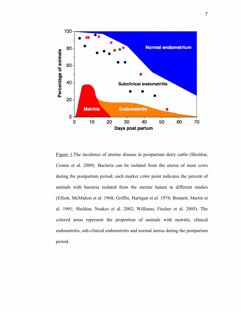
Figure 1.The incidence of uterine disease in postpartum dairy cattle (Sheldon,
Cronin et al. 2009). Bacteria can be isolated from the uterus of most cows
during the postpartum period; each marker color point indicates the percent of
animals with bacteria isolated from the uterine lumen in different studies
(Elliott, McMahon et al. 1968; Griffin, Hartigan et al. 1974; Bonnett, Martin et
al. 1991; Sheldon, Noakes et al. 2002; Williams, Fischer et al. 2005). The
colored areas represent the proportion of animals with metritis, clinical
endometritis, sub-clinical endometritis and normal uterus during the postpartum
period.
8
Uterine diseases
Metritis
Metritis is an inflammation of the endometrium and muscular layers of
the uterus, which can extend to the serosa and ligaments (causing peri and
parametritis) as well as to surfaces of other peritoneal viscera (Bondurant
1999). Histologically, evidence of severe edema, massive infiltration by
leukocytes, and myometrial degeneration is shown, resulting in systemic signs
of illness such as; fever, red-brown watery fetid vulvar discharge with a flaccid
uterus, dullness, anorexia, increase of heart rate, and decrease milk production
(Drillich, Beetz et al. 2001; Sheldon, Lewis et al. 2006). It is observed mostly
during the first two weeks post partum. Dystocia, metabolic imbalances around
parturition (Sandals, Curtis et al. 1979; Markusfeld 1987) and retained fetal
membranes have been the major risk factors for metritis (Drillich, Pfutzner et
al. 2003). There is also strong association between feed intake and cows’
behaviour and the subsequent risk of development of metritis (Huzzey, Veira et
al. 2007).
The bacterias most associated with metritis are; E. coli, A. pyogenes,
Fusobacterium necroforum, Provetella spp. Bacteroids spp and a variety of
anaerobic and gran-negative species that are eventually isolated (Drillich,
Pfutzner et al. 2003). Incidence of metritis varies between studies and it can
9
reach up to 40%. (Etherington, Bosu et al. 1984; Bartlett, Kirk et al. 1986;
Peeler, Otte et al. 1994; Sheldon, Cronin et al. 2009). In bovine metritis, there is
an initial decrease in phagocytic activity of neutrophils followed by an increase
2–3 weeks later (Vandeplassche 1981; Frank, Anderson et al. 1983).
Some cases of metritis are less severe than the acute toxic form and occur
after 3 weeks post partum and have been characterized by an enlarged uterus
with fetid vaginal discharge and mild signs of systemic illness.
Pyometra
Pyometra is defined by accumulation of a changeable amount of purulent
exudate within the endometrial lumen with persistence of a corpus luteum,
consequentially, prolonging the estrous cycle. This condition is more likely to
develop in cows that ovulate before the resolution of bacterial contamination.
The destruction of endometrial cells and the switch of PGF production to PGE
in the presence of bacterial contamination might prevent the luteolysis creating
a continuous dominancy of the uterus by progesterone that is known to suppress
the uterine defense mechanism (Sheldon, Cronin et al. 2009). The diagnosis of
pyometra depends on transrectal palpation of a distended uterus and/or
transrectal ultrasonography of a fluid-filled uterus with hyperechogenic content
and the presence of a persistent corpus luteum, with a history of anestrus. The
pyometra fluid can be shifted from one horn to the other, which cannot be done
10
with conceptus membranes, and often the uterine wall is thicker than that of a
pregnant uterus. Treatment of pyometra is PGF2α or an analogue causing
luteolysis and generating a switch in hormone dominancy (progesterone to
estradiol) improving the uterine defense system to resolve the infection
(Bretzlaff, Whitmore et al. 1982; Paisley, Mickelsen et al. 1986; Bretzlaff 1987;
Gilbert and Schwark 1992).
Endometritis
The endometritis is an inflammation limited to the endometrium. This
condition may follow parturition, copulation, artificial insemination, or infusion
of irritants into the uterine lumen. Usually, the uterine dimension is normal and
the presence of a purulent or mucopurulent discharge can be observed at the
vulva and/or cervical os by vaginoscopy. The affected animal is not
systemically sick.
In a study with a large number of dairy cows, the cervix with a diameter bigger
than 7.5 cm had a greater association with endometritis than uterine dimension.
Also, the presence of a purulent discharge at the vulva or cervical os after
21DIM or a mucopurulent discharge after 26DIM were considered positive
signs of endometritis (LeBlanc, Duffield et al. 2002).
Historically endometritis has been diagnosed mainly by transrectal
examination (TRE) 3 to 4 weeks after parturition. Diagnosis of endometritis by
TRE is subjective, revealed only 16.9% of the affected cows, and has little
11
association with reproductive perfomance (Lewis 1997; LeBlanc, Duffield et al.
2002). Vaginoscopy, which is a simple and rapid method, revealed 23.5% of
affected animals and has a better association to further reproductive
performance (LeBlanc, Duffield et al. 2002), but it fails to detect 9.1% of
clinically diagnosed cows (Kasimanickam, Duffield et al. 2004). Various
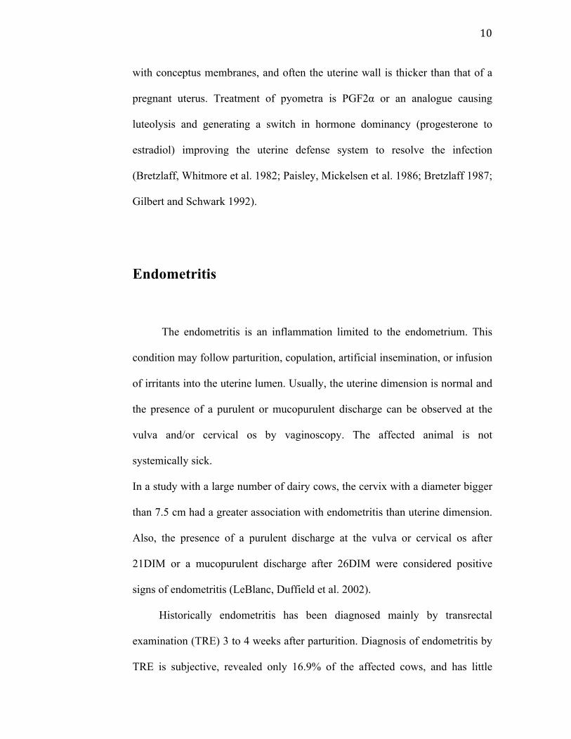
studies used a scoring system of the vaginal secretion (Dohmen, Lohuis et al.
1995; Sheldon and Noakes 1998; Huszenicza, Fodor et al. 1999) and odor
(Williams, Fischer et al. 2005) (Fig 2), which is correlated with bacterial culture
and infertility. In one study, Arcanobacterium pyogenes, Proteus and
Fusobacterium necrophorum were associated with mucopurulent or purulent
vaginal secretion. A. pyogenes, Escherichia coli, non-hemolytic Streptococci,
and Mannheimia haemolytica were associated with a fetid mucus odor
(Williams, Fischer et al. 2005).
12
Figure 2. Vaginal discharge from cows at post partum scored by its visual
characteristics with 0 - clear vaginal mucus, 1 - mucus with flecks of pus, 2 -
50% mucus 50% pus and 3 - >50% pus (Williams, Fischer et al. 2005)
13
Ultrasonography is another diagnostic method that can improve the
sensitivity and specificity when applied with another method (Kasimanickam,
Duffield et al. 2004; Barlund, Carruthers et al. 2008), however when used alone
it is able to detect only severe endometritis. (Mateus, Lopes da Costa et al.
2002).
Bacteriology can be used to diagnose endometritis but the cost, the
urgency to treat and the predominance of A. pyogenes and anaerobic bacteria as
pathogens; are reasons that do not justify this diagnostic practice. The definitive
diagnosis of endometritis is based on the histological examination of
endometrial biopsies. However, biopsies are expensive, time consuming, and
have been reported to be detrimental to future fertility when applied early on
postpartum. (Etherington, Bosu et al. 1984; Bonnett, Martin et al. 1993)
Cows with endometritis have their blood neutrophils counts augmented
two weeks before parturition and up to 4 weeks post partum when compared
with cows clear of uterine disease (Kim, Na et al. 2005). Also, neutrophils from
cows with endometritis have a decrease in phagocytic activity (Vandeplassche
and Bouters 1983; Anderson, Hemeida et al. 1985; Kim, Na et al. 2005),
suggesting that cows which develop endometritis are susceptible even before
parturition by a poorly understood mechanism where the immune system is
debilitated probably by internal or external stress factors like concomitant
diseases, malnutrition, poor housing, management, negative energy balance,
submissive behavior (Kim, Na et al. 2005).
14
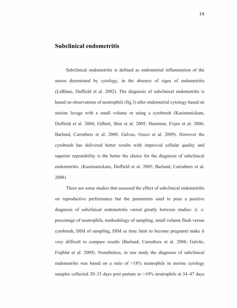
Subclinical endometritis
Subclinical endometritis is defined as endometrial inflammation of the
uterus determined by cytology, in the absence of signs of endometritis
(LeBlanc, Duffield et al. 2002). The diagnosis of subclinical endometritis is
based on observations of neutrophils (fig.3) after endometrial cytology based on
uterine lavage with a small volume or using a cytobrush (Kasimanickam,
Duffield et al. 2004; Gilbert, Shin et al. 2005; Hammon, Evjen et al. 2006;
Barlund, Carruthers et al. 2008; Galvao, Greco et al. 2009). However the
cytobrush has delivered better results with improved cellular quality and
superior repeatability is the better the choice for the diagnosis of subclinical
endometritis. (Kasimanickam, Duffield et al. 2005; Barlund, Carruthers et al.
2008)
There are some studies that assessed the effect of subclinical endometritis
on reproductive performance but the parameters used to pose a positive
diagnosis of subclinical endometritis varied greatly between studies: (i. e.
percentage of neutrophils, methodology of sampling, small volume flush versus
cytobrush, DIM of sampling, DIM as time limit to become pregnant) make it
very difficult to compare results (Barlund, Carruthers et al. 2008; Galvão,
Frajblat et al. 2009). Nonetheless, in one study the diagnosis of subclinical
endometritis was based on a ratio of >18% neutrophils in uterine cytology
samples collected 20–33 days post partum or >10% neutrophils at 34–47 days
15
post partum using 132 DIM to calculate the risk of pregnancy cutoff
(Kasimanickam, Duffield et al. 2004). In another study which used uterine
lavage, a count of >5% of neutrophils between 40 and 60 DIM was considered
positive, with 120DIM for the time limit for getting pregnant (Gilbert, Shin et
al. 2005).
The incidence of subclinical endometritis varies between 0 and 73% and
is associated with longer intervals to conception and a greater likelihood of
culling (Gilbert, Shin et al. 2005; Kasimanickam, Duffield et al. 2005; Barlund,
Carruthers et al. 2008). Studies on subclinical endometritis are at its beginning
though intrauterine infusion of cephapirin benzathine, has shown improvement
on reproductive performance of cows declared positive for subclinical
endometritis (Kasimanickam, Duffield et al. 2005).
16
A
B
C
Figure 3. Endometrial cytology slides. Picture A shows a large quantity of
neutrophils. Picture B shows endometrial cells without white blood cells.
Picture C shows endometrial cells with neutrophils scattered within the slide.
17
Uterine defense
The first response of the uterus against foreign bodies (bacteria, sperm) is
a neutrophilic influx into the endometrium and uterine lumen (Klucinski,
Dembele et al. 1995). Experimental replication of this event has been achieved
by several investigators (Kluciński, Targowski et al. 1990; Butt, Senger et al.
1991; Zerbe, Schuberth et al. 1996) utilizing an extended variety of substances
and microorganisms, inducing a significant influx of neutrophils into the uterine
lumen. Recruitment and activation of neutrophils was presumed to be the result
of cytokine release from lymphocytes induced by the prior immunization.
However, when nonspecific inflammation was induced in the endometrium,
intrauterine neutrophils influx was present, but with reduced activity when
compared to circulating neutrophils (Kluciński, Targowski et al. 1990;
Klucinski, Dembele et al. 1995). There are several mechanisms by which
neutrophils phagocytize and kill microorganisms. They can be directly attracted
to microbial products or they can be stimulated by complement chemotactic
substance like the LTB4 that is a potent leukotriene largely present in inflamed
uteri and by complement component C5a (Belluzzi, Galeotti et al. 1994;
Bondurant 1999). The opsonization (i.e., complement component C3b and
specific antibodies) of microorganisms in the intrauterine lumen improves
neutrophil phagocytosis due to the presence of receptors for the Fc component
of the antibody and receptors for C3b on the neutrophil. Such binding and
engulfing of opsonized microorganism is greatly enhanced (Watson 1985;
18
Watson 1989; Rysanek, Babak et al. 2001). The neutrophils then kill the
microorganisms by a mechanism of oxygen dependent generation of superoxide
anions(Kϋther, Audigè et al. 1998), lysozymes and proteolytic enzymes.
Neutrophils flushed from normal uteri on postestrus had a greater degree of
expression of Fc receptors for IgG1 and IgG2 antibodies than did peripheral
blood neutrophils. Neutrophils from cows with experimental endometritis
showed a decrease in the expression of Fc receptors for IgG antibodies and a
lower index of Fc-mediated phagocytosis (Klucinski, Niemialtowski et al.
1994).
In addition to neutrophil migration, other cellular components that are
eventually activated include macrophages, lymphocytes, eosinophils, and mast
cells. Inflammatory mediators are then released, such as tissue necrosis factor
(TNFα), histamine and prostaglandins, interleukins, and chemotactic factors for
neutrophils and eosinophils, including LTB4 and eosinophil chemotactic factor
A (ECF-A) (Miller 1996). The eosinophils also release inflammatory mediators
and chemicals such as superoxides and enzymes that kill microorganisms.
Uterine mast cell degranulation releases tryptase and other proteases that can
activate complement components C3 and C5 to generate anaphylatoxins. They
also release kallikreins that generate kinins (Kϋther, Audigè et al. 1998) whish
are potent vasoactive agents that increase vascular permeability.
It is important to understand that estrus alone is associated with a slight
migration of neutrophils into the endometrium (Studer and Morrow 1978) with
a maximum number of cells in the normal uterine lumen shortly after estrus and
ovulation. The great majority of these cells are neutrophils, which shows
19
augmented phagocytic and killing ability during estrus over diestrus (Watson
1985). Lymphocytes are generally present in the stratum compactum of the
endometrium. Their numbers change during the estrous cycle, being highest in
the periestrus period and lowest at diestrus (Vander Wielen and King 1984).
Leukocyte esterase
Leukocyte esterase (LE) is used to test for the presence of white blood
cells in the urine as indication of an inflammation process of the urinary tract.
In humans, it has been used for a rapid diagnosis of inflammation in many body
fluids such as urine, pleural fluid, peritoneal fluid, and cerebrospinal fluid
(Levy, Tournot et al. 1989; Azoulay, Fartoukh et al. 2000; Braga, Souza et al. ;
Rerknimitr, Rungsangmanoon et al. 2006). The principle of leukocyte esterase
(LE) test is that the esterase released from activated neutrophil cells reacts with
indoxil carbonic acid ester; indoxil is released by the esterase and reacts with
diazoniun salt and is oxidized yielding a violet azo dye. The intensity of the
color is correlated to leukocyte counts according to the fabricant (Kutter,
Figueiredo et al. 1987). Positive test results are clinically significant (Herlihy,
Wilkerson et al. 1984; Romero, Emamian et al. 1988; Levy, Tournot et al.
1989; Braga, Souza et al. 2006; Rerknimitr, Rungsangmanoon et al. 2006). The
LE test is also used to screen for gonorrhea. The combination of the LE test
with the urinary nitrite test provides an excellent screen for establishing the
20
presence of a urinary tract infection. Urine samples that test positive for both
nitrite and leukocyte esterase should be cultured for pathogenic bacteria.
In the dog where the esterase leukocyte test activity have been studied in
cases of pyuria, the correlation with esterase activity and the disease has not
been significant and the sensitivity and specificity appears to be low, suggesting
that canine urine may contain a esterase inhibitor or canine neutrophils do not
express the same quantity or type of esterase expressed by human leukocytes.
(Vail, Allen et al. 1986; Bauer, Rettig et al. 2008) In the cow, investigators have
used a commercial leukocyte esterase strip test in uterine lavage for diagnosis
of endometritis and showed a high correlation between endometrial cytology
and leukocyte esterase with 96% sensitivity and 98% specificity. (Santos,
Roman et al. 2006).
The objectives of this study are to:
1) Compare uterine leukocyte esterase activity and the endometrial
cytology (EC).
2) Compare leukocyte esterase activity of the cervix (CxLE) and the
uterus (UtLE).
3) Compare two methods of assessing the slides
We hypothesized that the diagnosis of subclinical endometritis can be
accomplished using the leukocyte esterase activity.
21
CHAPTER 2
VALIDATION OF LEUKOCYTE ESTERASE
ACTIVITY FOR THE DIAGNOSIS OF
SUBCLINICAL ENDOMETRITIS IN POSTPARTUM
DAIRY COWS
Couto GB, Vaillancourt DH, and Lefebvre RC
Department of Clinical Sciences, Faculté de médecine vétérinaire, Université de
Montréal, 3200 rue Sicotte, Saint-Hyacinthe, Québec, J2S 7C6.
Address of correspondence and reprint request to
Réjean Lefebvre
E-‐mail:
22
Abstract
The aim of this study was to compare leukocyte esterase activity and the
endometrial cytology (EC) for the diagnosis of subclinical endometritis in
Holstein cows (n=218) between 21 and 47 days in milk (DIM). The relationship
between the uterine and cervical leukocyte esterase activity was also
determined. In addition, two methods for assessing endometrial cytology were
compared. Cows from five commercial dairy herds were allocated to the study
based on the absence of vaginal discharge at vaginoscopy or genital anomalies
after transrectal examination. Cows included in the study were monitored
biweekly for at least 200 days. The percentage of polymorphonuclear cells (%
neutrophils) was correlated with esterase score measured either from the uterus
(UtLE, P=0.0001) or the cervix (CxLE, P=0.002). Both CxLE and UtLE
(P=0.0009 and P=0.0001 respectively) decreased with DIM, however they were
not statistically associated with pregnancy risk at 90 DIM. Between 32-47 DIM,
the % neutrophils and risk of pregnancy at 90 DIM were associated (P=0.04).
For the same period, survival analysis identified cows with > 2.6 % neutrophils
at EC as subclinical endometritis cows. In conclusion, the results indicate that
uterine leukocyte esterase activity is correlated to percentage of neutrophils at
endometrial cytology but is not reliable to predict the risk of pregnancy.
Subclinical endometritis (> 2.6 % neutrophils) diagnosed by EC between 32
and 47 DIM was associated with reduced risk of pregnancy.
23
Introduction
In the last decade, decrease of the fertility of dairy cows has gathered
considerable attention in veterinary science (Bousquet, Bouchard et al. 2004).
As the reproductive performance is associated with the health status of the
uterus at the end of the voluntary waiting period (VWP) (Ferguson and
Galligan 2000), assessment of uterine condition in postpartum cows has been
the focus of research. In cows, lochia is normally expelled from the
reproductive tract during the first two or three weeks after parturition (Gier and
Marion 1968). Discharges can persist longer depending on the virulence of the
causative organism and predisposing factors to the disease (Azawi 2008).
Uterine disease affects half of dairy cattle (Markusfeld 1987; Clay, Welper et
al. 2004) and causes infertility by disrupting uterine and ovarian functions
(Mateus, Lopes da Costa et al. 2002). As inflammation is normally present
during the physiological uterine involution in postpartum cows, rigorous
characterization of pathologic conditions become intricate. Therefore, the
complex relationship between postpartum diseases and reproductive
performance remains unclear (Fourichon, Seegers et al. 2000).
Clinical endometritis is defined as inflammation of the endometrium with
vaginal purulent discharge after 21 DIM or mucopurulent discharge after 26
DIM in absence of systemic clinical signs (LeBlanc, Duffield et al. 2002;
Sheldon, Lewis et al. 2006; LeBlanc 2008). Characterization of the vaginal
discharge is associated with uterine pathogens and a prognosis on the cow’s
24
future fertility (Sheldon, Cronin et al. 2009). The clinical endometritis has a
negative impact on reproductive performance increasing services per
conception, calving to first service interval, calving to conception interval
(Borsberry and Dobson 1989; Heuwieser, Tenhagen et al. 2000), risk of
pregnancy (LeBlanc, Duffield et al. 2002) and conception rate (Fourichon,
Seegers et al. 2000). Postpartum uterine disease represents a leading cause of
reproductive inefficiency (Sheldon, Lewis et al. 2006) and could mean
significant economic loss for the dairy industry (Bartlett, Kirk et al. 1986;
Guard 1994; Drillich, Beetz et al. 2001). The financial impact is driven by
infertility, excessive culling, reduced production, and cost of treatment.
Contrary to clinical endometritis, subclinical endometritis does not present any
clear evidence of endometrial inflammation at the genital examination
(LeBlanc, Duffield et al. 2002). As the more severe inflammation (clinical
endometritis) subsides, the drainage of abnormal uterine fluid is not obvious
(Sheldon and Dobson 2004). Even though the condition is much more difficult
to diagnose, subclinical endometritis could still impair reproductive
performance (Sheldon, Lewis et al. 2006). The incidence of subclinical
endometritis depends on the diagnostic technique and the day of the postpartum
period when the diagnosis is made, and may reach 74% in a herd (Gilbert, Shin
et al. 2005). In other studies, subclinical endometritis has been reported as high
as 43% for cows between 20 and 33 DIM, 45% for cows between 34 and 47
DIM and 53% for cows between 40 and 60 DIM. (Kasimanickam, Duffield et
al. 2004; Gilbert, Shin et al. 2005)

25
Once the endometrial cells recognized pathogens present in the uterus
(innate immune system), they secrete peptides like cytokines and chemokines
which attract inflammatory cells like neutrophils (Sheldon, Cronin et al. 2009).
Neutrophils are recruited from the circulation into the lumen, eliminate bacteria
and become an excellent indication of an inflammatory process (Tizard 1996;
Wade and Lewis 1996). Blood-derived neutrophils are the main effector cells
for removing bacteria from the uterus. After elimination of the pathogenic
organisms, inflammation subsides and neutrophils will be limited to the uterine
lumen fluid (Kluciński, Targowski et al. 1990). Persistent neutrophils in the
endometrium in the absence of bacteria may be an important characteristic of
subclinical endometritis and cause of infertility in cattle (Sheldon, Cronin et al.
2009). The neutrophils are the predominant inflammatory cells in the uterine
lumen and their relative proportion has been shown to be predictive of future
reproductive performance. In absence of clinical signs, subclinical endometritis
could be cytologically defined as > 8.0% neutrophils of total cells collected by
cytobrush in dairy cows between 28 and 41 DIM (Barlund, Carruthers et al.
2008) or >10% between 34 and 47 DIM (Kasimanickam, Duffield et al. 2004).
However, high incidence of a transient inflammation response in the
postpartum uterus, time variation of sampling the uterus and the lack of
standardization of different techniques sustain the controversy concerning the
case definition of subclinical endometritis.
Diagnosis of subclinical endometritis should depend on direct methods
that will reveal the presence of inflammatory cells in the lumen or within the
26
endometrium. In cows, moderately invasive methods to evaluate the
inflammatory process of the endometrium include uterine lavage and cytobrush
cytology. Both methods have been described and accepted as diagnostic
techniques. The cytobrush technique seems to be the most useful technique and
the most reliable diagnostic test compared with uterine lavage (Kasimanickam,
Duffield et al. 2005)and ultrasonography (Barlund, Carruthers et al. 2008).
Cytobrush cytology is considered the reference of cytological diagnosis for
subclinical endometritis because of the quality of cells and better repeatability.
However, it is not a cow-side test. Although neither complex nor expensive,
endometrial cytology requires special instruments and expertise and lacks of
immediacy. From a point of view of practicality and efficiency, the diagnostic
test for subclinical endometritis needs to be performed rapidly and the results
analysed on farm. With these characteristics, a treatment decision can be made
and executed at the time of the visit by the veterinarian.
An alternative method to assess inflammatory cells in the lumen of the
uterus is the leukocyte esterase test. It has been used for a rapid diagnosis of
inflammation in many body fluids such as urine, pleural fluid, peritoneal fluid,
and cerebrospinal fluid (Levy, Tournot et al. 1989; Azoulay, Fartoukh et al.
2000; Braga, Souza et al. ; Rerknimitr, Rungsangmanoon et al. 2006) and could
be used as an indirect method to detect neutrophils in the cow’s uterus. Santos
et al., (Santos, Roman et al. 2006) investigated the use of a commercial strip
test, used for urinary neutrophils, in uterine lavage for diagnosis of endometritis
and showed a high correlation between endometrial cytology and leukocyte
27
esterase activity with 96% sensitivity and 98% specificity. The leukocyte
esterase released from neutrophil cells reacts with indoxil carbonic acid ester.
Indoxil is released by the esterase reaction with diazoniun salt and is oxidized
yielding a violet azo dye (Kutter, Figueiredo et al. 1987). The intensity of the
color correlates to leukocyte counts according to the fabricant.
The aim of this study is to compare leukocyte esterase activity to the
endometrial cytology to diagnose subclinical endometritis in clinically normal
postpartum dairy cows. The relation between the uterine and cervical leukocyte
esterase activity was assessed and the impact of subclinical endometritis was
evaluated. We hypothesized that the diagnosis of subclinical endometritis can
be achieved using the leukocyte esterase activity.
Materials and Methods
Holstein cows (n=285) from 5 commercial herds monitored bimonthly by
the ambulatory clinic of the Faculty of veterinary medicine of l’Université de
Montréal (Quebec, Canada) were enrolled in the research project. Herd records
were compiled in a databank DSA@HR (Dossier de Santé Animal: DS@HR,
2725 boul. Western Casavant, St-Hyacinthe, QC, J2S 0E5) and validated for
reproduction. All herds were housed in tie-stall stanchion barns, milked twice
daily and artificially inseminated exclusively after a voluntary waiting period of
approximately 60 days and on heat detection. The total mixed ration diet is
28
composed mainly of corn silage, alfalfa/grass silage and corn meal and protein
supplements. A genital exam was performed by transrectal palpation and
vaginoscopy for all cows. Only cows without evidences of clinical endometritis
according to the criteria of Leblanc (LeBlanc, Duffield et al. 2002) and between
21-47 DIM were included in the study.
Cows were subjected to different diagnostic tests in succession.
Reproductive examination included measurement of the cervical size and
uterine size (cm) at the base of the horn, (left and right) and assessment of the
ovarian activity (presence of a corpus luteum, follicles and cysts) as determined
by transrectal palpation. Vaginal secretion was classified according to Williams
and al. (Williams, Fischer et al. 2005) with some variations. The vaginal
examination (VEx) was done with a disposable vaginoscope to visualize the
cervical os and secretion which were scored as; VEx0, no mucus, VEx1, clear
mucus, VEx2, mucus containing flecks of white or off-white pus, VEx3, ≤50%
white or off-white mucopurulent material, VEx4, purulent material, usually
white, occasionally yellow. Cows with VEx2, VEx3 and VEx4 were considered
as having clinical endometritis.
Once the cow met the enrollment criteria, the endometrial cytology and
the leukocyte esterase test were performed. For the leukocyte esterase test in the
cervix (CxLE), the reagent strip was partially introduced into a uterine infusion
pipette, (Continental Plastic Corp. Delavan, WI, USA) with the reactive pad
protruding outside the pipette. Through a disposable and sterile lubricated 50
cm long vaginoscope, the cervix was visualized and the pipette with the
29
leukocyte esterase pad Uristix® (Bayer Health Care L.L.C., Elkhart, IN) was
introduced into the first ring of the cervix for 5 seconds and the color pad
determined after two minutes according to the fabricant recommendations.
Lastly, uterine leukocyte esterase activity (UtLE) was measured and
cellular material were harvested for cytological evaluation by cytobrush (VWR
Canlab, Mississauga, ON, Canada) technique as previously described
(Kasimanickam, Duffield et al. 2004). Briefly, the cytobrush was screwed onto
a solid stainless steel rod and placed in a stainless steel tube of 65 cm long for
passage through the cervix. To protect the instrument from vaginal
contamination, the apparatus was inserted into a double pipette (Continental
Plastic Corp., Delavan, WI, USA). The instrument was introduced into the
vagina as the vaginoscope was removed, a sleeved arm was introduced into the
rectum to facilitate passage of the instrument and it was pushed through the
double pipette. The tube was advanced through the cervix into the body of the
uterus where the cytobrush was pushed out (1.0 cm) of the stainless steel tube
and exposed the lumen of the uterus. The cytobrush in contact with the
endometrium was gently rotated clockwise approximately one turn to obtain
cellular material from the adjacent endometrium. The cytobrush was retracted
into the stainless tube before removing the whole apparatus from the genital
tract.
Slides were prepared by rolling the cytobrush on a predetermined surface
area of a clean glass microscope slide and left to dry. After the slides were
made, the cytobrush was plunged into a 3ml glass tube containing 1ml of saline
30
0.9% and gently shaked for 30s. The leukocyte esterase strip was then inserted
into the glass tube. The strip was removed from the tube and the result (UtLE)
recorded after 2 minutes. The slides were transported to the laboratory where
they were stained with a modified Giemsa stain (PROTOCOL HEMA 3 STAIN
SET, Fisher Diagnostics, Ficher Scientific Company L.L.C. Valley Pike,
Middletown, VA, USA). Once stained and dried, cover slip was applied using
Histofluid ®mounting medium (Paul Mareinfield GmbH & Co., Lauda
Koenigshofen, Germany).
Slide evaluation
Two methods were used to evaluate slides; a first rigorous method (RM)
of reading the slides was done followed by a second simple assessment method
(SM) more likely to be used in the field. The RM consisted in drawing
imaginary lines of 0.5cm apart starting in the middle (line #1) of the slide. Line
#2 was drawing on the right side of line #1 and line #3 on the left side.
Following the same pattern, line #4 was the following right of the line #2 and
line #5 the following left to line # 3. Slides were assessed by reading the five
straight lines, bottom to top, starting from the central line (#1) to the exterior
one (1 to 5), under a 400X magnification by the same examiner (figure 1). A
differential count of all neutrophils, lymphocytes, macrophages and epithelial
cells using a minimum of 500 cells or all five lines was obtained to provide a
31
quantitative assessment of endometrial inflammation. Also, the number of
clumps of epithelial cells and the number of fields assessed were recorded. The
SM method consisted of assessing first the slide at 200X magnification to
localize the area with a reasonable quantity of recognizable cells (independent
of each kind). From these areas, ten microscopic fields were randomly selected
and total neutrophil counts were recorded at 400X.
32
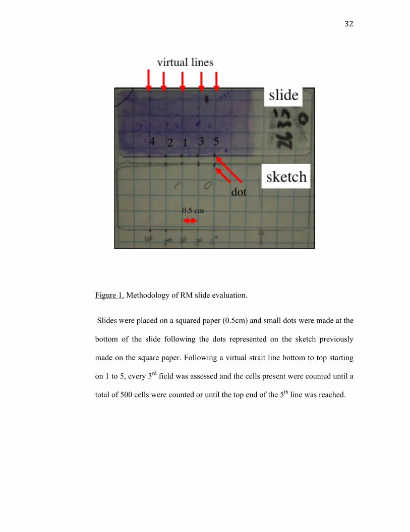
Figure 1. Methodology of RM slide evaluation.
Slides were placed on a squared paper (0.5cm) and small dots were made at the
bottom of the slide following the dots represented on the sketch previously
made on the square paper. Following a virtual strait line bottom to top starting
on 1 to 5, every 3rd field was assessed and the cells present were counted until a
total of 500 cells were counted or until the top end of the 5th line was reached.
33
Criteria of exclusion
Cows that received systemic antibiotics, intrauterine infusion or
reproductive hormone administration from a period of 10 days prior to the
sampling were excluded from the experiment. Furthermore, cows, with a
history of twins, metritis, clinical endometritis, displacement of abomasum and
retained placenta were also excluded. Cows identified to be culled by the owner
before 200 days in milk were not included in the study. Cows that were not
bred before 90 DIM were excluded for the analysis of conception rate. This
study did not control for any other treatment for which ever cause (i.e. mastitis,
pneumonia etc.) that may have been administered between the time of the test
and the time of pregnancy diagnosis.
Data analysis and Statistics
Data analysis was conducted with SAS v 9.1.3. A linear mixed model
with the site of ULE as fixed effect, dairy herd as a random effect and the DIM
as a cofactor was used to estimate a correlation between the UtLE or CxLE and
percentage of neutrophils. A Weighted kappa was plotted to measure the
correlation between UtLE and CxLE. To estimate the correlation between the
percentage of neutrophils in RM and SM a linear regression was used. Using a
mixed logistic regression with dairies as random effect and DIM as cofactor,
34
the percentage of neutrophils, UtLE and CxLE were analysed to predict
pregnancy on cows at risk to become pregnant at 90DIM (i.e inseminated
between 43-90 DIM) to all cows (GT), and the cows were divided in two
groups for further analysis; group 1 (G1) cows from 21 to 31 DIM and group 2
(G2) cows from 32 to 47 DIM. A Receiver/Response Operating Characteristics
(ROC) curve was plotted using the sensitivity and specificity for each possible
percentage of neutrophils in the G2 group against prevalence of gestation at
90DIM to find a cut off for the percentage of neutrophils. We used 90DIM as
the limit for calculating the success of insemination because it gives a majority
of cows (85%) the chance to be inseminated at least once if we consider a first
AI at 60DIM and a heat detection rate at 50%. Pregnancy diagnosis was made
by palpation after 30 days and reconfirmed after 45 days after AI.
Results
The study began in September 2007 and continued until October 2008.
From all 285 cows examined during this study, 85 (29.8%) cows had on vaginal
examination (VEx) a score VEx0; 133 (46.6%) cows had score VEx1; 27 cows
(9.47%) had score VEx2; 22 (7.7%) cows had score VEx3 and 18 (6.3%) cows
had score VEx4. Scores VEx2, VEx3 and VEx4 where considered clinical
endometritis resulting in an overall of 23.5% of positive cases of endometritis

35
that varied within herds from 11.5% to 38.6% (11.5; 16.6; 27.2; 38.2; 38.6).
Cows with clinical endometritis were treated and enrolled in another study.
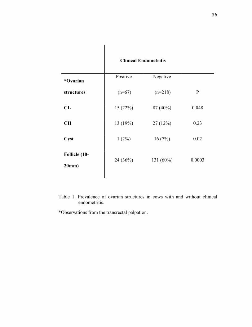
Table 1 describes the prevalence of ovarian findings relevant to
endometritis (n=285). Clinical endometritis cows showed less active ovaries
with less cows having corpus luteum (15 cows, 22%) compared to cows
without clinical endometritis (87 cows, 40%, P<0.05). Similarly, more cows
had normal follicular activity with follicles between 10 and 20 mm (131 cows,
60%, P<0.0003) on the ovaries compared to diseased ones. Among the
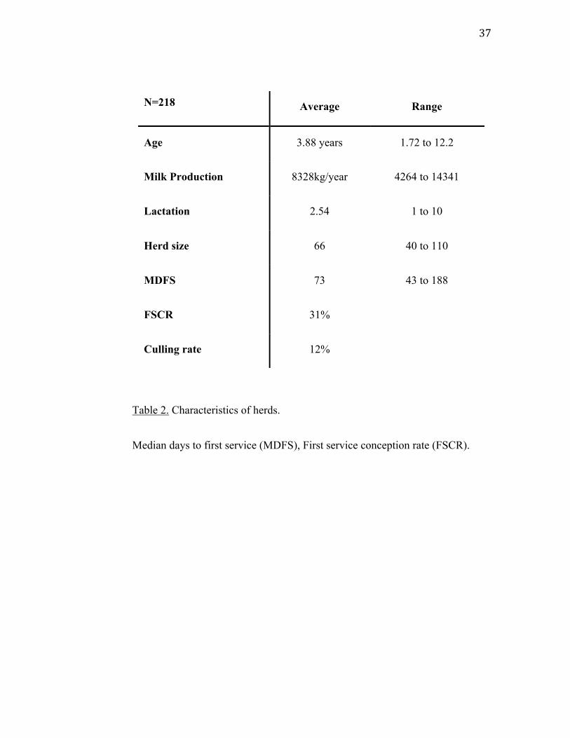
remaining 218 cows diagnosed without clinical endometritis, herd size ranged
from 40 to 110 cows in milk with an average milk production of 8328kg per
lactation and 2.4 lactations. The mean age was 3.9 years old (Table 2).
36
Clinical Endometritis
*Ovarian
structures
Positive
(n=67)
Negative
(n=218) P
CL 15 (22%) 87 (40%) 0.048
CH 13 (19%) 27 (12%) 0.23
Cyst 1 (2%) 16 (7%) 0.02
Follicle (10-
20mm) 24 (36%) 131 (60%) 0.0003
Table 1. Prevalence of ovarian structures in cows with and without clinical endometritis.
*Observations from the transrectal palpation.
37
Table 2. Characteristics of herds.
Median days to first service (MDFS), First service conception rate (FSCR).
N=218 Average Range
Age 3.88 years 1.72 to 12.2
Milk Production 8328kg/year 4264 to 14341
Lactation 2.54 1 to 10
Herd size 66 40 to 110
MDFS 73 43 to 188
FSCR 31%
Culling rate 12%
38
Leukocyte esterase activity and endometrial cytology
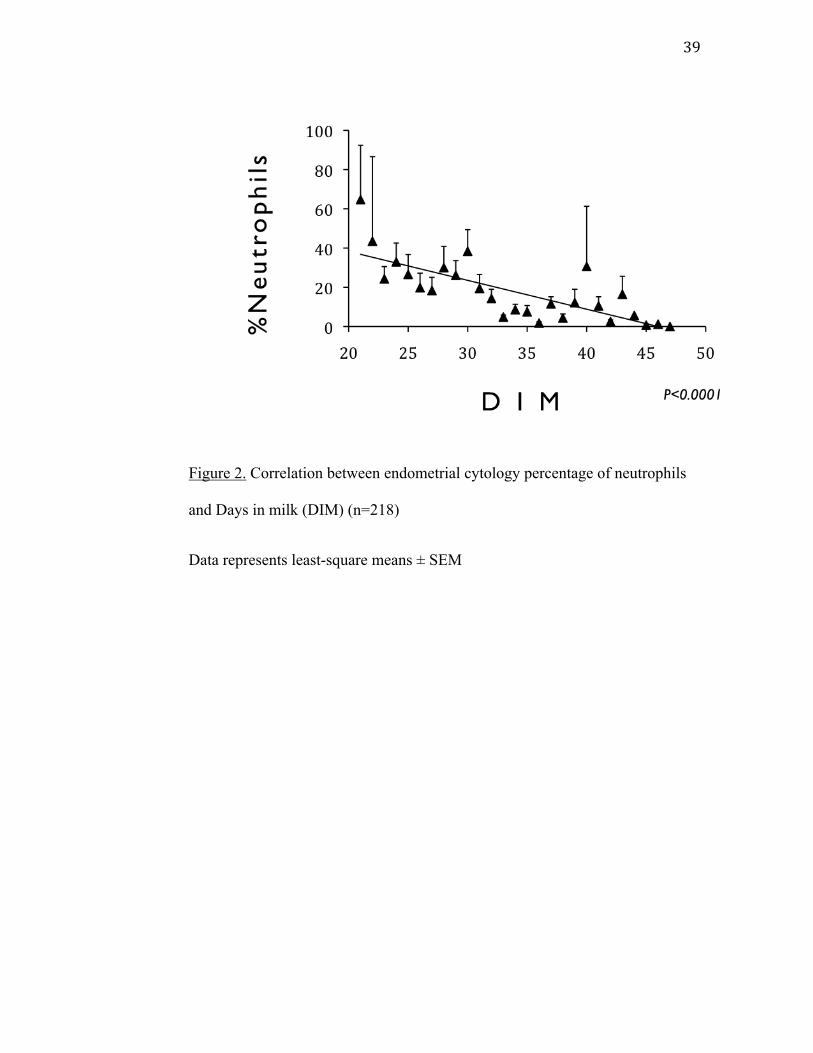
The percentage of neutrophils diminished with the number of DIM (fig.
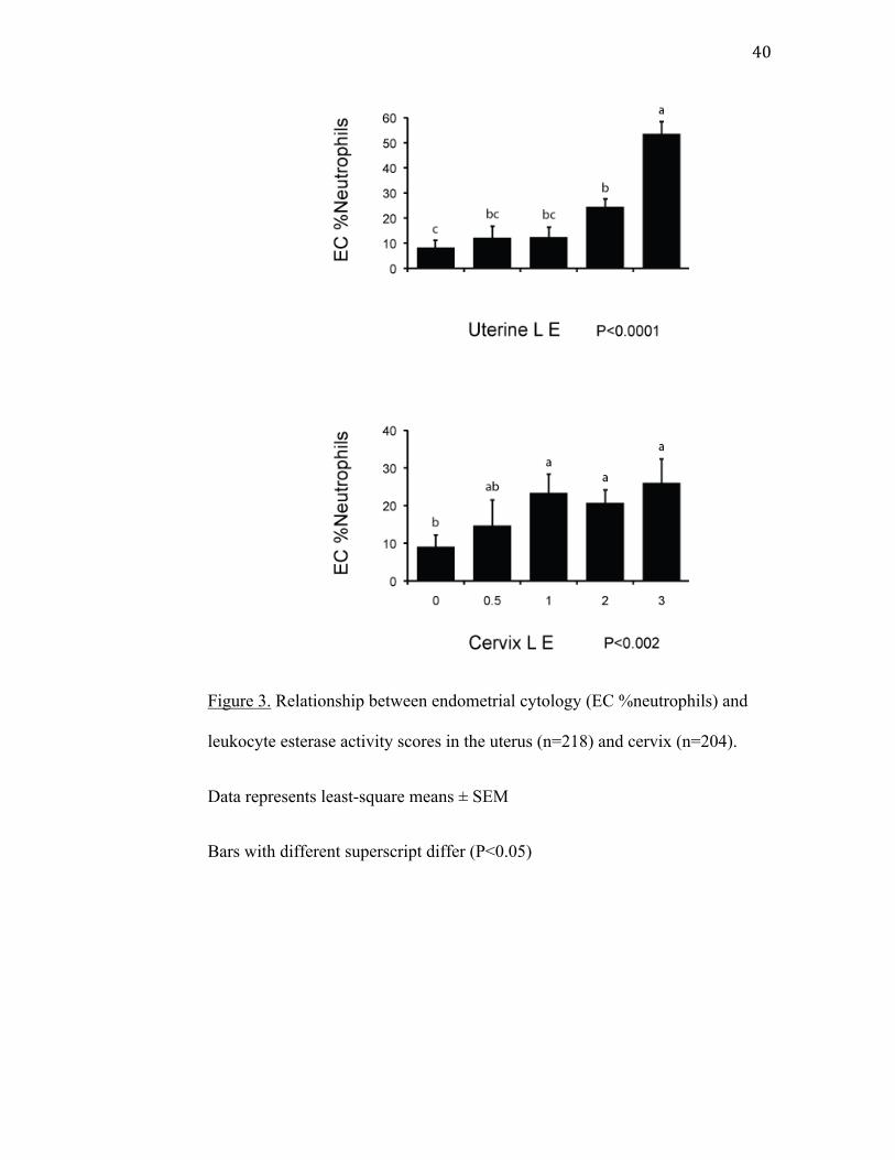
2). The esterase activity for both UtLE (P<0.0001) and CxLE correlates with
the increase of neutrophils (fig.3). For UtLE, the percentage of neutrophils was
significantly lower in cows with a score of 0 than in cows with score of 2 and 3
and significantly higher in cows with a score of 3 than in cows with a score of
0, 0.5, 1, and 2 (fig.3). For CxLE, neutrophil counts in cows with score 0 were
significantly lower than in cows with score 1, 2 and 3 and percentage of
neutrophils were not significantly different between cows with scores of 0.5, 1,
2 and 3 (fig.3). There was a slight agreement between UtLE and CxLE with a
weighted Kappa of 0.37.
39
Figure 2. Correlation between endometrial cytology percentage of neutrophils
and Days in milk (DIM) (n=218)
Data represents least-square means ± SEM
40
Figure 3. Relationship between endometrial cytology (EC %neutrophils) and
leukocyte esterase activity scores in the uterus (n=218) and cervix (n=204).
Data represents least-square means ± SEM
Bars with different superscript differ (P<0.05)
41
Impact of subclinical endometritis
Among the 218 cows used in this study, 32 cows were bred after 90 DIM
and were thus excluded from subsequent statistical analysis. The analyses
demonstrated that the risk of pregnancy by 90DIM was not significantly
associated with the UtLE and CxLE for any group GT, G1 and G2. Similarly,
the risk of pregnancy at 90DIM for all cows (GT, n=186) and the group G1
(n=92) was not associated (P=0.7) with the percentage of neutrophils on
endometrial cytology. However, in G2 (n=94) there was an association
(P=0.04) between endometrial percentage of neutrophils and pregnancy risk at
90DIM (fig. 4). The risk of getting the cows of group G2 pregnant was
diminished by 0.93 point (95%CI 0.89-0.98) each time the percentage of
neutrophils was increased by 1.0 point in G2. The ROC curve demonstrated
that the percentage of neutrophils that are considered positive for subclinical
endometritis in the G2 group was 2.6 which gives a prevalence of subclinical
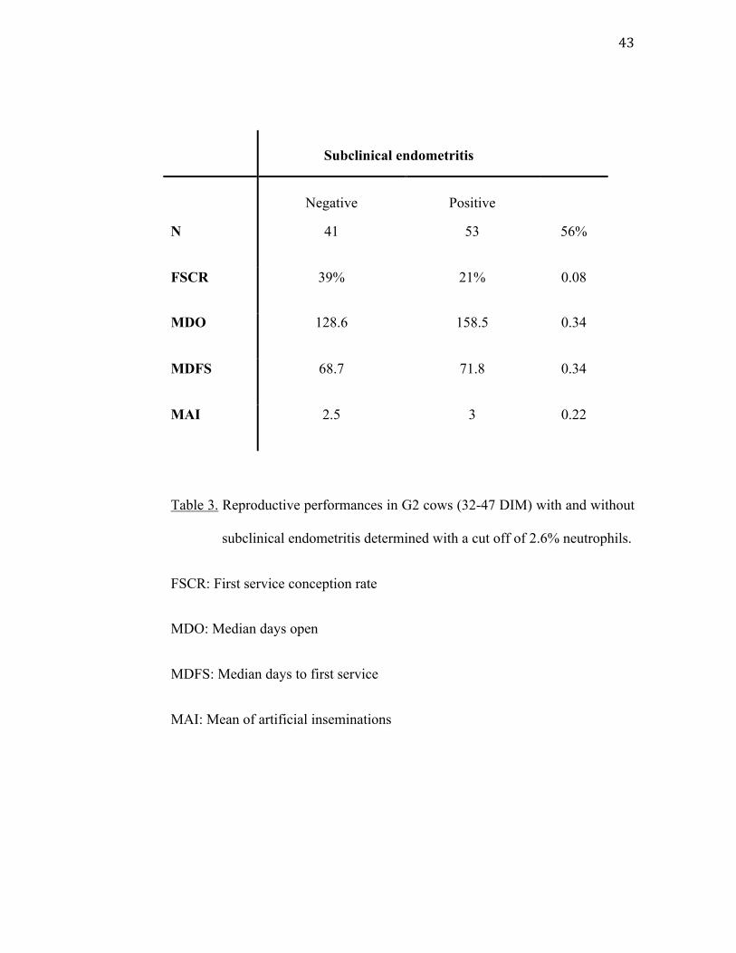
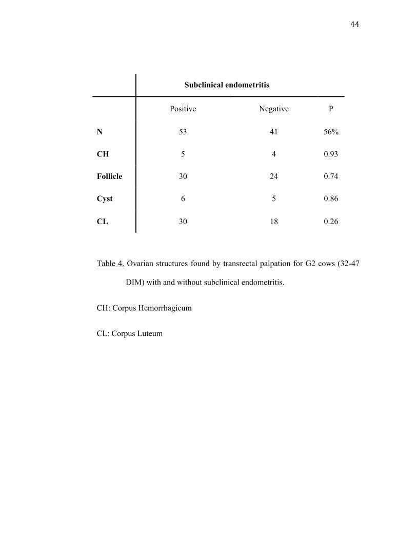
endometritis of 56% for cows in G2 (32-47 DIM). (Table 3)
Table 4 shows the prevalence of ovarian structures found by transrectal
palpation for cows in group G2. Statistical analysis demonstrated that there
were no differences for any of the ovarian structures (i.e.corpus
hemorrhagicum, corpus luteum, follicles and cysts) between the cows
diagnosed with subclinical endometritis and cows without subclinical
endometritis.
42
Figure 4. Percentage of neutrophils in pregnant (Gest) and open (Ngst) cows for
the group 21-47 DIM (GT, n=186), group 21-31 DIM (G1, n=92) and
32-47 DIM (G2, n=94).
Data represents Least-square means ±SEM
* significant difference.
43
Subclinical endometritis
Negative Positive
N 41 53 56%
FSCR 39% 21% 0.08
MDO 128.6 158.5 0.34
MDFS 68.7 71.8 0.34
MAI 2.5 3 0.22
Table 3. Reproductive performances in G2 cows (32-47 DIM) with and without
subclinical endometritis determined with a cut off of 2.6% neutrophils.
FSCR: First service conception rate
MDO: Median days open
MDFS: Median days to first service
MAI: Mean of artificial inseminations
44
Subclinical endometritis
Positive Negative P
N 53 41 56%
CH 5 4 0.93
Follicle 30 24 0.74
Cyst 6 5 0.86
CL 30 18 0.26
Table 4. Ovarian structures found by transrectal palpation for G2 cows (32-47
DIM) with and without subclinical endometritis.
CH: Corpus Hemorrhagicum
CL: Corpus Luteum
45
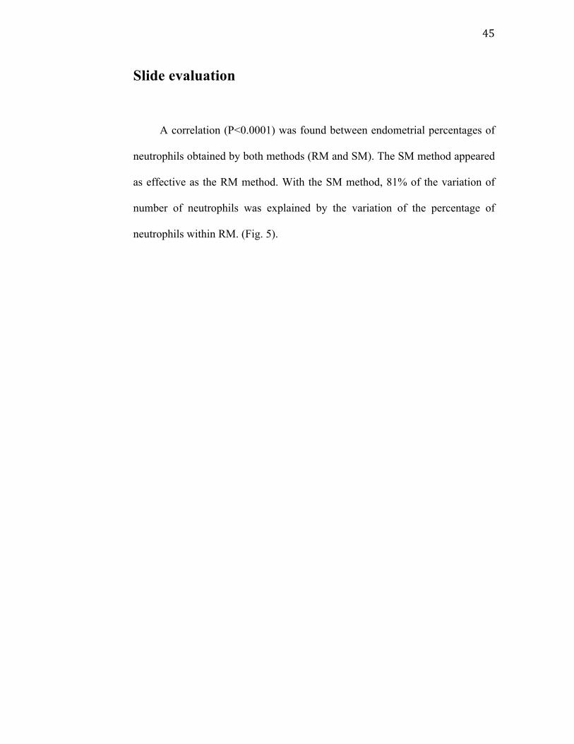
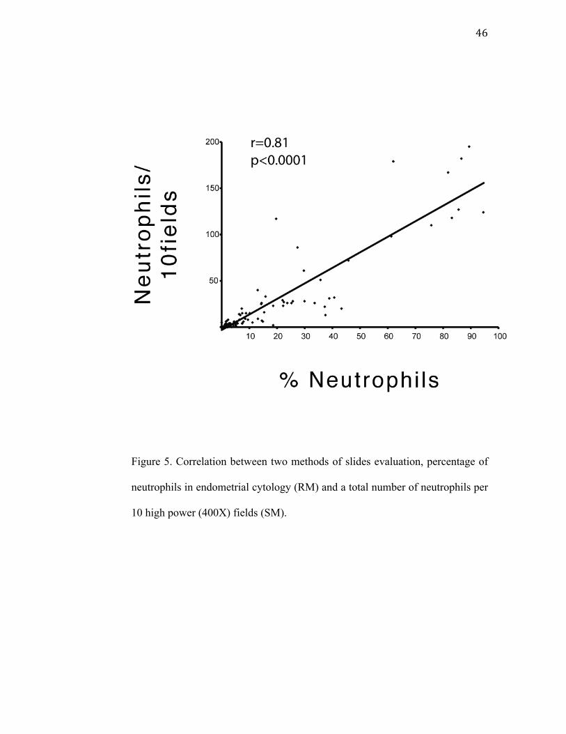
Slide evaluation
A correlation (P<0.0001) was found between endometrial percentages of
neutrophils obtained by both methods (RM and SM). The SM method appeared
as effective as the RM method. With the SM method, 81% of the variation of
number of neutrophils was explained by the variation of the percentage of
neutrophils within RM. (Fig. 5).
46
Figure 5. Correlation between two methods of slides evaluation, percentage of
neutrophils in endometrial cytology (RM) and a total number of neutrophils per
10 high power (400X) fields (SM).
47
Discussion
Subclinical endometritis is characterized by inflammation of the
endometrium without clinical signs and results in a significant reduction in
reproductive performance (Kasimanickam, Duffield et al. 2004; Gilbert, Shin et
al. 2005; Barlund, Carruthers et al. 2008; Santos, Lamb et al. 2009). The
prevalence of the disease is very variable, and depends on the diagnosis
technique; the DIM of the genital examination and the statistical method used
to determine the cut-off point of the neutrophil ratio obtained from endometrial
cytology (Guidry, Paape et al. 1976; Fourichon, Seegers et al. 2000;
Kasimanickam, Duffield et al. 2005; Santos, Lamb et al. 2009). In all cases, the
diagnosis is based on relative proportion of neutrophils. Cytobrush cytology
appeared to be the most consistent and reliable technique to diagnose
subclinical endometritis (Kasimanickam, Duffield et al. 2005; Barlund,
Carruthers et al. 2008). However, cytobrush cytology is not a cow-side test and
does not allow a rapid result and a treatment decision on the farm. Preliminary
data on leukocyte esterase activity of uterine neutrophils proposed the
technique as a cow-side test for subclinical endometritis allowing rapid decision
making at the farm (Santos, Roman et al. 2006).
In the present study, the correlation between subjective scores of uterine
leukocyte esterase activity and the percentage of neutrophils in the uterus
determined by cytobrush cytology was high (P<0.0001). Santos et al., (Santos,
48
Roman et al. 2006) had similar results with a high correlation (P<0.0001)
between leukocyte esterase score and the percentage of neutrophils from
samples obtained by uterine lavage. This was supported by a correlation
between vaginal discharge score and leukocyte esterase activity (data not
shown). The CxLE was also correlated to the percentage of neutrophils on
endometrial cytology (fig. 3). This is a good indication of uterine drainage of
exudate from the uterus and is in agreement with another study comparing
cytology from the cervix and uterus (Yavari, Haghkhah et al. 2009). The cervix
is easier to reach; the technique could mean a practical advantage to diagnose
subclinical endometritis on farm. However, even though the high correlation
between the leukocyte esterase activity (UtLE and CxLE) and the percentage of
neutrophils (EC), the pregnancy rate at 90DIM was not correlated to neither of
them in any of the groups (GT, G1 and G2) of postpartum cows. With a high
correlation between subjective score of UtLE and the percentage of neutrophils
in the uterus for G2 (32-47 DIM), one would have expected that leukocyte
esterase activity could be a predictor of pregnancy or infertility. Specially, that
a significant negative effect of the presence of neutrophils on fertility was
measured with the endometrial cytology for the same time window. The small
number of cows in G2 (n=94) and the lower capacity of the test to detect
esterase activity within an environment with a potentially smaller number of
neutrophils could explain the lack of significant effect on fertility. The fact that
two slides were produced with the same cytobrush before it was plunged in 1
ml of saline could probably explain the discrepancy of results between the EC
and esterase activity in terms of pregnancy risk. With the methodology, many

49
of the neutrophils, cellular debris and uterine fluids stayed behind on the slides
compromising the esterase activity assessment.
Because the presence of neutrophils and fluids in the cervix may be due
mostly to drainage from the uterus (Wray 1982; Földi, Kulcsár et al. 2006;
Azawi 2008) a high level of agreement was expected between UtLE and CxLE
However, only a slight agreement between UtLE and CxLE was observed in the
present study. The descrepency between UtLE and CxLE may be due to the
following. During postpartum uterine involution, occasional reduced
contractibility of the myometrium could temporally result in a poor clearance of
the uterus (Rigby, Barhoumi et al. 2001) and maintain a large amount of
neutrophils and fluid in the uterus, and not in the cervix. A good uterine
clearance may push a great amount of neutrophils into the cervix and result in a
relatively small quantity in the uterus increasing the disagreement between the
UtLE and CxLE. Stage of the estrous cycle can affect the myometrial
contractility and the neutrophil influx into the endometrial lumen and cervical
mucus (Bondurant 1999). Furthermore, the cervix has its own inflammatory
cell response to the presence of bacteria, which may not reach the uterus, with
influx of neutrophils to the cervical lumen (Kelly 2002). Finally, as the
leukocyte esterase test is based on hydrolytic reaction (Kutter, Figueiredo et al.
1987), a smaller amount of mucus or fluid in the cervix compared to the uterus
could inhibit or slow down the chemical reaction and could be responsible for
false negative results. During the clearance between the uterine lumen and the
outer cervical os, the esterase enzyme could be potentially degraded.

50
In the present study, the mean percentage of endometrial neutrophils,
when it was measured before 32DIM, did not predict pregnancy in cows at
90DIM. During that time, spontaneous resolution of uterine diseases occurs and
it is associated with neutrophils increasing their killing capacity (L Mateus
2002), suggesting that subclinical endometritis cows might spontaneously
recover. The fact that the present study was performed on several herds (n=5)
could have reduced the effects of certain confounding variables like
management strategies, body condition score, milk production and selected
herds with high prevalence of subclinical endometritis (Gilbert, Shin et al.
2005; Barlund, Carruthers et al. 2008) and it is not in agreement with another
study using a similar sampling technique (Kasimanickam, Duffield et al. 2004).
Survival analysis based on a statistical model showing a significant effect of
percentage of neutrophils on pregnancy risk is more likely to reflect
physiological events.
Evaluation of the cow reproductive tract is based on breeding history,
reproductive status, and physical examination findings, creating a more
complete database from which to make a better diagnosis and prognosis. For
more subtle disease like subclinical endometritis, additional and more invasive
diagnostic methods are required. Endometrial cytobrush appears to be a reliable
sampling technique showing more distinctively neutrophils, debris, mucus and
endometrial cells (Kasimanickam, Duffield et al. 2005; Barlund, Carruthers et
al. 2008). In mares, endometrial cytology is also useful to screen for an active
inflammatory response (Bowen, Ley et al. 1987; LeBlanc, Magsig et al. 2007).
51
In addition to the technique of sampling, the method of assessment of the
cytological smear could be important. The presence of mucus, debris, proteins
and clumps or aggregates of endometrial cells could potentially complicate and
influence the results and affect the interpretation. In the present study, the slides
were evaluated according to two different approaches. The RM was an
exhaustive method compared to the SM method, which is a more rapid and
simple approach that could appeal to practitioners. Previous studies did not
precisely and meticulously describe the way endometrial smears were analyzed.
With an agreement of 81% between the two methods, SM did not show any
disadvantages over the RM. Other researchers reported similar results in cows
(Santos, Lamb et al. 2009) and mares (Bourke, Mills et al. 1997). Therefore,
evaluation of endometrial slides should be initiated using low magnification
under bright-field microscope for a first evaluation of the quality of the smear
and to determine the areas for further analysis. A sufficient number of well
preserved and adequately stained endometrial cells and evenly dispersed
clumps or aggregates of endometrial cells would be a good indication of a good
quality smear. Analysis of ten representative microscopic fields was enough to
perform a good slide evaluation.
Conclusion
The present results support that the leukocyte esterase activity from
52
samples obtained by cytobrush cytology of the endometrium may have
potential utility for the diagnosis of subclinical endometritis in postpartum dairy
cows. Because of lack of power, further studies are required to test the full
potential of leukocyte esterase activity for the diagnosis of subclinical
endometritis in post partum cows. Cytobrush cytology assists in identifying
animals with subclinical endometritis after 32DIM and the assessment of slides
by a simple and more practical method provides a rapid and simple means of
assessing endometrial cytology slide of individual cows.
Acknowledgements
The Fond du Centenaire supported the present study. The authors
recognize the contributions of Mr. G Beauchamp for the statistical analysis and
of the enrolled herds owners.
53
References
Azawi, O. I. (2008). "Postpartum uterine infection in cattle." Animal
Reproduction Science 105(3-4): 187-208.
Azoulay, E., M. Fartoukh, et al. (2000). "Rapid diagnosis of infectious pleural
effusions by use of reagent strips." Clinical Infectious Diseases 31(4):
914-9.
Barlund, C. S., T. D. Carruthers, et al. (2008). "A comparison of diagnostic
techniques for postpartum endometritis in dairy cattle." Theriogenology
69(6): 714-723.
Bartlett, P., J. Kirk, et al. (1986). "Metritis complex in Michigan Holstein-
Friesian cattle: incidence, descriptive epidemiology and estimated
economic impact." Preventive Veterinary Medicine (Netherlands) 4(3):
235-248.
Bondurant, R. (1999). "Inflammation in the bovine female reproductive tract."
Journal of Animal Science 77(E-Suppl 2): 101.
Borsberry, S. and H. Dobson (1989). "Periparturient diseases and their effect on
reproductive performance in five dairy herds." Veterinary Record
124(9): 217-9.
Bourke, M., J. N. Mills, et al. (1997). "Collection of endometrial cells in the
mare." Aust Vet J 75(10): 755-8.
Bousquet, D., E. Bouchard, et al. (2004). "Decreasing fertility in dairy cows:
myth or reality?" Medecin Veterinaire du Quebec. 34: 59-60.
54
Bowen, J., W. Ley, et al. (1987). "Dynamic changes which accompany acute
and chronic uterine infections in the mare." J. Reprod. Fertil. Suppl 35:
675-677.
Braga, L. L. B. C., M. H. L. P. d. Souza, et al. (2006). "Diagnosis of
spontaneous bacterial peritonitis in cirrhotic patients in northeastern
Brazil by use of rapid urine-screening test." Sao Paulo Medical Journal
124: 141-144.
Clay, J. S., R. D. Welper, et al. (2004). "Genetic selection for health traits using
producer-recorded data. I. Incidence rates, heritability estimates, and
sire breeding values." J Dairy Sci 87(12): 4287-94.
Drillich, M., O. Beetz, et al. (2001). "Evaluation of a Systemic Antibiotic
Treatment of Toxic Puerperal Metritis in Dairy Cows." J. Dairy Sci.
84(9): 2010-2017.
Ferguson, J. and D. Galligan (2000). "Assessment of reproductive efficiency in
dairy herds." Vet. Med. Food Animal Compendium 22(11): 5150.
Földi, J., M. Kulcsár, et al. (2006). "Bacterial complications of postpartum
uterine involution in cattle." Animal Reproduction Science 96(3-4):
265-81.
Fourichon, C., H. Seegers, et al. (2000). "Effect of disease on reproduction in
the dairy cow: a meta-analysis." Theriogenology 53(9): 1729-59.
Gier, H. T. and G. B. Marion (1968). "Uterus of the cow after parturition:
involutional changes." Am J Vet Res 29(1): 83-96.
55
Gilbert, R. O., S. T. Shin, et al. (2005). "Prevalence of endometritis and its
effects on reproductive performance of dairy cows." Theriogenology
64(9): 1879-88.
Guard, C. L. (1994). Costs of clinical disease in dairy cows. In: Proceedings of
Annual Cornell Conference on Veternity, Cornell University, Ithaca,
NY,.
Guidry, A. J., M. J. Paape, et al. (1976). "Effects of parturition and lactation on
blood and milk cell concentrations, corticosteroids, and neutrophil
phagocytosis in the cow." American journal of veterinary research
37(10): 1195-200.
Heuwieser, W., B. A. Tenhagen, et al. (2000). "Effect of three programmes for
the treatment of endometritis on the reproductive performance of a dairy
herd." Veterinary Record 146(12): 338-41.
Kasimanickam, R., T. F. Duffield, et al. (2004). "Endometrial cytology and
ultrasonography for the detection of subclinical endometritis in
postpartum dairy cows." Theriogenology 62(1-2): 9-23.
Kasimanickam, R., T. F. Duffield, et al. (2005). "A comparison of the
cytobrush and uterine lavage techniques to evaluate endometrial
cytology in clinically normal postpartum dairy cows." Canadian
Veterinary Journal 46(3): 255-9.
Kelly, R. (2002). "Inflammatory mediators and cervical ripening." Journal of
reproductive immunology 57(1-2): 217-224.
56
Kluciński, W., S. P. Targowski, et al. (1990). "Immunological induction of
endometritis-model investigations in cows." Zentralblatt für
Veterinärmedizin. Reihe A 37(2): 148-53.
Kutter, D., G. Figueiredo, et al. (1987). "Chemical detection of leukocytes in
urine by means of a new multiple test strip." Journal of Clinical
Chemistry & Clinical Biochemistry 25(2): 91-4.
L Mateus, L. L. d. C., H Carvalho, P Serra, J Robalo Silva, (2002). "Blood and
Intrauterine Leukocyte Profile and Function in Dairy Cows that
Spontaneously Recovered from Postpartum Endometritis."
Reproduction in domestic animals 37(3): 176-180.
LeBlanc, M. M., J. Magsig, et al. (2007). "Use of a low-volume uterine flush
for diagnosing endometritis in chronically infertile mares."
Theriogenology 68(3): 403-12.
LeBlanc, S. J. (2008). "Postpartum uterine disease and dairy herd reproductive
performance: A review." The Veterinary Journal 176(1): 102-114.
LeBlanc, S. J., T. F. Duffield, et al. (2002). "Defining and Diagnosing
Postpartum Clinical Endometritis and its Impact on Reproductive
Performance in Dairy Cows." J. Dairy Sci. 85(9): 2223-2236.
Levy, M., F. Tournot, et al. (1989). "Evaluation of screening tests for urinary
infection in hospital patients." Lancet 2(8659): 384-5.
Markusfeld, O. (1987). "Periparturient traits in seven high dairy herds.
Incidence rates, association with parity, and interrelationships among
traits." J Dairy Sci 70(1): 158-66.
57
Mateus, L., L. Lopes da Costa, et al. (2002). "Influence of puerperal uterine
infection on uterine involution and postpartum ovarian activity in dairy
cows." Reproduction in domestic animals 37(1): 31-35.
Rerknimitr, R., W. Rungsangmanoon, et al. (2006). "Efficacy of leukocyte
esterase dipstick test as a rapid test in diagnosis of spontaneous bacterial
peritonitis." World Journal of Gastroenterology 12(44): 7183-7.
Rigby, S. L., R. Barhoumi, et al. (2001). "Mares with Delayed Uterine
Clearance Have an Intrinsic Defect in Myometrial Function." Biol
Reprod 65(3): 740-747.
Santos, N. R., G. C. Lamb, et al. (2009). "Postpartum endometrial cytology in
beef cows." Theriogenology 71(5): 739-745.
Santos, N. R., H. B. Roman, et al. (2006). "The use of Leukocyte esterase
reagent strips for diagnosis of subclinical endometritis in dairy cows."
Theriogenology 66(3): 663-687(Abstract).
Sheldon, I. and H. Dobson (2004). "Postpartum uterine health in cattle." Animal
Reproduction Science 82: 295-306.
Sheldon, I. M., J. Cronin, et al. (2009). "Defining Postpartum Uterine Disease
and the Mechanisms of Infection and Immunity in the Female
Reproductive Tract in Cattle." Biol Reprod 81(6): 1025-1032.
Sheldon, I. M., G. S. Lewis, et al. (2006). "Defining postpartum uterine disease
in cattle." Theriogenology 65(8): 1516-30.
Tizard, I. R. (1996). Veterinary Immunology: An Introduction. Philadelphia,
PA.
58
Wade, D. and G. Lewis (1996). "Exogenous prostaglandin F2 alpha stimulates
utero-ovarian release of prostaglandin F2 alpha in sheep: a possible
component of the luteolytic mechanism of action of exogenous
prostaglandin F2 alpha." Domest Anim Endocrinol 13(5): 383-98.
Williams, E. J., D. P. Fischer, et al. (2005). "Clinical evaluation of postpartum
vaginal mucus reflects uterine bacterial infection and the immune
response in cattle." Theriogenology 63(1): 102-17.
Wray, S. (1982). "The role of mechanical and hormonal stimuli on uterine
involution in the rat." The Journal of Physiology 328(1): 1-9.
Yavari, M., M. Haghkhah, et al. (2009). "Comparison of Cervical and Uterine
Cytology Between Different Classification of Postpartum Endometritis
and Bacterial Isolates in." International Journal of Dairy Science 4(1):
19-26.
59
CHAPTER 3 – GENERAL DISCUSSION
The leukocyte esterase activity test followed closely the cytology exams
as demonstrated on figure 2 and is in agreement with the study of Santos
(Santos, Roman et al. 2006). However, in our study we were not able to find an
esterase activity threshold score that could predict pregnancy risk and
subclinical endometritis even on G2 where the EC showed a difference in
pregnancy rate diagnosed at 90DIM with a cut off of 2.6% of neutrophils. From
the present study, it would appear that the esterase activity measurements by
commercial stick esterase test was not able to detect such low percentage of
neutrophils for assessing subclinical endometritis. This finding can be
explained by the lack of power or to details on the methodology (i.e., the
esterase activity was only verified after the production of two slides for
cytology evaluation) that may or may not have contributed to the esterase
ability to detect such low percentage of neutrophils.
In our study, we demonstrated that the presence of neutrophils in the
uterine lumen of cows after 32 DIM only, have a significant impact on
subsequent fertility. This seems reasonable since many cows diagnosed with
clinical endometritis are able to recover from this disease before 30 DIM (L
Mateus 2002) suggesting that cows diagnosed with subclinical endometritis
before 30 DIM can recover spontaneously. This study reported 2.6% of
leukocytes as a cut off for the diagnosis of subclinical endometritis sampled
between 32-47 days postpartum against a diagnosis of pregnancy at 90DIM.
60
The percentage of neutrophils used as a cut off in this study was much lower
than the 10 and 8% reported by others (Kasimanickam, Duffield et al. 2004;
Barlund, Carruthers et al. 2008). In these studies, the cytology samples were
collected at a similar interval postpartum than the present study, but the
pregnancy status time against which the cut off values were calculated from
was much longer (132 and 150 DIM) compared to 90 DIM in the present study.
Using 90 DIM as the limit for the diagnosis of pregnancy, we did not allow
cows to cycle naturally or by hormonal manipulation several times before
getting pregnant which could explain the lower cut off of neutrophils measured
in the present study to identify cows with subclinical endometritis.
The first service conception rate between endometritis positive and
endometritis negative cows sampled on G2 in this study was not significantly
different and had only a tendency with a P value of 0.08. It was expected to see
a significant difference in FSCR between endometritis positive and
endometritis negative cows sampled on G2 since the correlation between the
percentage of neutrophils measured on G2 and the pregnancy risk was
significant. Other studies demonstrated a significant difference in FSCR
between cows with or without subclinical endometritis without showing if there
was a significant correlation between neutrophil count and pregnancy risk
before defining the cut off that characterizes positive subclinical endometritis
(Kasimanickam, Duffield et al. 2004; Barlund, Carruthers et al. 2008).
Nevertheless, the number of cows used for analysis on our study was much
smaller and could have contributed to the lack of statistical power.
61
A high level of agreement was expected between UtLE and CxLE, which
did not occur. Sporadically contractions during uterine involution may be seen
and may lead to a poor clearance of the uterus (Rigby, Barhoumi et al. 2001)
keeping a large amount of fluid and neutrophils in the uterus. On the other hand
a good uterine clearance may push a great amount of neutrophils into the cervix
and result in a relatively small quantity in the uterus increasing the
disagreement between the UtLE and CxLE. Stage of the estrous cycle can affect
the myometrial contractility and the neutrophil influx into the endometrial
lumen and cervical mucus (Bondurant 1999). Furthermore, the cervix has its
own inflammatory cell response to the presence of bacteria, which may not
reach the uterus, with influx of neutrophils to the cervical lumen (Kelly 2002).
Finally, as the leukocyte esterase test is based on hydrolytic reaction (Kutter,
Figueiredo et al. 1987), a smaller amount of mucus or fluid in the cervix
compared to the uterus could inhibit or slow down the chemical reaction and
could be responsible for false negative results.
A systematic approach to assess the slides performing a thorough
scanning of slides and counting several fields and up to 500 cells (epithelial,
neutrophils, macrophages and lymphocytes) was used in parallel with a simple
method used routinely by clinical pathologists to diagnose the presence of
inflammatory cells on cytology. The correlation between the systematic
approach and the simple approach was high which indicates that a simple
method can be used for the diagnosis of subclinical endometritis.
62
A great variation in methodology, statistical analysis and long period
between the disease occurrence and the impact on fertility, make it difficult to
draw definitive conclusions. One study that today is a reference for many others
has found a cut off for the percentage of neutrophils without showing if the
mean percentage of neutrophils for pregnant and non-pregnant cows were
statistically significant, using this cut off for further analysis on fertility
parameters between positive and negative subclinical endometritis cows
(Kasimanickam, Duffield et al. 2004). Another study used a cut off based on
previous experience without further explanation (5% neutrophils) (Gilbert, Shin
et al. 2005). However in another study by the same research group, a different
cut off (5.5% neutrophils) was used (Santos, Lamb et al. 2009). The point is; if
a number of cows are divided into two groups, differences in reproduction
parameters (days open, first service conception rate) between the two groups
might be found. There are also many others variables that can be implicated in
this scenario such as; nutrition, stress, milk production, management, etc.
Studies of subclinical endometritis are somewhat recent and have
generated some data that is, unfortunately, not yet comparable. There is not, up
untill now, a clear definition of subclinical endometritis and a ”gold standard
test”.
63
The leukocyte esterase activity followed the percentage of neutrophils
present in the samples, but was not able to predict pregnancy risk at first service
between 60 to 90 DIM.
This study confirms the link between the percentage of neutrophils
measured from the uterine lumen between 32 and 47 days post partum and the
risk of pregnancy. However, there was no statistical difference in days open and
only a tendency to decrease first service conception rate in cows where
subclinical endometritis was diagnosed using a 2.6% cut off of neutrophils.
Future studies on subclinical endometritis should be concentrated in a
post partum period where the uterine involution (macroscopic and microscopic)
may have completely occurred. A much large number of cows should be used
for the experiment; blood may be withdrawn for differentiation between cows
with high and low concentration of progesterone and for analyses of in vitro
neutrophil activity that may differ between cows with and without subclinical
endometritis. As far as comparing leukocyte esterase and endometrial cytology
using cytobrush for the diagnosis of subclinical endometritis an exclusive
cytobrush should be used for the purpose of esterase activity measurements.
64
CONCLUSION
Studies of subclinical endometritis are somewhat recent and have
generated some data that is, unfortunately, not yet comparable. There is not, up
untill now, a clear definition of subclinical endometritis and a ”gold standard
test”.
The leukocyte esterase activity followed the percentage of neutrophils
present in the samples, but was not able to predict pregnancy risk at first service
between 60 to 90 DIM.
This study confirms the link between the percentage of neutrophils
measured from the uterine lumen between 32 and 47 days post partum and the
risk of pregnancy. However, there was no statistical difference in days open and
only a tendency to decrease first service conception rate in cows where
subclinical endometritis was diagnosed using a 2.6% cut off of neutrophils.
Future studies on subclinical endometritis should be concentrated in
a post partum period where the uterine involution (macroscopic and
microscopic) may have completely occurred. A much large number of cows
should be used for the experiment; blood may be withdrawn for
differentiation between cows with high and low concentration of
progesterone and for analyses of in vitro neutrophil activity that may differ
between cows with and without subclinical endometritis. As far as
comparing leukocyte esterase and endometrial cytology using cytobrush

65
for the diagnosis of subclinical endometritis an exclusive cytobrush should
be used for the purpose of esterase activity measurements.
66
REFERENCES
Anderson, K., N. Hemeida, et al. (1985). "Collection and phagocytic evaluation
of uterine neutrophilic leukocytes." Theriogenology 24(3): 305.
Azawi, O. I. (2008). "Postpartum uterine infection in cattle." Animal
Reproduction Science 105(3-4): 187-208.
Azoulay, E., M. Fartoukh, et al. (2000). "Rapid diagnosis of infectious pleural
effusions by use of reagent strips." Clinical Infectious Diseases 31(4):
914-9.
Barlund, C. S., T. D. Carruthers, et al. (2008). "A comparison of diagnostic
techniques for postpartum endometritis in dairy cattle." Theriogenology
69(6): 714-723.
Bartlett, P., J. Kirk, et al. (1986). "Metritis complex in Michigan Holstein-
Friesian cattle: incidence, descriptive epidemiology and estimated
economic impact." Preventive Veterinary Medicine (Netherlands) 4(3):
235-248.
Bauer, N., S. Rettig, et al. (2008). "Evaluation the Clinitek status™ automated
dipstick analysis device for semiquantitative testing of canine urine."
Research in Veterinary Science 85(3): 467-472.
67
Belluzzi, S., M. Galeotti, et al. (1994). "The relationship between histology and
chemical mediators in biopsy samples of endometrium from infertile
cattle." Proc., 18th World Buiatrics Cong., 26th Cong. Italian Assoc.
Buiatrics, Bologna, Italy, August: 313ñ316.
Bondurant, R. (1999). "Inflammation in the bovine female reproductive tract."
Journal of Animal Science 77(E-Suppl 2): 101.
Bonnett, B. N., S. W. Martin, et al. (1991). "Endometrial biopsy in Holstein-
Friesian dairy cows. III. Bacteriological analysis and correlations with
histological findings." Canadian Journal of Veterinary Research 55(2):
168-73.
Bonnett, B. N., R. B. Miller, et al. (1991). "Endometrial biopsy in Holstein-
Friesian dairy cows. II. Correlations between histological criteria."
Canadian Journal of Veterinary Research 55(2): 162.
Bonnett, B. N., S. W. Martin, et al. (1993). "Associations of clinical findings,
bacteriological and histological results of endometrial biopsy with
reproductive performance of postpartum dairy cows." Prev. Vet. Med
15: 205-220.
68
Borsberry, S. and H. Dobson (1989). "Periparturient diseases and their effect on
reproductive performance in five dairy herds." Veterinary Record
124(9): 217-9.
Bourke, M., J. N. Mills, et al. (1997). "Collection of endometrial cells in the
mare." Aust Vet J 75(10): 755-8.
Bousquet, D., E. Bouchard, et al. (2004). "Decreasing fertility in dairy cows:
myth or reality?" Medecin Veterinaire du Quebec. 34: 59-60.
Bowen, J., W. Ley, et al. (1987). "Dynamic changes which accompany acute
and chronic uterine infections in the mare." J. Reprod. Fertil. Suppl 35:
675-677.
Braga, L. L. B. C., M. H. L. P. d. Souza, et al. (2006). "Diagnosis of
spontaneous bacterial peritonitis in cirrhotic patients in northeastern
Brazil by use of rapid urine-screening test." Sao Paulo Medical Journal
124: 141-144.
Bretzlaff, K., H. Whitmore, et al. (1982). "Incidence and treatments of
postpartum reproductive problems in a dairy herd." Theriogenology
17(5): 527.
69
Bretzlaff, K. (1987). "Rationale for treatment of endometritis in the dairy cow."
The Veterinary clinics of North America. Food animal practice 3(3):
593.
Butt, B., P. Senger, et al. (1991). "Neutrophil migration into the bovine uterine
lumen following intrauterine inoculation with killed Haemophilus
somnus." Reproduction 93(2): 341.
Chwalisz, K., M. Benson, et al. (1994). "Cervical ripening with the cytokines
interleukin 8 (IL-8), interleukin 1 (IL-1 ) and tumor necrosis factor
alpha (TNF- ) in guinea pigs." Hum. Reprod 9: 273-281.
Clay, J. S., R. D. Welper, et al. (2004). "Genetic selection for health traits using
producer-recorded data. I. Incidence rates, heritability estimates, and
sire breeding values." J Dairy Sci 87(12): 4287-94.
Dohmen, M., J. Lohuis, et al. (1995). "The relationship between bacteriological
and clinical findings in cows with subacute/chronic endometritis."
Theriogenology 43(8): 1379-1388.
Drillich, M., O. Beetz, et al. (2001). "Evaluation of a Systemic Antibiotic
Treatment of Toxic Puerperal Metritis in Dairy Cows." J. Dairy Sci.
84(9): 2010-2017.
70
Drillich, M., A. Pfutzner, et al. (2003). "Comparison of two protocols for the
treatment of retained fetal membranes in dairy cattle." Theriogenology
59(3-4): 951-60.
Elliott, L., K. J. McMahon, et al. (1968). "Uterus of the cow after parturition:
bacterial content." Am J Vet Res 29(1): 77-81.
Etherington, W. G., W. T. Bosu, et al. (1984). "Reproductive performance in
dairy cows following postpartum treatment with gonadotrophin
releasing hormone and/or prostaglandin: a field trial." Can J Comp Med
48(3): 245-50.
Ferguson, J. and D. Galligan (2000). "Assessment of reproductive efficiency in
dairy herds." Vet. Med. Food Animal Compendium 22(11): 5150-58
Földi, J., M. Kulcsár, et al. (2006). "Bacterial complications of postpartum
uterine involution in cattle." Animal Reproduction Science 96(3-4):
265-81.
Fourichon, C., H. Seegers, et al. (2000). "Effect of disease on reproduction in
the dairy cow: a meta-analysis." Theriogenology 53(9): 1729-59.
Frank, T., K. Anderson, et al. (1983). "Phagocytosis in the uterus: A review."
Theriogenology 20(1): 103.
71
Galvao, K. N., L. F. Greco, et al. (2009). "Effect of intrauterine infusion of
ceftiofur on uterine health and fertility in dairy cows." J. Dairy Sci.
92(4): 1532-1542.
Galvão, K. N., M. Frajblat, et al. (2009). "Effect of Early Postpartum Ovulation
on Fertility in Dairy Cows." Reproduction in domestic animals: 1-5
Garverick, H. and M. Smith (1993). "Female reproductive physiology and
endocrinology of cattle." The Veterinary clinics of North America. Food
animal practice 9(2): 223.
Gier, H. T. and G. B. Marion (1968). "Uterus of the cow after parturition:
involutional changes." Am J Vet Res 29(1): 83-96.
Gilbert, R. and W. Schwark (1992). "Pharmacological considerations in the
management of peripartum conditions in the cow. Vet. Clin. N. Am."
Food Anim. Pract 8(1): 29-56.
Gilbert, R. O., S. T. Shin, et al. (2005). "Prevalence of endometritis and its
effects on reproductive performance of dairy cows." Theriogenology
64(9): 1879-88.
72
Griffin, J. F., P. J. Hartigan, et al. (1974). "Non-specific uterine infection and
bovine fertility. I. Infection patterns and endometritis during the first
seven weeks post-partum." Theriogenology 1(3): 91-106.
Guard, C. L. (1994). Costs of clinical disease in dairy cows. In: Proceedings of
Annual Cornell Conference on Veternity, Cornell University, Ithaca,
NY,.
Guidry, A. J., M. J. Paape, et al. (1976). "Effects of parturition and lactation on
blood and milk cell concentrations, corticosteroids, and neutrophil
phagocytosis in the cow." American journal of veterinary research
37(10): 1195-200.
Hammon, D. S., I. M. Evjen, et al. (2006). "Neutrophil function and energy
status in Holstein cows with uterine health disorders." Veterinary
Immunology and Immunopathology 113(1-2): 21-29.
Herlihy, R. E., R. Wilkerson, et al. (1984). "New and rapid method for
detection of pyuria by leukocyte esterase reaction." Urology 23(2): 148-
149.
Heuwieser, W., B. A. Tenhagen, et al. (2000). "Effect of three programmes for
the treatment of endometritis on the reproductive performance of a dairy
herd." Veterinary Record 146(12): 338-41.
73
Huszenicza, G., M. Fodor, et al. (1999). "Uterine bacteriology, resumption of
cyclic ovarian activity and fertility in postpartum cows kept in large-
scale dairy herds." Reproduction in domestic animals 34(3): 237-246.
Huzzey, J. M., D. M. Veira, et al. (2007). "Prepartum behavior and dry matter
intake identify dairy cows at risk for metritis." J Dairy Sci 90(7): 3220-
33.
Kasimanickam, R., T. F. Duffield, et al. (2004). "Endometrial cytology and
ultrasonography for the detection of subclinical endometritis in
postpartum dairy cows." Theriogenology 62(1-2): 9-23.
Kasimanickam, R., T. Duffield, et al. (2005). "The effect of a single
administration of cephapirin or cloprostenol on the reproductive
performance of dairy cows with subclinical endometritis."
Theriogenology 63(3): 818-830.
Kasimanickam, R., T. F. Duffield, et al. (2005). "A comparison of the
cytobrush and uterine lavage techniques to evaluate endometrial
cytology in clinically normal postpartum dairy cows." Canadian
Veterinary Journal 46(3): 255-9.
Kelly, R. (2002). "Inflammatory mediators and cervical ripening." Journal of
reproductive immunology 57(1-2): 217-224.
74
Kim, I., K. Na, et al. (2005). "Immune Responses during the Peripartum Period
in Dairy Cows with Postpartum Endometritis." Journal of Reproduction
and Development 51(6): 757-764.
King, A., H. O. D. Critchley, et al. (2003). "Innate immune defences in the
human endometrium." Reproductive biology and endocrinology 1: 116-
124.
Klucinski, W., M. Niemialtowski, et al. (1994). "FcRs-dependent phagocytic
activity of bovine neutrophils after induction of cellular immunity."
Proc. 18th World Buiatrics Cong, 26th Cong. Italian Assoc. Buiatrics,
Bologna, Italy. Societa Italiana di Buiatria, Bologna, Italy: 701-702.
Klucinski, W., K. Dembele, et al. (1995). "Evaluation of the Effect of
Experimental Cow Endometritis on Bacterial Capability of
Phagocytizing Cells Isolated from the Blood and Uterine Lumen."
Journal of Veterinary Medicine-Berlin-Series A- 42: 461-466.
Kluciński, W., S. P. Targowski, et al. (1990). "The phagocytic activity of
polymorphonuclear leucocytes isolated from normal uterus and that
with experimentally induced inflammation in cows." Zentralblatt für
Veterinärmedizin. Reihe A 37(7): 506-12.
75
Kluciński, W., S. P. Targowski, et al. (1990). "Immunological induction of
endometritis-model investigations in cows." Zentralblatt für
Veterinärmedizin. Reihe A 37(2): 148-53.
Kutter, D., G. Figueiredo, et al. (1987). "Chemical detection of leukocytes in
urine by means of a new multiple test strip." Journal of Clinical
Chemistry & Clinical Biochemistry 25(2): 91-4.
Kϋther, K., L. Audigè, et al. (1998). "Bovine mast cells: distribution, density,
heterogeneity, and influence of fixation techniques." Cell and tissue
research 293(1): 111-119.
L Mateus, L. L. d. C., H Carvalho, P Serra, J Robalo Silva, (2002). "Blood and
Intrauterine Leukocyte Profile and Function in Dairy Cows that
Spontaneously Recovered from Postpartum Endometritis."
Reproduction in domestic animals 37(3): 176-180.
LeBlanc, M. M., J. Magsig, et al. (2007). "Use of a low-volume uterine flush
for diagnosing endometritis in chronically infertile mares."
Theriogenology 68(3): 403-12.
LeBlanc, S. J., T. F. Duffield, et al. (2002). "Defining and Diagnosing
Postpartum Clinical Endometritis and its Impact on Reproductive
Performance in Dairy Cows." J. Dairy Sci. 85(9): 2223-2236.
76
LeBlanc, S. J. (2008). "Postpartum uterine disease and dairy herd reproductive
performance: A review." The Veterinary Journal 176(1): 102-114.
Leslie, K. E. (1983). "The Events of Normal and Abnormal Postpartum
Reproductive Endocrinology and Uterine Involution in Dairy Cows: A
Review." The Canadian Veterinary Journal 24(3): 67-71.
Levy, M., F. Tournot, et al. (1989). "Evaluation of screening tests for urinary
infection in hospital patients." Lancet 2(8659): 384-5.
Lewis, G. S. (1997). "Uterine health and disorders." J Dairy Sci 80(5): 984-994.
Marion, G. and H. Gier (1959). "Histological and cytological changes in the
bovine uterine epithelium." J Anim Sci 18: 1552-1553.
Markusfeld, O. (1987). "Periparturient traits in seven high dairy herds.
Incidence rates, association with parity, and interrelationships among
traits." J Dairy Sci 70(1): 158-66.
Mateus, L., L. Lopes da Costa, et al. (2002). "Influence of puerperal uterine
infection on uterine involution and postpartum ovarian activity in dairy
cows." Reproduction in domestic animals 37(1): 31-35.
77
Miller, H. (1996). "Mucosal mast cells and the allergic response against
nematode parasites." Veterinary Immunology and Immunopathology
54(1-4): 331-336.
Miller, H. V., P. B. Kimsey, et al. (1980). "Endometritis of dairy cattle:
diagnosis, treatment and fertility." Bov. Practit. 15: 13-23.
Olson, J. D., L. Ball, et al. (1984). "Aspects of bacteriology and endocrinology
of cows with pyometra and retained fetal membranes." American
Journal of Veterinary Research 45(11): 2251-5.
Paisley, L., W. Mickelsen, et al. (1986). "Mechanisms and therapy for retained
fetal membranes and uterine infections of cows: A review."
Theriogenology 25(3): 353.
Peeler, E. J., M. J. Otte, et al. (1994). "Inter-relationships of periparturient
diseases in dairy cows." Vet Rec 134(6): 129-32.
Rerknimitr, R., W. Rungsangmanoon, et al. (2006). "Efficacy of leukocyte
esterase dipstick test as a rapid test in diagnosis of spontaneous bacterial
peritonitis." World Journal of Gastroenterology 12(44): 7183-7.
78
Rigby, S. L., R. Barhoumi, et al. (2001). "Mares with Delayed Uterine
Clearance Have an Intrinsic Defect in Myometrial Function." Biol
Reprod 65(3): 740-747.
Romero, R., M. Emamian, et al. (1988). "The value of the leukocyte esterase
test in diagnosing intra-amniotic infection." Am J Perinatol 5: 64-69.
Ruder, C. A., R. G. Sasser, et al. (1981). "Uterine infections in the postpartum
cow: II. Possible synergistic effect of and." Theriogenology 15(6): 573-
580.
Rysanek, D., V. Babak, et al. (2001). "Variations among unbred heifers in the
activities of polymorphonuclear leucocytes from the mammary gland
and blood." Journal of Veterinary Medicine, Series B 48(1): 31-41.
Sandals, W. C., R. A. Curtis, et al. (1979). "The effect of retained placenta and
metritis complex on reproductive performance in dairy cattle -- a case
control study." Can Vet J 20(5): 131-5.
Santos, N. R., H. B. Roman, et al. (2006). "The use of Leukocyte esterase
reagent strips for diagnosis of subclinical endometritis in dairy cows."
Theriogenology 66(3): 663-687(Abstract).
79
Santos, N. R., G. C. Lamb, et al. (2009). "Postpartum endometrial cytology in
beef cows." Theriogenology 71(5): 739-745.
Sennstrom, M., G. Ekman, et al. (2000). "Human cervical ripening, an
inflammatory process mediated by cytokines." Molecular human
reproduction 6(4): 375.
Sheldon, I. M. and D. Noakes (1998). "Comparison of three treatments for
bovine endometritis." The Veterinary Record 142(21): 575.
Sheldon, I. M., D. E. Noakes, et al. (2002). "Effect of postpartum manual
examination of the vagina on uterine bacterial contamination in cows."
Veterinary Record 151(18): 531-4.
Sheldon, I. M., D. E. Noakes, et al. (2002). "Influence of uterine bacterial
contamination after parturition on ovarian dominant follicle selection
and follicle growth and function in cattle." Reproduction 123(6): 837-
845.
Sheldon, I. M. and H. Dobson (2004). "Postpartum uterine health in cattle."
Animal Reproduction Science 82: 295-306.
Sheldon, I. M., G. S. Lewis, et al. (2006). "Defining postpartum uterine disease
in cattle." Theriogenology 65(8): 1516-30.
80
Sheldon, I. M., J. Cronin, et al. (2009). "Defining Postpartum Uterine Disease
and the Mechanisms of Infection and Immunity in the Female
Reproductive Tract in Cattle." Biol Reprod 81(6): 1025-1032.
Studer, E. and D. Morrow (1978). "Postpartum evaluation of bovine
reproductive potential: comparison of findings from genital tract
examination per rectum, uterine culture, and endometrial biopsy."
Journal of the American Veterinary Medical Association 172(4): 489.
Tizard, I. R. (1996). Veterinary Immunology: An Introduction. Philadelphia,
PA.
Vail, D., T. Allen, et al. (1986). "Applicability of leukocyte esterase test strip in
detection of canine pyuria." Journal of the American Veterinary Medical
Association (USA) 189(11): 1451-1453.
Vaillancourt, D. (1987). "Physiopathologie et therapeutique de l'uterus en
periode puerperale chez la vache laitiere: revue." Can Vet J 28(6): 330-
337.
Vandeplassche, M. (1981). "New comparative aspects of involution and
puerperal metritis in mare, cow and sow." Montash Veterinaermedicine
36: 804.
81
Vandeplassche, M. and R. Bouters (1983). "Phagocytosis in the blood and
uterine exudates of mares, cows, and sows." Proc. 3rd Int Symp. World
Assoc. Vet Lab. Diag.: 183-189.
Vander Wielen, A. and G. King (1984). "Intraepithelial lymphocytes in the
bovine uterus during the oestrous cycle and early gestation." Journal of
reproduction and fertility 70(2): 457.
Wade, D. and G. Lewis (1996). "Exogenous prostaglandin F2 alpha stimulates
utero-ovarian release of prostaglandin F2 alpha in sheep: a possible
component of the luteolytic mechanism of action of exogenous
prostaglandin F2 alpha." Domest Anim Endocrinol 13(5): 383-98.
Watson, E. (1985). "Opsonising ability of bovine uterine secretions during the
oestrous cycle." Veterinary Record 117(11): 274-275.
Watson, E. (1989). "In vitro function of bovine neutrophils against
Actinomyces pyogenes." American journal of veterinary research
(USA) 50(4): 455-8.
Williams, E. J., D. P. Fischer, et al. (2005). "Clinical evaluation of postpartum
vaginal mucus reflects uterine bacterial infection and the immune
response in cattle." Theriogenology 63(1): 102-17.
82
Wood, C. (1999). "Control of parturition in ruminants." Journal of reproduction
and fertility. Supplement 54: 115.
Wray, S. (1982). "The role of mechanical and hormonal stimuli on uterine
involution in the rat." The Journal of Physiology 328(1): 1-9.
Yavari, M., M. Haghkhah, et al. (2009). "Comparison of Cervical and Uterine
Cytology Between Different Classification of Postpartum Endometritis
and Bacterial Isolates in." International Journal of Dairy Science 4(1):
19-26.
Zerbe, H., H. Schuberth, et al. (1996). "A new model system for endometritis:
basic concepts and characterization of phenotypic and functional
properties of bovine uterine neutrophils." Theriogenology 46(8): 1339-
1356.
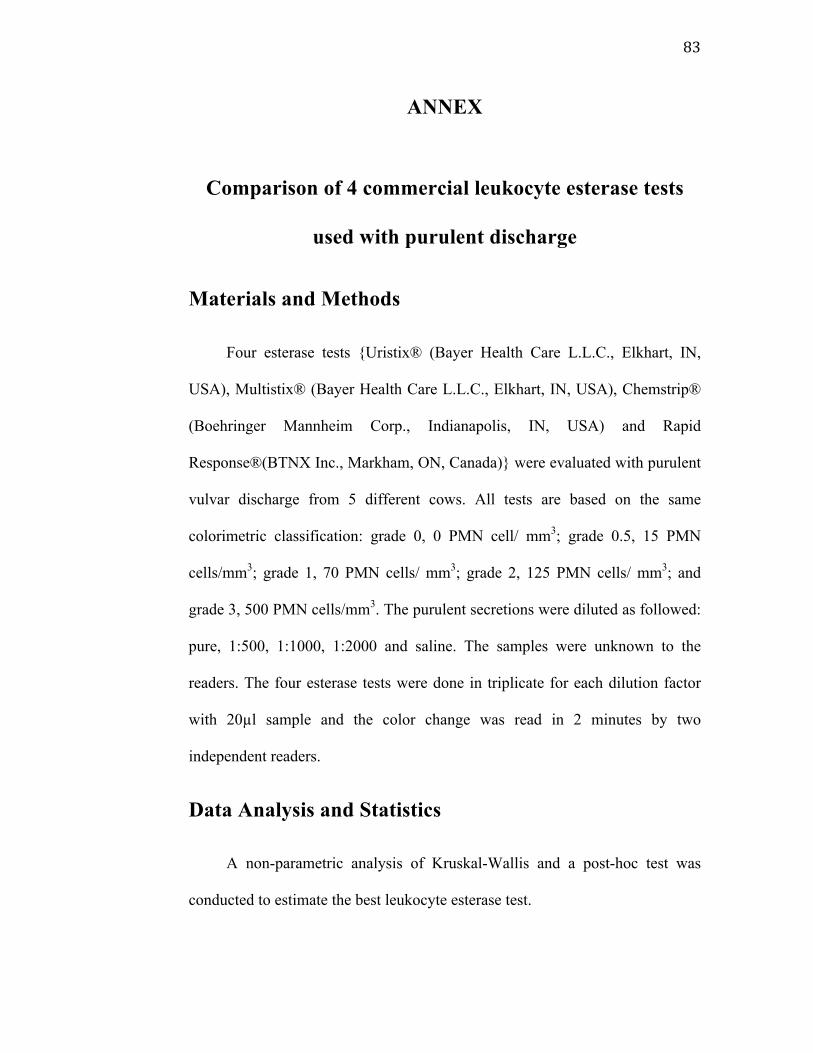
83
ANNEX
Comparison of 4 commercial leukocyte esterase tests
used with purulent discharge
Materials and Methods
Four esterase tests {Uristix® (Bayer Health Care L.L.C., Elkhart, IN,
USA), Multistix® (Bayer Health Care L.L.C., Elkhart, IN, USA), Chemstrip®
(Boehringer Mannheim Corp., Indianapolis, IN, USA) and Rapid
Response®(BTNX Inc., Markham, ON, Canada)} were evaluated with purulent
vulvar discharge from 5 different cows. All tests are based on the same
colorimetric classification: grade 0, 0 PMN cell/ mm3; grade 0.5, 15 PMN
cells/mm3; grade 1, 70 PMN cells/ mm3; grade 2, 125 PMN cells/ mm3; and
grade 3, 500 PMN cells/mm3. The purulent secretions were diluted as followed:
pure, 1:500, 1:1000, 1:2000 and saline. The samples were unknown to the
readers. The four esterase tests were done in triplicate for each dilution factor
with 20µl sample and the color change was read in 2 minutes by two
independent readers.
Data Analysis and Statistics
A non-parametric analysis of Kruskal-Wallis and a post-hoc test was
conducted to estimate the best leukocyte esterase test.
84
Results
There was a difference (P=0.0001) between the leukocyte esterase tests
with a purulent solution. The median score of the triplicates was significantly
lower for the RapidResponse® than for the 3 other tests and when the group
pure was considered positive and group saline negative, the sensitivity was only
21.4% (vs 100% for the other 3). At the dilution 1:1000, a difference (P=0.002)
between the leukocyte esterase tests indicated that the median score of the
RapidResponse® was significantly lower than the Multistix® and Uristix® and
there was no difference between the RapidResponse® and Chemstrip®. This
first experiment showed that 2 leukocyte esterase tests (Uristix® and
Multistix®) were similar in their responses and the leukocyte esterase test
Chemstrip® and RapidResponse® did not have a constant response. The
leukocyte esterase test Uristix® was chosen in this study.
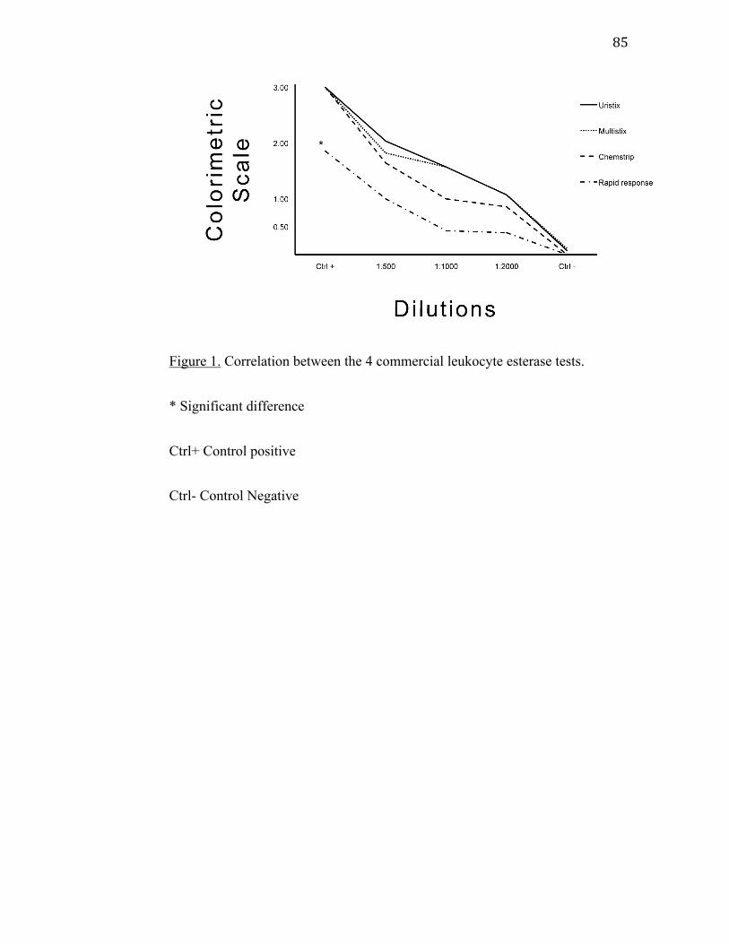
85
Figure 1. Correlation between the 4 commercial leukocyte esterase tests.
* Significant difference
Ctrl+ Control positive
Ctrl- Control Negative