The architecture of EMC reveals a path for membrane ...
30
*For correspondence: [email protected] † These authors contributed equally to this work Competing interest: See page 25 Funding: See page 25 Received: 15 April 2020 Accepted: 26 May 2020 Published: 27 May 2020 Reviewing editor: Volker Do ¨ tsch, Goethe University, Germany Copyright O’Donnell et al. This article is distributed under the terms of the Creative Commons Attribution License, which permits unrestricted use and redistribution provided that the original author and source are credited. The architecture of EMC reveals a path for membrane protein insertion John P O’Donnell 1† , Ben P Phillips 1† , Yuichi Yagita 1† , Szymon Juszkiewicz 1 , Armin Wagner 2 , Duccio Malinverni 1 , Robert J Keenan 3 , Elizabeth A Miller 1 , Ramanujan S Hegde 1 * 1 MRC Laboratory of Molecular Biology, Cambridge, United Kingdom; 2 Diamond Light Source, Didcot, United Kingdom; 3 Department of Biochemistry and Molecular Biology, The University of Chicago, Chicago, United States Abstract Approximately 25% of eukaryotic genes code for integral membrane proteins that are assembled at the endoplasmic reticulum. An abundant and widely conserved multi-protein complex termed EMC has been implicated in membrane protein biogenesis, but its mechanism of action is poorly understood. Here, we define the composition and architecture of human EMC using biochemical assays, crystallography of individual subunits, site-specific photocrosslinking, and cryo- EM reconstruction. Our results suggest that EMC’s cytosolic domain contains a large, moderately hydrophobic vestibule that can bind a substrate’s transmembrane domain (TMD). The cytosolic vestibule leads into a lumenally-sealed, lipid-exposed intramembrane groove large enough to accommodate a single substrate TMD. A gap between the cytosolic vestibule and intramembrane groove provides a potential path for substrate egress from EMC. These findings suggest how EMC facilitates energy-independent membrane insertion of TMDs, explain why only short lumenal domains are translocated by EMC, and constrain models of EMC’s proposed chaperone function. Introduction The endoplasmic reticulum (ER) is the site for biogenesis of nearly all eukaryotic integral membrane proteins (Shao and Hegde, 2011a). The defining feature of these proteins is the presence of one or more a-helical TMDs (von Heijne, 2007). Successful biogenesis requires each of these TMDs to be moved from the aqueous phase of the cytosol into the hydrophobic core of the lipid bilayer (Guna and Hegde, 2018; White and von Heijne, 2005). Although this insertion reaction can occur unassisted in vitro for some substrates (Brambillasca et al., 2005; Brambillasca et al., 2006), inser- tion in the crowded cellular environment typically requires factors that facilitate the reaction to mini- mize off-pathway outcomes such as aggregation, mislocalization, and degradation (Anghel et al., 2017; Guna et al., 2018; Heinrich et al., 2000; Samuelson et al., 2000; Wang et al., 2014). The best understood insertion factor is the protein translocation channel formed by the heterotri- meric Sec61 complex (Rapoport et al., 2017). Structural studies have demonstrated that the Sec61a subunit contains an hourglass pore across the membrane for polypeptide translocation (Van den Berg et al., 2004; Voorhees et al., 2014). The wall of this pore contains a lateral gate that opens to provide hydrophobic domains in a substrate access to the lipid bilayer (Gogala et al., 2014; Li et al., 2016; Voorhees and Hegde, 2016). Thus, Sec61 is thought to facilitate TMD insertion by its distinctive architecture that connects the aqueous environment in the cytosol to the hydrophobic environment inside the membrane. In addition to the Sec61 complex, the ER contains two other widely conserved insertases that both mediate the insertion of tail-anchored (TA) membrane proteins (Guna and Hegde, 2018). TA proteins contain a single TMD close to the C-terminus with a short unstructured domain translocated across the membrane (Kutay et al., 1993). This topology necessitates that the TMD is inserted post- O’Donnell et al. eLife 2020;9:e57887. DOI: https://doi.org/10.7554/eLife.57887 1 of 30 RESEARCH ARTICLE
Transcript of The architecture of EMC reveals a path for membrane ...
*For correspondence:
†These authors contributed
equally to this work
Competing interest: See
page 25
Funding: See page 25
Received: 15 April 2020
Accepted: 26 May 2020
Published: 27 May 2020
Reviewing editor: Volker
Dotsch, Goethe University,
Germany
Copyright O’Donnell et al.
This article is distributed under
the terms of the Creative
Commons Attribution License,
which permits unrestricted use
and redistribution provided that
the original author and source are
credited.
The architecture of EMC reveals a pathfor membrane protein insertionJohn P O’Donnell1†, Ben P Phillips1†, Yuichi Yagita1†, Szymon Juszkiewicz1,Armin Wagner2, Duccio Malinverni1, Robert J Keenan3, Elizabeth A Miller1,Ramanujan S Hegde1*
1MRC Laboratory of Molecular Biology, Cambridge, United Kingdom; 2DiamondLight Source, Didcot, United Kingdom; 3Department of Biochemistry and MolecularBiology, The University of Chicago, Chicago, United States
Abstract Approximately 25% of eukaryotic genes code for integral membrane proteins that are
assembled at the endoplasmic reticulum. An abundant and widely conserved multi-protein complex
termed EMC has been implicated in membrane protein biogenesis, but its mechanism of action is
poorly understood. Here, we define the composition and architecture of human EMC using
biochemical assays, crystallography of individual subunits, site-specific photocrosslinking, and cryo-
EM reconstruction. Our results suggest that EMC’s cytosolic domain contains a large, moderately
hydrophobic vestibule that can bind a substrate’s transmembrane domain (TMD). The cytosolic
vestibule leads into a lumenally-sealed, lipid-exposed intramembrane groove large enough to
accommodate a single substrate TMD. A gap between the cytosolic vestibule and intramembrane
groove provides a potential path for substrate egress from EMC. These findings suggest how EMC
facilitates energy-independent membrane insertion of TMDs, explain why only short lumenal
domains are translocated by EMC, and constrain models of EMC’s proposed chaperone function.
IntroductionThe endoplasmic reticulum (ER) is the site for biogenesis of nearly all eukaryotic integral membrane
proteins (Shao and Hegde, 2011a). The defining feature of these proteins is the presence of one or
more a-helical TMDs (von Heijne, 2007). Successful biogenesis requires each of these TMDs to be
moved from the aqueous phase of the cytosol into the hydrophobic core of the lipid bilayer
(Guna and Hegde, 2018; White and von Heijne, 2005). Although this insertion reaction can occur
unassisted in vitro for some substrates (Brambillasca et al., 2005; Brambillasca et al., 2006), inser-
tion in the crowded cellular environment typically requires factors that facilitate the reaction to mini-
mize off-pathway outcomes such as aggregation, mislocalization, and degradation (Anghel et al.,
2017; Guna et al., 2018; Heinrich et al., 2000; Samuelson et al., 2000; Wang et al., 2014).
The best understood insertion factor is the protein translocation channel formed by the heterotri-
meric Sec61 complex (Rapoport et al., 2017). Structural studies have demonstrated that the Sec61a
subunit contains an hourglass pore across the membrane for polypeptide translocation (Van den
Berg et al., 2004; Voorhees et al., 2014). The wall of this pore contains a lateral gate that opens to
provide hydrophobic domains in a substrate access to the lipid bilayer (Gogala et al., 2014;
Li et al., 2016; Voorhees and Hegde, 2016). Thus, Sec61 is thought to facilitate TMD insertion by
its distinctive architecture that connects the aqueous environment in the cytosol to the hydrophobic
environment inside the membrane.
In addition to the Sec61 complex, the ER contains two other widely conserved insertases that
both mediate the insertion of tail-anchored (TA) membrane proteins (Guna and Hegde, 2018). TA
proteins contain a single TMD close to the C-terminus with a short unstructured domain translocated
across the membrane (Kutay et al., 1993). This topology necessitates that the TMD is inserted post-
O’Donnell et al. eLife 2020;9:e57887. DOI: https://doi.org/10.7554/eLife.57887 1 of 30
RESEARCH ARTICLE
translationally. The ‘guided entry of TA proteins’ (GET) pathway (Chio et al., 2017; Hegde and
Keenan, 2011) culminates at a heterodimeric complex (made of the ER-resident membrane proteins
Get1 and Get2) that inserts TA proteins delivered to it by the targeting factor Get3
(Mariappan et al., 2011; Wang et al., 2014). More recently, the ten-subunit ‘ER membrane protein
complex’ (EMC) (Christianson et al., 2012; Jonikas et al., 2009) was shown to insert TA proteins
whose TMDs are insufficiently hydrophobic to effectively engage TRC40, the mammalian homolog
of Get3 (Guna et al., 2018).
In addition to TA proteins, EMC mediates co-translational insertion of TMDs close to the N-termi-
nus in the Nexo topology (defined by a translocated N-terminus) (Chitwood et al., 2018). Notably,
the translocated domain is short and unstructured. When the N-terminus is extended and preceded
by a signal peptide, insertion is no longer EMC-dependent and occurs instead via the Sec61a lateral
gate (Chitwood et al., 2018). Although the topology of Nexo TMDs is opposite to the TMDs of TA
proteins, they are both terminal TMDs whose insertion is not accompanied by appreciable polypep-
tide translocation. The EMC-mediated insertion reactions of both types of terminal TMDs has been
reconstituted with purified EMC in vitro (Chitwood et al., 2018; Guna et al., 2018), suggesting that
they might use similar mechanisms (Chitwood and Hegde, 2019).
The ten subunits of mammalian EMC (termed EMC1 through EMC10) are poorly understood
because they have very few clearly established domains or resemblance to proteins of known struc-
ture or biochemical activity (Wideman, 2015; Figure 1A). The one possible exception is the three-
TMD protein EMC3, which is predicted to be topologically and evolutionarily related to Get1 and a
subdomain of the prokaryotic insertase YidC (Anghel et al., 2017). It has been speculated that this
potential structural similarity reflects a similarity in molecular function.
The membrane subunit EMC3 is in complex with six other integral membrane EMC subunits (1, 4,
5, 6, 7, and 10) that together contain 12 predicted TMDs (Chitwood and Hegde, 2019;
Christianson et al., 2012; Wideman, 2015). The seven membrane subunits of EMC associate with
the cytosolic subunits EMC2, EMC8, and EMC9. EMC8 and EMC9 are ~44% identical in mammals,
and not all species contain both genes (Wideman, 2015). Whether they are both part of a single 10-
protein complex or substitute for each other in a 9-protein complex is not known. No free popula-
tion of individual subunits has been detected (Chitwood et al., 2018; Guna et al., 2018;
Volkmar et al., 2019), and disruption of most EMC subunits causes loss of EMC integrity and func-
tion (Volkmar et al., 2019). Thus, EMC is thought to function as a stable complex to mediate TMD
insertion. Notably, this reaction appears to be energy independent in reconstitution assays in vitro
(Guna et al., 2018), consistent with the absence of any nucleotide-binding domain in any of its subu-
nits (Wideman, 2015).
EMC has also been suggested to act as a co-translational chaperone that captures individual or
bundles of TMDs as they exit laterally from the Sec61 complex (Shurtleff et al., 2018). This postu-
lated function has been inferred from analysis of EMC-mediated co-translational proximity biotinyla-
tion of ribosomes translating membrane proteins. How EMC might act as a chaperone, and the
relationship of this function to its insertase activity, is not known. Consistent with either function,
many membrane proteins are partially or strongly impacted in their biogenesis by the loss of EMC in
numerous organisms including yeast, worms, flies, and mammals (Bircham et al., 2011;
Chitwood et al., 2018; Guna et al., 2018; Lakshminarayan et al., 2020; Louie et al., 2012;
Richard et al., 2013; Satoh et al., 2015; Shurtleff et al., 2018; Talbot et al., 2019; Taylor et al.,
2005; Volkmar et al., 2019). Thus, EMC is a highly abundant component of the ER needed for cellu-
lar and organism homeostasis.
Understanding the function(s) of EMC requires knowledge of its structure. By analogy to the
Sec61 complex, EMC’s insertase function might involve a path from the cytosol into the membrane.
To investigate this idea, we used a combination of biochemical, biophysical, and structural
approaches to determine the architecture of EMC. Our findings suggest that the cytosolic subunits
initially engage a TMD in a weakly hydrophobic vestibule that is contiguous with an intramembrane
groove open to the lipid bilayer. This work provides a mechanistic framework for how the highly
abundant and conserved EMC functions as a TMD insertase, explains why EMC acts preferentially on
terminal TMDs, and suggests that EMC is unlikely to chaperone TMDs released at the Sec61a lateral
gate.
O’Donnell et al. eLife 2020;9:e57887. DOI: https://doi.org/10.7554/eLife.57887 2 of 30
Research article Biochemistry and Chemical Biology Structural Biology and Molecular Biophysics
Results
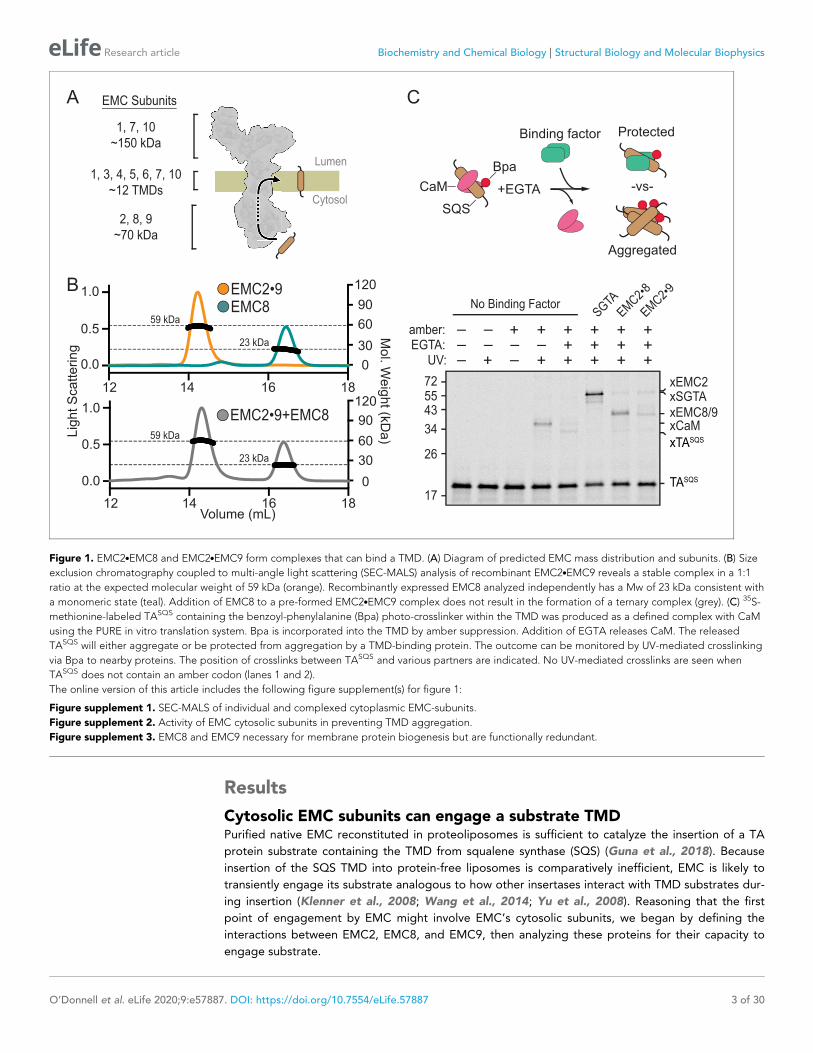
Cytosolic EMC subunits can engage a substrate TMDPurified native EMC reconstituted in proteoliposomes is sufficient to catalyze the insertion of a TA
protein substrate containing the TMD from squalene synthase (SQS) (Guna et al., 2018). Because
insertion of the SQS TMD into protein-free liposomes is comparatively inefficient, EMC is likely to
transiently engage its substrate analogous to how other insertases interact with TMD substrates dur-
ing insertion (Klenner et al., 2008; Wang et al., 2014; Yu et al., 2008). Reasoning that the first
point of engagement by EMC might involve EMC’s cytosolic subunits, we began by defining the
interactions between EMC2, EMC8, and EMC9, then analyzing these proteins for their capacity to
engage substrate.
A EMC Subunits
1, 7, 10~150 kDa
2, 8, 9~70 kDa
1, 3, 4, 5, 6, 7, 10~12 TMDs
Lumen
Cytosol
12 14 16 18
Lig
ht S
catte
rin
g
Mo
l. We
igh
t (kDa
)
1.0
0.5
120
90
60
30
00.0
12 14 16 18Volume (mL)
120
90
60
30
0
1.0
0.5
0.0
B EMC2•9EMC8
EMC2•9+EMC8
59 kDa
23 kDa
59 kDa
23 kDa
No Binding FactorSGTA
EMC2•8
EMC2•9
725543
34
26
17
xSGTAxEMC2
xEMC8/9xCaM
TASQS
xTASQS
EGTA:UV:
– –– –
– –++
amber: +– – + + + + ++ + + ++ + + +
C
Bpa
Binding factor
SQS
CaM +EGTA
Protected
-vs-
Aggregated
Figure 1. EMC2.EMC8 and EMC2.EMC9 form complexes that can bind a TMD. (A) Diagram of predicted EMC mass distribution and subunits. (B) Size
exclusion chromatography coupled to multi-angle light scattering (SEC-MALS) analysis of recombinant EMC2.EMC9 reveals a stable complex in a 1:1
ratio at the expected molecular weight of 59 kDa (orange). Recombinantly expressed EMC8 analyzed independently has a Mw of 23 kDa consistent with
a monomeric state (teal). Addition of EMC8 to a pre-formed EMC2.EMC9 complex does not result in the formation of a ternary complex (grey). (C) 35S-
methionine-labeled TASQS containing the benzoyl-phenylalanine (Bpa) photo-crosslinker within the TMD was produced as a defined complex with CaM
using the PURE in vitro translation system. Bpa is incorporated into the TMD by amber suppression. Addition of EGTA releases CaM. The released
TASQS will either aggregate or be protected from aggregation by a TMD-binding protein. The outcome can be monitored by UV-mediated crosslinking
via Bpa to nearby proteins. The position of crosslinks between TASQS and various partners are indicated. No UV-mediated crosslinks are seen when
TASQS does not contain an amber codon (lanes 1 and 2).
The online version of this article includes the following figure supplement(s) for figure 1:
Figure supplement 1. SEC-MALS of individual and complexed cytoplasmic EMC-subunits.
Figure supplement 2. Activity of EMC cytosolic subunits in preventing TMD aggregation.
Figure supplement 3. EMC8 and EMC9 necessary for membrane protein biogenesis but are functionally redundant.
O’Donnell et al. eLife 2020;9:e57887. DOI: https://doi.org/10.7554/eLife.57887 3 of 30
Research article Biochemistry and Chemical Biology Structural Biology and Molecular Biophysics

Purified recombinant EMC2 formed a stable complex with either EMC8 or EMC9. Size exclusion
chromatography coupled to multi-angle light scattering (SEC-MALS) showed that each individual
protein is monomeric and the EMC2.EMC8 and EMC2.EMC9 complexes are heterodimers
(Figure 1B; Figure 1—figure supplement 1). The EMC2.EMC9 heterodimer did not form a ternary
complex with excess EMC8, and the EMC2.EMC8 heterodimer did not form a ternary complex with
excess EMC9. Thus, the cytosolic domain of EMC is likely to be composed of EMC2 in complex with
either EMC8 or EMC9, but not both. The presence of only one of either EMC8 or EMC9 in native
EMC may explain why some species have only one of these two genes (Wideman, 2015).
To analyze substrate interaction, a 35S-labeled TA protein containing the TMD of SQS (TASQS)
was produced in vitro using a fully purified translation system derived from E. coli components
(Shimizu and Ueda, 2010). The photocrosslinking amino acid 4-Benzoylphenylalanine (Bpa) was
incorporated within the TMD by amber suppression and TASQS was kept soluble by including an
excess of the chaperone-like protein calmodulin (CaM) (Guna et al., 2018; Shao and Hegde,
2011b). UV irradiation of this complex produced a TASQS-CaM crosslinked product which was dimin-
ished if CaM was inactivated by chelation of Ca2+ with EGTA (Figure 1C, lanes 4, 5). Without a chap-
erone, SQS formed self-crosslinks due to its aggregation as documented previously (Guna et al.,
2018). Release of SQS from CaM in the presence of EMC2.EMC8 or EMC2.EMC9 showed crosslinks
to EMC2, EMC8, and EMC9 concomitant with a reduction of the TASQS aggregate crosslink
(Figure 1C, lanes 7,8). This effect of the EMC2.EMC8 and EMC2.EMC9 complexes is similar, but
less complete, than the effect seen with the TMD chaperone SGTA (Shao et al., 2017; Figure 1C,
lane 6). Analysis of individual subunits at various concentrations showed that EMC2 inhibited aggre-
gation with a similar potency as the heterodimeric complexes, whereas EMC8 showed somewhat
lower potency and EMC9 was largely inert (Figure 1—figure supplement 2).
These observations indicate that the cytosolic domain of EMC contains a TMD-binding hetero-
dimer of the EMC2.EMC8 subcomplex or the homologous EMC2.EMC9 subcomplex. Interchange-
ability of EMC8 and EMC9 explains why knockdown of EMC8 is accompanied by increased EMC9
(Volkmar et al., 2019), and why individual knockdowns do not impact EMC substrates but the dou-
ble-knockdown does (Figure 1—figure supplement 3). Whether there are functional differences
between EMC8- versus EMC9-containing EMC for certain substrates remains to be determined.
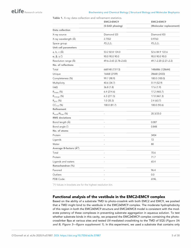
Crystal structure of the EMC2.EMC9 complexRecombinant cytosolic subunits and subcomplexes were screened for crystal formation, resulting in
well-diffracting crystals of a nearly full length complex containing EMC2 and EMC9. This complex,
lacking only short regions at the termini, was verified to be functional for substrate interaction by the
photo-crosslinking assay (data not shown). The EMC2.EMC9 structure was solved with experimental
phases from a single-wavelength anomalous diffraction (SAD) experiment using endogenous sulphur
atoms for anomalous signal (Wagner et al., 2016). An initial model was built de novo and subse-
quently used as a molecular replacement search model for a native crystal diffracting X-rays to 2.2 A
(Table 1).
EMC2 is largely alpha-helical and contains a curved tetratricopeptide repeat (TPR) motif but-
tressed on one side by EMC9 (Figure 2A). The core of EMC9 consists of a small b-barrel flanked by
alpha-helices. The ~1100 A2 EMC2-EMC9 interface contains a network of hydrogen bonds and salt
bridges (Krissinel and Henrick, 2007), explaining its high stability in vitro and the absence of any
appreciable free population of either protein in cells. EMC2 and EMC9 both contribute to the forma-
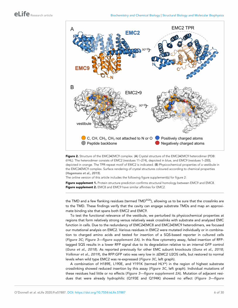
tion of a large, relatively shallow and moderately hydrophobic cavity that we term the cytosolic vesti-
bule (Figure 2B).
A reference-free model of EMC8 predicted by Rosetta (Rohl et al., 2004) possessed the same
core fold as the EMC9 structure with a RMSD of 2.3 A. When EMC9 was used as a reference
(Song et al., 2013), the alpha-carbon backbone of the EMC8 model was indistinguishable from
EMC9 with an RMSD of 0.6 A (Figure 2—figure supplement 1). This observation is consistent with
their high homology (~44% identity) and comparable affinity for EMC2 (Figure 2—figure supple-
ment 2). We therefore conclude that the EMC2.EMC8 structure is likely to be very similar to
EMC2.EMC9 explaining why the absence of either one has no obvious phenotype in cells.
O’Donnell et al. eLife 2020;9:e57887. DOI: https://doi.org/10.7554/eLife.57887 4 of 30
Research article Biochemistry and Chemical Biology Structural Biology and Molecular Biophysics
Functional analysis of the vestibule in the EMC2.EMC9 complexBased on the ability of a substrate TMD to photo-crosslink with both EMC2 and EMC9, we posited
that a TMD might bind to the vestibule in the EMC2.EMC9 complex. The moderate hydrophobicity
of this region in both the EMC2.EMC9 structure and EMC2.EMC8 model is consistent with the mod-
erate potency of these complexes in preventing substrate aggregation in aqueous solution. To test
whether substrate binds in this cavity, we prepared the EMC2.EMC9 complex containing the photo-
crosslinker Bpa at various sites and tested UV-mediated crosslinking to the TMD of SQS (Figure 3A
and B; Figure 3—figure supplement 1). In this experiment, we used a substrate that contains only
Table 1. X-ray data collection and refinement statistics.
EMC2.EMC9 EMC2.EMC9
(S-SAD phasing) (Molecular replacement)
Data collection
X-ray source Diamond I23 Diamond I03
X-ray wavelength (A) 2.7552 0.9763
Space group P212121 P212121
Unit cell parameters
a, b, c (A) 53.2 82.8 124.0 52.6 84.9 122.6
a, b, g (˚) 90.0 90.0 90.0 90.0 90.0 90.0
Resolution range (A) 49.6–2.65 (2.78–2.65) 49.7–2.20 (2.27–2.2)
No. of reflections
Total 668148 (73113) 1486886 (128644)
Unique 16468 (2109) 28668 (2433)
Completeness (%) 99.7 (98.9) 100.0 (100.0)
Multiplicity 40.6 (34.7) 51.9 (52.9)
I/s(I) 36.8 (1.8) 17.6 (1.9)
Rmeas (%) 6.4 (214.6) 17.2 (465.7)
Rmerge (%) 6.3 (211.5) 17.0 (461.3)
Rpim (%) 1.0 (35.3) 2.4 (63.7)
CC1/2 (%) 100.0 (81.7) 100.0 (92.6)
Refinement
Rwork/Rfree (%) - 20.3/25.0
RMS deviations
Bond length (A) - 0.007
Bond angle (˚) - 0.848
No. of atoms
Protein - 3454
Ligands - 72
Water - 80
Average B-factors (A2)
Total - 70.6
Protein - 71.7
Ligands and waters - 65.4
Ramachandran (%)
Favored - 96.4
Outliers - 0.0
PDB Code: - 6Y4L
*(*) Values in brackets are for the highest resolution bin.
O’Donnell et al. eLife 2020;9:e57887. DOI: https://doi.org/10.7554/eLife.57887 5 of 30
Research article Biochemistry and Chemical Biology Structural Biology and Molecular Biophysics
the TMD and a few flanking residues (termed TMDSQS), allowing us to be sure that the crosslinks are
to the TMD. These findings verify that the cavity can engage substrate TMDs and map an approxi-
mate binding site that spans both EMC2 and EMC9.
To test the functional relevance of the vestibule, we perturbed its physicochemical properties at
regions that form relatively strong versus relatively weak crosslinks with substrate and analyzed EMC
function in cells. Due to the redundancy of EMC2.EMC8 and EMC2.EMC9 heterodimers, we focused
our mutational analysis on EMC2. Various residues in EMC2 were mutated individually or in combina-
tion to charged amino acids and tested for insertion of a SQS-based reporter in cultured cells
(Figure 3C; Figure 3—figure supplement 2A). In this flow cytometry assay, failed insertion of RFP-
tagged SQS results in a lower RFP signal due to its degradation relative to an internal GFP control
(Guna et al., 2018). As reported previously for other EMC subunit knockouts (Guna et al., 2018;
Volkmar et al., 2019), the RFP:GFP ratio was very low in DEMC2 U2OS cells, but restored to normal
levels when wild type EMC2 was re-expressed (Figure 3C, left graph).
A combination of H189E, L190E, and Y191K (termed HLY*) in the region of highest substrate
crosslinking showed reduced insertion by this assay (Figure 3C, left graph). Individual mutations of
these residues had little or no effects (Figure 3—figure supplement 2A). Mutation of adjacent resi-
dues that were already hydrophilic (Q193E and Q194K) showed no effect (Figure 3—figure
BEMC2•9
vestibule
C, CH, CH2, CH3 not attached to N or O
Peptide backbone Negatively charged atoms
Positively charged atoms
AAEMC2
EMC9
N
C
C
C
N
N
EMC2 TPR
90º
90º
Figure 2. Structure of the EMC2.EMC9 complex. (A) Crystal structure of the EMC2.EMC9 heterodimer (PDB:
6Y4L). The heterodimer consists of EMC2 (residues 11–274), depicted in blue, and EMC9 (residues 1–200),
depicted in orange. The TPR-repeat motif of EMC2 is indicated. (B) Physicochemical properties of a vestibule in
the EMC2.EMC9 complex. Surface rendering of crystal structures coloured according to chemical properties
(Hagemans et al., 2015).
The online version of this article includes the following figure supplement(s) for figure 2:
Figure supplement 1. Protein structure prediction confirms structural homology between EMC9 and EMC8.
Figure supplement 2. EMC8 and EMC9 have similar affinities for EMC2.
O’Donnell et al. eLife 2020;9:e57887. DOI: https://doi.org/10.7554/eLife.57887 6 of 30
Research article Biochemistry and Chemical Biology Structural Biology and Molecular Biophysics
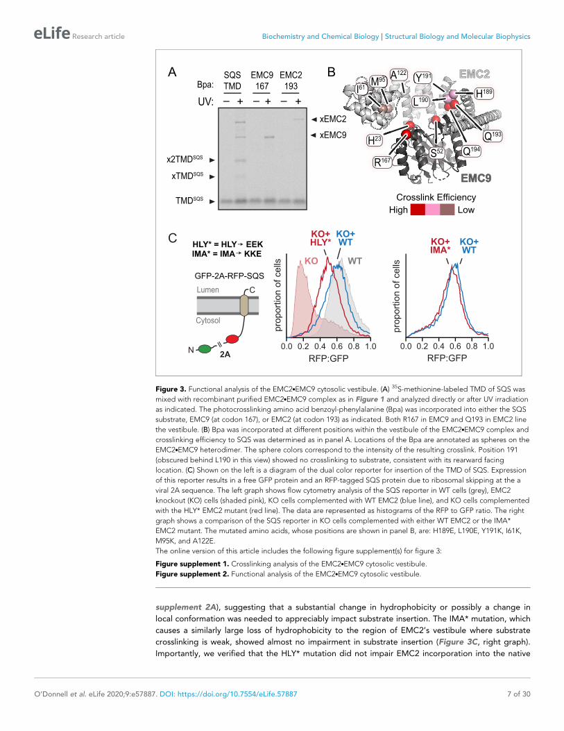
supplement 2A), suggesting that a substantial change in hydrophobicity or possibly a change in
local conformation was needed to appreciably impact substrate insertion. The IMA* mutation, which
causes a similarly large loss of hydrophobicity to the region of EMC2’s vestibule where substrate
crosslinking is weak, showed almost no impairment in substrate insertion (Figure 3C, right graph).
Importantly, we verified that the HLY* mutation did not impair EMC2 incorporation into the native
GFP-2A-RFP-SQS
RFP:GFP
1.00.80.60.40.20.0
KO WT
KO+WT
KO+HLY*
pro
po
rtio
n o
f ce
lls
pro
po
rtio
n o
f ce
lls
HLY* = HLY EEKIMA* = IMA KKE
Cytosol
Lumen
N
C
2A
C
A
UV:
Bpa:
– + – + – +
EMC9167
EMC2193
SQSTMD
xEMC2
xEMC9
TMDSQS
xTMDSQS
x2TMDSQS
B
MC2
MC9
Crosslink Efficiency
High Low
H189
Q193Q
Q194
L190
R167S52
I6161M95
H23
Y19195 YA122
KO+IMA*
KO+WT
RFP:GFP1.00.80.60.40.20.0
EMC2
EMC9
Figure 3. Functional analysis of the EMC2.EMC9 cytosolic vestibule. (A) 35S-methionine-labeled TMD of SQS was
mixed with recombinant purified EMC2.EMC9 complex as in Figure 1 and analyzed directly or after UV irradiation
as indicated. The photocrosslinking amino acid benzoyl-phenylalanine (Bpa) was incorporated into either the SQS
substrate, EMC9 (at codon 167), or EMC2 (at codon 193) as indicated. Both R167 in EMC9 and Q193 in EMC2 line
the vestibule. (B) Bpa was incorporated at different positions within the vestibule of the EMC2.EMC9 complex and
crosslinking efficiency to SQS was determined as in panel A. Locations of the Bpa are annotated as spheres on the
EMC2.EMC9 heterodimer. The sphere colors correspond to the intensity of the resulting crosslink. Position 191
(obscured behind L190 in this view) showed no crosslinking to substrate, consistent with its rearward facing
location. (C) Shown on the left is a diagram of the dual color reporter for insertion of the TMD of SQS. Expression
of this reporter results in a free GFP protein and an RFP-tagged SQS protein due to ribosomal skipping at the a
viral 2A sequence. The left graph shows flow cytometry analysis of the SQS reporter in WT cells (grey), EMC2
knockout (KO) cells (shaded pink), KO cells complemented with WT EMC2 (blue line), and KO cells complemented
with the HLY* EMC2 mutant (red line). The data are represented as histograms of the RFP to GFP ratio. The right
graph shows a comparison of the SQS reporter in KO cells complemented with either WT EMC2 or the IMA*
EMC2 mutant. The mutated amino acids, whose positions are shown in panel B, are: H189E, L190E, Y191K, I61K,
M95K, and A122E.
The online version of this article includes the following figure supplement(s) for figure 3:
Figure supplement 1. Crosslinking analysis of the EMC2.EMC9 cytosolic vestibule.
Figure supplement 2. Functional analysis of the EMC2.EMC9 cytosolic vestibule.
O’Donnell et al. eLife 2020;9:e57887. DOI: https://doi.org/10.7554/eLife.57887 7 of 30
Research article Biochemistry and Chemical Biology Structural Biology and Molecular Biophysics
EMC (Figure 3—figure supplement 2B), unlike the A129K mutation whose strong phenotype (Fig-
ure 3—figure supplement 2A) could be ascribed to poor assembly (Figure 3—figure supplement
2C). Thus, there is concordance between EMC2 regions of the vestibule that interact with a TMD
substrate in vitro and mutations that perturb substrate insertion in cells.
Position of the EMC2.EMC9 subcomplex within native EMCOnce a TMD substrate binds to the cytosolic subunits of EMC, subsequent insertion requires access
to the lipid bilayer. To understand how the substrate-binding cavity within the cytosolic subunits is
oriented relative to the membrane, we sought to place our EMC2.EMC9 structure within the archi-
tecture of native EMC. Affinity-purified EMC representing a mixture of EMC8- and EMC9-containing
complexes (Guna et al., 2018) were analyzed by single-particle cryo-EM. The map clearly shows
density for the lumenal, transmembrane, and cytoplasmic regions of EMC (Figure 4A). Due to pref-
erential orientation, the resulting density map was limited to modest resolution of (6.5 A) throughout
the structure (Table 2; Figure 4—figure supplement 1).
Although atomic models could not be built de novo from the EM map, this resolution was suffi-
cient to dock the EMC2.EMC9 crystal structure. The only region of EMC2.EMC9 that did not pre-
cisely align with the EM-density was the first three alpha-helices of EMC2 comprising residues 11–66
(Figure 4—figure supplement 2). Low frequency normal mode analysis (Suhre and Sanejouand,
2004) predicted that these three helices undergo structural movement that would be compatible
with the EM-density. Therefore, the EMC2.EMC9 structure was refined against the EM-density using
Flex-EM, (Topf et al., 2008), Coot (Emsley et al., 2010), and PHENIX real-space refinement
(Afonine et al., 2018), resulting in a slightly rotated position that fits into the EM-density
(Figure 4B; Figure 4—figure supplement 2).
The plane of the membrane was evident from the detergent micelle surrounding the TMD region
of EMC (Figure 4A). Relative to the membrane, the EMC2.EMC9 complex is oriented such that the
TPR-repeats of EMC2 are proximal to the membrane but angled at ~30˚. In this configuration, the
substrate binding cavity of EMC2.EMC9 has access to both the bulk cytosol and the membrane
domain of EMC (Figure 4B). The surface of EMC2 that faces the membrane domain is also highly
conserved, consistent with this region making contacts with the membrane-embedded subunits of
EMC (Figure 4C). Thus, the cytosolic subunits of EMC are arranged so the cavity capable of binding
substrate forms a vestibule that links the cytosol to the integral membrane subunits that would act
next to mediate TMD insertion.
The region of the vestibule that binds substrates as determined in crosslinking assays is occupied
in the cryo-EM map by density that is contributed from another EMC subunit (possibly EMC6, as dis-
cussed below). Intramolecular placeholders that temporarily shield the substrate-binding pockets are
also observed in the membrane protein targeting factors SRP and Get3 (Mateja et al., 2015;
Voorhees and Hegde, 2015). In both of these other examples, the placeholders are less hydropho-
bic than substrate TMDs, allowing their displacement by bona fide substrates but presumably not
other proteins. EMC may therefore operate similarly. Thus the putative placeholder density might
provide an approximation of what a substrate-bound intermediate of EMC looks like. From this posi-
tion, an inserting substrate would next have to engage the region of EMC embedded in the
membrane.
Architecture of the membrane-embedded and lumenal regions of EMCA cross section through the detergent micelle of the EMC map in the plane of the membrane
showed the arrangement of thirteen putative TMD helices (Figure 5A). To assign the intramembrane
densities to individual EMC subunits, we first generated starting models for those that contain two
or more TMDs. Using trRosetta (Yang et al., 2020), which employs co-evolutionary data, deep learn-
ing, and inter-residue contacts for energy minimization of structural models, we produced models
for EMC3, EMC4, EMC5, and EMC6 (Figure 5—figure supplement 1). Even though trRosetta does
not consider biological membranes or topology, the predicted TMDs of EMC3 and EMC5 pack
together as helices whose lengths match the thickness of a lipid bilayer. Strikingly, trRosetta accu-
rately predicted the structures of EMC2, EMC9, and Sec61a, providing confidence in its capacity to
produce starting models for both soluble and integral membrane domains.
O’Donnell et al. eLife 2020;9:e57887. DOI: https://doi.org/10.7554/eLife.57887 8 of 30
Research article Biochemistry and Chemical Biology Structural Biology and Molecular Biophysics

Although we had previously predicted two TMDs for EMC6 based on hydrophobicity profiles
(Krogh et al., 2001), trRosetta generated a three-helix bundle (Figure 5—figure supplement 1)
that matched the consensus of other topology prediction algorithms (Tsirigos et al., 2015). More
surprisingly, EMC4 also is predicted by trRosetta to have three TMD-like helices, not two as previ-
ously thought based on topology algorithms. Protease-protection assays resolved this discrepancy in
favor of the trRosetta model because we found that the N-terminus of EMC4 faces the cytosol and
the C-terminus is in the ER lumen (Figure 5—figure supplement 2).
EMC2
EMC9
B
90º
A
Lumen
TM region
Cytosol
detergent micelle
C
90º
ConservationLow High
90º
C
90º
ConserervaLow
Membrane Plane
potentialplaceholder
Figure 4. The position of EMC2.EMC9 within native EMC. (A) Cryo-EM map of the EMC at 6.4 A resolution reveals
the architecture of the complex. The map is shown at two contour levels: a stringent contour that illustrates
secondary structure features (0.21) superimposed with a liberal contour that shows the detergent micelle (0.15). (B)
Refinement of the EMC2.EMC9 crystal structure (blue and orange) into the cytosolic density (grey) using Flex-EM,
Coot and PHENIX. (C) Surface rendering of the EMC2.EMC9 crystal structure coloured by residue conservation
(Ashkenazy et al., 2016) from highly conserved (purple) to weakly conserved (green). The top rim of EMC2’s TPR
is highly conserved. This surface faces the membrane and regions of it interact with other EMC-subunits. The
substrate binding vestibule also exhibits high conservation in comparison to the remaining solvent exposed
surface of the EMC2.EMC9 heterodimer.
The online version of this article includes the following figure supplement(s) for figure 4:
Figure supplement 1. Cryo-electron microscopy data processing.
Figure supplement 2. Normal mode analysis and flexible fitting of the EMC2.EMC9 crystal structure into the full
EMC cryo-EM map.
O’Donnell et al. eLife 2020;9:e57887. DOI: https://doi.org/10.7554/eLife.57887 9 of 30
Research article Biochemistry and Chemical Biology Structural Biology and Molecular Biophysics
The trRosetta models for EMC3, EMC6, and EMC5 could be docked at distinctive positions into
the EM map based on their helix lengths and relative tilts (Figure 5B). The main remaining region
that could accommodate a three-helix bundle was therefore assigned to EMC4. This left two TMD-
like densities and three single-spanning EMC subunits (EMC1, EMC7, and EMC10). Because EMC10
can be depleted with no functional consequences (Volkmar et al., 2019), we suspected this subunit
was probably peripheral and least likely to be visualized in the map. We therefore assigned the two
remaining TMD-like densities to EMC1 and EMC7, distinguishing between them using a site-specific
photo-crosslinking approach.
The photo-crosslinking amino acid 3’-azibutyl-N-carbamoyl-lysine (AbK) was introduced by amber
suppression (Ai et al., 2011) into specific sites of individual FLAG-tagged EMC subunits. The cells
were then UV-irradiated to induce crosslinks with nearby proteins (with a backbone-to-backbone dis-
tance of ~10–15 A) and the samples were analyzed by immunoprecipitation and immunoblotting.
Three and four sequential positions were tested within a TMD to sample different radial directions
(McCormick et al., 2003). Introduction of AbK at position I23AbK in the first TMD of EMC3 showed a
strong high-molecular weight crosslink consistent with the size of EMC1. EMC1 immunoblotting of
natively purified EMC via the FLAG-tagged EMC3 verified that the UV-dependent EMC3 crosslinking
partner is EMC1. Denaturing IP of EMC1 followed by anti-FLAG immunoblotting further validated
this assignment. The EMC1 crosslink is sharply diminished at positions T24AbK and F25AbK, but is par-
tially recovered at position F26AbK, thereby defining the face of this TMD helix that is adjacent to
EMC1 (Figure 5C).
Table 2. Cryo-EM data collection and processing.
Dataset 1 Dataset 2 Dataset 3 Dataset 4 Dataset 5
Microscope Titan Krios (m06 eBIC) Titan Krios (m06 eBIC) Titan Krios (m06 eBIC) Titan Krios(MRC-LMB)
Titan Krios(MRC-LMB)
Pixel Size 1.380 1.380 1.380 1.179 1.390
Voltage 300 300 300 300 300
Spherical Aberation 2.7 2.7 2.7 2.7 2.7
Total exposure (e-/A2) 39.60 42.50 37.77 39.36 44.36
Exposure Length (s) 5.0 11.02 14 11 11
Frames 25 44 40 44 40
Defocus Range (mm) �0.5 to �1.5 �0.5 to �1.5 �0.5 to �1.5 �0.5 to �1.5 �0.5 to �1.5
Micrographs 2776 2484 1206 4228 932
Microscope tilt (degrees) 0 0 30 20 20
Volta Phase Plate [ [ [ [ [
Pre-merge processing Dataset 1 Dataset 2 Dataset 3 Dataset 4 Dataset 5
Motion Correction and CTF estimation (micrographs) 2776 2484 1206 4228 932
Blob-based autopicking (particles) 1,298,488 541,233 319,989 1,145,157 223,613
2x iterations of 2D classification (particles) 63,180 63,315 51,371 305,685 28,662
Template based autopicking (particles) 467,323 687,645 826,006 N/A 273,852
2D classification (particles) 11,862 71,977 63,881 113,852 66,799
Ab Initio 3D classification (particles) 103,105 71,977 46,349 113,852 50,232
Post-merge processing Combined Datasets
2x Iterations ab initio3D classification (particles)
405,515
Non-uniform Refinement (particles) 167,294
Per-particle CTF refinement 6.71 A map
Non-uniform refinementwith local resolution estimation and filtering
6.4 A map
EMDB Deposition code EMD-11058
O’Donnell et al. eLife 2020;9:e57887. DOI: https://doi.org/10.7554/eLife.57887 10 of 30
Research article Biochemistry and Chemical Biology Structural Biology and Molecular Biophysics

90º
A
D
1TM1
3TM1
3TM2
3TM3
4TM3
4TM2
4TM1
7TM1
6TM2
6TM3
5TM1
5TM2
view from
ER lumen
6TM1
UV: – +
EMC3-3xFLAG
72
55
43
34
95130
α-FLAG
– + – +
WB:
Denat. IPα-EMC1
α-FLAG
Nat. IPα-FLAG
α-EMC1
EMC1
1 x 3
Lysate
– + – + – +
23 24 25 26 23 23
C
EMC3
amb:
detergentmicelle
BBBBBBBBBBB
90º
β-propsE
β-props
90º
crossbar
EMC7
EMC10
EMC10EMC7
Fview from
ER lumen
crossbar
EMC1crossbarER lumen
cytosol
EMC1
EMC5
EMC4
EMC6
EMC3
Figure 5. A composite model of EMC’s membrane and lumenal domains. (A) Cross section through the TMD region of the cryo-EM map of EMC (light
grey) identifies 13 helix-like densities (dark blue) that define an intramembrane groove (red star) open to the lipid bilayer. Density corresponding to the
annular detergent micelle is indicated. (B) Ab initio trRosetta models fitted into cryo-EM density for the EMC membrane domain are shown for EMC1
(wheat), EMC3 (orange), EMC4 (light pink), EMC5 (red), EMC6 (teal), and EMC7 (green). The horizontal density at the membrane-lumen interface
termed the ‘crossbar’ is assigned to an amphipathic helix in EMC1. (C) EMC3-3xFLAG constructs containing amber codons (amb.) at the indicated
positions were expressed together with an amber suppressor tRNA and cognate aminoacyl-tRNA synthetase that accepts the UV-activated crosslinking
amino acid 3’-azibutyl-N-carbamoyl-lysine (AbK). Cells were left untreated or irradiated with UV and analyzed by immunoblotting for EMC3-3xFLAG. A
prominent UV-dependent crosslink is seen from position 23 and to a lesser extent, position 26. Native FLAG immunoprecipitation (IP) recovers EMC1
(indicating that EMC3-3xFLAG is incorporated into EMC), which shifts with UV. Denaturing EMC1 IP confirms the crosslinked product contains both
EMC1 and EMC3. (D) TMD helices positioned based on docking of ab initio models overlayed with AbK-mediated crosslinks (see Figure 5—figure
supplement 3). The positions where AbK was incorporated are shown as spheres, with magenta lines showing the closest point of the target protein in
the model. (E) Composite model of the EMC lumenal domain generated by ab initio modelling in trRosetta and real-space refinement in PHENIX. Cryo-
EM density has been colored according to subunit identity and the composite lumenal domain model accounts for almost all the lumenal EM density.
(F) Cross-section through EMC at the plane of the membrane-lumen interface illustrating that a pore is not evident across the membrane. All EM data
visualized in UCSF ChimeraX with EM maps contoured at 0.15 (panel A) and 0.21 (panels B, E, and F) with hide dust setting of 10. Panel D was
generated in PyMOL.
The online version of this article includes the following figure supplement(s) for figure 5:
Figure supplement 1. Ab initio prediction of EMC subunit structure and flexible fitting using real-space refinement.
Figure supplement 2. Protease-protection analysis of EMC4 topology.
Figure supplement 3. Site-specific photocrosslinking between EMC subunits.
Figure supplement 4. Provisional assignment of non-EMC2.EMC9 cytosolic density.
Figure 5 continued on next page
O’Donnell et al. eLife 2020;9:e57887. DOI: https://doi.org/10.7554/eLife.57887 11 of 30
Research article Biochemistry and Chemical Biology Structural Biology and Molecular Biophysics

Similar experiments placing AbK in other parts of EMC3 showed that TMD2, but not TMD3, is
near EMC1 (Figure 5—figure supplement 3; results summarized in Figure 5D). These results not
only validated the trRosetta model for EMC3, but also assigned the position of EMC1’s sole TMD
within the EM density. The remaining TMD-like density in the EM map was assigned to the TMD of
EMC7 and supported by Abk-mediated crosslinks seen from TMD2 of EMC3. Additional crosslinking
data between EMC5 and EMC6 supported our overall placements for the TMD regions of EMC sub-
units, resulting in a provisional model for the membrane domain of EMC (Figure 5D).
The EM density in the cytosolic domain contains regions not accounted by EMC2 or EMC8/9 (Fig-
ure 5—figure supplement 4). The regions of additional density in the cytosolic vestibule extend
from the TMDs of EMC1 and possibly EMC6. Consistent with these being core features of EMC, the
C-terminal tail of EMC1 and the N-terminal tail of EMC6 are highly conserved. Most of the remaining
density could be fitted with minor adjustments to the trRosetta model for the cytosolic domains of
EMC3. In particular, a three-helix bundle formed of the coiled-coil between TMD1 and TMD2 in
complex with a helix in the C-terminal tail fit well into a pyramidal density on the backside of EMC2.
This three-helix bundle is predicted based on co-evolution analysis using trRosetta. Consistent with
this placement, AbK positioned in the EMC3 coiled coil where it approaches the three-helix bundle
of EMC4 forms a UV-mediated EMC3-EMC4 crosslink. The remainder of the cytosolic density might
correspond to EMC3’s C-terminal tail. Thus, EMC2 organizes EMC’s membrane subunits via its
multi-pronged interactions with EMC1, EMC3, and EMC6, explaining why the membrane domain of
EMC falls apart completely in the absence of EMC2 (Volkmar et al., 2019).
The lumenal region of EMC is almost entirely composed of the lumenal domains of EMC1, EMC7,
and EMC10. Predictions of their structures (Figure 5—figure supplement 1) generated models that
could be unambiguously docked into the lumenal region of the EM map (Figure 5E). In these mod-
els, EMC1 contains two beta-propellers, while EMC7 and EMC10 each contain a small beta-barrel of
different sizes. Using these distinctive features to guide docking, other regions of continuous density
could be assigned to other parts of the predicted models with minor adjustments. The most promi-
nent additional feature was a long conserved amphipathic helix flanked by two long unstructured
linkers. We assigned this EMC1 helix to a conspicuous density (termed the crossbar) that sits at the
interface between the lumen and membrane. Inspection of the density at the plane of the crossbar
where the TMDs interact with the lumenal domain shows that EMC does not have a pore across the
membrane that connects the cytosolic vestibule to the ER lumen (Figure 5F).
In sum, all of the membrane-embedded and lumenal density in the EM map could be accounted
for by rigid body fitting the independently generated trRosetta models (Figure 5—figure supple-
ment 1). These models were subsequently real-spaced refined into the density using PHENIX. The
combination of atomic resolution structures, contact-informed modeling, photocrosslinking, and
moderate resolution EM-density allow the positioning of individual subunits to produce a composite
EMC structure (Figure 5—figure supplement 5; Table 3). The composite structural model accounts
for the majority of EMC polypeptides with the exception of the TMD of EMC10 and flexible loops
and termini of many subunits.
The vestibule leads into a lipid-exposed intramembrane grooveA TMD substrate in the cytosolic vestibule of EMC needs to access the membrane interior for inser-
tion. Furthermore, any route into the membrane should not only be accessible from the vestibule,
but also be separated from the vestibule by a gap exposed to the cytosol. The gap is crucial for
release of substrate from EMC; without it, the polypeptide segment between the substrate’s TMD
and cytosolic domain could not pass into the cytosol. This is analogous to the Sec61 complex, where
a gap of ~10 A separates the cytosolic end of the lateral gate and the ribosome surface surrounding
the mouth of the polypeptide exit tunnel (Voorhees et al., 2014).
The cytosolic vestibule is continuous with a large horseshoe-shaped groove lined by the TMDs of
EMC1, EMC3, EMC6, and EMC5 (Figure 6A; red star in Figure 5A). The lumenal side of this groove
contains the crossbar contributed by EMC1. The groove stretches deep into the membrane and is
Figure 5 continued
Figure supplement 5. Views of the composite EMC model.
O’Donnell et al. eLife 2020;9:e57887. DOI: https://doi.org/10.7554/eLife.57887 12 of 30
Research article Biochemistry and Chemical Biology Structural Biology and Molecular Biophysics
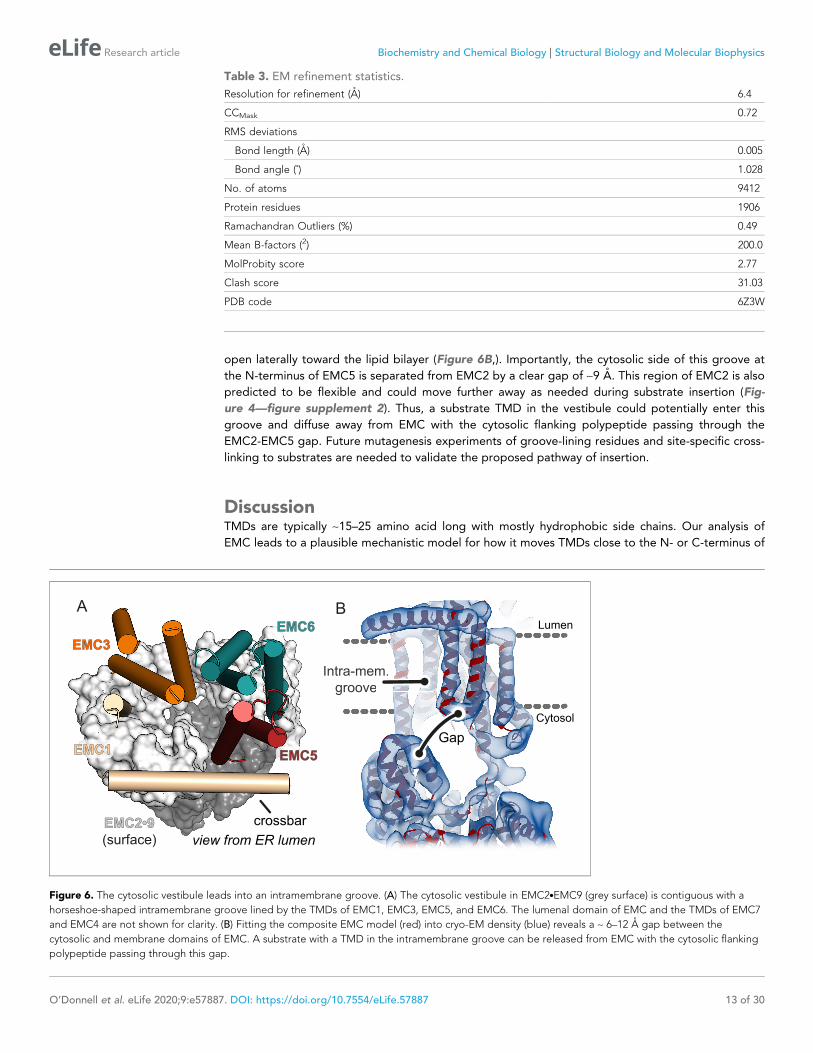
open laterally toward the lipid bilayer (Figure 6B,). Importantly, the cytosolic side of this groove at
the N-terminus of EMC5 is separated from EMC2 by a clear gap of ~9 A. This region of EMC2 is also
predicted to be flexible and could move further away as needed during substrate insertion (Fig-
ure 4—figure supplement 2). Thus, a substrate TMD in the vestibule could potentially enter this
groove and diffuse away from EMC with the cytosolic flanking polypeptide passing through the
EMC2-EMC5 gap. Future mutagenesis experiments of groove-lining residues and site-specific cross-
linking to substrates are needed to validate the proposed pathway of insertion.
DiscussionTMDs are typically ~15–25 amino acid long with mostly hydrophobic side chains. Our analysis of
EMC leads to a plausible mechanistic model for how it moves TMDs close to the N- or C-terminus of
Table 3. EM refinement statistics.
Resolution for refinement (A) 6.4
CCMask 0.72
RMS deviations
Bond length (A) 0.005
Bond angle (˚) 1.028
No. of atoms 9412
Protein residues 1906
Ramachandran Outliers (%) 0.49
Mean B-factors (2) 200.0
MolProbity score 2.77
Clash score 31.03
PDB code 6Z3W
crossbar
EMC6
EMC3
EMC5EMC1
EMC2•9
(surface) view from ER lumen
A B
Gap
m.m.m.Intra-mem.Intra-mem.tra-mem.m.m.m.groovegroove
Lumen
Cytosol
Figure 6. The cytosolic vestibule leads into an intramembrane groove. (A) The cytosolic vestibule in EMC2.EMC9 (grey surface) is contiguous with a
horseshoe-shaped intramembrane groove lined by the TMDs of EMC1, EMC3, EMC5, and EMC6. The lumenal domain of EMC and the TMDs of EMC7
and EMC4 are not shown for clarity. (B) Fitting the composite EMC model (red) into cryo-EM density (blue) reveals a ~ 6–12 A gap between the
cytosolic and membrane domains of EMC. A substrate with a TMD in the intramembrane groove can be released from EMC with the cytosolic flanking
polypeptide passing through this gap.
O’Donnell et al. eLife 2020;9:e57887. DOI: https://doi.org/10.7554/eLife.57887 13 of 30
Research article Biochemistry and Chemical Biology Structural Biology and Molecular Biophysics
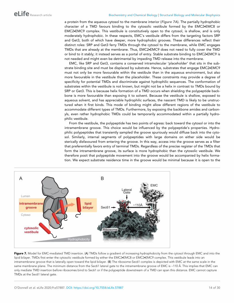
a protein from the aqueous cytosol to the membrane interior (Figure 7A). The partially hydrophobic
character of a TMD favours binding to the cytosolic vestibule formed by the EMC2.EMC8 or
EMC2.EMC9 complex. This vestibule is constitutively open to the cytosol, is shallow, and is only
moderately hydrophobic. In these respects, EMC’s vestibule differs from the targeting factors SRP
and Get3, both of which have deeper, more hydrophobic grooves. These differences reflect their
distinct roles: SRP and Get3 ferry TMDs through the cytosol to the membrane, while EMC engages
TMDs that are already at the membrane. Thus, EMC2.EMC9 does not need to fully cover the TMD
or bind to it stably; it instead serves as a portal of entry. Stable substrate binding to EMC2.EMC9 is
not needed and might even be detrimental by impeding TMD release into the membrane.
EMC, like SRP and Get3, contains a conserved intramolecular ‘placeholder’ that sits in the sub-
strate binding site and must be displaced by substrate. Hence, substrates that engage EMC2.EMC9
must not only be more favourable within the vestibule than in the aqueous environment, but also
more favourable in the vestibule than the placeholder. These constraints may provide a degree of
specificity for potential TMDs and discriminate against hydrophilic sequences. The conformation of
substrates within the vestibule is not known, but might not be a helix in contrast to TMDs bound by
SRP or Get3. This is because helix formation of a TMD occurs when shielding the polypeptide back-
bone is more favourable than exposing it to solvent. Because the vestibule is shallow, exposed to
aqueous solvent, and has appreciable hydrophilic surfaces, the nascent TMD is likely to be unstruc-
tured when it first binds. This mode of binding might allow different regions of the vestibule to
accommodate different types of TMDs. Furthermore, by exposing the backbone amides and carbon-
yls, even rather hydrophobic TMDs could be temporarily accommodated within a partially hydro-
philic vestibule.
From the vestibule, the polypeptide has two points of egress: back toward the cytosol or into the
intramembrane groove. This choice would be influenced by the polypeptide’s properties. Hydro-
philic polypeptides that transiently sampled the groove spuriously would diffuse back into the cyto-
sol. Similarly, internal segments of polypeptides with large domains on either side would be
sterically disfavoured from entering the groove. In this way, access into the groove serves as a filter
that preferentially favors entry of terminal TMDs. Regardless of the precise register of the TMDs that
form the intramembrane groove, its surface is more hydrophobic than the cytosolic vestibule. We
therefore posit that polypeptide movement into the groove would be accompanied by helix forma-
tion. We expect substrate residence time in the groove would be minimal because it is open to the
B
LateralGate
Sec61
EMC
~110Å
Ribosome
A
Hydrophobicity
Cytosol
Lumen
cytosolic
vestibule
intramembrane
groove
lipid
bilayer~110Å11111010101010ÅÅÅÅÅ
Figure 7. Model for EMC-mediated TMD insertion. (A) TMDs follow a gradient of increasing hydrophobicity from the cytosol through EMC and into the
lipid bilayer. TMDs first enter the cytosolic vestibule formed by either the EMC2.EMC8 or EMC2.EMC9 complex. This vestibule leads into an
intramembrane groove that is laterally open toward the lipid bilayer. (B) The ribosome-Sec61 complex is depicted with EMC at the same scale in the
same membrane plane. The minimum distance from the Sec61 lateral gate to the intramembrane groove of EMC is ~110 A. This implies that EMC can
only mediate TMD insertion before ribosomes bind to Sec61 or if the polypeptide downstream of a TMD can span this distance. EMC cannot capture
TMDs at the Sec61 lateral gate.
O’Donnell et al. eLife 2020;9:e57887. DOI: https://doi.org/10.7554/eLife.57887 14 of 30
Research article Biochemistry and Chemical Biology Structural Biology and Molecular Biophysics
lipid bilayer. It is likely that TMD movement into the groove, helix formation, and partitioning into
the membrane interior occur in rapid succession. Thus, the cytosol, vestibule, groove, and mem-
brane interior are connected via a gradient of hydrophobicity that a TMD follows during its energy-
independent insertion by EMC (Figure 7A).
The topology of the TMD would be influenced by steric constraints imposed by its flanking
regions. Because the groove is not open toward the lumen, the side of the substrate containing only
a short flexible segment can be translocated while the side with a bulky domain cannot. Transloca-
tion of the short terminus could be promoted despite the absence of a trans-bilayer pore by thermo-
dynamically favorable release of a TMD into the lipid environment (Engelman and Steitz, 1981).
This driving force can facilitate translocation of up to ~100 amino acid long polypeptide segments in
the absence of a translocon (Brambillasca et al., 2006). In cases where both sides are short (e.g.,
very small single-TMD proteins), one orientation might be favoured over the other by flanking
charges on the substrate being interpreted by charged residues on EMC.
It is noteworthy that EMC2 at the entry point into the intramembrane groove contains well-con-
served solvent-exposed basic amino acids (see Figure 2B) such as Arg26 and Arg91. Furthermore,
the ordered cytosolic tail of EMC1 at this key junction contains four conserved basic residues, at
least some of which would face the vestibule. Finally, a conserved basic residue at position six in
EMC5, and possibly its N-terminus, may also contribute net positive charge in this region. This
enrichment of positive charge (and relatively fewer exposed negative charges) would disfavor entry
into the groove of polypeptide segments that are enriched in basic amino acids. Such a mechanism
would select against mitochondrial TA proteins, cleavable signal peptides, and Ncyt signal anchors.
In each case, these otherwise suitable EMC substrates (based on their terminal location and hydro-
phobicity) are enriched in basic amino acids in the short unstructured domain flanking the hydropho-
bic region (Beltzer et al., 1991; Costello et al., 2017; von Heijne, 1985; Kalbfleisch et al., 2007).
Conversely, bona fide EMC substrates (ER-targeted TA proteins and Nexo signal anchors) are typi-
cally dis-enriched in basic amino acids in the translocated tail. Thus, the mechanism of substrate
selection by EMC might be part of the explanation for the ‘positive-inside rule’, a long-observed
preference for positive flanking charges facing the cytosol (Gafvelin et al., 1997). This is an impor-
tant area for future investigation.
The architecture and dimensions of EMC have major implications for when it acts during co-trans-
lational insertion of N-terminal TMDs and how it could function as a putative chaperone. EMC can-
not feasibly approach closer than ~100 A to a ribosome-bound Sec61 complex (Figure 7B). This
means that EMC can act co-translationally on TMDs only before the ribosome docks on the Sec61
complex or only after at least ~100 A of polypeptide downstream of the TMD has emerged from the
ribosomal tunnel. Because ribosome-nascent chain complexes in which a TMD has just emerged
from the ribosome can be inserted in an EMC-dependent manner, we have postulated that EMC
acts after SRP-mediated targeting but before ribosomes bind to the Sec61 complex (Chitwood and
Hegde, 2019). Due to SRP’s position at the ribosome exit tunnel (Halic et al., 2004;
Schaffitzel et al., 2006; Voorhees and Hegde, 2015), Sec61 cannot engage its binding site unless
SRP releases the TMD and dissociates from the ribosome. Thus, there is a brief window when a TMD
is available for EMC-mediated insertion before Sec61 binds to its position at the exit tunnel
(Jomaa et al., 2017; Kobayashi et al., 2018). We posit that this time constraint helps minimize inap-
propriate insertion of N-terminal TMDs intended for the Ncyt topology so they can be subsequently
inserted by the Sec61 complex.
Although the intramembrane groove is well suited to serve as a chaperone for a single TMD, such
a function seems unlikely to occur adjacent to the Sec61 translocon as proposed on the basis of ribo-
some profiling experiments (Shurtleff et al., 2018). Co-translational engagement of EMC by TMDs
emerging from the ribosome-Sec61 complex could only occur once a TMD can diffuse at least 100 A
away. The very rigid structure of the EMC2.EMC9 complex and the absence of subcomplexes lack-
ing these proteins makes it unlikely that the steric constraints shown in Figure 7B could be over-
come. Furthermore, the intramembrane groove is sufficiently large to house one TMD, and possibly
a second just outside the groove. This means that EMC is not likely to bind multiple TMDs simulta-
neously. Thus, a chaperone function would seem limited to post-translationally engaging an isolated
TMD that has not yet assembled with its intra- or inter-molecular partners. Direct evidence for this
function is currently lacking, but merits future study.
O’Donnell et al. eLife 2020;9:e57887. DOI: https://doi.org/10.7554/eLife.57887 15 of 30
Research article Biochemistry and Chemical Biology Structural Biology and Molecular Biophysics
The role of the lumenal domain of EMC is currently unclear. The simplest possibility is that it
serves a crucial structural role in stabilizing the seven integral membrane subunits while sealing the
intramembrane groove toward the lumen. Maintaining the intramembrane groove configuration
might be energetically unfavourable without structural caps on both sides. Other examples of pro-
teins with a large lipid-exposed groove have most or all of their TMDs within a single polypeptide in
contrast to EMC’s multi-protein assembly (Kumazaki et al., 2014; Ramasamy et al., 2013;
Rollauer et al., 2012; Schoebel et al., 2017). Thus, the lumenal domain, like EMC2, might nucleate
EMC assembly, an early structural solution that was maintained across all eukaryotes.
Our proposed mechanism for EMC-mediated TMD insertion is conceptually similar to how the
prokaryotic protein YidC is thought to function (Dalbey et al., 2014). YidC is a much simpler protein
with a 5-TMD core whose arrangement forms an intramembrane ‘hydrophobic slide’ lined by TMD1,
TMD2, and TMD5 (Kumazaki et al., 2014). The hydrophobic slide in YidC is appreciably smaller
than the intramembrane groove in EMC and could not house a substrate TMD helix without confor-
mational changes. Nevertheless, biochemical evidence supports the notion that substrate TMDs are
near the hydrophobic slide during insertion (Klenner et al., 2008; Yu et al., 2008). Our structural
model for the EMC3 subunit is consistent with an evolutionary relationship to YidC (Anghel et al.,
2017). Notably however, EMC3 in our model is oriented such that the surface corresponding to
YidC’s hydrophobic slide is opposite to the surface lining EMC’s intramembrane groove (see
Figure 5D). This observation raises the possibility that the putative hydrophobic slide in EMC3 might
be an alternative or additional route for substrate TMD insertion.
One way this could occur without risk of substrate entanglement in EMC is if the TMD
approaches the hydrophobic slide from behind the EMC2.EMC9 vestibule. Upon insertion into the
hydrophobic slide, the TMD would then release from EMC in the same direction from which it
approached. Unlike in YidC however, access to lipid from the hydrophobic slide in EMC3 is partially
occluded by EMC4 (Figure 5D). Thus, insertion via this alternative ‘backside’ route may require con-
formational changes to create more space between EMC3, EMC4, and EMC6 so substrates can
enter the lipid bilayer.
Regardless of the exact route(s) of insertion through EMC, it appears that a simplified insertase
originating from a YidC-like protein (Borowska et al., 2015) has been elaborated during evolution
to form EMC. As Get1 appears to be a homolog of EMC3, the Get1/Get2 complex may be a simpli-
fied version of EMC that co-opted a different binding partner. High resolution structures of EMC’s
membrane region and the Get1/Get2 complex, together with structure-guided mutagenesis and
substrate crosslinking experiments, will be important for fully elucidating and comparing their poten-
tially shared mechanisms.
Materials and methods
Key resources table
Reagent type(species) or resource Designation Source or reference Identifiers Additional information
Strain, strainbackground (E. coli)
BL21 NEB Expresscompetent cells
New EnglandBiolabs
Cat# C2523
Cell line(Homo-sapiens)
HEK293 TRexEMC5-FLAG
Guna et al., 2018 MBP04 EMC5-FLAG (see below)integrated into FRT site ofHEK293 TRex-Flp-in cellline. Adapted for growth inFreeStyle suspension media
Cell line(Homo-sapiens)
HEK293 TRexGFP-P2A-RFP-SQS
Chitwood et al., 2018
Cell line(Homo-sapiens)
HEK293 TRexOPRK-GFP-P2A-RFP
Chitwood et al., 2018
Cell line(Homo-sapiens)
HEK293T ATCC ATCC-CRL-3216
cell line(Homo-sapiens)
U2OS Flp-in TRex Volkmar et al., 2019
Continued on next page
O’Donnell et al. eLife 2020;9:e57887. DOI: https://doi.org/10.7554/eLife.57887 16 of 30
Research article Biochemistry and Chemical Biology Structural Biology and Molecular Biophysics

Continued
Reagent type(species) or resource Designation Source or reference Identifiers Additional information
Cell line(Homo-sapiens)
U2OS Flp-inTRex DEMC2
Volkmar et al., 2019 EMC2 disrupted usingCRISPR-Cas9
Antibody ANTI-FLAG M2 AffinityGel (mouse monoclonal)
Sigma Cat# A4596
Antibody Mouse monoclonalANTI-FLAG M2-HRPconjugated
Sigma Cat# A8592,RRID:AB_439702
WB (1:10000)
Antibody Rabbit polyclonalEMC1
Thermo FisherScientific
Cat# A305-605A-M,RRID:AB_2782763
WB (1:1000),IP (0.1 mg/sample)
Antibody Rabbit polyclonalEMC4
Thermo FisherScientific
Cat# A305-752A-M,RRID:AB_2782909
IP (0.4 mg/sample)
Antibody Rabbit polyclonalEMC4 (TMEM85)
Abcam Cat# ab123719,RRID:AB_10951091
WB (1:1000)
Antibody Rabbit polyclonalEMC5 (MMGT1)
Abcam Cat# ab174366,RRID:AB_2750837
WB (1:1000)
Antibody Rabbit polyclonalEMC6 (TMEM93)
Abcam Cat# ab84902,RRID:AB_1925516
WB (1:1000)
Antibody Rabbit polyclonalEMC7 (C15orf24)
Proteintech Cat# 27550–1-AP WB (1:3000)
Antibody Rabbit polyclonalEMC7
Thermo FisherScientific
Cat# A305-678A-M,RRID:AB_2782836
WB (1:1000)
Antibody Rabbit polyclonalCalnexin N-terminus
Enzo Life Sciences Cat# ADI-SPA-865,RRID:AB_10618434
WB (1:5000)
Antibody Rabbit polyclonalHA tag
This paper Custom antibody raisedagainst HA peptideconjugated to KLH.
WB (1:5000)
RecombinantDNA reagent
pET28a-6xHIS-SUMO-EMC2
This paper Human EMC2,Residues 1–297
RecombinantDNA reagent
pET21-EMC2-6xHIS This paper Human EMC2,Residues 1–297
RecombinantDNA reagent
pET28a-6xHIS-SUMO-EMC8
This paper Human EMC8,Residues 1–210
RecombinantDNA reagent
pET21-EMC9-6xHIS This paper Human EMC9,Residues 1–208
RecombinantDNA reagent
pET21-EMC2-6xHIS This paper Human EMC2,Residues 11–274,for crystallization
RecombinantDNA reagent
pET21-EMC9-6xHIS This paper Human EMC9,Residues 1–200for crystallization
RecombinantDNA reagent
pET28a-6xHIS-ULP This paper Yeast ULP1 protease(Uniprot: Q02724),Residues 403–621,for SUMO cleavage
RecombinantDNA reagent
SEC61-SQS(amber)�3 F4
Guna et al., 2018 T7-based PURExpressplasmid (New EnglandBiolabs), SQS TMDresidues 378–410, Ambermutation at F389
RecombinantDNA reagent
SQS(amber)�3 F4 This paper T7-based PURExpressplasmid (New EnglandBiolabs), SQS TMDresidues 378–410, Ambermutation at F389
RecombinantDNA reagent
Clonetech-GFP-P2A-RFP-SQS
Guna et al., 2018
Continued on next page
O’Donnell et al. eLife 2020;9:e57887. DOI: https://doi.org/10.7554/eLife.57887 17 of 30
Research article Biochemistry and Chemical Biology Structural Biology and Molecular Biophysics
Continued
Reagent type(species) or resource Designation Source or reference Identifiers Additional information
RecombinantDNA reagent
pcDNA5-EMC2-3xHA This paper Human EMC2, Residues1–297, Tet operatorremoved with Sac1
RecombinantDNA reagent
pEVOL-pBpF Chin et al., 2002
RecombinantDNA reagent
pcDNA5/FRT/TO-EMC3-Xamb-3xFLAG
This paper Human EMC3 with anamber codon at position Xand a C-terminal 3xFLAG tag.Individual constructswhere X = 23, 24, 25, 26,71, 124, 125, 126, 127, 172,173, 174, and 175were produced.
RecombinantDNA reagent
pcDNA5/FRT/TO-EMC5-Xamb-3xFLAG
This paper Human EMC5 with an ambercodon at position X anda C-terminal 3xFLAG tag.Individual constructswhere X = 14, 15, 16, 17,54, 55, 56, and 57were produced.
RecombinantDNA reagent
pcDNA5/FRT/TO-3xFLAG-EMC6-Xamber
This paper Human EMC6 with an ambercodon at position X andan N-terminal 3xFLAG tag.Individual constructswhere X = 58, 59, 60, 61,94, 95, 96, and 97were produced.
RecombinantDNA reagent
pAS-Pyl-AF This paper Methanosarcina mazeipyrrolysyl-tRNA synthetasewith Y306A/Y384F mutationsand its cognate tRNAcarrying a U25C mutation
RecombinantDNA reagent
pcDNA5/FRT/TO-3xHA-EMC4
This paper Human EMC4 with anN-terminal 3xHA tag
RecombinantDNA reagent
pcDNA5/FRT/TO-EMC4-3xHA
This paper Human EMC4 with aC-terminal 3xHA tag
Sequence-based reagent
siRNA #1against EMC3
Ambion Custom synthesis GGCACUAGAUGAUGUCGAAtt
Sequence-based reagent
siRNA #2against EMC3
Ambion Custom synthesis CCUACUAUGUUGACAGACAtt
Sequence-based reagent
siRNA #1against EMC8
Ambion Custom synthesis AGAUCAUAGCUACGUGAUUtt
Sequence-based reagent
siRNA #2against EMC8
Ambion Custom synthesis GCUGGUUAUUAUCAAGCUAtt
Sequence-based reagent
siRNA #1against EMC9
Ambion Custom synthesis GUACUUAUUAUGUUGGAUAtt
Sequence-based reagent
siRNA #2against EMC9
Ambion Custom synthesis AUGCAGCUGUGAACGAUCAtt
Peptide,recombinant protein
3X FLAG Peptide Sigma-Aldrich Cat# F4799
Peptide,recombinant protein
Human SGTA Shao et al., 2017
Peptide,recombinant protein
Human CaM Shao et al., 2017
Peptide,recombinant protein
Human EMC2 This paper Purified from NEB BL21express cells
Continued on next page
O’Donnell et al. eLife 2020;9:e57887. DOI: https://doi.org/10.7554/eLife.57887 18 of 30
Research article Biochemistry and Chemical Biology Structural Biology and Molecular Biophysics
Continued
Reagent type(species) or resource Designation Source or reference Identifiers Additional information
Peptide,recombinant protein
Human EMC8 This paper Purified from NEB BL21express cells
Peptide,recombinant protein
Human EMC9 This paper Purified from NEB BL21express cells
Peptide,recombinant protein
ULP1 Protease This paper Purified from NEB BL21express cells
Commercialassay or kit
PURE in vitrotranslation system
Shao et al., 2017
Chemicalcompound, drug
deoxy big CHAP(DBC)
Anatrace Cat# 256455
Chemicalcompound, drug
Lauryl MaltoseNeopentylGlycol (LMNG)
Anatrace Cat# NG310 5 GM
Chemicalcompound, drug
Bpa BACHEM Cat# 4017646
Chemicalcompound, drug
AbK Iris Biotech GmbH Cat# HAA3110
Software, algorithm CryoSPARCversion 2.12.4.
Punjani et al., 2017 RRID:SCR_016501
Software, algorithm UCSF ChimeraXVersion 1.0
Goddard et al., 2018 RRID:SCR_015872
Software, algorithm trRosetttaStructurePrediction Server
Yang et al., 2020
Software, algorithm PHENIXVersion 1.17
Adams et al., 2010;Afonine et al., 2018
RRID:SCR_014224
Software, algorithm CootVersion 0.9-pre
Emsley et al., 2010 RRID:SCR_014222
Software, algorithm XDSVersion 20190315
Kabsch, 2010 RRID:SCR_015652
Software, algorithm PyMolVersion 2.1
DeLano Scientific LLC RRID:SCR_000305
Software, algorithm DIALSVersion 2.2
Winter et al., 2018
Software, algorithm BLENDCCP4i 7.0.076
Foadi et al., 2013
Software, algorithm PointlessCCP4i 7.0.076
Evans and Murshudov, 2013 RRID:SCR_014218
Software, algorithm AimlessCCP4i 7.0.076
Evans, 2011 RRID:SCR_015747
Software, algorithm SHELXDVersion 2013/2
Sheldrick, 2008
Software, algorithm FlowJoVersion 9.9.6
FlowJo, LLC RRID:SCR_008520
Software, algorithm FlexEM Topf et al., 2008
Other 1.2/1.3 UltrAu Foil grids Quantifoil Direct OrderForm System
Other Au 200 2/2 grids Quantifoil Direct OrderForm System
Other HEK293 FreeStyleculture media
ThermoFisherScientific
Cat# 12338001 For HEK293 suspensionadapted cell growth
O’Donnell et al. eLife 2020;9:e57887. DOI: https://doi.org/10.7554/eLife.57887 19 of 30
Research article Biochemistry and Chemical Biology Structural Biology and Molecular Biophysics

Protein expression and purificationBacterial over-expression plasmids for EMC2, EMC8, EMC9, SGTA, calmodulin, subsequent point
mutations, and amber suppression mutations were produced by standard molecular biology techni-
ques and listed in the Key Resources Table. Proteins were expressed in E. coli BL21 NEB express
cells at 18˚C for 18 hr following induction with 0.5 mM IPTG at an OD 600 of 0.6. For non-natural
amino acid incorporation, amber codons were introduced at desired sites using site directed muta-
genesis. Constructs were co-expressed with pEVOL-pBpF (Chin et al., 2002) in E. coli BL21 NEB
express cells at 37˚C. At OD 600 of 0.3, 0.2% L-arabinose was added to cultures and continued to
grow to an OD 600 of 0.6 and cultures were cooled to 16˚C. Once cultures were cooled, 0.15 mM
IPTG and 1 mM benzoyl-phenylalanine (from a 1 M stock in 1 M NaOH) was added. Cells were then
cultured at 16˚C overnight. For both standard and amber suppression protein expression, cells were
harvested via centrifugation, resuspended in ice cold Ni2+-NTA buffer A (25 mM Tris pH 8.4, 500
mM NaCl, 20 mM Imidazole), lysed using sonication at 4˚C, and subjected to centrifugation at
39,000 x g for 1 hr at 4˚C. The supernatant containing hexahistidine-tagged proteins was passed
over a column of Ni2+-NTA matrix (Qiagen) at 0.5 mL matrix/1 L of culture. The matrix was washed
with 20 column volumes of Ni2+-NTA buffer A and eluted in a minimum of 3 column volumes using
Ni2+-NTA buffer A supplemented with imidazole to a final concentration 500 mM. Samples were
buffer exchanged into 25 mM HEPES pH 7.5, 100–400 mM NaCl (depending on the protein) with
either PD-10 or HiPrep 26/10 desalting columns (GE Healthcare). SGTA, calmodulin, and EMC
amber suppression samples for in vitro assays were used directly and subsequently were supple-
mented with 10% glycerol and flash frozen in liquid nitrogen. EMC8 expression required an amino
terminal SUMO solubilization tag, which was subsequently cleaved using ULP1 protease overnight at
4˚C. Excess His-tagged SUMO and His-tagged ULP1 protease were removed via passage through a
column of Ni2+-NTA matrix. EMC2, EMC8, and EMC9 were concentrated and subjected to gel filtra-
tion using a GE 200 16/60 liquid chromatography column equilibrated in 25 mM HEPES pH 7.5 and
400 mM NaCl. Complex formation of EMC2.EMC8 or EMC2.EMC9 complexes was achieved by mix-
ing a 1 to 1.2 molar ratio of EMC2 and either EMC8 or EMC9. Excess EMC8 or EMC9 was removed
with gel filtration as indicated above.
Size-Exclusion chromatography coupled to Multi-Angle light scatteringProtein samples at 1 mg/mL (15–40 mM) were injected onto an S200 10/300 Increase SEC column
(GE, Marlborough, MA), equilibrated with 25 mM HEPES pH 7.5, 400 mM NaCl. The SEC column
was coupled to a static 18-angle light scattering detector (DAWN HELEOS-II) and a refractive index
detector (Optilab T-rEX) (Wyatt Technology, Goleta, CA). Data were collected every second at a
flow rate of 0.5 mL/min. Data analysis was carried out using ASTRA VI, yielding the molar mass for
each sample. The light scattering detectors were normalized and data quality were assessed by test-
ing a BSA standard (Pierce).
Microscale thermophoresisSolvent exposed cysteine labeling of EMC2 was conducted with 100 mM EMC2 and 120 mM malei-
mide OG-488 (Molecular Probes, Eugene, OR) for 30 min on ice. Excess dye was removed from the
labeling reaction with a NAP-5 column (GE, Marlborough, MA) pre-equilibrated with 25 mM HEPES
pH 7.5, 500 mM NaCl. The labeling efficiency ranged from 55–65%. The concentration of
EMC2.OG-488 was fixed at 50 nM and a dilution series of EMC8 or EMC9 ranging from 4 nM to 150
mM was used in the hydrophilic treated capillaries (NanoTemper Technologies). A Monolith NT.115
instrument (NanoTemper Technologies) was used with a 50% LED power and laser pulses at both
60% and 100% power. Data were analyzed using the NT Analysis software (NanoTemper
Technologies).
Crystallization of EMC2.EMC9 complexEMC2.EMC9 complex comprising EMC2 residues 11–274 (of the 297 residue full length protein) and
EMC9 residues 1–200 (of the 208 residue full length protein) were gel filtered into a buffer contain-
ing 25 mM Tris pH 8.5 and 400 mM NaCl. Protein at a concentration of 30 mg/mL was screened
through sitting drop vapor diffusion using 200 nL protein mixed with 200 nL of reservoir solution and
incubated at 20˚C. Initial crystals were optimized via hanging drop vapor diffusion by mixing 800 nL
O’Donnell et al. eLife 2020;9:e57887. DOI: https://doi.org/10.7554/eLife.57887 20 of 30
Research article Biochemistry and Chemical Biology Structural Biology and Molecular Biophysics

of protein (30 mg/mL) and 600 nL reservoir solution followed by incubation at 20˚C. The crystal used
for experimental phasing had a reservoir solution containing 28% PEG4000, 0.1 M Tris pH 8.3, and
0.2 M lithium sulfate. Crystals appeared and reached maximum dimensions without visual defects
between 1–2 days. Crystals for experimental phasing were transferred to reservoir solution contain-
ing 20% glycerol for cryoprotection. Crystals used for native data set collection were grown in a res-
ervoir solution containing 34% PEG4000, 0.1 M Tris pH 8.3, and 0.2 M lithium sulfate. Crystals for
native collection were harvested within 24 hr directly from the drop by adding 4 mL of reservoir solu-
tion containing 20% ethylene glycol and incubating 1 min. Cryopreserved crystals were flash frozen
in liquid nitrogen.
Crystallographic data collection and analysisData were collected on EMC2.EMC9 crystals using I23 long-wavelength beamline at Diamond. Four
native sulfur-SAD experiments with varied kappa goniometer orientation were conducted with a sin-
gle crystal and each data set contained 3600 images collected at l = 2.755 A with 0.1˚ oscillations
on a curved Pilatus 12M detector. Images were integrated and scaled with XDS and XSCALE
(Kabsch, 2010). The substructure consisting of 27 sulfur sites was solved using SHELXD (Shel-
drick, 2008) and phases were obtained with Autosol (Adams et al., 2010). The initial model was
built manually and refined using Coot (Emsley et al., 2010) and PHENIX (Adams et al., 2010)
respectively. The space group was P212121 and a single EMC2.EMC9 molecule was observed in the
asymmetric unit.
A native non-isomorphous P212121 dataset was collected collected at I03 with a l = 0.9763 A,
3600 images with a 0.1˚ oscillation on an Eiger2 XE 16M detector. Data reduction was carried out
with DIALS where four lattices offset by less than 5˚ were determined using multi-lattice processing
(Winter et al., 2018), and each of the four lattices contained an equivalent number of reflections.
The deconvolved datasets were analyzed in BLEND (Foadi et al., 2013) and merged/scaled using
Pointless (Evans and Murshudov, 2013) and Aimless (Evans, 2011). The structure was determined
by molecular replacement using the software package PHENIX (Adams et al., 2010). Iterative
rounds of building and refinement were completed using Coot (Emsley et al., 2010) and PHENIX
(Afonine et al., 2018), which resulted in the final model (PDB: 6Y4L). Data collection and refinement
statistics are summarized in Table 1. Illustrations were made in Pymol versions 1.8.6 and 2.3.2
(Schrodinger, LLC). Surface chemical properties and conservation were displayed as previously
described (Ashkenazy et al., 2016; Hagemans et al., 2015).
Mammalian cell cultureAll cell lines were cultured in DMEM containing 10% fetal calf serum and 2 mM L-glutamine. HEK293
T-REx Flp-In cells overexpressing EMC5-FLAG have been described (Guna et al., 2018). Wild type
U2OS Flp-In TRex cells and DEMC2 U2OS Flp-In TRex cells have been characterized previously
(Volkmar et al., 2019). HEK293T cells were originally from ATCC (ATCC CRL-3216). Cell line identi-
ties were verified by a combination of an integrated Frt site and doxycycline inducibility (distinctive
to Flp-in T-Rex cells), by antibiotic resistance markers, and by immunoblotting for the product of
knockout alleles. Further genetic analysis was not performed. Cells were checked approximately
monthly for mycoplasma contamination using the MycoAlert Mycoplasma Detection Kit (Lonza) and
found to be negative.
Purification of native mammalian EMC40 g pellets of suspension adapted HEK293 T-REx Flp-In cells overexpressing EMC5-FLAG were sol-
ubilized in 40 mL of 2x solubilization buffer [100 mM HEPES pH 7.4, 400 mM NaCl, 4 mM MgAc2,
1.6% deoxy big CHAP (DBC; Merck – 256455)] for 1 hr with gentle shaking on ice. All subsequent
steps apart from FLAG elution were conducted on ice or at 4˚C. Solubilized cells were clarified for
20 min at 21,000 x g at 4˚C and incubated for 1 hr with 1 mL bed volume of anti-FLAG M2 affinity
gel (Sigma A4596) which had been pre-equilibrated in DBC wash buffer (0.3% DBC, 50 mM HEPES
pH 7.4, 200 mM NaCl, 2 mM MgAc2). FLAG resin was collected by centrifugation (5 min at 1500 x g)
and washed twice with 4 mL DBC wash buffer. Resin was transferred to 10 mL gravity flow column
and washed with 3 � 8 mL of 0.1% Lauryl Maltose Neopentyl Glycol (LMNG) wash buffer (0.1%
LMNG, 50 mM HEPES pH 7.4, 200 mM NaCl, 2 mM MgAc2) allowing 10 min between washes to
O’Donnell et al. eLife 2020;9:e57887. DOI: https://doi.org/10.7554/eLife.57887 21 of 30
Research article Biochemistry and Chemical Biology Structural Biology and Molecular Biophysics

permit detergent exchange. Resin was then washed with 3 � 8 mL of 0.01% LMNG wash buffer
(0.01% LMNG, 50 mM HEPES pH 7.4, 200 mM NaCl, 2 mM MgAc2) allowing 10 min between washes
for detergent exchange. Two FLAG elutions were conducted for 25 min each at room temperature
by incubating the resin in 2 mL of FLAG elution buffer (0.01% LMNG, 50 mM HEPES pH 7.4, 200
mM NaCl, 2 mM MgAc2, 0.25 mg/mL 3X FLAG peptide -Sigma F4799) with gentle end-over-end
mixing. Combined FLAG elutions were diluted with 4 mL low-salt dilution buffer (0.01% LMNG, 50
mM HEPES pH 7.4, 2 mM magnesium acetate) and were bound to 150 mL bed volume of fast-flow
SP sepharose, which had been pre-equilibrated with 2 mL ion exchange buffer A (0.01% LMNG, 50
mM HEPES pH 7.4, 50 mM sodium chloride, 2 mM magnesium acetate). SP sepharose was washed
with 3 � 1 mL of ion exchange buffer A before 3 � 150 mL rounds of elution in ion exchange buffer
B (0.01% LMNG, 50 mM HEPES pH 7.4, 400 mM sodium chloride, 2 mM magnesium acetate). The
first elution was run over the resin twice and the resulting fractions were checked by A280. The peak
fraction was then centrifuged for 30 min at 175,000 x g to remove insoluble aggregates.
Cryo-EM sample preparation and data acquisitionGrid preparation consists of adding 3 mL of mammalian EMC complex at a concentration of 0.8–0.9
mg/mL to Quantifoil 1.2/1.3 UltrAu Foil grids or Quantifoil gold Au 200 2/2 grids. The Quantifoil
1.2/1.3 UltrAu Foil grids or Quantifoil gold Au 200 2/2 grids were first hydrophilized using a
Fischione model 1070 plasma cleaner with a 9:1 ratio of Argon to Oxygen for 2 min and 30 s respec-
tively. Grids were blotted for 9 s and plunged into liquid ethane using a Leica EM GP2 plunge
freezer at 100% humidity. Particles were screened on a Technai F20 at 200 kV and data was col-
lected on two FEI Titan Krios microscopes operating at 300 kV and equipped with Gatan K2 summit
direct electron detectors, energy filters and volta phase plates. Data were collected at pixel sizes
between 1.18 and 1.39 A per pixel. Exposures were between 5 and 11 s long resulting total accumu-
lated dose of 38 to 45 e-/A2 and were fractioned into 25 to 44 frames. All data were collected using
FEI’s EPU software at the MRC-LMB or eBIC.
Cryo-EM data processingCryo-EM data processing was performed in the CryoSPARC processing suite (Punjani et al., 2017)
using version 2.12.4. Initially five independent datasets were processed individually. Movies were
motion corrected using multi-patch motion correction and CTF values estimated using multi-patch
CTF estimation in CryoSPARC. Particles were picked using a blob picker before extraction with a
box size of 224 pixels and 2D classification to produce templates for 2D-template based auto pick-
ing. Picked particles were subject to 2D and 3D reference-free classification and selected particles
were refined using non-uniform refinement and local resolution estimation in CryoSPARC. Maps
from datasets with differing nominal pixel sizes were then re-scaled to a range of pixel sizes and
matched to a reference map in UCSF Chimera, yielding a cross correlation coefficient (Tan et al.,
2017). Titration of these cross correlation values was used to identify an optimal nominal pixel size
relative to the reference dataset (Wilkinson et al., 2019). The calculated values were used to re-
scale the motion corrected micrographs from two datasets to match the nominal pixel size of 1.38 A
per pixel. The re-scaled datasets were then reprocessed as detailed above. Rescaled particles were
merged with particles from three datasets originally collected at 1.38 A per pixel, resulting in a mas-
ter pool of 405,515 particles, which were subject to reference-free 3D classification. The final dataset
of 167,294 particles was subject to non-uniform refinement followed by per-particle CTF refinement.
CTF refined particles were subject to a final round of non-uniform refinement followed by local reso-
lution estimation and filtering to produce a final map with a resolution of 6.4 A. Refinement was lim-
ited to a maximum alignment resolution of 6.5 A to prevent overfitting. During filtering a
conservative ad hoc B-Factor of �150 was applied to the map to prevent over-sharpening. Full
details of processing of individual and merged datasets are provided in Table 2.
PURE in vitro translation reactionsA homemade PURE translation system (Shimizu and Ueda, 2010) was modified to accommodate
amber-suppression as previously described (Shao et al., 2017). Changes that enable nearly quantita-
tive amber-suppression are the omission of RF1, use of E. coli tRNA purified from a strain over-
expressing the amber suppressor tRNA, addition of recombinant amber suppressor tRNA synthetase
O’Donnell et al. eLife 2020;9:e57887. DOI: https://doi.org/10.7554/eLife.57887 22 of 30
Research article Biochemistry and Chemical Biology Structural Biology and Molecular Biophysics
(Bpa-RS) at 50 mg/mL, and addition of 0.1 mM of the photo-crosslinking non-natural amino acid ben-
zoyl-phenylalanine (Bpa). Translation reactions of 25 mL were supplemented with 10 ng/mL DNA tem-
plate, 24 mM CaM, 100 nM CaCl2, and 2.5 mL 35S-methionine (from a stock of ~10 mCi/mL).
Reactions were incubated at 37˚C for 30 min, returned to ice, and diluted with 35 mL of ice-cold
physiologic salt buffer (PSB: 50 mM HEPES pH 7.5, 100 mM KAc, 5 mM MgAc2, 100 nM CaCl2).
Translated products were isolated via sucrose cushion by layering 50 mL of the diluted translation on
top of 50 mL PSB supplemented with 20% sucrose. Centrifugation was conducted for 60 min at
55,000 RPM in a TLS-55 rotor with the slowest acceleration and deceleration possible. The top 80 mL
of the sample were removed and snap frozen in liquid N2 for storage prior to subsequent use in
crosslinking reactions.
Bpa crosslinking reactionsThe translation products prepared as described above were diluted 1:4 in reaction buffer (25 mM
HEPES pH 7.5, 500 mM NaCl, 100 nM CaCl2) and mixed 1:1 with reaction buffer, SGTA, or EMC
components at concentrations indicated in the figure legends. Samples were incubated at 37˚C for 5
min, returned to ice and UV-irradiated using a UVP B-100 series lamp (UVP LLC) at a distance of 10
cm for a duration of 10 min.
Protease protection assayPlasmids encoding human EMC4 with an N- or C-terminal 3xHA tag in the pcDNA5/FRT/TO vector
were transiently transfected into HEK293T cells using PEI ‘MAX’ (Polysciences). At 3 days after trans-
fection, cells were collected, resuspended in ice-cold homogenizing buffer (20 mM HEPES pH 7.4,
250 mM sucrose, 2 mM MgCl2, and EDTA-free protease inhibitor cocktail [Roche]), and homoge-
nized on ice by 25 passages through a 26-guage needle fitted to a 1 mL syringe. Homogenates were
centrifuged at 800 x g for 5 min at 4˚C to pellet nuclei and debris, and the postnuclear supernatants
were further centrifuged at 4000 x g for 10 min at 4˚C to pellet heavy membranes. The supernatants
were then subjected to ultracentrifugation at 100,000 x g for 30 min at 4˚C, and the resulting micro-
some pellets were resuspended in ice-cold physiologic salt buffer-2 (50 mM HEPES pH 7.4, 100 mM
KOAc, 2 mM MgAc2) and placed on ice. Aliquots were left untreated or treated with 0.5 mg/mL of
proteinase K in the presence or absence of 0.5% Triton X-100. After a 30 min incubation on ice, the
digestion reactions were stopped by incubation with 5 mM PMSF on ice for 5 min and transferred to
10-volumes of boiling SDS lysis buffer (1% SDS and 0.1 M Tris-HCl pH 8.0). Samples were directly
analyzed by SDS-PAGE and immunoblotting.
Site-specific photo-crosslinking in mammalian cellsPosition-specific incorporation of the photo-crosslinking amino acid AbK into proteins in mammalian
cells was achieved by amber suppression using the Methanosarcina mazei pyrrolysyl-tRNA synthe-
tase (PylRS) and tRNAPylCUA pair (Ai et al., 2011). Plasmids used for the in vivo amber suppression
were constructed as follows. The plasmid SE315 (Oller-Salvia et al., 2018), which encodes WT PylRS
and the U25C mutant of tRNAPylCUA, was modified to express V5-tagged PylRS carrying Y306A and
Y384F mutations [known as PylRS-AF mutant (Yanagisawa et al., 2008) instead of WT PylRS. The
resultant plasmid was termed pAS-Pyl-AF and used throughout this study. The coding regions of
human EMC subunits were inserted into a pcDNA5/FRT/TO vector containing an N-terminal or
C-terminal 3xFLAG tag, and the codons for the selected positions were individually mutated to an
amber codon in order to allow AbK incorporation. To express AbK-modified EMC subunits,
HEK293T cells were co-transfected with pAS-Pyl-AF and an amber codon-containing EMC construct
at a ratio of 4:1 using PEI ‘MAX’ (Polysciences) and were grown for 2 days in the presence of 0.5
mM AbK (Iris Biotech). After washing cells with PBS, ice-cold PBS was added, and photo-crosslinking
was induced by placing cells on ice ~12.5 cm away from a UVP B-100 series lamp (UVP LLC) for 15
min. Cells were then harvested, frozen in liquid nitrogen, and stored at �80˚C prior to analysis. For
immunoprecipitation under non-denaturing conditions, cell pellets were lysed in native IP buffer (50
mM HEPES pH 7.4, 200 mM NaCl, 2 mM MgAc2, 1% Triton X-100) supplemented with EDTA-free
protease inhibitor cocktail (Roche). Cell lysates were cleared by centrifugation and incubated at 4˚C
for 1.5–2 hr with anti-FLAG M2 affinity gel (Sigma). After extensive washing with native IP buffer,
proteins were eluted with SDS-PAGE sample buffer. Immunoprecipitation was also done under
O’Donnell et al. eLife 2020;9:e57887. DOI: https://doi.org/10.7554/eLife.57887 23 of 30
Research article Biochemistry and Chemical Biology Structural Biology and Molecular Biophysics
denaturing conditions using antibodies against EMC subunits. For this, cell pellets were resuspended
in SDS lysis buffer (1% SDS and 0.1 M Tris-HCl pH 8.0) and heat-denatured at 95˚C for ~10 min with
occasional vortex mixing. After cooling to room temperature, the denatured lysates were diluted 10-
fold with IP buffer (50 mM HEPES pH 7.4, 100 mM NaCl, 1% Triton X-100) and incubated at 4˚C for
1.5–2 hr with Protein A agarose beads plus the appropriate antibody, followed by extensive washing
with IP buffer and elution with SDS-PAGE sample buffer.
Flow cytometry analysisWild type U2OS Flp-In TRex cells and DEMC2 U2OS Flp-In TRex cells (Volkmar et al., 2019) were
used in flow cytometry experiments and were maintained in DMEM supplemented with 10% fetal
calf serum and 2 mM L-glutamine. Inverse transfections were conducted as follows: 3 mL GeneJuice
(Merk Millipore) was added dropwise to 100 mL Opti-MEM (Gibco), vortexed and incubated at room
temperature for 5 min. SQS-reporter DNA (250 ng) and either EMC2 DNA or empty vector (750 ng)
were added to the Opti-MEM/GeneJuice mixture, solution was pipetted up and down to mix, and
incubated at room temperature for 20 min. Cells were seeded into a six well dish at 40% confluency
concurrent with the addition of 100 mL of the transfection/DNA mixture. Cells were grown for 3 days
and then detached with trypsin/EDTA in 250 mL volume, neutralized with 500 mL complete media,
and pelleted by centrifugation at 5000 x g in a table top centrifuge at 4˚C. Cells were washed by
resuspension in 500 mL ice cold PBS and re-pelleted as above. Pelleted cells were resuspended in a
final volume of 600 mL ice cold PBS and filtered through a 70 mm cell strainer for analysis. Cells were
analyzed using a BD LSR II flow cytometer (BD Biosciences, Franklin Lakes, NJ). The flow cytometry
experiment and data analysis using FlowJo software packages were conducted as previously
described (Itakura et al., 2016). Knockdowns of EMC3, EMC8 and EMC9 with siRNAs were for 3
days in HEK 293 Trex cells with stably integrated inducible reporters (GFP-P2A-RFP-SQS or OPRK-
GFP-P2A-RFP) using Lipofectamine RNAiMAX reagent (Thermo Fisher). siRNA sequences are avail-
able in the Key Resources Table). Expression of the reporters was induced with 1 mg/ml doxycycline
for ~20 hr before analysis by flow cytometry performed as described above.
Statistics and reproducibilityThis study did not employ any statistical analysis or comparisons, no grouping, and no power calcula-
tions. Biochemical and cell biological experiments include internal controls that were performed as
part of the same experiment and are shown within the respective figures. Each result or conclusion
from any given figure panel represents one example of at least two biologically independent experi-
ments, both of which contained the same or similar internal controls. No attempts at replication with
valid controls failed. Where data are compiled from multiple measurements (Figure 2—figure sup-
plement 2), the number of replicates are indicated in the figure legend with error bars to indicate
standard deviation.
AcknowledgementsWe thank P Chitwood for helpful discussions, MM Babu for co-evolution advice, RL Williams and D
Bellini for advice on crystallography analysis, J Chin and I Gregor for amber suppression reagents, A
Guna for help with protein preparations at an early stage of this project, N Volkmar and J Christian-
son for discussions and the DEMC2 cells, G Cannone for EM support, J Grimmett and T Darling for
scientific computing, the EM, crystallization, mass spectrometry, and biophysics core facilities at the
LMB, and the staff on beamlines I03 and I23 at the Diamond Light Source for assistance with crystal
screening and data collection. We acknowledge access and support of the Cryo-EM facilities at the
UK national electron bio-imaging centre (eBIC), proposal EM17434-54, funded by the Wellcome
Trust, MRC and BBSRC. We acknowledge Diamond Light Source for beamtime under proposal
MX21426. Funding was provided by the Medical Research Council of the UK (MC_UP_A022_1007 to
RSH and MC_UP_1201/10 to EAM) and the US National Institutes of Health (R01 GM078186 to EAM
and R01 GM130051 to RJK). JPO was funded by an EMBO long term fellowship (ALTF 18–2018),
BPP was supported by Boehringer Ingelheim Fonds, and YY was funded by fellowships from the
Naito Foundation and the Osamu Hayaishi Memorial Scholarship for Study Abroad. DM acknowl-
edges the support of the Swiss National Science foundation (SNF) under grant P2ELP3_18910.
O’Donnell et al. eLife 2020;9:e57887. DOI: https://doi.org/10.7554/eLife.57887 24 of 30
Research article Biochemistry and Chemical Biology Structural Biology and Molecular Biophysics

Additional information
Competing interests
Ramanujan S Hegde: Reviewing editor, eLife. Elizabeth A Miller: Reviewing editor, eLife. The other
authors declare that no competing interests exist.
Funding
Funder Grant reference number Author
Medical Research Council MC_UP_A022_1007 Ramanujan S Hegde
Medical Research Council MC_UP_1201/10 Elizabeth A Miller
National Institutes of Health R01 GM078186 Elizabeth A Miller
National Institutes of Health R01 GM130051 Robert J Keenan
European Molecular BiologyOrganization
ALTF 18-2018 John P O’Donnell
Boehringer Ingelheim Fonds Ben Photon Phillips
Naito Foundation Yuichi Yagita
Japanese Biochemical Society Yuichi Yagita
Swiss National Science Foun-dation
P2ELP3_18910 Duccio Malinverni
The funders had no role in study design, data collection and interpretation, or the
decision to submit the work for publication.
Author contributions
John P O’Donnell, Conceptualization, Funding acquisition, Investigation, Methodology, Writing -
original draft, Writing - review and editing, Performed the biochemical and biophysical analysis of
EMC cytosolic subunits, solved the EMC2.EMC9 crystal structure, analyzed EMC mutants in cells,
and contributed to model generation, building, and interpretation; Ben P Phillips, Conceptualization,
Investigation, Methodology, Writing - review and editing, Initiated and performed the cryo-EM anal-
ysis (with help from JPO at later stages) and contributed to the building and interpretation of mod-
els; Yuichi Yagita, Investigation, Methodology, Writing - review and editing, Set up the site-specific
photo-crosslinking assay in cells and analysed EMC4 topology; Szymon Juszkiewicz, Investigation,
Writing - review and editing, Performed flow cytometry analysis of EMC subunit knockdowns; Armin
Wagner, Duccio Malinverni, Investigation, Methodology, Writing - review and editing; Robert J
Keenan, Formal analysis, Methodology, Writing - review and editing; Elizabeth A Miller, Conceptuali-
zation, Supervision, Funding acquisition, Project administration, Writing - review and editing; Rama-
nujan S Hegde, Conceptualization, Supervision, Funding acquisition, Writing - original draft, Project
administration, Writing - review and editing
Author ORCIDs
John P O’Donnell https://orcid.org/0000-0002-0571-3603
Ben P Phillips https://orcid.org/0000-0001-9117-4695
Szymon Juszkiewicz http://orcid.org/0000-0002-3361-7264
Armin Wagner http://orcid.org/0000-0001-8995-7324
Robert J Keenan http://orcid.org/0000-0003-1466-0889
Elizabeth A Miller http://orcid.org/0000-0002-1033-8369
Ramanujan S Hegde https://orcid.org/0000-0001-8338-852X
Decision letter and Author response
Decision letter https://doi.org/10.7554/eLife.57887.sa1
Author response https://doi.org/10.7554/eLife.57887.sa2
O’Donnell et al. eLife 2020;9:e57887. DOI: https://doi.org/10.7554/eLife.57887 25 of 30
Research article Biochemistry and Chemical Biology Structural Biology and Molecular Biophysics
Additional files
Supplementary files. Transparent reporting form
Data availability
Structural coordinates have been deposited in PDB under accession codes 6Y4L and 6Z3W. Cryo-
EM data had been deposited to EMDB under accession code EMD-11058. All other data in this
study are provided within the manuscript.
The following datasets were generated:
Author(s) Year Dataset title Dataset URLDatabase andIdentifier
O’Donnell JP,Wagner A, PhillipsBP, Hegde RS
2020 Crystal structure of the humanEMC2�EMC9 complex
https://www.rcsb.org/structure/6Y4L
RCSB Protein DataBank, 6Y4L
O’Donnell JP, Phil-lips BP, Hegde RS
2020 Cryo-EM structure of human EMC https://www.rcsb.org/structure/6Z3W
RCSB Protein DataBank, 6Z3W
Hegde RS, PhillipsBP, O’Donnell JP,Miller EA
2020 Human ER Membrane proteinComplex (EMC)
https://www.ebi.ac.uk/pdbe/emdb/11058
Electron MicroscopyData Bank, 11058
The following previously published datasets were used:
Author(s) Year Dataset title Dataset URLDatabase andIdentifier
Voorhees RM,Hegde RS
2016 The structure of the mammalianSec61 channel opened by a signalsequence
https://www.rcsb.org/structure/3JC2
RCSB Protein DataBank, 3JC2
Voorhees RM, Fer-nandez IS, ScheresSHW, Hegde RS
2014 Structure of the idle mammalianribosome-Sec61 complex
https://www.rcsb.org/structure/3J7Q
RCSB Protein DataBank, 3J7Q
ReferencesAdams PD, Afonine PV, Bunkoczi G, Chen VB, Davis IW, Echols N, Headd JJ, Hung LW, Kapral GJ, Grosse-Kunstleve RW, McCoy AJ, Moriarty NW, Oeffner R, Read RJ, Richardson DC, Richardson JS, Terwilliger TC,Zwart PH. 2010. PHENIX: a comprehensive Python-based system for macromolecular structure solution. ActaCrystallographica Section D Biological Crystallography 66:213–221. DOI: https://doi.org/10.1107/S0907444909052925, PMID: 20124702
Afonine PV, Poon BK, Read RJ, Sobolev OV, Terwilliger TC, Urzhumtsev A, Adams PD. 2018. Real-spacerefinement in PHENIX for cryo-EM and crystallography. Acta Crystallographica Section D Structural Biology 74:531–544. DOI: https://doi.org/10.1107/S2059798318006551
Ai HW, Shen W, Sagi A, Chen PR, Schultz PG. 2011. Probing Protein-Protein interactions with a geneticallyencoded Photo-crosslinking amino acid. Chembiochem : A European Journal of Chemical Biology 12:1854–1857. DOI: https://doi.org/10.1002/cbic.201100194, PMID: 21678540
Anghel SA, McGilvray PT, Hegde RS, Keenan RJ. 2017. Identification of Oxa1 homologs operating in theeukaryotic endoplasmic reticulum. Cell Reports 21:3708–3716. DOI: https://doi.org/10.1016/j.celrep.2017.12.006, PMID: 29281821
Ashkenazy H, Abadi S, Martz E, Chay O, Mayrose I, Pupko T, Ben-Tal N. 2016. ConSurf 2016: an improvedmethodology to estimate and visualize evolutionary conservation in macromolecules. Nucleic Acids Research44:W344–W350. DOI: https://doi.org/10.1093/nar/gkw408, PMID: 27166375
Beltzer JP, Fiedler K, Fuhrer C, Geffen I, Handschin C, Wessels HP, Spiess M. 1991. Charged residues are majordeterminants of the transmembrane orientation of a signal-anchor sequence. The Journal of BiologicalChemistry 266:973–978. DOI: https://doi.org/10.5167/uzh-165, PMID: 1985975
Bircham PW, Maass DR, Roberts CA, Kiew PY, Low YS, Yegambaram M, Matthews J, Jack CA, Atkinson PH.2011. Secretory pathway genes assessed by high-throughput microscopy and synthetic genetic array analysis.Molecular BioSystems 7:2589–2598. DOI: https://doi.org/10.1039/c1mb05175j, PMID: 21731954
Borowska MT, Dominik PK, Anghel SA, Kossiakoff AA, Keenan RJ. 2015. A YidC-like protein in the archaealplasma membrane. Structure 23:1715–1724. DOI: https://doi.org/10.1016/j.str.2015.06.025, PMID: 26256539
O’Donnell et al. eLife 2020;9:e57887. DOI: https://doi.org/10.7554/eLife.57887 26 of 30
Research article Biochemistry and Chemical Biology Structural Biology and Molecular Biophysics
Brambillasca S, Yabal M, Soffientini P, Stefanovic S, Makarow M, Hegde RS, Borgese N. 2005. Transmembranetopogenesis of a tail-anchored protein is modulated by membrane lipid composition. The EMBO Journal 24:2533–2542. DOI: https://doi.org/10.1038/sj.emboj.7600730, PMID: 15973434
Brambillasca S, Yabal M, Makarow M, Borgese N. 2006. Unassisted translocation of large polypeptide domainsacross phospholipid bilayers. Journal of Cell Biology 175:767–777. DOI: https://doi.org/10.1083/jcb.200608101, PMID: 17130291
Chin JW, Martin AB, King DS, Wang L, Schultz PG. 2002. Addition of a photocrosslinking amino acid to thegenetic code of Escherichia coli. PNAS 99:11020–11024. DOI: https://doi.org/10.1073/pnas.172226299,PMID: 12154230
Chio US, Cho H, Shan SO. 2017. Mechanisms of Tail-Anchored membrane protein targeting and insertion.Annual Review of Cell and Developmental Biology 33:417–438. DOI: https://doi.org/10.1146/annurev-cellbio-100616-060839, PMID: 28992441
Chitwood PJ, Juszkiewicz S, Guna A, Shao S, Hegde RS. 2018. EMC is required to initiate accurate membraneprotein topogenesis. Cell 175:1507–1519. DOI: https://doi.org/10.1016/j.cell.2018.10.009, PMID: 30415835
Chitwood PJ, Hegde RS. 2019. The role of EMC during membrane protein biogenesis. Trends in Cell Biology 29:371–384. DOI: https://doi.org/10.1016/j.tcb.2019.01.007, PMID: 30826214
Christianson JC, Olzmann JA, Shaler TA, Sowa ME, Bennett EJ, Richter CM, Tyler RE, Greenblatt EJ, WadeHarper J, Kopito RR. 2012. Defining human ERAD networks through an integrative mapping strategy. NatureCell Biology 14:93–105. DOI: https://doi.org/10.1038/ncb2383
Costello JL, Castro IG, Camoes F, Schrader TA, McNeall D, Yang J, Giannopoulou EA, Gomes S, Pogenberg V,Bonekamp NA, Ribeiro D, Wilmanns M, Jedd G, Islinger M, Schrader M. 2017. Predicting the targeting of tail-anchored proteins to subcellular compartments in mammalian cells. Journal of Cell Science 130:1675–1687.DOI: https://doi.org/10.1242/jcs.200204, PMID: 28325759
Dalbey RE, Kuhn A, Zhu L, Kiefer D. 2014. The membrane insertase YidC. Biochimica Et Biophysica Acta (BBA) -Molecular Cell Research 1843:1489–1496. DOI: https://doi.org/10.1016/j.bbamcr.2013.12.022
Emsley P, Lohkamp B, Scott WG, Cowtan K. 2010. Features and development of coot. Acta Crystallographica.Section D, Biological Crystallography 66:486–501. DOI: https://doi.org/10.1107/S0907444910007493,PMID: 20383002
Engelman DM, Steitz TA. 1981. The spontaneous insertion of proteins into and across membranes: the helicalhairpin hypothesis. Cell 23:411–422. DOI: https://doi.org/10.1016/0092-8674(81)90136-7, PMID: 7471207
Evans PR. 2011. An introduction to data reduction: space-group determination, scaling and intensity statistics.Acta Crystallographica Section D Biological Crystallography 67:282–292. DOI: https://doi.org/10.1107/S090744491003982X, PMID: 21460446
Evans PR, Murshudov GN. 2013. How good are my data and what is the resolution? Acta CrystallographicaSection D Biological Crystallography 69:1204–1214. DOI: https://doi.org/10.1107/S0907444913000061,PMID: 23793146
Foadi J, Aller P, Alguel Y, Cameron A, Axford D, Owen RL, Armour W, Waterman DG, Iwata S, Evans G. 2013.Clustering procedures for the optimal selection of data sets from multiple crystals in Macromolecularcrystallography. Acta Crystallographica Section D Biological Crystallography 69:1617–1632. DOI: https://doi.org/10.1107/S0907444913012274, PMID: 23897484
Gafvelin G, Sakaguchi M, Andersson H, von Heijne G. 1997. Topological rules for membrane protein assembly ineukaryotic cells. The Journal of Biological Chemistry 272:6119–6127. DOI: https://doi.org/10.1074/jbc.272.10.6119, PMID: 9045622
Goddard TD, Huang CC, Meng EC, Pettersen EF, Couch GS, Morris JH, Ferrin TE. 2018. UCSF ChimeraX:meeting modern challenges in visualization and analysis. Protein Science 27:14–25. DOI: https://doi.org/10.1002/pro.3235, PMID: 28710774
Gogala M, Becker T, Beatrix B, Armache JP, Barrio-Garcia C, Berninghausen O, Beckmann R. 2014. Structures ofthe Sec61 complex engaged in nascent peptide translocation or membrane insertion. Nature 506:107–110.DOI: https://doi.org/10.1038/nature12950, PMID: 24499919
Guna A, Volkmar N, Christianson JC, Hegde RS. 2018. The ER membrane protein complex is a transmembranedomain insertase. Science 359:470–473. DOI: https://doi.org/10.1126/science.aao3099, PMID: 29242231
Guna A, Hegde RS. 2018. Transmembrane domain recognition during membrane protein biogenesis and qualitycontrol. Current Biology : CB 28:R498–R511. DOI: https://doi.org/10.1016/j.cub.2018.02.004, PMID: 29689233
Hagemans D, van Belzen IA, Moran Luengo T, Rudiger SG. 2015. A script to highlight hydrophobicity and chargeon protein surfaces. Frontiers in Molecular Biosciences 2:56. DOI: https://doi.org/10.3389/fmolb.2015.00056,PMID: 26528483
Halic M, Becker T, Pool MR, Spahn CM, Grassucci RA, Frank J, Beckmann R. 2004. Structure of the signalrecognition particle interacting with the elongation-arrested ribosome. Nature 427:808–814. DOI: https://doi.org/10.1038/nature02342, PMID: 14985753
Hegde RS, Keenan RJ. 2011. Tail-anchored membrane protein insertion into the endoplasmic reticulum. NatureReviews Molecular Cell Biology 12:787–798. DOI: https://doi.org/10.1038/nrm3226, PMID: 22086371
Heinrich SU, Mothes W, Brunner J, Rapoport TA. 2000. The Sec61p complex mediates the integration of amembrane protein by allowing lipid partitioning of the transmembrane domain. Cell 102:233–244.DOI: https://doi.org/10.1016/S0092-8674(00)00028-3, PMID: 10943843
Itakura E, Zavodszky E, Shao S, Wohlever ML, Keenan RJ, Hegde RS. 2016. Ubiquilins chaperone and triagemitochondrial membrane proteins for degradation. Molecular Cell 63:21–33. DOI: https://doi.org/10.1016/j.molcel.2016.05.020, PMID: 27345149
O’Donnell et al. eLife 2020;9:e57887. DOI: https://doi.org/10.7554/eLife.57887 27 of 30
Research article Biochemistry and Chemical Biology Structural Biology and Molecular Biophysics

Jomaa A, Fu YH, Boehringer D, Leibundgut M, Shan SO, Ban N. 2017. Structure of the quaternary complexbetween SRP, SR, and translocon bound to the translating ribosome. Nature Communications 8:15470.DOI: https://doi.org/10.1038/ncomms15470, PMID: 28524878
Jonikas MC, Collins SR, Denic V, Oh E, Quan EM, Schmid V, Weibezahn J, Schwappach B, Walter P, WeissmanJS, Schuldiner M. 2009. Comprehensive characterization of genes required for protein folding in theendoplasmic reticulum. Science 323:1693–1697. DOI: https://doi.org/10.1126/science.1167983, PMID: 19325107
Kabsch W. 2010. XDS. Acta Crystallographica. Section D, Biological Crystallography 66:125–132. DOI: https://doi.org/10.1107/S0907444909047337, PMID: 20124692
Kalbfleisch T, Cambon A, Wattenberg BW. 2007. A bioinformatics approach to identifying tail-anchored proteinsin the human genome. Traffic 8:1687–1694. DOI: https://doi.org/10.1111/j.1600-0854.2007.00661.x, PMID: 17892534
Klenner C, Yuan J, Dalbey RE, Kuhn A. 2008. The Pf3 coat protein contacts TM1 and TM3 of YidC duringmembrane biogenesis. FEBS Letters 582:3967–3972. DOI: https://doi.org/10.1016/j.febslet.2008.10.044,PMID: 18996118
Kobayashi K, Jomaa A, Lee JH, Chandrasekar S, Boehringer D, Shan SO, Ban N. 2018. Structure of aprehandover mammalian ribosomal SRP�SRP receptor targeting complex. Science 360:323–327. DOI: https://doi.org/10.1126/science.aar7924, PMID: 29567807
Krissinel E, Henrick K. 2007. Inference of macromolecular assemblies from crystalline state. Journal of MolecularBiology 372:774–797. DOI: https://doi.org/10.1016/j.jmb.2007.05.022, PMID: 17681537
Krogh A, Larsson B, von Heijne G, Sonnhammer EL. 2001. Predicting transmembrane protein topology with ahidden markov model: application to complete genomes. Journal of Molecular Biology 305:567–580.DOI: https://doi.org/10.1006/jmbi.2000.4315, PMID: 11152613
Kumazaki K, Chiba S, Takemoto M, Furukawa A, Nishiyama K, Sugano Y, Mori T, Dohmae N, Hirata K, Nakada-Nakura Y, Maturana AD, Tanaka Y, Mori H, Sugita Y, Arisaka F, Ito K, Ishitani R, Tsukazaki T, Nureki O. 2014.Structural basis of Sec-independent membrane protein insertion by YidC. Nature 509:516–520. DOI: https://doi.org/10.1038/nature13167, PMID: 24739968
Kutay U, Hartmann E, Rapoport TA. 1993. A class of membrane proteins with a C-terminal anchor. Trends in CellBiology 3:72–75. DOI: https://doi.org/10.1016/0962-8924(93)90066-a, PMID: 14731773
Lakshminarayan R, Phillips BP, Binnian IL, Gomez-Navarro N, Escudero-Urquijo N, Warren AJ, Miller EA. 2020.Pre-emptive quality control of a misfolded membrane protein by Ribosome-Driven effects. Current Biology 30:854–864. DOI: https://doi.org/10.1016/j.cub.2019.12.060, PMID: 31956032
Li L, Park E, Ling J, Ingram J, Ploegh H, Rapoport TA. 2016. Crystal structure of a substrate-engaged SecYprotein-translocation channel. Nature 531:395–399. DOI: https://doi.org/10.1038/nature17163, PMID: 26950603
Louie RJ, Guo J, Rodgers JW, White R, Shah N, Pagant S, Kim P, Livstone M, Dolinski K, McKinney BA, Hong J,Sorscher EJ, Bryan J, Miller EA, Hartman JL. 2012. A yeast phenomic model for the gene interaction networkmodulating CFTR-DF508 protein biogenesis. Genome Medicine 4:103. DOI: https://doi.org/10.1186/gm404,PMID: 23270647
Mariappan M, Mateja A, Dobosz M, Bove E, Hegde RS, Keenan RJ. 2011. The mechanism of membrane-associated steps in tail-anchored protein insertion. Nature 477:61–66. DOI: https://doi.org/10.1038/nature10362, PMID: 21866104
Mateja A, Paduch M, Chang HY, Szydlowska A, Kossiakoff AA, Hegde RS, Keenan RJ. 2015. Protein targetingstructure of the Get3 targeting factor in complex with its membrane protein cargo. Science 347:1152–1155.DOI: https://doi.org/10.1126/science.1261671, PMID: 25745174
McCormick PJ, Miao Y, Shao Y, Lin J, Johnson AE. 2003. Cotranslational protein integration into the ERmembrane is mediated by the binding of nascent chains to translocon proteins. Molecular Cell 12:329–341.DOI: https://doi.org/10.1016/S1097-2765(03)00304-6, PMID: 14536073
Oller-Salvia B, Kym G, Chin JW. 2018. Rapid and efficient generation of stable Antibody-Drug conjugates via anencoded cyclopropene and an Inverse-Electron-Demand Diels-Alder reaction. Angewandte ChemieInternational Edition 57:2831–2834. DOI: https://doi.org/10.1002/anie.201712370, PMID: 29356244
Punjani A, Rubinstein JL, Fleet DJ, Brubaker MA. 2017. cryoSPARC: algorithms for rapid unsupervised cryo-EMstructure determination. Nature Methods 14:290–296. DOI: https://doi.org/10.1038/nmeth.4169, PMID: 28165473
Ramasamy S, Abrol R, Suloway CJ, Clemons WM. 2013. The glove-like structure of the conserved membraneprotein TatC provides insight into signal sequence recognition in twin-arginine translocation. Structure 21:777–788. DOI: https://doi.org/10.1016/j.str.2013.03.004, PMID: 23583035
Rapoport TA, Li L, Park E. 2017. Structural and mechanistic insights into protein translocation. Annual Review ofCell and Developmental Biology 33:369–390. DOI: https://doi.org/10.1146/annurev-cellbio-100616-060439,PMID: 28564553
Richard M, Boulin T, Robert VJ, Richmond JE, Bessereau JL. 2013. Biosynthesis of ionotropic acetylcholinereceptors requires the evolutionarily conserved ER membrane complex. PNAS 110:E1055–E1063. DOI: https://doi.org/10.1073/pnas.1216154110, PMID: 23431131
Rohl CA, Strauss CE, Misura KM, Baker D. 2004. Protein structure prediction using rosetta. Methods inEnzymology 383:66–93. DOI: https://doi.org/10.1016/S0076-6879(04)83004-0, PMID: 15063647
Rollauer SE, Tarry MJ, Graham JE, Jaaskelainen M, Jager F, Johnson S, Krehenbrink M, Liu SM, Lukey MJ,Marcoux J, McDowell MA, Rodriguez F, Roversi P, Stansfeld PJ, Robinson CV, Sansom MS, Palmer T, Hogbom
O’Donnell et al. eLife 2020;9:e57887. DOI: https://doi.org/10.7554/eLife.57887 28 of 30
Research article Biochemistry and Chemical Biology Structural Biology and Molecular Biophysics

M, Berks BC, Lea SM. 2012. Structure of the TatC core of the twin-arginine protein transport system. Nature492:210–214. DOI: https://doi.org/10.1038/nature11683, PMID: 23201679
Samuelson JC, Chen M, Jiang F, Moller I, Wiedmann M, Kuhn A, Phillips GJ, Dalbey RE. 2000. YidC mediatesmembrane protein insertion in Bacteria. Nature 406:637–641. DOI: https://doi.org/10.1038/35020586
Satoh T, Ohba A, Liu Z, Inagaki T, Satoh AK. 2015. dPob/EMC is essential for biosynthesis of rhodopsin andother multi-pass membrane proteins in Drosophila photoreceptors. eLife 4:e06306. DOI: https://doi.org/10.7554/eLife.06306
Schaffitzel C, Oswald M, Berger I, Ishikawa T, Abrahams JP, Koerten HK, Koning RI, Ban N. 2006. Structure ofthe E. coli signal recognition particle bound to a translating ribosome. Nature 444:503–506. DOI: https://doi.org/10.1038/nature05182, PMID: 17086205
Schoebel S, Mi W, Stein A, Ovchinnikov S, Pavlovicz R, DiMaio F, Baker D, Chambers MG, Su H, Li D, RapoportTA, Liao M. 2017. Cryo-EM structure of the protein-conducting ERAD channel Hrd1 in complex with Hrd3.Nature 548:352–355. DOI: https://doi.org/10.1038/nature23314, PMID: 28682307
Shao S, Rodrigo-Brenni MC, Kivlen MH, Hegde RS. 2017. Mechanistic basis for a molecular triage reaction.Science 355:298–302. DOI: https://doi.org/10.1126/science.aah6130, PMID: 28104892
Shao S, Hegde RS. 2011a. Membrane protein insertion at the endoplasmic reticulum. Annual Review of Cell andDevelopmental Biology 27:25–56. DOI: https://doi.org/10.1146/annurev-cellbio-092910-154125, PMID: 21801011
Shao S, Hegde RS. 2011b. A calmodulin-dependent translocation pathway for small secretory proteins. Cell 147:1576–1588. DOI: https://doi.org/10.1016/j.cell.2011.11.048, PMID: 22196732
Sheldrick GM. 2008. A short history of SHELX. Acta Crystallographica. Section A, Foundations of Crystallography64:112–122. DOI: https://doi.org/10.1107/S0108767307043930, PMID: 18156677
Shimizu Y, Ueda T. 2010. PURE technology. Methods in Molecular Biology 607:11–21. DOI: https://doi.org/10.1007/978-1-60327-331-2_2, PMID: 20204844
Shurtleff MJ, Itzhak DN, Hussmann JA, Schirle Oakdale NT, Costa EA, Jonikas M, Weibezahn J, Popova KD, JanCH, Sinitcyn P, Vembar SS, Hernandez H, Cox J, Burlingame AL, Brodsky JL, Frost A, Borner GH, Weissman JS.2018. The ER membrane protein complex interacts cotranslationally to enable biogenesis of multipassmembrane proteins. eLife 7:e37018. DOI: https://doi.org/10.7554/eLife.37018, PMID: 29809151
Song Y, DiMaio F, Wang RY, Kim D, Miles C, Brunette T, Thompson J, Baker D. 2013. High-resolutioncomparative modeling with RosettaCM. Structure 21:1735–1742. DOI: https://doi.org/10.1016/j.str.2013.08.005, PMID: 24035711
Suhre K, Sanejouand YH. 2004. ElNemo: a normal mode web server for protein movement analysis and thegeneration of templates for molecular replacement. Nucleic Acids Research 32:W610–W614. DOI: https://doi.org/10.1093/nar/gkh368, PMID: 15215461
Talbot BE, Vandorpe DH, Stotter BR, Alper SL, Schlondorff JS. 2019. Transmembrane insertases and N-glycosylation critically determine synthesis, trafficking, and activity of the nonselective cation channel TRPC6.The Journal of Biological Chemistry 294:12655–12669. DOI: https://doi.org/10.1074/jbc.RA119.008299,PMID: 31266804
Tan YZ, Baldwin PR, Davis JH, Williamson JR, Potter CS, Carragher B, Lyumkis D. 2017. Addressing preferredspecimen orientation in single-particle cryo-EM through tilting. Nature Methods 14:793–796. DOI: https://doi.org/10.1038/nmeth.4347, PMID: 28671674
Taylor MR, Kikkawa S, Diez-Juan A, Ramamurthy V, Kawakami K, Carmeliet P, Brockerhoff SE. 2005. Thezebrafish pob gene encodes a novel protein required for survival of red cone photoreceptor cells. Genetics170:263–273. DOI: https://doi.org/10.1534/genetics.104.036434, PMID: 15716502
Topf M, Lasker K, Webb B, Wolfson H, Chiu W, Sali A. 2008. Protein structure fitting and refinement guided bycryo-EM density. Structure 16:295–307. DOI: https://doi.org/10.1016/j.str.2007.11.016, PMID: 18275820
Tsirigos KD, Peters C, Shu N, Kall L, Elofsson A. 2015. The TOPCONS web server for consensus prediction ofmembrane protein topology and signal peptides. Nucleic Acids Research 43:W401–W407. DOI: https://doi.org/10.1093/nar/gkv485, PMID: 25969446
Van den Berg B, Clemons WM, Collinson I, Modis Y, Hartmann E, Harrison SC, Rapoport TA. 2004. X-raystructure of a protein-conducting channel. Nature 427:36–44. DOI: https://doi.org/10.1038/nature02218,PMID: 14661030
Volkmar N, Thezenas M-L, Louie SM, Juszkiewicz S, Nomura DK, Hegde RS, Kessler BM, Christianson JC. 2019.The ER membrane protein complex promotes biogenesis of sterol-related enzymes maintaining cholesterolhomeostasis. Journal of Cell Science 132:jcs223453. DOI: https://doi.org/10.1242/jcs.223453
von Heijne G. 1985. Signal sequences the limits of variation. Journal of Molecular Biology 184:99–105.DOI: https://doi.org/10.1016/0022-2836(85)90046-4, PMID: 4032478
von Heijne G. 2007. The membrane protein universe: what’s out there and why bother? Journal of InternalMedicine 261:543–557. DOI: https://doi.org/10.1111/j.1365-2796.2007.01792.x, PMID: 17547710
Voorhees RM, Fernandez IS, Scheres SH, Hegde RS. 2014. Structure of the mammalian ribosome-Sec61 complexto 3.4 a resolution. Cell 157:1632–1643. DOI: https://doi.org/10.1016/j.cell.2014.05.024, PMID: 24930395
Voorhees RM, Hegde RS. 2015. Structures of the scanning and engaged states of the mammalian SRP-ribosomecomplex. eLife 4:e07975. DOI: https://doi.org/10.7554/eLife.07975
Voorhees RM, Hegde RS. 2016. Structure of the Sec61 channel opened by a signal sequence. Science 351:88–91. DOI: https://doi.org/10.1126/science.aad4992, PMID: 26721998
O’Donnell et al. eLife 2020;9:e57887. DOI: https://doi.org/10.7554/eLife.57887 29 of 30
Research article Biochemistry and Chemical Biology Structural Biology and Molecular Biophysics
Wagner A, Duman R, Henderson K, Mykhaylyk V. 2016. In-vacuum long-wavelength macromolecularcrystallography. Acta Crystallographica Section D Structural Biology 72:430–439. DOI: https://doi.org/10.1107/S2059798316001078
Wang F, Chan C, Weir NR, Denic V. 2014. The Get1/2 transmembrane complex is an endoplasmic-reticulummembrane protein insertase. Nature 512:441–444. DOI: https://doi.org/10.1038/nature13471, PMID: 25043001
White SH, von Heijne G. 2005. Transmembrane helices before, during, and after insertion. Current Opinion inStructural Biology 15:378–386. DOI: https://doi.org/10.1016/j.sbi.2005.07.004, PMID: 16043344
Wideman JG. 2015. The ubiquitous and ancient ER membrane protein complex (EMC): tether or not?F1000Research 4:624. DOI: https://doi.org/10.12688/f1000research.6944.1, PMID: 26512320
Wilkinson ME, Kumar A, Casanal A. 2019. Methods for merging data sets in electron cryo-microscopy. ActaCrystallographica Section D Structural Biology 75:782–791. DOI: https://doi.org/10.1107/S2059798319010519
Winter G, Waterman DG, Parkhurst JM, Brewster AS, Gildea RJ, Gerstel M, Fuentes-Montero L, Vollmar M,Michels-Clark T, Young ID, Sauter NK, Evans G. 2018. DIALS : implementation and evaluation of a newintegration package . Acta Crystallographica Section D Structural Biology 74:85–97. DOI: https://doi.org/10.1107/S2059798317017235
Yanagisawa T, Ishii R, Fukunaga R, Kobayashi T, Sakamoto K, Yokoyama S. 2008. Multistep engineering ofpyrrolysyl-tRNA synthetase to genetically encode N(epsilon)-(o-azidobenzyloxycarbonyl) lysine for site-specificprotein modification. Chemistry & Biology 15:1187–1197. DOI: https://doi.org/10.1016/j.chembiol.2008.10.004, PMID: 19022179
Yang J, Anishchenko I, Park H, Peng Z, Ovchinnikov S, Baker D. 2020. Improved protein structure predictionusing predicted interresidue orientations. PNAS 117:1496–1503. DOI: https://doi.org/10.1073/pnas.1914677117
Yu Z, Koningstein G, Pop A, Luirink J. 2008. The conserved third transmembrane segment of YidC contactsnascent Escherichia coli inner membrane proteins. Journal of Biological Chemistry 283:34635–34642.DOI: https://doi.org/10.1074/jbc.M804344200, PMID: 18840604
O’Donnell et al. eLife 2020;9:e57887. DOI: https://doi.org/10.7554/eLife.57887 30 of 30
Research article Biochemistry and Chemical Biology Structural Biology and Molecular Biophysics