Suppression of costimulation by human cytomegalovirus ...
6
Suppression of costimulation by human cytomegalovirus promotes evasion of cellular immune defenses Eddie C. Y. Wang a,1,2 , Mariana Pjechova a,b,1 , Katie Nightingale c , Virginia-Maria Vlahava a , Mihil Patel a , Eva Ruckova a,b , Simone K. Forbes a , Luis Nobre c , Robin Antrobus c , Dawn Roberts a , Ceri A. Fielding a , Sepehr Seirafian a , James Davies a , Isa Murrell a , Betty Lau d , Gavin S. Wilkie d , Nicolás M. Suárez d , Richard J. Stanton a , Borivoj Vojtesek b , Andrew Davison d , Paul J. Lehner c , Michael P. Weekes c,1 , Gavin W. G. Wilkinson a,1 , and Peter Tomasec a,1,3 a Division of Infection and Immunity, Cardiff University School of Medicine, Cardiff CF14 4XN, United Kingdom; b Regional Centre for Applied Molecular Oncology, Masaryk Memorial Cancer Institute, 65653 Brno, Czech Republic; c Cambridge Institute for Medical Research, University of Cambridge, Cambridge CB2 0XY, United Kingdom; and d Institute of Infection, Immunity & Inflammation, Medical Research Council–University of Glasgow Centre for Virus Research, Glasgow G61 1QH, United Kingdom Edited by Lewis L. Lanier, University of California, San Francisco, CA, and approved April 3, 2018 (received for review December 4, 2017) CD58 is an adhesion molecule that is known to play a critical role in costimulation of effector cells and is intrinsic to immune synapse structure. Herein, we describe a virally encoded gene that inhibits CD58 surface expression. Human cytomegalovirus (HCMV) UL148 was necessary and sufficient to promote intracellular retention of CD58 during HCMV infection. Blocking studies with antagonistic anti- CD58 mAb and an HCMV UL148 deletion mutant (HCMVΔUL148) with restored CD58 expression demonstrated that the CD2/CD58 axis was essential for the recognition of HCMV-infected targets by CD8 + HCMV- specific cytotoxic T lymphocytes (CTLs). Further, challenge of peripheral blood mononuclear cells ex vivo with HCMVΔUL148 increased both CTL and natural killer (NK) cell degranulation against HCMV-infected cells, including NK-driven antibody-dependent cellular cytotoxicity, showing that UL148 is a modulator of the function of multiple effector cell subsets. Our data stress the effect of HCMV immune evasion func- tions on shaping the immune response, highlighting the capacity for their potential use in modulating immunity during the development of anti-HCMV vaccines and HCMV-based vaccine vectors. human cytomegalovirus | immune modulation | CTLs | NK cells | CD58 H uman cytomegalovirus (HCMV; species Human beta- herpesvirus 5) is the major viral cause of congenital birth defects and an important pathogen capable of causing severe disease in immunocompromised and immune-naïve individuals. HCMV is noted for inducing the most potent cellular immune responses ob- served for any human pathogen. These responses, including ex- pansions of cytotoxic T lymphocytes (CTLs) and natural killer (NK) cells of a specific phenotype, are large and are maintained for life (reviewed in refs. 1 and 2). HCMV, however, is not cleared by the host following primary infection. The mechanisms that underpin the ability of the virus to endure in the presence of such immunity has been the target of intense study, with the hope that the knowledge gained will inform the generation of anti-HCMV vaccines and also vaccine design, for which the maintenance of induced immune responses is paramount. Indeed, HCMV is being pursued as a vaccine vector in its own right because redirection of anti-CMV immunity can clear pathogens otherwise capable of persisting in their host (3, 4). The study of HCMV-encoded genes and proteins has revealed many strategies designed to avoid innate and adaptive immunity, which have defined a number of basic immune pathways essential to CTL and NK activity. These include at least 4 functions that inhibit HLA-I expression and 10 that impair NK cell activation (reviewed in refs. 5 and 6). CTLs and NK cells use the supramolecular adhesion complex (SMAC) at the immune synapse (IS) to interact with their targets. The SMAC is a tightly packed intercellular complex of adhesion molecules and receptor/ligand pairs where antigen presentation and signaling take place, which regulate the secretion of cytotoxic granules and cytokines from the effector cells (7–9). There are many descriptions of HCMV acting to prevent expression of ac- tivating ligands and promote the expression of inhibitory receptors on the target cell surface, but reports of immune evasion mech- anisms able to impede formation of IS structure have been limited to remodeling of the target cell actin cytoskeleton (10). CD58 (LFA-3) on target cells acts to promote cell-to-cell ad- hesion and IS formation and to provide a costimulatory signal through its receptor, CD2, on effectors (11–23). Recent studies have highlighted the importance of CD2 engagement for cos- timulation of CD4 + T cells in HCMV infection (24) and adaptive NK cells (25, 26) and have identified the CD2/CD58 axis as the primary costimulatory pathway for CD28 − CD8 + CTLs (19, 27, 28). Cell surface expression of CD58 has been reported to be either up- regulated (29, 30) or down-regulated (31) by HCMV infection Significance Human cytomegalovirus (HCMV) is the major infectious cause of developmental disorders in babies due to its capacity to cross the placenta. HCMV is also a major pathogen in trans- plant recipients and HIV–AIDS patients. Despite inducing the strongest immune responses observed for any human path- ogen, HCMV evades host defenses and persists for life. Herein, we report another viral stealth strategy. HCMV UL148 reduces surface expression of a key cell adhesion molecule (CD58), impairing the ability of NK and T cells to be activated by HCMV- infected cells. Our findings highlight a role for CD58 in recogni- tion of HCMV-infected cells and may be relevant for develop- ment of future antiviral therapies. Author contributions: E.C.Y.W., P.J.L., M.P.W., G.W.G.W., and P.T. designed research; E.C.Y.W., M. Pjechova, K.N., V.-M.V., M. Patel, S.K.F., L.N., R.A., B.L., G.S.W., N.M.S., M.P.W., and P.T. performed research; E.R., D.R., C.A.F., S.S., J.D., I.M., R.J.S., B.V., A.D., P.J.L., G.W.G.W., and P.T. contributed new reagents/analytic tools; E.C.Y.W., M. Pjechova, K.N., V.-M.V., M. Patel, S.K.F., L.N., R.A., B.L., G.S.W., N.M.S., A.D., M.P.W., and P.T. ana- lyzed data; and E.C.Y.W., A.D., M.P.W., G.W.G.W., and P.T. wrote the paper. The authors declare no conflict of interest. This article is a PNAS Direct Submission. Published under the PNAS license. Data deposition: The sequences reported in this paper (Fig. S1) have been deposited in the GenBank database, www.ncbi.nlm.nih.gov/nucleotide/ (accession nos. MH036939, MH036940, KM192398, KP973361, KP973625–KP973630, and KP973632–KP973642). 1 E.C.Y.W., M. Pjechova, M.P.W., G.W.G.W., and P.T. contributed equally to this work. 2 To whom correspondence should be addressed. Email: [email protected]. 3 Deceased March 10, 2017. This article contains supporting information online at www.pnas.org/lookup/suppl/doi:10. 1073/pnas.1720950115/-/DCSupplemental. Published online April 24, 2018. 4998–5003 | PNAS | May 8, 2018 | vol. 115 | no. 19 www.pnas.org/cgi/doi/10.1073/pnas.1720950115 Downloaded by guest on January 25, 2022
Transcript of Suppression of costimulation by human cytomegalovirus ...
Suppression of costimulation by humancytomegalovirus promotes evasion ofcellular immune defensesEddie C. Y. Wanga,1,2, Mariana Pjechovaa,b,1, Katie Nightingalec, Virginia-Maria Vlahavaa, Mihil Patela, Eva Ruckovaa,b,Simone K. Forbesa, Luis Nobrec, Robin Antrobusc, Dawn Robertsa, Ceri A. Fieldinga, Sepehr Seirafiana, James Daviesa,Isa Murrella, Betty Laud, Gavin S. Wilkied, Nicolás M. Suárezd, Richard J. Stantona, Borivoj Vojtesekb, Andrew Davisond,Paul J. Lehnerc, Michael P. Weekesc,1, Gavin W. G. Wilkinsona,1, and Peter Tomaseca,1,3
aDivision of Infection and Immunity, Cardiff University School of Medicine, Cardiff CF14 4XN, United Kingdom; bRegional Centre for Applied MolecularOncology, Masaryk Memorial Cancer Institute, 65653 Brno, Czech Republic; cCambridge Institute for Medical Research, University of Cambridge, CambridgeCB2 0XY, United Kingdom; and dInstitute of Infection, Immunity & Inflammation, Medical Research Council–University of Glasgow Centre for VirusResearch, Glasgow G61 1QH, United Kingdom
Edited by Lewis L. Lanier, University of California, San Francisco, CA, and approved April 3, 2018 (received for review December 4, 2017)
CD58 is an adhesion molecule that is known to play a critical role incostimulation of effector cells and is intrinsic to immune synapsestructure. Herein, we describe a virally encoded gene that inhibitsCD58 surface expression. Human cytomegalovirus (HCMV) UL148was necessary and sufficient to promote intracellular retention ofCD58 during HCMV infection. Blocking studies with antagonistic anti-CD58 mAb and an HCMV UL148 deletion mutant (HCMVΔUL148) withrestored CD58 expression demonstrated that the CD2/CD58 axis wasessential for the recognition of HCMV-infected targets by CD8+ HCMV-specific cytotoxic T lymphocytes (CTLs). Further, challenge of peripheralblood mononuclear cells ex vivo with HCMVΔUL148 increased bothCTL and natural killer (NK) cell degranulation against HCMV-infectedcells, including NK-driven antibody-dependent cellular cytotoxicity,showing that UL148 is a modulator of the function of multiple effectorcell subsets. Our data stress the effect of HCMV immune evasion func-tions on shaping the immune response, highlighting the capacity fortheir potential use inmodulating immunity during the development ofanti-HCMV vaccines and HCMV-based vaccine vectors.
human cytomegalovirus | immune modulation | CTLs | NK cells | CD58
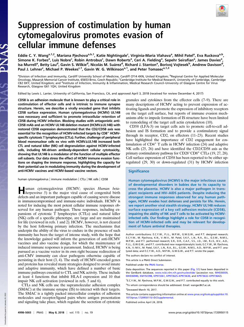
Human cytomegalovirus (HCMV; species Human beta-herpesvirus 5) is the major viral cause of congenital birth
defects and an important pathogen capable of causing severe diseasein immunocompromised and immune-naïve individuals. HCMV isnoted for inducing the most potent cellular immune responses ob-served for any human pathogen. These responses, including ex-pansions of cytotoxic T lymphocytes (CTLs) and natural killer(NK) cells of a specific phenotype, are large and are maintainedfor life (reviewed in refs. 1 and 2). HCMV, however, is not clearedby the host following primary infection. The mechanisms thatunderpin the ability of the virus to endure in the presence of suchimmunity has been the target of intense study, with the hope thatthe knowledge gained will inform the generation of anti-HCMVvaccines and also vaccine design, for which the maintenance ofinduced immune responses is paramount. Indeed, HCMV is beingpursued as a vaccine vector in its own right because redirection ofanti-CMV immunity can clear pathogens otherwise capable ofpersisting in their host (3, 4). The study of HCMV-encoded genesand proteins has revealed many strategies designed to avoid innateand adaptive immunity, which have defined a number of basicimmune pathways essential to CTL and NK activity. These includeat least 4 functions that inhibit HLA-I expression and 10 thatimpair NK cell activation (reviewed in refs. 5 and 6).CTLs and NK cells use the supramolecular adhesion complex
(SMAC) at the immune synapse (IS) to interact with their targets.The SMAC is a tightly packed intercellular complex of adhesionmolecules and receptor/ligand pairs where antigen presentationand signaling take place, which regulate the secretion of cytotoxic
granules and cytokines from the effector cells (7–9). There aremany descriptions of HCMV acting to prevent expression of ac-tivating ligands and promote the expression of inhibitory receptorson the target cell surface, but reports of immune evasion mech-anisms able to impede formation of IS structure have been limitedto remodeling of the target cell actin cytoskeleton (10).CD58 (LFA-3) on target cells acts to promote cell-to-cell ad-
hesion and IS formation and to provide a costimulatory signalthrough its receptor, CD2, on effectors (11–23). Recent studieshave highlighted the importance of CD2 engagement for cos-timulation of CD4+ T cells in HCMV infection (24) and adaptiveNK cells (25, 26) and have identified the CD2/CD58 axis as theprimary costimulatory pathway for CD28−CD8+ CTLs (19, 27, 28).Cell surface expression of CD58 has been reported to be either up-regulated (29, 30) or down-regulated (31) by HCMV infection
Significance
Human cytomegalovirus (HCMV) is the major infectious causeof developmental disorders in babies due to its capacity tocross the placenta. HCMV is also a major pathogen in trans-plant recipients and HIV–AIDS patients. Despite inducing thestrongest immune responses observed for any human path-ogen, HCMV evades host defenses and persists for life. Herein,we report another viral stealth strategy. HCMV UL148 reducessurface expression of a key cell adhesion molecule (CD58),impairing the ability of NK and T cells to be activated by HCMV-infected cells. Our findings highlight a role for CD58 in recogni-tion of HCMV-infected cells and may be relevant for develop-ment of future antiviral therapies.
Author contributions: E.C.Y.W., P.J.L., M.P.W., G.W.G.W., and P.T. designed research;E.C.Y.W., M. Pjechova, K.N., V.-M.V., M. Patel, S.K.F., L.N., R.A., B.L., G.S.W., N.M.S.,M.P.W., and P.T. performed research; E.R., D.R., C.A.F., S.S., J.D., I.M., R.J.S., B.V., A.D.,P.J.L., G.W.G.W., and P.T. contributed new reagents/analytic tools; E.C.Y.W., M. Pjechova,K.N., V.-M.V., M. Patel, S.K.F., L.N., R.A., B.L., G.S.W., N.M.S., A.D., M.P.W., and P.T. ana-lyzed data; and E.C.Y.W., A.D., M.P.W., G.W.G.W., and P.T. wrote the paper.
The authors declare no conflict of interest.
This article is a PNAS Direct Submission.
Published under the PNAS license.
Data deposition: The sequences reported in this paper (Fig. S1) have been deposited inthe GenBank database, www.ncbi.nlm.nih.gov/nucleotide/ (accession nos. MH036939,MH036940, KM192398, KP973361, KP973625–KP973630, and KP973632–KP973642).1E.C.Y.W., M. Pjechova, M.P.W., G.W.G.W., and P.T. contributed equally to this work.2To whom correspondence should be addressed. Email: [email protected] March 10, 2017.
This article contains supporting information online at www.pnas.org/lookup/suppl/doi:10.1073/pnas.1720950115/-/DCSupplemental.
Published online April 24, 2018.
4998–5003 | PNAS | May 8, 2018 | vol. 115 | no. 19 www.pnas.org/cgi/doi/10.1073/pnas.1720950115
Dow
nloa
ded
by g
uest
on
Janu
ary
25, 2
022
depending on the HCMV strain used. The aim of our study was todissect this phenomenon, thereby determining the functional rele-vance of regulating CD58 expression on HCMV-infected cells. Wedescribe the identification of a viral-encoded gene responsible fordown-regulating CD58 and detail the broad effect of this function onboth CTL and NK cell recognition in the context of HCMV infection.
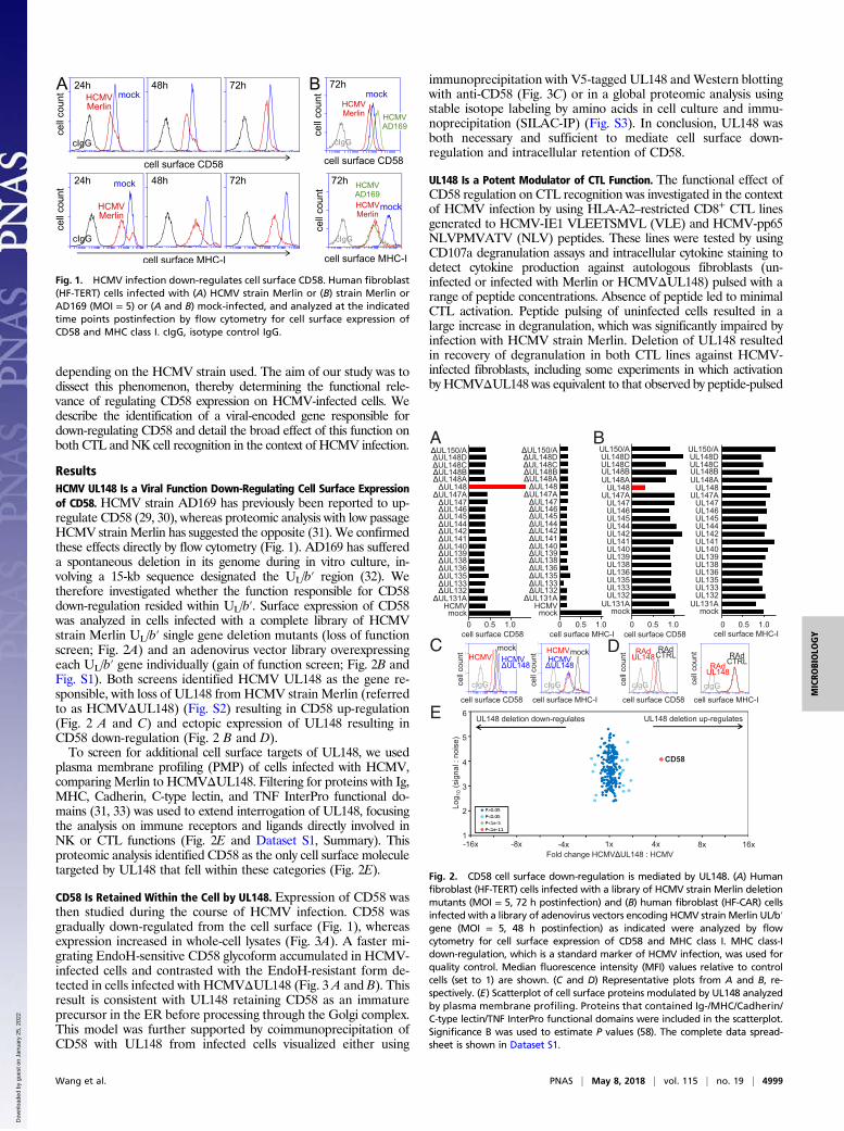
ResultsHCMV UL148 Is a Viral Function Down-Regulating Cell Surface Expressionof CD58. HCMV strain AD169 has previously been reported to up-regulate CD58 (29, 30), whereas proteomic analysis with low passageHCMV strainMerlin has suggested the opposite (31). We confirmedthese effects directly by flow cytometry (Fig. 1). AD169 has suffereda spontaneous deletion in its genome during in vitro culture, in-volving a 15-kb sequence designated the UL/b′ region (32). Wetherefore investigated whether the function responsible for CD58down-regulation resided within UL/b′. Surface expression of CD58was analyzed in cells infected with a complete library of HCMVstrain Merlin UL/b′ single gene deletion mutants (loss of functionscreen; Fig. 2A) and an adenovirus vector library overexpressingeach UL/b′ gene individually (gain of function screen; Fig. 2B andFig. S1). Both screens identified HCMV UL148 as the gene re-sponsible, with loss of UL148 from HCMV strain Merlin (referredto as HCMVΔUL148) (Fig. S2) resulting in CD58 up-regulation(Fig. 2 A and C) and ectopic expression of UL148 resulting inCD58 down-regulation (Fig. 2 B and D).To screen for additional cell surface targets of UL148, we used
plasma membrane profiling (PMP) of cells infected with HCMV,comparing Merlin to HCMVΔUL148. Filtering for proteins with Ig,MHC, Cadherin, C-type lectin, and TNF InterPro functional do-mains (31, 33) was used to extend interrogation of UL148, focusingthe analysis on immune receptors and ligands directly involved inNK or CTL functions (Fig. 2E and Dataset S1, Summary). Thisproteomic analysis identified CD58 as the only cell surface moleculetargeted by UL148 that fell within these categories (Fig. 2E).
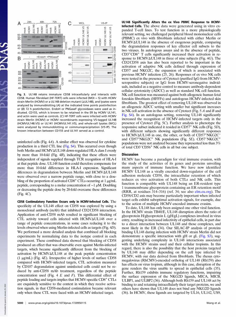
CD58 Is Retained Within the Cell by UL148. Expression of CD58 wasthen studied during the course of HCMV infection. CD58 wasgradually down-regulated from the cell surface (Fig. 1), whereasexpression increased in whole-cell lysates (Fig. 3A). A faster mi-grating EndoH-sensitive CD58 glycoform accumulated in HCMV-infected cells and contrasted with the EndoH-resistant form de-tected in cells infected with HCMVΔUL148 (Fig. 3 A and B). Thisresult is consistent with UL148 retaining CD58 as an immatureprecursor in the ER before processing through the Golgi complex.This model was further supported by coimmunoprecipitation ofCD58 with UL148 from infected cells visualized either using
immunoprecipitation with V5-tagged UL148 and Western blottingwith anti-CD58 (Fig. 3C) or in a global proteomic analysis usingstable isotope labeling by amino acids in cell culture and immu-noprecipitation (SILAC-IP) (Fig. S3). In conclusion, UL148 wasboth necessary and sufficient to mediate cell surface down-regulation and intracellular retention of CD58.
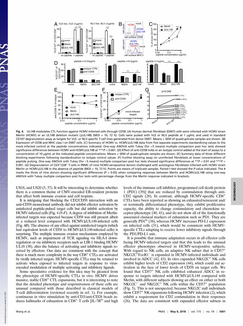
UL148 Is a Potent Modulator of CTL Function. The functional effect ofCD58 regulation on CTL recognition was investigated in the contextof HCMV infection by using HLA-A2–restricted CD8+ CTL linesgenerated to HCMV-IE1 VLEETSMVL (VLE) and HCMV-pp65NLVPMVATV (NLV) peptides. These lines were tested by usingCD107a degranulation assays and intracellular cytokine staining todetect cytokine production against autologous fibroblasts (un-infected or infected with Merlin or HCMVΔUL148) pulsed with arange of peptide concentrations. Absence of peptide led to minimalCTL activation. Peptide pulsing of uninfected cells resulted in alarge increase in degranulation, which was significantly impaired byinfection with HCMV strain Merlin. Deletion of UL148 resultedin recovery of degranulation in both CTL lines against HCMV-infected fibroblasts, including some experiments in which activationby HCMVΔUL148 was equivalent to that observed by peptide-pulsed
BA
Fig. 1. HCMV infection down-regulates cell surface CD58. Human fibroblast(HF-TERT) cells infected with (A) HCMV strain Merlin or (B) strain Merlin orAD169 (MOI = 5) or (A and B) mock-infected, and analyzed at the indicatedtime points postinfection by flow cytometry for cell surface expression ofCD58 and MHC class I. cIgG, isotype control IgG.
cell
coun
t
cell
coun
t UL150/AUL148DUL148CUL148BUL148A
UL148UL147A
UL147UL146UL145UL144UL142UL141UL140UL139UL138UL136UL135UL133UL132
UL131AHCMVmock
cell surface CD58 cell surface MHC-I
D
B
C
A
cell surface CD58 cell surface MHC-I ce
ll co
unt RAd
UL148RAd
CTRL
cIgG
050
0200
400
050
0200
400
cell surface MHC-I cell surface CD58
cell
coun
t
cIgG
mockHCMV HCMV
UL148
cIgG
mockHCMVHCMVUL148
cIgG
cell surface CD58 cell surface MHC-I0 0.5 1.0 0 0.5 1.0 0 0.5 1.0 0 0.5 1.0
RAdCTRLRAd
UL148
-8x 1x 4x 8x-16x 16x
5
4
3
2
1
6
Log 1
0(s
igna
l : n
oise
)
UL148 deletion up-regulatesUL148 deletion down-regulates
Fold change HCMV UL148 : HCMV
CD58
-4x
E
UL150/AUL148DUL148CUL148BUL148A
UL148UL147A
UL147UL146UL145UL144UL142UL141UL140UL139UL138UL136UL135UL133UL132
UL131AHCMVmock
UL150/AUL148DUL148CUL148BUL148A
UL148UL147A
UL147UL146UL145UL144UL142UL141UL140UL139UL138UL136UL135UL133UL132
UL131Amock
UL150/AUL148DUL148CUL148BUL148A
UL148UL147A
UL147UL146UL145UL144UL142UL141UL140UL139UL138UL136UL135UL133UL132
UL131Amock
Fig. 2. CD58 cell surface down-regulation is mediated by UL148. (A) Humanfibroblast (HF-TERT) cells infected with a library of HCMV strain Merlin deletionmutants (MOI = 5, 72 h postinfection) and (B) human fibroblast (HF-CAR) cellsinfected with a library of adenovirus vectors encoding HCMV strain Merlin UL/b′gene (MOI = 5, 48 h postinfection) as indicated were analyzed by flowcytometry for cell surface expression of CD58 and MHC class I. MHC class-Idown-regulation, which is a standard marker of HCMV infection, was used forquality control. Median fluorescence intensity (MFI) values relative to controlcells (set to 1) are shown. (C and D) Representative plots from A and B, re-spectively. (E) Scatterplot of cell surface proteins modulated by UL148 analyzedby plasma membrane profiling. Proteins that contained Ig-/MHC/Cadherin/C-type lectin/TNF InterPro functional domains were included in the scatterplot.Significance B was used to estimate P values (58). The complete data spread-sheet is shown in Dataset S1.
Wang et al. PNAS | May 8, 2018 | vol. 115 | no. 19 | 4999
MICRO
BIOLO
GY
Dow
nloa
ded
by g
uest
on
Janu
ary
25, 2
022
uninfected cells (Fig. 4A). A similar effect was observed for cytokineproduction in a third CTL line (Fig. S4). This occurred even thoughboth Merlin and HCMVΔUL148 down-regulated HLA class I evenlyby more than 10-fold (Fig. 4B), indicating that these effects wereindependent of signals supplied through TCR recognition of HLA-Iat that peptide dose. UL148 function could therefore compensate formore than 10-fold differences in HLA-I expression. Significantdiferences in degranulation between Merlin and HCMVΔUL148were observed over a narrow peptide range, with close to a dou-bling of the proportion of activated CD8+ CTLs occurring at 1 μg/mLpeptide, corresponding to a molar concentration of ∼1 μM. Doublingor decreasing the peptide dose by 20-fold overcame these differences(Fig. 4C).
CD58 Costimulatory Function Occurs only in HCMV-Infected Cells. Thespecificity of the UL148 effect on CD58 was explored by using amonoclonal antibody (mAb) that inhibited CD2/CD58 interaction.Application of anti-CD58 mAb resulted in significant blocking ofCTL activity toward cells infected with HCMVΔUL148 over arange of peptide concentrations, in some cases reducing it to thelevels observed when usingMerlin-infected cells as targets (Fig. 4D).We performed a more detailed analysis that combined all blockingexperiments by normalizing data to the isotype control in eachexperiment. These combined data showed that blocking of CD58produced an effect that was observable even against Merlin-infectedtargets, which became significantly different from the blocking ofactivation by HCMVΔUL148 at the peak peptide concentration(1 μg/mL) (Fig. 4E). Irrespective of higher levels of surface CD58compared with HCMV-infected targets, CTL activation measuredby CD107 degranulation against uninfected cells could not be re-duced by anti-CD58 mAb treatment, regardless of the peptideconcentration used (Fig. 4 E and F). This differential effect ofpeptide loading and targets implies that HCMV-specific CD8+CTLsare exquisitely sensitive to the context in which they receive activa-tion signals, in that CD58-mediated costimulation became relevantonly when these CTL were faced with an HCMV-infected target.
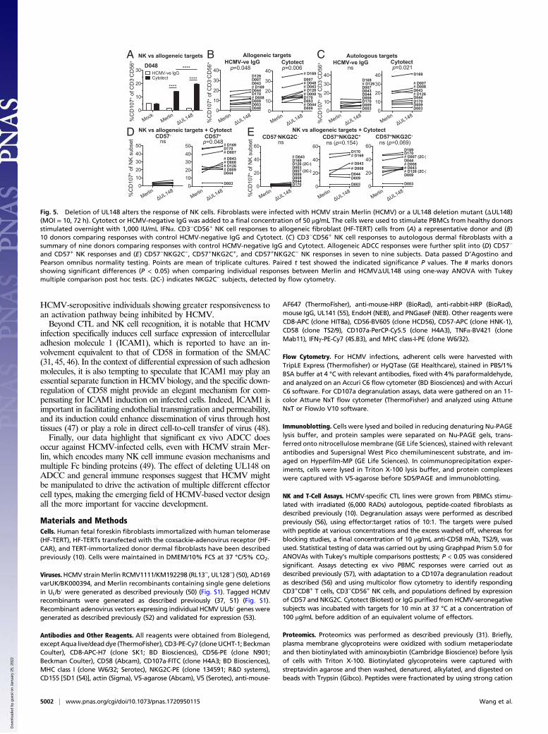
UL148 Significantly Alters the ex Vivo PBMC Response to HCMV-Infected Cells. The above data were generated using in vitro ex-panded T-cell lines. To test function in a more physiologicallyrelevant setting, we challenged peripheral blood mononuclear cells(PBMC) ex vivo with fibroblasts infected with either Merlin orHCMVΔUL148 in the absence of exogenous peptide, comparingthe degranulation responses of key effector cell subsets to thetwo viruses. In autologous assays and in the absence of peptide,CD3+CD8+ T cells significantly increased their activation in re-sponse to HCMVΔUL148 in three of nine subjects (Fig. 4G). TheCD2/CD58 axis has also been reported to be important in theactivation of adaptive NK cells defined through expression ofCD57 and NKG2C, the expansion of which is associated withprevious HCMV infection (25, 26). Responses of ex vivo NK cellswere tested in the presence of Cytotect (purified IgG fromHCMV-seropositive subjects) or IgG from HCMV-seronegative individ-uals, included as a negative control to measure antibody-dependentcellular cytotoxicity (ADCC) as well as standard NK cell function.NK cell function was measured against both allogeneic human fetalforeskin fibroblasts (HFFFs) and autologous HCMV-infected skinfibroblasts. The greatest effect of removing UL148 was observed inan allogeneic ADCC setting with smaller but significant increasesin NK cell activation in the absence of Cytotect (Fig. 5 A and B andFig. S4). In an autologous setting, removing UL148 significantlyincreased the recognition of HCMV-infected targets only in thepresence of Cytotect (Fig. 5C). Further analysis to phenotype theresponsive subset indicated it resided in CD57+ NK cells (Fig. 5D),with different subjects showing significantly different responsesto HCMVΔUL148 in one, the other, or both of CD57+NKG2C−
and CD57+NKG2C+ NK populations (Fig. 5E). CD57−NKG2C+
populations were not analyzed because they represented less than 3%of total CD3−CD56+ NK cells in all but one subject.
DiscussionHCMV has become a paradigm for viral immune evasion, withthe study of the activities of its genes and proteins unveilingmany aspects of immune function. We have now identifiedHCMV UL148 as a virally encoded down-regulator of the celladhesion molecule CD58, the intracellular retention of whichreduces ex vivo activation of both CTLs and NK cells. Thisfunction is compatible with UL148 being an ER-resident type1 transmembrane glycoprotein containing an ER retention motif(RRR, at residues 314–316) (ref. 34; see also elm.eu.org). TheCD58/CD2 axis may become particularly important when infectedtarget cells exhibit suboptimal activation signals, for example, dueto the action of multiple HCMV-encoded immune evasins.To date, UL148 has only been assigned one other viral function.
In the HCMV strain TB40/E, UL148 disruption alters the ratio ofglycoprotein H/glycoprotein L (gH/gL) complexes involved in virusentry, resulting in increased infectivity of epithelial cells, in part dueto a direct interaction between UL148 and those complexes, andmost likely in the ER (34). Our SILAC-IP analysis of proteinsbinding UL148 during infection with HCMV strain Merlin did notdemonstrate a specific interaction with gH or gL (Fig. S3), sug-gesting underlying complexity in UL148 interactions associatedwith the HCMV strains used and their cellular tropisms. In thisregard, there is also the possibility that the host proteins targetedby UL148 may differ depending on the cell type infected byHCMV, with our data derived from fibroblasts. The rhesus cyto-megalovirus (RhCMV)-encoded ortholog of UL148 (Rh159) alsohas effects on virus tropism, although in this case, disruption of thegene renders the virus unable to spread in epithelial cells (35).Further, Rh159 exhibits immune regulatory functions, impairingthe surface expression of the NKG2D ligands, MICA, MICB,ULBP1, and ULBP2 (36). Although both Rh159 and UL148 act bybinding to and retaining intracellularly their target proteins, we andothers have shown that UL148 does not bind any NKG2D ligands(36). In HCMV, these ligands are targeted by UL16, UL142, US9,
A C
B
Fig. 3. UL148 retains immature CD58 intracellularly and interacts withCD58. Human fibroblast (HF-TERT) cells were infected (MOI = 5) with HCMVstrain Merlin (HCMV) or a UL148 deletion mutant (ΔUL148), and lysates wereanalyzed by immunoblotting (A) at the indicated time points postinfectionor (B) 72 h postinfection. EndoH or PNGaseF glycosidases were used as in-dicated. CD155, which is known to be retained in the ER by HCMV UL141,and actin were used as controls. (C) HF-TERT cells were infected with HCMVstrain Merlin (HCMV) or HCMV recombinants expressing V5-tagged UL148(HCMVUL148.V5) or UL141 (HCMVUL141.V5), and whole-cell lysates (WCL)were analyzed by immunoblotting or coimmunoprecipitation (V5-IP). Theknown interaction between CD155 and UL141 served as a control.
5000 | www.pnas.org/cgi/doi/10.1073/pnas.1720950115 Wang et al.
Dow
nloa
ded
by g
uest
on
Janu
ary
25, 2
022
US18, and US20 (5, 37). It will be interesting to determine whetherthere is a common theme of CMV-encoded ER-resident proteinsthat affect both immune evasion and cell tropism.It is intriguing that blocking the CD2/CD58 interaction with an
anti-CD58 monoclonal antibody did not inhibit effector activation byuninfected peptide-pulsed target cells but did inhibit activation byHCMV-infected cells (Fig. 4D–F). A degree of inhibition of Merlin-infected targets was expected because CD58 was still present albeitat a reduced level compared with HCMVΔUL148-infected cells(Fig. 4B). The absence of any effect against uninfected targets (whichhad equivalent levels of CD58 to HCMVΔUL148-infected cells) issurprising. The multiple immune evasion mechanisms employed byHCMV, such as impairment of TCR signaling via HLA-I down-regulation or via inhibitory receptors such as LIR-1 binding HCMVUL18 (38), alter the balance of activating and inhibitory signals re-ceived by effectors. Our data are consistent with the concept thatthere is much more complexity in the way CD8+ CTLs are activatedby virally infected targets. HCMV-specific CTLs may be retuned toactivate when exposed to infected targets in response to virus-encoded modulation of multiple activating and inhibitory ligands.Some speculative evidence for this idea may be gleaned from
the phenotype of HCMV-specific CTLs in vivo. HCMV drivesmassive, stable CD8+ CTL expansions, but it is interesting to notethat the detailed phenotype and responsiveness of these cells areunusual compared with those described in classical models ofT-cell differentiation (reviewed recently in ref. 1). For example,continuous in vitro stimulation by anti-CD3/anti-CD28 beads in-duces hallmarks of exhaustion in CD8+ T cells [IL-7Rlo and high
levels of the immune cell inhibitor, programmed cell death protein1 (PD1) (39)] that are reduced by costimulation through anti-CD2 signals (28). In contrast, although HCMV-specific CD8+
CTLs have been reported as showing an exhausted/senescent and/or terminally differentiated phenotype, they exhibit proliferativecapacity, the ability to change costimulatory and chemokine re-ceptor-phenotype (40, 41), and do not show all of the functionallyassociated classical markers of exhaustion such as PD1. They aregenerally PD1lo (38), whereas HCMV increases PD-L1 expressionon infected cells (31), which would be consistent with HCMV-specific CTLs adapting to receive fewer inhibitory signals throughthe PD1/PD-L1 axis.It is possible that immune adaptation occurs in all effector cells
facing HCMV-infected targets and that this leads to the unusualeffector phenotypes observed in HCMV-seropositive subjects.With regard to NK cells, an adaptive NK subset that is CD57+
NKG2C+FceR1− is expanded in HCMV-infected individuals andinvolved in ADCC (42, 43). In vitro expanded NKG2C+ NK cellsexhibit higher levels of CD2 expression (44), which could aid ac-tivation in the face of lower levels of CD58 on target cells. Wefound that CD57+ NK cells exhibited enhanced ADCC in re-sponse to targets infected with HCMVΔUL148 compared withMerlin, with different subjects showing an effect on either or bothNKG2C− and NKG2C+ NK cells within the CD57+ population(Fig. 5). This is not unexpected, because NKG2C null individualsshow CD57+ NK expansions following HCMV infection (2), whichexhibit a requirement for CD2 costimulation in their responses(26). The data are consistent with expanded effector subsets in
A B C
D
E F G
Fig. 4. UL148 modulates CTL function against HCMV-infected cells through CD58. (A) Human dermal fibroblast (D007) cells were infected with HCMV strainMerlin (HCMV) or an UL148 deletion mutant (ΔUL148) (MOI = 10, 72 h). Cells were pulsed with VLE or NLV peptide at 1 μg/mL and used in standardCD107 degranulation assay as targets for VLE- or NLV-specific T-cell lines generated from donor D007. Means + SEM of quadruplicate samples are shown. (B)Expression of CD58 and MHC class I on D007 cells. (C) Summary of HCMV vs. HCMVΔUL148 data from five separate experiments standardizing values to themock-infected control at the peptide concentrations indicated. One-way ANOVA with Tukey (for >5 means) multiple comparison post hoc tests showedsignificance differences between HCMV and HCMVΔUL148 at ***P < 0.001. (D) Effect of anti-CD58 mAb or an isotype control added at the start of assays to aconcentration of 10 μg/mL at the indicated peptide concentrations. Means + SEM of quadruplicate samples are shown. (E) Summary data of three differentblocking experiments following standardization to isotype control values. (F) Further blocking assay on uninfected fibroblasts at lower concentrations ofpeptide pulsing. One-way ANOVA with Tukey (for >5 means) multiple comparison post hoc tests showed significance differences at **P < 0.01 and ***P <0.001. (G) Degranulation of CD3+CD8+ T-cells in PBMC of nine HCMV-seropositive donors challenged with autologous fibroblasts infected with HCMV strainMerlin or HCMVΔUL148 in the absence of peptide (MOI = 10, 72 h). Points are means of triplicate samples. Paired t test showed the P value indicated. The #marks the three of nine donors showing significant differences (P < 0.05) when comparing responses between Merlin and HCMVΔUL148 using one-wayANOVA with Tukey multiple comparison post hoc tests with percentage change from the Merlin response indicated in brackets.
Wang et al. PNAS | May 8, 2018 | vol. 115 | no. 19 | 5001
MICRO
BIOLO
GY
Dow
nloa
ded
by g
uest
on
Janu
ary
25, 2
022
HCMV-seropositive individuals showing greater responsiveness toan activation pathway being inhibited by HCMV.Beyond CTL and NK cell recognition, it is notable that HCMV
infection specifically induces cell surface expression of intercellularadhesion molecule 1 (ICAM1), which is reported to have an in-volvement equivalent to that of CD58 in formation of the SMAC(31, 45, 46). In the context of differential expression of such adhesionmolecules, it is also tempting to speculate that ICAM1 may play anessential separate function in HCMV biology, and the specific down-regulation of CD58 might provide an elegant mechanism for com-pensating for ICAM1 induction on infected cells. Indeed, ICAM1 isimportant in facilitating endothelial transmigration and permeability,and its induction could enhance dissemination of virus through hosttissues (47) or play a role in direct cell-to-cell transfer of virus (48).Finally, our data highlight that significant ex vivo ADCC does
occur against HCMV-infected cells, even with HCMV strain Mer-lin, which encodes many NK cell immune evasion mechanisms andmultiple Fc binding proteins (49). The effect of deleting UL148 onADCC and general immune responses suggest that HCMV mightbe manipulated to drive the activation of multiple different effectorcell types, making the emerging field of HCMV-based vector designall the more important for vaccine development.
Materials and MethodsCells. Human fetal foreskin fibroblasts immortalized with human telomerase(HF-TERT), HF-TERTs transfected with the coxsackie-adenovirus receptor (HF-CAR), and TERT-immortalized donor dermal fibroblasts have been describedpreviously (10). Cells were maintained in DMEM/10% FCS at 37 °C/5% CO2.
Viruses.HCMV strainMerlin RCMV1111/KM192298 (RL13−, UL128−) (50), AD169varUK/BK000394, and Merlin recombinants containing single gene deletionsin UL/b′ were generated as described previously (50) (Fig. S1). Tagged HCMVrecombinants were generated as described previously (37, 51) (Fig. S1).Recombinant adenovirus vectors expressing individual HCMV UL/b′ genes weregenerated as described previously (52) and validated for expression (53).
Antibodies and Other Reagents. All reagents were obtained from Biolegend,exceptAqua live/dead dye (ThermoFisher), CD3-PE-Cy7 (cloneUCHT-1; BeckmanCoulter), CD8-APC-H7 (clone SK1; BD Biosciences), CD56-PE (clone N901;Beckman Coulter), CD58 (Abcam), CD107a-FITC (clone H4A3; BD Biosciences),MHC class I (clone W6/32; Serotec), NKG2C-PE (clone 134591; R&D systems),CD155 [5D1 (54)], actin (Sigma), V5-agarose (Abcam), V5 (Serotec), anti-mouse-
AF647 (ThermoFisher), anti-mouse-HRP (BioRad), anti-rabbit-HRP (BioRad),mouse IgG, UL141 (55), EndoH (NEB), and PNGaseF (NEB). Other reagents wereCD8-APC (clone HIT8a), CD56-BV605 (clone HCD56), CD57-APC (clone HNK-1),CD58 (clone TS2/9), CD107a-PerCP-Cy5.5 (clone H4A3), TNFα-BV421 (cloneMab11), IFNγ-PE-Cy7 (4S.B3), and MHC class-I-PE (clone W6/32).
Flow Cytometry. For HCMV infections, adherent cells were harvested withTripLE Express (Thermofisher) or HyQTase (GE Healthcare), stained in PBS/1%BSA buffer at 4 °C with relevant antibodies, fixed with 4% paraformaldehyde,and analyzed on an Accuri C6 flow cytometer (BD Biosciences) and with AccuriC6 software. For CD107a degranulation assays, data were gathered on an 11-color Attune NxT flow cytometer (ThermoFisher) and analyzed using AttuneNxT or FlowJo V10 software.
Immunoblotting. Cells were lysed and boiled in reducing denaturing Nu-PAGElysis buffer, and protein samples were separated on Nu-PAGE gels, trans-ferred onto nitrocellulose membrane (GE Life Sciences), stained with relevantantibodies and Supersignal West Pico chemiluminescent substrate, and im-aged on Hyperfilm-MP (GE Life Sciences). In coimmunoprecipitation exper-iments, cells were lysed in Triton X-100 lysis buffer, and protein complexeswere captured with V5-agarose before SDS/PAGE and immunoblotting.
NK and T-Cell Assays. HCMV-specific CTL lines were grown from PBMCs stimu-lated with irradiated (6,000 RADs) autologous, peptide-coated fibroblasts asdescribed previously (10). Degranulation assays were performed as describedpreviously (56), using effector:target ratios of 10:1. The targets were pulsedwith peptide at various concentrations and the excess washed off, whereas forblocking studies, a final concentration of 10 μg/mL anti-CD58 mAb, TS2/9, wasused. Statistical testing of data was carried out by using Graphpad Prism 5.0 forANOVAs with Tukey’s multiple comparisons posttests; P < 0.05 was consideredsignificant. Assays detecting ex vivo PBMC responses were carried out asdescribed previously (57), with adaptation to a CD107a degranulation readoutas described (56) and using multicolor flow cytometry to identify respondingCD3+CD8+ T cells, CD3−CD56+ NK cells, and populations defined by expressionof CD57 and NKG2C. Cytotect (Biotest) or IgG purified from HCMV-seronegativesubjects was incubated with targets for 10 min at 37 °C at a concentration of100 μg/mL before addition of an equivalent volume of effectors.
Proteomics. Proteomics was performed as described previously (31). Briefly,plasma membrane glycoproteins were oxidized with sodium metaperiodateand then biotinylated with aminoxybiotin (Cambridge Bioscience) before lysisof cells with Triton X-100. Biotinylated glycoproteins were captured withstreptavidin agarose and then washed, denatured, alkylated, and digested onbeads with Trypsin (Gibco). Peptides were fractionated by using strong cation
A B C
D E
Fig. 5. Deletion of UL148 alters the response of NK cells. Fibroblasts were infected with HCMV strain Merlin (HCMV) or a UL148 deletion mutant (ΔUL148)(MOI = 10, 72 h). Cytotect or HCMV-negative IgG was added to a final concentration of 50 μg/mL The cells were used to stimulate PBMCs from healthy donorsstimulated overnight with 1,000 IU/mL IFNα. CD3−CD56+ NK cell responses to allogeneic fibroblast (HF-TERT) cells from (A) a representative donor and (B)10 donors comparing responses with control HCMV-negative IgG and Cytotect. (C) CD3−CD56+ NK cell responses to autologous dermal fibroblasts with asummary of nine donors comparing responses with control HCMV-negative IgG and Cytotect. Allogeneic ADCC responses were further split into (D) CD57−
and CD57+ NK responses and (E) CD57−NKG2C−, CD57+NKG2C+, and CD57+NKG2C− NK responses in seven to nine subjects. Data passed D’Agostino andPearson omnibus normality testing. Points are mean of triplicate cultures. Paired t test showed the indicated significance P values. The # marks donorsshowing significant differences (P < 0.05) when comparing individual responses between Merlin and HCMVΔUL148 using one-way ANOVA with Tukeymultiple comparison post hoc tests. (2C-) indicates NKG2C− subjects, detected by flow cytometry.
5002 | www.pnas.org/cgi/doi/10.1073/pnas.1720950115 Wang et al.
Dow
nloa
ded
by g
uest
on
Janu
ary
25, 2
022
exchange. Enriched peptides were labeled with tandem mass tag (TMT) re-agents, combined at a 1:1 ratio, and then prefractionated by offline high-pH-reversed-phase chromatography (Agilent). Mass spectrometry and data anal-ysis were performed as described previously by using an Orbitrap Fusion. Pvalues were estimated using Benjamini-Hochberg–corrected significance A orsignificance B values from Perseus version 1.2.0.16 (58). SILAC-IP experimentswere conducted and analyzed as described previously (10).
Ethics Statement. Healthy adult volunteers provided blood and dermal fi-broblasts for this study after providing written informed consent. The study
was approved by the Cardiff University School of Medicine Research EthicsCommittee, reference numbers 10/20 and 16/52.
ACKNOWLEDGMENTS. The study was supported by Medical ResearchCouncil and Wellcome Trust Grants G1000236, MR/P001602/1, WT090323MA(to E.C.Y.W., G.W.G.W., and P.T.), MR/L008734/1 (to E.C.Y.W., R.J.S., G.W.G.W.,and P.T.), MC_UU_12014/3 (to A.D.), MEYS-NPS I-L01413, Czech ScienceFoundation Project P206/12/G151 (to B.V.), Wellcome Trust Senior ClinicalResearch Fellowship 108070 (to M.P.W.), and Wellcome Trust PrincipalResearch Fellowship 101835 (to P.J.L.).
1. Klenerman P, Oxenius A (2016) T cell responses to cytomegalovirus. Nat Rev Immunol16:367–377.
2. Goodier MR, et al. (2014) Rapid NK cell differentiation in a population with near-universal human cytomegalovirus infection is attenuated by NKG2C deletions. Blood124:2213–2222.
3. Hansen SG, et al. (2013) Cytomegalovirus vectors violate CD8+ T cell epitope recog-nition paradigms. Science 340:1237874.
4. Hansen SG, et al. (2013) Immune clearance of highly pathogenic SIV infection. Nature502:100–104.
5. Wilkinson GW, et al. (2008) Modulation of natural killer cells by human cytomega-lovirus. J Clin Virol 41:206–212.
6. Halenius A, Gerke C, Hengel H (2015) Classical and non-classical MHC I moleculemanipulation by human cytomegalovirus: So many targets—But how many arrows inthe quiver? Cell Mol Immunol 12:139–153.
7. Davis DM, et al. (1999) The human natural killer cell immune synapse. Proc Natl AcadSci USA 96:15062–15067.
8. Grakoui A, et al. (1999) The immunological synapse: A molecular machine controllingT cell activation. Science 285:221–227.
9. Monks CR, Freiberg BA, Kupfer H, Sciaky N, Kupfer A (1998) Three-dimensional seg-regation of supramolecular activation clusters in T cells. Nature 395:82–86.
10. Stanton RJ, et al. (2014) HCMV pUL135 remodels the actin cytoskeleton to impairimmune recognition of infected cells. Cell Host Microbe 16:201–214.
11. Beyers AD, Spruyt LL, Williams AF (1992) Molecular associations between the T-lym-phocyte antigen receptor complex and the surface antigens CD2, CD4, or CD8 andCD5. Proc Natl Acad Sci USA 89:2945–2949.
12. Bierer BE, Hahn WC (1993) T cell adhesion, avidity regulation and signaling: A mo-lecular analysis of CD2. Semin Immunol 5:249–261.
13. Brown MH, Cantrell DA, Brattsand G, Crumpton MJ, Gullberg M (1989) TheCD2 antigen associates with the T-cell antigen receptor CD3 antigen complex on thesurface of human T lymphocytes. Nature 339:551–553.
14. Davis SJ, Ikemizu S, Wild MK, van der Merwe PA (1998) CD2 and the nature of proteininteractions mediating cell-cell recognition. Immunol Rev 163:217–236.
15. Dustin ML, Selvaraj P, Mattaliano RJ, Springer TA (1987) Anchoring mechanisms forLFA-3 cell adhesion glycoprotein at membrane surface. Nature 329:846–848.
16. Gassmann M, Amrein KE, Flint NA, Schraven B, Burn P (1994) Identification of a signalingcomplex involving CD2, zeta chain and p59fyn in T lymphocytes. Eur J Immunol 24:139–144.
17. Holter W, Schwarz M, Cerwenka A, Knapp W (1996) The role of CD2 as a regulator ofhuman T-cell cytokine production. Immunol Rev 153:107–122.
18. Kaizuka Y, Douglass AD, Vardhana S, Dustin ML, Vale RD (2009) The coreceptorCD2 uses plasma membrane microdomains to transduce signals in T cells. J Cell Biol185:521–534.
19. Leitner J, Herndler-Brandstetter D, Zlabinger GJ, Grubeck-Loebenstein B, Steinberger P(2015) CD58/CD2 is the primary costimulatory pathway in human CD28-CD8+ T cells.J Immunol 195:477–487.
20. Meuer SC, et al. (1984) An alternative pathway of T-cell activation: A functional rolefor the 50 kd T11 sheep erythrocyte receptor protein. Cell 36:897–906.
21. Selvaraj P, et al. (1987) The T lymphocyte glycoprotein CD2 binds the cell surface li-gand LFA-3. Nature 326:400–403.
22. Siliciano RF, Pratt JC, Schmidt RE, Ritz J, Reinherz EL (1985) Activation of cytolytic Tlymphocyte and natural killer cell function through the T11 sheep erythrocytebinding protein. Nature 317:428–430.
23. Yang JJ, Ye Y, Carroll A, Yang W, Lee HW (2001) Structural biology of the cell ad-hesion protein CD2: Alternatively folded states and structure-function relation. CurrProtein Pept Sci 2:1–17.
24. Shiao SL, et al. (2007) Human effector memory CD4+ T cells directly recognize allo-geneic endothelial cells in vitro and in vivo. J Immunol 179:4397–4404.
25. Rölle A, et al. (2016) CD2-CD58 interactions are pivotal for the activation and functionof adaptive natural killer cells in human cytomegalovirus infection. Eur J Immunol 46:2420–2425.
26. Liu LL, et al. (2016) Critical role of CD2 co-stimulation in adaptive natural killer cellresponses revealed in NKG2C-deficient humans. Cell Rep 15:1088–1099.
27. Bruns T, et al. (2015) CMV infection of human sinusoidal endothelium regulates he-patic T cell recruitment and activation. J Hepatol 63:38–49.
28. McKinney EF, Lee JC, Jayne DR, Lyons PA, Smith KG (2015) T-cell exhaustion, co-stimulation and clinical outcome in autoimmunity and infection. Nature 523:612–616.
29. Grundy JE, Downes KL (1993) Up-regulation of LFA-3 and ICAM-1 on the surface offibroblasts infected with cytomegalovirus. Immunology 78:405–412.
30. Fletcher JM, Prentice HG, Grundy JE (1998) Natural killer cell lysis of cytomegalovirus(CMV)-infected cells correlates with virally induced changes in cell surface lymphocyte
function-associated antigen-3 (LFA-3) expression and not with the CMV-induceddown-regulation of cell surface class I HLA. J Immunol 161:2365–2374.
31. Weekes MP, et al. (2014) Quantitative temporal viromics: An approach to investigatehost-pathogen interaction. Cell 157:1460–1472.
32. Cha TA, et al. (1996) Human cytomegalovirus clinical isolates carry at least 19 genesnot found in laboratory strains. J Virol 70:78–83.
33. Vivier E, Tomasello E, Baratin M, Walzer T, Ugolini S (2008) Functions of natural killercells. Nat Immunol 9:503–510.
34. Li G, Nguyen CC, Ryckman BJ, Britt WJ, Kamil JP (2015) A viral regulator of glyco-protein complexes contributes to human cytomegalovirus cell tropism. Proc Natl AcadSci USA 112:4471–4476.
35. Lilja AE, Chang WL, Barry PA, Becerra SP, Shenk TE (2008) Functional genetic analysisof rhesus cytomegalovirus: Rh01 is an epithelial cell tropism factor. J Virol 82:2170–2181.
36. Sturgill ER, et al. (2016) Natural killer cell evasion is essential for infection by rhesuscytomegalovirus. PLoS Pathog 12:e1005868.
37. Fielding CA, et al. (2014) Two novel human cytomegalovirus NK cell evasion functionstarget MICA for lysosomal degradation. PLoS Pathog 10:e1004058.
38. Hertoghs KM, et al. (2010) Molecular profiling of cytomegalovirus-induced humanCD8+ T cell differentiation. J Clin Invest 120:4077–4090.
39. Francisco LM, Sage PT, Sharpe AH (2010) The PD-1 pathway in tolerance and auto-immunity. Immunol Rev 236:219–242.
40. Gandhi MK, et al. (2003) Late diversification in the clonal composition of human cy-tomegalovirus-specific CD8+ T cells following allogeneic hemopoietic stem celltransplantation. Blood 102:3427–3438.
41. Waller EC, et al. (2007) Differential costimulation through CD137 (4-1BB) restoresproliferation of human virus-specific “effector memory” (CD28(-) CD45RA(HI)) CD8(+)T cells. Blood 110:4360–4366.
42. Gumá M, et al. (2004) Imprint of human cytomegalovirus infection on the NK cellreceptor repertoire. Blood 104:3664–3671.
43. Gumá M, et al. (2006) Expansion of CD94/NKG2C+ NK cells in response to humancytomegalovirus-infected fibroblasts. Blood 107:3624–3631.
44. Béziat V, et al. (2013) NK cell responses to cytomegalovirus infection lead to stableimprints in the human KIR repertoire and involve activating KIRs. Blood 121:2678–2688.
45. Kronschnabl M, Stamminger T (2003) Synergistic induction of intercellular adhesionmolecule-1 by the human cytomegalovirus transactivators IE2p86 and pp71 is medi-ated via an Sp1-binding site. J Gen Virol 84:61–73.
46. Huppa JB, Davis MM (2003) T-cell-antigen recognition and the immunological syn-apse. Nat Rev Immunol 3:973–983.
47. Bentz GL, et al. (2006) Human cytomegalovirus (HCMV) infection of endothelial cellspromotes naive monocyte extravasation and transfer of productive virus to enhancehematogenous dissemination of HCMV. J Virol 80:11539–11555.
48. Murrell I, et al. (2017) The pentameric complex drives immunologically covert cell-celltransmission of wild-type human cytomegalovirus. Proc Natl Acad Sci USA 114:6104–6109.
49. Corrales-Aguilar E, et al. (2014) Human cytomegalovirus Fcγ binding proteinsgp34 and gp68 antagonize Fcγ receptors I, II and III. PLoS Pathog 10:e1004131.
50. Stanton RJ, et al. (2010) Reconstruction of the complete human cytomegalovirusgenome in a BAC reveals RL13 to be a potent inhibitor of replication. J Clin Invest 120:3191–3208.
51. Murrell I, et al. (2016) Genetic stability of bacterial artificial chromosome-derivedhuman cytomegalovirus during culture in vitro. J Virol 90:3929–3943.
52. Stanton RJ, McSharry BP, Armstrong M, Tomasec P, Wilkinson GW (2008) Re-engi-neering adenovirus vector systems to enable high-throughput analyses of genefunction. Biotechniques 45:659–662, 664–668.
53. Seirafian S (2012) . An analysis of human cytomegalovirus gene usage. PhD thesis(Cardiff University, Cardiff, UK).
54. Aoki J, Koike S, Ise I, Sato-Yoshida Y, Nomoto A (1994) Amino acid residues on humanpoliovirus receptor involved in interaction with poliovirus. J Biol Chem 269:8431–8438.
55. Tomasec P, et al. (2005) Downregulation of natural killer cell-activating ligandCD155 by human cytomegalovirus UL141. Nat Immunol 6:181–188.
56. Prod’homme V, et al. (2007) The human cytomegalovirus MHC class I homologUL18 inhibits LIR-1+ but activates LIR-1- NK cells. J Immunol 178:4473–4481.
57. Wang EC, et al. (2002) UL40-mediated NK evasion during productive infection withhuman cytomegalovirus. Proc Natl Acad Sci USA 99:7570–7575.
58. Cox J, Mann M (2008) MaxQuant enables high peptide identification rates, in-dividualized p.p.b.-range mass accuracies and proteome-wide protein quantification.Nat Biotechnol 26:1367–1372.
Wang et al. PNAS | May 8, 2018 | vol. 115 | no. 19 | 5003
MICRO
BIOLO
GY
Dow
nloa
ded
by g
uest
on
Janu
ary
25, 2
022