Review Article Gut Microbial Flora, Prebiotics, and...
10
Hindawi Publishing Corporation BioMed Research International Volume 2013, Article ID 435268, 9 pages http://dx.doi.org/10.1155/2013/435268 Review Article Gut Microbial Flora, Prebiotics, and Probiotics in IBD: Their Current Usage and Utility Franco Scaldaferri, 1 Viviana Gerardi, 1 Loris Riccardo Lopetuso, 1 Fabio Del Zompo, 1 Francesca Mangiola, 1 Ivo Boškoski, 1 Giovanni Bruno, 1 Valentina Petito, 1 Lucrezia Laterza, 1 Giovanni Cammarota, 1 Eleonora Gaetani, 1 Alessandro Sgambato, 2 and Antonio Gasbarrini 1 1 Department of Internal Medicine, Gastroenterology Division, Catholic University of Sacred Hearth, Policlinico “A. Gemelli” Hospital, lgo Gemelli 8, 00168 Roma, Italy 2 Institute of Pathology, Catholic University of Sacred Hearth, lgo Gemelli 8, 00168 Rome, Italy Correspondence should be addressed to Franco Scaldaferri; [email protected] Received 19 April 2013; Revised 29 June 2013; Accepted 8 July 2013 Academic Editor: Michael Mahler Copyright © 2013 Franco Scaldaferri et al. is is an open access article distributed under the Creative Commons Attribution License, which permits unrestricted use, distribution, and reproduction in any medium, provided the original work is properly cited. Inflammatory bowel diseases are chronic diseases affecting the gastrointestinal tract, whose major forms are represented by Crohn’s disease (CD) and ulcerative colitis (UC). eir etiology is still unclear, although several factors have been identified as major determinants for induction or relapses. Among these, the role of the “forgotten organ”, gut microbiota, has become more appreciated in recent years. e delicate symbiotic relationship between the gut microbiota and the host appears to be lost in IBD. In this perspective, several studies have been conducted to assess the role of prebiotics and probiotics in gut microbiota modulation. is is a minireview aimed to address in an easy format (simple questions-simple answers) some common issues about the theme. An update on the role of selected constituents of gut microbiota in the pathogenesis of IBD is presented together with the analysis of the efficacy of gut microbiota modulation by prebiotics and probiotics administration in the management of IBD. 1. Introduction e human microflora, known as “microbiota”, includes bacteria, fungi, bacteriophages, and viruses and acts as an “organ” synergistically with the host, creating an ecosystem. It is able to colonize skin, the genitourinary system, the respiratory system, and, above all, the gut. Gut microbiota includes around a thousand different species and more than 15,000 different strains of bacteria, for a total weight of about 1 Kg. Stomach and small intestine are relatively poor of bacteria, whilst the colon hosts about 10 12 microorganisms [1], mainly belonging to the Firmicutes and Bacteroidetes phyla [2]. Other domains represented are those of Archaea [3] and Eukarya, plus many viruses and bacteriophages [4]. Gut microbiota is harbored also by several yeast families, whose role in gastrointestinal physiology as well as in diseases still remains unclear. Since most species seem to be refractory to cultivation with usual methods, culture-independent molecular tech- niques, such as 16S rDNA genotyping, are used to charac- terize the gut microflora from both fecal samples and bowel biopsies [5–8]. At birth, the human gut is sterile, and the first coloniza- tion occurs during childbirth and the first feed. Subsequently, the microbiota changes under the influence of age, sex, state of immune maturation, and environmental factors. Aſter the first two years, the microbiota becomes more stable, although a stable endogen flora could be differentiated by a transient one, which is, on the contrary, more sensitive to external stimuli, as the gut mucosa is the first line of communication with exogenous agents [9, 10]. During the first years of life, gut microbiota stimulates the innate immunity, by inducing gut-associated lymphoid system, and the acquired immunity, by stimulating specific
Transcript of Review Article Gut Microbial Flora, Prebiotics, and...

Hindawi Publishing CorporationBioMed Research InternationalVolume 2013, Article ID 435268, 9 pageshttp://dx.doi.org/10.1155/2013/435268
Review ArticleGut Microbial Flora, Prebiotics, and Probiotics in IBD:Their Current Usage and Utility
Franco Scaldaferri,1 Viviana Gerardi,1 Loris Riccardo Lopetuso,1
Fabio Del Zompo,1 Francesca Mangiola,1 Ivo Boškoski,1 Giovanni Bruno,1
Valentina Petito,1 Lucrezia Laterza,1 Giovanni Cammarota,1 Eleonora Gaetani,1
Alessandro Sgambato,2 and Antonio Gasbarrini1
1 Department of Internal Medicine, Gastroenterology Division, Catholic University of Sacred Hearth,Policlinico “A. Gemelli” Hospital, lgo Gemelli 8, 00168 Roma, Italy
2 Institute of Pathology, Catholic University of Sacred Hearth, lgo Gemelli 8, 00168 Rome, Italy
Correspondence should be addressed to Franco Scaldaferri; [email protected]
Received 19 April 2013; Revised 29 June 2013; Accepted 8 July 2013
Academic Editor: Michael Mahler
Copyright © 2013 Franco Scaldaferri et al. This is an open access article distributed under the Creative Commons AttributionLicense, which permits unrestricted use, distribution, and reproduction in any medium, provided the original work is properlycited.
Inflammatory bowel diseases are chronic diseases affecting the gastrointestinal tract, whosemajor forms are represented by Crohn’sdisease (CD) and ulcerative colitis (UC). Their etiology is still unclear, although several factors have been identified as majordeterminants for induction or relapses. Among these, the role of the “forgotten organ”, gutmicrobiota, has becomemore appreciatedin recent years. The delicate symbiotic relationship between the gut microbiota and the host appears to be lost in IBD. In thisperspective, several studies have been conducted to assess the role of prebiotics and probiotics in gut microbiota modulation. Thisis a minireview aimed to address in an easy format (simple questions-simple answers) some common issues about the theme. Anupdate on the role of selected constituents of gut microbiota in the pathogenesis of IBD is presented together with the analysis ofthe efficacy of gut microbiota modulation by prebiotics and probiotics administration in the management of IBD.
1. Introduction
The human microflora, known as “microbiota”, includesbacteria, fungi, bacteriophages, and viruses and acts as an“organ” synergistically with the host, creating an ecosystem.It is able to colonize skin, the genitourinary system, therespiratory system, and, above all, the gut.
Gut microbiota includes around a thousand differentspecies and more than 15,000 different strains of bacteria,for a total weight of about 1 Kg. Stomach and small intestineare relatively poor of bacteria, whilst the colon hosts about1012 microorganisms [1], mainly belonging to the Firmicutesand Bacteroidetes phyla [2]. Other domains represented arethose of Archaea [3] and Eukarya, plus many viruses andbacteriophages [4].Gutmicrobiota is harbored also by severalyeast families, whose role in gastrointestinal physiology aswell as in diseases still remains unclear.
Since most species seem to be refractory to cultivationwith usual methods, culture-independent molecular tech-niques, such as 16S rDNA genotyping, are used to charac-terize the gut microflora from both fecal samples and bowelbiopsies [5–8].
At birth, the human gut is sterile, and the first coloniza-tion occurs during childbirth and the first feed. Subsequently,the microbiota changes under the influence of age, sex, stateof immune maturation, and environmental factors. After thefirst two years, themicrobiota becomesmore stable, althougha stable endogen flora could be differentiated by a transientone, which is, on the contrary, more sensitive to externalstimuli, as the gut mucosa is the first line of communicationwith exogenous agents [9, 10].
During the first years of life, gut microbiota stimulatesthe innate immunity, by inducing gut-associated lymphoidsystem, and the acquired immunity, by stimulating specific
2 BioMed Research International
systemic and local immune responses [11]. In the gut, bac-terial fragments stimulate specific receptors, like TLR9 (toll-like receptor 9, expressed on epithelial and immune cells) andthe inflammasome that are able to recognize bacterial DNA[12].
In normal conditions, stimulation of the mucosalimmune system by gut microbiota determines a state of“low-grade physiological inflammation” [13], a status ofcontinues activation of the mucosal immune system inresponse to commensals, and, in case of needs, also towardspathogens. Mucosal homeostasis requires a continuousbalance between pro- and anti-inflammatory components.In recent years several studies investigated the correlationbetween dysbiosis and intestinal and extraintestinal diseases,including immune system alteration, obesity, allergies,autoimmune diseases, irritable bowel syndrome (IBS), andinflammatory bowel disease (IBD) [14, 15].
IBD are chronic, relapsing, multifactorial conditionsaffecting the digestive tract. These majorly include ulcerativecolitis (UC) and Crohn’s disease (CD). Although the etiologyof these diseases is still unclear, the main hypothesis isthat IBD are a result of an excessive immune response toendogenous bacteria, which occurs in genetically predis-posed individuals [16, 17].
Most of conventional IBD therapies aim to modulateimmune system. 5-aminosalicylic acid (ASA) compounds,corticosteroids, azathioprine/6-mercaptopurine, methotrex-ate, cyclosporine, and anti-TNF𝛼 agents are constantly usedto manage these diseases. Several and probably less charac-terized therapies, as additive to or alternative to conventionaltherapies, in milder cases, aim to modulate gut microbiota,directly or indirectly. For example, antibiotics are used in IBD,and they are considered particularly effective in perianal andpostoperative CD and in pouchitis [18].
Probiotics contain viable organisms, sufficient amounts ofwhich reach the intestine in an active state, thus exert positivehealth effects [19]. They mostly include lactic acid-producingbacteria and yeasts that reach the gut unaltered, withoutproviding damage to the host [18, 20]. Their mechanisms ofaction are still unclear; they probably modulate the mem-brane permeability and themucosal immune system, keepingaway pathogens from intestinal mucosa surface. Lactobacillusand Bifidobacteria produce harmful substances for Gram-positive and Gram-negative bacteria, and they compete withpathogens (i.e., Bacteriodetes, Clostridium, Staphylococcus,and Enterobacter) for cell adhesion [18, 21, 22].
On the other side, prebiotics are selectively fermentedingredients that allow specific changes both in the compo-sition and/or in the activity of gastrointestinal microflora,conferring benefits upon the host well-being and health[19]. They are nondigestible oligosaccharides, such as fruc-tooligosaccharides (FOS), galactooligosaccharides (GOS),lactulose, and inulin, and they have the potential to stimulategrowth of selective and beneficial gut bacteria [18]. Because oftheir composition, they cannot be adsorbed until they reachcolon, where they can be fermented by a specific microflorainto small chain fatty acid (SCFA) and lactate [23].
Their exact mechanism of action is still unclear. Recentevidences hypothesized that they are able to increase the
production of SCFA and to modulate cytokines productionwithin the gut mucosa, by modulating the gut flora compo-sition. The synergistic combination of pro- and prebiotics iscalled “synbiotic” [19] (Figure 1).
2. Is There a Role for Specific Pathogensin IBD?
It was originally suspected that IBD depend on a singlepathogenic strain of bacteria. In 1984, Chiodini et al. [24]showed a strong association between Mycobacterium aviumsubspecies paratubercalosis (MAP) and CD, but this hypoth-esis was confirmed only by few studies. Furthermore, theinefficiency of antituberculosis antibiotics in CD patientsreinforced the criticism towards MAP [25, 26].
Escherichia coli is usually isolated inmany intestinal biop-sies of CD patients. In particular, adherent-invasive strains(AIEC) are found in patients with ileal CD [27]. Also Yersiniaand Pseudomonas are supposed to act as triggers in CDdisease [28]. On the other side, Salmonella, Campylobacterjejuni, Clostridiumdifficile, Adenovirus, andMycoplasmahavebeen identified as agents associated to disease relapsing butnot to induction [29, 30].
A study conducted by Willing et al. [31] showed that,in ileal CD patients, Faecalibacterium and Roseburia areunderrepresented whilst Enterobacteriaceae (such as E. coli)and Ruminococcus gnavus are increased.
Fusobacterium varium has been localized in the colon ofUC patients and causes UC in mice when injected by enema[32, 33]. Moreover, it has been assessed an overgrowth ofE. coli in UC patients, suggesting a possible role on genesisand/or maintenance of the disease [34].
2.1. Is There a Role of the Commensal Flora in IBD Patho-genesis? Several evidences support the hypothesis that gutmicrobiota plays a role in the pathogenesis of IBD, particu-larly studies involving animalmodels or in vitromodels. Herewe decided to present limited data coming from experimentalmodels, while focusing more on human studies.
Themost inflamed intestinal areas in IBD patients are thesame displaying the highest amount of intestinal bacteria.Theevidence that germ-free mice do not develop severe colitissupports this finding [35]. Furthermore, recurrence rate ofpostoperative CD and pouchitis is higher when the fecalstream is reestablished [15, 36].
IBD patients display a reduced amount of dominant com-mensal bacteria, such as Firmicutes (in particularClostridiumclusters IX and IV) and Bacteriodetes, facing an increasednumber of Proteobacteria and Actinobacteria. This observa-tion is associated with a decreased SCFA level in feces of IBDpatients. Among SCFAs, a decrease in butyrate level has beenassociated with IBD as it is able to inhibit proinflammatorycytokines release to increase the production of mucin andantimicrobial peptides and to provide energy to colonocytes[37–39].
Among Firmicutes, the reduction of Faecalibacteriumprausnitzii has recently emerged as a very frequent finding
BioMed Research International 3
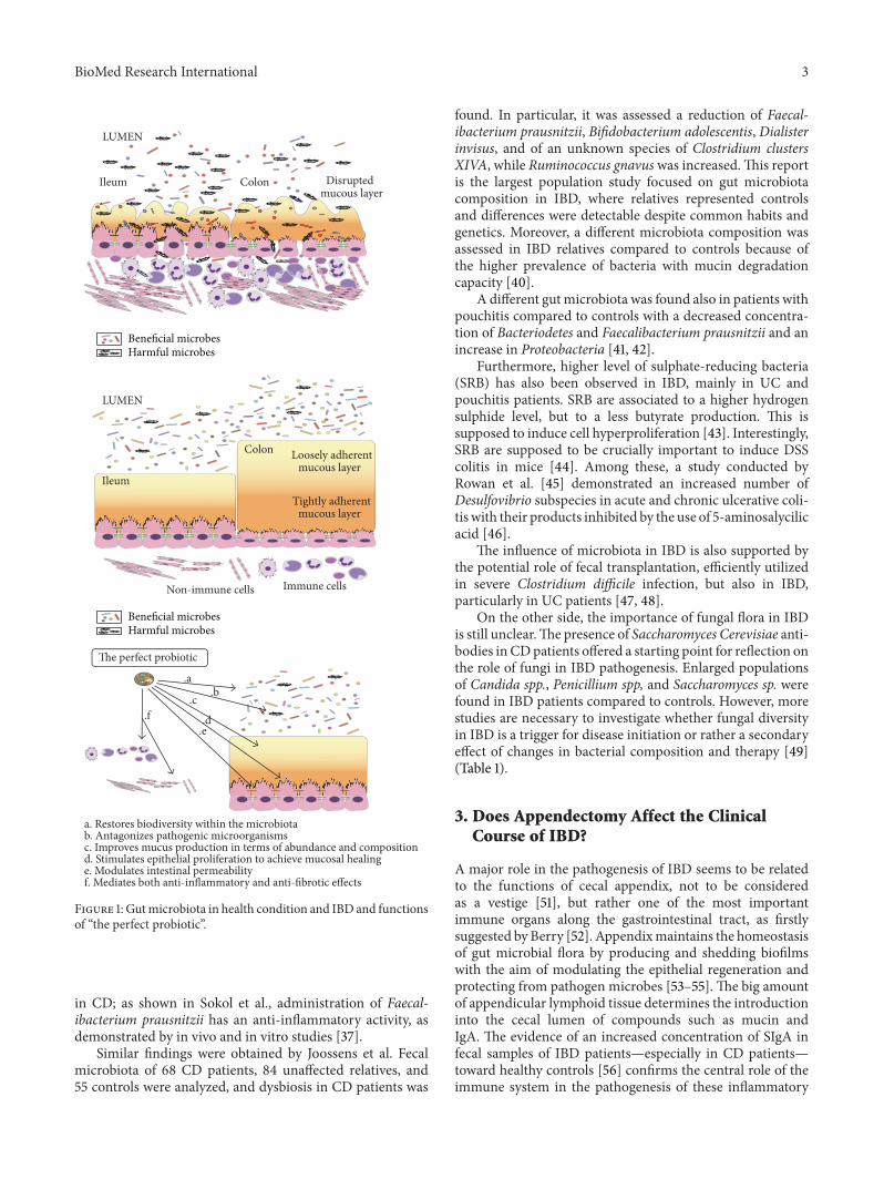
ColonIleum Disruptedmucous layer
LUMEN
Ileum
Colon Loosely adherentmucous layer
Tightly adherentmucous layer
LUMEN
Non-immune cells Immune cells
The perfect probiotic
.a.b
.c
.d.e
.f
a. Restores biodiversity within the microbiotab. Antagonizes pathogenic microorganismsc. Improves mucus production in terms of abundance and compositiond. Stimulates epithelial proliferation to achieve mucosal healinge. Modulates intestinal permeabilityf. Mediates both anti-inflammatory and anti-fibrotic effects
Beneficial microbesHarmful microbes
Beneficial microbesHarmful microbes
Figure 1: Gutmicrobiota in health condition and IBD and functionsof “the perfect probiotic”.
in CD; as shown in Sokol et al., administration of Faecal-ibacterium prausnitzii has an anti-inflammatory activity, asdemonstrated by in vivo and in vitro studies [37].
Similar findings were obtained by Joossens et al. Fecalmicrobiota of 68 CD patients, 84 unaffected relatives, and55 controls were analyzed, and dysbiosis in CD patients was
found. In particular, it was assessed a reduction of Faecal-ibacterium prausnitzii, Bifidobacterium adolescentis, Dialisterinvisus, and of an unknown species of Clostridium clustersXIVA, while Ruminococcus gnavuswas increased.This reportis the largest population study focused on gut microbiotacomposition in IBD, where relatives represented controlsand differences were detectable despite common habits andgenetics. Moreover, a different microbiota composition wasassessed in IBD relatives compared to controls because ofthe higher prevalence of bacteria with mucin degradationcapacity [40].
A different gutmicrobiota was found also in patients withpouchitis compared to controls with a decreased concentra-tion of Bacteriodetes and Faecalibacterium prausnitzii and anincrease in Proteobacteria [41, 42].
Furthermore, higher level of sulphate-reducing bacteria(SRB) has also been observed in IBD, mainly in UC andpouchitis patients. SRB are associated to a higher hydrogensulphide level, but to a less butyrate production. This issupposed to induce cell hyperproliferation [43]. Interestingly,SRB are supposed to be crucially important to induce DSScolitis in mice [44]. Among these, a study conducted byRowan et al. [45] demonstrated an increased number ofDesulfovibrio subspecies in acute and chronic ulcerative coli-tis with their products inhibited by the use of 5-aminosalycilicacid [46].
The influence of microbiota in IBD is also supported bythe potential role of fecal transplantation, efficiently utilizedin severe Clostridium difficile infection, but also in IBD,particularly in UC patients [47, 48].
On the other side, the importance of fungal flora in IBDis still unclear.The presence of Saccharomyces Cerevisiae anti-bodies in CDpatients offered a starting point for reflection onthe role of fungi in IBD pathogenesis. Enlarged populationsof Candida spp., Penicillium spp, and Saccharomyces sp. werefound in IBD patients compared to controls. However, morestudies are necessary to investigate whether fungal diversityin IBD is a trigger for disease initiation or rather a secondaryeffect of changes in bacterial composition and therapy [49](Table 1).
3. Does Appendectomy Affect the ClinicalCourse of IBD?
A major role in the pathogenesis of IBD seems to be relatedto the functions of cecal appendix, not to be consideredas a vestige [51], but rather one of the most importantimmune organs along the gastrointestinal tract, as firstlysuggested by Berry [52]. Appendixmaintains the homeostasisof gut microbial flora by producing and shedding biofilmswith the aim of modulating the epithelial regeneration andprotecting from pathogen microbes [53–55]. The big amountof appendicular lymphoid tissue determines the introductioninto the cecal lumen of compounds such as mucin andIgA. The evidence of an increased concentration of SIgA infecal samples of IBD patients—especially in CD patients—toward healthy controls [56] confirms the central role of theimmune system in the pathogenesis of these inflammatory
4 BioMed Research International
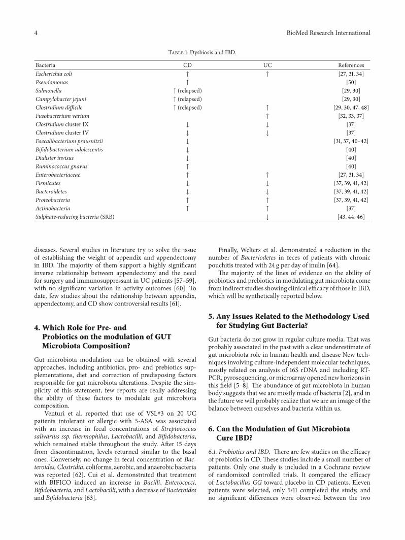
Table 1: Dysbiosis and IBD.
Bacteria CD UC ReferencesEscherichia coli ↑ ↑ [27, 31, 34]Pseudomonas ↑ [50]Salmonella ↑ (relapsed) [29, 30]Campylobacter jejuni ↑ (relapsed) [29, 30]Clostridium difficile ↑ (relapsed) ↑ [29, 30, 47, 48]Fusobacterium varium ↑ [32, 33, 37]Clostridium cluster IX ↓ ↓ [37]Clostridium cluster IV ↓ ↓ [37]Faecalibacterium prausnitzii ↓ [31, 37, 40–42]Bifidobacterium adolescentis ↓ [40]Dialister invisus ↓ [40]Ruminococcus gnavus ↑ [40]Enterobacteriaceae ↑ ↑ [27, 31, 34]Firmicutes ↓ ↓ [37, 39, 41, 42]Bacteroidetes ↓ ↓ [37, 39, 41, 42]Proteobacteria ↑ ↑ [37, 39, 41, 42]Actinobacteria ↑ ↑ [37]Sulphate-reducing bacteria (SRB) ↓ [43, 44, 46]
diseases. Several studies in literature try to solve the issueof establishing the weight of appendix and appendectomyin IBD. The majority of them support a highly significantinverse relationship between appendectomy and the needfor surgery and immunosuppressant in UC patients [57–59],with no significant variation in activity outcomes [60]. Todate, few studies about the relationship between appendix,appendectomy, and CD show controversial results [61].
4. Which Role for Pre- andProbiotics on the modulation of GUTMicrobiota Composition?
Gut microbiota modulation can be obtained with severalapproaches, including antibiotics, pro- and prebiotics sup-plementations, diet and correction of predisposing factorsresponsible for gut microbiota alterations. Despite the sim-plicity of this statement, few reports are really addressingthe ability of these factors to modulate gut microbiotacomposition.
Venturi et al. reported that use of VSL#3 on 20 UCpatients intolerant or allergic with 5-ASA was associatedwith an increase in fecal concentrations of Streptococcussalivarius ssp. thermophilus, Lactobacilli, and Bifidobacteria,which remained stable throughout the study. After 15 daysfrom discontinuation, levels returned similar to the basalones. Conversely, no change in fecal concentration of Bac-teroides, Clostridia, coliforms, aerobic, and anaerobic bacteriawas reported [62]. Cui et al. demonstrated that treatmentwith BIFICO induced an increase in Bacilli, Enterococci,Bifidobacteria, and Lactobacilli, with a decrease of Bacteroidesand Bifidobacteria [63].
Finally, Welters et al. demonstrated a reduction in thenumber of Bacteriodetes in feces of patients with chronicpouchitis treated with 24 g per day of inulin [64].
The majority of the lines of evidence on the ability ofprobiotics and prebiotics in modulating gut microbiota comefrom indirect studies showing clinical efficacy of those in IBD,which will be synthetically reported below.
5. Any Issues Related to the Methodology Usedfor Studying Gut Bacteria?
Gut bacteria do not grow in regular culture media. That wasprobably associated in the past with a clear underestimate ofgut microbiota role in human health and disease New tech-niques involving culture-independent molecular techniques,mostly related on analysis of 16S rDNA and including RT-PCR, pyrosequencing, ormicroarray opened newhorizons inthis field [5–8]. The abundance of gut microbiota in humanbody suggests that we are mostly made of bacteria [2], and inthe future we will probably realize that we are an image of thebalance between ourselves and bacteria within us.
6. Can the Modulation of Gut MicrobiotaCure IBD?
6.1. Probiotics and IBD. There are few studies on the efficacyof probiotics in CD. These studies include a small number ofpatients. Only one study is included in a Cochrane reviewof randomized controlled trials. It compared the efficacyof Lactobacillus GG toward placebo in CD patients. Elevenpatients were selected, only 5/11 completed the study, andno significant differences were observed between the two
BioMed Research International 5
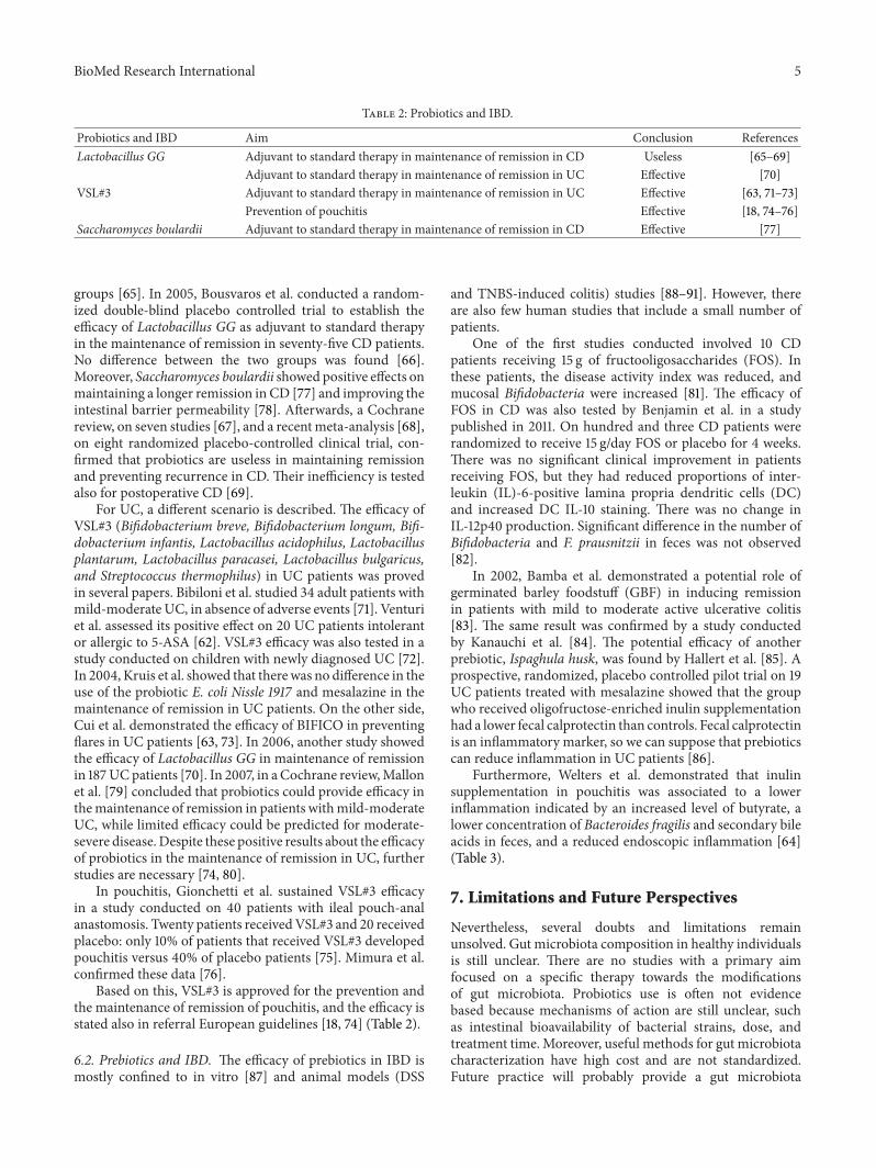
Table 2: Probiotics and IBD.
Probiotics and IBD Aim Conclusion ReferencesLactobacillus GG Adjuvant to standard therapy in maintenance of remission in CD Useless [65–69]
Adjuvant to standard therapy in maintenance of remission in UC Effective [70]VSL#3 Adjuvant to standard therapy in maintenance of remission in UC Effective [63, 71–73]
Prevention of pouchitis Effective [18, 74–76]Saccharomyces boulardii Adjuvant to standard therapy in maintenance of remission in CD Effective [77]
groups [65]. In 2005, Bousvaros et al. conducted a random-ized double-blind placebo controlled trial to establish theefficacy of Lactobacillus GG as adjuvant to standard therapyin the maintenance of remission in seventy-five CD patients.No difference between the two groups was found [66].Moreover, Saccharomyces boulardii showed positive effects onmaintaining a longer remission in CD [77] and improving theintestinal barrier permeability [78]. Afterwards, a Cochranereview, on seven studies [67], and a recentmeta-analysis [68],on eight randomized placebo-controlled clinical trial, con-firmed that probiotics are useless in maintaining remissionand preventing recurrence in CD. Their inefficiency is testedalso for postoperative CD [69].
For UC, a different scenario is described. The efficacy ofVSL#3 (Bifidobacterium breve, Bifidobacterium longum, Bifi-dobacterium infantis, Lactobacillus acidophilus, Lactobacillusplantarum, Lactobacillus paracasei, Lactobacillus bulgaricus,and Streptococcus thermophilus) in UC patients was provedin several papers. Bibiloni et al. studied 34 adult patients withmild-moderate UC, in absence of adverse events [71]. Venturiet al. assessed its positive effect on 20 UC patients intolerantor allergic to 5-ASA [62]. VSL#3 efficacy was also tested in astudy conducted on children with newly diagnosed UC [72].In 2004, Kruis et al. showed that therewas no difference in theuse of the probiotic E. coli Nissle 1917 and mesalazine in themaintenance of remission in UC patients. On the other side,Cui et al. demonstrated the efficacy of BIFICO in preventingflares in UC patients [63, 73]. In 2006, another study showedthe efficacy of Lactobacillus GG in maintenance of remissionin 187UCpatients [70]. In 2007, in a Cochrane review,Mallonet al. [79] concluded that probiotics could provide efficacy inthemaintenance of remission in patients withmild-moderateUC, while limited efficacy could be predicted for moderate-severe disease.Despite these positive results about the efficacyof probiotics in the maintenance of remission in UC, furtherstudies are necessary [74, 80].
In pouchitis, Gionchetti et al. sustained VSL#3 efficacyin a study conducted on 40 patients with ileal pouch-analanastomosis. Twenty patients receivedVSL#3 and 20 receivedplacebo: only 10% of patients that received VSL#3 developedpouchitis versus 40% of placebo patients [75]. Mimura et al.confirmed these data [76].
Based on this, VSL#3 is approved for the prevention andthe maintenance of remission of pouchitis, and the efficacy isstated also in referral European guidelines [18, 74] (Table 2).
6.2. Prebiotics and IBD. The efficacy of prebiotics in IBD ismostly confined to in vitro [87] and animal models (DSS
and TNBS-induced colitis) studies [88–91]. However, thereare also few human studies that include a small number ofpatients.
One of the first studies conducted involved 10 CDpatients receiving 15 g of fructooligosaccharides (FOS). Inthese patients, the disease activity index was reduced, andmucosal Bifidobacteria were increased [81]. The efficacy ofFOS in CD was also tested by Benjamin et al. in a studypublished in 2011. On hundred and three CD patients wererandomized to receive 15 g/day FOS or placebo for 4 weeks.There was no significant clinical improvement in patientsreceiving FOS, but they had reduced proportions of inter-leukin (IL)-6-positive lamina propria dendritic cells (DC)and increased DC IL-10 staining. There was no change inIL-12p40 production. Significant difference in the number ofBifidobacteria and F. prausnitzii in feces was not observed[82].
In 2002, Bamba et al. demonstrated a potential role ofgerminated barley foodstuff (GBF) in inducing remissionin patients with mild to moderate active ulcerative colitis[83]. The same result was confirmed by a study conductedby Kanauchi et al. [84]. The potential efficacy of anotherprebiotic, Ispaghula husk, was found by Hallert et al. [85]. Aprospective, randomized, placebo controlled pilot trial on 19UC patients treated with mesalazine showed that the groupwho received oligofructose-enriched inulin supplementationhad a lower fecal calprotectin than controls. Fecal calprotectinis an inflammatory marker, so we can suppose that prebioticscan reduce inflammation in UC patients [86].
Furthermore, Welters et al. demonstrated that inulinsupplementation in pouchitis was associated to a lowerinflammation indicated by an increased level of butyrate, alower concentration of Bacteroides fragilis and secondary bileacids in feces, and a reduced endoscopic inflammation [64](Table 3).
7. Limitations and Future Perspectives
Nevertheless, several doubts and limitations remainunsolved. Gut microbiota composition in healthy individualsis still unclear. There are no studies with a primary aimfocused on a specific therapy towards the modificationsof gut microbiota. Probiotics use is often not evidencebased because mechanisms of action are still unclear, suchas intestinal bioavailability of bacterial strains, dose, andtreatment time. Moreover, useful methods for gut microbiotacharacterization have high cost and are not standardized.Future practice will probably provide a gut microbiota
6 BioMed Research International
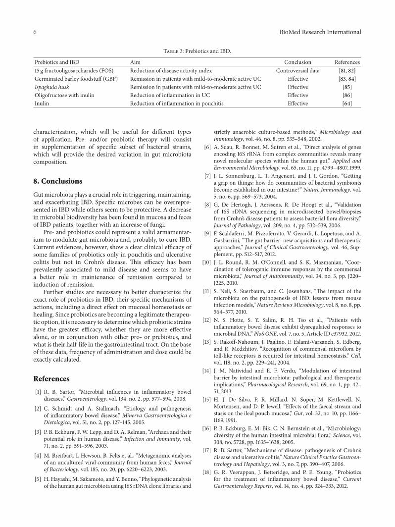
Table 3: Prebiotics and IBD.
Prebiotics and IBD Aim Conclusion References15 g fructooligosaccharides (FOS) Reduction of disease activity index Controversial data [81, 82]Germinated barley foodstuff (GBF) Remission in patients with mild-to-moderate active UC Effective [83, 84]Ispaghula husk Remission in patients with mild-to-moderate active UC Effective [85]Oligofructose with inulin Reduction of inflammation in UC Effective [86]Inulin Reduction of inflammation in pouchitis Effective [64]
characterization, which will be useful for different typesof application. Pre- and/or probiotic therapy will consistin supplementation of specific subset of bacterial strains,which will provide the desired variation in gut microbiotacomposition.
8. Conclusions
Gutmicrobiota plays a crucial role in triggering,maintaining,and exacerbating IBD. Specific microbes can be overrepre-sented in IBD while others seem to be protective. A decreasein microbial biodiversity has been found in mucosa and fecesof IBD patients, together with an increase of fungi.
Pre- and probiotics could represent a valid armamentar-ium to modulate gut microbiota and, probably, to cure IBD.Current evidences, however, show a clear clinical efficacy ofsome families of probiotics only in pouchitis and ulcerativecolitis but not in Crohn’s disease. This efficacy has beenprevalently associated to mild disease and seems to havea better role in maintenance of remission compared toinduction of remission.
Further studies are necessary to better characterize theexact role of probiotics in IBD, their specific mechanisms ofactions, including a direct effect on mucosal homeostasis orhealing. Since probiotics are becoming a legitimate therapeu-tic option, it is necessary to determinewhich probiotic strainshave the greatest efficacy, whether they are more effectivealone, or in conjunction with other pro- or prebiotics, andwhat is their half-life in the gastrointestinal tract. On the baseof these data, frequency of administration and dose could beexactly calculated.
References
[1] R. B. Sartor, “Microbial influences in inflammatory boweldiseases,” Gastroenterology, vol. 134, no. 2, pp. 577–594, 2008.
[2] C. Schmidt and A. Stallmach, “Etiology and pathogenesisof inflammatory bowel disease,” Minerva Gastroenterologica eDietologica, vol. 51, no. 2, pp. 127–145, 2005.
[3] P. B. Eckburg, P. W. Lepp, and D. A. Relman, “Archaea and theirpotential role in human disease,” Infection and Immunity, vol.71, no. 2, pp. 591–596, 2003.
[4] M. Breitbart, I. Hewson, B. Felts et al., “Metagenomic analysesof an uncultured viral community from human feces,” Journalof Bacteriology, vol. 185, no. 20, pp. 6220–6223, 2003.
[5] H. Hayashi, M. Sakamoto, and Y. Benno, “Phylogenetic analysisof the human gutmicrobiota using 16S rDNAclone libraries and
strictly anaerobic culture-based methods,” Microbiology andImmunology, vol. 46, no. 8, pp. 535–548, 2002.
[6] A. Suau, R. Bonnet, M. Sutren et al., “Direct analysis of genesencoding 16S rRNA from complex communities reveals manynovel molecular species within the human gut,” Applied andEnvironmentalMicrobiology, vol. 65, no. 11, pp. 4799–4807, 1999.
[7] J. L. Sonnenburg, L. T. Angenent, and J. I. Gordon, “Gettinga grip on things: how do communities of bacterial symbiontsbecome established in our intestine?” Nature Immunology, vol.5, no. 6, pp. 569–573, 2004.
[8] G. De Hertogh, J. Aerssens, R. De Hoogt et al., “Validationof 16S rDNA sequencing in microdissected bowel/biopsiesfrom Crohn’s disease patients to assess bacterial flora diversity,”Journal of Pathology, vol. 209, no. 4, pp. 532–539, 2006.
[9] F. Scaldaferri, M. Pizzoferrato, V. Gerardi, L. Lopetuso, and A.Gasbarrini, “The gut barrier: new acquisitions and therapeuticapproaches,” Journal of Clinical Gastroenterology, vol. 46, Sup-plement, pp. S12–S17, 2012.
[10] J. L. Round, R. M. O’Connell, and S. K. Mazmanian, “Coor-dination of tolerogenic immune responses by the commensalmicrobiota,” Journal of Autoimmunity, vol. 34, no. 3, pp. J220–J225, 2010.
[11] S. Nell, S. Suerbaum, and C. Josenhans, “The impact of themicrobiota on the pathogenesis of IBD: lessons from mouseinfectionmodels,”Nature ReviewsMicrobiology, vol. 8, no. 8, pp.564–577, 2010.
[12] N. S. Hotte, S. Y. Salim, R. H. Tso et al., “Patients withinflammatory bowel disease exhibit dysregulated responses tomicrobial DNA,” PloS ONE, vol. 7, no. 5, Article ID e37932, 2012.
[13] S. Rakoff-Nahoum, J. Paglino, F. Eslami-Varzaneh, S. Edberg,and R. Medzhitov, “Recognition of commensal microflora bytoll-like receptors is required for intestinal homeostasis,” Cell,vol. 118, no. 2, pp. 229–241, 2004.
[14] J. M. Natividad and E. F. Verdu, “Modulation of intestinalbarrier by intestinal microbiota: pathological and therapeuticimplications,” Pharmacological Research, vol. 69, no. 1, pp. 42–51, 2013.
[15] H. J. De Silva, P. R. Millard, N. Soper, M. Kettlewell, N.Mortensen, and D. P. Jewell, “Effects of the faecal stream andstasis on the ileal pouch mucosa,” Gut, vol. 32, no. 10, pp. 1166–1169, 1991.
[16] P. B. Eckburg, E. M. Bik, C. N. Bernstein et al., “Microbiology:diversity of the human intestinal microbial flora,” Science, vol.308, no. 5728, pp. 1635–1638, 2005.
[17] R. B. Sartor, “Mechanisms of disease: pathogenesis of Crohn’sdisease and ulcerative colitis,”Nature Clinical Practice Gastroen-terology and Hepatology, vol. 3, no. 7, pp. 390–407, 2006.
[18] G. R. Veerappan, J. Betteridge, and P. E. Young, “Probioticsfor the treatment of inflammatory bowel disease,” CurrentGastroenterology Reports, vol. 14, no. 4, pp. 324–333, 2012.

BioMed Research International 7
[19] M. De Vrese and J. Schrezenmeir, “Probiotics, prebiotics, andsynbiotics,”Advances in Biochemical Engineering/Biotechnology,vol. 111, pp. 1–66, 2008.
[20] M. A. C. Looijer-van Langen and L. A. Dieleman, “Prebiotics inchronic intestinal inflammation,” Inflammatory Bowel Diseases,vol. 15, no. 3, pp. 454–462, 2009.
[21] A. L. Servin, “Antagonistic activities of lactobacilli and bifi-dobacteria against microbial pathogens,” FEMS MicrobiologyReviews, vol. 28, no. 4, pp. 405–440, 2004.
[22] M. C. Collado, J. Meriluoto, and S. Salminen, “Role of com-mercial probiotic strains against human pathogen adhesion tointestinal mucus,” Letters in Applied Microbiology, vol. 45, no. 4,pp. 454–460, 2007.
[23] Y. Bouhnik, L. Raskine, G. Simoneau et al., “The capacity ofnondigestible carbohydrates to stimulate fecal bifidobacteriain healthy humans: a double-blind, randomized, placebo-controlled, parallel-group, dose-response relation study,”Amer-ican Journal of Clinical Nutrition, vol. 80, no. 6, pp. 1658–1664,2004.
[24] R. J. Chiodini, H. J. Van Kruiningen, and R. Thayer, “Possiblerole of mycobacteria in inflammatory bowel disease. I. AnunclassifiedMycobacterium species isolated from patients withCrohn’s disease,” Digestive Diseases and Sciences, vol. 29, no. 12,pp. 1073–1079, 1984.
[25] K. Hulten, A. Almashhrawi, F. A. K. El-Zaatari, and D. Y.Graham, “Antibacterial therapy for Crohn’s disease: a reviewemphasizing therapy directed against mycobacteria,” DigestiveDiseases and Sciences, vol. 45, no. 3, pp. 445–456, 2000.
[26] W. Selby, P. Pavli, B. Crotty et al., “Two-year combination antibi-otic therapy with clarithromycin, rifabutin, and clofazimine forcrohn’s disease,” Gastroenterology, vol. 132, no. 7, pp. 2313–2319,2007.
[27] B. Chassaing and A. Darfeuillemichaud, “The commensalmicrobiota and enteropathogens in the pathogenesis of inflam-matory bowel diseases,” Gastroenterology, vol. 140, no. 6, pp.1720–1728, 2011.
[28] N. A. Nagalingam and S. V. Lynch, “Role of the microbiotain inflammatory bowel diseases,” Inflammatory Bowel Diseases,vol. 18, no. 5, pp. 968–980, 2012.
[29] G. De Hertogh and K. Geboes, “Crohn’s disease and infec-tions: a complex relationship,” MedGenMed Medscape GeneralMedicine, vol. 6, no. 3, 2004.
[30] D. Comito and C. Romano, “Dysbiosis in the pathogenesis ofpediatric inflammatory bowel diseases,” International Journal ofInflammation, vol. 2012, Article ID 687143, 7 pages, 2012.
[31] B. P.Willing, J. Dicksved, J. Halfvarson et al., “A pyrosequencingstudy in twins shows that gastrointestinal microbial profilesvary with inflammatory bowel disease phenotypes,” Gastroen-terology, vol. 139, no. 6, pp. 1844.e1–1854.e1, 2010.
[32] T. Ohkusa, N. Sato, T. Ogihara, K. Morita, M. Ogawa, andI. Okayasu, “Fusobacterium varium localized in the colonicmucosa of patients with ulcerative colitis stimulates species-specific antibody,” Journal of Gastroenterology and Hepatology,vol. 17, no. 8, pp. 849–853, 2002.
[33] T. Ohkusa, I. Okayasu, T. Ogihara, K. Morita, M. Ogawa,and N. Sato, “Induction of experimental ulcerative colitis byFusobacteriumvarium isolated from colonicmucosa of patientswith ulcerative colitis,” Gut, vol. 52, no. 1, pp. 79–83, 2003.
[34] H. Sokol, P. Lepage, P. Seksik, J. Dore, and P. Marteau,“Temperature gradient gel electrophoresis of fecal 16S rRNAreveals active Escherichia coli in themicrobiota of patients with
ulcerative colitis,” Journal of Clinical Microbiology, vol. 44, no. 9,pp. 3172–3177, 2006.
[35] R. K. Sellon, S. Tonkonogy, M. Schultz et al., “Resident entericbacteria are necessary for development of spontaneous coli-tis and immune system activation in interleukin-10-deficientmice,” Infection and Immunity, vol. 66, no. 11, pp. 5224–5231,1998.
[36] G. R. D’Haens, K. Geboes, M. Peeters, F. Baert, F. Penninckx,and P. Rutgeerts, “Early lesions of recurrent Crohn’s diseasecaused by infusion of intestinal contents in excluded ileum,”Gastroenterology, vol. 114, no. 2, pp. 262–267, 1998.
[37] H. Sokol, B. Pigneur, L. Watterlot et al., “Faecalibacteriumprausnitzii is an anti-inflammatory commensal bacterium iden-tified by gut microbiota analysis of Crohn disease patients,”Proceedings of the National Academy of Sciences of the UnitedStates of America, vol. 105, no. 43, pp. 16731–16736, 2008.
[38] F. Fava and S. Danese, “Intestinal microbiota in inflammatorybowel disease: friend of foe?”World Journal of Gastroenterology,vol. 17, no. 5, pp. 557–566, 2011.
[39] C. Manichanh, N. Borruel, F. Casellas, and F. Guarner, “The gutmicrobiota in IBD,” Nature Reviews, vol. 9, no. 10, pp. 599–608,2012.
[40] M. Joossens, G. Huys, M. Cnockaert et al., “Dysbiosis of thefaecal microbiota in patients with Crohn’s disease and theirunaffected relatives,” Gut, vol. 60, no. 5, pp. 631–637, 2011.
[41] B. Shen, “Acute and chronic pouchitis-pathogenesis, diagnosisand treatment,” Nature Reviews Gastroenterology and Hepatol-ogy, vol. 9, pp. 323–333, 2012.
[42] S. D. McLaughlin, A. W. Walker, C. Churcher et al., “Thebacteriology of pouchitis: a molecular phylogenetic analysisusing 16s rRNA gene cloning and sequencing,” Annals ofSurgery, vol. 252, no. 1, pp. 90–98, 2010.
[43] W. E. W. Roediger, A. Duncan, O. Kapaniris, and S. Millard,“Reducing sulfur compounds of the colon impair colonocytenutrition: implications for ulcerative colitis,” Gastroenterology,vol. 104, no. 3, pp. 802–809, 1993.
[44] T. Ohkusa, M. Yamada, T. Takenaga et al., “Protective effectof metronidazole in experimental ulcerative colitis induced bydextran sulfate sodium,” Japanese Journal of Gastroenterology,vol. 84, no. 10, pp. 2337–2346, 1987.
[45] F. Rowan, N. G. Docherty, M. Murphy, B. Murphy, J. C.Coffey, and P. R. O’Connell, “Desulfovibrio bacterial species areincreased in ulcerative colitis,”Diseases of theColon andRectum,vol. 53, no. 11, pp. 1530–1536, 2010.
[46] M. C. L. Pitcher, E. R. Beatty, and J. H. Cummings, “Thecontribution of sulphate reducing bacteria and 5-aminosalicylicacid to faecal sulphide in patients with ulcerative colitis,” Gut,vol. 46, no. 1, pp. 64–72, 2000.
[47] C. J. Damman, S. I. Miller, C. M. Surawicz, and T. L. Zisman,“The microbiome and inflammatory bowel disease: is therea therapeutic role for fecal microbiota transplantation?” TheAmerican Journal of Gastroenterology, vol. 107, no. 10, pp. 1452–1459, 2012.
[48] S. A. Kahn, R. Gorawara-Bhat, and D. T. Rubin, “Fecal bac-teriotherapy for ulcerative colitis: patients are ready, are we?”Inflammatory Bowel Diseases, vol. 18, no. 4, pp. 676–684, 2012.
[49] S. J. Ott, T. Kuhbacher,M.Musfeldt et al., “Fungi and inflamma-tory bowel diseases: alterations of composition and diversity,”Scandinavian Journal of Gastroenterology, vol. 43, no. 7, pp. 831–841, 2008.
8 BioMed Research International
[50] Q. Wang, G. M. Garrity, J. M. Tiedje, and J. R. Cole, “NaıveBayesian classifier for rapid assignment of rRNA sequencesinto the new bacterial taxonomy,” Applied and EnvironmentalMicrobiology, vol. 73, no. 16, pp. 5261–5267, 2007.
[51] M. Laurin, M. L. Everett, and W. Parker, “The cecal appendix:one more immune component with a function disturbed bypost-industrial culture,” Anatomical Record, vol. 294, no. 4, pp.567–579, 2011.
[52] R. J. Berry, “The true caecal apex, or the vermiform appendix:its minute and comparative anatomy,” Journal of Anatomy andPhysiology, vol. 35, pp. 83–100, 1900.
[53] R. Randal Bollinger, A. S. Barbas, E. L. Bush, S. S. Lin, and W.Parker, “Biofilms in the large bowel suggest an apparent func-tion of the human vermiform appendix,” Journal of TheoreticalBiology, vol. 249, no. 4, pp. 826–831, 2007.
[54] J. W. Costerton, “Overview of microbial biofilms,” Journal ofIndustrial Microbiology, vol. 15, no. 3, pp. 137–140, 1995.
[55] J. W. Costerton, “Introduction to biofilm,” International Journalof Antimicrobial Agents, vol. 11, no. 3-4, pp. 217–221, 1999.
[56] E. Schoof, M. R. John, B. Arndt et al., “Solid phase competitiveluminescence immunoassay for immunoglobulin A in faeces:development and clinical validation,” Clinica Chimica Acta, vol.261, no. 1, pp. 1–17, 1997.
[57] G. L. Radford-Smith, J. E. Edwards, D. M. Purdie et al.,“Protective role of appendicectomy on onset and severity ofulcerative colitis and Crohn’s disease,” Gut, vol. 51, no. 6, pp.808–813, 2002.
[58] J. Cosnes, F. Carbonnel, L. Beaugerie, A. Blain, D. Reijasse,and J.-P. Gendre, “Effects of appendicectomy on the course ofulcerative colitis,” Gut, vol. 51, no. 6, pp. 803–807, 2002.
[59] M. Naganuma, B.-E. Iizuka, A. Torii et al., “Appendectomyprotects against the development of ulcerative colitis andreduces its recurrence: results of a multicenter case-controlledstudy in japan,” American Journal of Gastroenterology, vol. 96,no. 4, pp. 1123–1126, 2001.
[60] W. S. Selby, S. Griffin, N. Abraham, and M. J. Solomon,“Appendectomy protects against the development of ulcerativecolitis but does not affect its course,” American Journal ofGastroenterology, vol. 97, no. 11, pp. 2834–2838, 2002.
[61] G. L. Radford-Smith, “What is the importance of appendectomyin the natural history of IBD?” Inflammatory Bowel Diseases,vol. 14, pp. S72–S74, 2008.
[62] A. Venturi, P. Gionchetti, F. Rizzello et al., “Impact on thecomposition of the faecal flora by a new probiotic preparation:preliminary data on maintenance treatment of patients withulcerative colitis,” Alimentary Pharmacology and Therapeutics,vol. 13, no. 8, pp. 1103–1108, 1999.
[63] H.-H. Cui, C.-L. Chen, J.-D. Wang et al., “Effects of probioticon intestinal mucosa of patients with ulcerative colitis,” WorldJournal of Gastroenterology, vol. 10, no. 10, pp. 1521–1525, 2004.
[64] C. F. M.Welters, E. Heineman, F. B. J. M.Thunnissen, A. E. J. M.Van den Bogaard, P. B. Soeters, and C. G. M. I. Baeten, “Effectof dietary inulin supplementation on inflammation of pouchmucosa in patients with an ileal pouch-anal anastomosis,”Diseases of the Colon and Rectum, vol. 45, no. 5, pp. 621–627,2002.
[65] M. Schultz, A. Timmer, H. H. Herfarth, R. B. Sartor, J. A.Vanderhoof, andH. C. Rath, “Lactobacillus GG in inducing andmaintaining remission of Crohn’s disease,” BMC Gastroenterol-ogy, vol. 4, article 5, 2004.
[66] A. Bousvaros, S. Guandalini, R. N. Baldassano et al., “A ran-domized, double-blind trial of lactobacillus GG versus placeboin addition to standard maintenance therapy for children withCrohn’s disease,” Inflammatory Bowel Diseases, vol. 11, no. 9, pp.833–839, 2005.
[67] V. E. Rolfe, P. J. Fortun, C. J. Hawkey, and F. Bath-Hextall,“Probiotics for maintenance of remission in Crohn’s disease,”Cochrane Database of Systematic Reviews, no. 4, Article IDCD004826, 2006.
[68] R. Rahimi, S. Nikfar, F. Rahimi et al., “A meta-analysis on theefficacy of probiotics for maintenance of remission and pre-vention of clinical and endoscopic relapse in Crohn’s disease,”Digestive Diseases and Sciences, vol. 53, no. 9, pp. 2524–2531,2008.
[69] G. Doherty, G. Bennett, S. Patil, A. Cheifetz, and A. C. Moss,“Interventions for prevention of post-operative recurrence ofCrohn’s disease,” Cochrane Database of Systematic Reviews, no.4, Article ID CD006873, 2009.
[70] M. A. Zocco, L. Z. Dal Verme, F. Cremonini et al., “Efficacy ofLactobacillus GG inmaintaining remission of ulcerative colitis,”Alimentary Pharmacology and Therapeutics, vol. 23, no. 11, pp.1567–1574, 2006.
[71] R. Bibiloni, R. N. Fedorak, G. W. Tannock et al., “VSL#3probiotic-mixture induces remission in patients with activeulcerative colitis,” American Journal of Gastroenterology, vol.100, no. 7, pp. 1539–1546, 2005.
[72] E. Miele, F. Pascarella, E. Giannetti, L. Quaglietta, R. N.Baldassano, and A. Staiano, “Effect of a probiotic preparation(VSL#3) on induction andmaintenance of remission in childrenwith ulcerative colitis,” American Journal of Gastroenterology,vol. 104, no. 2, pp. 437–443, 2009.
[73] W. Kruis, P. Fric, J. Pokrotnieks et al., “Maintaining remission ofulcerative colitis with the probiotic Escherichia coli Nissle 1917is as effective as with standard mesalazine,” Gut, vol. 53, no. 11,pp. 1617–1623, 2004.
[74] M.H. Floch,W.A.Walker, K.Madsen et al., “Recommendationsfor probiotic use—2011 update,” Journal of Clinical Gastroen-terology, vol. 45, no. 3, pp. S168–S171, 2011.
[75] P. Gionchetti, F. Rizzello, U. Helwig et al., “Prophylaxis ofpouchitis onset with probiotic therapy: a double-blind, placebo-controlled trial,”Gastroenterology, vol. 124, no. 5, pp. 1202–1209,2003.
[76] T. Mimura, F. Rizzello, U. Helwig et al., “Once daily highdose probiotic therapy (VSL#3) for maintaining remission inrecurrent or refractory pouchitis,” Gut, vol. 53, no. 1, pp. 108–114, 2004.
[77] M. Guslandi, G. Mezzi, M. Sorghi, and P. A. Testoni, “Sac-charomyces boulardii in maintenance treatment of Crohn’sdisease,”Digestive Diseases and Sciences, vol. 45, no. 7, pp. 1462–1464, 2000.
[78] E. Garcia Vilela, M. De Lourdes De Abreu Ferrari, H. OswaldoDa Gama Torres et al., “Influence of Saccharomyces boulardiion the intestinal permeability of patients with Crohn’s diseasein remission,” Scandinavian Journal of Gastroenterology, vol. 43,no. 7, pp. 842–848, 2008.
[79] P. Mallon, D. McKay, S. Kirk, and K. Gardiner, “Probiotics forinduction of remission in ulcerative colitis,” Cochrane Databaseof Systematic Reviews, no. 4, Article ID CD005573, 2007.
[80] B. J. Meijer and L. A. Dieleman, “Probiotics in the treatmentof human inflammatory bowel diseases: update 2011,” Journal ofClinical Gastroenterology, vol. 45, no. 3, pp. S139–S144, 2011.
BioMed Research International 9
[81] J. O. Lindsay, K. Whelan, A. J. Stagg et al., “Clinical, microbio-logical, and immunological effects of fructo-oligosaccharide inpatients with Crohn’s disease,” Gut, vol. 55, no. 3, pp. 348–355,2006.
[82] J. L. Benjamin, C. R. H. Hedin, A. Koutsoumpas et al.,“Randomised, double-blind, placebo-controlled trial of fructo-oligosaccharides in active Crohn’s disease,” Gut, vol. 60, no. 7,pp. 923–929, 2011.
[83] T. Bamba, O. Kanauchi, A. Andoh, and Y. Fujiyama, “A newprebiotic from germinated barley for nutraceutical treatment ofulcerative colitis,” Journal of Gastroenterology and Hepatology,vol. 17, no. 8, pp. 818–824, 2002.
[84] O. Kanauchi, T. Suga, M. Tochihara et al., “Treatment ofulcerative colitis by feeding with germinated barley foodstuff:first report of a multicenter open control trial,” Journal ofGastroenterology, vol. 37, no. 14, pp. 67–72, 2002.
[85] C. Hallert, M. Kaldma, and B.-G. Petersson, “Ispaghula huskmay relieve gastrointestinal symptoms in ulcerative colitis inremission,” Scandinavian Journal of Gastroenterology, vol. 26,no. 7, pp. 747–750, 1991.
[86] F. Casellas, N. Borruel, A. Torrejon et al., “Oral oligofructose-enriched inulin supplementation in acute ulcerative colitis iswell tolerated and associated with lowered faecal calprotectin,”Alimentary Pharmacology and Therapeutics, vol. 25, no. 9, pp.1061–1067, 2007.
[87] S. J. Langlands, M. J. Hopkins, N. Coleman, and J. H. Cum-mings, “Prebiotic carbohydrates modify the mucosa associatedmicroflora of the human large bowel,” Gut, vol. 53, no. 11, pp.1610–1616, 2004.
[88] S. Videla, J. Vilaseca, M. Antolın et al., “Dietary inulin improvesdistal colitis induced by dextran sodium sulfate in the rat,”American Journal of Gastroenterology, vol. 96, no. 5, pp. 1486–1493, 2001.
[89] J. Winkler, R. Butler, and E. Symonds, “Fructo-oligosaccharidereduces inflammation in a dextran sodium sulphate mousemodel of colitis,” Digestive Diseases and Sciences, vol. 52, no. 1,pp. 52–58, 2007.
[90] C. Cherbut, C. Michel, and G. Lecannu, “The prebiotic charac-teristics of fructooligosaccharides are necessary for reduction ofTNBS-induced colitis in rats,” Journal of Nutrition, vol. 133, no.1, pp. 21–27, 2003.
[91] D. Camuesco, L. Peran,M. Comalada et al., “Preventative effectsof lactulose in the trinitrobenzenesulphonic acid model of ratcolitis,” Inflammatory Bowel Diseases, vol. 11, no. 3, pp. 265–271,2005.

Submit your manuscripts athttp://www.hindawi.com
Stem CellsInternational
Hindawi Publishing Corporationhttp://www.hindawi.com Volume 2014
Hindawi Publishing Corporationhttp://www.hindawi.com Volume 2014
MEDIATORSINFLAMMATION
of
Hindawi Publishing Corporationhttp://www.hindawi.com Volume 2014
Behavioural Neurology
EndocrinologyInternational Journal of
Hindawi Publishing Corporationhttp://www.hindawi.com Volume 2014
Hindawi Publishing Corporationhttp://www.hindawi.com Volume 2014
Disease Markers
Hindawi Publishing Corporationhttp://www.hindawi.com Volume 2014
BioMed Research International
OncologyJournal of
Hindawi Publishing Corporationhttp://www.hindawi.com Volume 2014
Hindawi Publishing Corporationhttp://www.hindawi.com Volume 2014
Oxidative Medicine and Cellular Longevity
Hindawi Publishing Corporationhttp://www.hindawi.com Volume 2014
PPAR Research
The Scientific World JournalHindawi Publishing Corporation http://www.hindawi.com Volume 2014
Immunology ResearchHindawi Publishing Corporationhttp://www.hindawi.com Volume 2014
Journal of
ObesityJournal of
Hindawi Publishing Corporationhttp://www.hindawi.com Volume 2014
Hindawi Publishing Corporationhttp://www.hindawi.com Volume 2014
Computational and Mathematical Methods in Medicine
OphthalmologyJournal of
Hindawi Publishing Corporationhttp://www.hindawi.com Volume 2014
Diabetes ResearchJournal of
Hindawi Publishing Corporationhttp://www.hindawi.com Volume 2014
Hindawi Publishing Corporationhttp://www.hindawi.com Volume 2014
Research and TreatmentAIDS
Hindawi Publishing Corporationhttp://www.hindawi.com Volume 2014
Gastroenterology Research and Practice
Hindawi Publishing Corporationhttp://www.hindawi.com Volume 2014
Parkinson’s Disease
Evidence-Based Complementary and Alternative Medicine
Volume 2014Hindawi Publishing Corporationhttp://www.hindawi.com